Embed Size (px)
Citation preview
This article appeared in a journal published by Elsevier. The attachedcopy is furnished to the author for internal non-commercial researchand education use, including for instruction at the authors institution
and sharing with colleagues.
Other uses, including reproduction and distribution, or selling orlicensing copies, or posting to personal, institutional or third party
websites are prohibited.
In most cases authors are permitted to post their version of thearticle (e.g. in Word or Tex form) to their personal website orinstitutional repository. Authors requiring further information
regarding Elsevier’s archiving and manuscript policies areencouraged to visit:
http://www.elsevier.com/copyright
Author's personal copy
Schistosoma mansoni: Molecular characterization of Alkaline Phosphataseand expression patterns across life cycle stages
B.O. Araujo-Montoya a, H.K. Rofatto a, C.A. Tararam a, L.P. Farias a, K.C. Oliveira b, S. Verjovski-Almeida b,R.A. Wilson c, L.C.C. Leite a,⇑a Centro de Biotecnologia, Instituto Butantan, Av. Vital Brazil 1500, São Paulo, SP, Brazilb Instituto de Química, Universidade de São Paulo, Av. Prof. Lineu Prestes 748, São Paulo, SP, Brazilc Department of Biology, University of York, P.O. Box 373, York YO10 5YW, UK
a r t i c l e i n f o
Article history:Received 16 November 2010Received in revised form 3 June 2011Accepted 7 July 2011Available online 18 July 2011
Keywords:Alkaline PhosphataseSchistosoma mansoniReal time RT-PCRWestern blotGlycosylationImmunolocalization
a b s t r a c t
Here we describe the cloning and characterization of the Schistosoma mansoni Alkaline Phosphatase(SmAP), previously identified in the tegument of adult worms. SmAP encodes a complete sequence com-posed of 536 amino acids containing an N-terminal signal peptide, five N-glycosylation sites, and a GPIanchor signal, similar to that described for mammalian orthologs. Real-time RT-PCR and Western blotexperiments suggest a rapid translation as soon as cercariae are transformed into schistosomula. Immu-nolocalization analysis shows that the protein is widely distributed in the worm tissues, with increasedconcentration in the vitelline glands of female parasites. Furthermore, the surface localization of thisenzyme was quantitatively supported by its enzymatic activity in live ex vivo or cultured parasitesthroughout the life cycle stages. The fact that cercariae accumulate large amounts of SmAP mRNA, whichrapidly translates into protein upon schistosomula transformation, indicates it may have an importantrole in host invasion.
� 2011 Elsevier Inc. All rights reserved.
1. Introduction
Schistosomiasis is considered a neglected tropical disease byWHO and the search for a vaccine is one of the main goals ofTropical Disease Research (Morel, 2000). The currently used anti-schistosomal drugs (e.g. praziquantel and/or oxamniquine) donot prevent reinfection (Bergquist et al., 2002) and potentially in-crease the likeliness for the parasite to acquire resistance (Al-Sherbiny et al., 2003). The Schistosoma mansoni and Schistosomajaponicum transcriptomes (Hu et al., 2003; Verjovski-Almeidaet al., 2003) and genomic sequencing projects (Berriman et al.,2009; Zhou et al., 2009) opened new opportunities for diagnosis,drug discovery, and vaccine research.
The Alkaline Phosphatase from S. mansoni (SmAP) is so far oneof the most studied molecules on the parasite–host interface(Ballen et al., 2002; Bhardwaj and Skelly, 2011; Cesari, 1974; Dus-anic, 1959; Nimmo-Smith and Standen, 1963; Payares et al., 1984),although it has not been well characterized at the molecular level.It is a glycosylated enzyme (Payares et al., 1984) associated withthe cell membrane (Cesari, 1974). It seems to have a fundamentalrole in the parasite’s physiology, removing phosphate groups fromorganic molecules (Cesari et al., 1981). This protein may be impor-
tant in the context of immune evasion within the host, by metab-olizing the intrinsic host’s molecular signals of danger releasedduring skin and blood vein invasion (Bhardwaj and Skelly, 2009,2011). The Alkaline Phosphatase activity was initially detected indifferent tissues of the adult parasite’s body (Cesari, 1974). It wasused as a marker for tegument surface plasma membrane enrich-ment (Braschi et al., 2006; Roberts et al., 1983). Additionally, ithas been characterized as a surface-exposed tegument protein byproteomic studies (Braschi et al., 2006; Braschi and Wilson,2006). This protein becomes even more exposed on the parasite’ssurface after treatment with praziquantel and the co-administra-tion of an antisera that inhibits Alkaline Phosphatase activityincreases the killing of female worms in vivo (Fallon et al., 1994).A more recent study showed that SmAP, among others, is recog-nized by antisera from infected Rhesus macaques which succeededin eliminating adult worms. The concentration of antibodies wasconsidered as a remarkable feature for worm elimination in high-responder individuals (Wilson et al., 2008). It was assumed thatthese protein targets should be continuously recognized and func-tionally inhibited by neutralizing antibodies. A study has beenrecently published in which the role of SmAP in degrading AMPwas demonstrated (Bhardwaj and Skelly, 2011). With these andother pieces of evidence, SmAP has been proposed as a vaccinecandidate (Braschi and Wilson, 2006; Verjovski-Almeida et al.,2003). In this context, we have characterized the molecular traits,
0014-4894/$ - see front matter � 2011 Elsevier Inc. All rights reserved.doi:10.1016/j.exppara.2011.07.008
⇑ Corresponding author.E-mail address: [email protected] (L.C.C. Leite).
Experimental Parasitology 129 (2011) 284–291
Contents lists available at ScienceDirect
Experimental Parasitology
journal homepage: www.elsevier .com/locate /yexpr
Author's personal copy
produced the recombinant protein and evaluated its expressionduring the parasite’s life cycle. We have also investigated the sur-face enzymatic activity in different parasite stages and its putativeinhibition by antisera.
2. Materials and methods
2.1. Biological material
The S. mansoni adult worms (BH strain) were obtained from per-fusion of infected hamsters. Eggs, miracidia, cercariae, and schisto-somula were obtained as previously described (Verjovski-Almeidaet al., 2003). Experimental animal protocols were approved by theAnimal Use Ethics Committee of Instituto Butantan (CEUAIB Proto-col N� 597/09, São Paulo, Brazil).
2.2. Cloning and sequence analysis
Total RNA was isolated from adult worms using TRIzol reagent(Life Technologies), followed by mRNA purification with oligo(dT)-cellulose columns according to the manufacturer’s instructions (GEHealthcare). Specific oligonucleotide was designed based on SmAE607243 contig sequence (Verjovski-Almeida et al., 2003) to per-form rapid amplification of 30 cDNA End (RACE) (Life Technologies)according to manufacturer’s instructions. After the determinationof the 30 end, the SuperScriptTM First-Strand Synthesis Systemfor RT-PCR (Life Technologies) was used for SmAP full-length cDNAgeneration following the manufacturer’s protocol. The 50 ATG CTTCCA ACT GTC TTA TCG AC 30 forward and 50 GAT TAC ATA CTTTCA CAT GTT TTA TTA TG 30 reverse primers were used to amplifythe complete open reading frame of SmAP. The obtained PCR frag-ment was cloned into pGEM-T easy vector (Promega) and se-quenced to confirm its identity. Sequence alignments were doneusing the ClustalW software (Higgins, 1994); determination ofthe molecular weight and pI were done with the Compute pI/MwTool algorithm available at the Swiss Institute of Bioinformatics(http://www.expasy.org/cgi-bin/pi_tool); detection of the signalpeptide and transmembrane region were performed with the Sig-nalP algorithm, (Bendtsen et al., 2004) and TMHMM version 2.0(Sonnhammer et al., 1998) respectively, and N- and O-glycosyla-tion sites with the NetNGlyc version 1.0 and YingOYang algorithms(Gupta and Brunak, 2002). Homology modeling was done usingSwiss-PDB Viewer 4.0.1 software (Guex and Peitsch, 1997) andputative conformational B-cell epitope prediction was done withthe Discotope 1.2 algorithm (Haste Andersen et al., 2006).
2.3. Real-time RT-PCR
Total RNA from eggs, miracidia, cercariae, 7-day old culturedschistosomula, adult worms, male and female were extracted usingTRIzol (Life Technologies) according to manufacturer’s protocol.RNA quantitation was carried out with ND-1000 spectrophotome-ter (NanoDrop Technologies) and its quality verified with anAgilent 2100 Bioanalyzer. For the cDNA synthesis, RNA samplesfrom at least three different extractions, were first treated withRNase-free DNase RQ1 (Promega), and then the Superscript IIIreverse transcriptase was added for the cDNA synthesis, usingrandomic primers, following instructions from the manufacturer(Life Technologies).
For quantitative PCR (qPCR), primers were designed using thePrimer Express algorithm annealing right above splicing sites toavoid genomic DNA amplification. The primers designed this waywere: forward, 50 CGC CTC TAA AGC AGG ATT TTC TAC 30, and re-verse, 50 GTC AAA AGT TCC ATC AAA CCA GC 30, which togetherwith the synthesized cDNA and SYBR Green (Applied Biosystems)
were used for amplification following 10 min at 95 �C, 40 cyclesof 10 s at 95 �C, followed by 1 min at 60 �C and monitored for fluo-rescence emission from SYBR Green only when intercalated indsDNA in a 7300 Real Time PCR System (Applied Biosystems).
Each cycle’s fluorescence was collected, analyzed and resultsshown as fluorescence (DRn) vs. cycles. The amplification curveswere used to determine the number of cycles that reached thethreshold (Ct). With these values we could compare the results ob-tained with SmAP expression and the control housekeeping gene,a-tubulin (Accession: M80214) for all the stages analyzed, usingthe following formula: fold change = 2�DDCt (Livak and Schmittgen,2001; Pfaffl, 2001). Graphics are shown as relative fold change vs.life cycle stages, in order to show how many times a gene is moreexpressed in a stage compared to any other stage. These differ-ences were statistically analyzed by ANOVA and later, by Tukey’swith help of the statistical analysis software GraphPad Prism 4.
2.4. Expression and purification of recombinant Alkaline Phosphatase
The coding sequence of the S. mansoni Alkaline Phosphatase(SmAP) was first amplified from a cDNA obtained from total mRNAfrom S. mansoni adult worms by PCR with the primers P1: 50 CACCTC GAG AAA TCG TCC TTA TTG AAT 30, and P2: 50 CAT GGT ACCTCT ATC GAG ATC CAT TGT TTC C 30. The amplified fragments wereinserted directionally into the pAE expression vector (Ramos et al.,2004) and confirmed by automatic sequencing (ABI 377, PE Ap-plied Biosystems). Escherichia coli BL21 Star (DE3) pLysS straintransformed with pAE-smAP was grown overnight as a pre-inocu-lum in 10 mL of 2YT medium. This culture was then transferred toa 1 L flask containing 600 mL of 2YT medium, and allowed to growuntil log phase and then induced with 1 mM IPTG. After induction,cells were collected by centrifugation and resuspended in lysis buf-fer (50 mM Tris–HCl, pH 8.5 plus Protease Inhibitor Cocktail 1�(Sigma)), and sonicated in an ice bath (40 Hz, 1s pulse, 10 min).After centrifugation at 20,000g, inclusion bodies were separatedand resuspended in washing buffer (50 mM Tris–HCl pH 8.8,10 mM EDTA, 2% Triton X-100) and centrifuged again at 20,000g,and after two more washing steps, the resulting inclusion bodieswere finally solubilized with 8 M urea. These were then refoldedovernight by pulsed dilution (100�) in a refolding buffer (Tris50 mM pH 8.5 and b-mercaptoethanol 5 mM), filtered through0.8 lm-pore filters (Millipore), and finally nickel-affinity purifiedusing a 5 mL HisTrap™ HP column (GE Healthcare). Protein con-centrations were determined by Lowry’s method (Bio-Rad DC Pro-tein Assay) using bovine serum albumin as a standard. In order tomonitor the purity of the recombinant protein along the wholepurification process, equal amount of samples (20 lg) were elec-trophoresed in 12% SDS–PAGE and stained afterwards with Coo-massie Brilliant Blue R-250 (Sigma). The protein purity wasdetermined by densitometric analysis using ImageJ software(http://rsweb.nih.gov/ij/).
2.5. Polyclonal antibody production
Rats were immunized with 100 lg of the purified recombinantprotein with Freund’s adjuvant according to the following sche-dule: a first immunization, and three subsequent boosts at every14 days, and a final bleeding 2 weeks after the third boost. Thesesera obtained were immunoadsorbed with an extract of E. colitransformed with empty pAE vector and utilized in the Westernblot and immunolocalization assays.
2.6. Protein expression profile along life cycle stages
Total protein extracts from eggs, miracidia, cercariae, schisto-somula, and adult worms were prepared in 40 mM Tris, pH 7.4,
B.O. Araujo-Montoya et al. / Experimental Parasitology 129 (2011) 284–291 285
Author's personal copy
2% SDS plus protease inhibitor through sonication, as describedabove. The samples were centrifuged at 20,000g for 30 min at4 �C and the supernatant was recovered and used for the assays.The tegument extract from adult worms was obtained by afreeze/thaw/vortex procedure, as previously described (Robertset al., 1983). Briefly, frozen worms (1000) were thawed on ice inthe presence of 1 mL ice-cold RPMI medium plus protease inhibi-tors (Protease inhibitor cocktail; Sigma); 10 vortex pulses at max-imum speed during 1 s each, to detach the tegument. After thestripped worms had settled, the supernatant containing pieces oftegument was collected and sonicated. Protein concentrationswere determined with a DC Protein Assay (Bio-Rad) using BSA asa standard. For Western blot, purified recombinant SmAP (rSmAP,25 ng, �80% purity) and total parasite protein extracts (5 lg each)were subjected to SDS–PAGE (12%) 1D gels under reducing condi-tions. After transfer onto PVDF membranes (GE Healthcare) andblocking (Tris 10 mM pH 8.0, 0.3% Tween 20, and 5% [wt/vol]skimmed milk powder), blots were incubated with immunoad-sorbed rat antisera against rSmAP in a 1:5000 dilution. After threewashes with Tris 10 mM pH 8.0, blots were incubated with HRP-conjugated goat anti-rat IgG antibody in a 1:6000 dilution (PierceBiotechnology), then washed three times as before and specificantibody binding was visualized using the ECL Western BlottingDetection System (GE Healthcare).
2.7. Deglycosylation experiment
Deglycosylation of native SmAP was carried out as previouslydescribed (Plummer and Tarentino, 1991). Briefly, 20 lg of tegu-ment or egg extract was denatured for 10 min at 100 �C with 1�glycoprotein denaturing buffer (10�: 5% SDS, 0.4 M DTT) and thenincubated for 16 h at 37 �C with PNGaseF (New England Biolabs)followed or not by treatment with a set of enzymes for determin-ing potential O-glycosylations (EDEGLY Kit, Sigma). Negative con-trols contained the same reagents as samples minus enzyme.
2.8. Immunolocalization of SmAP in adult worms
Freshly perfused adult worms were embedded in OCT medium(Sakura) in a pre-cooled beaker of isopentane, frozen in liquid N2.Eight micrometer cryostat sections of adult worms were adheredto silanized glass slides (DakoCytomation), fixed in acetone for30 min at �20 �C and rehydrated with PBS overnight at 4 �C. Sliceswere then blocked 4 h with PBS, 0.1% Tween 20 and 10% normalrabbit serum. Following this, incubations were made with the ratanti-rSmAP serum (1:200 dilution in the same blocking buffer)and rhodamine–falloidin (1:100) for 3 h at room temperature.After washing six times with PBS, 0.1% Tween 20, the slides wereincubated with Alexa Fluor� 488 rabbit anti-rat IgG (H + L) (LifeTechonologies) (1:200 dilution). E. coli-immunoadsorbed serumfrom naïve rats was used as negative control. Images were ac-quired in a Zeiss LSM 510 Meta Confocal System, attached to aZeiss Axiovert 100 microscope using a 1.2 NA 40� PlanApochro-matic objective with differential interference contrast and a BandPass filter (BP 500-550 IR) to avoid autofluorescence.
2.9. Activity assay of Alkaline Phosphatase on live stages of theparasite
We used the synthetic substrate p-Nitrophenyl Phosphate(pNPP) in a adapted protocol (Cesari et al., 1981) to assess the sur-face activity of live parasites. We followed two criteria: the firstone compared enzymatic activity per surface area of the life cyclestages and the second one compared activity per parasite numberbetween male and female adult worms. For the first analysis, wemade an estimate of the surface areas of the different stages of in-
tact parasites, to roughly normalize them. We considered the sur-face area of cercaria to be 10,682–20,000 lm2 (Crabtree andWilson, 1980; Samuelson and Caulfield, 1985), that from eggs tobe around 16,304 lm2 using an approach to measure the surfacearea of a hen egg (Narushin, 2005), and male adult worms to bearound 12,000,000 lm2 without considering the tegument pits(Smith et al., 1969). Based on these calculations, we used a totalnumber of 2000 cercariae, 4000 eggs, 1500 7-day old culturedschistosomula and three male adult worms. In the second analysis,we used three individuals of each, male and female live ex vivoadult parasites. All of the specimens were incubated in 100 lL ofthe pNPP substrate (Sigma), pH 9.5, for 30 min, after which para-sites were visually inspected for viability, and finally the absor-bance at 405 nm was read on a multiplate ELISA reader(Multiskan EX, LabSystems). As a control, 100 lL of pNPP wereincubated with 100 lL of PBS; each sample was assessed in tripli-cate and statistical analysis was performed using ANOVA. For theantibody blocking assay, adult parasites were incubated in 50 lLof anti-rSmAP sera for 1 h at 37 �C, and then assayed for surfaceactivity using the same parameters and calculations as mentionedabove.
3. Results
3.1. Sequence analysis of Alkaline Phosphatase from S. mansoni
To clone the full-length sequence of the SmAP cDNA, we per-formed a 30 RACE experiment based on SmAE 607243 contig (Ver-jovski-Almeida et al., 2003) to determine the 30 end of the gene.With this information we designed specific oligonucleotides to am-plify the full-length SmAP by RT-PCR from adult worm total mRNA.The resulting full-length cDNA (GenBank Accession HM045783)proved to be 99% identical to a previously reported SmAP cDNA[GenBank Accession EU040139, (Ndegwa et al., 2007)], with somedivergences, possibly due to the differences between the two par-asite strains compared (BH in this study and Puerto Rican in theformer) or to sequencing errors (Supplementary Fig. 1).
Searching the genomic sequence of the corresponding cDNA atthe S. mansoni genome database (www.schistodb.org/Homepage/Smansoni), we identified two predicted Alkaline Phosphatase pro-teins (Smp_155890 and Smp_145290). The sequence identified inSmp_155890 turned out to be the same sequence as SmAP,although with a shorter and divergent 30 end, probably becauseits last exon was not correctly predicted (Supplementary Fig. 1).Subsequent re-alignment analysis of these two sequences on thecorresponding scaffold (supercontig Smp_scaff000188) revealed apotential misassembly as being responsible for this artifact (datanot shown). On the other hand, Smp_145290 (supercontigSmp_scaff000103; 881397–886893) shares 88% identity withSmAP, revealing the presence of polymorphisms all over the se-quence and differing greatly in the 5th exon; however, this pre-dicted protein could not be validated with any EST from S.mansoni databases (data not shown).
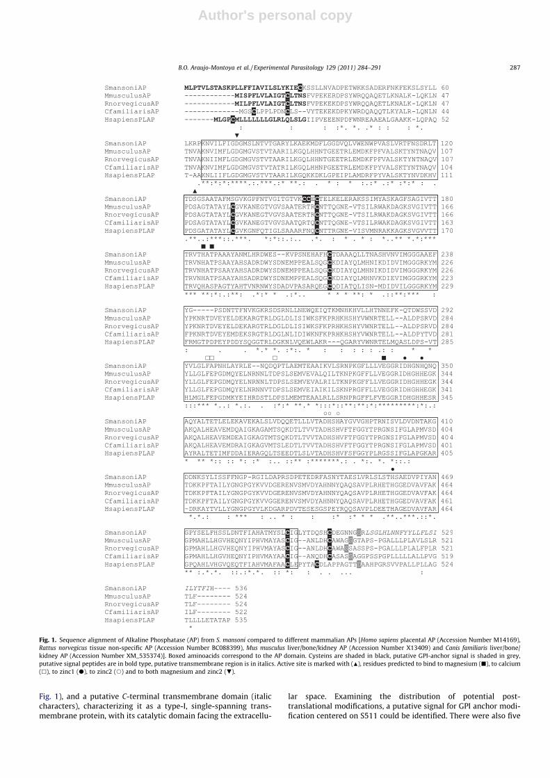
The full-length cloned cDNA displayed an ORF of 1698 bp,encoding a protein of 536 amino acids with a predicted molecularmass of approximately 59.3 kDa and an isoeletric point of 5.92. TheSmAP protein sequence showed an overall aminoacid sequenceidentity of 33–37% to those of Homo sapiens placental AlkalinePhosphatase, Rattus norvegicus tissue non-specific Alkaline Phos-phatase, Mus musculus liver Alkaline Phosphatase, and Canis famili-aris Alkaline Phosphatase (Fig. 1). SmAP contains the AP signaturesequence (residues 64-495) (outlined by a continuous box inFig. 1), and is recognized as part of the Pfam Alkaline Phosphatasefamily (PF00245) with an E-value of 8.60 � 10�109. SmAP also con-tains a putative N-terminal signal peptide (bold characters in
286 B.O. Araujo-Montoya et al. / Experimental Parasitology 129 (2011) 284–291
Author's personal copy
Fig. 1), and a putative C-terminal transmembrane domain (italiccharacters), characterizing it as a type-I, single-spanning trans-membrane protein, with its catalytic domain facing the extracellu-
lar space. Examining the distribution of potential post-translational modifications, a putative signal for GPI anchor modi-fication centered on S511 could be identified. There were also five
Fig. 1. Sequence alignment of Alkaline Phosphatase (AP) from S. mansoni compared to different mammalian APs [Homo sapiens placental AP (Accession Number M14169),Rattus norvegicus tissue non-specific AP (Accession Number BC088399), Mus musculus liver/bone/kidney AP (Accession Number X13409) and Canis familiaris liver/bone/kidney AP (Accession Number XM_535374)]. Boxed aminoacids correspond to the AP domain. Cysteins are shaded in black, putative GPI-anchor signal is shaded in grey,putative signal peptides are in bold type, putative transmembrane region is in italics. Active site is marked with (N), residues predicted to bind to magnesium (j), to calcium(h), to zinc1 (d), to zinc2 (s) and to both magnesium and zinc2 (.).
B.O. Araujo-Montoya et al. / Experimental Parasitology 129 (2011) 284–291 287
Author's personal copy
potential N-glycosylation sites (Asn223, Asn244, Asn398, Asn413,and Asn445) and a few potential O-glycosylation sites with low-probability. Comparing the SmAP amino acid sequence with thoseof mammalian AP’s (Kozlenkov et al., 2002), we further identifiedthree metal atom binding sites: one for magnesium (at positionsAsp73, His185, Thr187, and Glu 338) and two for zinc (Asp 343,His 347, His 459 for Zinc1; Asp 73, Asp 374, His375, His 377 forZinc2). In the mammalian sequence there is a calcium binding sitewhich is not totally conserved in the SmAP (Phe 298, Ala 299, andAsp 311). The catalytic center would be a serine residue at position123 (Kozlenkov et al., 2002) (Fig. 1).
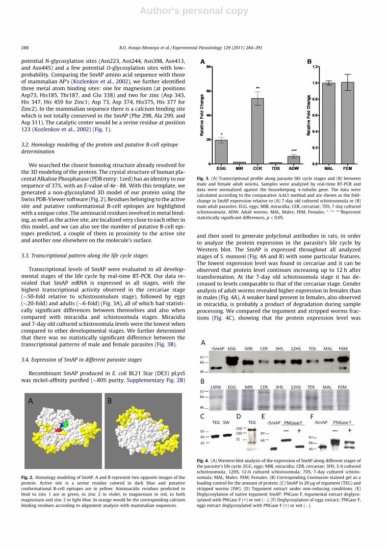
3.2. Homology modeling of the protein and putative B-cell epitopedetermination
We searched the closest homolog structure already resolved forthe 3D modeling of the protein. The crystal structure of human pla-cental Alkaline Phosphatase (PDB entry: 1zed) has an identity to oursequence of 37%, with an E-value of 4e�88. With this template, wegenerated a non-glycosylated 3D model of our protein using theSwiss PDB-Viewer software (Fig. 2). Residues belonging to the activesite and putative conformational B-cell epitopes are highlightedwith a unique color. The aminoacid residues involved in metal bind-ing, as well as the active site, are localized very close to each other inthis model, and we can also see the number of putative B-cell epi-topes predicted, a couple of them in proximity to the active siteand another one elsewhere on the molecule’s surface.
3.3. Transcriptional pattern along the life cycle stages
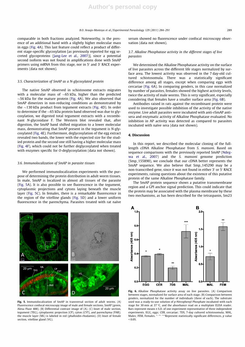
Transcriptional levels of SmAP were evaluated in all develop-mental stages of the life cycle by real-time RT-PCR. Our data re-vealed that SmAP mRNA is expressed in all stages, with thehighest transcriptional activity observed in the cercariae stage(�50-fold relative to schistosomulum stage), followed by eggs(�20-fold) and adults (�6-fold) (Fig. 3A), all of which had statisti-cally significant differences between themselves and also whencompared with miracidia and schistosomula stages. Miracidiaand 7-day old cultured schistosomula levels were the lowest whencompared to other developmental stages. We further determinedthat there was no statistically significant difference between thetranscriptional patterns of male and female parasites (Fig. 3B).
3.4. Expression of SmAP in different parasite stages
Recombinant SmAP produced in E. coli BL21 Star (DE3) pLysSwas nickel-affinity purified (�80% purity, Supplementary Fig. 2B)
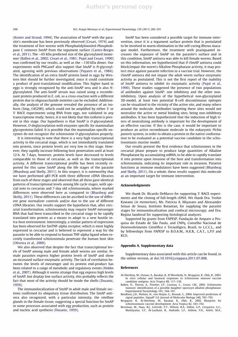
and then used to generate polyclonal antibodies in rats, in orderto analyze the protein expression in the parasite’s life cycle byWestern blot. The SmAP is expressed throughout all analyzedstages of S. mansoni (Fig. 4A and B) with some particular features.The lowest expression level was found in cercariae and it can beobserved that protein level continues increasing up to 12 h aftertransformation. At the 7-day old schistosomula stage it has de-creased to levels comparable to that of the cercariae stage. Genderanalysis of adult worms revealed higher expression in females thanin males (Fig. 4A). A weaker band present in females, also observedin miracidia, is probably a product of degradation during sampleprocessing. We compared the tegument and stripped worms frac-tions (Fig. 4C), showing that the protein expression level was
Fig. 2. Homology modeling of SmAP. A and B represent two opposite images of theprotein. Active site is a serine residue colored in dark blue and putativeconformational B-cell epitopes are in yellow. Aminoacidic residues predicted tobind to zinc 1 are in green, to zinc 2 in violet, to magnesium in red, to bothmagnesium and zinc 2 in light blue. In orange would be the corresponding calciumbinding residues according to alignment analysis with mammalian sequences.
Fig. 3. (A) Transcriptional profile along parasite life cycle stages and (B) betweenmale and female adult worms. Samples were analyzed by real-time RT-PCR anddata were normalized against the housekeeping a-tubulin gene. The data werecalculated according to the comparative DDCt method and are shown as the fold-change in SmAP expression relative to (A) 7-day old cultured schistosomula or (B)male adult parasites. EGG, eggs; MIR, miracidia; CER, cercariae; 7DS, 7-day culturedschistosomula; ADW, Adult worms; MAL, Males; FEM, Females. ⁄, ⁄⁄, ⁄⁄⁄Representstatistically significant differences, p 6 0.05.
Fig. 4. (A) Western blot analysis of the expression of SmAP along different stages ofthe parasite’s life cycle. EGG, eggs; MIR, miracidia; CER, cercariae; 3HS, 3-h culturedschistosomula; 12HS, 12-h cultured schistosomula; 7DS, 7-day cultured schisto-somula; MAL, Males; FEM, Females. (B) Corresponding Coomassie-stained gel as aloading control for the amount of protein. (C) SmAP in 20 lg of tegument (TEG) andstripped worms (SW). (D) Tegument extract under non-reducing conditions. (E)Deglycosylation of native tegument SmAP; PNGase F, tegumental extract deglyco-sylated with PNGase F (+) or not (�). (F) Deglycosylation of eggs extract; PNGase F,eggs extract deglycosylated with PNGase F (+) or not (�).
288 B.O. Araujo-Montoya et al. / Experimental Parasitology 129 (2011) 284–291
Author's personal copy
comparable in both fractions analyzed. Noteworthy, is the pres-ence of an additional band with a slightly higher molecular massin eggs (Fig. 4A). This last feature could reflect a product of differ-ent stage-specific glycosylation [as previously reported for egg se-creted glycoproteins (Jang-Lee et al., 2007)], since a potentialsecond isoform was not found in amplifications done with SmAPprimers using mRNA from this stage, nor in 50 and 30 RACE exper-iments (data not shown).
3.5. Characterization of SmAP as a N-glycosylated protein
The native SmAP observed in schistosome extracts migrateswith a molecular mass of �65 kDa, higher than the predicted�56 kDa for the mature protein (Fig. 4A). We also observed thatSmAP dimerizes in non-reducing conditions as demonstrated bythe �130 kDa product from tegument extracts (Fig. 4D). In orderto determine if the �65 kDa protein band observed was due to gly-cosylation, we digested total tegument extracts with a recombi-nant N-glycosidase F. The Western blot revealed that, afterdigestion, the SmAP band shifted migration to a lower molecularmass, demonstrating that SmAP present in the tegument is N-gly-cosylated (Fig. 4E). Furthermore, deglycosylation of the egg extractrevealed two bands, the lower with the expected size for the stud-ied protein and the second one still having a higher molecular mass(Fig. 4F), which could not be further deglycosylated when treatedwith enzymes specific for O-deglycosylation (data not shown).
3.6. Immunolocalization of SmAP in parasite tissues
We performed immunolocalization experiments with the pur-pose of determining the protein distribution in adult worm tissues.In male, SmAP is localized in almost all tissues of the parasite(Fig. 5A). It is also possible to see fluorescence in the tegument,cytoplasmic projections and cytons laying beneath the musclelayer (Fig. 5C). In females, there is a remarkable fluorescence inthe region of the vitelline glands (Fig. 5D) and a lower uniformfluorescence in the parenchyma. Parasites treated with rat naïve
serum showed no fluorescence under confocal microscopy obser-vation (data not shown).
3.7. Alkaline Phosphatase activity in the different stages of liveparasites
We determined the Alkaline Phosphatase activity on the surfaceof live parasites across the different life stages normalized by sur-face area. The lowest activity was observed in the 7 day-old cul-tured schistosomula. There was a statistically significantdifference among all stages, except when comparing eggs withcercariae (Fig. 6A). In comparing genders, in this case normalizedby number of parasites, females showed the highest activity levels,twice the activity of male worms. This is very significant, especiallyconsidering that females have a smaller surface area (Fig. 6B).
Antibodies raised in rats against the recombinant protein wereused to investigate possible inhibition of the activity of the nativeenzyme. Live adult parasites were incubated with anti-rSmAP anti-sera and enzymatic activity of Alkaline Phosphatase evaluated. Noinhibition in AP activity was detected as compared to parasitesincubated with naïve sera (data not shown).
4. Discussion
In this report, we described the molecular cloning of the full-length cDNA Alkaline Phosphatase from S. mansoni. Based onsequence comparisons with the previously reported SmAP (Ndeg-wa et al., 2007) and the S. mansoni genome prediction(Smp_155890), we conclude that our cDNA better represents theSmAP sequence. We also believe that Smp_145290 may be anon-transcribed gene, since it was not found in either 30 or 50 RACEexperiments, raising questions about the existence of this putativeprotein of the same Alkaline Phosphatase family.
The SmAP protein sequence shows a putative transmembraneregion and a GPI anchor signal prediction. This could indicate thatthe protein may be associated with the plasma membrane by thesetwo mechanisms, as has been described for the tetraspanin, Sm23
Fig. 5. Immunolocalization of SmAP in transversal section of adult worms. (A)Fluorescence confocal microscopy image of male and female sections, SmAP (green,Alexa Fluor 488). (B) Differential contrast image of (A). (C) Inset of male section,tegument (TEG), cytoplasmic projection (CP), cyton (CYT), and parenchyma (PAR);the muscle layer (ML) is labeled in red (phalloidin-rhodamin). (D) Inset of femalesection, vitelline gland (VG).
Fig. 6. Alkaline Phosphatase activity assay on live parasites. (A) Comparisonbetween stages, normalized for surface area of each stage. (B) Comparison betweengenders, normalized for the number of individuals (three of each). The substrateused was a ready-to-use solution of p-Nitrophenyl Phosphate incubated with eachstage for 30 min at 37 �C, and the absorbance read on a multiplate ELISA reader.Bars represent means ± S.D. of one experiment representative of three independentexperiments. EGG, eggs; CER, cercariae; 7DS, 7-day cultured schistosomula; MAL,Males; FEM, Females. ⁄, ⁄⁄, ⁄⁄⁄Represent statistically significant differences, p value< 0.05.
B.O. Araujo-Montoya et al. / Experimental Parasitology 129 (2011) 284–291 289
Author's personal copy
(Koster and Strand, 1994). The association of SmAP with the para-site’s membrane has been previously observed (Cesari, 1974) andthe treatment of live worms with Phosphatidylinositol-Phospholi-pase C removes SmAP from the tegument surface (Castro-Borgeset al., 2011). The �65 kDa predicted size of the glycosylated mono-mer (Ballen et al., 2002; Cesari et al., 1981; Pujol and Cesari, 1990)was confirmed by our results, as well as the �130 kDa dimer. Ourexperiments with PNGaseF also support that SmAP is N-glycosyl-ated, agreeing with previous observations (Payares et al., 1984).The identification of an extra SmAP protein band in eggs by Wes-tern blot should be further investigated, since it could constitutea product of post-translational modification. This higher band ineggs is strongly recognized by the anti-SmAP sera and is also N-glycosylated. The anti-SmAP serum was raised using a recombi-nant protein produced in E. coli, so a cross-reaction with a differentprotein due to oligosaccharide moieties can be excluded. Addition-ally, the analysis of the genome revealed the presence of an iso-form (Smp_145290), which could not be amplified by either 30 or50 RACE experiments using RNA from eggs, nor validated in anytranscriptome study; hence, it is not likely that this isoform is pres-ent in this stage. Our hypothesis is that SmAP is O-glycosylated.However, O-deglycosylation with enzymes specific for mammalianglycoproteins failed. It is possible that the mammalian-specific en-zymes do not recognize the schistosome O-glycosylation properly.
It is interesting to note that there is a very high transcriptionalactivity in the cercarial stage, which is not immediately translatedinto protein, since protein levels are very low in this stage. How-ever, they rapidly increase following host penetration until at least12 h. At 7 days, however, protein levels have decreased to levelscomparable to those of cercariae, as well as the transcriptionalactivity. A different transcriptional profile has been recently re-ported for this same SmAP along the life stages of the parasite(Bhardwaj and Skelly, 2011). In this respect, it is noteworthy thatwe have performed qRT-PCR with three different cDNA librariesfrom each of these stages and observed that all three gave identicalpatterns of transcriptional levels among life cycle stages, with spe-cial note to cercariae and 7-day old schistosomula, where markeddifferences were observed here as compared to (Bhardwaj andSkelly, 2011). These differences can be ascribed to the use of differ-ent gene normalizer controls and/or due to the use of differentcDNA libraries. Our results support the hypothesis that, after cerc-arial transformation, schistosomula may require SmAP messengerRNA that had been transcribed in the cercarial stage to be rapidlytranslated into protein as a means to adapt to a new hostile in-tra-host environment. Interestingly, a similar pattern of expressionhas been observed for SmTNF-alpha receptor, which is most highlyexpressed in cercariae and is believed to represent a way for theparasite to be able to respond to human TNF-alpha ligand when re-cently transformed schistosomula penetrate the human host skin(Oliveira et al., 2009).
We also observed that despite the fact that transcriptional lev-els of SmAP among male and female adult worms are similar, fe-male parasites express higher protein levels of SmAP and showan increased surface enzymatic activity. The lack of correlation be-tween the levels of messenger and its protein end-product hasbeen related to a range of metabolic and regulatory events (Hokkeet al., 2007). Although it seems strange that egg express high levelsof SmAP, but display low surface activity, this probably reflects thefact that most of the activity should be inside the shells (Dusanic,1959).
The immunolocalization of SmAP in adult male and female sec-tions confirmed its ubiquitary tissue distribution. The SmAP anti-sera also recognized, with a particular intensity, the vitellineglands in the female tissue, suggesting a special function for SmAPin some processes associated with egg production, such as proteinand nucleic acid synthesis (Dusanic, 1959).
SmAP has been considered a possible target for immune inter-vention, since it is a tegument surface protein that is postulatedto be involved in worm elimination in the self-curing Rhesus maca-que model. Furthermore, the treatment with praziquantel in-creases the exposure of SmAP on the parasite’s surface and inthis condition, SmAP antisera was able to kill female worms. Basedon this information, we hypothesized that if rSmAP antisera couldblock/impair the worm’s Alkaline Phosphatase activity, it may pro-tect mice against parasite infection in a vaccine trial. However, therSmAP antisera did not impair the adult worm surface enzymaticactivity as postulated. This is not the first report of the inabilityof SmAP antisera to inhibit its enzymatic activity (Pujol et al.,1990). These studies suggested the presence of two populationsof antibodies against SmAP: one inhibitory and the other non-inhibitory. Upon analysis of the hypothetical non-glycosylated3D-model, at least two potential B-cell discontinuous epitopescan be visualized in the vicinity of the active site, and many othersaround the molecule. Antibodies targeting these sites would notneutralize the active or metal binding sites, being non-inhibitoryantibodies. It has been hypothesized that the induction of high ti-ters of neutralizing antibody is important for the development ofan effective vaccine. If this is the case, it would be important toproduce an active recombinant molecule in the eukaryotic Pichiapastoris system, in order to obtain a protein in the native conforma-tion to be evaluated as a potential vaccine candidate in the schis-tosomiasis murine model.
Our results present the first evidence that schistosomes in thecercarial phase prepare to produce large quantities of AlkalinePhosphatase by accumulating mRNA, to be able to rapidly translateit into protein upon invasion of the host and transformation intoschistosomula, indicating its important role in invasion. Putativefunctions as immune modulators have been suggested (Bhardwajand Skelly, 2011). On a whole, these results support this moleculeas an important target for immune intervention.
Acknowledgments
We thank Dr. Ricardo DeMarco for supporting 30 RACE experi-ments and the cloning of full-length cDNA. We thank Dra. ToshieKawano (in memorium), Ms. Patricia A. Miyasato and AlexsanderSeixas de Souza, Instituto Butantan, for supplying the parasitestages for immunolocalization and confocal microscopy; and Dra.Regina Sandoval for supporting histological analyzes.
Supported by grants from FAPESP, Fundação de Amparo a Pes-quisa do Estado de São Paulo, and CNPq, Conselho Nacional deDesenvolvimento Científico e Tecnológico, Brasil, to L.C.C.L., andby fellowships from FAPESP to B.O.A.M., H.K.R., C.A.T., L.P.F andKCO.
Appendix A. Supplementary data
Supplementary data associated with this article can be found, inthe online version, at doi:10.1016/j.exppara.2011.07.008.
References
Al-Sherbiny, M., Osman, A., Barakat, R., El Morshedy, H., Bergquist, R., Olds, R., 2003.In vitro cellular and humoral responses to Schistosoma mansoni vaccinecandidate antigens. Acta Tropica 88, 117–130.
Ballen, D., Theron, A., Pointier, J.P., Coustau, C., Cesari, I.M., 2002. Schistosomamansoni: identification of a possible daughter sporocyst alkaline phosphatase.Experimental Parasitology 101, 164–167.
Bendtsen, J.D., Nielsen, H., von Heijne, G., Brunak, S., 2004. Improved prediction ofsignal peptides: SignalP 3.0. Journal of Molecular Biology 340, 783–795.
Bergquist, R., Al-Sherbiny, M., Barakat, R., Olds, R., 2002. Blueprint forschistosomiasis vaccine development. Acta Tropica 82, 183–192.
Berriman, M., Haas, B.J., LoVerde, P.T., Wilson, R.A., Dillon, G.P., Cerqueira, G.C.,Mashiyama, S.T., Al-Lazikani, B., Andrade, L.F., Ashton, P.D., Aslett, M.A.,
290 B.O. Araujo-Montoya et al. / Experimental Parasitology 129 (2011) 284–291
Author's personal copy
Bartholomeu, Blandin, G., Caffrey, C.R., Coghlan, A., Coulson, R., Day, T.A.,Delcher, A., DeMarco, R., Djikeng, A., Eyre, T., Gamble, J.A., Ghedin, E., Gu, Y.,Hertz-Fowler, C., Hirai, H., Hirai, Y., Houston, R., Ivens, A., Johnston, D.A.,Lacerda, D., Macedo, C.D., McVeigh, P., Ning, Z., Oliveira, G., Overington, J.P.,Parkhill, J., Pertea, M., Pierce, R.J., Protasio, A.V., Quail, M.A., Rajandream, M.A.,Rogers, J., Sajid, M., Salzberg, S.L., Stanke, M., Tivey, A.R., White, O., Williams,D.L., Wortman, J., Wu, W., Zamanian, M., Zerlotini, A., Fraser-Liggett, C.M.,Barrell, B.G., El-Sayed, N.M., . The genome of the blood fluke Schistosomamansoni. Nature 460, 352–358.
Bhardwaj, R., Skelly, P.J., 2009. Purinergic signaling and immune modulation at theschistosome surface? Trends in Parasitology 25, 256–260.
Bhardwaj, R., Skelly, P.J., 2011. Characterization of Schistosome TegumentalAlkaline Phosphatase (SmAP). PLoS Neglected Tropical Diseases 5, e1011.
Braschi, S., Wilson, R.A., 2006. Proteins exposed at the adult schistosome surfacerevealed by biotinylation. Molecular and Cellular Proteomics 5, 347–356.
Braschi, S., Curwen, R.S., Ashton, P.D., Verjovski-Almeida, S., Wilson, A., 2006. Thetegument surface membranes of the human blood parasite Schistosomamansoni: a proteomic analysis after differential extraction. Proteomics 6,1471–1482.
Castro-Borges, W., Dowle, A., Curwen, R.S., Thomas-Oates, J., Wilson, R.A., 2011.Enzymatic shaving of the tegument surface of live schistosomes for proteomicanalysis: a rational approach to select vaccine candidates. PLoS NeglectedTropical Diseases 5, e993.
Cesari, I.M., 1974. Schistosoma mansoni: distribution and characteristics of alkalineand acid phosphatase. Experimental Parasitology 36, 405–414.
Cesari, I.M., Simpson, A.J., Evans, W.H., 1981. Properties of a series of tegumentalmembrane-bound phosphohydrolase activities of Schistosoma mansoni.Biochemical Journal 198, 467–473.
Crabtree, J.E., Wilson, R.A., 1980. Schistosoma mansoni: a scanning electronmicroscope study of the developing schistosomulum. Parasitology 81, 553–564.
Dusanic, D.G., 1959. Histochemical observations of alkaline phosphatase inSchistosoma mansoni. Journal of Infectious Diseases 105, 1–8.
Fallon, P.G., Smith, P., Nicholls, T., Modha, J., Doenhoff, M.J., 1994. Praziquantel-induced exposure of Schistosoma mansoni alkaline phosphatase: drug-antibodysynergy which acts preferentially against female worms. Parasite Immunology16, 529–535.
Guex, N., Peitsch, M.C., 1997. SWISS-MODEL and the Swiss-PdbViewer: anenvironment for comparative protein modeling. Electrophoresis 18, 2714–2723.
Gupta, R., Brunak, S., 2002. Prediction of glycosylation across the human proteomeand the correlation to protein function. Pacific Symposium on Biocomputing310, 322.
Haste Andersen, P., Nielsen, M., Lund, O., 2006. Prediction of residues indiscontinuous B-cell epitopes using protein 3D structures. Protein Science 15,2558–2567.
Higgins, D.G., 1994. CLUSTAL V: multiple alignment of DNA and protein sequences.Methods in Molecular Biology 25, 307–318.
Hokke, C.H., Fitzpatrick, J.M., Hoffmann, K.F., 2007. Integrating transcriptome,proteome and glycome analyses of Schistosoma biology. Trends in Parasitology23, 165–174.
Hu, W., Yan, Q., Shen, D.K., Liu, F., Zhu, Z.D., Song, H.D., Xu, X.R., Wang, Z.J., Rong, Y.P.,Zeng, L.C., Wu, J., Zhang, X., Wang, J.J., Xu, X.N., Wang, S.Y., Fu, G., Zhang, X.L.,Wang, Z.Q., Brindley, P.J., McManus, D.P., Xue, C.L., Feng, Z., Chen, Z., Han, Z.G.,2003. Evolutionary and biomedical implications of a Schistosoma japonicumcomplementary DNA resource. Nature Genetics 35, 139–147.
Jang-Lee, J., Curwen, R.S., Ashton, P.D., Tissot, B., Mathieson, W., Panico, M., Dell, A.,Wilson, R.A., Haslam, S.M., 2007. Glycomics analysis of Schistosoma mansoni eggand cercarial secretions. Molecular and Cellular Proteomics 6, 1485–1499.
Koster, B., Strand, M., 1994. Schistosoma mansoni: Sm23 is a transmembrane proteinthat also contains a glycosylphosphatidylinositol anchor. Archives ofBiochemistry and Biophysics 310, 108–117.
Kozlenkov, A., Manes, T., Hoylaerts, M.F., Millan, J.L., 2002. Function assignment toconserved residues in mammalian alkaline phosphatases. The Journal ofBiological Chemistry 277, 22992–22999.
Livak, K.J., Schmittgen, T.D., 2001. Analysis of relative gene expression data usingreal-time quantitative PCR and the 2(-Delta Delta C(T)) Method. Methods 25,402–408.
Morel, C.M., 2000. Reaching maturity–25 years of the TDR. Parasitology Today 16,522–528.
Narushin, V.G., 2005. Egg geometry calculation using the measurements of lengthand breadth. Poultry Science 84, 482–484.
Ndegwa, D., Krautz-Peterson, G., Skelly, P.J., 2007. Protocols for gene silencing inschistosomes. Experimental Parasitology 117, 284–291.
Nimmo-Smith, R.H., Standen, O.D., 1963. Phosphomonoesterases of Schistosomamansoni. Experimental Parasitology 13, 305–322.
Oliveira, K.C., Carvalho, M.L., Venancio, T.M., Miyasato, P.A., Kawano, T., DeMarco, R.,Verjovski-Almeida, S., 2009. Identification of the Schistosoma mansoni TNF-alpha receptor gene and the effect of human TNF-alpha on the parasite geneexpression profile. PLoS Neglected Tropical Diseases 3, e556.
Payares, G., Smithers, S.R., Evans, W.H., 1984. Purification and topographicallocation of tegumental alkaline phosphatase from adult Schistosoma mansoni.Molecular and Biochemical Parasitology 13, 343–360.
Pfaffl, M.W., 2001. A new mathematical model for relative quantification in real-time RT-PCR. Nucleic Acids Research 29, e45.
Plummer Jr., T.H., Tarentino, A.L., 1991. Purification of the oligosaccharide-cleavingenzymes of Flavobacterium meningosepticum. Glycobiology 1, 257–263.
Pujol, F.H., Cesari, I.M., 1990. Antigenicity of adult Schistosoma mansoni alkalinephosphatase. Parasite Immunology 12, 189–198.
Pujol, F.H., Liprandi, F., Rodriguez, M., Cesari, I.M., 1990. Production of a mousemonoclonal antibody against the alkaline phosphatase of adult Schistosomamansoni. Molecular and Biochemical Parasitology 40, 43–52.
Ramos, C.R., Abreu, P.A., Nascimento, A.L., Ho, P.L., 2004. A high-copy T7 Escherichiacoli expression vector for the production of recombinant proteins with aminimal N-terminal His-tagged fusion peptide. Brazilian Journal of Medical andBiological Research 37, 1103–1109.
Roberts, S.M., MacGregor, A.N., Vojvodic, M., Wells, E., Crabtree, J.E., Wilson, R.A.,1983. Tegument surface membranes of adult Schistosoma mansonik:development of a method for their isolation. Molecular and BiochemicalParasitology 9, 105–127.
Samuelson, J.C., Caulfield, J.P., 1985. The cercarial glycocalyx of Schistosomamansoni. The Journal of Cell Biology 100, 1423–1434.
Smith, J.H., Reynolds, E.S., Von Lichtenberg, F., 1969. The integument of Schistosomamansoni. The American Journal of Tropical Medicine and Hygiene 18, 28–49.
Sonnhammer, E.L., von Heijne, G., Krogh, A., 1998. A hidden Markov model forpredicting transmembrane helices in protein sequences. Proceedings/.International Conference on Intelligent Systems for Molecular Biology 6, 175–182.
Verjovski-Almeida, S., DeMarco, R., Martins, E.A., Guimaraes, P.E., Ojopi, E.P.,Paquola, A.C., Piazza, J.P., Nishiyama Jr., M.Y., Kitajima, J.P., Adamson, R.E.,Ashton, P.D., Bonaldo, M.F., Coulson, P.S., Dillon, G.P., Farias, L.P., Gregorio, S.P.,Ho, P.L., Leite, R.A., Malaquias, L.C., Marques, R.C., Miyasato, P.A., Nascimento,A.L., Ohlweiler, F.P., Reis, E.M., Ribeiro, M.A., Sa, R.G., Stukart, G.C., Soares, M.B.,Gargioni, C., Kawano, T., Rodrigues, V., Madeira, A.M., Wilson, R.A., Menck, C.F.,Setubal, J.C., Leite, L.C., Dias-Neto, E., 2003. Transcriptome analysis of theacoelomate human parasite Schistosoma mansoni. Nature Genetics 35, 148–157.
Wilson, R.A., Langermans, J.A., van Dam, G.J., Vervenne, R.A., Hall, S.L., Borges, W.C.,Dillon, G.P., Thomas, A.W., Coulson, P.S., 2008. Elimination of Schistosomamansoni adult worms by Rhesus macaques: basis for a therapeutic vaccine?PLoS Neglected Tropical Diseases 2, e290.
Zhou, Y., Zheng, H., Liu, F., Hu, W., Wang, Z.Q., Gang, L., Ren, S., 2009. TheSchistosoma japonicum genome reveals features of host–parasite interplay.Nature 460, 345–351.
B.O. Araujo-Montoya et al. / Experimental Parasitology 129 (2011) 284–291 291


