Embed Size (px)
Citation preview

Acta Tropica 76 (2000) 107–117
Tegumental changes in adult Schistosoma mansoniharboured in mice treated with praziquantel enantiomers
Xiao Shuhua a, Shen Binggui a, Jacques Chollet b, Marcel Tanner b,*a Institute of Parasitic Diseases, Chinese Academy of Pre6enti6e Medicine, Shanghai 200025, China
b Swiss Tropical Institute, PO Box, CH–4002 Basel, Switzerland
Received 21 October 1999; received in revised form 20 February 2000; accepted 12 March 2000
Abstract
Praziquantel administered to the host causes damage to the tegument of Schistosoma mansoni. In this study, theeffects of racemic praziquantel (Pra) and its enantiomers, levo-praziquantel (L-Pra) and dextro-praziquantel (D-Pra)were compared using scanning electron microscopy (SEM). Mice infected with S. mansoni for 49 days were treatedwith a single dose of Pra (300 mg/kg), L-Pra (150 mg/kg) or D-Pra (150 or 600 mg/kg). Groups of three mice werekilled after 4 and 24 h, and schistosomes collected by perfusion and examined by SEM. Treatment with Pra or L-Pra,for 4 or 24 h, caused tegumental damage to S. mansoni including severe swelling, vacuolization, fusion of thetegumental ridges and loss or shortening of the spines on the tubercles, collapse and peeling. After treatment withD-Pra at 150 mg/kg, no apparent damage was observed. When the dosage was increased to 600 mg/kg, after 4 hlesions on the tegument similar to those induced by Pra or L-Pra were seen, but less severe. After 24 h, there wasevidence of recovery. The study thus clearly showed that L-Pra was more active than D-Pra in causing tegumentaldamage. D-Pra showed a qualitatively similar activity at a higher concentration. It is possible that this effect was dueat least to some extent to the small amount of L-Pra (B2%) which was present in the preparation of D-Pra used.© 2000 Elsevier Science B.V. All rights reserved.
Keywords: Schistosomiasis; Schistosoma mansoni ; Praziquantel; Enantiomers; Scanning electron microscopy
www.elsevier.com/locate/actatropica
1. Introduction
Racemic praziquantel (Pra), which is effectiveagainst all five human schistosome species, iscomposed of equal parts of its optical isomers,levo-praziquantel (L-Pra) and dextro-praziquantel
(D-Pra) (Andrews et al., 1983). Experimental andclinical studies with Schistosoma japonicum havedemonstrated that L-Pra is the component activeagainst this species (Andrews, 1985; Liu et al.,1986a,b; Wang et al., 1987; Liu et al., 1988;Tanaka et al., 1989; Wu et al., 1991; Xiao et al.,1998), but there has been some dispute aboutwhether this also applies to Schistosoma mansoni.Most studies with S. mansoni, both in vitro and invivo, also identified L-Pra as the active component
* Corresponding author. Tel.: +41-61-2848287; fax: +41-61-2717951.
E-mail address: [email protected] (M. Tanner).
0001-706X/00/$ - see front matter © 2000 Elsevier Science B.V. All rights reserved.
PII: S 0001 -706X(00 )00076 -0

X. Shuhua et al. / Acta Tropica 76 (2000) 107–117108
(Andrews, 1985; Xiao and Catto, 1989; Staudt etal., 1992; Xiao et al., 1999), though the oppositeconclusion was drawn from another in vivo exper-iment (Tanaka et al., 1989). The present studywas based on the previous assessment of efficacyof L-Pra and D-Pra (Xiao et al., 1999) and com-pared the activity of Pra and its enantiomers incausing tegumental damage to adult worms ofS. mansoni using scanning electron microscopy(SEM).
2. Materials and methods
2.1. Parasites and mice
S. mansoni cercariae (Liberia strain), releasedfrom infected Biomphalaria glabrata snails wereprovided by the Swiss Tropical Institute (STI).Female MORO strain mice, weighing 18–22 g,were obtained from Biotechnology & AnimalBreeding Division (Fullinsdorf, Switzerland). Allmice were maintained on Rodent Blox obtainedfrom Eberle NAFAG (Gossau, Switzerland) inthe animal care facilities of the STI.
2.2. Drugs
Pra was the product of Shanghai No. 6 Phar-maceutical Factory. L-Pra and D-Pra were synthe-sized by the Institute of Parasitic Diseases,Chinese Academy of Preventive Medicine. Theoptical rotation of L-Pra was −132.43 and ofD-Pra +133.21 in ethanol at 11°C. The opticalspecificity of the enantiomers was confirmed usinghigh pressure liquid chromatography. The L-Prawas pure, whereas the D-Pra was slightly contam-inated (B2%) with L-Pra (Xiao and Shen, 1995;Xiao et al., 1999). For administration, all drugswere suspended in 7% Tween 80 and 3% ethanolat a final concentration of 15–60 g/l. The volumeof the dose given to each mouse was 10 ml/kg.
2.3. Infection and treatment
Mice were infected subcutaneously with 120cercariae each. Forty-nine days after infection,groups of five mice were treated intragastricallywith Pra at 300 mg/kg, L-Pra at 150 mg/kg, orD-Pra at 150 or 600 mg/kg. After 4 and 24 h,groups of 2–3 mice were killed by blood-lettingand schistosomes collected by perfusion with ice-cold Hanks’ balanced salt solution (HBSS). Twountreated mice served as controls. The wormswere rinsed three times with HBSS and thenfixed in 2.5% glutaraldehyde-phosphate buffer(0.1 mol/l, pH 7.4).
2.4. SEM obser6ation
The worm samples were processed routinely(Xiao et al., 1982) and examined with a JoelJSM-820 SEM. In each group, 6–8 male or fe-male worms were examined.
3. Results
3.1. Effects of Pra
Four hours after treatment with 300 mg/kg ofPra, two out of six male worms showed swelling,erosion and focal peeling of the discoid or outersurface of the oral sucker (untreated control: Fig.Fig. 1. Oral sucker of untreated control male worm (×600).

X. Shuhua et al. / Acta Tropica 76 (2000) 107–117 109
Fig. 2. Pra (300 mg/kg) for 4 h. Male worm with damaged oralsucker (×550).
female worms, there was no or only slight swellingof the tegument, except in one worm, where therewas vacuolization in the anterior portion of thedorsal surface with focal collapse (Fig. 7).
A total of 24 h after treatment, damage wasgenerally more severe, though patches of normal
Fig. 4. Pra (300 mg/kg) for 4 h. Male worm with tegumentdamage appearing as shortened spines on the tubercles, severeswelling of tubercles resulting in a smooth surface or emer-gence of vesicles around the tubercles (×2000).
Fig. 3. Tegument of untreated control male worm (×2000).
Fig. 5. Pra (300 mg/kg) for 4 h. Male worm with severeswelling and fusion of tegumental ridges resulting in formationof a large mass of material protruding from the worm surface(×850).
1, treated: Fig. 2). The male worms showedchanges in the tubercles, namely shortening orloss of the spines. In the severely damaged ones,there was disruption of the tubercles (untreatedcontrol: Fig. 3, treated: Fig. 4), and vacuolizationon the tegumental ridges or on the tubercles. Inone worm, the focal swelling led to the tegumentalridges fusing together and protruding from thesurface (Fig. 5). Extensive peeling of the tail ormiddle dorsal surface was also seen (Fig. 6). In

X. Shuhua et al. / Acta Tropica 76 (2000) 107–117110
Fig. 6. Pra (300 mg/kg) for 4 h. Male worm, showing extensivepeeling in the middle dorsal tegument. (×1200).
portion (Fig. 10). The alteration of the tegumenton the ventral surface was similar to that on thedorsal surface, and in one worm extensive peelingwas seen in the anterior portion (Fig. 11). Femaleworms also showed light or moderate swelling,and some sensory structures appeared to be swol-len or even collapsed (Fig. 12). In one worm,severe peeling and erosion were seen on the dorsalsurface (Fig. 13).
Fig. 8. Pra (300 mg/kg) for 24 h. Male worm, showing one orseveral vesicles on the surface of swollen tubercles (×2000).
Fig. 7. Pra (300 mg/kg) for 4 h. Female worm with vesicula-tion in anterior portion of dorsal tegument accompanied bycollapse of large vesicles (×3000).
Fig. 9. Pra (300 mg/kg) for 24 h. Male worm, showingnumerous vesicles emerging on the tegument around the tuber-cles (×2000).
tegument remained. In male worms, some of thetubercles appeared to be swollen, especially on theupper surface, with vesicles protruding from thesurface (Fig. 8), or numerous small blebs emerg-ing from the tegument around the tubercles (Fig.9). Some worms showed collapse of tubercles andfocal peeling of the surface along the middle

X. Shuhua et al. / Acta Tropica 76 (2000) 107–117 111
Fig. 10. Pra (300 mg/kg) for 24 h. Male worm with collapse ofdamaged tubercles (×2000).
the tegumental ridges, was usually seen accompa-nied by vacuolization. The tubercles showed lossof spines, swelling and collapse (Fig. 14). Severedamage to the oral sucker and the tegument be-tween oral sucker and acetabulum was also seen(Fig. 15). In female worms, the tegument revealedsevere focal or extensive damage in areas wherethe sensory structures are usually distributed. Thealteration was characterized by swelling and fu-sion of tegumental ridges, accompanied by col-lapse of the damaged sensory structures, resulting
Fig. 12. Pra (300 mg/kg) for 24 h. Female worm with collapseof damaged sensory structures (×2000).
Fig. 11. Pra (300 mg/kg) for 24 h. Ventral surface of maleworm, showing extensive peeling and damaged tubercles(×500).
Fig. 13. Pra (300 mg/kg) for 24 h. Female worm, showingpeeling of dorsal surface (×600).
3.2. Effects of L-Pra
Four hours after treatment with 150 mg/kg ofL-Pra, all worms showed moderate or severe focalor extensive tegumental damage. In male worms,the type of damage was similar to that induced byPra, but more prominent. Swelling and fusion of

X. Shuhua et al. / Acta Tropica 76 (2000) 107–117112
Fig. 14. L-Pra (150 mg/kg) for 4 h. Male worm, showingdamaged tubercles and vesicles on the tegumental surface(×2000).
extensive swelling of tubercles, accompanied byvacuolization or even collapse (Fig. 19). In femaleworms, the most prominent alteration was severeswelling and usually, the swollen tegument fusedto form a mass with some cracks (Fig. 20) orsuperficial peeling (Fig. 21).
Fig. 16. L-Pra (150 mg/kg) for 4 h. Female worm with focallydamaged tegumental surface (×1000).
Fig. 15. L-Pra (150 mg/kg) for 4 h. Male worm with damagedoral sucker (×1000).
Fig. 17. L-Pra (150 mg/kg) for 4 h. Female worm with focalswelling and fusion of tegumental ridges (×4000).
in deformation of the local tegument (Figs. 16and 17). Focal or extensive peeling and erosion ofthe tegument were also seen (Fig. 18).
A total of 24 h after treatment, it became moredifficult to remove severely damaged worms.Those that could be perfused out still showed

X. Shuhua et al. / Acta Tropica 76 (2000) 107–117 113
Fig. 18. L-Pra (150 mg/kg) for 4 h. Female worm, showingextensive peeling and erosion of tegument (×1300).
generally seen, was slight swelling or fusion of thetegumental ridges (Fig. 24).
Four hours after treatment with D-Pra at thehigher dose of 600 mg/kg, the tubercles on thetegument of male worms showed slight damage,or even severe damage similar to that induced byPra or L-Pra. Usually, shortening or loss of spineswas observed, together with swelling, formationof blebs or collapse (Figs. 25 and 26). Although
Fig. 20. L-Pra (150 mg/kg) for 24 h. Female worm, showingextensive swelling and fusion of tegumental ridges (×900).
Fig. 19. L-Pra (150 mg/kg) for 24 h. Male worm, showingswelling of tubercles with collapse or vesicle formation on thetubercle surface (×2000).
Fig. 21. L-Pra (150 mg/kg) for 24 h. Female worm withsuperficial focal peeling of tegument (×3000).
3.3. Effects of D-Pra
After 4 or 24 h after treatment with 150 mg/kgof D-Pra, all worms showed no or only slight focaldamage to the tegument. In male worms, thetubercles usually appeared to be swollen, with afew blebs (Fig. 22), or shortened spines on thesurface (Fig. 23). In female worms, the alteration

X. Shuhua et al. / Acta Tropica 76 (2000) 107–117114
Fig. 22. D-Pra (150 mg/kg) for 4 or 24 h. Male worm withsevere swelling of tubercles with shortened spines or even lossof spines (×4000).
ridges. Focal peeling was seen in two out of eightfemale worms (Fig. 28).
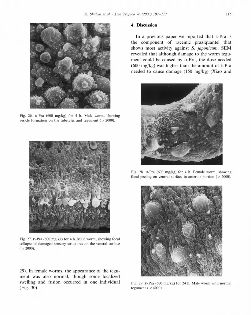
Twenty four hours after treatment, all wormsexamined showed apparent recovery from thedamage to the tegument induced by D-Pra.Swelling of the tegument subsided in most of themale worms, and apart from a few, which showedslight swelling and loss of spines, most of thetubercles showed a normal intact appearance (Fig.
Fig. 24. D-Pra (150 mg/kg) for 4 or 24 h. Female worm,showing slight swelling and fusion of tegumental ridges(×6000).
Fig. 23. D-Pra (150 mg/kg) for 4 or 24 h. Male worm showingextensive swelling of tubercles and formation of small vesicleson the tegument (×2000).
Fig. 25. D-Pra (600 mg/kg) for 4 h. Male worm with swellingtubercles accompanied by shortened spines and formation ofvesicles on the tegument (×2000).
some slight or moderate swelling of tegumentalridges was seen, there were no large massesformed by fusion of swollen ridges, and only oneworm showed focal collapse of sensory structures(Fig. 27). In female worms, the tegumental alter-ations were swelling and fusion of tegumental
X. Shuhua et al. / Acta Tropica 76 (2000) 107–117 115
Fig. 26. D-Pra (600 mg/kg) for 4 h. Male worm, showingvesicle formation on the tubercles and tegument (×2000).
4. Discussion
In a previous paper we reported that L-Pra isthe component of racemic praziquantel thatshows most activity against S. japonicum. SEMrevealed that although damage to the worm tegu-ment could be caused by D-Pra, the dose needed(600 mg/kg) was higher than the amount of L-Praneeded to cause damage (150 mg/kg) (Xiao and
Fig. 28. D-Pra (600 mg/kg) for 4 h. Female worm, showingfocal peeling on ventral surface in anterior portion (×2000).
Fig. 27. D-Pra (600 mg/kg) for 4 h. Male worm, showing focalcollapse of damaged sensory structures on the ventral surface(×2000).
Fig. 29. D-Pra (600 mg/kg) for 24 h. Male worm with normaltegument (×4000).
29). In female worms, the appearance of the tegu-ment was also normal, though some localizedswelling and fusion occurred in one individual(Fig. 30).

X. Shuhua et al. / Acta Tropica 76 (2000) 107–117116
Fig. 30. D-Pra (600 mg/kg) for 24 h. Female worm with slightfocal swelling and fusion of tegument (×6000).
The results of the present study showed thatL-Pra is substantially more active than D-Pra incausing damage to the tegument of adult S. man-soni. The observation that D-Pra caused a qualita-tively similar but less severe damage to thetegument is not consistent with an earlier report(Irie et al., 1989). However, it is possible that thedamage observed with D-Pra in the present studywas actually due to the low amounts of L-Pra(B2%, leading to a maximum concentration of12 mg/kg in a dose of 600 mg/kg) owing to thesynthesis and purification process. Solutions of anisomeric compound often contain small amountsof the other enantiomer, and this has been ob-served before with praziquantel (Xiao et al.,1999).
Acknowledgements
This investigation received financial supportfrom the UNDP/World Bank/WHO Special Pro-gramme for Research and Training in TropicalDiseases (TDR), the Swiss Tropical Institute, andthe Ninth Five-Year Key Research Programme ofChina. We would like to thank J.M. Jenkins andDr J. Utzinger for useful suggestions and com-ments on the manuscript.
References
Andrews, P., Thomas, H., Pohlke, R., Seubert, J., 1983.Praziquantel. Med. Res. Rev. 3, 147–200.
Andrews, P., 1985. Praziquantel: mechanisms of anti-schisto-somal activity. Pharmacol. Ther. 29, 129–156.
Irie, Y., Utsunomiya, H., Tanaka, M., Ohmae, H., Nara, T.,Yasuraoka, K., 1989. Schistosoma japonicum and S. man-soni : ultrastructural damage in the tegument and reproduc-tive organs after treatment with levo- anddextro-praziquantel. Am. J. Trop. Med. Hyg. 41, 204–211.
Liu, Y.H., Qian, M.X., Wang, X.G., Wang, R.Q., Lu, S.Z.,Liu, J., Li, G.H., Chen, Y.D., 1986a. Efficacy of single oraldoses of praziquantel in treatment of Schistosomajaponicum infection. Chin. Med. J. 99, 470–472.
Liu, Y.H., Qian, M.X., Wang, X.G., Jia, J., Wang, Q.N.,Jiang, Y.F., Wang, R.Q., Yan, S.H., Chen, B.Y., Li, J.S.,1986b. Comparative efficacy of praziquantel and its opticisomers in experimental therapy of schistosomiasis in rab-bits. Chin. Med. J. 99, 935–940.
Shen, 1995). Recently, we demonstrated thatwhen mice infected with S. mansoni were treatedwith L-Pra at 150 mg/kg, the reduction in wormnumbers was similar to that of Pra at 300 mg/kg,and that D-Pra showed no effect even at a dosageof 600 mg/kg (Xiao et al., 1999).
The current study indicated that a similar typeof tegumental lesion could be caused in S. man-soni by L-Pra and at a high dose of D-Pra. Thedamage was always less severe with D-Pra, espe-cially at the lower dose of 150 mg/kg. A lowerdose of L-Pra than that used here (50 mg/kg) alsocaused damage (data not shown), but at this dosethe worm reduction rate was only about 50%(Xiao et al., 1999). Tegumental damage may notalways result in death, but killing of the schisto-somes appears to be related to the intensity of thedamage. In worms exposed to Pra and L-Pra,extensive peeling of the tegument occurredrapidly, resulting in exposure of worm surfaceantigen and severe disruption of tegumental func-tions. In vivo, such damage may render wormsvulnerable to attacks by the host’s immune sys-tem. In the case of D-Pra, severe tegumental dam-age with extensive peeling was only seenoccasionally at 4 h after treatment. The wormsisolated 24 h later were less damaged, indicatingthat recovery may occur.

X. Shuhua et al. / Acta Tropica 76 (2000) 107–117 117
Liu, Y.H., Wang, X.G., Qian, M.X., Yan, S.H., Chen, B.Y.,Li, J.S., Jin, J.M., Zhang, G.S., 1988. A comparative trialof single dose treatment with praziquantel and levoprazi-quantel in human schistosomiasis japonica. Jap. J. Para-sitol. 37, 331–334.
Staudt, U., Schmahl, G., Blaschke, G., Mehlhorn, H., 1992.Light and scanning electron microscopy studies on theeffects of the enantiomers of praziquantel and its mainmetabolite on Schistosoma mansoni in vitro. Parasitol. Res.78, 392–397.
Tanaka, M., Ohmae, H., Utsunomiya, H., Nara, T., Irie, Y.,Yasuraoka, K., 1989. A comparison of the antischistoso-mal effect of levo- and dextro-praziquantel on Schistosomajaponicum and S. mansoni in mice. Am. J. Trop. Med. Hyg.41, 198–203.
Wang, X.G., Liu, Y.H., Tan, S.H., Yan, S.H., Chen, B.Y.,Qian, M.X., Zheng, X.P., Wang, Q.N., 1987. Comparativeefficacy of levopraziquantel and praziquantel in the treat-ment of Schistosoma japonicum infection. Acta Univer. Sci.Med. Chongquine 12, 9–12.
Wu, M.H., Wei, C.C., Xu, Z.Y., Yuan, H.C., Lian, W.N.,Yang, Q.J., Chen, M., Jiang, Q.Q., Wang, C.Z., Zhang,S.J., 1991. Comparison of the therapeutic efficacy and sideeffects of a single dose of levo-praziquantel with mixed
isomer praziquantel in 278 cases of schistosomiasis japon-ica. Am. J. Trop. Med. Hyg. 45, 345–349.
Xiao, S.H., Dai, Z.Q., Zhang, R.Q., Xu, H.C., Shao, B.R.,1982. Scanning electron microscope observation on tegu-mental surface alterations of Schistosoma japonicum in-duced by pyquiton. Acta Pharm. Sin. 17, 498–502.
Xiao, S.H., Catto, B.A., 1989. Comparative in vitro and invivo activity of racemic praziquantel and its levorotatedisomer on Schistosoma mansoni. J. Infect. Dis. 159, 589–592.
Xiao, S.H., Shen, B.G., 1995. Scanning electron microscopeobservation on tegumental alteration of Schistosomajaponicum induced by levo- and dextro-praziquantel. Chin.J. Parasitol. Parasitic Dis. 13, 46–50.
Xiao, S.H., You, J.Q., Mei, J.Y., Hu, Y.Q., Zhou, D.H.,Catto, B.A., 1998. In vitro and in vivo effect of levoprazi-quantel, dextropraziquantel versus racemic praziquantel ondifferent developmental stages of Schistosoma japonicum.Chin. J. Parasitol. Parasitic Dis. 16, 335–341.
Xiao, S., Chollet, J., Booth, M., Weiss, N.A., Tanner, M.,1999. Therapeutic effect of praziquantel enantiomers inmice infected with Schistosoma mansoni. Trans. Roy. Soc.Trop. Med. Hyg. 93, 324–325.
.


