Embed Size (px)
Citation preview
�����������������
Citation: Dacheux, M.A.; Lee, S.C.;
Shin, Y.; Norman, D.D.; Lin, K.-H.; E,
S.; Yue, J.; Benyó, Z.; Tigyi, G.J.
Prometastatic Effect of ATX Derived
from Alveolar Type II Pneumocytes
and B16-F10 Melanoma Cells. Cancers
2022, 14, 1586. https://doi.org/
10.3390/cancers14061586
Academic Editors: Jozsef Tímar and
Andrea Ladányi
Received: 26 January 2022
Accepted: 17 March 2022
Published: 21 March 2022
Publisher’s Note: MDPI stays neutral
with regard to jurisdictional claims in
published maps and institutional affil-
iations.
Copyright: © 2022 by the authors.
Licensee MDPI, Basel, Switzerland.
This article is an open access article
distributed under the terms and
conditions of the Creative Commons
Attribution (CC BY) license (https://
creativecommons.org/licenses/by/
4.0/).
cancers
Article
Prometastatic Effect of ATX Derived from Alveolar Type IIPneumocytes and B16-F10 Melanoma CellsMélanie A. Dacheux 1,†, Sue Chin Lee 1,†, Yoojin Shin 1, Derek D. Norman 1, Kuan-Hung Lin 1, Shuyu E 2,Junming Yue 2 , Zoltán Benyó 3 and Gábor J. Tigyi 1,*
1 Department of Physiology, College of Medicine, University of Tennessee Health Science Center (UTHSC),Memphis, TN 38163, USA; [email protected] (M.A.D.); [email protected] (S.C.L.);[email protected] (Y.S.); [email protected] (D.D.N.); [email protected] (K.-H.L.)
2 Department of Pathology and Laboratory Medicine, College of Medicine, University of Tennessee HealthScience Center (UTHSC), Memphis, TN 38163, USA; [email protected] (S.E.); [email protected] (J.Y.)
3 Institute of Translational Medicine, Semmelweis University, H-1428 Budapest, Hungary;[email protected]
* Correspondence: [email protected]; Tel.: +1-901-448-4793† These authors contributed equally to this work.
Simple Summary: Only a few studies have reported a role of alveolar type II epithelial (ATII)pneumocytes in the development of the lung metastatic niche. In the present study, we investigatedthe contribution of autotaxin (ATX) produced by ATII cells in driving the progression of B16-F10melanoma-derived lung metastases. We found that the metastatic burden is reduced when the ATXgene is deleted from both ATII and B16-F10 cells. We detected increased levels of cytokines such asIFNγ and TNFα, which could favor the increase in infiltrating CD8+ T cells observed in the tumorregions. Our findings suggest that a concomitant inhibition of ATX from both stromal and cancercells may, in part, modulate the antitumor response to better control metastatic progression.
Abstract: Although metastases are the principal cause of cancer-related deaths, the molecular aspectsof the role of stromal cells in the establishment of the metastatic niche remain poorly understood.One of the most prevalent sites for cancer metastasis is the lungs. According to recent research, lungstromal cells such as bronchial epithelial cells and resident macrophages secrete autotaxin (ATX), anenzyme with lysophospholipase D activity that promotes cancer progression. In fact, several studieshave shown that many cell types in the lung stroma could provide a rich source of ATX in diseases.In the present study, we sought to determine whether ATX derived from alveolar type II epithelial(ATII) pneumocytes could modulate the progression of lung metastasis, which has not been evaluatedpreviously. To accomplish this, we used the B16-F10 syngeneic melanoma model, which readilymetastasizes to the lungs when injected intravenously. Because B16-F10 cells express high levels ofATX, we used the CRISPR-Cas9 technology to knock out the ATX gene in B16-F10 cells, eliminatingthe contribution of tumor-derived ATX in lung metastasis. Next, we used the inducible Cre/loxPsystem (Sftpc-CreERT2/Enpp2fl/fl) to generate conditional knockout (KO) mice in which ATX isspecifically deleted in ATII cells (i.e., Sftpc-KO). Injection of ATX-KO B16-F10 cells into Sftpc-KOor Sftpc-WT control littermates allowed us to investigate the specific contribution of ATII-derivedATX in lung metastasis. We found that targeted KO of ATX in ATII cells significantly reduced themetastatic burden of ATX-KO B16-F10 cells by 30% (unpaired t-test, p = 0.028) compared to Sftpc-WTcontrol mice, suggesting that ATX derived from ATII cells could affect the metastatic progression.We detected upregulated levels of cytokines such as IFNγ (unpaired t-test, p < 0.0001) and TNFα(unpaired t-test, p = 0.0003), which could favor the increase in infiltrating CD8+ T cells observed inthe tumor regions of Sftpc-KO mice. Taken together, our results highlight the contribution of hostATII cells as a stromal source of ATX in the progression of melanoma lung metastasis.
Keywords: autotaxin; lysophosphatidic acid; metastasis; B16-F10; melanoma; alveolar type II cells;tumor microenvironment; tumor immunity
Cancers 2022, 14, 1586. https://doi.org/10.3390/cancers14061586 https://www.mdpi.com/journal/cancers
Cancers 2022, 14, 1586 2 of 18
1. Introduction
The most common cancers in the United States are skin cancers, among whichmelanoma is the deadliest form. Metastatic melanoma has a poor prognosis with a five-year survival rate of 27%. Although targeted treatments and immune checkpoint therapieshave improved the long-term survival rate of patients with metastatic melanoma, themechanisms governing metastasis and treatment failure remain poorly understood [1,2].
During the metastatic process, select cancer cells disseminate from a primary lesionand migrate to a distant tissue. In this mechanism, these cancer cells need to successfullyinvade the basement membrane, extravasate the blood vessel, escape immunosurveillance,seed at a secondary site, and promote the development of a metastatic niche that favorstheir growth [3]. The development of the metastatic niche is influenced by the tumormicroenvironment (TME), where complex interactions occur between cancer cells and thesurrounding tissue components [4]. Increasing evidence suggests that the TME forms acritical barrier to successful treatment [5–7]. Autotaxin (ATX), also known as ectonucleotidepyrophosphatase/phosphodiesterase 2 (ENPP2), generates lysophosphatidic acid (LPA),a growth-factor-like lipid mediator that serves as a ligand for six G-protein-coupled re-ceptors (LPAR; LPAR1-6). This signaling pathway regulates TME biology [8,9]. ATX wasfirst identified in melanoma cells as a motility-promoting factor [10]. The level of ATXis upregulated in many types of cancers, and the ATX–LPA–LPAR pathway promotescancer cell proliferation [11], migration, survival, invasion, angiogenesis, metastasis, andtherapeutic resistance [8,12,13], in both humans and mice.
Although some cancer cell lines secrete high levels of ATX protein, recent investi-gations have turned to focus on the role of stromal-derived ATX in tumorigenesis andmetastasis. For example, in breast cancer, mammary adipose tissue and cancer-associatedfibroblasts secrete ATX, which in turn drives breast cancer progression [11]. Moreover, ATXsecreted by platelets promotes breast cancer colonization to the bone [14]. In pancreatic can-cer, pancreatic stellate cells promote pancreatic ductal adenocarcinoma progression throughthe release of lysophosphatidylcholine (LPC), which is hydrolyzed into LPA by ATX [15].Conditional genetic deletion of ATX from bronchial epithelial cells and macrophages dimin-ishes neoplastic lesions in a urethane-induced lung cancer mouse model. Moreover, ATXexpression is increased in the lungs of patients with lung cancer and has been linked to thepathogenesis of lung cancer [16]. In the C57BL/6-B16-F10 syngeneic murine melanomalung metastasis model, we previously demonstrated that host LPAR1 and LPAR5 promotethe seeding of metastases and that ATX produced by cancer cells enhances melanoma cellmigration and invasion [17,18].
Lungs represent one of the most common sites for metastasis of multiple types ofcancers [19]. Although ATII cells represent approximately 5% of the alveolar surface area,they play an essential role as the precursors of type I pneumocytes [20] and in maintainingpulmonary tissue homeostasis via the coordination of the host defense response and thesecretion of lipid and protein surfactants [21]. In a lung injury model, endogenous ATX issecreted by alveolar epithelial cells in vivo and by the adenocarcinoma A549 cell line andprimary human small airway epithelial cells in vitro [22]. In the lung tissue of patients withidiopathic pulmonary fibrosis, increased ATX expression was detected in both bronchiolarand alveolar epithelium, alveolar macrophages, and fibroblastoid cells [23]. These studieshighlight the hypothesis that many cell types of lung stroma could provide a rich source ofATX in diseases.
Although we showed previously that rat alveolar epithelial type II cells (ATII) expressATX [18], the contribution of this source of ATX has not yet been investigated in thecontext of lung metastasis. Therefore, we set out to determine the contribution of ATII-derived ATX in B16-F10 metastasis by crossing Sftpc-CreERT2 mice with Enpp2fl/fl miceto generate conditional knockout (KO) mice in which ATX is specifically deleted in ATIIcells (i.e., Sftpc-KO). To eliminate the contribution of tumor-derived ATX in the metastaticprocess, we injected B16-F10 tumor cells lacking ATX (ATX-KO B16-F10) into Sftpc-KOmice and Sftpc-WT control littermates. We found that deletion of ATX in ATII cells reduced
Cancers 2022, 14, 1586 3 of 18
the number of metastatic lung nodules of ATX-KO B16-F10 cells by 30% compared to Sftpc-WT control mice, suggesting that ATX derived from ATII cells could affect the metastaticprogression. We also found that the levels of IFNγ and TNFα cytokines and the infiltrationby CD8+ cytotoxic T lymphocytes show characteristic changes in B16-F10 lung metastasis.
2. Materials and Methods2.1. CRISPR-Cas9-Edited B16-F10 Cells and Isolation of Murine ATII Cells
B16-F10 melanoma cells (ATCC; CRL-6475) were used to generate a pool of wild-type (WT)and CRISPR-Cas9-edited ATX-knockout (KO) B16-F10 cells by Synthego Inc. (Menlo Park, CA,USA). Briefly, the single guide RNA (sgRNA) sequence 5′-UCUCCAUGGACCAACACAUC-3′
was used to create frameshift mutations in the coding region of Enpp2 (Atx) exon 3, chro-mosome 15. To generate ATX-KO B16-F10 cells, ribonucleoproteins containing Cas9 en-donuclease and sgRNA were electroporated into these cells; to generate WT cells, theelectroporation was performed without sgRNA. The end products were two mixed pop-ulations, a CRISPR-Cas9-edited ATX-KO pool (ATX-KO B16-F10) and a control WT pool(WT B16-F10), respectively. The editing efficiency was analyzed through Synthego Perfor-mance Analysis using the ICE analysis program (2019.v2.0. Synthego). WT and ATX-KOB16-F10 cells were cultured in Gibco Minimum Essential Media (MEM; Thermo FisherScientific, Pittsburgh, PA, USA), supplemented with 5% heat-inactivated Hyclone FBS(Thermo Fisher Scientific), 2 mM L-glutamine, 1X MEM vitamins (Gibco, 11120-053),1X MEM non-essential amino acids (Gibco, 11140-050), and 1 mM sodium pyruvate(Gibco, 11360-070). To verify the genotypes by PCR amplification, we used Enpp2 primers:(F) 5′-GTGTCACTGAGCCTTTTGCT-3′ and (R) 5′-CTATACTGACATGCATTTCACAC-3′.
Primary mouse ATII cells were isolated according to published protocols [24,25]with minor modifications. Briefly, mice were anesthetized with a cocktail of ketamineand xylazine (Zoetis Inc., Parsippany, NJ, USA and Covetrus Inc., Portland, ME, USArespectively), and the hearts were perfused with 10 mL of sterile PBS. The lungs wereinflated with 2 mL dispase solution (Corning, Inc., Corning, NY, USA) followed by 0.5 mLof 1% low melting point agarose (Sigma-Aldrich, Burlington, MA, USA). Lungs wereremoved and digested in 3 mL dispase solution with gentle agitation at room temperaturefor 45 min. Cells were dissociated mechanically in DMEM dissociation medium, consistingof DMEM (Thermo Fisher Scientific, Waltham, MA, USA) supplemented with 10% heat-inactivated FBS, 2 mM L-glutamine, 1% penicillin–streptomycin, 10 µg/mL gentamicin(Thermo Fisher Scientific), 2.5 µg/mL amphotericin (Sigma-Aldrich), and 0.1 mg/mLDNase I (Qiagen, Venlo, The Netherlands). The supernatant was sequentially filteredthrough 70 µm, 40 µm (Falcon Inc., Daytona Beach, FL, USA), and 30 µm mesh filters(MACS Miltenyi Biotec, Bergisch Gladbach, Germany). The cell suspension was platedfor 3 h on a cell culture dish that was coated with biotinylated anti-mouse anti-CD45,anti-Ter119, anti-CD16/32, and anti-CD31 antibodies (all from Becton-Dickinson, FranklinLakes, NJ, USA), and subsequently cultured in mouse ATII growth medium consisting ofDMEM supplemented with 10% heat-inactivated FBS, 10 mM HEPES, 4 mM L-glutamine,1% penicillin–streptomycin, 10 µg/mL Primocin (InvivoGen), and 2.5 µg/mL amphotericin.The resulting ATII cells were plated in 35 mm wells coated with fibronectin (Sigma-Aldrich)and laminin (Corning) and cultured up to 7 days in mouse ATII growth medium that wasreplaced daily.
2.2. Animal Models
All animal procedures were approved by the University of Tennessee Health ScienceCenter (UTHSC) Institutional Animal Care and Use Committee (IACUC) and were inaccordance with the Guide for the Care and Use of Laboratory Animals.
C57BL/6J and B6.129S-Sftpctm1(cre/ERT2)Blh/J (Sftpc-CreERT2) mice were obtained fromThe Jackson Laboratories (Bar Harbor, ME, USA). Sftpc-CreERT2 mice express a tamoxifen-inducible Cre recombinase, where the Cre activity is specifically expressed in ATII cells.Enpp2 floxed mice (Enpp2fl/fl) that possess loxP sites flanking exons 6 and 7 of Enpp2 [26]
Cancers 2022, 14, 1586 4 of 18
were a gift from Dr. Wouter H. Moolenaar (The Netherlands Cancer Institute, Amster-dam, Netherlands). To generate conditional ATX-KO ATII mice, we crossed Sftpc-CreERT2
and Enpp2fl/fl mice. Genotypes used for the experiments were (Sftpc-Cre+, Enpp2fl/fl),abbreviated as Sftpc-KO, for the ATX-deleted ATII mice, and (Sftpc-Cre+, Enpp2WT), ab-breviated as Sftpc-WT, for the WT littermates. Genotyping of mice was performed byPCR amplification on tail genomic DNA using Platinum Direct PCR Universal Master Mix(Invitrogen, San Diego, CA, USA), according to the manufacturer’s protocol. The followingprimers were used: Sftpc-common allele: (F) 5′-TGCTTCACAGGGTCGGTAG-3′; Sftpc-mutant allele: (R) 5′-ACACCGGCCTTATTCCAAG-3′; Sftpc-WT allele: (R) 5′-CATTACCTGGGGTAGGA CCA-3′; Enpp2-common allele: (R) 5′-ACAGACTTCTCTGAAGCTGAC-3′;Enpp2-flox allele: (F) 5′-GCACATACCTTTAATTCCAGCAC-3′; Enpp2-WT allele:(F) 5′-CATTTCCATTCCCTGCTCC-3′.
In the metastasis model, WT or CRISPR-Cas9-edited ATX-KO B16-F10 cells (1 × 10⁵)were inoculated into WT C57BL/6J mice via the tail vein. For the experiments investigatingthe contribution of ATX-producing ATII cells, 1.5 × 10⁵ cells were inoculated into the tailvein of either Sftpc-WT or Sftpc-KO mice. All mice were euthanized using an overdoseof inhaled isoflurane (Pivetal, Henry Schein) on post-inoculation day 21. Terminal bloodcollection was performed in heparin-coated tubes (Sarstedt) and EDTA-coated tubes, andplasma was rapidly isolated and stored at −80 ◦C. Bronchoalveolar lavage fluid (BALF)was collected using 2× 600 µL of PBS. Subsequently, lungs were inflated with 10% bufferedformalin (Thermo-Fisher), the metastatic nodules on the lung surface were counted, andthe excised lungs were stored in 10% buffered formalin. Necropsy was also performed toidentify the presence of extrapulmonary metastases.
2.3. Tamoxifen Treatment
To induce the deletion of ATX in ATII cells, Sftpc-KO mice were administered dailyintraperitoneal (IP) injections of 100 mg/kg/day tamoxifen (TAM, Sigma-Aldrich) dilutedin absolute ethanol and corn oil (Sigma-Aldrich; v/v 1:9) for 5 days. The Sftpc-WT controlmice were treated the same way.
2.4. Measurement of ATX Activity
ATX activity was measured in conditioned media (CM) of WT and ATX-KO B16-F10cells or BALF from mice injected with WT or ATX-KO B16-F10 cells, as described in [18].Briefly, the fluorogenic LPC analog, FS-3 (Echelon Biosciences, Salt Lake City, UT, USA) wasused as a substrate for ATX. For measurement of ATX activity in culture medium, CM wascollected after 18 h of incubation in serum-free medium, clarified by centrifugation, filteredthrough a 0.22-µm filter, and concentrated using an Amicon Ultra 30,000 Centrifugal FilterUnit (Millipore; Billerica, MA, USA). For measurement of the activity in biological fluids,BALF was rapidly stored at −80 ◦C after collection and thawed on ice just before the assay.Five hundred microliters of BALF was then concentrated to 150 µL using an Amicon Ultra10,000 Centrifugal Filter Unit (Millipore; Billerica, MA, USA). Activity was measured byincubating 30 µL of cell CM or murine BALF with 2 µM FS-3 substrate and 10 µM BSA(Sigma-Aldrich) in assay buffer consisting of 50 mM Tris, (pH = 8.0), 140 mM NaCl, 5 mMKCl, 1 mM CaCl2, and 1 mM MgCl2. A 10 nM final concentration of purified recombinanthuman ATX was used as a positive control. Fluorescence was read every 2 min for 4 h at37 ◦C at wavelengths of 485 nm and 538 nm using a FlexStation3 plate reader (MolecularDevices, San Jose, CA, USA).
2.5. Western Blotting
We verified the presence of ATX protein in cell lysates using Western blotting (WB).WT and ATX-KO B16-F10 cells or ATII cells were plated to confluency (2 × 10⁶ cells/T75flask or 5 × 10⁵ cells/well in a 6-well plate, respectively) in their respective serum-freemedium for 18 h at 37 ◦C. Cells were washed twice with ice-cold PBS and lysed withM-PER Mammalian Protein Extraction Reagent (Thermo-Fisher) supplemented with 1%
Cancers 2022, 14, 1586 5 of 18
protease inhibitor cocktail (Sigma-Aldrich). Protein concentrations were estimated usinga BCA kit (Thermo Fisher Scientific). A total of 100 µg (B16-F10 cells) or 150 µg protein(ATII cells) per lane was separated on 8% SDS-PAGE gels and transferred to a nitrocellulosemembrane. The membrane was blocked with 5% non-fat milk in TBST (Bio-Rad) for 2 h andincubated overnight at 4 ◦C with a 1:1000 dilution of primary rat anti-4F1 ATX antibodies(gift from Dr. Junken Aoki, University of Tokyo). The membrane was rinsed and incubatedwith a 1:2000 dilution of HRP-conjugated goat anti-rat secondary antibodies (Thermo-Fisher) for 1 h at room temperature. ATX was detected using enhanced chemiluminescence(ECL) solution from a Super Signal West Pico Chemiluminescent Substrate kit (Thermo-Fisher) for B16-F10 cells or a Super Signal West Atto Maximum Sensitivity Substrate kit(Thermo-Fisher) for ATII cells and visualized using a Bio-Rad Gel Imager. Detected ATXsignals were normalized to actin as a loading control, as follows: The membrane wassubsequently stripped of antibodies with Restore Western Blot Stripping Buffer (Thermo-Fisher) according to the manufacturer’s protocol and incubated with mouse anti-actinprimary antibody (Chemicon, 1:600,000 dilution) overnight at 4 ◦C. Secondary goat anti-mouse HPR antibody (Sigma-Aldrich) was used at a dilution of 1:5000 and incubated for1 h at room temperature.
For ATII cells, a TNFα-stimulation step was added prior to the 18 h incubation. To doso, 10 ng/mL of recombinant mouse TNFα (Biolegend, San Diego, CA, USA) was added tothe serum-free medium to stimulate ATX production.
2.6. Histology and Immunostaining
Excised lungs were paraffinized and cut into 5 µm sections. Sections were stained witha Hematoxylin & Eosin Stain kit (Vector Laboratories, Burlingame, CA, USA) according tothe manufacturer’s protocol. Immunohistology was used to identify the spatial distributionof CD4+ and CD8+ T cells or CD68+ macrophage cells. After deparaffinization and rehydra-tion, antigen retrieval was performed for 20 min at sub-boiling temperature with 10 mM ofcitrate buffer (pH = 6.0). Immunostaining was performed using VectaStain Elite ABC andAvidin/Biotin Blocking Kits, biotinylated anti-rat IgG, and ImmPACT DAB kit (all from Vec-tor Laboratories), according to the manufacturers’ instructions. The dilutions of the primaryantibodies used were as follows: CD68 (DAKO) 1:200, CD4 (Roche, Basel, Switzerland)1:200, and CD8 (Invitrogen) 1:100. Sections were counterstained with hematoxylin.
2.7. Immunofluorescence
B16-F10 cells were cultured in a chamber slide system, washed twice with ice-coldPBS, and fixed in ice-cold methanol for 5 min. Slides were washed three times in PBS(5 min/wash), blocked using PBS containing 3% serum for 1 h at room temperature, andwashed three times in PBS. Slides were incubated with anti-4F1 ATX primary antibodies(1:100 in PBS containing 0.1% BSA) overnight at 4 ◦C. Slides were washed three times inPBS and incubated in a dark chamber with AlexaFluor488-conjugated anti-rat secondaryantibodies (Abcam; 1:1000 in PBS containing 0.1% BSA) for 1 h at room temperature. Slideswere washed three times in PBS and counter-stained with Hoechst (Life Technologies;1:5000 in PBS) for 5 min at room temperature in a dark chamber and washed again threetimes in PBS. Coverslips were affixed to slides with mounting medium. Slides werevisualized using a Nikon Eclipse Ti microscope.
2.8. Mass Spectrometry
All lysophospholipids (16:0-LPA, 17:0-LPA, 18:0-LPA, 18:1-LPA, 18:2-LPA, and 20:4-LPA)were purchased from Avanti Polar lipids, Inc. Blood samples were collected in EDTA-coatedtubes, and plasma samples were isolated and stored at −80 ◦C. Where indicated, bloodwas allowed to clot for serum collection and stored at −80 ◦C. Fifty microliters of plasmaor serum was mixed with nine volumes of acidic methanol, homogenized for 3 min inan ultrasonic bath, and centrifuged at 14,000× g for 10 min at 4 ◦C. The supernatant wasseparated, dried, and resuspended in 100 µL of methanol. Ten microliters was injected into
Cancers 2022, 14, 1586 6 of 18
the AB Sciex Qtrap 4500 LC-MS/MS for analysis according to [27], with minor modifications.Briefly, LC separation was performed with a Capcell Park ACR column (Shiseido, Tokyo,Japan) with a gradient of elution of solvent A (5 mM ammonium formate in water) andsolvent B (5 mM of ammonium formate in acetonitrile). pH of both solvents was adjustedto 4 using formic acid. The elution program was as follows: Starting condition was set at55% of solvent B for 10 min. Then, from 10 to 40 min, a linear gradient to 90% of solvent Bwas applied and held for 7 min. Finally, the mobile phase was reset to 55% of solvent B for3 min.
2.9. Transwell Cell Migration Assay
Cell transmigration assays were performed using the 24-well CellQart transwell insertswith 8 µm pore size (Sterlitech Corporation, Auburn, WA, USA) that were coated withfibronectin for 2 h at 37 ◦C prior to use. Fibronectin solution was then removed and2 × 105 cells were plated per insert and incubated for 1 h at 37 ◦C in 5% CO2. The lowerchambers were filled with 600 µL of medium, and transmigration was allowed to proceedfor 6 h at 37 ◦C. After incubation, inserts were washed in PBS, fixed in methanol, stainedwith 1% crystal violet solution (Sigma-Aldrich) for 15 min, and air-dried. Images of eachinsert were collected using a Nikon Eclipse80i microscope. Transmigrated ATX-KO B16-F10cells were counted and compared to the transmigrated control WT cells. Each experimentwas performed in quadruplicate. To identify any effect of ATX-producing ATII cells onB16-F10 transmigration, 5 × 10⁵ WT or ATX-KO ATII cells were plated in a 24-well plateafter isolation and maintained in culture for 6 days before performing transmigration assay.
2.10. Cell Proliferation Assay
WT or ATX-KO B16-F10 cells were plated in a 24-well plate (2.5 × 10³ cells per well)in growth medium. CellTiter-Blue reagent (Promega, Madison, WI, USA) was added at theindicated time points (24 h, 48 h, 72 h, and 96 h) according to the manufacturer’s protocoland incubated for 2 h. Fluorescent signal from the CellTiter-Blue reagent was measuredon a FlexStation3 plate reader (Molecular Devices) at wavelengths of 560 nm and 590 nm.Growth medium was replenished daily throughout the entire experiment.
2.11. Cytokine Measurement
Thirteen cytokines (IL-23, IL-1α, IFN-γ, TNF-α, MCP-1, IL-12p70, IL-1β, IL-10, IL-6,IL-27, IL-17A, IFN-β, and GM-CSF) were detected in the plasma from WT and Sftpc-KOmice, using the LEGENDPlex Mouse Inflammation Panel (Biolegend, San Diego, CA, USA),according to the manufacturer’s protocol, and quantified by flow cytometry using a ZE5cell analyzer (Bio-Rad, Hercules, CA, USA).
2.12. Statistical Analysis
Data were analyzed using GraphPad Prism version 9.1.2 software (San Diego, CA,USA). The choice of statistical tests was based on normality and sample size. Where indi-cated, data from several independent experiments were pooled. Normality determinationwas performed using Shapiro–Wilk test when the n-number was superior to 6. If thesample distribution was normal, the parametric unpaired t-test with a two-tailed p-valuewas applied. If the n-number was too small (less than 6), a non-parametric Mann–Whitneytest was used. p-values < 0.05 (denoted by *), <0.01 (**), <0.001 (***), and <0.0001 (****) wereconsidered to be statistically significant.
3. Results3.1. Generation and Validation of an Inducible Conditional KO Mice in Which ATX Is SpecificallyDeleted in ATII Cells
The B16-F10 murine melanoma model is a widely used model to study melanomaprogression and metastasis in immunocompetent mice. When injected intravenously, B16-F10 cells readily metastasize to the lungs, allowing the study of the late events associated
Cancers 2022, 14, 1586 7 of 18
with metastasis. Previously, we demonstrated that shRNA-mediated silencing of ATX,a motility-promoting factor that is highly expressed in melanoma, partially reduced thenumber of B16-F10 metastatic nodules in the lungs [17]. Our findings suggest that tumor-derived ATX is only partially responsible for the metastatic progression of B16-F10 andthat other factors may be involved in mediating metastasis. We considered the possibilitythat lung stromal-derived ATX may play a role in regulating the metastatic progression ofB16-F10. To address our hypothesis, we used a dual approach where ATX was conditionallydeleted in the lung TME’s key cell types and generation of an ATX-KO melanoma cell line.
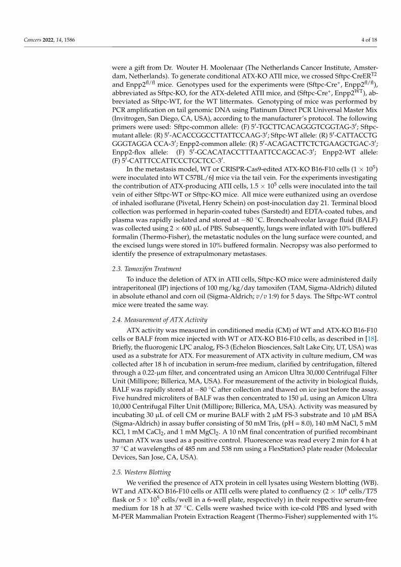
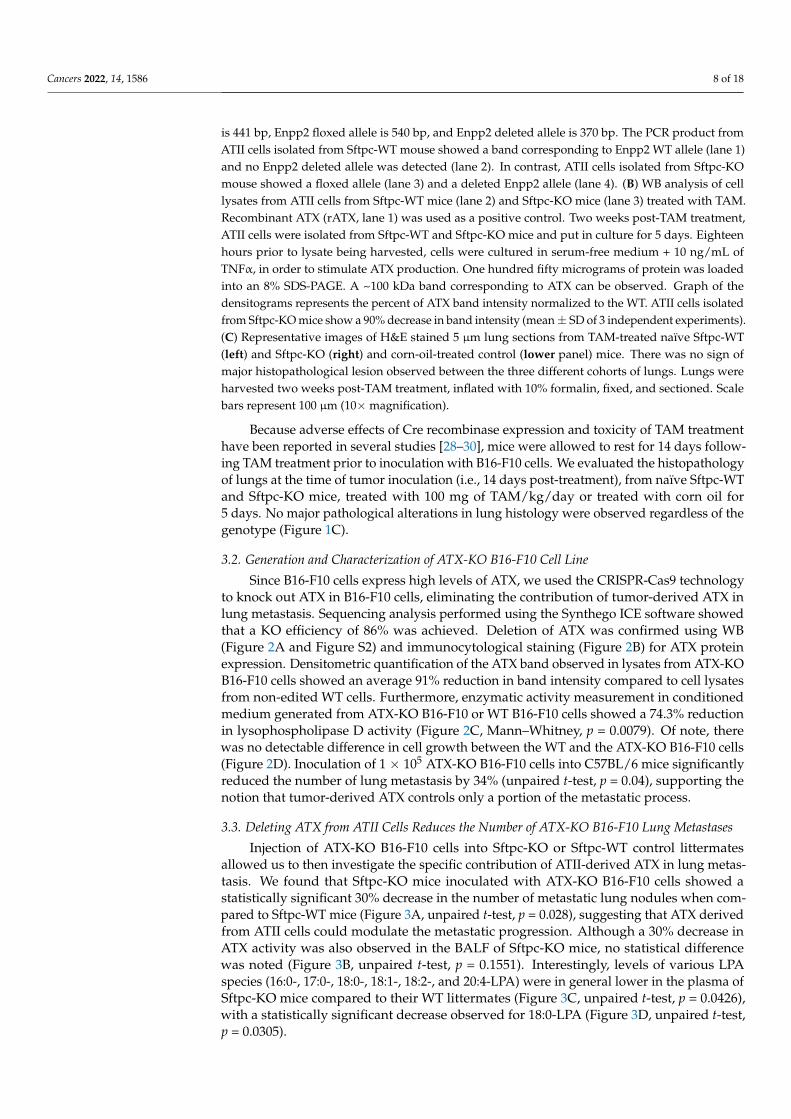
In order to determine whether ATX derived from ATII cells modulates B16-F10 metas-tasis to the lung, we crossed Sftpc-CreERT2 mice with Enpp2fl/fl mice to specifically deletethe ATX gene in ATII cells (i.e., Sftpc-KO mice). We validated the KO efficiency of ATX inthis model by comparing the ATX expression in isolated ATII cells after in vivo inductionby TAM. Following PCR amplification, Cre-mediated deletion of the floxed ATX allele wasdetected only in ATII cells isolated from Sftpc-KO mice (Figure 1A). Similarly, we detecteda 90% reduction in ATX protein expression by immunoblot in cells isolated from Sftpc-KOmice (Figures 1B and S1) compared to those from WT littermates, after TNFα stimulation.
Cancers 2022, 14, x FOR PEER REVIEW 8 of 19
Figure 1. In vitro characterization of Sftpc-WT and Sftpc-KO mice. (A) Agarose electrophoresis of PCR-amplified Enpp2 allele of ATII cells isolated from Sftpc-WT (lanes 1 and 2) and Sftpc-KO (lanes 3 and 4) mice, treated in vivo with TAM (100 mg/kg/day for 5 days). The size of the Enpp2 WT allele is 441 bp, Enpp2 floxed allele is 540 bp, and Enpp2 deleted allele is 370 bp. The PCR product from ATII cells isolated from Sftpc-WT mouse showed a band corresponding to Enpp2 WT allele (lane 1) and no Enpp2 deleted allele was detected (lane 2). In contrast, ATII cells isolated from Sftpc-KO mouse showed a floxed allele (lane 3) and a deleted Enpp2 allele (lane 4). (B) WB analysis of cell lysates from ATII cells from Sftpc-WT mice (lane 2) and Sftpc-KO mice (lane 3) treated with TAM. Recombinant ATX (rATX, lane 1) was used as a positive control. Two weeks post-TAM treatment, ATII cells were isolated from Sftpc-WT and Sftpc-KO mice and put in culture for 5 days. Eighteen hours prior to lysate being harvested, cells were cultured in serum-free medium + 10 ng/mL of TNFα, in order to stimulate ATX production. One hundred fifty micrograms of protein was loaded into an 8% SDS-PAGE. A ~100 kDa band corresponding to ATX can be observed. Graph of the densitograms represents the percent of ATX band intensity normalized to the WT. ATII cells isolated from Sftpc-KO mice show a 90% decrease in band intensity (mean ± SD of 3 independent experiments). (C) Representative images of H&E stained 5 μm lung sections from TAM-treated naïve Sftpc-WT (left) and Sftpc-KO (right) and corn-oil-treated control (lower panel) mice. There was no sign of major histopathological lesion observed between the three different cohorts of lungs. Lungs were harvested two weeks post-TAM treatment, inflated with 10% formalin, fixed, and sectioned. Scale bars represent 100 μm (10× magnification).
3.2. Generation and Characterization of ATX-KO B16-F10 Cell Line
Figure 1. In vitro characterization of Sftpc-WT and Sftpc-KO mice. (A) Agarose electrophoresis ofPCR-amplified Enpp2 allele of ATII cells isolated from Sftpc-WT (lanes 1 and 2) and Sftpc-KO (lanes 3and 4) mice, treated in vivo with TAM (100 mg/kg/day for 5 days). The size of the Enpp2 WT allele
Cancers 2022, 14, 1586 8 of 18
is 441 bp, Enpp2 floxed allele is 540 bp, and Enpp2 deleted allele is 370 bp. The PCR product fromATII cells isolated from Sftpc-WT mouse showed a band corresponding to Enpp2 WT allele (lane 1)and no Enpp2 deleted allele was detected (lane 2). In contrast, ATII cells isolated from Sftpc-KOmouse showed a floxed allele (lane 3) and a deleted Enpp2 allele (lane 4). (B) WB analysis of celllysates from ATII cells from Sftpc-WT mice (lane 2) and Sftpc-KO mice (lane 3) treated with TAM.Recombinant ATX (rATX, lane 1) was used as a positive control. Two weeks post-TAM treatment,ATII cells were isolated from Sftpc-WT and Sftpc-KO mice and put in culture for 5 days. Eighteenhours prior to lysate being harvested, cells were cultured in serum-free medium + 10 ng/mL ofTNFα, in order to stimulate ATX production. One hundred fifty micrograms of protein was loadedinto an 8% SDS-PAGE. A ~100 kDa band corresponding to ATX can be observed. Graph of thedensitograms represents the percent of ATX band intensity normalized to the WT. ATII cells isolatedfrom Sftpc-KO mice show a 90% decrease in band intensity (mean± SD of 3 independent experiments).(C) Representative images of H&E stained 5 µm lung sections from TAM-treated naïve Sftpc-WT(left) and Sftpc-KO (right) and corn-oil-treated control (lower panel) mice. There was no sign ofmajor histopathological lesion observed between the three different cohorts of lungs. Lungs wereharvested two weeks post-TAM treatment, inflated with 10% formalin, fixed, and sectioned. Scalebars represent 100 µm (10×magnification).
Because adverse effects of Cre recombinase expression and toxicity of TAM treatmenthave been reported in several studies [28–30], mice were allowed to rest for 14 days follow-ing TAM treatment prior to inoculation with B16-F10 cells. We evaluated the histopathologyof lungs at the time of tumor inoculation (i.e., 14 days post-treatment), from naïve Sftpc-WTand Sftpc-KO mice, treated with 100 mg of TAM/kg/day or treated with corn oil for5 days. No major pathological alterations in lung histology were observed regardless of thegenotype (Figure 1C).
3.2. Generation and Characterization of ATX-KO B16-F10 Cell Line
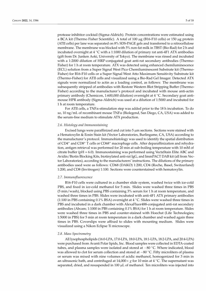
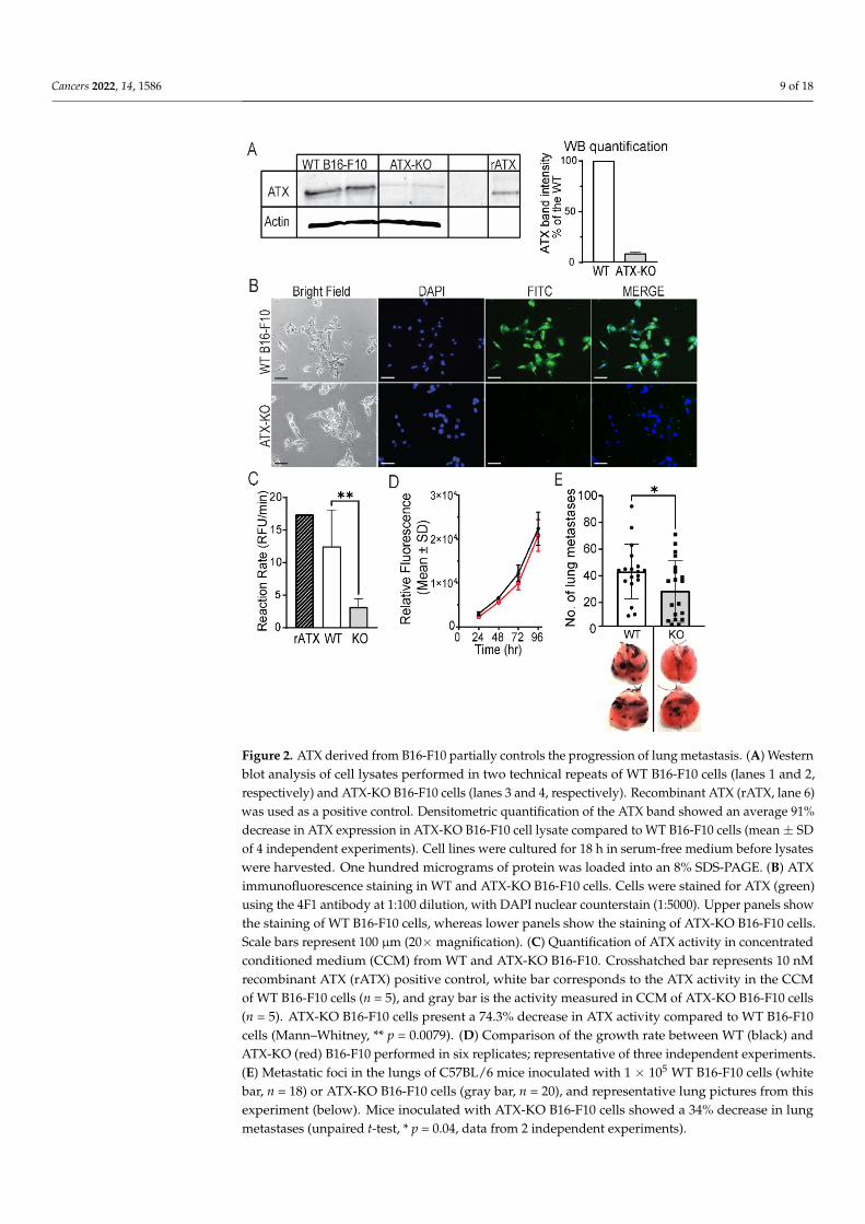
Since B16-F10 cells express high levels of ATX, we used the CRISPR-Cas9 technologyto knock out ATX in B16-F10 cells, eliminating the contribution of tumor-derived ATX inlung metastasis. Sequencing analysis performed using the Synthego ICE software showedthat a KO efficiency of 86% was achieved. Deletion of ATX was confirmed using WB(Figure 2A and Figure S2) and immunocytological staining (Figure 2B) for ATX proteinexpression. Densitometric quantification of the ATX band observed in lysates from ATX-KOB16-F10 cells showed an average 91% reduction in band intensity compared to cell lysatesfrom non-edited WT cells. Furthermore, enzymatic activity measurement in conditionedmedium generated from ATX-KO B16-F10 or WT B16-F10 cells showed a 74.3% reductionin lysophospholipase D activity (Figure 2C, Mann–Whitney, p = 0.0079). Of note, therewas no detectable difference in cell growth between the WT and the ATX-KO B16-F10 cells(Figure 2D). Inoculation of 1 × 10⁵ ATX-KO B16-F10 cells into C57BL/6 mice significantlyreduced the number of lung metastasis by 34% (unpaired t-test, p = 0.04), supporting thenotion that tumor-derived ATX controls only a portion of the metastatic process.
3.3. Deleting ATX from ATII Cells Reduces the Number of ATX-KO B16-F10 Lung Metastases
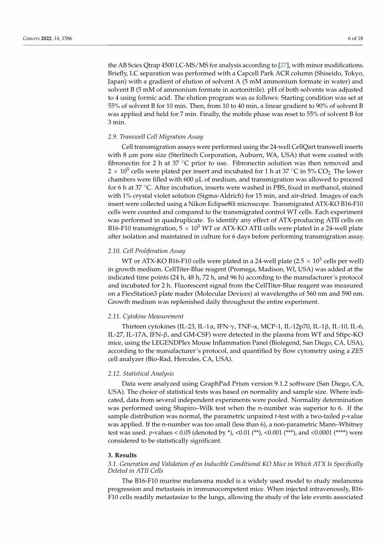
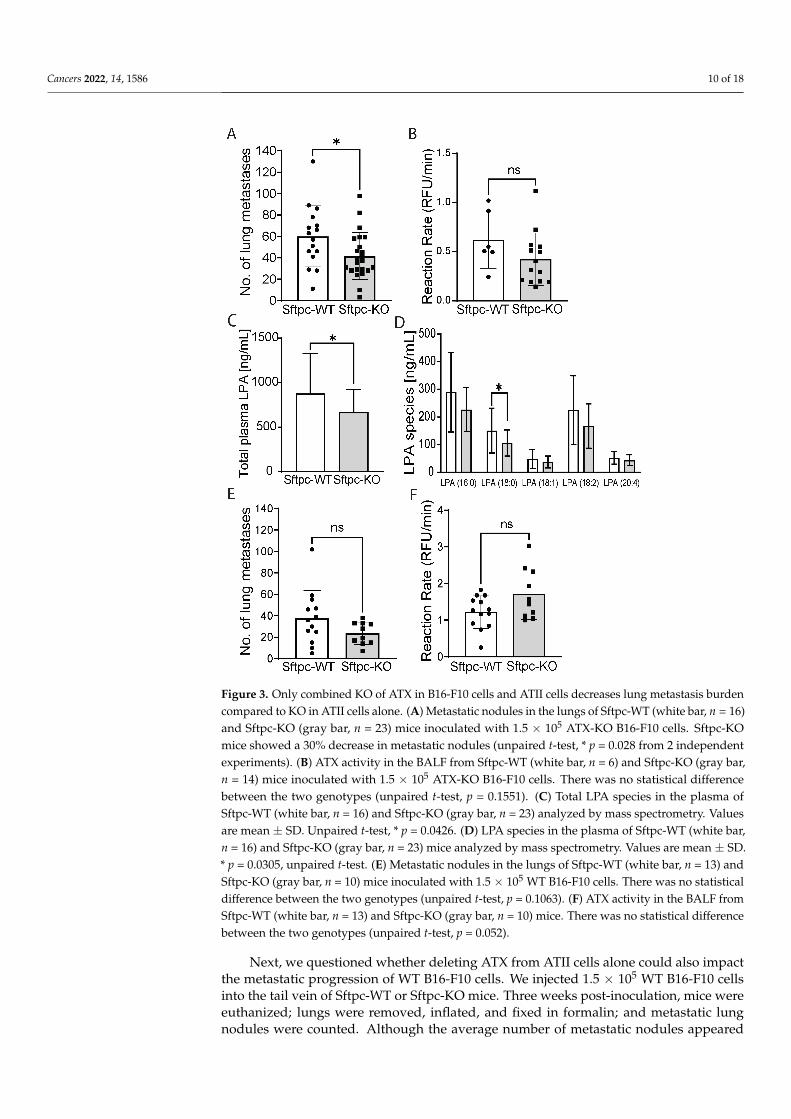
Injection of ATX-KO B16-F10 cells into Sftpc-KO or Sftpc-WT control littermatesallowed us to then investigate the specific contribution of ATII-derived ATX in lung metas-tasis. We found that Sftpc-KO mice inoculated with ATX-KO B16-F10 cells showed astatistically significant 30% decrease in the number of metastatic lung nodules when com-pared to Sftpc-WT mice (Figure 3A, unpaired t-test, p = 0.028), suggesting that ATX derivedfrom ATII cells could modulate the metastatic progression. Although a 30% decrease inATX activity was also observed in the BALF of Sftpc-KO mice, no statistical differencewas noted (Figure 3B, unpaired t-test, p = 0.1551). Interestingly, levels of various LPAspecies (16:0-, 17:0-, 18:0-, 18:1-, 18:2-, and 20:4-LPA) were in general lower in the plasma ofSftpc-KO mice compared to their WT littermates (Figure 3C, unpaired t-test, p = 0.0426),with a statistically significant decrease observed for 18:0-LPA (Figure 3D, unpaired t-test,p = 0.0305).
Cancers 2022, 14, 1586 9 of 18
Cancers 2022, 14, x FOR PEER REVIEW 9 of 19
Since B16-F10 cells express high levels of ATX, we used the CRISPR-Cas9 technology to knock out ATX in B16-F10 cells, eliminating the contribution of tumor-derived ATX in lung metastasis. Sequencing analysis performed using the Synthego ICE software showed that a KO efficiency of 86% was achieved. Deletion of ATX was confirmed using WB (Figures 2A and S2) and immunocytological staining (Figure 2B) for ATX protein expression. Densitometric quantification of the ATX band observed in lysates from ATX-KO B16-F10 cells showed an average 91% reduction in band intensity compared to cell lysates from non-edited WT cells. Furthermore, enzymatic activity measurement in conditioned medium generated from ATX-KO B16-F10 or WT B16-F10 cells showed a 74.3% reduction in lysophospholipase D activity (Figure 2C, Mann–Whitney, p = 0.0079). Of note, there was no detectable difference in cell growth between the WT and the ATX-KO B16-F10 cells (Figure 2D). Inoculation of 1 × 10⁵ ATX-KO B16-F10 cells into C57BL/6 mice significantly reduced the number of lung metastasis by 34% (unpaired t-test, p = 0.04), supporting the notion that tumor-derived ATX controls only a portion of the metastatic process.
Figure 2. ATX derived from B16-F10 partially controls the progression of lung metastasis. (A) Western blot analysis of cell lysates performed in two technical repeats of WT B16-F10 cells (lanes 1 and 2, respectively) and ATX-KO B16-F10 cells (lanes 3 and 4, respectively). Recombinant ATX (rATX, lane 6) was used as a positive control. Densitometric quantification of the ATX band showed an average 91% decrease in ATX expression in ATX-KO B16-F10 cell lysate compared to WT B16-F10 cells (mean ± SD of 4 independent experiments). Cell lines were cultured for 18 h in serum-free
Figure 2. ATX derived from B16-F10 partially controls the progression of lung metastasis. (A) Westernblot analysis of cell lysates performed in two technical repeats of WT B16-F10 cells (lanes 1 and 2,respectively) and ATX-KO B16-F10 cells (lanes 3 and 4, respectively). Recombinant ATX (rATX, lane 6)was used as a positive control. Densitometric quantification of the ATX band showed an average 91%decrease in ATX expression in ATX-KO B16-F10 cell lysate compared to WT B16-F10 cells (mean ± SDof 4 independent experiments). Cell lines were cultured for 18 h in serum-free medium before lysateswere harvested. One hundred micrograms of protein was loaded into an 8% SDS-PAGE. (B) ATXimmunofluorescence staining in WT and ATX-KO B16-F10 cells. Cells were stained for ATX (green)using the 4F1 antibody at 1:100 dilution, with DAPI nuclear counterstain (1:5000). Upper panels showthe staining of WT B16-F10 cells, whereas lower panels show the staining of ATX-KO B16-F10 cells.Scale bars represent 100 µm (20×magnification). (C) Quantification of ATX activity in concentratedconditioned medium (CCM) from WT and ATX-KO B16-F10. Crosshatched bar represents 10 nMrecombinant ATX (rATX) positive control, white bar corresponds to the ATX activity in the CCMof WT B16-F10 cells (n = 5), and gray bar is the activity measured in CCM of ATX-KO B16-F10 cells(n = 5). ATX-KO B16-F10 cells present a 74.3% decrease in ATX activity compared to WT B16-F10cells (Mann–Whitney, ** p = 0.0079). (D) Comparison of the growth rate between WT (black) andATX-KO (red) B16-F10 performed in six replicates; representative of three independent experiments.(E) Metastatic foci in the lungs of C57BL/6 mice inoculated with 1 × 10⁵ WT B16-F10 cells (whitebar, n = 18) or ATX-KO B16-F10 cells (gray bar, n = 20), and representative lung pictures from thisexperiment (below). Mice inoculated with ATX-KO B16-F10 cells showed a 34% decrease in lungmetastases (unpaired t-test, * p = 0.04, data from 2 independent experiments).
Cancers 2022, 14, 1586 10 of 18Cancers 2022, 14, x FOR PEER REVIEW 11 of 19
Figure 3. Only combined KO of ATX in B16-F10 cells and ATII cells decreases lung metastasis burden compared to KO in ATII cells alone. (A) Metastatic nodules in the lungs of Sftpc-WT (white bar, n = 16) and Sftpc-KO (gray bar, n = 23) mice inoculated with 1.5 × 10⁵ ATX-KO B16-F10 cells. Sftpc-KO mice showed a 30% decrease in metastatic nodules (unpaired t-test, * p = 0.028 from 2 independent experiments). (B) ATX activity in the BALF from Sftpc-WT (white bar, n = 6) and Sftpc-KO (gray bar, n = 14) mice inoculated with 1.5 × 10⁵ ATX-KO B16-F10 cells. There was no statistical difference between the two genotypes (unpaired t-test, p = 0.1551). (C) Total LPA species in the plasma of Sftpc-WT (white bar, n = 16) and Sftpc-KO (gray bar, n = 23) analyzed by mass spectrometry. Values are mean ± SD. Unpaired t-test, * p = 0.0426. (D) LPA species in the plasma of Sftpc-WT (white bar, n = 16) and Sftpc-KO (gray bar, n = 23) mice analyzed by mass spectrometry. Values are mean ± SD. * p = 0.0305, unpaired t-test. (E) Metastatic nodules in the lungs of Sftpc-WT (white bar, n = 13) and Sftpc-KO (gray bar, n = 10) mice inoculated with 1.5 × 10⁵ WT B16-F10 cells. There was no statistical difference between the two genotypes (unpaired t-test, p = 0.1063). (F) ATX activity in the BALF from Sftpc-WT (white bar, n = 13) and Sftpc-KO (gray bar, n = 10) mice. There was no statistical difference between the two genotypes (unpaired t-test, p = 0.052).
3.4. Changes in Immunological Response Associated with Deleting ATX in ATII Cells To understand the mechanism(s) of the reduction in lung metastasis, we first
compared the relative transmigration abilities of WT and ATX-KO B16-F10 cells in the presence of ATII cells isolated from Sftpc-WT and Sftpc-KO mice. However, no difference was observed in the transmigration of either ATX-KO B16-F10 cells compared to WT B16-F10 cells alone (Figure 4A, Mann–Whitney, p = 0.083) or WT and ATX-KO B16-F10 cells in
Figure 3. Only combined KO of ATX in B16-F10 cells and ATII cells decreases lung metastasis burdencompared to KO in ATII cells alone. (A) Metastatic nodules in the lungs of Sftpc-WT (white bar, n = 16)and Sftpc-KO (gray bar, n = 23) mice inoculated with 1.5 × 10⁵ ATX-KO B16-F10 cells. Sftpc-KOmice showed a 30% decrease in metastatic nodules (unpaired t-test, * p = 0.028 from 2 independentexperiments). (B) ATX activity in the BALF from Sftpc-WT (white bar, n = 6) and Sftpc-KO (gray bar,n = 14) mice inoculated with 1.5 × 10⁵ ATX-KO B16-F10 cells. There was no statistical differencebetween the two genotypes (unpaired t-test, p = 0.1551). (C) Total LPA species in the plasma ofSftpc-WT (white bar, n = 16) and Sftpc-KO (gray bar, n = 23) analyzed by mass spectrometry. Valuesare mean ± SD. Unpaired t-test, * p = 0.0426. (D) LPA species in the plasma of Sftpc-WT (white bar,n = 16) and Sftpc-KO (gray bar, n = 23) mice analyzed by mass spectrometry. Values are mean ± SD.* p = 0.0305, unpaired t-test. (E) Metastatic nodules in the lungs of Sftpc-WT (white bar, n = 13) andSftpc-KO (gray bar, n = 10) mice inoculated with 1.5 × 10⁵ WT B16-F10 cells. There was no statisticaldifference between the two genotypes (unpaired t-test, p = 0.1063). (F) ATX activity in the BALF fromSftpc-WT (white bar, n = 13) and Sftpc-KO (gray bar, n = 10) mice. There was no statistical differencebetween the two genotypes (unpaired t-test, p = 0.052).
Next, we questioned whether deleting ATX from ATII cells alone could also impactthe metastatic progression of WT B16-F10 cells. We injected 1.5 × 10⁵ WT B16-F10 cellsinto the tail vein of Sftpc-WT or Sftpc-KO mice. Three weeks post-inoculation, mice wereeuthanized; lungs were removed, inflated, and fixed in formalin; and metastatic lungnodules were counted. Although the average number of metastatic nodules appeared
Cancers 2022, 14, 1586 11 of 18
to be trending lower in Sftpc-KO mice, it did not reach statistical significance (Figure 3E,unpaired t-test, p = 0.1063). One possible explanation could be that the total amount of ATXpresent in the lungs, both from the WT B16-F10 cells which highly express ATX and fromother stromal cell types, may mask or compensate for the reduction in ATX generated byATII cells, and the latter pool alone failed to significantly impact the metastatic progressionof WT B16-F10 cells. Indeed, when we measured the ATX activity in the BALF of Sftpc-WTand Sftpc-KO mice inoculated with WT B16-F10 cells, we found no statistically significantdifference (Figure 3F). Taken together, our data suggest that ATX derived from ATII cellsmay influence metastatic progression, particularly in tumors with low ATX expression.
3.4. Changes in Immunological Response Associated with Deleting ATX in ATII Cells
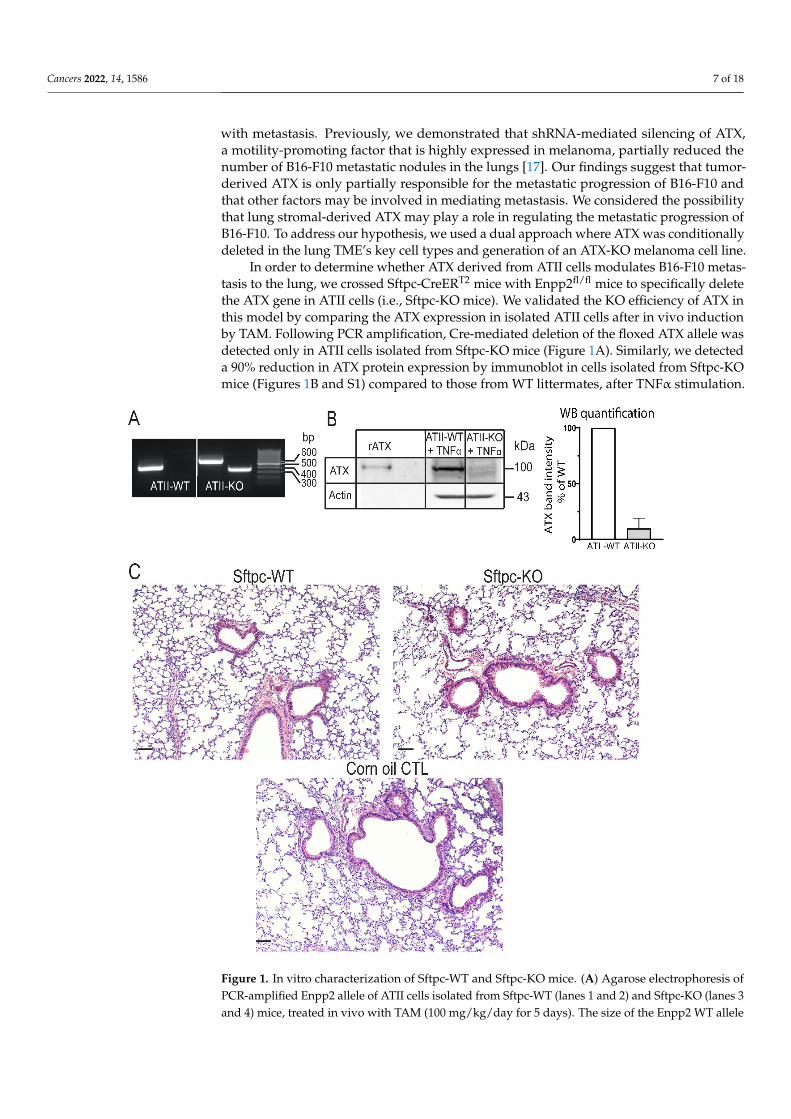
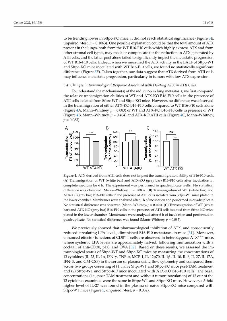
To understand the mechanism(s) of the reduction in lung metastasis, we first comparedthe relative transmigration abilities of WT and ATX-KO B16-F10 cells in the presence ofATII cells isolated from Sftpc-WT and Sftpc-KO mice. However, no difference was observedin the transmigration of either ATX-KO B16-F10 cells compared to WT B16-F10 cells alone(Figure 4A, Mann–Whitney, p = 0.083) or WT and ATX-KO B16-F10 cells in presence of WT(Figure 4B, Mann–Whitney, p = 0.404) and ATX-KO ATII cells (Figure 4C, Mann–Whitney,p = 0.083).
Cancers 2022, 14, x FOR PEER REVIEW 12 of 19
presence of WT (Figure 4B, Mann–Whitney, p = 0.404) and ATX-KO ATII cells (Figure 4C, Mann–Whitney, p = 0.083).
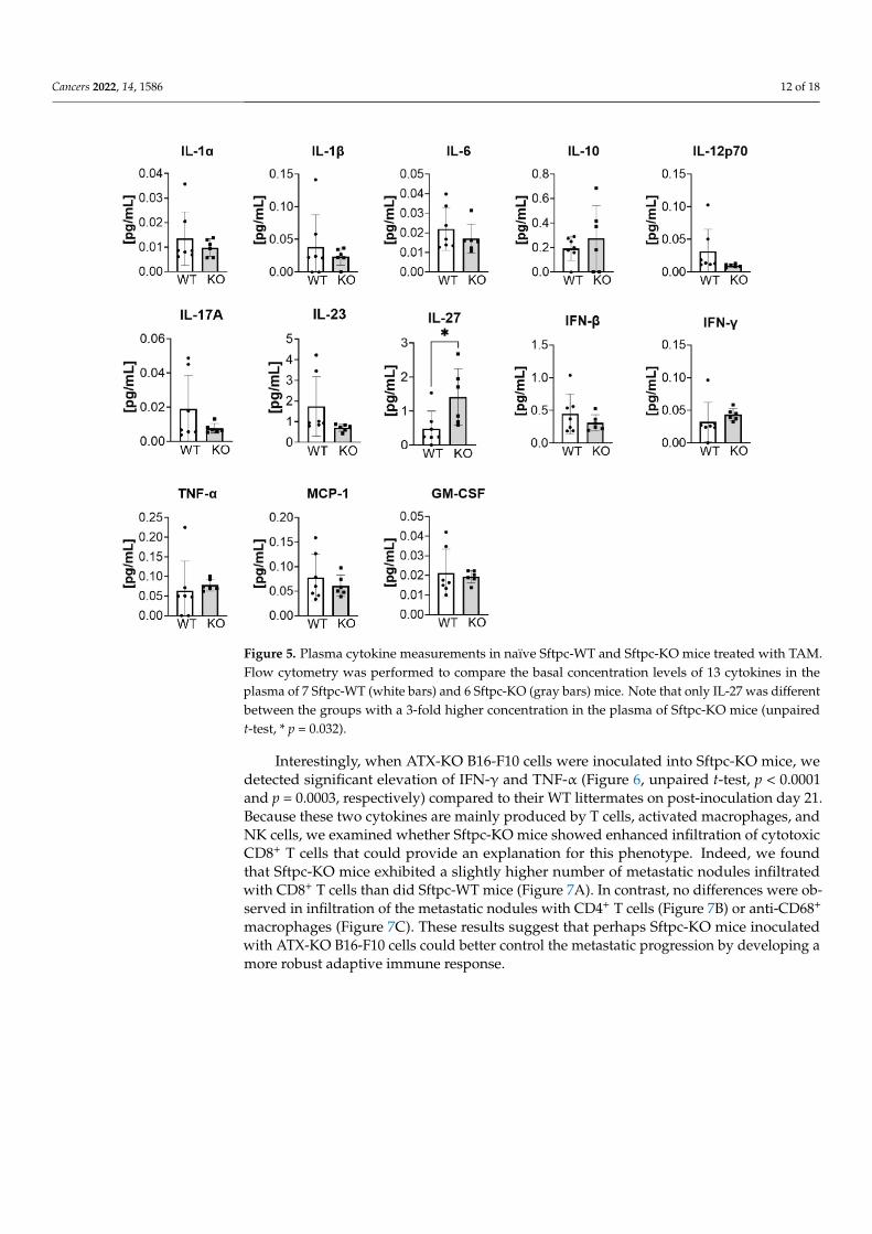
We previously showed that pharmacological inhibition of ATX, and consequently reduced circulating LPA levels, diminished B16-F10 metastases in mice [31]. Moreover, enhanced effector functions of CD8+ T cells are observed in heterozygous ATX+/− mice, where systemic LPA levels are approximately halved, following immunization with a cocktail of anti-CD30, pl:C, and OVA [32]. Based on these results, we assessed the immunological status of Sftpc-WT and Sftpc-KO mice by measuring the concentrations of 13 cytokines (IL-23, IL-1α, IFN-γ, TNF-α, MCP-1, IL-12p70, IL-1β, IL-10, IL-6, IL-27, IL-17A, IFN-β, and GM-CSF) in the serum or plasma using flow cytometry and compared them across two groups consisting of (1) naïve Sftpc-WT and Sftpc-KO mice post-TAM treatment and (2) Sftpc-WT and Sftpc-KO mice inoculated with ATX-KO B16-F10 cells. The basal concentrations (i.e., post-TAM treatment and without tumor inoculation) of 12 out of the 13 cytokines examined were the same in Sftpc-WT and Sftpc-KO mice. However, a 3-fold higher level of IL-27 was found in the plasma of naïve Sftpc-KO mice compared with Sftpc-WT mice (Figure 5, unpaired t-test, p = 0.032).
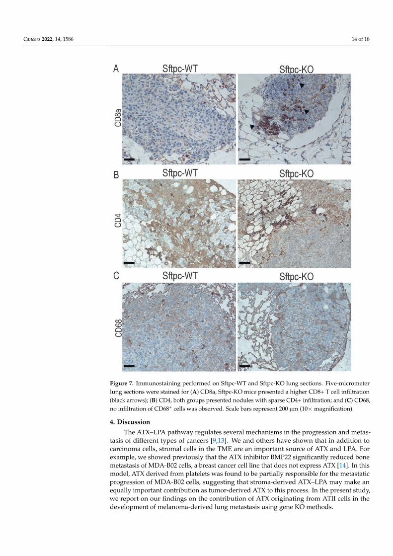
Interestingly, when ATX-KO B16-F10 cells were inoculated into Sftpc-KO mice, we detected significant elevation of IFN-γ and TNF-α (Figure 6, unpaired t-test, p < 0.0001 and p = 0.0003, respectively) compared to their WT littermates on post-inoculation day 21. Because these two cytokines are mainly produced by T cells, activated macrophages, and NK cells, we examined whether Sftpc-KO mice showed enhanced infiltration of cytotoxic CD8+ T cells that could provide an explanation for this phenotype. Indeed, we found that Sftpc-KO mice exhibited a slightly higher number of metastatic nodules infiltrated with CD8+ T cells than did Sftpc-WT mice (Figure 7A). In contrast, no differences were observed in infiltration of the metastatic nodules with CD4+ T cells (Figure 7B) or anti-CD68+ macrophages (Figure 7C). These results suggest that perhaps Sftpc-KO mice inoculated with ATX-KO B16-F10 cells could better control the metastatic progression by developing a more robust adaptive immune response.
Figure 4. ATX derived from ATII cells does not impact the transmigration ability of B16-F10 cells. (A) Transmigration of WT (white bar) and ATX-KO (gray bar) B16-F10 cells after incubation in complete medium for 6 h. The experiment was performed in quadruplicate wells. No statistical difference was observed (Mann–Whitney, p = 0.083). (B) Transmigration of WT (white bar) and ATX-KO (gray bar) B16-F10 cells in the presence of ATII cells isolated from Sftpc-WT mice plated in the lower chamber. Membranes were analyzed after 6 h of incubation and performed in quadruplicate. No statistical difference was observed (Mann–Whitney, p = 0.404). (C) Transmigration of WT (white bar) and ATX-KO (gray bar) B16-F10 cells in the presence of ATII cells isolated from Sftpc-KO mice plated in the lower chamber. Membranes were analyzed after 6 h of incubation and performed in quadruplicate. No statistical difference was found (Mann–Whitney, p = 0.083).
Figure 4. ATX derived from ATII cells does not impact the transmigration ability of B16-F10 cells.(A) Transmigration of WT (white bar) and ATX-KO (gray bar) B16-F10 cells after incubation incomplete medium for 6 h. The experiment was performed in quadruplicate wells. No statisticaldifference was observed (Mann–Whitney, p = 0.083). (B) Transmigration of WT (white bar) andATX-KO (gray bar) B16-F10 cells in the presence of ATII cells isolated from Sftpc-WT mice plated inthe lower chamber. Membranes were analyzed after 6 h of incubation and performed in quadruplicate.No statistical difference was observed (Mann–Whitney, p = 0.404). (C) Transmigration of WT (whitebar) and ATX-KO (gray bar) B16-F10 cells in the presence of ATII cells isolated from Sftpc-KO miceplated in the lower chamber. Membranes were analyzed after 6 h of incubation and performed inquadruplicate. No statistical difference was found (Mann–Whitney, p = 0.083).
We previously showed that pharmacological inhibition of ATX, and consequentlyreduced circulating LPA levels, diminished B16-F10 metastases in mice [31]. Moreover,enhanced effector functions of CD8+ T cells are observed in heterozygous ATX+/− mice,where systemic LPA levels are approximately halved, following immunization with acocktail of anti-CD30, pl:C, and OVA [32]. Based on these results, we assessed the im-munological status of Sftpc-WT and Sftpc-KO mice by measuring the concentrations of13 cytokines (IL-23, IL-1α, IFN-γ, TNF-α, MCP-1, IL-12p70, IL-1β, IL-10, IL-6, IL-27, IL-17A,IFN-β, and GM-CSF) in the serum or plasma using flow cytometry and compared themacross two groups consisting of (1) naïve Sftpc-WT and Sftpc-KO mice post-TAM treatmentand (2) Sftpc-WT and Sftpc-KO mice inoculated with ATX-KO B16-F10 cells. The basalconcentrations (i.e., post-TAM treatment and without tumor inoculation) of 12 out of the13 cytokines examined were the same in Sftpc-WT and Sftpc-KO mice. However, a 3-foldhigher level of IL-27 was found in the plasma of naïve Sftpc-KO mice compared withSftpc-WT mice (Figure 5, unpaired t-test, p = 0.032).
Cancers 2022, 14, 1586 12 of 18Cancers 2022, 14, x FOR PEER REVIEW 13 of 19
Figure 5. Plasma cytokine measurements in naïve Sftpc-WT and Sftpc-KO mice treated with TAM. Flow cytometry was performed to compare the basal concentration levels of 13 cytokines in the plasma of 7 Sftpc-WT (white bars) and 6 Sftpc-KO (gray bars) mice. Note that only IL-27 was different between the groups with a 3-fold higher concentration in the plasma of Sftpc-KO mice (unpaired t-test, * p = 0.032).
Figure 5. Plasma cytokine measurements in naïve Sftpc-WT and Sftpc-KO mice treated with TAM.Flow cytometry was performed to compare the basal concentration levels of 13 cytokines in theplasma of 7 Sftpc-WT (white bars) and 6 Sftpc-KO (gray bars) mice. Note that only IL-27 was differentbetween the groups with a 3-fold higher concentration in the plasma of Sftpc-KO mice (unpairedt-test, * p = 0.032).
Interestingly, when ATX-KO B16-F10 cells were inoculated into Sftpc-KO mice, wedetected significant elevation of IFN-γ and TNF-α (Figure 6, unpaired t-test, p < 0.0001and p = 0.0003, respectively) compared to their WT littermates on post-inoculation day 21.Because these two cytokines are mainly produced by T cells, activated macrophages, andNK cells, we examined whether Sftpc-KO mice showed enhanced infiltration of cytotoxicCD8+ T cells that could provide an explanation for this phenotype. Indeed, we foundthat Sftpc-KO mice exhibited a slightly higher number of metastatic nodules infiltratedwith CD8+ T cells than did Sftpc-WT mice (Figure 7A). In contrast, no differences were ob-served in infiltration of the metastatic nodules with CD4+ T cells (Figure 7B) or anti-CD68+
macrophages (Figure 7C). These results suggest that perhaps Sftpc-KO mice inoculatedwith ATX-KO B16-F10 cells could better control the metastatic progression by developing amore robust adaptive immune response.

Cancers 2022, 14, 1586 13 of 18
Cancers 2022, 14, x FOR PEER REVIEW 14 of 19
Figure 6. Cytokine measurements in the plasma of Sftpc-WT and Sftpc-KO mice on post-inoculation day 21. Flow cytometry was performed to compare the concentration of 13 cytokines in the plasma of Sftpc-WT (white bars) and Sftpc-KO (gray bars) mice, on day 21 post-inoculation, inoculated with 1.5 × 10⁵ ATX-KO B16-F10 cells. Sftpc-KO mice presented an increase in 2 out of the 13 cytokines (unpaired t-test, IFNγ, **** p < 0.0001; TNFα, *** p = 0.0003).
Figure 6. Cytokine measurements in the plasma of Sftpc-WT and Sftpc-KO mice on post-inoculationday 21. Flow cytometry was performed to compare the concentration of 13 cytokines in the plasma ofSftpc-WT (white bars) and Sftpc-KO (gray bars) mice, on day 21 post-inoculation, inoculated with1.5 × 105 ATX-KO B16-F10 cells. Sftpc-KO mice presented an increase in 2 out of the 13 cytokines(unpaired t-test, IFNγ, **** p < 0.0001; TNFα, *** p = 0.0003).
Cancers 2022, 14, 1586 14 of 18Cancers 2022, 14, x FOR PEER REVIEW 15 of 19
Figure 7. Immunostaining performed on Sftpc-WT and Sftpc-KO lung sections. Five-micrometer lung sections were stained for (A) CD8a, Sftpc-KO mice presented a higher CD8+ T cell infiltration (black arrows); (B) CD4, both groups presented nodules with sparse CD4+ infiltration; and (C) CD68, no infiltration of CD68+ cells was observed. Scale bars represent 200 μm (10× magnification).
4. Discussion The ATX–LPA pathway regulates several mechanisms in the progression and
metastasis of different types of cancers [9,13]. We and others have shown that in addition to carcinoma cells, stromal cells in the TME are an important source of ATX and LPA. For example, we showed previously that the ATX inhibitor BMP22 significantly reduced bone metastasis of MDA-B02 cells, a breast cancer cell line that does not express ATX [14]. In this model, ATX derived from platelets was found to be partially responsible for the metastatic progression of MDA-B02 cells, suggesting that stroma-derived ATX–LPA may make an equally important contribution as tumor-derived ATX to this process. In the present study, we report on our findings on the contribution of ATX originating from ATII cells in the development of melanoma-derived lung metastasis using gene KO methods.
Figure 7. Immunostaining performed on Sftpc-WT and Sftpc-KO lung sections. Five-micrometerlung sections were stained for (A) CD8a, Sftpc-KO mice presented a higher CD8+ T cell infiltration(black arrows); (B) CD4, both groups presented nodules with sparse CD4+ infiltration; and (C) CD68,no infiltration of CD68+ cells was observed. Scale bars represent 200 µm (10×magnification).
4. Discussion
The ATX–LPA pathway regulates several mechanisms in the progression and metas-tasis of different types of cancers [9,13]. We and others have shown that in addition tocarcinoma cells, stromal cells in the TME are an important source of ATX and LPA. Forexample, we showed previously that the ATX inhibitor BMP22 significantly reduced bonemetastasis of MDA-B02 cells, a breast cancer cell line that does not express ATX [14]. In thismodel, ATX derived from platelets was found to be partially responsible for the metastaticprogression of MDA-B02 cells, suggesting that stroma-derived ATX–LPA may make anequally important contribution as tumor-derived ATX to this process. In the present study,we report on our findings on the contribution of ATX originating from ATII cells in thedevelopment of melanoma-derived lung metastasis using gene KO methods.
Cancers 2022, 14, 1586 15 of 18
Although ATII cells represent a small portion of the lung stroma (~5%), they alsosynthesize and secrete ATX, and after lung injury and remodeling, they serve as progenitorsof type I pneumocytes. In addition to these important roles, we show for the first time thatdeleting ATX from ATII cells led to a significant 30% decrease in ATX-KO B16-F10 lungmetastases. While ATX activity in the BALF of Sftpc-KO mice appeared to trend lower, itwas not statistically significant. Nonetheless, we observed a slight decrease in total plasmaLPA levels, with a significant decrease in 18:0-LPA. On the contrary, deleting ATX fromATII cells alone was not sufficient to significantly decrease the number of WT B16-F10lung metastatic nodules. We note that the Cre/loxP TAM-inducible KO system used inthis study does not provide 100% efficient recombination. Furthermore, we must take intoconsideration that ATII cells represent a low fraction of the total lung cell population. Thus,the lack of an effect of ATX KO in ATII cells alone on reducing the metastatic burden ofWT B16-F10 cells could be explained by the hypothesis that WT B16-F10 cells and possiblyother stromal cells in the lung TME produce substantially more ATX and LPA than doATII cells alone. This hypothesis could also explain why we failed to detect a statisticallysignificant difference in ATX activity in the BALF from Sftpc-WT compared with that ofSftpc-KO mice.
We did not observe differences in the transmigration of ATX-KO B16-F10 cells towardsATII cells isolated from either Sftpc-WT or Sftpc-KO mice that could account for the re-duced metastatic burden observed in Sftpc-KO mice. Therefore, our attention turned to therelative levels of antitumor immunity in Sftpc-WT versus Sftpc-KO mice. We performedimmunohistological staining for CD8-, CD4-, and CD68-positive tumor-infiltrating im-mune cells and measured relative levels of cytokines produced in Sftpc-WT and Sftpc-KOmice. At the basal level, mice from both genotypes treated with TAM and without tumorinoculation showed no difference in 12 out of the 13 cytokines tested, with the exceptionbeing IL-27, which was increased by ~3-fold in Sftpc-KO mice. A possible explanationfor this observation might be that the increase in IL-27 was caused by a natural reactiveresponse to TAM treatment. Indeed, Bockerstett et al. demonstrated that gastric lesionscaused by TAM administration can be reduced by administration of recombinant IL-27,leading to suppressed expression of proinflammatory genes [33]. However, this alone couldnot account for the nearly 3-fold increase in IL-27 being observed only in Sftpc-KO miceand not in Sftpc-WT mice, since both received the same TAM treatment. Therefore, wesuggest that this difference might be attributed to the model itself, where the KO of ATX inATII cells could directly or indirectly increase IL-27 production. It is also possible that thegenetic recombination in the Sftpc-KO mice might delay and/or prolong IL-27 productionbecause TAM can modulate the production of some cytokines [34]. These open questionswill have to be addressed experimentally in the future.
Examination of cytokine levels in tumor-bearing mice on post-inoculation day 21showed that inoculation of ATX-KO B16-F10 cells led to a selective increase in IFNγ andTNFα levels only in Sftpc-KO mice. Although these two cytokines play paradoxical rolesin cancer [35,36], several reports have demonstrated their anticancer activities [37–39]. Forexample, IFNγ is mainly produced by CD8+ and CD4+ T cells and can directly enhanceCD8+ T cell motility and cytotoxicity [40]. In support of this, we observed an increasedrecruitment of CD8+ T cells to the sites of metastases in Sftpc-KO mice that could beattributed to the increase in IFNγ levels. This could provide an explanation for why thesemice presented decreased numbers of metastatic nodules. It is interesting to note thatthis notion is in agreement with our previous reports demonstrating that stromal LPAhas a profound role in inhibiting tumor immunity [41] by blocking the activation of the Tcell [42] and B cell receptors [43], in turn attenuating cancer surveillance and tumor cellkilling. However, other reports have implicated the ATX–LPA axis in the polarization ofnaïve T cells [44], T cell motility [45], and increasing T cell entry to secondary lymphoidorgans [44,46]. Taken together, these point to the complexity and the spatial dependence ofthe actions of the ATX–LPA axis.
Cancers 2022, 14, 1586 16 of 18
Alternatively, the reduced number of lung metastases could also be attributed to theincreased levels of IL-27 in Sftpc-KO mice following TAM treatment. Indeed, IL-27 hasbeen shown to enhance NK cell activity in the B16-F10 model [47]. Therefore, the earlyoverexpression of IL-27 observed at the basal level in Sftpc-KO mice might establish a fasteractivation of NK cells, leading to a better control of lung metastasis development. Futureand extensive studies are needed to fully delineate the underlying mechanisms that willallow us to better understand how the modulation of these cytokines by the ATX–LPA axisimpacts immune regulation in the TME.
5. Conclusions
Taken together, our data generated using cell-type-specific KO methods suggest thatATX produced by ATII cells together with ATX synthesized by B16-F10 melanoma cells canimpair the adaptive antitumor immune response. They further suggest that inhibiting bothsources of ATX reduces the metastatic burden by increasing cytotoxic CD8+ T cell infiltra-tion. This in turn lends support to new arguments in favor of the therapeutic exploration ofATX inhibitors for the control of metastatic spread. This is a key finding as it suggests thatATX derived from ATII cells could affect the metastatic progress, particularly in tumorswith low ATX expression. More importantly, it indicates that therapeutic targeting of ATXcould still be a viable option even for patients whose tumors do not express high levelsof ATX. This is a new finding, and an extension from our previous work using a differentcancer model, where ATX derived from platelets modulates the metastatic progression ofbreast cancer cells that do not express ATX to the bone.
Supplementary Materials: The following are available online at https://www.mdpi.com/article/10.3390/cancers14061586/s1, Figure S1: Western blot analysis of cell lysates from ATII cells isolatedfrom Sftpc-WT and Sftpc-KO mice, Figure S2: Western blot analysis of cell lysates from WT andATX-KO B16-F10 cells.
Author Contributions: Conceptualization, M.A.D., S.C.L. and G.J.T.; methodology, M.A.D., S.C.L.,Y.S., D.D.N. and G.J.T.; formal analysis, M.A.D. and S.C.L.; investigation, M.A.D., S.C.L., Y.S., D.D.N.and G.J.T.; resources, S.C.L., K.-H.L., S.E. and G.J.T.; data curation, M.A.D., S.C.L., and Y.S.; writingand preparation of the original draft, M.A.D., S.C.L. and G.J.T.; writing, review and editing, M.A.D.,S.C.L., Y.S., D.D.N., K.-H.L., S.E., J.Y., Z.B. and G.J.T.; supervision, S.C.L. and G.J.T.; funding acqui-sition, S.C.L., K.-H.L., S.E. and G.J.T. All authors have read and agreed to the published version ofthe manuscript.
Funding: This research was funded by grants from the NCI (CA092160, G.J.T.), the Van VleetOncologic Research Fund (G.J.T.), the UTHSC Office of Research D3 Cornet Fund (G.J.T.), the Williamand Ella Owens Foundation (S.C.L.), and the Ministry of Science and Technology of Taiwan (MOST-109-2917I-564-029, K.H.L) and a grant from the Residency Program of the Department of Pathologyof UTHSC (S.E).
Institutional Review Board Statement: The study was conducted according to the guidelines of theInstitutional Animal Care and Use Committee (IACUC) at the University of Tennessee Health ScienceCenter (PHS assurance number A3325-01).
Informed Consent Statement: Not applicable.
Data Availability Statement: The data presented in this study are available on request from thecorresponding author.
Acknowledgments: The authors thank Jorge Solares and Cindy Lazar for their assistance with theimmunostaining, Louisa Balazs for her help with their interpretations, and Kyle Johnson-Moore fromthe UTHSC Office of Scientific Writing for editing the manuscript. This work was supported bygrants from the NCI (CA092160, G.T.), the Van Vleet Oncologic Research Fund (G.T.), the UTHSCOffice of Research D3 Cornet Fund (G.T.), the William and Ella Owens Foundation (S.C.L.), and theMinistry of Science and Technology (MOST-109-2917I-564-029, K.H.L.) of Taiwan and a grant fromthe Residency Program of the Department of Pathology of UTHSC (S.E.).
Conflicts of Interest: The authors declare no conflict of interest.
Cancers 2022, 14, 1586 17 of 18
References1. Fares, C.M.; Van Allen, E.M.; Drake, C.G.; Allison, J.P.; Hu-Lieskovan, S. Mechanisms of Resistance to Immune Checkpoint
Blockade: Why Does Checkpoint Inhibitor Immunotherapy Not Work for All Patients? Am. Soc. Clin. Oncol. Educ. Book 2019, 39,147–164. [CrossRef] [PubMed]
2. Michielin, O.; Atkins, M.B.; Koon, H.B.; Dummer, R.; Ascierto, P.A. Evolving impact of long-term survival results on metastaticmelanoma treatment. J. Immunother. Cancer 2020, 8, e000948. [CrossRef] [PubMed]
3. van Zijl, F.; Krupitza, G.; Mikulits, W. Initial steps of metastasis: Cell invasion and endothelial transmigration. Mutat. Res. Mutat.Res. 2011, 728, 23–34. [CrossRef]
4. Zhuyan, J.; Chen, M.; Zhu, T.; Bao, X.; Zhen, T.; Xing, K.; Wang, Q.; Zhu, S. Critical steps to tumor metastasis: Alterations oftumor microenvironment and extracellular matrix in the formation of pre-metastatic and metastatic niche. Cell Biosci. 2020, 10, 89.[CrossRef] [PubMed]
5. Brodt, P. Role of the Microenvironment in Liver Metastasis: From Pre- to Prometastatic Niches. Clin. Cancer Res. 2016, 22,5971–5982. [CrossRef] [PubMed]
6. Hirata, E.; Sahai, E. Tumor Microenvironment and Differential Responses to Therapy. Cold Spring Harb. Perspect. Med. 2017,7, a026781. [CrossRef]
7. Benavente, S.; Sánchez-García, A.; Naches, S.; Lleonart, M.E.; Lorente, J. Therapy-Induced Modulation of the Tumor Microenvi-ronment: New Opportunities for Cancer Therapies. Front. Oncol. 2020, 10, 582884. [CrossRef]
8. Tigyi, G.J.; Yue, J.; Norman, D.D.; Szabo, E.; Balogh, A.; Balazs, L.; Zhao, G.; Lee, S.C. Regulation of tumor cell-Microenvironmentinteraction by the autotaxin-lysophosphatidic acid receptor axis. Adv. Biol. Regul. 2019, 71, 183–193. [CrossRef]
9. Aiello, S.; Casiraghi, F. Lysophosphatidic Acid: Promoter of Cancer Progression and of Tumor Microenvironment Development.A Promising Target for Anticancer Therapies? Cells 2021, 10, 1390. [CrossRef]
10. Stracke, M.L.; Krutzsch, H.C.; Unsworth, E.J.; Arestad, A.; Cioce, V.; Schiffmann, E.; Liotta, L.A. Identification, purification, andpartial sequence analysis of autotaxin, a novel motility-stimulating protein. J. Biol. Chem. 1992, 267, 2524–2529. [CrossRef]
11. Benesch, M.G.K.; Tang, X.; Dewald, J.; Dong, W.-F.; Mackey, J.R.; Hemmings, D.G.; McMullen, T.P.W.; Brindley, D.N. Tumor-induced inflammation in mammary adipose tissue stimulates a vicious cycle of autotaxin expression and breast cancer progression.FASEB J. 2015, 29, 3990–4000. [CrossRef]
12. Brindley, D.N.; Lin, F.-T.; Tigyi, G.J. Role of the autotaxin–lysophosphatidate axis in cancer resistance to chemotherapy andradiotherapy. Biochim. Biophys. Acta (BBA)-Mol. Cell Biol. Lipids 2013, 1831, 74–85. [CrossRef]
13. Zhang, X.; Li, M.; Yin, N.; Zhang, J. The Expression Regulation and Biological Function of Autotaxin. Cells 2021, 10, 939. [CrossRef]14. Leblanc, R.; Lee, S.-C.; David, M.; Bordet, J.-C.; Norman, D.D.; Patil, R.; Miller, D.; Sahay, D.; Ribeiro, J.; Clézardin, P.; et al.
Interaction of platelet-derived autotaxin with tumor integrin αVβ3 controls metastasis of breast cancer cells to bone. Blood 2014,124, 3141–3150. [CrossRef]
15. Auciello, F.R.; Bulusu, V.; Oon, C.; Tait-Mulder, J.; Berry, M.; Bhattacharyya, S.; Tumanov, S.; Allen-Petersen, B.L.; Link, J.;Kendsersky, N.D.; et al. A Stromal Lysolipid–Autotaxin Signaling Axis Promotes Pancreatic Tumor Progression. Cancer Discov.2019, 9, 617–627. [CrossRef]
16. Magkrioti, C.; Oikonomou, N.; Kaffe, E.; Mouratis, M.-A.; Xylourgidis, N.; Barbayianni, I.; Megadoukas, P.; Harokopos, V.;Valavanis, C.; Chun, J.; et al. The autotaxin-lysophosphatidic acid axis promotes lung carcinogenesis. Cancer Res. 2018, 78,3634–3644. [CrossRef]
17. Gotoh, M.; Fujiwara, Y.; Yue, J.; Liu, J.; Lee, S.; Fells, J.; Uchiyama, A.; Murakami-Murofushi, K.; Kennel, S.; Wall, J.; et al.Controlling cancer through the autotaxin-lysophosphatidic acid receptor axis. Biochem. Soc. Trans. 2012, 40, 31–36. [CrossRef]
18. Lee, S.-C.; Fujiwara, Y.; Liu, J.; Yue, J.; Shimizu, Y.; Norman, D.D.; Wang, Y.; Tsukahara, R.; Szabo, E.; Patil, R.; et al. Autotaxin andLPA1 and LPA5 Receptors Exert Disparate Functions in Tumor Cells versus the Host Tissue Microenvironment in MelanomaInvasion and Metastasis. Mol. Cancer Res. 2015, 13, 174–185. [CrossRef]
19. Minn, A.J.; Gupta, G.P.; Siegel, P.M.; Bos, P.D.; Shu, W.; Giri, D.D.; Viale, A.; Olshen, A.B.; Gerald, W.L.; Massagué, J. Genes thatmediate breast cancer metastasis to lung. Nature 2005, 436, 518–524. [CrossRef]
20. Ruaro, B.; Salton, F.; Braga, L.; Wade, B.; Confalonieri, P.; Volpe, M.C.; Baratella, E.; Maiocchi, S.; Confalonieri, M. The Historyand Mystery of Alveolar Epithelial Type II Cells: Focus on Their Physiologic and Pathologic Role in Lung. Int. J. Mol. Sci. 2021,22, 2566. [CrossRef]
21. Castranova, V.; Rabovsky, J.; Tucker, J.; Miles, P. The alveolar type II epithelial cell: A multifunctional pneumocyte. Toxicol. Appl.Pharmacol. 1988, 93, 472–483. [CrossRef]
22. Zhao, J.; He, D.; Berdyshev, E.; Zhong, M.; Salgia, R.; Morris, A.J.; Smyth, S.S.; Natarajan, V.; Zhao, Y. Autotaxin induces lungepithelial cell migration through lysoPLD activity-dependent and -independent pathways. Biochem. J. 2011, 439, 45–55. [CrossRef]
23. Oikonomou, N.; Mouratis, M.-A.; Tzouvelekis, A.; Kaffe, E.; Valavanis, C.; Vilaras, G.; Karameris, A.; Prestwich, G.D.; Bouros, D.;Aidinis, V. Pulmonary Autotaxin Expression Contributes to the Pathogenesis of Pulmonary Fibrosis. Am. J. Respir. Cell Mol. Biol.2012, 47, 566–574. [CrossRef]
24. Sinha, M.; Lowell, C. Isolation of Highly Pure Primary Mouse Alveolar Epithelial Type II Cells by Flow Cytometric Cell Sorting.Bio-Protocol 2016, 6, e2013. [CrossRef]
25. Sun, F.; Xiao, G.; Qu, Z. Isolation of Murine Alveolar Type II Epithelial Cells. Bio-Protocol 2017, 7, 2288. [CrossRef]
Cancers 2022, 14, 1586 18 of 18
26. Van Meeteren, L.A.; Ruurs, P.; Stortelers, C.; Bouwman, P.; van Rooijen, M.A.; Pradère, J.P.; Pettit, T.R.; Wakelam, M.J.O.;Saulnier-Blache, J.S.; Mummery, C.L.; et al. Autotaxin, a Secreted Lysophospholipase D, Is Essential for Blood Vessel Formationduring Development. Mol. Cell. Biol. 2006, 26, 5015–5022. [CrossRef]
27. Okudaira, M.; Inoue, A.; Shuto, A.; Nakanaga, K.; Kano, K.; Makide, K.; Saigusa, D.; Tomioka, Y.; Aoki, J. Separation andquantification of 2-acyl-1-lysophospholipids and 1-acyl-2-lysophospholipids in biological samples by LC-MS/MS. J. Lipid Res.2014, 55, 2178–2192. [CrossRef]
28. Huh, W.J.; Khurana, S.S.; Geahlen, J.H.; Kohli, K.; Waller, R.A.; Mills, J.C. Tamoxifen Induces Rapid, Reversible Atrophy, andMetaplasia in Mouse Stomach. Gastroenterology 2012, 142, 21–24.e7. [CrossRef]
29. Etori, S.; Nakano, R.; Kamada, H.; Hosokawa, K.; Takeda, S.; Fukuhara, M.; Kenmotsu, Y.; Ishimine, A.; Sato, K. Tamoxifen-induced Lung Injury. Intern. Med. 2017, 56, 2903–2906. [CrossRef]
30. Donocoff, R.S.; Teteloshvili, N.; Chung, H.; Shoulson, R.; Creusot, R.J. Optimization of tamoxifen-induced Cre activity and itseffect on immune cell populations. Sci. Rep. 2020, 10, 15244. [CrossRef]
31. Gupte, R.; Patil, R.; Liu, J.; Wang, Y.; Lee, S.C.; Fujiwara, Y.; Fells, J.; Bolen, A.L.; Emmons-Thompson, K.; Yates, C.R.; et al. Benzyland Naphthalene Methylphosphonic Acid Inhibitors of Autotaxin with Anti-invasive and Anti-metastatic Activity. ChemMedChem2011, 6, 922–935. [CrossRef]
32. Mathew, D.; Kremer, K.N.; Strauch, P.; Tigyi, G.; Pelanda, R.; Torres, R.M. LPA5 Is an Inhibitory Receptor That Suppresses CD8T-Cell Cytotoxic Function via Disruption of Early TCR Signaling. Front. Immunol. 2019, 10, 1159. [CrossRef]
33. Bockerstett, K.A.; Petersen, C.P.; Noto, C.N.; Kuehm, L.M.; Wong, C.F.; Ford, E.L.; Teague, R.M.; Mills, J.C.; Goldenring, J.R.;DiPaolo, R.J. Interleukin 27 Protects From Gastric Atrophy and Metaplasia During Chronic Autoimmune Gastritis. Cell. Mol.Gastroenterol. Hepatol. 2020, 10, 561–579. [CrossRef]
34. Behjati, S.; Frank, M. The Effects of Tamoxifen on Immunity. Curr. Med. Chem. 2009, 16, 3076–3080. [CrossRef]35. Wang, X.; Lin, Y. Tumor necrosis factor and cancer, buddies or foes? Acta Pharmacol. Sin. 2008, 29, 1275–1288. [CrossRef]36. Jorgovanovic, D.; Song, M.; Wang, L.; Zhang, Y. Roles of IFN-γ in tumor progression and regression: A review. Biomark. Res. 2020,
8, 49. [CrossRef]37. Fenton, S.; Saleiro, D.; Platanias, L. Type I and II Interferons in the Anti-Tumor Immune Response. Cancers 2021, 13, 1037.
[CrossRef]38. Burke, J.D.; Young, H.A. IFN-γ: A cytokine at the right time, is in the right place. Semin. Immunol. 2019, 43, 101280. [CrossRef]39. Montfort, A.; Colacios, C.; Levade, T.; Andrieu-Abadie, N.; Meyer, N.; Ségui, B. The TNF Paradox in Cancer Progression and
Immunotherapy. Front. Immunol. 2019, 10, 1818. [CrossRef]40. Bhat, P.; Leggatt, G.; Waterhouse, N.; Frazer, I. Interferon-γ derived from cytotoxic lymphocytes directly enhances their motility
and cytotoxicity. Cell Death Dis. 2017, 8, e2836. [CrossRef]41. Lee, S.C.; Dacheux, M.A.; Norman, D.D.; Balázs, L.; Torres, R.M.; Augelli-Szafran, C.E.; Tigyi, G.J. Regulation of Tumor Immunity
by Lysophosphatidic Acid. Cancers 2020, 12, 1202. [CrossRef] [PubMed]42. Oda, S.K.; Strauch, P.; Fujiwara, Y.; Al-Shami, A.; Oravecz, T.; Tigyi, G.; Pelanda, R.; Torres, R.M. Lysophosphatidic Acid Inhibits
CD8 T-cell Activation and Control of Tumor Progression. Cancer Immunol. Res. 2013, 1, 245–255. [CrossRef] [PubMed]43. Hu, J.; Oda, S.K.; Shotts, K.; Donovan, E.E.; Strauch, P.; Pujanauski, L.M.; Victorino, F.; Al-Shami, A.; Fujiwara, Y.; Tigyi, G.; et al.
Lysophosphatidic Acid Receptor 5 Inhibits B Cell Antigen Receptor Signaling and Antibody Response. J. Immunol. 2014, 193,85–95. [CrossRef] [PubMed]
44. Zhang, Y.; Chen, Y.-C.M.; Krummel, M.F.; Rosen, S.D. Autotaxin through Lysophosphatidic Acid Stimulates Polarization, Motility,and Transendothelial Migration of Naive T Cells. J. Immunol. 2012, 189, 3914–3924. [CrossRef] [PubMed]
45. Knowlden, S.A.; Capece, T.; Popovic, M.; Chapman, T.; Rezaee, F.; Kim, M.; Georas, S.N. Regulation of T Cell Motility In Vitroand In Vivo by LPA and LPA2. PLoS ONE 2014, 9, e101655. [CrossRef]
46. Kanda, H.; Newton, R.; Klein, R.; Morita, Y.; Gunn, M.D.; Rosen, S.D. Autotaxin, an ectoenzyme that produces lysophosphatidicacid, promotes the entry of lymphocytes into secondary lymphoid organs. Nat. Immunol. 2008, 9, 415–423. [CrossRef]
47. Liu, Z.; Yu, J.; Carson, W.E., 3rd; Bai, X.F. The role of IL-27 in the induction of anti-tumor cytotoxic T lymphocyte response. Am. J.Transl. Res. 2013, 5, 470–480.