Embed Size (px)
Citation preview
Available online www.ijpras.com
International Journal of Pharmaceutical Research & Allied Sciences, 2019, 8(4):60-70
Review Article ISSN : 2277-3657
CODEN(USA) : IJPRPM
Investigating the Causes and Effects of Alveolar Bone Loss and the Impact of
Restoration Types; A Systematic Review Analysis
Mohamed Tharwat Hamed1, 2*, Hisham Abdullah Mously3
1BDS, MSD, CAGS, MPH, MPHE, DMSc, Associate Professor, Department of Oral & Maxillofacial
Prosthodontics, King Abdulaziz University Faculty of Dentistry, Jeddah, Saudi Arabia. 2 Associate Professor, Department of Fixed Prosthodontics, Cairo University School of Dentistry, Cairo, Egypt.
3BDS, MSc, Assistant Professor and Consultant, Department of Oral & Maxillofacial Prosthodontics, King
Abdulaziz University Faculty of Dentistry, Jeddah, Saudi Arabia.
*Email: mohtharwat @ hotmail.com
ABSTRACT
Alveolar bone loss has now been identified as a growing problem among various periodontal diseases. Several
restoration methods are being adopted depending upon the type of periodontal diseases. The present study aimed
to investigate different causes and effects of alveolar bone loss along with the impact of restoration types on
alveolar bone loss. A systematic review approach is followed to propose results for the intended objectives. A
total of 14 studies were selected based on the inclusion and exclusion criteria for systematic review analysis that
falls in the duration between Jan 2000 to Jan 2019. Ovid database, EMBASE, and Web of Science were used to
search the given studies. Keywords such as; prostheses or restoration, loss of alveolar bone and types of
restoration, orthodontic tooth movement or alveolar ridge preservation were selected. The articles providing
information about the preservation of alveolar bone in clinical context, periodontal health conditions, and bone
density around implants were included in the review; whereas, the articles with abstracts only and the remodeling
process of implants were excluded. The risk of biasness was analyzed through Cochrane Collaboration’s tool.
Reasons such as overhang fillings, impact of statins, subgingival calculus, oxidative stress, etc. were identified.
Besides, the given studies are significant in indicating the effectiveness of different restoration types. The study
concluded that there is a need to provide clinical trials regarding the impact of restoration types and causes of
alveolar bone loss.
Key words: Alveolar Bone Loss, Restoration Types, Periodontal Diseases.
INTRODUCTION
Defects in alveolar bones are mainly restored through different methods that help in managing the sustainability
of alveolar bones. Patients undergoing through problems in alveolar bone are usually treated through orthodontic
tooth movements, which according to Lee et al. [1] is one of the most successful methods of bone restoration.
Tamimi et al. [2] suggested three important restoration types that can be successfully used for treating problems
of alveolar bone, such as alveolar ridge. The types include bone augmentation [3], guided bone regeneration [4,
5], and distraction osteogenesis [6]. In certain cases, periodontitis or trauma results in the reduction of bone volume
as a result of tooth loss. [3]
Hamed and Mously Int.J. Pharm. Res. Allied Sci., 2019, 8(4):60-70
61
AlJehani [7] demonstrated that the prevalence of bone loss is more frequent among individuals aged above 70,
provided that the mean rate of bone loss is up to 0.288 mm in comparison to the annual rate of bone loss. The
occurrence of alveolar bone loss is due to the increasing time of bacterial plaque that is in continuous contact of
periodontal tissues. After extracting tooth, an average of 1.5mm – 2mm vertical and 40 – 50% horizontal alveolar
bone loss occur within the time period of 6 months. [8] Moimaz et al. [9] indicated another significant cause of
alveolar bone loss, according to which the problem is mostly found in long term and habitual smokers. Since
smoking causes different bacteria in the oral cavity, this leads towards the excessive bone loss along with the
formation of periodontal pockets. [10, 11] Trauma, periodontal disease, and periapical pathology may lead to loss
of the alveolar bone volume.
For various scholars, tooth extraction causes problems related to periodontal tissues resulting in alveolar bone
loss. Barone et al. [12] suggested some other causes of bone loss that includes trauma, congenital alveolar defects,
atrophy, periodontal diseases and tumor resection.
Mostly changes in the alveolar dimensions take place in the first 3 months after restoration. The bone loss leads
to 40 – 60% loss of ridge volume during the first three years if no treatment is provided for restoring the dentition.
[3] With respect to the success of implant and its survival, lack of sufficient bone height and volume is considered
detrimental to the final treatment outcome. The success of esthetic or functional restoration used in rehabilitation
of partially and completely edentulous patients is dependent on its optimal placement. However, its placement is
significantly affected due to height, alveolar ridge dimensions, and buccolingual position. [13]
Disruption of the alveolar bone leads to restoration of functional conditions that are integrated through the
endosseous implants. [14] There is a negative impact of implant placement on the alveolar ridge after removal of
tooth, causing bone resorption and remodeling that is a natural healing process. [13] There is a prominent root
position in the anterior region of maxilla that is accompanied by fine and fragile vestibular wall. The vestibular
wall is likely to get damaged during tooth extraction. The vestibular walls play an important role in managing the
process of alveolar bone restoration. Therefore, restoration of the remaining alveolar ridge is to fulfil the
contemporary requirements of prosthetic implant placement. The fixed prosthesis after interproximal amalgam
restorations is a major cause of periodontal disease. However, wrong placement of margins during amalgam
restoration may create an adverse effect on the alveolar bone. [15]
There is a direct association between type of restoration and its placement, and prevalence of periodontal disease.
The development of periodontal disease is also associated with materials used for implant restoration. Majority
of the restorative materials are biocompatible and do not have a negative impact on the periodontal tissues, except
for the self-curing acrylics. [15] The accumulation of plaque is prevented through a highly polished surface
inhibiting the initiation of periodontal disease. Chen et al. [16] determined the influence of the abutment height
over peri-implant marginal bone loss. The study elaborated that the success of tooth implant is mainly based on
marginal bone loss. Initial bone loss within the process is largely dependent on psychological factors. The findings
of the study indicated no significant association between early marginal bone loss and cofounding factors. Dal
Piva et al. [17] conducted a study to determine the influence of the alveolar bone loss along with thickness of
layer on the biochemical behavior of endodontically treated maxillary incisors. A finite element analysis was
conducted. Examinations were made under two different thickness and lengths of bone loss. The findings of the
study indicated that more cement concentration, dentin and fiberglass posts were associated to the bones with
increased alveolar bone loss. Moreover, maximum thickness of cement layer is associated with more stress on the
alveolar bone.
Studies have shown that various surgical materials and techniques are used for successful placement of the dental
implants within the resorbed alveolar bone. It is believed that the denture foundation and patient satisfaction is
improved due to the presence of implant beneath partial or complete denture. The information regarding the
influence of restoration methods on the loss of alveolar bone. Therefore, the present study aims to investigate the
impact of restoration methods and their influence on alveolar bone loss. It further outlines different causes of
alveolar bone loss.
METHODOLOGY:
A systematic review of literature was conducted to investigate the impact of restoration and their influence on
alveolar bone loss, along with different causes of the alveolar bone loss. The overall methodological practices
involved in this study are held through the researcher’s compliance to Preferred Reporting Items for Systematic
Hamed and Mously Int.J. Pharm. Res. Allied Sci., 2019, 8(4):60-70
62
Reviews and Meta-analysis (PRISMA). Since only few studies provided similar outcomes, the study failed to
perform meta-analysis.
Procedure
Studies from Jan 2000 to Jan 2019 were included for the systematic review. Ovid databases, EMBASE and Web
of Science were used for searching relevant studies. Furthermore, bibliography of publications that fall within the
same time duration and below provided inclusion and exclusion criteria was studied to expand the sources of
researching. The keywords used for searching databases included loss of alveolar bone and types of restorations,
prostheses or restoration, orthodontic tooth movement or alveolar ridge preservation. The filters of English article
and dental journals based on human studies were applied in all databases.
Identifying and Collecting Articles
The inclusion criteria for different publications was based on study designs. However, randomized controlled
trials, cohort studies, case control studies, experimental studies, case reports along with full text articles written
in English language were included in the inclusion criteria. The articles reporting about preservation of alveolar
bone in clinical contexts were included in the study analysis, which helped us identify the causes of alveolar bone
loss and the techniques of preservation. [18] There was extensive involvement of the studies that were related to
the improved bone density around the implants, periodontal health conditions and systemic diseases. Studies in
relation to the given issues are important as they cause bone loss. In contrast to this, the articles with only abstracts
and remodeling process of implant body were excluded, as the prime focus is to provide the causes of alveolar
bone loss along with the impact created by different restoration techniques. Moreover, the articles concerning
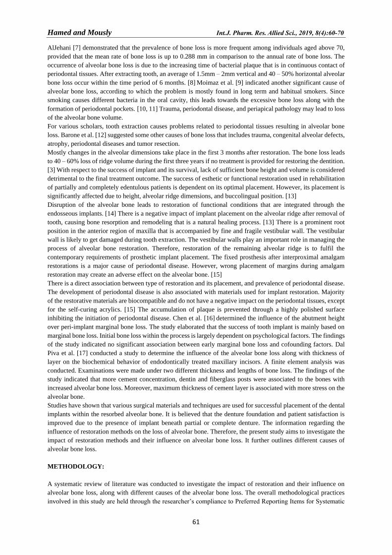
problems other than alveolar bone were excluded from the searched criteria. Figure 1 illustrates the procedure of
data collection through review of different related studies.
Figure 1: PRISMA Flow Diagram
Hamed and Mously Int.J. Pharm. Res. Allied Sci., 2019, 8(4):60-70
63
Data Extraction
Data were independently extracted by the contribution of two reviewers, in case of an ambiguity, a third reviewer
assisted the study. The studies that were totally complied on the inclusion criteria were included, and those that
consisted sufficient information in their abstracts were involved in the full text assessment carried by the reviewer
to identify and determine the eligibility of the study. The selected studies further underwent the process of validity
assessment along with extraction of data. The reasons that were considered for the rejection of different studies
were also recorded. The extraction and recording of data were based on the following variables; a) study type b)
participants’ characteristics, c) duration of participants follow-up d) intervention characteristics e) sample size f)
source of funding and conflicts of interest, methods of randomization.
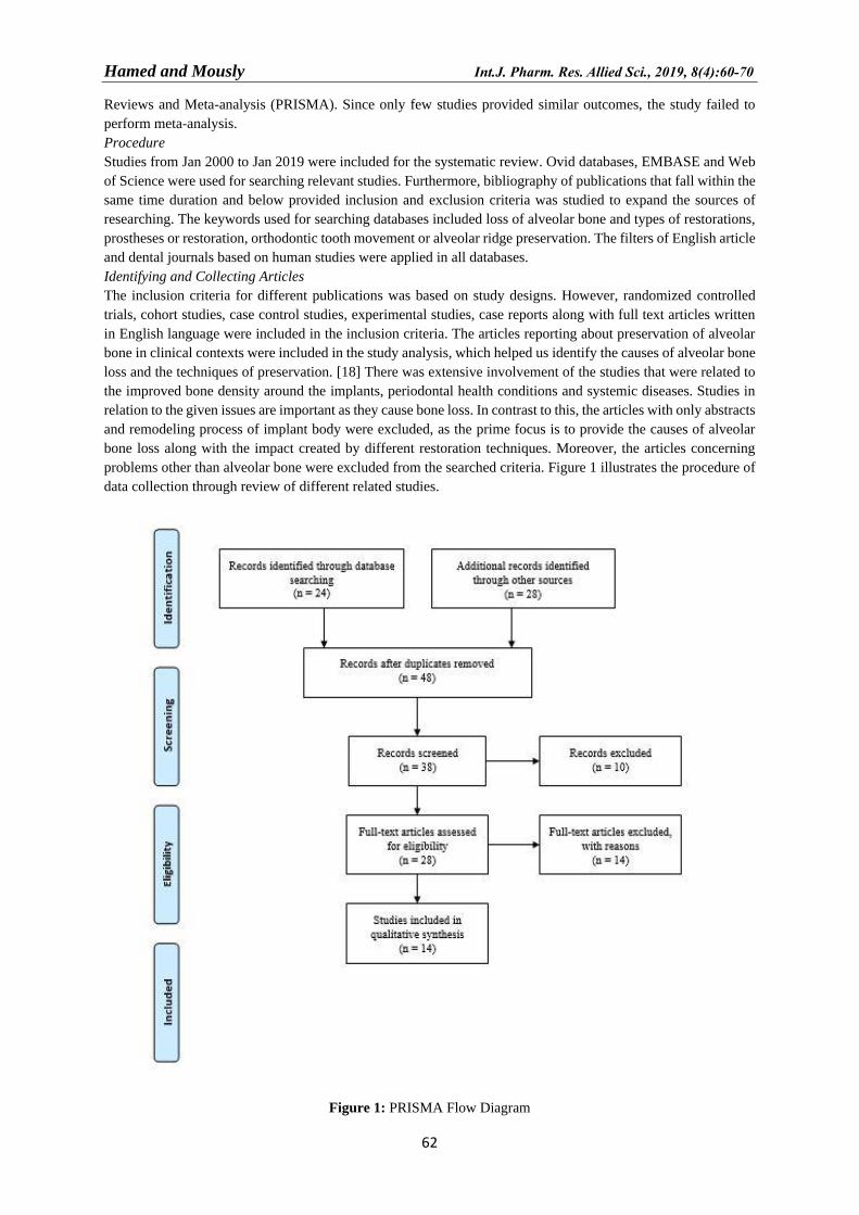
Risk of Bias and Qualitative Analysis
The study was designed and reviewed by two reviewers to ensure that the study is in accordance to PRISMA
guidelines to avoid risk of biasness while providing the quality evidences. Besides, the evaluation of the included
articles was also undertaken to maintain the originality of this review. The methodological quality of the
observational studies that were included, were assessed through the Newcastle Ottawa Scale (NOS). The study
guidelines provided by the National Institute of Health Research PROS-PRES were also followed. Risk of
biasness of the studies were tested using Cochrane Collaboration’s Tool as indicated in Figure 2.
Different techniques associated with the restoration methods of loss of alveolar bone were studied in the selected
studies. The primary outcome of the analyses provided that overhang, implant restorations loading, and restoration
timings are some considerable factors that are associated to the alveolar bone loss.
Figure 2: Risk of Biasness
RESULTS:
To identify different causes of alveolar bone loss along with the impact of treatment methods for alveolar bone
restorations, 14 studies were selected based on the inclusion criteria of the study. Among these, 5 were original
articles, 4 were review articles and remaining 5 included, case studies, cohort study, prospective study, narrative
review and longitudinal study.
Najm et al. [19] assessed the relationship between overhang filling and alveolar bone loss. Total of 900 digital
panoramic radiographs were examined to identify the presence of overhang amalgam. Among the selected images,
only 80 were found with overhang fillings. The overhang filling surface of the alveolar bone loss was compared
with normal surface of similar tooth. The results indicated through radiographical images suggested that the
overhang amalgam was found mostly in mandibles in firsts molars. Besides, the overhang filling in alveolar bone
Hamed and Mously Int.J. Pharm. Res. Allied Sci., 2019, 8(4):60-70
64
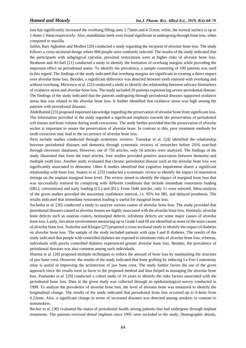
loss has significantly increased the overhang filling unto 3.73mm and 4.31mm; while, the normal surface is up to
1.4mm-1.9mm respectively. Also, mandibular teeth were found significant in undergoing through bone loss, when
compared to maxilla.
Julihn, Barr Agholme and Modéer [20] conducted a study regarding the incipient of alveolar bone loss. The study
follows a cross-sectional design where 800 people were randomly selected. The results of the study indicated that
the participants with subgingival calculus, proximal restorations were at higher risks of alveolar bone loss.
Ibraheem and Al-Safi [21] conducted a study to identify the formation of overhang margins while providing the
important effect on periodontal status. To identify the prevalence, a sample consisting of 100 patients was used
in this regard. The findings of the study indicated that overhang margins are significant in creating a direct impact
over alveolar bone loss. Besides, a significant difference was detected between tooth restored with overhang and
without overhang. Miricescu et al. [22] conducted a study to identify the relationship between salivary biomarkers
of oxidative stress and alveolar bone loss. The study included 20 patients experiencing severe periodontal disease.
The findings of the study indicated that the patients undergoing through periodontal diseases supported oxidative
stress that was related to the alveolar bone loss. It further identified that oxidative stress was high among the
patients with periodontal diseases.
Abdelhamid [23] proposed important knowledge regarding the preservation of alveolar bone from significant loss.
The information provided in the study regarded a significant emphasis towards the preservation of periodontal
soft tissues and bone volume during tooth extractions. The study further provided that the preservation of alveolar
socket is important to assure the preservation of alveolar bone. In contrast to this, poor treatment methods for
tooth extraction may lead to the occurrence of alveolar bone loss.
Next include studies conducted through systematic reviews. Tonsekar et al. [24] identified the relationship
between periodontal diseases and dementia through systematic reviews of researches before 2016 searched
through electronic databases. However, out of 756 articles, only 16 articles were analyzed. The findings of the
study illustrated that from the total articles, four studies provided positive association between dementia and
multiple tooth loss. Another study evaluated that chronic periodontal disease such as the alveolar bone loss was
significantly associated to dementia. Other 8 studies identified that cognitive impairment shares a significant
relationship with bone loss. Suarez et al. [25] conducted a systematic review to identify the impact of restoration
timings on the implant marginal bone level. The review aimed to identify the impact of marginal bone loss that
was successfully restored by complying with different conditions that include immediate restoration loading
(IRL), conventional and early loading (CL) and (EL). From 1640 articles, only 11 were selected. Meta-analysis
of the given studies provided the maximum confidence interval, i.e. 95% for IRL and delayed prosthesis. The
results indicated that immediate restoration loading is useful for marginal bone loss.
Suchetha et al. [26] conducted a study to analyze various causes of alveolar bone loss. The study provided that
periodontal diseases caused in alveolar tissues are highly associated with the alveolar bone loss. Similarly, alveolar
bone defects such as osseous craters, hemiseptal defects, infrabony defects are some major causes of alveolar
bone loss. Lastly, furcation involvements measuring up to Grade I and III are identified as some of the main causes
of alveolar bone loss. Soskolne and Klinger [27] proposed a cross-sectional study to identify the impact of diabetes
on alveolar bone loss. The sample of the study included patients with type I and II diabetes. The results of the
study indicated that people with controlled diabetes are exposed to minimum risks of alveolar bone loss; whereas,
individuals with poorly controlled diabetes experienced greater alveolar bone loss. Besides, the prevalence of
periodontal diseases was also common among such individuals.
Sharma et al. [28] proposed multiple techniques to reduce the amount of bone loss by maintaining the structure
of jaw bone crest. However, the results of the study indicated that bone grafting by inducing Le Fort I osteotomy
inlay is useful in improving the architecture of jaw bone crest. The study further favors the use of the given
approach since the results were in favor to the proposed method and thus helped in managing the alveolar bone
loss. Paulander et al. [29] conducted a cohort study of 10 years to identify the risks factors associated with the
periodontal bone loss. Data in the given study was collected through an epidemiological survey conducted in
1998. To analyze the prevalence of alveolar bone loss, the level of alveolar bone was measured to identify the
longitudinal change. The results of the study indicated that periodontal bone loss occurred up to 0.4mm from
0.22mm. Also, a significant change in terms of increased diseases was detected among smokers in contrast to
nonsmokers.
Becker et al. [30] evaluated the status of periodontal health among patients that had undergone through implant
treatments. The patients received dental implants since 1995 were included in the study. Demographic details,
Hamed and Mously Int.J. Pharm. Res. Allied Sci., 2019, 8(4):60-70
65
quality and quantity of bones, implant location along with the type of study were recorded in the database. The
patients aged between 66-93 years were included. NIH (National Institute of Health) images were used to measure
the changes in bone levels. The results of the study concluded that the patients with the given provided outstanding
survival rates of dental implants, with limited changes in the interproximal bone levels. The treatment was
significant in maintaining patient’s oral health. De Monès et al. [31] conducted an important study to provide the
causes of alveolar bone loss. The aim of the study however, was to provide the impact of statins in minimizing
the resorption in alveolar bones. A systematic search was performed through databases such as MEDline and
Pubmed, resulting into the selection of 21 studies. The results of the study indicated that the use of statins was
highly significant in reducing the oral cavity and the prevalence of periodontal diseases after tooth extraction.
Therefore, statins were highly useful in controlling the alveolar bone loss.
Another study undertaken by Kim et al. [32] examined the relationship between alveolar bone loss and root
proximity. The study followed a longitudinal cohort design by including a sample of 1231 individuals. Results
were proposed by measuring the interradicular distances and alveolar base levels through digitalized radiographs.
The findings of the study indicated a significant nonlinear relationship between alveolar bone levels and
interradicular distance.
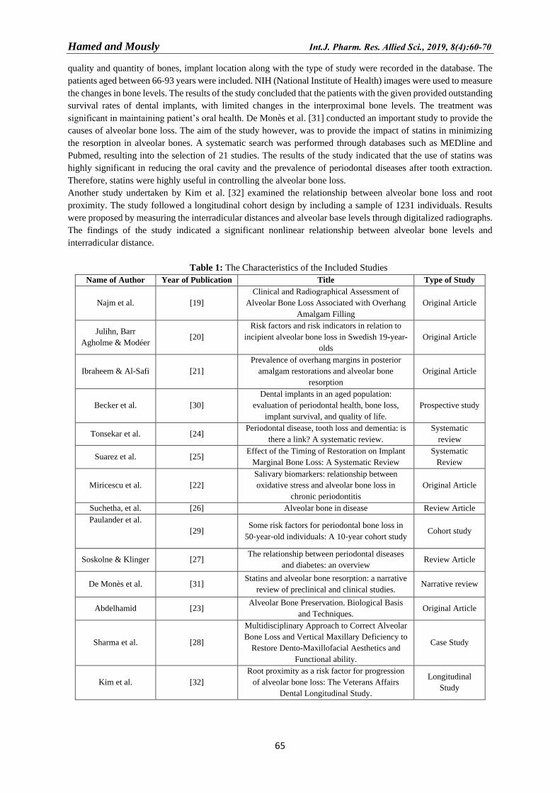
Table 1: The Characteristics of the Included Studies
Name of Author Year of Publication Title Type of Study
Najm et al. [19]
Clinical and Radiographical Assessment of
Alveolar Bone Loss Associated with Overhang
Amalgam Filling
Original Article
Julihn, Barr
Agholme & Modéer [20]
Risk factors and risk indicators in relation to
incipient alveolar bone loss in Swedish 19-year-
olds
Original Article
Ibraheem & Al-Safi [21]
Prevalence of overhang margins in posterior
amalgam restorations and alveolar bone
resorption
Original Article
Becker et al. [30]
Dental implants in an aged population:
evaluation of periodontal health, bone loss,
implant survival, and quality of life.
Prospective study
Tonsekar et al. [24] Periodontal disease, tooth loss and dementia: is
there a link? A systematic review.
Systematic
review
Suarez et al. [25] Effect of the Timing of Restoration on Implant
Marginal Bone Loss: A Systematic Review
Systematic
Review
Miricescu et al. [22]
Salivary biomarkers: relationship between
oxidative stress and alveolar bone loss in
chronic periodontitis
Original Article
Suchetha, et al. [26] Alveolar bone in disease Review Article
Paulander et al.
[29] Some risk factors for periodontal bone loss in
50‐year‐old individuals: A 10‐year cohort study Cohort study
Soskolne & Klinger [27] The relationship between periodontal diseases
and diabetes: an overview Review Article
De Monès et al. [31] Statins and alveolar bone resorption: a narrative
review of preclinical and clinical studies. Narrative review
Abdelhamid [23] Alveolar Bone Preservation. Biological Basis
and Techniques. Original Article
Sharma et al. [28]
Multidisciplinary Approach to Correct Alveolar
Bone Loss and Vertical Maxillary Deficiency to
Restore Dento-Maxillofacial Aesthetics and
Functional ability.
Case Study
Kim et al. [32]
Root proximity as a risk factor for progression
of alveolar bone loss: The Veterans Affairs
Dental Longitudinal Study.
Longitudinal
Study
Hamed and Mously Int.J. Pharm. Res. Allied Sci., 2019, 8(4):60-70
66
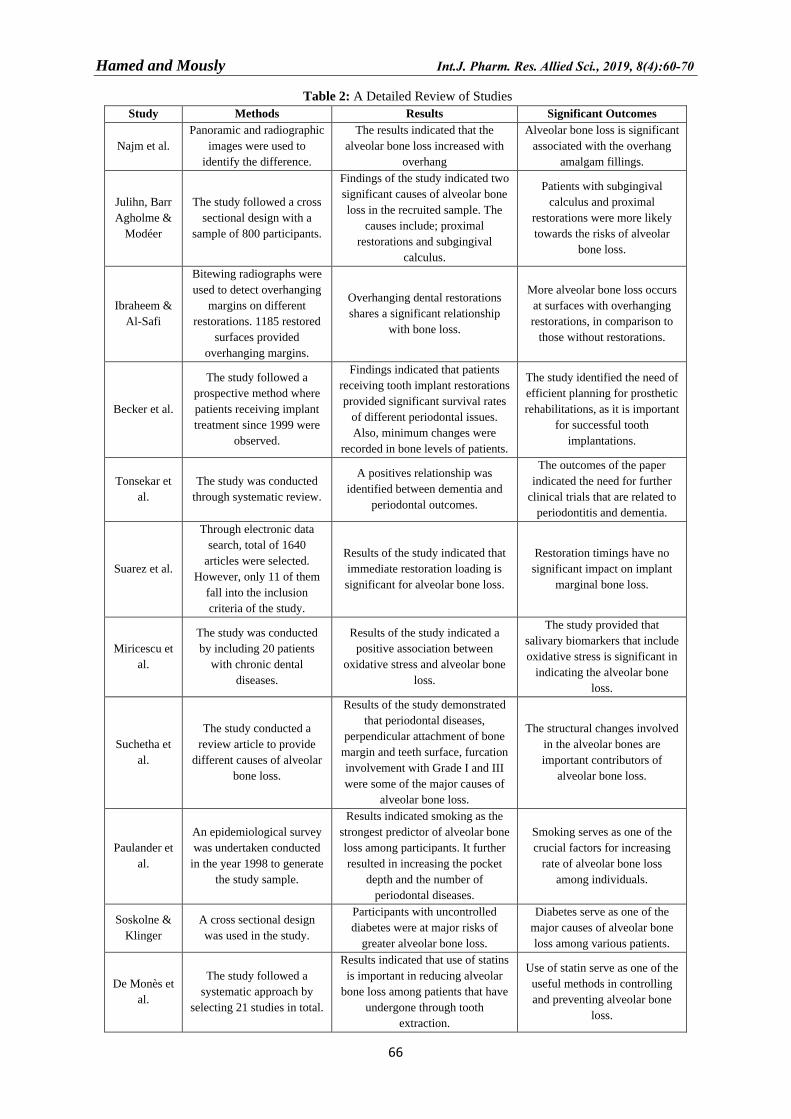
Table 2: A Detailed Review of Studies
Study Methods Results Significant Outcomes
Najm et al.
Panoramic and radiographic
images were used to
identify the difference.
The results indicated that the
alveolar bone loss increased with
overhang
Alveolar bone loss is significant
associated with the overhang
amalgam fillings.
Julihn, Barr
Agholme &
Modéer
The study followed a cross
sectional design with a
sample of 800 participants.
Findings of the study indicated two
significant causes of alveolar bone
loss in the recruited sample. The
causes include; proximal
restorations and subgingival
calculus.
Patients with subgingival
calculus and proximal
restorations were more likely
towards the risks of alveolar
bone loss.
Ibraheem &
Al-Safi
Bitewing radiographs were
used to detect overhanging
margins on different
restorations. 1185 restored
surfaces provided
overhanging margins.
Overhanging dental restorations
shares a significant relationship
with bone loss.
More alveolar bone loss occurs
at surfaces with overhanging
restorations, in comparison to
those without restorations.
Becker et al.
The study followed a
prospective method where
patients receiving implant
treatment since 1999 were
observed.
Findings indicated that patients
receiving tooth implant restorations
provided significant survival rates
of different periodontal issues.
Also, minimum changes were
recorded in bone levels of patients.
The study identified the need of
efficient planning for prosthetic
rehabilitations, as it is important
for successful tooth
implantations.
Tonsekar et
al.
The study was conducted
through systematic review.
A positives relationship was
identified between dementia and
periodontal outcomes.
The outcomes of the paper
indicated the need for further
clinical trials that are related to
periodontitis and dementia.
Suarez et al.
Through electronic data
search, total of 1640
articles were selected.
However, only 11 of them
fall into the inclusion
criteria of the study.
Results of the study indicated that
immediate restoration loading is
significant for alveolar bone loss.
Restoration timings have no
significant impact on implant
marginal bone loss.
Miricescu et
al.
The study was conducted
by including 20 patients
with chronic dental
diseases.
Results of the study indicated a
positive association between
oxidative stress and alveolar bone
loss.
The study provided that
salivary biomarkers that include
oxidative stress is significant in
indicating the alveolar bone
loss.
Suchetha et
al.
The study conducted a
review article to provide
different causes of alveolar
bone loss.
Results of the study demonstrated
that periodontal diseases,
perpendicular attachment of bone
margin and teeth surface, furcation
involvement with Grade I and III
were some of the major causes of
alveolar bone loss.
The structural changes involved
in the alveolar bones are
important contributors of
alveolar bone loss.
Paulander et
al.
An epidemiological survey
was undertaken conducted
in the year 1998 to generate
the study sample.
Results indicated smoking as the
strongest predictor of alveolar bone
loss among participants. It further
resulted in increasing the pocket
depth and the number of
periodontal diseases.
Smoking serves as one of the
crucial factors for increasing
rate of alveolar bone loss
among individuals.
Soskolne &
Klinger
A cross sectional design
was used in the study.
Participants with uncontrolled
diabetes were at major risks of
greater alveolar bone loss.
Diabetes serve as one of the
major causes of alveolar bone
loss among various patients.
De Monès et
al.
The study followed a
systematic approach by
selecting 21 studies in total.
Results indicated that use of statins
is important in reducing alveolar
bone loss among patients that have
undergone through tooth
extraction.
Use of statin serve as one of the
useful methods in controlling
and preventing alveolar bone
loss.
Hamed and Mously Int.J. Pharm. Res. Allied Sci., 2019, 8(4):60-70
67
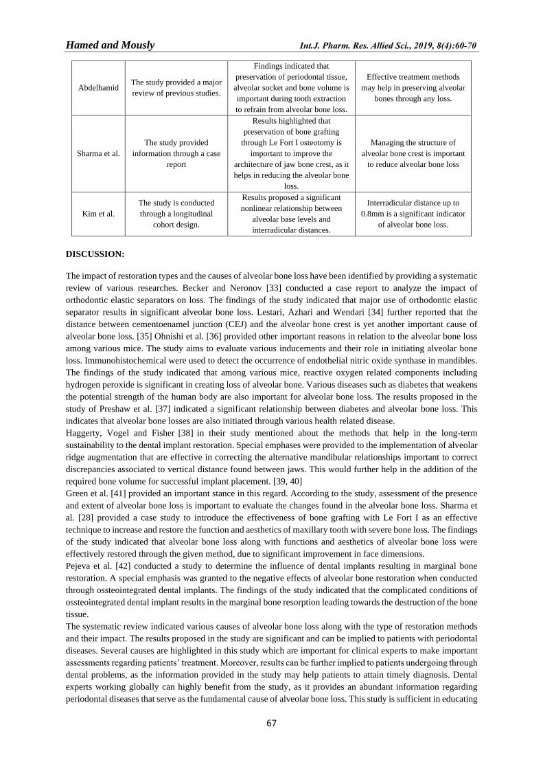
Abdelhamid The study provided a major
review of previous studies.
Findings indicated that
preservation of periodontal tissue,
alveolar socket and bone volume is
important during tooth extraction
to refrain from alveolar bone loss.
Effective treatment methods
may help in preserving alveolar
bones through any loss.
Sharma et al.
The study provided
information through a case
report
Results highlighted that
preservation of bone grafting
through Le Fort I osteotomy is
important to improve the
architecture of jaw bone crest, as it
helps in reducing the alveolar bone
loss.
Managing the structure of
alveolar bone crest is important
to reduce alveolar bone loss
Kim et al.
The study is conducted
through a longitudinal
cohort design.
Results proposed a significant
nonlinear relationship between
alveolar base levels and
interradicular distances.
Interradicular distance up to
0.8mm is a significant indicator
of alveolar bone loss.
DISCUSSION:
The impact of restoration types and the causes of alveolar bone loss have been identified by providing a systematic
review of various researches. Becker and Neronov [33] conducted a case report to analyze the impact of
orthodontic elastic separators on loss. The findings of the study indicated that major use of orthodontic elastic
separator results in significant alveolar bone loss. Lestari, Azhari and Wendari [34] further reported that the
distance between cementoenamel junction (CEJ) and the alveolar bone crest is yet another important cause of
alveolar bone loss. [35] Ohnishi et al. [36] provided other important reasons in relation to the alveolar bone loss
among various mice. The study aims to evaluate various inducements and their role in initiating alveolar bone
loss. Immunohistochemical were used to detect the occurrence of endothelial nitric oxide synthase in mandibles.
The findings of the study indicated that among various mice, reactive oxygen related components including
hydrogen peroxide is significant in creating loss of alveolar bone. Various diseases such as diabetes that weakens
the potential strength of the human body are also important for alveolar bone loss. The results proposed in the
study of Preshaw et al. [37] indicated a significant relationship between diabetes and alveolar bone loss. This
indicates that alveolar bone losses are also initiated through various health related disease.
Haggerty, Vogel and Fisher [38] in their study mentioned about the methods that help in the long-term
sustainability to the dental implant restoration. Special emphases were provided to the implementation of alveolar
ridge augmentation that are effective in correcting the alternative mandibular relationships important to correct
discrepancies associated to vertical distance found between jaws. This would further help in the addition of the
required bone volume for successful implant placement. [39, 40]
Green et al. [41] provided an important stance in this regard. According to the study, assessment of the presence
and extent of alveolar bone loss is important to evaluate the changes found in the alveolar bone loss. Sharma et
al. [28] provided a case study to introduce the effectiveness of bone grafting with Le Fort I as an effective
technique to increase and restore the function and aesthetics of maxillary tooth with severe bone loss. The findings
of the study indicated that alveolar bone loss along with functions and aesthetics of alveolar bone loss were
effectively restored through the given method, due to significant improvement in face dimensions.
Pejeva et al. [42] conducted a study to determine the influence of dental implants resulting in marginal bone
restoration. A special emphasis was granted to the negative effects of alveolar bone restoration when conducted
through ossteointegrated dental implants. The findings of the study indicated that the complicated conditions of
ossteointegrated dental implant results in the marginal bone resorption leading towards the destruction of the bone
tissue.
The systematic review indicated various causes of alveolar bone loss along with the type of restoration methods
and their impact. The results proposed in the study are significant and can be implied to patients with periodontal
diseases. Several causes are highlighted in this study which are important for clinical experts to make important
assessments regarding patients’ treatment. Moreover, results can be further implied to patients undergoing through
dental problems, as the information provided in the study may help patients to attain timely diagnosis. Dental
experts working globally can highly benefit from the study, as it provides an abundant information regarding
periodontal diseases that serve as the fundamental cause of alveolar bone loss. This study is sufficient in educating
Hamed and Mously Int.J. Pharm. Res. Allied Sci., 2019, 8(4):60-70
68
the target population that includes patients with periodontal diseases and alveolar bone loss along with those
intending to undergo restoration treatments, and further serves as a guideline for the effectiveness of different
restoration methods.
CONCLUSION:
Different forms of restoration methods have been evolved by the time which creates both positive and negative
effects on the strength and placement of the restored tooth. The study is significant in providing valuable outcomes
regarding the causes of alveolar bone loss and impact of restoration methods. Information provided in the study
is important for professional experts belonging to the given field.
Several reasons have been identified regarding the causes of alveolar bone loss along with the impact of type of
restoration treatment. Outcomes such as the overhang amalgamation filling, impact of statins, periodontal
diseases, furcation involvement, oxidative stress, overhanging dental restorations, etc. are some important
problems that are associated to alveolar bone loss. Other than this, several studies were included to identify the
impact of different restoration methods are presented. The results however indicated that certain restoration
methods are insignificant in providing maximum survival rate.
The present study is important in shedding light on different types of restoration methods along with their impact.
A number of studies are provided in the review to outline some of the major findings that have been developed in
the past few years. The findings of the study are useful for clinical expertise, as it provides a variety of knowledge
considering the loss of alveolar bone and restoration methods. The results are further critical as they explicitly
highlight the pros and cons of different restoration methods along with the causes that ultimately result in the
tooth restoration failure. As the study follows a systematic review, it is invalid to propose any accurate results.
The study has certain limitations, as only few studies are added in the review. Therefore, a more detailed
discussion is required to cover the topic further, through cross-sectional study design.
ACKNOWLEDGEMENT
The author is very thankful to all the associated personnel in any reference that contributed in/for the purpose of
this research. Further, this research holds no conflict of interest and is not funded through any source.
REFERENCES
1. Lee KJ, Joo E, Yu HS, Park YC. Restoration of an alveolar bone defect caused by an ankylosed mandibular
molar by root movement of the adjacent tooth with miniscrew implants. Am J Orthod Dentofacial Orthop.
2009; 136: 440-9. https://doi.org/10.1016/j.ajodo.2007.05.019
2. Tamimi F, Torres J, Al-Abedalla K, Lopez-Cabarcos E, Alkhraisat MH, Bassett DC, Gbureck U, Barralet
JE. Osseointegration of dental implants in 3D-printed synthetic onlay grafts customized according to bone
metabolic activity in recipient site. Biomaterials. 2014; 35: 5436-45. 10.1016/j.biomaterials.2014.03.050
3. Sheikh Z, Sima C, Glogauer M. Bone replacement materials and techniques used for achieving vertical
alveolar bone augmentation. Materials. 2015; 8: 2953-93. https://doi.org/10.3390/ma8062953
4. Harsas NA, Irwan A. Guided bone regeneration in periodontology. Makassar Dent. J. 2015; 4.
https://doi.org/10.1016/j.ajodo.2007.05.028
5. Khojasteh A, Kheiri L, Motamedian SR, Khoshkam V. Guided bone regeneration for the reconstruction of
alveolar bone defects. Ann Maxillofac Surg. 2017; 7: 263. https://doi.org/10.4103/ams.ams_76_17
6. Levin BP. Alveolar ridge augmentation: combining bioresorbable scaffolds with osteoinductive bone grafts
in atrophic sites. A follow-up to an evolving technique. Compend Contin Educ Dent. 2013; 34: 178-86.
7. AlJehani YA. Risk factors of periodontal disease: review of the literature. Int. J. Dent. 2014; 2014.
8. Liu J, Kerns DG. Suppl 1: Mechanisms of guided bone regeneration: A review. Open Dent J. 2014; 8: 56.
https://doi.org/10.2174/1874210601408010056
9. Moimaz SA, Zina LG, Saliba O, Garbin CA. Smoking and periodontal disease: clinical evidence for an
association. Oral Hlth Prev Dent. 2009;7.
10. Grossi SG, Genco RJ, Machtet EE, Ho AW, Koch G, Dunford R, Zambon JJ, Hausmann E. Assessment of
risk for periodontal disease. II. Risk indicators for alveolar bone loss. J Periodontol. 1995; 66: 23-9.
Hamed and Mously Int.J. Pharm. Res. Allied Sci., 2019, 8(4):60-70
69
11. Grossi SG, Zambon JJ, Ho AW, Koch G, Dunford RG, Machtei EE, Norderyd OM, Genco RJ. Assessment
of risk for periodontal disease. I. Risk indicators for attachment loss. J Periodontol. 1994; 65: 260-7.
12. Barone A, Ricci M, Tonelli P, Santini S, Covani U. Tissue changes of extraction sockets in humans: a
comparison of spontaneous healing vs. ridge preservation with secondary soft tissue healing. Clin Oral
Implants Res. 2013; 24: 1231-7.
13. Mezzomo LA, Shinkai RS, Mardas N, Donos N. Alveolar ridge preservation after dental extraction and
before implant placement: a literature review. Revista Odonto Ciência. 2011; 26: 77-83. 10.1590/s1980-
65232011000100017
14. Tonelli P, Duvina M, Barbato L, Biondi E, Nuti N, Brancato L, Delle Rose G. Bone regeneration in
dentistry. Clin Cases Miner Bone Metab. 2011; 8: 24. https://doi.org/10.1111/ger.12261
15. Rajan K, Ramamurthy J. Effect of restorations on periodontal health. JDMS. 2014; 13: 2279-0861.
10.9790/0853-13747173
16. Chen Z, Lin CY, Li J, Wang HL, Yu H. Influence of abutment height on peri-implant marginal bone loss:
A systematic review and meta-analysis. The Journal of prosthetic dentistry. 2019.
https://doi.org/10.1016/j.prosdent.2018.10.003
17. Dal Piva AM, Tribst JP, e Souza RO, Borges AL. Influence of alveolar bone loss and cement layer thickness
on the biomechanical behavior of endodontically treated maxillary incisors: a 3-dimensional finite element
analysis. Journal of endodontics. 2017; 43: 791-5. https://doi.org/10.1016/j.joen.2016.11.020
18. Oral Health and Alveolar Bone Disease. National Institutes of Health. 2018
19. Najm AA, Akram HM, Mahdi AS, Ali OH. Clinical and Radiographical Assessment of Alveolar Bone
Loss Associated with Overhang Amalgam Filling. Health Sciences. 2018; 7: 11-6.
20. Julihn A, Barr Agholme M, Modéer T. Risk factors and risk indicators in relation to incipient alveolar bone
loss in Swedish 19-year-olds. Acta Odontologica Scandinavica. 2008; 66: 139-47.
https://doi.org/10.1080/00016350802087024
21. Ibraheem AF, Al-Safi KA. Prevalence of overhang margins in posterior amalgam restorations and alveolar
bone resorption. Journal of Baghdad College of Dentistry. 2005; 17: 11-3.
22. Miricescu D, Totan A, Calenic B, Mocanu B, Didilescu A, Mohora M, Spinu T, Greabu M. Salivary
biomarkers: relationship between oxidative stress and alveolar bone loss in chronic periodontitis. Acta
Odontologica Scandinavica. 2014; 72: 42-7. https://doi.org/10.3109/00016357.2013.795659
23. Abdelhamid A. Alveolar Bone Preservation. Biological Basis and Techniques. Int J. 2017; 5: 56-68.
24. Tonsekar PP, Jiang SS, Yue G. Periodontal disease, tooth loss and dementia: is there a link? A systematic
review. Gerodontology. 2017; 34: 151-63.
25. Suarez F, Chan HL, Monje A, Galindo‐Moreno P, Wang HL. Effect of the timing of restoration on implant
marginal bone loss: a systematic review. J Periodontol. 2013; 84: 159-69.
https://doi.org/10.1902/jop.2012.120099
26. Suchetha A, Tanwar E, Darshan BM, Apoorva SM, Salman K. Alveolar bone in disease.
27. Soskolne WA, Klinger A. The relationship between periodontal diseases and diabetes: an overview. Ann
Periodontol. 2001; 6: 91-8.
28. Sharma P, Shu L, Tao W, Dawazeh R, Sharma A. Multidisciplinary Approach to Correct Alveolar Bone
Loss and Vertical Maxillary Deficiency to Restore Dento-Maxillofacial Aesthetics and Functional Ability.
Int J Case Rep. 2018; 2: 5.
29. Paulander J, Wennström JL, Axelsson P, Lindhe J. Some risk factors for periodontal bone loss in 50‐year‐
old individuals: A 10‐year cohort study. J Clin Periodontol. 2004; 31: 489-96.
30. Becker W, Hujoel P, Becker BE, Wohrle P. Dental implants in an aged population: evaluation of
periodontal health, bone loss, implant survival, and quality of life. Clinical implant dentistry and related
research. 2016; 18: 473-9. https://doi.org/10.1590/1678-7757-2017-0084
31. De Monès E, Schlaubitz S, Catros S, Fricain JC. Statins and alveolar bone resorption: a narrative review
of preclinical and clinical studies. Oral surgery, oral medicine, oral pathology and oral radiology. 2015;
119: 65-73.
32. Kim T, Miyamoto T, Nunn ME, Garcia RI, Dietrich T. Root proximity as a risk factor for progression of
alveolar bone loss: The Veterans Affairs Dental Longitudinal Study. Journal of periodontology. 2008; 79:
654-9.
Hamed and Mously Int.J. Pharm. Res. Allied Sci., 2019, 8(4):60-70
70
33. Becker T, Neronov, A. Orthodontic elastic separator-induced periodontal abscess: a case report. Case
reports in Dentistry, 2012.
34. Lestari AD, Azhari A, Wendari S. The measurement of the alveolar bone crest in aggressive periodontitis
using Cone Beam Computed Tomography imaging. Padjadjaran Journal of Dentistry. 2012; 24.
https://doi.org/10.24198/pjd.vol24no1.15373
35. Abrahamsson KH, Koch G, Norderyd O, Romao C, Wennström JL. Periodontal conditions in a Swedish
city population of adolescents: a cross-sectional study. J Periodontal Res. 2006; 30: 25-34.
36. Ohnishi T, Bandow K, Kakimoto K, Machigashira M, Matsuyama T, Matsuguchi T. Oxidative stress causes
alveolar bone loss in metabolic syndrome model mice with type 2 diabetes. J Periodontal Res. 2009; 44:
43-51.
37. Preshaw PM, Alba AL, Herrera D, Jepsen S, Konstantinidis A, Makrilakis K, Taylor R. Periodontitis and
diabetes: a two-way relationship. Diabetologia. 2012; 55: 21-31.
38. Haggerty CJ, Vogel CT, Fisher GR. Simple bone augmentation for alveolar ridge defects. Oral and
Maxillofacial Surgery Clinics. 2015; 27: 203-26. https://doi.org/10.1016/j.coms.2015.01.011
39. Felice P, Iezzi G, Lizio G, Piattelli A, Marchetti C. Reconstruction of atrophied posterior mandible with
inlay technique and mandibular ramus block graft for implant prosthetic rehabilitation. Journal of Oral and
Maxillofacial Surgery. 2009; 67: 372-80.
40. Barone A, Covani U. Maxillary alveolar ridge reconstruction with nonvascularized autogenous block bone:
clinical results. Journal of Oral and Maxillofacial Surgery. 2007; 65: 2039-46.
https://doi.org/10.1016/j.joms.2007.05.017
41. Green PT, Mol A, Moretti AJ, Tyndall DA, Kohltfarber HB. Comparing the diagnostic efficacy of intraoral
radiography and cone beam computed tomography volume registration in the detection of mandibular
alveolar bone defects. Oral surgery, oral medicine, oral pathology and oral radiology. 2019.
https://doi.org/10.1016/j.oooo.2018.12.018
42. Pejeva E, Papakoca K, Ambarkova V, Todorovska G. Marginal Bone Resorption at Dental Implant–12
Clinical Cases. of. 2018; 11: 2.


