Embed Size (px)
Citation preview

Murine Alveolar Macrophages Limit Replication of Vaccinia Virus
Rachel Rivera1, Martha Hutchens2, Kathryn E. Luker3, Joanne Sonstein4, Jeffrey L.Curtis2,4,5, and Gary D. Luker1,2,3,61 Department of Microbiology and Immunology, University of Michigan Medical School
2 Graduate Program in Immunology, University of Michigan Medical School
3 Department of Radiology, University of Michigan Medical School
4 Division of Pulmonary and Critical Care Medicine, Department of Internal Medicine, University ofMichigan Medical School
5 Department of Veterans Affairs Health System, Ann Arbor, MI 48109
AbstractBecause of concerns about zoonotic transmission of monkeypox to humans and the bioterrorismthreat posed by Orthopoxviruses, there is renewed interest in probing cellular and molecularmechanisms of host defense to these pathogens. In particular, it is essential to understand viral-hostinteractions in the respiratory tract, which is the route of infection for smallpox and a likely route oftransmission for monkeypox. In this study, we analyze functions of alveolar macrophages in poxvirusinfection, using a recombinant vaccinia virus expressing firefly luciferase to quantify infection inmice and cell culture. Depletion of alveolar macrophages with liposomal clodronate worsens theoverall severity of infection in mice, including greater replication and systemic dissemination ofvaccinia as determined by bioluminescence imaging. Absence of alveolar macrophages increasestotal numbers of granulocytes and granulocytes/monocyte progenitor cells in the lung during vacciniainfection, indicating that protective effects of alveolar macrophages may be mediated in part byreducing the host inflammation. Alveolar macrophages also limit vaccinia infection in respiratoryepithelium, as shown by a co-culture model of cell lines derived from alveolar macrophages and lungepithelium. Collectively, these data demonstrate that alveolar macrophages are key determinants ofhost defense against local and systemic infection with poxviruses.
The respiratory tract is a key route of spread for orthopoxviruses, including the viruses thatcause smallpox and monkeypox (Fenner et al., 1988). In addition, the potential use of theseviruses as agents of biological warfare raises the possibility for exposure to levels of aerosolizedviruses that markedly exceed doses encountered during normal infection. Therefore, definingcellular and molecular determinants of the host response to poxviruses in the respiratory systemis essential for understanding basic questions about host immunity to these pathogens andlimiting the bioterrorism threat posed by poxviruses.
Host defense against poxviruses in the respiratory tract begins at the level of infected cells,which likely are epithelial cells. Infected cells initiate an intracellular anti-viral response tolimit viral replication and secrete interferons and other anti-viral cytokines as part of the localinnate immune response. Local immunity is amplified through macrophages and dendritic
6To whom correspondence should be addressed: Gary D. Luker, University of Michigan Medical School, Department of Radiology, 109Zina Pitcher Place, A526 BSRB, Ann Arbor, MI 48109; [email protected]'s Disclaimer: This is a PDF file of an unedited manuscript that has been accepted for publication. As a service to our customerswe are providing this early version of the manuscript. The manuscript will undergo copyediting, typesetting, and review of the resultingproof before it is published in its final citable form. Please note that during the production process errors may be discovered which couldaffect the content, and all legal disclaimers that apply to the journal pertain.
NIH Public AccessAuthor ManuscriptVirology. Author manuscript; available in PMC 2008 June 20.
Published in final edited form as:Virology. 2007 June 20; 363(1): 48–58.
NIH
-PA Author Manuscript
NIH
-PA Author Manuscript
NIH
-PA Author Manuscript

cells, which also migrate to draining lymph nodes to activate adaptive immunity. Ultimately,local innate immunity and systemic adaptive immunity combine to control acute infection andestablish lasting immunity against subsequent poxvirus infection.
Alveolar macrophages are the predominant resident phagocytic cells in the lungs that respondto inhaled organisms in the respiratory tract, initiate local immune responses to pathogens, andmaintain functional integrity of lung epithelium. Under basal conditions, alveolar macrophagesappear to promote an immunosuppressive microenvironment in the lung. These cells suppressactivation of adaptive immunity by limiting access of antigens to dendritic cells and reducingfunctions of dendritic cells, T cells, and B cells (MacLean et al., 1996;Thepen, Kraal, and Holt,1994). In response to bacterial pathogens, alveolar macrophages decrease the overall severityof disease, in part by reducing levels of pro-inflammatory cytokines and recruitment ofleukocytes to the lung (Broug-Holub et al., 1997;Knapp et al., 2003). However, functions ofthese cells in the context of viral infections remain poorly defined, particularly for respiratoryinfection with poxviruses.
We investigated alveolar macrophages in host defense to poxviruses, using a recombinantvaccinia virus that expresses firefly luciferase as a model for analyzing functions of these cellsin local and systemic responses to infection. In mice, we found that depletion of alveolarmacrophages increases local and systemic replication of vaccinia virus and overall severity ofdisease. Absence of alveolar macrophages increases recruitment of inflammatory cells to thelung during vaccinia infection, which likely contributes to more severe disease in these animals.Alveolar macrophages also significantly limit infection of lung epithelial cells in an in vitroco-culture model system. Collectively, our results demonstrate functions of alveolarmacrophages to limit viral replication and disease severity in response to infection with anorthopoxvirus.
ResultsDepletion of alveolar macrophages with liposomal clodronate
Treatment of mice with liposomes containing the bisphosphonate compound clodronate is awell-established method for depleting alveolar macrophages, a cell population that comprises> 95% of cells in the normal airway. This technique does not damage respiratory epitheliumor cause toxicity to neutrophils, another phagocytic cell type (van Rooijen, 1989) (Qian et al.,1994).
To establish the kinetics of macrophage depletion, we intratracheally injected 100 μL ofliposomes containing clodronate or saline as a control (Clodronate Liposomes) into Sv129/Evmice. Sv129/Ev mice have been used by our laboratory and others for studies of vacciniapathogenesis (van den Broek et al., 1995) (Luker et al., 2005). Numbers of alveolarmacrophages recovered by bronchoalveolar lavage were quantified at various time aftertreatment and compared with alveolar macrophages in untreated, strain-matched mice. In micetreated with clodronate liposomes, only ≈ 5% of alveolar macrophages remained on day 2, andnumbers of alveolar macrophages were ≤ 25% of normal through 7 days (Fig 1). Bycomparison, treatment with saline liposomes had only a minimal effect on numbers of alveolarmacrophages. These data show that we are able to effectively deplete alveolar macrophagesfor most of the expected period of acute infection with vaccinia virus.
Some populations of dendritic cells may be depleted by clodronate liposomes (Leenen et al.,1998), although effects of this treatment on lung dendritic cells remain poorly defined. Wequantified numbers of dendritic cells in lungs of mice treated with clodronate or control salineliposomes. Clodronate liposomes did not affect total numbers of CD11c(+), MHC class II(+)dendritic cells in the lung interstitium as compared with control liposomes. In both groups,
Rivera et al. Page 2
Virology. Author manuscript; available in PMC 2008 June 20.
NIH
-PA Author Manuscript
NIH
-PA Author Manuscript
NIH
-PA Author Manuscript
dendritic cells comprised 0.8% of total cells in the lung, which is consistent with our previousdata from untreated mice. Because antigen presenting cells such as dendritic cells andmacrophages migrate to regional lymph nodes to initiate adaptive immunity, we also quantifiednumbers of these cell types in the tracheobronchial lymph node of mice 2 days after injectionof liposomes. There were no significant differences between clodronate and saline control micein numbers or percentages of CD11c(+), MHC class II(+) dendritic cells, Mac-3(+)macrophages, or ERMP-20(+) late monocyte progenitors in these lymph nodes (data notshown). Furthermore, numbers or percentages of dendritic cells and macrophages in peripheralblood, as determined by these same cell surface markers, did not differ between mice 2 daysfollowing treatment with clodronate or saline liposomes (data not shown). Collectively, thesedata demonstrate that intratracheal administration of clodronate liposomes depleted onlyalveolar macrophages.
Alveolar macrophages limit vaccinia infection in vivoWe treated mice with intratracheal administration of clodronate liposomes to investigate effectsof alveolar macrophages on host defense against respiratory infection with vaccinia virus. Micewere injected with clodronate liposomes and then infected with 4 × 10⁵ pfu Vac-FL intranasallytwo days later at a time when alveolar macrophages are depleted maximally as shown above.Control mice received liposomes filled with saline two days prior to infection. Mice wereinoculated with vaccinia intranasally to reproduce infection through the upper respiratory tract,the typical route of transmission for smallpox. In this and subsequent experiments, two miceeach treated with clodronate or saline liposomes were euthanized at the time of infection withvaccinia to confirm effective depletion of alveolar macrophages relative to saline control (datanot shown).
Depletion of alveolar macrophages increased the overall severity of vaccinia infection relativeto saline controls, as evidenced by more pronounced weight loss over the course of infection(Fig 2A). Differences in weight loss became apparent by day 3 after infection and persistedthrough day 7. Notably, mice treated with clodronate liposomes to deplete alveolarmacrophages died on day 7 post-infection, while control animals survived through day 8 ofacute infection.
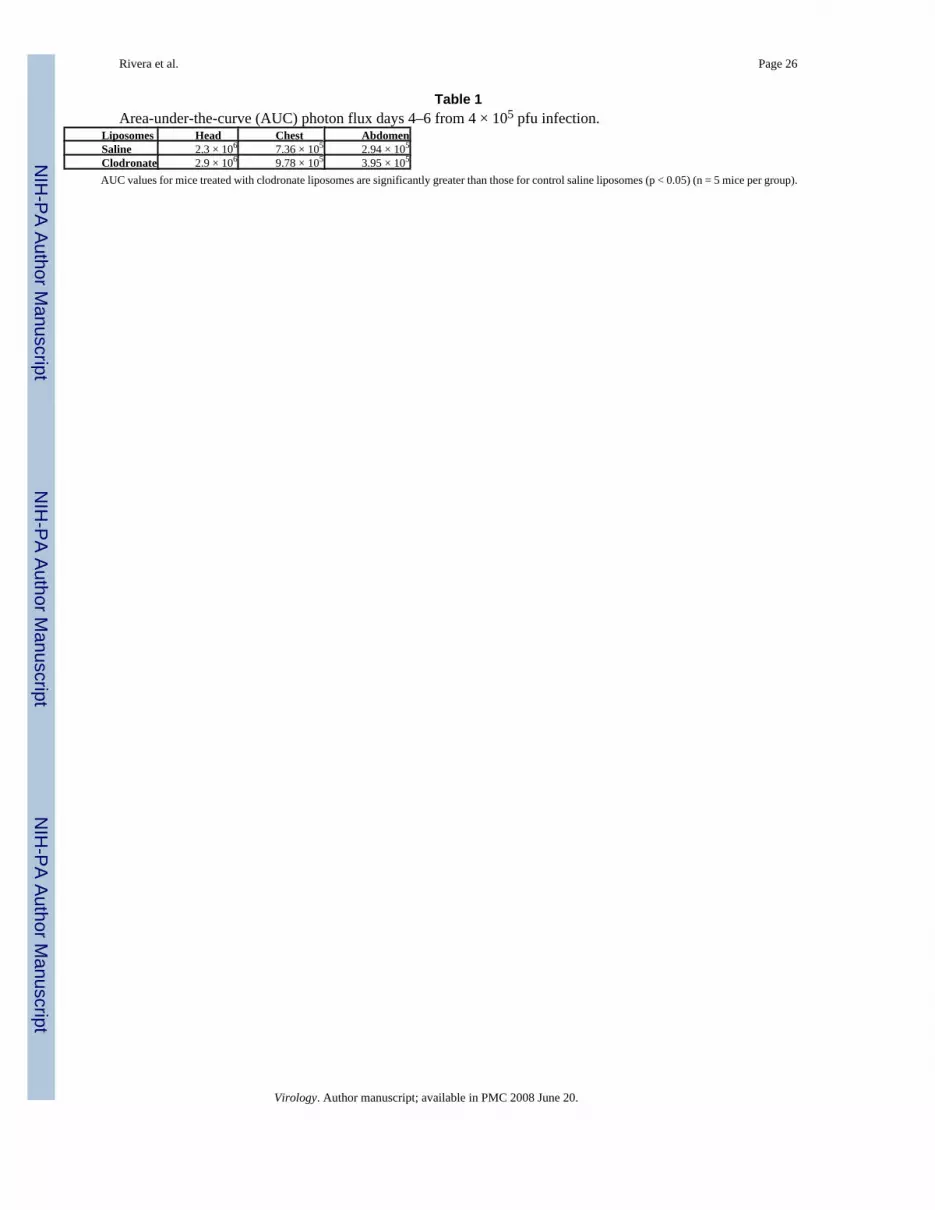
We used bioluminescence imaging to quantify viral replication and dissemination over thecourse of infection. Mice treated with clodronate liposomes had higher levels of luciferaseactivity in head, chest, and abdomen on days 4–6 post-infection than control mice (Fig 2B–E).By area-under-curve (AUC) analysis of these days, total bioluminescence for each region ofinterest was significantly greater in mice treated with clodronate liposomes (Table 1) (p < 0.05).On day 7 after infection, amounts of luciferase activity at these various sites were lower inmice treated with clodronate liposomes compared with control animals. However, this decreasein bioluminescence may be due to poor perfusion of luciferin to sites of Vac-FL in these micebecause of severe systemic illness in mice treated with clodronate liposomes rather than anactual decrease in vaccinia virus. Collectively, these data show that alveolar macrophagesdecrease the overall extent of vaccinia viral infection after respiratory inoculation.
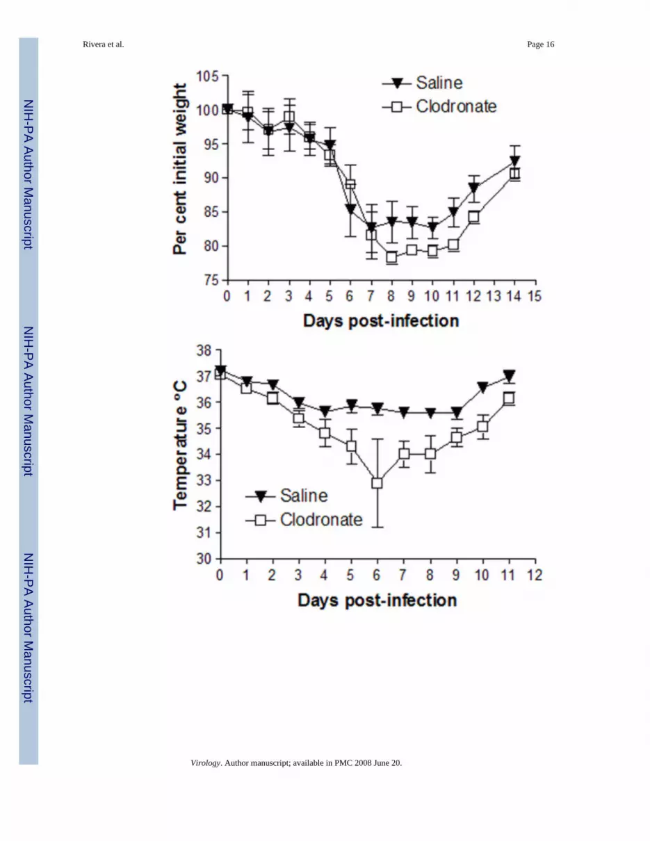
To build upon the data presented above with a lethal inoculum of vaccinia virus, we theninvestigated functions of alveolar macrophages in the context of a non-lethal infection. Micewere treated with clodronate or control saline liposomes and infected with 4 × 10⁴ pfu Vac-FL two days later at a time coinciding with maximum depletion of alveolar macrophages byclodronate. Under these conditions, mice lost comparable amounts of weight until day 8 post-infection, when weight loss was greater in animals treated with clodronate liposomes. Thesedifferences in weight loss persisted through day 12 (Fig 3A).
Rivera et al. Page 3
Virology. Author manuscript; available in PMC 2008 June 20.
NIH
-PA Author Manuscript
NIH
-PA Author Manuscript
NIH
-PA Author Manuscript

In addition to weight, we measured rectal temperatures as another determinant of overallseverity of disease produced by Vac-FL. Mice depleted of alveolar macrophages had a greaterloss of body temperature over the course of infection, and differences between groups occurredearlier than we observed with body weight (Fig 3B). By day 6, mean body temperatures inthese mice had dropped by > 4° C, while control animals lost < 2° C. Collectively, these datademonstrate that alveolar macrophages reduce the overall severity of system disease producedby non-lethal infection with vaccinia virus.
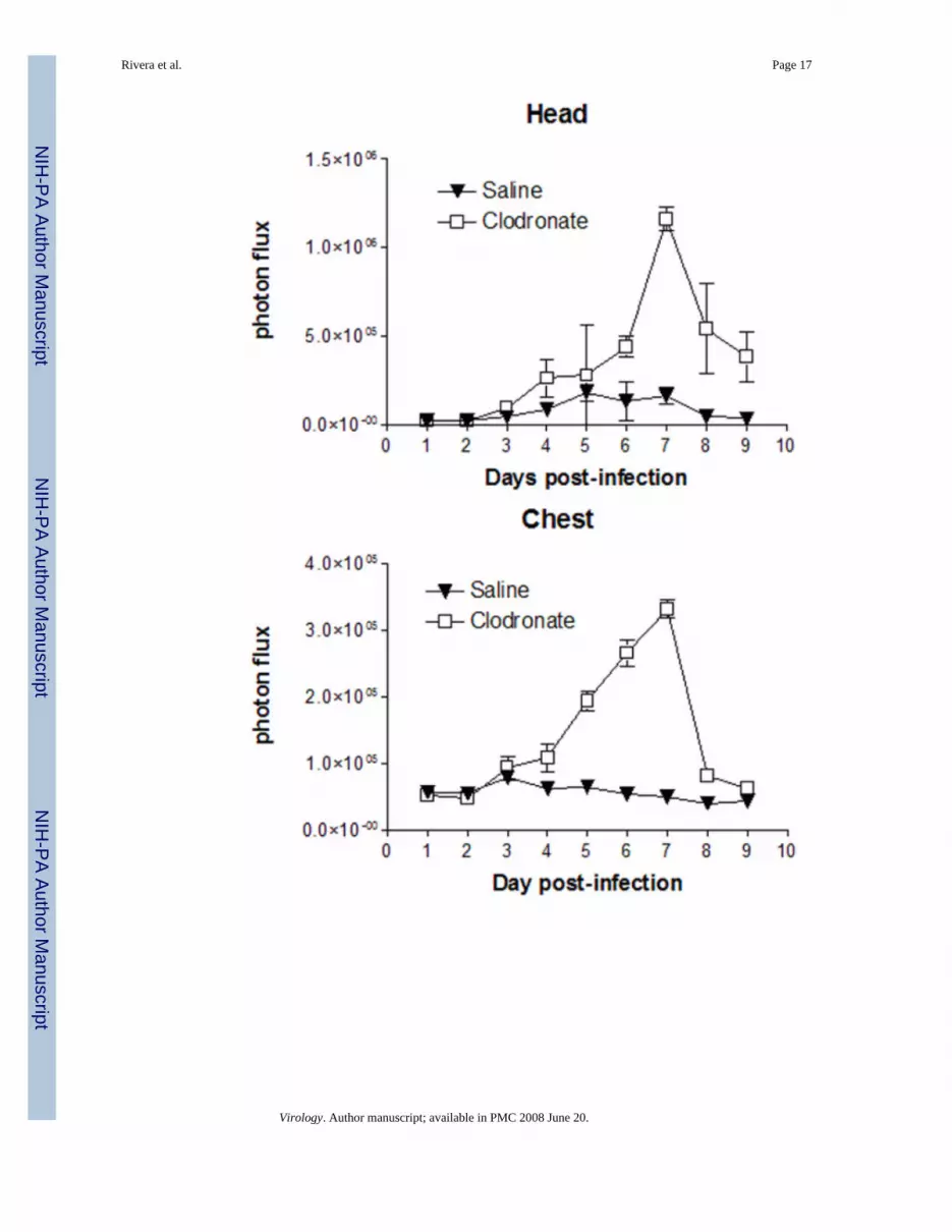
Differences in viral replication as quantified by bioluminescence imaging and plaque assaycorresponded with protective effects of alveolar macrophages in vaccinia infection. Luciferaseactivity produced by Vac-FL in head, chest, and abdomen regions-of-interest was higher inmice treated with clodronate liposomes, with differences becoming apparent by days 4–5 post-infection (Fig 3C–E). In the abdomen, images showed that Vac-FL localized predominantlyto the spleen (data not shown). By AUC analysis of photon flux over days 4–9 post-infection,bioluminescence from Vac-FL was significantly greater in these three regions of interest forinfection (Table 2) (p < 0.01).
To confirm imaging data, we euthanized three mice from each cohort on days 3 and 5 post-infection to quantify amounts of virus in selected organs by plaque assay. Titers of Vac-FL intracheas were approximately 2 logs greater in mice treated with clodronate liposomes comparedwith control animals on days 3 and 5 post-infection (Fig 3F–I). Interestingly, viral titers in thelungs were comparable between both groups, implying that differences in bioluminescence inthe chest region-of-interest may be the result of greater amounts of Vac-FL in tracheas.Systemic spread of Vac-FL to the spleen was detected in mice treated with clodronateliposomes on day 5, while animals treated with saline liposomes had no detectable virus in thespleen on either day. Neither group had detectable amounts of Vac-FL in liver. Amounts ofVac-FL in brain were comparable on day 5, which is consistent with our previous datasuggesting that vaccinia spreads to the brain by local extension from the nasopharynx ratherthan systemic dissemination (Luker et al., 2005). We also analyzed bronchoalveolar lavagefluids from these mice for levels of interferon β, TNF-α, IL-6, and MCP-1, but we detected noconsistent differences in levels of these cytokines produced by mice treated with clodronateversus saline liposomes (data not shown). Overall, these data further demonstrate that alveolarmacrophages limit local infection with vaccinia virus and emphasize that protective effects ofthese cells extend beyond the lung and impact the overall severity of infection.
Depletion of alveolar macrophages enhances recruitment of leukocytes to the lungAlveolar macrophages have been shown to regulate recruitment of inflammatory cells to thelung during infection with bacterial pathogens including Klebsiella pneumoniae andStreptococcus pneumoniae (Broug-Holub et al., 1997) (Knapp et al., 2003). To determine towhat extent alveolar macrophages regulate leukocytes in the lung during vaccinia infection,we depleted alveolar macrophages with liposomal clodronate and then analyzed total numbersand types of immune cells in the lung interstitium on days 3 and 5 after infection with 4 ×10⁵ pfu Vac-FL. Numbers of cells in uninfected mice also were determined and subtractedfrom values obtained in the infected animals.
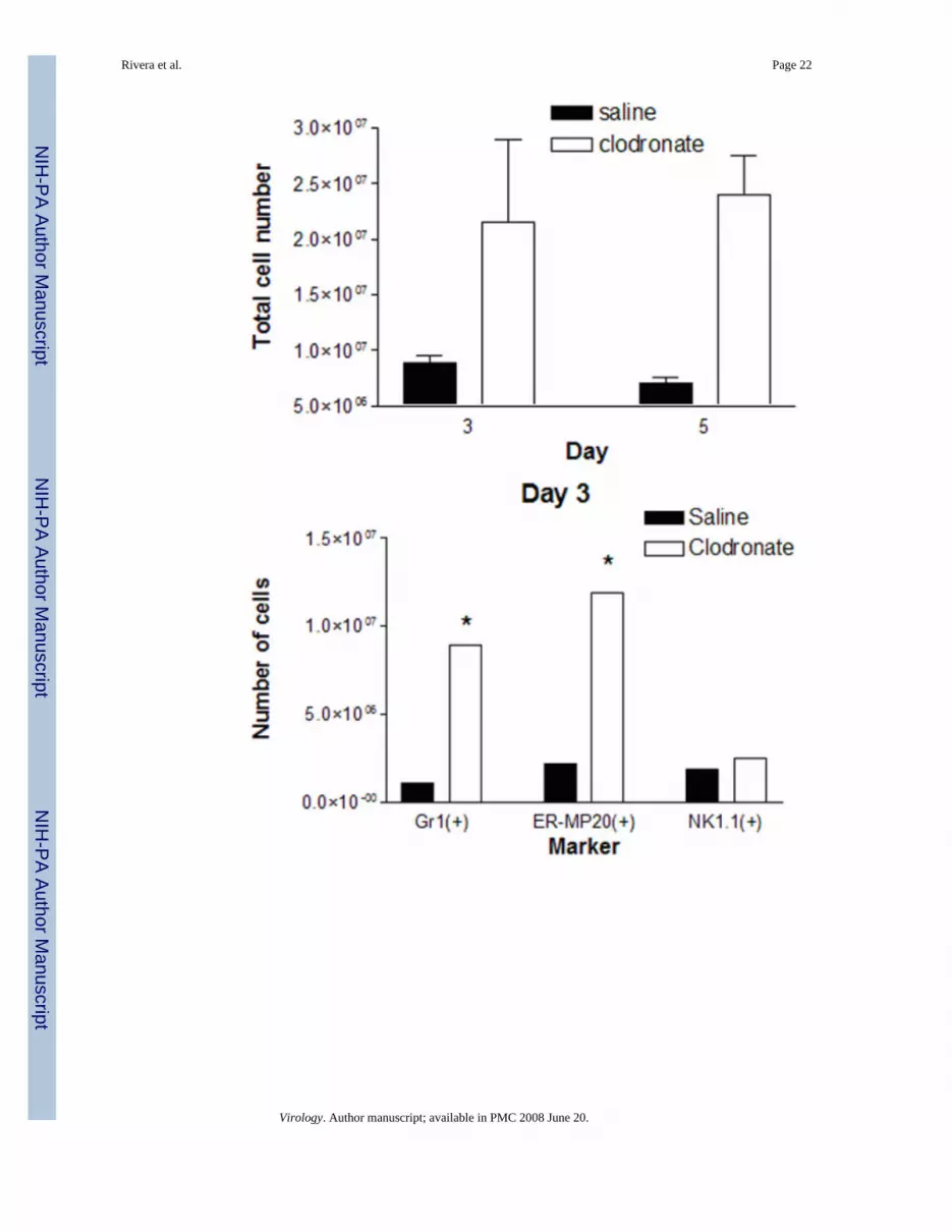
Depletion of alveolar macrophages with liposomal clodronate significantly increased totalnumbers of CD45+ leukocytes recruited to lungs of infected mice (Fig 4A). On both days 3and 5 post-infection, there were approximately 3-fold more total leukocytes present in micetreated with liposomal clodronate and then infected with Vac-FL. By comparison, totalnumbers of leukocytes in uninfected lungs did not differ between mice injected with clodronateor saline liposomes (data not shown).
Rivera et al. Page 4
Virology. Author manuscript; available in PMC 2008 June 20.
NIH
-PA Author Manuscript
NIH
-PA Author Manuscript
NIH
-PA Author Manuscript

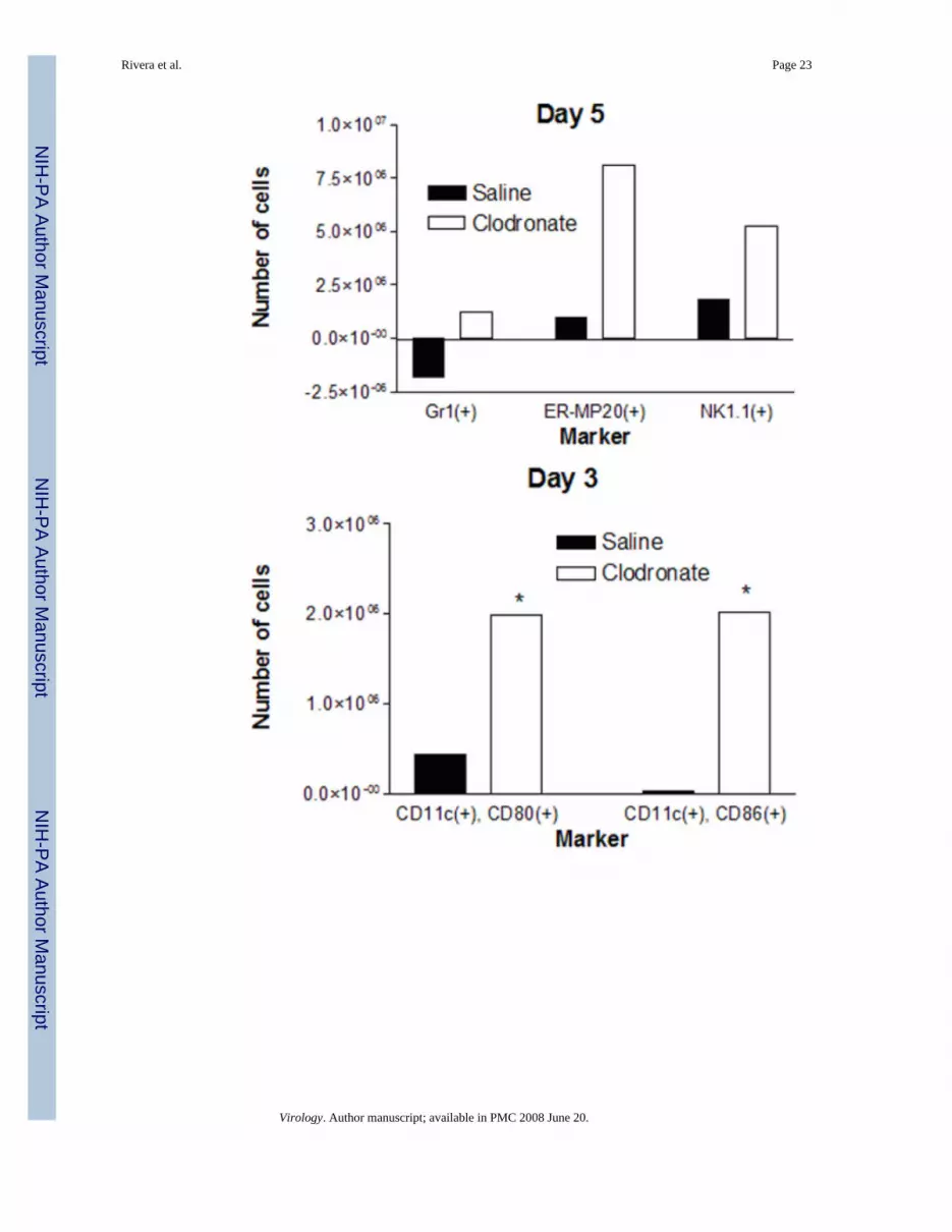
We used flow cytometry to identify specific types of leukocytes present in the lungs and effectsof alveolar macrophages on numbers of various cells present during vaccinia infrction with 4× 10⁵ pfu. On day 3 post-infection, mice treated with clodronate liposomes had significantlygreater numbers of Gr1(+) granulocytes relative to saline control (Fig 4B). Depletion ofalveolar macrophages also increased numbers of cells expressing ER-MP20, a marker ofprecursor cells for granulocytes/monocytes, on day 3 after infection with Vac-FL. Numbers ofGr1(+) and ER-MP20(+) cells on day 5 of infection remained higher in mice treated withclodronate liposomes as compared with control, although total numbers of cells expressingeither marker decreased from day 3 in both experimental groups (Fig 4C). In addition, therewere significantly greater numbers of NK1.1(+) natural killer cells present in lungs of micetreated with clodronate liposomes on day 5. We did not detect differences in numbers of cellswith markers of T or B lymphocytes on either day (data not shown). Although the alveolarspace ultimately is repopulated by cells recruited from the circulation, this process does notmake a significant impact on leukocytes in the lung at these early times. Overall, these datashow that alveolar macrophages directly or indirectly limit influx of selected populations ofleukocytes into the lung during the course of infection with vaccinia virus.
Because alveolar macrophages have been proposed to limit antigen presentation by residentdendritic cells in the pulmonary interstitium, we also used flow cytometry to analyze expressionof co-stimulatory molecules on CD11c(+) cells in lung tissue. Using CD11c as a marker ofpulmonary dendritic cells, there were no differences in total numbers of these cells in lungbetween mice treated with clodronate or saline liposomes, respectively (data not shown).However, depletion of alveolar macrophages greatly enhanced the number of activated CD11c(+) cells on both days 3 and 5 post-infection, as determined by increased expression of co-stimulatory molecules CD80 or CD86 (Fig 4D, E). These data for CD11c(+) dendritic cellssuggest that alveolar macrophages limit activation of dendritic cells during vaccinia infection,potentially through direct suppression or indirectly by reducing amounts of antigen availableto dendritic cells.
Co-culture of alveolar macrophages with respiratory epithelium reduces vaccinia infectionand replication in vitro
Because alveolar macrophages limited replication of vaccinia virus in vivo, we further analyzedeffects of these cells on viral infection in respiratory epithelium in cultured cell lines derivedfrom alveolar macrophages and lung epithelium. MH-S alveolar macrophages and LA-4 lungepithelial cells, which are murine cell lines that have been used previously to analyze hostresponses to infection (Farbermann et al., 2004) (Tuthill et al., 2003), were co-cultured in directcontact to model the dynamic interactions between these cell types during infection in vivo.For these experiments, we focused on co-cultures with 1% and 5% MH-S cells added to LA-4cells for 48 hours prior to infection. Although it is difficult to determine the actual ratio ofalveolar macrophages to respiratory epithelium at a site of infection, these percentages arelower than those published previously for co-culture models and may more accuratelyreproduce conditions in vivo (Hjort et al., 2003) (Ishii et al., 2005).
Cells were infected with Vac-FL, adjusting the total amount of virus to account for differingtotal numbers of LA-4 and MH-S cells in various wells, and we then quantifiedbioluminescence 24 hours after infection. Wells with co-cultured MH-S and LA-4 cells hadsignificantly less bioluminescence from Vac-FL as compared with LA-4 cells alone (Fig 5A).Addition of as few as 1% MH-S cells significantly limited expression of the reporter gene, andprotective effects were evident under both co-culture conditions (p < 0.05). To establish thatco-cultures of respiratory epithelium and alveolar macrophages also limited production of newviruses, we quantified viral replication by plaque assay 24 hours after infection with Vac-FLat an MOI of 0.1. Addition of either 1% or 5% MH-S macrophages to LA-4 epithelial cells
Rivera et al. Page 5
Virology. Author manuscript; available in PMC 2008 June 20.
NIH
-PA Author Manuscript
NIH
-PA Author Manuscript
NIH
-PA Author Manuscript
significantly decreased replication of Vac-FL relative to LA-4 cells alone (p< 0.01) (Fig 5B).Collectively, these data demonstrate that alveolar macrophages limit vaccinia infection inrespiratory epithelium, which may contribute in part to protective effects of alveolarmacrophages during vaccinia infection in mice.
Type I interferons are key components of host defense against vaccinia, which suggests thatproduction of these cytokines or genes activated by type I interferons could account forprotective effects of MH-S cells on infection of respiratory epithelium by vaccinia. Wemeasured levels of interferon β, a key type I interferon regulated by transcription factorsincluding IRF-3 and IRF-7 and produced early in infection (Wathelet et al., 1998). Byquantitative RT-PCR, MH-S cell increased expression of interferon β by 2–3 fold in responseto vaccinia infection at MOI 0.1. However, there was no change in expression of ISG15,RANTES, or IP10, each of which can be induced primarily by IRF-3 or induced secondarilyby type I interferon signaling through its cognate receptor (Nakaya et al., 2001). These datasuggest that activation of a type I interferon response to vaccinia infection is incomplete inMH-S cells, which is consistent with previous work showing that primary alveolarmacrophages from mice can respond to but not produce interferon β (Punturieri et al., 2004).Therefore, other anti-viral signaling molecules and/or mechanisms of host defense, such asphagocytosis, likely enable MH-S cells to reduce vaccinia infection in co-culture withrespiratory epithelium.
DiscussionWe demonstrate that alveolar macrophages confer protection against vaccinia virus infection,functioning to limit local and systemic replication of virus as quantified by bioluminescenceimaging and plaque assays on excised tissues. Depletion of alveolar macrophages increasesweight loss and changes in body temperature, showing that these cells limit the overall severityof disease produced by vaccinia. In the setting of lethal infection, alveolar macrophages alsohad a modest effect to prolong survival of mice. Although images from selected mice showthat alveolar macrophages limit vaccinia infection in lung, data from plaque assays for theentire cohort of mice demonstrate that alveolar macrophages consistently reduce viralreplication in the trachea and not the lung. Alveolar macrophages limited systemicdissemination of virus to spleen and lymph nodes in the abdomen. Viral titers in the brain werenot affected by treatment with clodronate versus saline liposomes, although our previous worksuggests that brain infection with vaccinia is due to direct spread through the olfactory tractrather than systemic dissemination (Luker et al., 2005). Collectively, these data emphasize thecritical function of alveolar macrophages to regulate effective host immunity against vaccinia.
Alveolar macrophages function in part by limiting the total number of inflammatory cellsrecruited to the lung in response to vaccinia infection. In particular, depletion of alveolarmacrophages increases Gr1(+) granulocytes and ER-MP20(+) cells, the latter of which areprogenitors of granulocytes and monocytes. Prior studies performed predominantly in vitroshow that monocytes/macrophages and potentially neutrophils have direct roles in controllingreplication of vaccinia virus (Jones, 1982;Karupiah and Harris, 1995;West et al., 1987).However, our data show that the enhanced inflammatory response is associated with moresevere disease, indicating that enhanced recruitment of these cells is detrimental to the host.Our results are consistent with previous studies showing that the inflammatory response toparticulate materials and pathogens in the lungs disrupts integrity of respiratory epithelium andcapillaries, thereby impairing lung exchange and increasing severity of disease (Moraes,Zurawska, and Downey, 2006). In addition, depletion of alveolar macrophages increasesnumbers of CD11c(+) dendritic cells in the lung that express co-stimulatory molecules CD80and CD86 as markers of activation. It has been demonstrated previously that depletion ofpulmonary macrophages from rats enhances the ability of dendritic cells to present a model
Rivera et al. Page 6
Virology. Author manuscript; available in PMC 2008 June 20.
NIH
-PA Author Manuscript
NIH
-PA Author Manuscript
NIH
-PA Author Manuscript

antigen to T lymphocytes (Holt et al., 1993). Overall, our results establish that one key functionof alveolar macrophages is to limit the inflammatory response to vaccinia in the lung, whichreduces viral dissemination of systemic effects of infection.
In addition to reducing numbers of leukocytes in infected lungs, alveolar macrophages alsointeract with lung epithelium to limit infection with vaccinia. Using an in vitro co-culture modelof alveolar macrophages and lung epithelium, we demonstrated that addition of as few as 1%alveolar macrophages to lung epithelium significantly reduced viral gene expression andreplication after infection at low MOI. Although it is difficult to estimate relative proportionsof alveolar macrophages to respiratory epithelium at sites of infection, alveolar macrophagesare known to be recruited actively to areas of infection and inflammation in the lung. Therefore,the ability of alveolar macrophages to reduce vaccinia infection of lung epithelium in a cellculture model likely represents another critical function of alveolar macrophages in hostdefense against poxviruses in vivo.
Secretion of cytokines and/or phagocytosis are two possible mechanisms through whichalveolar macrophages limit infection with vaccinia virus. We analyzed expression of interferonβ and selected interferon stimulated genes in alveolar macrophages as candidate molecules toreduce viral replication. Although MH-S alveolar macrophages infected with vaccinia virusupregulated expression of interferon β, we did not detect changes in expression of otherinterferon-regulated genes. These data are consistent with previous research showing thatalveolar macrophages lack autocrine stimulation and feed-forward amplification of interferonsignaling in response to stimulation with poly I:C, although these cells are able to respond toexogenous type I interferon (Punturieri et al., 2004). Because we were unable to reliablyseparate small numbers of alveolar macrophages from lung epithelial cells, we could notanalyze expression of interferon-responsive genes in alveolar macrophages in co-cultureconditions. While lung epithelial cells may secrete type I interferon to activate interferonsignaling in alveolar macrophages, these data suggest that protective effects of alveolarmacrophages may be mediated by other cytokines or molecules such as nitric oxide (Karupiahand Harris, 1995). Protective effects of alveolar macrophages also may be mediated throughphagocytosis of virus particles, thereby sequestering vaccinia from lung epithelium andlimiting infection. This mechanism of host defense is supported by previous research showingthat both immune and non-immune macrophages ingest vaccinia virus (Greer, Delfs, andMcElree, 1974). Further studies are needed to establish to what extent secretion of cytokines,phagocytosis, and/or other mechanisms such as cell-to-cell contact promote protective effectsof alveolar macrophages in vitro and in vivo.
In conclusion, the current study establishes that alveolar macrophages have an essentialprotective function against infection with orthopoxviruses. These cells limit the inflammatoryresponse to vaccinia in the lung, which reduces viral replication, dissemination, and overallseverity of disease. Modulating the recruitment of neutrophils and other inflammatory cellsinto the lung potentially may be effective in reducing disease severity produced by a naturalor intentional infection with poxviruses.
Materials and MethodsCells
MH-S cells (ATCC) are an SV40 transformed murine alveolar macrophage cell line derivedfrom Balb/c mice. These cells retain many morphologic and functional features of alveolarmacrophages and express surface antigens MHC class I H2d, class II Ia, and CD11B(Mbawuike and Herscowitz, 1989). LA-4 cells are a murine lung epithelial cell line withcharacteristics of type II pneumocytes (Stoner et al., 1975). LA-4 cells were derived from anA/He mouse and have MHC class I haplotype H2k. MH-S and Vero cells were cultured in
Rivera et al. Page 7
Virology. Author manuscript; available in PMC 2008 June 20.
NIH
-PA Author Manuscript
NIH
-PA Author Manuscript
NIH
-PA Author Manuscript
DMEM medium (Invitrogen) with 10% heat-inactivated fetal bovine serum, 1% L-glutamine,and 0.1% penicillin-streptomycin in a 5% CO2 incubator at 37° C. LA-4 cells were culturedin DMEM medium containing 15% heat-inactivated fetal bovine serum.
Vaccinia virusStocks of Vac-FL, a recombinant Western Reserve vaccinia virus that expresses fireflyluciferase, were prepared and titered as described previously (Earl et al., 1998;Luker et al.,2005).
Monoclonal antibodiesThe following mAbs were used for flow cytometry (BD Pharmingen): RM4-4 (anti-murineCD4, rat IgG2b), 53–6.72 (anti-murine CD8, rat IgG2b), M1/70 (anti-murine CD11b, ratIgG2b), HL3 (anti-murine CD11c, hamster IgG1), 2.4G2 (anti-murine CD16/CD32 Fc block,rat IgG2b), 3/23 (anti-murine CD40, rat IgG2a), RA-36B2 (anti-murine CD45R/B220, ratIgG2a), 30-F11 (anti-murine CD45, rat IgG2b), 16–10A1 (anti-murine CD80, hamster IgG2),GL1 (anti-murine CD86, rat IgG2a), AF6-120.1 (anti-murine I-Ab MHC class II, mouseIgG2a), and RB6-8C5 (anti-murine Ly6G Gr-1, rat IgG2b). mAbs were primarily conjugatedwith FITC, biotin, or PE; biotinylated Abs were visualized using streptavidin-PerCP (BDPharmingen). Isotype-matched irrelevant control mAbs (BD Pharmingen) were testedsimultaneously in all experiments.
In vitro co-culture experiments1 × 104 LA-4 cells were plated in 96 well plates, allowed to adhere for ≈ 4 hours, and then 0– 5 × 10² MH-S cells were added to wells for 48 hours prior to infection. Cells were infectedwith Vac-FL at MOI 0.1, adjusting the inoculum for the total number of cells in wells.Bioluminescence from Vac-FL was quantified using the IVIS (Xenogen) 24 hours afterinfection (Luker et al., 2005). Viral titers were determined by plaque assay at these same timepoints.
Mouse proceduresAll animal procedures were approved by the University Committee on Use and Care of Animals(UCUCA) at University of Michigan. 7–10 week-old male 129 Ev/Sv mice (Taconic) wereused for all experiments. To deplete alveolar macrophages, mice were injected intratracheallywith 100 μl of liposomes containing clodronate (Clodronate liposomes). Control mice receivedliposomes containing saline. Mice were infected intranasally with various amounts of Vac-FLin 20 μL sterile DMEM as described in figure legends. Anesthetized mice were shaved withclippers to decrease absorption and scattering of light for bioluminescence imaging. Animalweights were recorded daily following infection, and rectal temperatures were measured every24 hours in selected experiments. Bronchoalveolar lavage and removal of lungs for analysisof cells in the pulmonary interstitium were performed as described previously (Curtis andKaltreider, 1989) (Osterholzer et al., 2005).
Analysis of BAL samplesTotal cells recovered from BAL were counted manually with a hemocytometer and then stainedwith Wright-Giemsa for differential cell counts. The first one ml of BAL fluid recovered fromlungs was used for ELISA studies of cytokines.
Antibody staining and flow-cytometric analysisStaining of cells in the pulmonary interstitium, including blockade of FcRs, and analysis byflow cytometry were performed as described previously (Osterholzer et al., 2005). Data were
Rivera et al. Page 8
Virology. Author manuscript; available in PMC 2008 June 20.
NIH
-PA Author Manuscript
NIH
-PA Author Manuscript
NIH
-PA Author Manuscript

collected on a FACScan flow cytometer using CellQuest software (both from BDImmunocytometry Systems) and analyzed using FlowJo software (Tree Star). A minimum of10,000 cells were analyzed per sample. For all analyses, percentages for matched isotypecontrol antibodies were subtracted from values obtained for staining with specific antibodiesfor individual markers.
Quantitative RT-PCRRNA was isolated from cultured cells using Trizol (Invitrogen) and RNeasy columns (Qiagen)according to the manufacturers’ protocols. Quantitative RT-PCR using syber green detectionwas performed with the iScript One-Step Kit (Bio-Rad) on a MX3000P instrument(Stratagene). Primer sequences for amplified mouse genes are listed below:
Interferon β: forward 5′ AGCTCCAAGAAAGGACGAACAT 3′reverse 5′ GCCCTGTAGGTGAGGTTGATCT 3′
ISG15: forward 5′ CAGGACGGTCTTACCCTTTCC 3′reverse 5′ AGGCTCGCTGCAGTTCTGTAC 3′
IP10: forward 5′ CCTGCCCACGTGTTGAGAT 3′reverse 5′ TGATGGTCTTAGATTCCGGATTC 3′
RANTES: forward 5′ GCCCACGTCAAGGAGTATTTCTA 3′reverse 5′ ACACACTTGGCGGTTCCTTC 3′
GAPDH: forward 5′ TATGTCGTGGAGTCTACTGGT 3′reverse 5′ GAGTTGTCATATTTCTCGTGG 3′
Data for target gene expression were normalized to GAPDH as a control.
Virus titrationViral titers in cells and organs were analyzed by serial dilution on Vero cells as describedpreviously (Luker et al., 2005).
Bioluminescence imagingBioluminescence imaging was performed with a cryogenically-cooled CCD camera (IVIS)(Xenogen) as described previously (Luker et al., 2002). ROI’s corresponding to the head, chest,and abdomen of infected mice were used to quantify bioluminescence as photon flux usingsoftware provided with the IVIS.
StatisticsPairs of data points were analyzed by t test for statistically significant differences (p < 0.05).Statistics for correlation coefficients and area-under-the-curve analyses were performed withcommercially available software (Graphpad, Prism).
Acknowledgements
Research was supported by R21AI066192 and RO1 HL082480 from the UHPHS, and Merit Review funds from theDepartment of Veterans Affairs. Support for imaging experiments was provided by NIH R24CA083099 for theUniversity of Michigan Small Animal Imaging Resource.
ReferencesBroug-Holub E, Toews G, van Iwaarden J, Strieter R, Kunkel S, Paine Rr, Standiford T. Alveolar
macrophages are required for protective pulmonary defenses in murine Klebsiella pneumonia:elimination of alveolar macrophages increases neutrophil recruitment but decreases bacterial clearanceand survival. Infect Immun 1997;65(4):1139–1146. [PubMed: 9119443]
Curtis J, Kaltreider H. Characterization of bronchoalveolar lymphocytes during a specific antibody-forming cell response in the lungs of mice. Am Rev Respir Dis 1989;139(2):393–400. [PubMed:2464296]
Rivera et al. Page 9
Virology. Author manuscript; available in PMC 2008 June 20.
NIH
-PA Author Manuscript
NIH
-PA Author Manuscript
NIH
-PA Author Manuscript

Earl, P.; Cooper, N.; Wyatt, L.; Moss, B. Preparation of cell cultures and vaccinia virus stocks. In:Ausubel, FM.; Brent, R.; Kingston, RE.; Moore, DD.; Seidman, JG.; Smith, JA.; Struhl, K., editors.Current Protocols in Molecular Biology. John Wiley & Sons, Inc.; 1998. p. 16.16.1-16.16.11.
Farbermann M, Hoffmann J, Ryerse J, Demello D. fusible signal to murine alveolar macrophages fromlipopolysaccharide- and Escherichia coli-stimulated lung Type II epithelial cells. Inflamm Res 2004;53(9):475–483. [PubMed: 15551001]
Fenner F, Henderson D, Arita I, Jezek Z, Ladnyi I. Smallpox & its eradication. World HealthOrganization. 1988
Greer B, Delfs D, McElree H. Electron microscope study of the interaction of vaccinia virus withmacrophages from immunized and nonimmunized rabbits. Infect Immun 1974;9(2):452–459.[PubMed: 4816467]
Hjort M, Brenyo A, Finkelstein J, Frampton M, LoMonaco M, Stewart J, Johnston C, D’Angio C.Alveolar epithelial cell-macrophage interactions affect oxygen-stimulated interleukin-8 release.Inflammation 2003;27(3):137–145. [PubMed: 12875367]
Holt P, Oliver J, Bilyk N, McMenamin C, McMenamin P, Kraal G, Thepen T. Downregulation of theantigen presenting cell function(s) of pulmonary dendritic cells in vivo by resident alveolarmacrophages. J Exp Med 1993;177(2):397–407. [PubMed: 8426110]
Ishii H, Hayashi S, Hogg J, Fujii T, Goto Y, Sakamoto N, Mukae H, Vincent R, van Eeden S. Alveolarmacrophage-epithelial cell interaction following exposure to atmospheric particles induces the releaseof mediators involved in monocyte mobilization and recruitment. Respir Res 2005;6:87. [PubMed:16053532]
Jones J. Interactions between human neutrophils and vaccinia virus: induction of oxidative metabolismand virus inactivation. Pediatr Res 1982;16(7):525–529. [PubMed: 7110771]
Karupiah G, Harris N. Inhibition of viral replication by nitric oxide and its reversal by ferrous sulfate andtricarboxylic acid cycle metabolites. J Exp Med 1995;181(6):2171–2179. [PubMed: 7539042]
Knapp S, Leemans J, Florquin S, Branger J, Maris N, Pater J, van Rooijen N, van der Poll T. Alveolarmacrophages have a protective antiinflammatory role during murine pneumococcal pneumonia. AmJ Respir Crit Care Med 2003;167:171–179. [PubMed: 12406830]
Leenen P, Radosevic K, Voerman J, Salomon B, van Rooijen N, Klatzmann D, van Ewijk W.Heterogeneity of mouse spleen dendritic cells: in vivo phagocytic activity, expression of macrophagemarkers, and subpopulation turnover. J Immunol 1998;160(5):2166–2173. [PubMed: 9498754]
Luker G, Bardill J, Prior J, Pica C, Piwnica-Worms D, Leib D. Noninvasive bioluminescence imagingof herpes simplex virus type 1 infection and therapy in living mice. J Virol 2002;76(23):12149–12161. [PubMed: 12414955]
Luker K, Hutchens M, Schultz T, Pekosz A, Luker G. Bioluminescence imaging of vaccinia virus: effectsof interferon on viral replication and spread. Virology 2005;341(2):284–300. [PubMed: 16095645]
MacLean J, Zia W, Pinto C, Zhao L, Liu H, Kradin R. Sequestration of inhaled particulate antigens bylung phagocytes. A mechanism for the effective inhibition of pulmonary cell-mediated immunity.Am J Pathol 1996;148:657–666. [PubMed: 8579128]
Mbawuike I, Herscowitz H. MH-S, a murine alveolar macrophage cell line: morphological, cytochemical,and functional characteristics. J Leukoc Biol 1989;46(2):119–127. [PubMed: 2787372]
Moraes T, Zurawska J, Downey G. Neutrophil granule contents in the pathogenesis of lung injury. CurrOpin Hematol 2006;13(1):21–27. [PubMed: 16319683]
Nakaya T, Sato M, Hata N, Asagiri M, Suemori H, Noguchi S, Tanaka N, Taniguchi T. Gene inductionpathways mediated by distinct IRFs during viral infection. Biochem Biophys Res Commun 2001;283(5):1150–1156. [PubMed: 11355893]
Osterholzer J, Ames T, Polak T, Sonstein J, Moore B, Chensue S, Toews G, Curtis J. CCR2 and CCR6,but not endothelial selectins, mediate the accumulation of immature dendritic cells within the lungsof mice in response to particulate antigen. J Immunol 2005;175(2):874–883. [PubMed: 16002685]
Punturieri A, Alviani R, Polak T, Copper P, Sonstein J, Curtis J. Specific engagement of TLR4 or TLR3does not lead to IFN-beta-mediated innate signal amplification and STAT1 phosphorylation inresident murine alveolar macrophages. J Immunol 2004;173(2):1033–1042. [PubMed: 15240691]
Rivera et al. Page 10
Virology. Author manuscript; available in PMC 2008 June 20.
NIH
-PA Author Manuscript
NIH
-PA Author Manuscript
NIH
-PA Author Manuscript

Qian Q, Jutila M, van Rooijen N, Cutler J. Elimination of mouse splenic macrophages correlates withincreased susceptibility to experimental disseminated candidiasis. J Immunol 1994;152(10):5000–5008. [PubMed: 8176217]
Stoner G, Kikkawa Y, Kniazeff A, Miyai K, Wagner R. Clonal isolation of epithelial cells from mouselung adenoma. Cancer Res 1975;35(8):2177–2185. [PubMed: 167947]
Thepen T, Kraal G, Holt P. The role of alveolar macrophages in regulation of lung inflammation. AnnN Y Acad Sci 1994;725:200–206. [PubMed: 8030991]
Tuthill T, Papadopoulos N, Jourdan P, Challinor L, Sharp N, Plumpton C, Shah K, Barnard S, Dash L,Burnet J, Killington R, Rowlands D, Clarke N, Blair E, Johnston S. Mouse respiratory epithelial cellssupport efficient replication of human rhinovirus. J Gen Virol 2003;84:2829–2836. [PubMed:13679617]
van den Broek M, Muller U, Huang S, Aguet M, Zinkernagel R. Antiviral defense in mice lacking bothalpha/beta and gamma interferon receptors. J Virol 1995;69(8):4792–4796. [PubMed: 7609046]
van Rooijen N. The liposome-mediated macrophage ‘suicide’ technique. J Immunol Methods 1989;124(1):1–6. [PubMed: 2530286]
Wathelet M, Lin C, Parekh B, Ronco L, Howley P, Maniatis T. Virus infection induces the assembly ofcoordinately activated transcription factors on the IFN-beta enhancer in vivo. Mol Cell 1998;1(4):507–518. [PubMed: 9660935]
West B, Eschete M, Cox M, King J. Neutrophil uptake of vaccinia virus in vitro. J Infect Dis 1987;156(4):597–606. [PubMed: 3624906]
Rivera et al. Page 11
Virology. Author manuscript; available in PMC 2008 June 20.
NIH
-PA Author Manuscript
NIH
-PA Author Manuscript
NIH
-PA Author Manuscript
Figure 1.Depletion of alveolar macrophages with clodronate liposomes. 129 Ev/Sv mice were injectedintratracheally with 100 μl of liposomes containing clodronate or saline as a control. Totalnumbers of cells recovered from bronchoalveolar lavage fluid were quantified at different timepoints after treatment (n = 3 per time point). Data are expressed as the per cent cells recoveredrelative to mice that did not receive either clodronate or saline liposomes (n = 3). Error barsrepresent SEM.
Rivera et al. Page 12
Virology. Author manuscript; available in PMC 2008 June 20.
NIH
-PA Author Manuscript
NIH
-PA Author Manuscript
NIH
-PA Author Manuscript

Rivera et al. Page 13
Virology. Author manuscript; available in PMC 2008 June 20.
NIH
-PA Author Manuscript
NIH
-PA Author Manuscript
NIH
-PA Author Manuscript

Rivera et al. Page 14
Virology. Author manuscript; available in PMC 2008 June 20.
NIH
-PA Author Manuscript
NIH
-PA Author Manuscript
NIH
-PA Author Manuscript

Figure 2.Alveolar macrophages limit vaccinia virus infection. Mice were treated with intratrachealinjection of clodronate or saline liposomes 2 days prior to infection with 4 × 10⁵ pfu Vac-FLintranasally (n = 5 mice per group). (A) Mice were weighed daily to monitor overall progressionof disease. Data are presented as mean ± SEM per cent loss of initial weight. (B) Representativebioluminescent images on day 4 of Vac-FL infection in mice treated with clodronate or salineliposomes. Viral bioluminescence in lungs (arrow) and spread to inguinal lymph node (asterisk)is greater in mice treated with clodronate liposomes to deplete alveolar macrophages. (C–E).Quantified photon flux data from head (C), chest (D), and abdomen (E) regions-of-interest.Data are shown as mean values of background subtracted photon flux ± SEM.
Rivera et al. Page 15
Virology. Author manuscript; available in PMC 2008 June 20.
NIH
-PA Author Manuscript
NIH
-PA Author Manuscript
NIH
-PA Author Manuscript
Rivera et al. Page 16
Virology. Author manuscript; available in PMC 2008 June 20.
NIH
-PA Author Manuscript
NIH
-PA Author Manuscript
NIH
-PA Author Manuscript
Rivera et al. Page 17
Virology. Author manuscript; available in PMC 2008 June 20.
NIH
-PA Author Manuscript
NIH
-PA Author Manuscript
NIH
-PA Author Manuscript

Rivera et al. Page 18
Virology. Author manuscript; available in PMC 2008 June 20.
NIH
-PA Author Manuscript
NIH
-PA Author Manuscript
NIH
-PA Author Manuscript

Rivera et al. Page 19
Virology. Author manuscript; available in PMC 2008 June 20.
NIH
-PA Author Manuscript
NIH
-PA Author Manuscript
NIH
-PA Author Manuscript

Rivera et al. Page 20
Virology. Author manuscript; available in PMC 2008 June 20.
NIH
-PA Author Manuscript
NIH
-PA Author Manuscript
NIH
-PA Author Manuscript
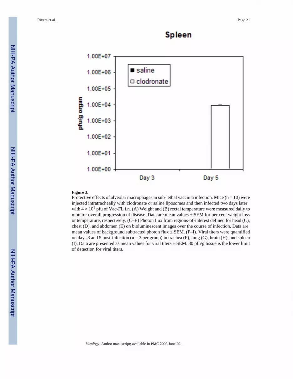
Figure 3.Protective effects of alveolar macrophages in sub-lethal vaccinia infection. Mice (n = 10) wereinjected intratracheally with clodronate or saline liposomes and then infected two days laterwith 4 × 10⁴ pfu of Vac-FL i.n. (A) Weight and (B) rectal temperature were measured daily tomonitor overall progression of disease. Data are mean values ± SEM for per cent weight lossor temperature, respectively. (C–E) Photon flux from regions-of-interest defined for head (C),chest (D), and abdomen (E) on bioluminescent images over the course of infection. Data aremean values of background subtracted photon flux ± SEM. (F–I). Viral titers were quantifiedon days 3 and 5 post-infection (n = 3 per group) in trachea (F), lung (G), brain (H), and spleen(I). Data are presented as mean values for viral titers ± SEM. 30 pfu/g tissue is the lower limitof detection for viral titers.
Rivera et al. Page 21
Virology. Author manuscript; available in PMC 2008 June 20.
NIH
-PA Author Manuscript
NIH
-PA Author Manuscript
NIH
-PA Author Manuscript
Rivera et al. Page 22
Virology. Author manuscript; available in PMC 2008 June 20.
NIH
-PA Author Manuscript
NIH
-PA Author Manuscript
NIH
-PA Author Manuscript
Rivera et al. Page 23
Virology. Author manuscript; available in PMC 2008 June 20.
NIH
-PA Author Manuscript
NIH
-PA Author Manuscript
NIH
-PA Author Manuscript
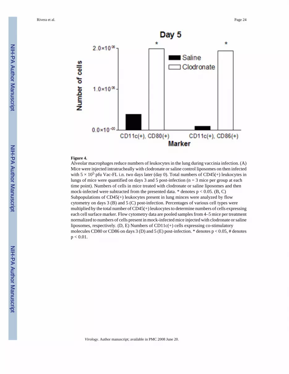
Figure 4.Alveolar macrophages reduce numbers of leukocytes in the lung during vaccinia infection. (A)Mice were injected intratracheally with clodronate or saline control liposomes on then infectedwith 5 × 10⁵ pfu Vac-FL i.n. two days later (day 0). Total numbers of CD45(+) leukocytes inlungs of mice were quantified on days 3 and 5 post-infection (n = 3 mice per group at eachtime point). Numbers of cells in mice treated with clodronate or saline liposomes and thenmock-infected were subtracted from the presented data. * denotes p < 0.05. (B, C)Subpopulations of CD45(+) leukocytes present in lung minces were analyzed by flowcytometry on days 3 (B) and 5 (C) post-infection. Percentages of various cell types weremultiplied by the total number of CD45(+) leukocytes to determine numbers of cells expressingeach cell surface marker. Flow cytometry data are pooled samples from 4–5 mice per treatmentnormalized to numbers of cells present in mock-infected mice injected with clodronate or salineliposomes, respectively. (D, E) Numbers of CD11c(+) cells expressing co-stimulatorymolecules CD80 or CD86 on days 3 (D) and 5 (E) post-infection. * denotes p < 0.05, # denotesp < 0.01.
Rivera et al. Page 24
Virology. Author manuscript; available in PMC 2008 June 20.
NIH
-PA Author Manuscript
NIH
-PA Author Manuscript
NIH
-PA Author Manuscript

Figure 5.Co-culture of lung epithelial cells with alveolar macrophages limits vaccinia infection. LA-4lung epithelial cells were cultured alone or with 1% or 5% added MH-S alveolar macrophagesfor 2 days prior to infection with Vac-FL at MOI of 0.1. Numbers of viral pfu were adjustedto account for differing total numbers of cells in various wells. (A) Photon flux data are shownfor Vac-FL 24 hours after infection at MOI of 0.1. Data are expressed as mean values ± SEMfor background subtracted photon flux normalized to bioluminescence produced from infectionof LA-4 cells alone. (B) Titers of Vac-FL produced 24 hours after infection of LA-4 cells orLA-4 cells cultured with 1% or 5% MH-S cells. Data are presented as mean values for viralpfu/ml ± SEM (n = 4 samples per condition). The lower limits of detection for the assay are30 pfu/ml. Results are representative of two independent experiments.
Rivera et al. Page 25
Virology. Author manuscript; available in PMC 2008 June 20.
NIH
-PA Author Manuscript
NIH
-PA Author Manuscript
NIH
-PA Author Manuscript
NIH
-PA Author Manuscript
NIH
-PA Author Manuscript
NIH
-PA Author Manuscript
Rivera et al. Page 26
Table 1Area-under-the-curve (AUC) photon flux days 4–6 from 4 × 10⁵ pfu infection.
Liposomes Head Chest AbdomenSaline 2.3 × 10⁶ 7.36 × 10⁵ 2.94 × 10⁵
Clodronate 2.9 × 10⁶ 9.78 × 10⁵ 3.95 × 10⁵
AUC values for mice treated with clodronate liposomes are significantly greater than those for control saline liposomes (p < 0.05) (n = 5 mice per group).
Virology. Author manuscript; available in PMC 2008 June 20.

NIH
-PA Author Manuscript
NIH
-PA Author Manuscript
NIH
-PA Author Manuscript
Rivera et al. Page 27
Table 2AUC photon flux days 4–9 from 4 × 10⁴ pfu infection.
Liposomes Head Chest AbdomenSaline 5.82 × 10⁵ 2.61 × 10⁵ 2.09 × 10⁵
Clodronate 2.74 × 10⁶ 9.59 × 10⁵ 3.03 × 10⁵
AUC values for mice treated with clodronate liposomes are significantly greater than those for control saline liposomes (p < 0.01) (n = 7 mice per groupthrough day 5, then n = 4 per group on days 6–9).
Virology. Author manuscript; available in PMC 2008 June 20.


