Embed Size (px)
Citation preview

Probing the Binding of Coumarins and Cyclothialidines to DNA Gyrase†
Sotirios C. Kampranis, Niall A. Gormley,‡ Rebecca Tranter,‡ George Orphanides,§ and Anthony Maxwell*
Department of Biochemistry, UniVersity of Leicester, Leicester LE1 7RH, U.K.
ReceiVed September 25, 1998; ReVised Manuscript ReceiVed NoVember 30, 1998
ABSTRACT: DNA gyrase is the target of a number of antibacterial agents, including the coumarins and thecyclothialidines. To extend our understanding of the mechanism of action of these compounds, we haveexamined the previously published crystal structures of the complexes between the 24 kDa fragment ofGyrB and coumarin and cyclothialidine drugs and made mutations by site-directed mutagenesis. We usedproteolysis as a probe of drug binding to wild-type and mutant proteins. Limited proteolysis of gyraserevealed that binding of these antibiotics is associated with a characteristic proteolytic fingerprint, suggestinga drug-induced conformational change. The ability of the mutants to bind the drugs was studied by testingtheir ability to induce the coumarin-associated proteolytic signature and to bind to a novobiocin-affinitycolumn. To analyze further the interaction of the drugs with gyrase, we studied the binding using surfaceplasmon resonance. Mutation of Asn46 to Asp has only a modest effect on the binding of coumarins,while an Asn46 to Leu mutation results in a 10-fold decrease in the affinity. Mutation of Asp73 to Asncompletely abolishes binding to both coumarins and cyclothialidines. Mutations at these residues alsoabolish ATP hydrolysis, explaining the inability of such mutations to occur spontaneously.
DNA gyrase is the target of a number of antibacterialagents (1). The most prominent of these are the quinolones,an entirely synthetic class of compounds, which have foundwide application in clinical practice (2, 3). The coumarinsand the cyclothialidines (Figure 1), which are naturallyoccurring antibiotics, have failed to become clinicallysuccessful due to poor cell penetration, low solubility, andtoxicity in eukaryotes (1). However, the fact that thesecompounds are significantly more potent in inhibiting DNAgyrase in vitro than the quinolones has stimulated interestwith respect to improving their properties in order to producestructurally related compounds suitable for clinical practice.
DNA gyrase is the bacterial type II topoisomerase whichcan introduce negative supercoils into DNA using the freeenergy of ATP hydrolysis (4, 5). Mechanistic studies haverevealed the steps involved in the supercoiling reaction.Briefly, this process involves the wrapping of DNA aroundthe enzyme, cleavage of this DNA in both strands (involvingthe formation of DNA-protein covalent bonds), and passageof a segment of DNA through this double-stranded break.This process is coupled to ATP hydrolysis and results in theintroduction of two negative supercoils. The enzyme fromEscherichia coli consists of two proteins, A and B, ofmolecular masses 97 and 90 kDa respectively; the active
enzyme is an A2B2 complex. Both of these proteins havebeen shown to contain distinct domains. The A protein(GyrA1) consists of an N-terminal domain (59-64 kDa)involved in DNA breakage and reunion and a C-terminaldomain (33 kDa) involved in DNA-protein interactions (4,5). The structure of the 59 kDa N-terminal fragment(GyrA59) has been solved to 2.8 Å resolution by X-raycrystallography (6). The B protein (GyrB) consists of anN-terminal domain (43 kDa; GyrB43) containing the ATPaseactivity and a C-terminal domain (47 kDa; GyrB47) involvedin interactions with the A protein and DNA. The structureof the 43 kDa N-terminal domain complexed with ADPNPhas been solved to 2.5 Å resolution by X-ray crystallography(7).
Since gyrase is an essential enzyme in prokaryotes but isnot found in eukaryotes, it is an ideal target for antibiotics.The coumarins (e.g., novobiocin and coumermycin A1;Figure 1) are naturally occurring compounds that have beenshown to inhibit the ATPase reaction of gyrase (8, 9).Sequencing of mutations ingyrB conferring coumarinresistance has suggested that the coumarin-binding site liesin the N-terminal portion of GyrB (10, 11). Indeed, a 24kDa N-terminal fragment of GyrB (GyrB24; residues 2-220)has been cloned and expressed and shown to contain thecoumarin-binding site (12). The cyclothialidines are anotherfamily of naturally occurring antibiotics that target DNAgyrase. In vitro studies have shown that cyclothialidines alsoact by inhibiting the ATPase reaction of gyrase (13).Moreover, Staphylococcus aureusstrains resistant to cy-clothialidines carry mutations that map near the ATP-binding
† S.C.K. was supported by a CASE studentship funded by theBBSRC and Glaxo-Wellcome, and a grant from the Alexander S.Onassis Public Benefit Foundation, N.A.G. was supported by theWellcome Trust, G.O. was supported by a CASE studentship fundedby the BBSRC and Zeneca Pharmaceuticals, and A.M. was a ListerInstitute Jenner Fellow.
* To whom correspondence should be addressed.‡ Present address: Department of Biochemistry, University of Bristol,
Bristol BS8 1TD, U.K.§ Present address: Howard Hughes Medical Institute, UMDNJ,
Robert Wood Johnson Medical School, Dept. of Biochemistry, 663Hoes Lane, Piscataway, NJ 08854.
1 Abbreviations: ADPNP, 5′-adenylylâ,γ-imidodiphosphate; GyrA,DNA gyrase A protein; GyrB, DNA gyrase B protein; PAGE,polyacrylamide gel electrophoresis; SDS, sodium dodecyl sulfate; SPR,surface plasmon resonance.
1967Biochemistry1999,38, 1967-1976
10.1021/bi982320p CCC: $18.00 © 1999 American Chemical SocietyPublished on Web 01/28/1999
Dow
nloa
ded
by T
UFT
S U
NIV
on
Nov
embe
r 3,
200
9 | h
ttp://
pubs
.acs
.org
P
ublic
atio
n D
ate
(Web
): J
anua
ry 2
8, 1
999
| doi
: 10.
1021
/bi9
8232
0p

site in GyrB (14), while binding studies suggested that thecyclothialidine-binding site overlaps with those of ATP andcoumarins (15). However, coumarin-resistant mutants are notnecessarily resistant to cyclothialidines (14-16).
The complexes of the 24 kDa GyrB fragment with thecoumarin drugs novobiocin and chlorobiocin and a memberof the cyclothialidine family (GR122222X) have beencrystallized and the structures solved to high resolution (17-19). These crystal structures show that the binding sites forATP and coumarins partially overlap with the sugar ring ofthe drugs at the binding site of the adenine ring of ATP.The cyclothialidine-binding site is distinct from that ofcoumarins but also overlaps with that of ATP, with theresorcinol ring of the drug occupying the position of theadenine ring of the nucleotide. Clearly, these drugs act bypreventing access of ATP to its binding site.
Although the solution of the crystal structure of theenzyme-drug complexes has improved our understanding
of the molecular details of the interaction of coumarins andcyclothialidines with DNA gyrase, a number of outstandingissues remain to be resolved. Spontaneous mutations tocoumarin resistance in GyrB have only been found at oneresidue (Arg136 to Cys, His, Leu, and Ser), despite consider-able efforts (10, 11, 20). A temperature-sensitive mutationconferring chlorobiocin resistance has been found at Gly164
(11, 21). This mutation is likely to be a folding mutationand has been shown to lie near the pyrrole ring in theGyrB24-chlorobiocin structure (18). (Mutations conferringcoumarin resistance inHaloferaxhave been found in GyrBat amino acids corresponding to Gly81, Ser121, and Arg136 inthe E. coli sequence (22), but it is not clear whether themutations at 81 and 121 contribute significantly to resis-tance.) In the crystal structure the drugs appear to beinteracting with a number of residues, and it seems surprisingthat mutation in only one of these amino acids (Arg136)occurs in spontaneous coumarin-resistant mutants (Figure
A
B
FIGURE 1: (A) The chemical structures of some members of the coumarin and cyclothialidine drug families. (B) Proposed hydrogen bonds(dashed lines) between novobiocin and amino acid residues in GyrB24 (MC) main chain) (17).
1968 Biochemistry, Vol. 38, No. 7, 1999 Kampranis et al.
Dow
nloa
ded
by T
UFT
S U
NIV
on
Nov
embe
r 3,
200
9 | h
ttp://
pubs
.acs
.org
P
ublic
atio
n D
ate
(Web
): J
anua
ry 2
8, 1
999
| doi
: 10.
1021
/bi9
8232
0p
1B). To corroborate the crystal structure data and to elucidatemore details of the drug-protein interaction, we havedetermined the drug-binding properties of proteins bearingother point mutations in this region.
Coumarins have a high affinity for the gyrase B proteinand fragments derived from it. This is manifested byKi
values from ATPase experiments which are in the range of10-7-10-9 M (9, 23, 24) and the tight binding of GyrB andthe 43 and 24 kDa fragments to coumarin-affinity columns(12, 24, 25). Rapid gel filtration binding experimentsmeasured dissociation rate constants for the drugs in therange of 10-3 s-1, while titration calorimetry determined theKd for coumarin binding to be∼10-8 M. Taken together theseresults suggest that the drugs bind with association rateconstants of>105 M-1 s-1. However, none of the techniquesemployed so far has been able to generate precise data, sothat it has proven difficult to establish definitive values forthe binding parameters and to discern differences betweenthe binding of different drugs to the protein. It wouldtherefore be desirable to apply a technique capable ofaccurately measuringKd values in the 10-7-10-9 M rangeand able to measure fast “on” rates. Moreover, establishinga technique that would allow the fast and accurate determi-nation of the affinity of coumarin and cyclothialidinederivatives for DNA gyrase would greatly facilitate theprocess of screening for novel inhibitors.
Limited proteolysis has been used successfully in theidentification of conformational changes occurring in DNAgyrase in the presence of inhibitors or nucleotide analogues(26). In this paper we have applied limited proteolysis inthe study of the interaction of coumarins with DNA gyrasein order to identify proteolytic signatures characteristic ofdrug binding. We have generated novel mutations in thedrug-binding site of GyrB and assessed the drug-binding andenzymic properties of proteins bearing these mutations. Wehave tested drug and nucleotide binding by the wild-typeand mutant proteins using proteolysis, and drug binding hasalso been analyzed by affinity column chromatography andquantitatively using surface plasmon resonance (SPR). In sodoing we have established SPR as a technique that can beused to accurately determine binding parameters for theinteraction of coumarins and cylothialidine with gyrase.
EXPERIMENTAL PROCEDURES
Materials.Novobiocin was purchased from Sigma; coumer-mycin and cyclothialidine were generous gifts from F.Hoffman-La Roche; and chlorobiocin was kindly suppliedby Rhone-Poulenc Rorer. The 43 kDa N-terminal domainof GyrB was purified from E. coli JM109[pAJ1] andderivatives thereof as described by Ali et al. (27). The 24kDa N-terminal subdomain of GyrB and a derivative carryingthe mutation Arg136 to Cys (12) were provided by Dr. G.Walford (University Leicester). DNA gyrase A and Bproteins were prepared as described previously (28).
Mutagenesis.Mutagenesis was carried out on plasmidpAJ1 (27) using Quikchange site-directed mutagenesis(Stratagene). The double-stranded mutagenic oligonucleotidesused were Asp46, 5′-AGG TGG TAG ATG ATG CTA TCGACG A-3′; Leu46, 5′-AGG TGG TAG ATC TTG CTA TCGACG A-3′; and Asn73, 5′-GTC TCT GTA CAG AAC GAC
GGG CGC G-3′ (only the top strands of each pair ofoligonucleotide is shown and the mutated codon is under-lined). PCR was carried out using Pfu DNA polymerase(Stratagene), and after the end of the reaction, 10 units ofDpn I restriction enzyme (Stratagene) was added directly toeach sample. After incubation at 37°C for 1 h, samples wereused to transform Epicurian Coli XL1-Blue supercompetentcells (Stratagene). Positive clones were identified by DNAsequencing.
CD Spectroscopy.To establish whether the mutatedproteins were correctly folded, they were analyzed by CDspectroscopy (carried out by Prof. N. C. Price and Dr. S. M.Kelly, University of Stirling). Spectra were recorded usinga JASCO J-600 spectropolarimeter at 20°C. Cell path lengthswere 0.01 cm (far UV) and 1 cm (near UV). Molar ellipticityvalues were calculated assuming a mean residue weight of110, calculated from the sequence of the protein. Spectrawere recorded at a scan rate of 10 nm/min, and the averagesof 4 scans were plotted.
Electrospray Mass Spectroscopy.Analysis was carried outby Dr. K. S. Lilley (PNACL, University of Leicester) on aMicromass Platform electrospray mass spectrometer. Priorto analysis, samples were desalted to 1% formic acid usingrapid gel filtration through a Sephadex G-50 spin column(Nick spin columns, Pharmacia).
Limited Proteolysis.Samples contained 0.3 mg/mL GyrB,43 or 24 kDa proteins, and 0.3 mg/mL GyrA (whereappropriate), in 50 mM Tris‚HCl (pH 7.5), 50 mM KCl, 4mM MgCl2, 4 mM dithiothreitol, 6.5% (w/v) glycerol. Whereindicated, ATP, ADPNP, and/or drug were also added.Samples were incubated for 1 h at 25°C. Proteolysis wascarried out using 10µg/mL trypsin at 37°C for 1 h (unlessotherwise stated), and the reaction was quenched by addingan equal volume of 62 mM Tris‚HCl (pH 6.8), 10% (v/v)glycerol, 2% (w/v) SDS, 5% (w/v)â-mercaptoethanol,0.001% (w/v) bromophenol blue and boiling for 5 min. Theproducts were analyzed by SDS-polyacrylamide gel elec-trophoresis (PAGE).
Surface Plasmon Resonance.Surface plasmon resonance(SPR) experiments were performed on a BIAcore 2000system (BIAcore). Protein was immobilized on a CM5 sensorchip (BIAcore) via amine coupling in 10 mM HEPES (pH7.4), 150 mM NaCl, 3.4 mM EDTA, and 0.05% Tween 20at a flow rate of 5µL/min. The immobilization procedurewas as follows. Flow cells were activated by a 7 min injectionof a solution containing 0.2 MN-ethyl-N′-(3-(diethylamino)propyl) carbodiimide and 0.05 MN-hydroxysuccinimide.Approximately 2000-3000 resonance units (RU) of proteinin 10 mM sodium acetate (pH 4.5) was immobilized, andthen the surface was blocked by a 7 min injection of 1 Methanolamine hydrochloride. The buffer was then changed,and the system was primed for the kinetic measurements.
Kinetic experiments were performed at 25°C in 35 mMTris‚HCl (pH 7.5), 24 mM KCl, 4 mM MgCl2, 5 mMdithiothreitol, 6.5% (w/v) glycerol, 0.02% Tween 20. Theflow rate was set at 50µL/min and the data collection at 2Hz. Various concentrations of the drug in the above bufferwere injected over the immobilized proteins, and the as-sociation phase was observed for 210 s. After the end of theinjection the flow was switched back to drug-free buffer andthe dissociation phase was observed for 240 s. At the end ofthe dissociation phase the surface was regenerated by a 10
Gyrase-Drug Interaction Biochemistry, Vol. 38, No. 7, 19991969
Dow
nloa
ded
by T
UFT
S U
NIV
on
Nov
embe
r 3,
200
9 | h
ttp://
pubs
.acs
.org
P
ublic
atio
n D
ate
(Web
): J
anua
ry 2
8, 1
999
| doi
: 10.
1021
/bi9
8232
0p

min injection of 2 M KCl (for novobiocin and cyclothiali-dine) and the same procedure was repeated for a differentdrug concentration. In the case of chlorobiocin and coumer-mycin the surface was regenerated first by competition withnovobiocin followed by a 2 M KCl wash. Control experi-ments showed no loss in binding capacity of the surface aftersuccessive regeneration steps, suggesting that there wasneither any loss in protein activity nor residual bound drugafter the end of each regeneration step.
The kinetic data were analyzed using a global-fittingalgorithm in the program BIAevaluation 3.0 (BIAcore) whilesimulation of mass transport effects was performed usingBIAsimulation program (BIAcore) and models of bindingdescribed therein.
Other Methods.ATPase reactions were carried out usinga pyruvate kinase/lactate dehydrogenase-linked assay asdescribed by Ali et al. (27). Drug-binding studies werecarried out using a novobiocin-affinity column as describedby Staudenbauer and Orr (24) and using rapid gel filtrationas described previously (29).
RESULTS
Site-Directed Mutagenesis.Examination of the crystalstructures of the 24 kDa N-terminal fragment of GyrBcomplexed with the coumarin drug novobiocin and thecyclothialidine analogue GR122222X (17) shows that thereare certain residues which make key hydrogen bonds to theligands (Figure 1B). These include Asn46, Asp73, and Arg136.These residues are also important in the interaction ofchlorobiocin with this protein (18). Arg136has frequently beenfound to be mutated in bacterial strains which are coumarin-resistant (10, 11, 14, 22, 30), but it is not presently clearwhy mutations do not arise at other loci. To improve ourunderstanding of the molecular details of drug binding andto corroborate the crystal structure, we have made mutationsat Asn46 (to Asp and Leu) and Asp73 (to Asn). Thesemutations were introduced into plasmid pAJ1 which carriesthe gene encoding the 43 kDa domain of GyrB. Wild-typeand mutant proteins were purified to>95% purity and werefound to behave as expected during the purification proce-dures. This suggested that the mutant proteins were unlikelyto be substantially misfolded. To ascertain whether themutant proteins were native in structure, they were subjectedto CD analysis as described in Experimental Procedures. Thefar- and near-UV spectra of wild-type and mutant proteinswere found to be virtually identical, suggesting that therewas no change in secondary structure and that the tertiarystructures were essentially the same (data not shown).
Coumarin Binding Results in a Characteristic ProteolyticFingerprint. To probe coumarin binding to wild-type andmutant gyrase proteins, we have examined drug-dependentchanges in proteolysis patterns. Proteolysis of gyrase bytrypsin produces two major products, a∼62 kDa fragmentbelonging to the N-terminal domain of GyrA and a∼25 kDafragment which is part of the C-terminal 47 kDa domain ofGyrB (26, 31). When the enzyme is incubated with novo-biocin (100 µM) prior to treatment with trypsin, a majorfragment of∼13 kDa and minor fragments of∼10 kDa areproduced (Figure 2A). Proteolysis of the complex between
the drug and either GyrB or the 43 kDa domain (GyrB43)alone revealed the same fingerprint, whereas proteolysis ofthe complex between the 24 kDa subdomain and drugproduces a smaller major fragment, most likely a truncatedversion of the 13 kDa fragment (Figures 2 and 3A).N-terminal sequencing of the 13 kDa peptide produced thesequence VSGGL which corresponds to residues 111-115of GyrB, identifying it as part of the 24 kDa subdomain ofthe B protein, in agreement with previous data that map thecoumarin-binding site to this domain (12, 17, 18). N-terminalsequencing of the most prominent∼10 kDa minor fragmenthas identified this peptide as starting at residue 21 of GyrB(peptide sequencing produced the sequence KRPGMY whichcorresponds to residues 21-26 of GyrB). To accuratelydefine the identity of these proteolytic fragments, weanalyzed the products of the proteolysis reactions by elec-trospray mass spectroscopy. Analysis of the∼13 kDafragment determined its size to be 12 780.3( 10.2 Da.Trypsin cleaves the peptide backbone C-terminal to a lysineor an arginine residue, and a peptide of this size could beproduced if cleavage occurred after Lys223. This peptide(comprising residues 111-223) has a calculated mass of12 726 Da in reasonable agreement with the results of massspectrometry. The major∼13 kDa fragment in the case ofthe 24 kDa domain is slightly smaller (Figures 2B and 3A);given that GyrB24 finishes at residue 220, this fragment islikely to comprise residues 111-220. The exact size of the
A
B
FIGURE 2: The coumarin-characteristic tryptic fingerprint. (A)GyrA, GyrB, or the A2B2 complex was incubated for 1 h at 25°Cwith novobiocin (100µM) or ADPNP (2 mM) as indicated. Sampleswere then treated with 10µg/mL trypsin for 1 h at 37°C and theproducts analyzed by SDS-PAGE. On the right is a diagrammaticrepresentation of the gyrase fragment corresponding to each band.(B) GyrB24 and GyrB43 were incubated as described in A in thepresence of the concentrations of novobiocin indicated.
1970 Biochemistry, Vol. 38, No. 7, 1999 Kampranis et al.
Dow
nloa
ded
by T
UFT
S U
NIV
on
Nov
embe
r 3,
200
9 | h
ttp://
pubs
.acs
.org
P
ublic
atio
n D
ate
(Web
): J
anua
ry 2
8, 1
999
| doi
: 10.
1021
/bi9
8232
0p

minor ∼10 kDa fragment could not be determined by massspectrometry, but this is likely to comprise residues 21-110 since such a peptide would have a calculated molecularweight of 9580 Da. This signature appears to be characteristicof all coumarin drugs since binding of coumermycin alsoresults in the same proteolytic fingerprint (data not shown),and as such presents a means of assessing the formation ofenzyme-drug complexes by proteolysis.
To test the ability of novobiocin to stay associated withthe protein during and after treatment with trypsin, weremoved samples at regular intervals from a proteolysisreaction (containing the 43 kDa fragment) and analyzed themby rapid gel filtration. By using radiolabeled drug, wedetermined that the protein bound novobiocin equally wellthroughout the proteolysis experiment (data not shown).Furthermore, when stripped of the bound novobiocin bydenaturation in 6 M urea and then refolded by dialysis, thetryptic fragments can once again bind novobiocin and areprotected from further degradation by trypsin. Moreover,these refolded peptides can be retained on a novobiocin-affinity column requiring 4 M urea for elution (data notshown).
The Molecular Basis of the Coumarin-CharacteristicFingerprint.At the molecular level, the protection of the 13kDa fragment (Figures 2 and 3) could be attributed to twomechanisms. One involves direct interaction of the drug withan arginine or lysine residue of the protein so that it cannotbe recognized by trypsin. Alternatively, binding of the drugcould stabilize a conformational change that results in theprotection of a certain tryptic site in the 13 kDa peptide.The fact that the coumarin drugs appear from the crystalstructure to be making contact with an arginine residue inthe protein (Arg136) makes the first mechanism a possibility.This can be addressed directly using a protein in which Arg136
has been mutated to a residue that is not targeted by trypsinand at the same time confers resistance to coumarins. Onesuch protein is the variant GyrB24Cys136 (12); if the firstmechanism is correct then treatment of this mutant withtrypsin would reveal the coumarin-characteristic signaturein the presence or absence of the drugs. However, this mutantwas found to be essentially completely degraded by trypsinirrespective of the presence of novobiocin (Figure 3A),suggesting that the coumarin-characteristic signature is notthe result of direct protection of Arg136 by the drugs.
However, the possibility still exists that the drugs directlyprotect another exposed arginine or lysine residue in theproximity of the drug-binding site. Therefore proteolysisexperiments were performed using two other proteases,chymotrypsin andStaphylococcus aureusV8 protease. Withboth of these proteases, a characteristic proteolytic signaturewas apparent in the presence of novobiocin. With chymo-trypsin this was very similar to that obtained in the case oftrypsin, comprising two major fragments of∼32 and∼13kDa and a minor fragment of∼10 kDa, the 32 kDa fragmentbeing completely degraded to the 13 kDa peptide at laterstages of the reaction (Figure 3B). Peptide sequencingrevealed that the 32 and 13 kDa fragments start at Lys110 ofGyrB, suggesting that they are the result of chymotrypticattack of the peptide backbone after Tyr109. By usingelectrospray mass spectroscopy, we measured the size of the∼13 kDa peptide to be 13 077( 6 Da. Since chymotrypsincleaves C-terminal to aromatic residues, it appears that this
fragment comprises residues 110-225, that is, cleavage hasoccurred C-terminal to Phe225. The calculated size of thispeptide is 13 072 Da, which agrees well with the massspectroscopy results. The exact size of the∼32 kDa fragmentcould not be measured by mass spectroscopy but it is likelyto comprise residues 110-393; this peptide has a calculatedsize of 31 773 Da. Cleavage of this fragment after Phe225
would give rise to the∼13 kDa peptide. N-terminalsequencing of the 10 kDa fragment revealed that this startsat Lys14. This fragment probably includes residues 14-109since the predicted mass of this peptide (10 191 Da) is ingood agreement with the size measured with mass spec-trometry (10 187( 5 Da). To produce this 10 kDa fragmentchymotrypsin would have to cleave C-terminal to Leu13.Although Leu is not usually a target for chymotrypsin, ithas been noted that under certain conditions this enzyme cancleave next to Leu or Ile residues (K. S. Lilley, personalcommunication). In the case of V8 protease, drug bindinginduces an overall protection of the 43 kDa domain (datanot shown). These results suggest that the drug-characteristicsignature is due to a drug-induced conformational changerather than direct protection of a proteolytic site by the drug.
Cyclothialidine Binding Results in the Same ProteolyticFingerprint. Cyclothialidines are another class of naturallyoccurring antibiotics that inhibit the ATPase reaction ofgyrase. Solution of the crystal structure of the cyclothiali-dine-GyrB24 complex has shown that the binding site forthese drugs overlaps with that of coumarins (17). Weaddressed the question of whether cyclothialidines stabilizethe same conformation of the enzyme by looking at theability of these drugs to induce a characteristic proteolytic
A
B
FIGURE 3: Tryptic and chymotryptic digests of GyrB fragments.(A) Wild-type GyrB24 or GyrB24 carrying the Arg136 to Cysmutation was incubated with novobiocin or cyclothialidine (CTD),as indicated, and treated with trypsin as described in the legend toFigure 2A. (B) Time course of a digestion of GyrB43 withchymotrypsin (20µg/mL) in the presence of novobiocin (100µM).
Gyrase-Drug Interaction Biochemistry, Vol. 38, No. 7, 19991971
Dow
nloa
ded
by T
UFT
S U
NIV
on
Nov
embe
r 3,
200
9 | h
ttp://
pubs
.acs
.org
P
ublic
atio
n D
ate
(Web
): J
anua
ry 2
8, 1
999
| doi
: 10.
1021
/bi9
8232
0p

fingerprint. Indeed, cyclothialidine binding to wild-typeGyrB24 revealed the same proteolytic signature as novo-biocin (Figure 3A). However, mutation of Arg136 to Cys didnot appear to abolish cyclothialidine binding since incubationof GyrB24Cys136with cyclothialidine revealed low levels ofthe drug-characteristic footprint (Figure 3A). Since thebinding sites for coumarins and cyclothialidine overlap butare not identical, this result adds further support to the notionthat the characteristic proteolytic fingerprint is due to aconformational change rather than a direct protection by thedrugs.
Probing Coumarin Binding to Mutants of Gyrase.Havingestablished that limited proteolysis can be used as a probefor coumarin binding to gyrase, we have used this techniqueto test the ability of the mutant 43 kDa proteins to bindnovobiocin. The mutants were incubated with 10 or 100µMnovobiocin and then treated with trypsin to reveal thecoumarin-characteristic signature. Mutation of asparagine 46does not seem to affect significantly the binding of the drugsince both 43 kDa mutant proteins, Asn46 to Leu and Asn46
to Asp, can be protected by the drug (Figure 4). By contrast,Asp73 appears to be crucial for drug binding. Mutation ofthis residue to asparagine results in failure of the mutantprotein to give the coumarin signature even at 100µMnovobiocin. These results were tested more directly bylooking at the retention of the mutants by a novobiocin-affinity column. The wild-type protein was retained on thecolumn and eluted at 4-6 M urea whereas the Asn46 mutantswere also retained but eluted at somewhat lower ureaconcentrations. However, the Asp73 to Asn mutant did notshow any appreciable binding to the column (data notshown). These data indicate that mutations at Asn46 have amodest effect on novobiocin binding whereas the Asp73 toAsn mutation has a profound effect.
ATP Binding and Hydrolysis.The 43 kDa N-terminaldomain of GyrB contains the active site for ATPase activityand on its own shows DNA-independent ATPase activity(27). We have measured the ATPase activity of the wild-type and mutant proteins (Figure 5A) and found that the wild-type protein turns over ATP at rates consistent with previousdata (27), whereas the mutant proteins show very littleactivity. As the gyrase ATPase reaction is specificallyinhibited by novobiocin, the ATPase activity was alsomeasured in the presence of an excess of this drug (Figure5A). We found that, whereas the wild-type ATPase activitywas virtually abolished by novobiocin, the low ATPaseactivity of the mutants was largely unaffected. As was
described earlier, the Asn73 protein binds novobiocin veryweakly and therefore any intrinsic ATPase activity wouldnot necessarily be affected by the drug. However, the twomutants at Asn46 show significant drug binding and wouldbe expected to be inhibitable in the presence of excess drug.The low ATPase activity observed may therefore be due toa contaminating ATPase activity. Taken together, it seemsthat the mutant proteins have little or no ATPase activity;that is, the mutations in the coumarin-binding site haveblocked the ATPase reaction. As CD spectroscopy indicatesthat the proteins are correctly folded, the loss of activity canbe directly attributed to the point mutations.
The inability of the mutant proteins to hydrolyze ATPcould be due to either impaired nucleotide binding or failureto perform the actual hydrolysis step. This issue can beaddressed using limited proteolysis. It was shown previouslythat binding of ADPNP results in a conformational changethat could be observed by proteolysis (26). In the case ofA2B2, ADPNP-induced dimerization protected the 43 kDadomains, giving rise to a 33 kDa fragment after extensiveproteolysis. This 33 kDa fragment was the only product of
FIGURE 4: Tryptic digests of mutant GyrB43 proteins. Proteins wereincubated with the indicated concentrations of novobiocin anddigested with trypsin as described in the legend to Figure 2A.
A
B
FIGURE 5: Interaction of mutant GyrB43 proteins with ATP andADPNP. (A) ATPase activity of wild-type and mutant proteins.The bar chart shows the relative rates of ATP hydrolysis by wild-type and mutant proteins at 25µM protein. Dark shading indicateswithout, and light shading with, 387µM novobiocin. The insetshows the ATPase activities as a function of protein concentration(open squares: wild-type, open circles, Asp46; filled squares, Leu46;and open triangles, Asn73 mutants). (B) Tryptic digest of mutantproteins in the presence of 2 mM ATP or ADPNP as indicated.Reactions were as described in the legend to Figure 2A.
1972 Biochemistry, Vol. 38, No. 7, 1999 Kampranis et al.
Dow
nloa
ded
by T
UFT
S U
NIV
on
Nov
embe
r 3,
200
9 | h
ttp://
pubs
.acs
.org
P
ublic
atio
n D
ate
(Web
): J
anua
ry 2
8, 1
999
| doi
: 10.
1021
/bi9
8232
0p

trypsin treatment of the 43 kDa domains in the presence ofthe nucleotide. We confirmed the ability of coumarins toinhibit binding of ADPNP by looking at the appearance ofthe ADPNP-characteristic fingerprint. ADPNP and novo-biocin revealed the respective characteristic proteolyticfingerprint when incubated separately with gyrase (Figure2A). However, when novobiocin was incubated together withA2B2 and ADPNP, only the coumarin-associated protectioncould be observed (data not shown). Experiments with aGyrB mutant (Glu42Ala) that binds ATP but is unable topromote hydrolysis (32) revealed that in the presence of ATPthis mutant also reveals the dimerization-associated pro-teolytic protection (33). Since with wild-type enzyme ATPcannot induce the dimerization-characteristic fingerprint,proteolysis can be used to identify proteins that can still bindbut not hydrolyze ATP. Wild-type and mutant 43 kDaproteins were incubated with ATP and ADPNP and treatedwith trypsin. As expected, the wild-type protein was lockedin the dimerized form by ADPNP but not by ATP, thusprotecting the 33 kDa fragment only in the presence of theATP analogue. However, none of the mutants was able tobind ATP or ADPNP tightly enough to reveal any protection(Figure 5B; some data not shown). It is therefore likely thatthe inability of these mutants to hydrolyze ATP stems fromtheir deficiency in nucleotide binding. Moreover, whenADPNP was incubated with the mutants for 1 h prior toincubation with novobiocin for another 1 h, and vice-versa,subsequent trypsin treatment revealed that ADPNP wasunable to displace bound drug in the case of the asparagine46 mutants. The Asp73 to Asn mutant was unable to bindeither of the two molecules. This is in contrast to the wild-type protein where preincubation with ADPNP first, thenwith novobiocin, yielded a mixture of the ADPNP-protected33 kDa fragment and the novobiocin-protected 13 kDafragment, and when the order of incubation is reversed, onlythe novobiocin-protected 13 kDa fragment was apparent (datanot shown).
Kinetic Analysis of Coumarin Binding by Surface PlasmonResonance (SPR).Coumarin drugs exhibit high affinity forDNA gyrase with equilibrium dissociation constants (Kd) forwild-type protein previously measured by titration calorim-etry in the order of 10-8-10-9 M (18, 29). Dissociation rateconstants (koff) for novobiocin of 10-3 s-1 have also beenmeasured using rapid gel filtration (29), but association rateconstants (kon) could not be directly measured and wereestimated fromkoff andKd data to be>105 M s-1. Neitherof these techniques has been able to produce accurate valuesfor the kinetic parameters, nor was the interaction with anydrug other than [3H]-labeled dihydronovobiocin measureddirectly. To accurately measure the binding of coumarindrugs and cyclothialidine to wild-type and mutant gyraseproteins, we have applied the technique of SPR.
Wild-type and mutant proteins (GyrB43 and GyrB24) werecoupled to the surface of SPR flow-cell chips, then solutionsof novobiocin, chlorobiocin, coumermycin, and cyclothia-lidine at different concentrations were passed over theimmobilized protein. The kinetic traces obtained wereanalyzed using global fitting to a 1:1 interaction betweenthe drug and the protein (Figure 6). Due to the high levelsof immobilized protein these experiments could be subjectto mass transport limitations. To accommodate this possibil-ity, global fitting was performed to a 1:1 model accounting
for mass transport. The kinetic parameters derived were nomore than 20% different from those observed without themass transport component. Moreover, simulation of theinteraction using the BIAsimulation software (BIAcore)revealed that mass transport would have very little effect onthe binding curves under the conditions used in theseexperiments (data not shown). Taking mass transport intoaccount resulted in lower standard error andø2 values;therefore the values for the kinetic constants reported herehave been determined using this model. Thekoff values forthe coumarins are in the order 10-3 s-1 while kon values areall >105 M s-1 (Table 1). These yield values forKd in therange 10-8-10-9 M. The order of affinity for both wild-type GyrB43 and wild-type GyrB24 is chlorobiocin>cyclothialidine>novobiocin, consistent with previous data(16, 29). Coumermycin binding to coupled proteins did notseem to be a simple interaction. Fitting of the associationphase to a model describing a simple 1:1 interaction revealedsystematic deviation of residuals, most likely caused by thisdrug dimerizing two protein molecules (27). Thus, onlyempirical values ofkon andkoff were determined by fittingto a simple 1:1 binding model. Residual coumermycinbinding to protein following the dissociation phase could notbe removed with high-salt washes (2 M KCl). A similareffect was observed with chlorobiocin but not with novo-biocin or cyclothialidine. As a consequence, an initialcompetitive elution with novobiocin followed by a high-saltwash was required to regenerate the bound protein. Thisobservation may be a manifestation of the greater hydro-phobic character of the binding of these two drugs to theprotein. Indeed, examination of the crystal structure of theGyrB24-chlorobiocin complex reveals that the 5-methyl-2-pyrrolylcarbonyl group, which is present in chlorobiocin
FIGURE 6: Drug binding measured by SPR. (A) Typical SPRexperiment with GyrB43 and novobiocin. The sensograms wereobtained with the following range of drug concentrations: 20(lowest curve), 40, 60, 80, and 100 nM (highest curve). (B) TypicalSPR experiment with GyrBAsp46 and cyclothialidine (CTD). Drugconcentrations are as described in A.
Gyrase-Drug Interaction Biochemistry, Vol. 38, No. 7, 19991973
Dow
nloa
ded
by T
UFT
S U
NIV
on
Nov
embe
r 3,
200
9 | h
ttp://
pubs
.acs
.org
P
ublic
atio
n D
ate
(Web
): J
anua
ry 2
8, 1
999
| doi
: 10.
1021
/bi9
8232
0p
and coumermycin but not in novobiocin, sits in a hydro-phobic pocket in the protein formed by residues Val43, Val71,Val120, Val167, Ile78, and Ala47 (18).
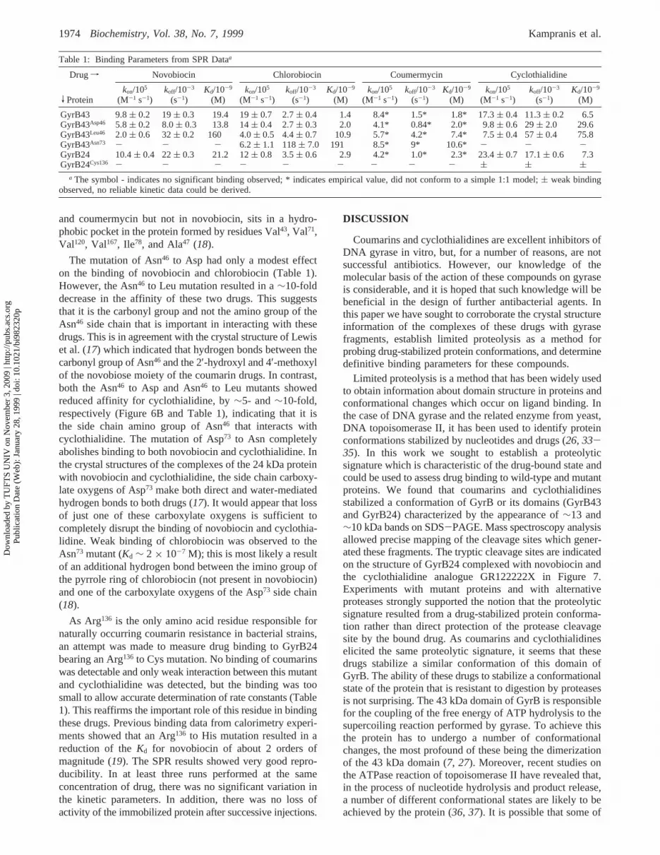
The mutation of Asn46 to Asp had only a modest effecton the binding of novobiocin and chlorobiocin (Table 1).However, the Asn46 to Leu mutation resulted in a∼10-folddecrease in the affinity of these two drugs. This suggeststhat it is the carbonyl group and not the amino group of theAsn46 side chain that is important in interacting with thesedrugs. This is in agreement with the crystal structure of Lewiset al. (17) which indicated that hydrogen bonds between thecarbonyl group of Asn46 and the 2′-hydroxyl and 4′-methoxylof the novobiose moiety of the coumarin drugs. In contrast,both the Asn46 to Asp and Asn46 to Leu mutants showedreduced affinity for cyclothialidine, by∼5- and∼10-fold,respectively (Figure 6B and Table 1), indicating that it isthe side chain amino group of Asn46 that interacts withcyclothialidine. The mutation of Asp73 to Asn completelyabolishes binding to both novobiocin and cyclothialidine. Inthe crystal structures of the complexes of the 24 kDa proteinwith novobiocin and cyclothialidine, the side chain carboxy-late oxygens of Asp73 make both direct and water-mediatedhydrogen bonds to both drugs (17). It would appear that lossof just one of these carboxylate oxygens is sufficient tocompletely disrupt the binding of novobiocin and cyclothia-lidine. Weak binding of chlorobiocin was observed to theAsn73 mutant (Kd ∼ 2 × 10⁻⁷ M); this is most likely a resultof an additional hydrogen bond between the imino group ofthe pyrrole ring of chlorobiocin (not present in novobiocin)and one of the carboxylate oxygens of the Asp73 side chain(18).
As Arg136 is the only amino acid residue responsible fornaturally occurring coumarin resistance in bacterial strains,an attempt was made to measure drug binding to GyrB24bearing an Arg136 to Cys mutation. No binding of coumarinswas detectable and only weak interaction between this mutantand cyclothialidine was detected, but the binding was toosmall to allow accurate determination of rate constants (Table1). This reaffirms the important role of this residue in bindingthese drugs. Previous binding data from calorimetry experi-ments showed that an Arg136 to His mutation resulted in areduction of theKd for novobiocin of about 2 orders ofmagnitude (19). The SPR results showed very good repro-ducibility. In at least three runs performed at the sameconcentration of drug, there was no significant variation inthe kinetic parameters. In addition, there was no loss ofactivity of the immobilized protein after successive injections.
DISCUSSION
Coumarins and cyclothialidines are excellent inhibitors ofDNA gyrase in vitro, but, for a number of reasons, are notsuccessful antibiotics. However, our knowledge of themolecular basis of the action of these compounds on gyraseis considerable, and it is hoped that such knowledge will bebeneficial in the design of further antibacterial agents. Inthis paper we have sought to corroborate the crystal structureinformation of the complexes of these drugs with gyrasefragments, establish limited proteolysis as a method forprobing drug-stabilized protein conformations, and determinedefinitive binding parameters for these compounds.
Limited proteolysis is a method that has been widely usedto obtain information about domain structure in proteins andconformational changes which occur on ligand binding. Inthe case of DNA gyrase and the related enzyme from yeast,DNA topoisomerase II, it has been used to identify proteinconformations stabilized by nucleotides and drugs (26, 33-35). In this work we sought to establish a proteolyticsignature which is characteristic of the drug-bound state andcould be used to assess drug binding to wild-type and mutantproteins. We found that coumarins and cyclothialidinesstabilized a conformation of GyrB or its domains (GyrB43and GyrB24) characterized by the appearance of∼13 and∼10 kDa bands on SDS-PAGE. Mass spectroscopy analysisallowed precise mapping of the cleavage sites which gener-ated these fragments. The tryptic cleavage sites are indicatedon the structure of GyrB24 complexed with novobiocin andthe cyclothialidine analogue GR122222X in Figure 7.Experiments with mutant proteins and with alternativeproteases strongly supported the notion that the proteolyticsignature resulted from a drug-stabilized protein conforma-tion rather than direct protection of the protease cleavagesite by the bound drug. As coumarins and cyclothialidineselicited the same proteolytic signature, it seems that thesedrugs stabilize a similar conformation of this domain ofGyrB. The ability of these drugs to stabilize a conformationalstate of the protein that is resistant to digestion by proteasesis not surprising. The 43 kDa domain of GyrB is responsiblefor the coupling of the free energy of ATP hydrolysis to thesupercoiling reaction performed by gyrase. To achieve thisthe protein has to undergo a number of conformationalchanges, the most profound of these being the dimerizationof the 43 kDa domain (7, 27). Moreover, recent studies onthe ATPase reaction of topoisomerase II have revealed that,in the process of nucleotide hydrolysis and product release,a number of different conformational states are likely to beachieved by the protein (36, 37). It is possible that some of
Table 1: Binding Parameters from SPR Dataa
Drug f Novobiocin Chlorobiocin Coumermycin Cyclothialidine
V Proteinkon/105
(M-1 s-1)koff/10-3
(s-1)Kd/10-9
(M)kon/105
(M-1 s-1)koff/10-3
(s-1)Kd/10-9
(M)kon/105
(M-1 s-1)koff/10-3
(s-1)Kd/10-9
(M)kon/105
(M-1 s-1)koff/10-3
(s-1)Kd/10-9
(M)
GyrB43 9.8( 0.2 19( 0.3 19.4 19( 0.7 2.7( 0.4 1.4 8.4* 1.5* 1.8* 17.3( 0.4 11.3( 0.2 6.5GyrB43Asp46 5.8( 0.2 8.0( 0.3 13.8 14( 0.4 2.7( 0.3 2.0 4.1* 0.84* 2.0* 9.8( 0.6 29( 2.0 29.6GyrB43Leu46 2.0( 0.6 32( 0.2 160 4.0( 0.5 4.4( 0.7 10.9 5.7* 4.2* 7.4* 7.5( 0.4 57( 0.4 75.8GyrB43Asn73 - - - 6.2( 1.1 118( 7.0 191 8.5* 9* 10.6* - - -GyrB24 10.4( 0.4 22( 0.3 21.2 12( 0.8 3.5( 0.6 2.9 4.2* 1.0* 2.3* 23.4( 0.7 17.1( 0.6 7.3GyrB24Cys136 - - - - - - - - - ( ( (
a The symbol - indicates no significant binding observed; * indicates empirical value, did not conform to a simple 1:1 model;( weak bindingobserved, no reliable kinetic data could be derived.
1974 Biochemistry, Vol. 38, No. 7, 1999 Kampranis et al.
Dow
nloa
ded
by T
UFT
S U
NIV
on
Nov
embe
r 3,
200
9 | h
ttp://
pubs
.acs
.org
P
ublic
atio
n D
ate
(Web
): J
anua
ry 2
8, 1
999
| doi
: 10.
1021
/bi9
8232
0p

these conformational states are more susceptible to proteasesthan others. Binding of the drug could therefore stabilizethe protein in a conformation that is resistant to proteolysis,while in the absence of the drugs, the protein would be freeto achieve the conformations that are more susceptible todegradation. Indeed, this idea is further supported by thesolution of the crystal structure of the GyrB24-chlorobiocincomplex, where it is found that the conformation of GyrB24in the chlorobiocin complex is different than that in thecomplex with ADPNP (18).
Examination of the crystal structures of GyrB24 com-plexed with novobiocin and the cyclothialidine GR122222X(17) suggested Asn46 and Asp73 as residues which contributeto drug binding (Figures 1B and 7). Naturally occurringcoumarin-resistance mutations have not been found at eitherof these positions. Limited proteolysis experiments suggestedthat the Asp73 to Asn mutation in GyrB43 abolished drugbinding but that mutations at Asn46 (to Asp and Leu) didnot significantly affect drug binding. This result was sup-ported by experiments using a novobiocin-affinity columnwhich suggested that the Asn46 mutants bound the drug lesstightly and that the Asp73 mutant showed no binding. ATPaseexperiments showed that none of the mutants could hydrolyzeATP, and proteolysis experiments suggested that none couldbind ATP or ADPNP. As the mutant proteins were shownto be folded by CD spectroscopy, it seems that the mutationshad abolished nucleotide binding. Hence such mutationswould not occur spontaneously in bacteria as they wouldrender gyrase inactive and the cell inviable. In relation tothe crystal structure of the GyrB43-ADPNP complex (7),Asn46 makes a key contact to the Mg2+ ion which is boundto the 3 phosphates of ATP, and Asp73 interacts with the N6
amino group of the adenine ring. Thus it is not unexpectedthat mutations at these amino acids should have such adeleterious effect on ATPase activity.
To accurately assess the binding of the coumarin andcyclothialidine drugs to the GyrB43 and GyrB24 domains,we used surface plasmon resonance. Previously, we usedrapid gel filtration and titration calorimetry to measure theseinteractions, but we found that neither of these techniqueswas sufficiently sensitive to yield accurate binding parameters(29). SPR is a technique for measuring association anddissociation rate constants of biological macromolecules andtheir ligands in real time (38). It relies on changes in therefractive index on a metal surface to which one of themolecules of interest is covalently coupled. In this case wecoupled the proteins (GyrB43 or GyrB24) and passed varyingconcentrations of drug over the immobilized protein. Al-though the change in molecular mass upon binding of thedrug was as little as∼1% in some cases, reproducible datawith a low signal-to-noise ratio were generated (e.g., Figure6). This allowed accurate determinations of the bindingparameters for all drugs (Table 1). Consistent with previouswork (29), association rate constants (kon values) were foundto be around 106 M-1 s-1, and dissociation rate constants(koff values) were found to be in the 10-2-10-3 s-1 range,yielding equilibrium dissociation constants (Kd values) of10-8-10-9 M. The order of binding affinity was found tobe chlorobiocin> cyclothialidine> novobiocin. The bindingdata for these drugs could be fitted to a model describing asimple 1:1 complex with the protein, as would be predictedfrom the crystal structures. In the case of coumermycin thedata were not consistent with a simple 1:1 binding modeland the binding parameters quoted in Table 1 are empirical.It has previously been found that coumermycin binds the24 and 43 kDa proteins with a stoichiometry of 1:2 (27, 29),in effect coumermycin cross-links two protein monomers.Due to the potential complexities of dimerization of proteinon the chip surface no attempt was made to further interpretthe coumermycin data.
FIGURE 7: Ribbon representations of the crystal structures of GyrB24 (17) complexed with novobiocin (left) and GR122222X (right).Amino acid side chains discussed in this paper are shown in red, and the drug molecules are in white. Sites of trypsin digestion are marked.The bar indicates diagrammatically the locations of the sites of trypsin cleavage.
Gyrase-Drug Interaction Biochemistry, Vol. 38, No. 7, 19991975
Dow
nloa
ded
by T
UFT
S U
NIV
on
Nov
embe
r 3,
200
9 | h
ttp://
pubs
.acs
.org
P
ublic
atio
n D
ate
(Web
): J
anua
ry 2
8, 1
999
| doi
: 10.
1021
/bi9
8232
0p
As expected from the proteolysis data, the Asn46 to Aspmutation had only a modest effect on the binding parametersof the coumarin drugs but a more profound effect on thebinding of cyclothialidine (Table 1). The Asn46 to Leumutation has a more drastic effect on the binding parametersfor both coumarins and cyclothialidine, reflecting the moreextreme change of the amino acid side chain. The Asp73 toAsn mutation effectively abolishes binding of novobiocinand cyclothialidine and supports only weak binding ofchlorobiocin, reflecting the contribution of the carboxylategroup to hydrogen bonding to the drugs. As predicted fromthe crystal structures and previous work, the Arg136 to Cysmutation abolishes coumarin binding and supports only weakbinding of cyclothialidine, underlining the importance of thisresidue.
In summary, we have shown that limited proteolysis canbe used to probe coumarin interaction with DNA gyrase,and the results support a drug-induced conformational changein the N-terminal domain of GyrB. Proteolysis and coumarin-affinity chromatography showed that point mutations at Asn46
and Asp73 disrupt coumarin binding, and ATPase experimentsshowed that proteins bearing these mutations are inactive.The loss of activity was found by proteolysis to be due tothe inability of the mutant proteins to bind ATP. SPRexperiments have allowed the accurate determination ofkinetic parameters for the binding of wild-type and mutantproteins to several coumarin drugs and cyclothialidine.
ACKNOWLEDGMENT
We thank Dr. R. Loris for assistance with moleculargraphics, Prof. N. C. Price and Dr. S. M. Kelly for carryingout the CD spectroscopy, and Dr. K. S. Lilley for performingthe peptide sequencing and mass spectroscopy. We acknowl-edge the Wellcome Trust for funding the purchase of theBIAcore 2000 system.
REFERENCES
1. Maxwell, A. (1997)Trends Microbiol. 5, 102-109.2. Drlica, K., and Zhao, X. (1997)Microbiol. Mol. Biol. ReV.
61, 377-392.3. Maxwell, A., and Critchlow, S. E. (1997) Mode of action, in
Handbook of Experimental Pharmacology-Quinolone Anti-bacterials(Kuhlmann, J., Dalhoff, A., and Zeiler, H.-J., Eds.)Vol. 127, pp 119-166, Springer-Verlag, Berlin, Germany.
4. Reece, R. J., and Maxwell, A. (1991)CRC Crit. ReV. Biochem.Mol. Biol. 26, 335-375.
5. Wigley, D. B. (1995)Annu. ReV. Biophys. Biomol. Struct. 24,185-208.
6. Morais Cabral, J. H., Jackson, A. P., Smith, C. V., Shikotra,N., Maxwell, A., and Liddington, R. C. (1997)Nature 388,903-906.
7. Wigley, D. B., Davies, G. J., Dodson, E. J., Maxwell, A., andDodson, G. (1991)Nature 351, 624-629.
8. Mizuuchi, K., O’Dea, M. H., and Gellert, M. (1978)Proc.Natl. Acad. Sci. U.S.A. 75, 5960-5963.
9. Sugino, A., Higgins, N. P., Brown, P. O., Peebles, C. L., andCozzarelli, N. R. (1978)Proc. Natl. Acad. Sci. U.S.A. 75,4838-4842.
10. del Castillo, I., Vizan, J., Rodriguez-Sainz, M., and Moreno,F. (1991)Proc. Natl. Acad. Sci. U.S.A. 88, 8860-8864.
11. Contreras, A., and Maxwell, A. (1992)Mol. Microbiol. 6,1617-1624.
12. Gilbert, E. J., and Maxwell, A. (1994)Mol. Microbiol. 12,365-373.
13. Nakada, N., Gmu¨nder, H., Hirata, T., and Arisawa, M. (1994)Antimicrob. Agents Chemother. 38, 1966-1973.
14. Stieger, M., Angehrn, P., Wohlgensinger, B., and Gmu¨nder,H. (1996)Antimicrob. Agents Chemother. 40, 1060-1062.
15. Nakada, N., Gmu¨nder, H., Hirata, T., and Arisawa, M. (1995)J. Biol. Chem. 270, 14286-14291.
16. Oram, M., Dosanjh, B., Gormley, N. A., Smith, C. V., Fisher,L. M., Maxwell, A., and Duncan, K. (1996)Antimicrob. AgentsChemother. 40, 473-476.
17. Lewis, R. J., Singh, O. M. P., Smith, C. V., Skarynski, T.,Maxwell, A., Wonacott, A. J., and Wigley, D. B. (1996)EMBO J. 15, 1412-1420.
18. Tsai, F. T. F., Singh, O. M. P., Skarzynski, T., Wonacott, A.J., Weston, S., Tucker, A., Pauptit, R. A., Breeze, A. L.,Poyser, J. P., O’Brien, R., Ladbury, J. E., and Wigley, D. B.(1997)Proteins: Struct., Funct., Genet. 28, 41-52.
19. Holdgate, G. A., Tunnicliffe, A., Ward, W. H. J., Weston, S.A., Rosenbrock, G., Barth, P. T., Taylor, I. W. F., Pauptit, R.A., and Timms, D. (1997)Biochemistry 36, 9663-9673.
20. Maxwell, A. (1993)Mol. Microbiol. 9, 681-686.21. Orr, E., Fairweather, N. F., Holland, I. B., and Pritchard, R.
H. (1979)Mol. Gen. Genet. 177, 103-112.22. Holmes, M. L., and Dyall-Smith, M. L. (1991)J. Bacteriol.
173, 642-648.23. Sugino, A., and Cozzarelli, N. R. (1980)J. Biol. Chem. 255,
6299-6306.24. Staudenbauer, W. L., and Orr, E. (1981)Nucleic Acids Res.
9, 3589-3603.25. Ali, J. A., Orphanides, G., and Maxwell, A. (1995)Biochem-
istry 34, 9801-9808.26. Kampranis, S. C., and Maxwell, A. (1998)J. Biol. Chem. 273,
22606-22614.27. Ali, J. A., Jackson, A. P., Howells, A. J., and Maxwell, A.
(1993)Biochemistry 32, 2717-2724.28. Maxwell, A., and Howells, A. J. (1998) Overexpression and
purification of bacterial DNA gyrase, inProtocols for DNAtopoisomerases I. DNA topology and enzyme purification(Bjornsti, M.-A., and Osheroff, N., Eds.) pp 135-144, HumanaPress, Towata, New Jersey.
29. Gormley, N. A., Orphanides, G., Meyer, A., Cullis, P. M.,and Maxwell, A. (1996)Biochemistry35, 5083-5092.
30. Samuels, D. S., Marconi, R. T., Huang, W. M., and Garon,C. F. (1994)J. Bacteriol.176, 3072-3075.
31. Reece, R. J., and Maxwell, A. (1989) J. Biol. Chem. 264,19648-19653.
32. Jackson, A. P., and Maxwell, A. (1993)Proc. Natl. Acad. Sci.U.S.A. 90, 11232-11236.
33. Kampranis, S. C., and Maxwell, A. (1998)J. Biol. Chem. 273,26305-26309.
34. Lindsley, J. E., and Wang, J. C. (1991)Proc. Natl. Acad. Sci.U.S.A. 88, 10485-10489.
35. Lindsley, J. E., and Wang, J. C. (1993)Nature 361, 749-750.
36. Harkins, T. T., and Lindsley, J. E. (1998)Biochemistry 37,7292-7298.
37. Harkins, T. T., Lewis, T. J., and Lindsley, J. E. (1998)Biochemistry 37, 7299-7312.
38. Szabo, A., Stolz, L., and Granzow, R. (1995)Curr. Opin.Struct. Biol. 5, 699-705.
BI982320P
1976 Biochemistry, Vol. 38, No. 7, 1999 Kampranis et al.
Dow
nloa
ded
by T
UFT
S U
NIV
on
Nov
embe
r 3,
200
9 | h
ttp://
pubs
.acs
.org
P
ublic
atio
n D
ate
(Web
): J
anua
ry 2
8, 1
999
| doi
: 10.
1021
/bi9
8232
0p


![Al(OTf)3-Catalyzed Preparation of 4-Hydroxy-3-propargylic Coumarins and Subsequent Regioselective Cyclization towards Furo- or Pyrano[3,2-c]coumarins](https://img.dokumen.tips/doc/110x75/63605c660f34b5e42206861e/alotf3-catalyzed-preparation-of-4-hydroxy-3-propargylic-coumarins-and-subsequent.jpg)


