Embed Size (px)
Citation preview
Kao et al. EJNMMI Research 2013, 3:57http://www.ejnmmires.com/content/3/1/57
ORIGINAL RESEARCH Open Access
Post-radioembolization yttrium-90 PET/CT - part 2:dose-response and tumor predictive dosimetry forresin microspheresYung-Hsiang Kao1,2,3*, Jeffrey D Steinberg4, Young-Soon Tay1, Gabriel KY Lim1, Jianhua Yan5, David W Townsend5,Charley A Budgeon6,7, Jan A Boucek2, Roslyn J Francis2,13, Timothy ST Cheo8, Mark C Burgmans9,14, Farah G Irani9,Richard HG Lo9, Kiang-Hiong Tay9, Bien-Soo Tan9, Pierce KH Chow10,11,12, Somanesan Satchithanantham1,Andrew EH Tan1, David CE Ng1 and Anthony SW Goh1
Abstract
Background: Coincidence imaging of low-abundance yttrium-90 (90Y) internal pair production by positronemission tomography with integrated computed tomography (PET/CT) achieves high-resolution imaging ofpost-radioembolization microsphere biodistribution. Part 2 analyzes tumor and non-target tissue dose-response by90Y PET quantification and evaluates the accuracy of tumor 99mTc macroaggregated albumin (MAA) single-photonemission computed tomography with integrated CT (SPECT/CT) predictive dosimetry.
Methods: Retrospective dose quantification of 90Y resin microspheres was performed on the same 23-patient data setin part 1. Phantom studies were performed to assure quantitative accuracy of our time-of-flight lutetium-yttrium-oxyorthosilicate system. Dose-responses were analyzed using 90Y dose-volume histograms (DVHs) by PET voxeldosimetry or mean absorbed doses by Medical Internal Radiation Dose macrodosimetry, correlated to follow-upimaging or clinical findings. Intended tumor mean doses by predictive dosimetry were compared to doses by 90Y PET.
Results: Phantom studies demonstrated near-perfect detector linearity and high tumor quantitative accuracy. Forhepatocellular carcinomas, complete responses were generally achieved at D70 > 100 Gy (D70, minimum dose to 70%tumor volume), whereas incomplete responses were generally at D70 < 100 Gy; smaller tumors (<80 cm3) achievedD70 > 100 Gy more easily than larger tumors. There was complete response in a cholangiocarcinoma at D70 90 Gy andpartial response in an adrenal gastrointestinal stromal tumor metastasis at D70 53 Gy. In two patients, a mean dose of18 Gy to the stomach was asymptomatic, 49 Gy caused gastritis, 65 Gy caused ulceration, and 53 Gy caused duodenitis.In one patient, a bilateral kidney mean dose of 9 Gy (V20 8%) did not cause clinically relevant nephrotoxicity. Undernear-ideal dosimetric conditions, there was excellent correlation between intended tumor mean doses by predictivedosimetry and those by 90Y PET, with a low median relative error of +3.8% (95% confidence interval, −1.2% to +13.2%).
Conclusions: Tumor and non-target tissue absorbed dose quantification by 90Y PET is accurate and yieldsradiobiologically meaningful dose-response information to guide adjuvant or mitigative action. Tumor 99mTc MAASPECT/CT predictive dosimetry is feasible. 90Y DVHs may guide future techniques in predictive dosimetry.
Keywords: Yttrium-90 radioembolization; Selective internal radiation therapy; PET/CT; Voxel dosimetry; Dose-volumehistogram; Predictive dosimetry
* Correspondence: [email protected] of Nuclear Medicine and PET, Singapore General Hospital,Outram Road, Singapore 169608, Singapore2Department of Nuclear Medicine, Sir Charles Gairdner Hospital, Hospital Ave,Perth, Western Australia 6009, AustraliaFull list of author information is available at the end of the article
© 2013 Kao et al.; licensee Springer. This is an OAttribution License (http://creativecommons.orin any medium, provided the original work is p
pen Access article distributed under the terms of the Creative Commonsg/licenses/by/2.0), which permits unrestricted use, distribution, and reproductionroperly cited.

Kao et al. EJNMMI Research 2013, 3:57 Page 2 of 12http://www.ejnmmires.com/content/3/1/57
BackgroundCoincidence imaging of low-abundance yttrium-90 (90Y)internal pair production by 90Y positron emission tomo-graphy with integrated computed tomography (PET/CT)achieves high-resolution imaging of post-radioembolizationmicrosphere biodistribution. In part 1, we reviewed the re-cent literature supporting the use of post-radioembolization90Y PET/CT, described our scan protocol, patient cohort,diagnostic reporting guidelines, and results of qualitativeanalysis [1]. In brief, we showed that with proper diagnosticreporting technique and emphasis on continuity of care,the presence of background noise did not pose a problemand 90Y PET/CT consistently out-performed 90Y brems-strahlung single-photon emission computed tomographywith integrated CT (SPECT/CT) in all aspects of qualitativeanalysis [1].The terms ‘predictive dosimetry’, ‘target’, ‘non-target’,
‘technical success’, and the ‘planning-therapy continuum’were also defined in part 1 [1]. It is important for theunderstanding of part 2 to reiterate that our definitionof technical success, an adaptation from conventionalreporting standards [2], refers to the qualitative as-sessment of a satisfactory 90Y activity biodistribution inaccordance with radiation planning expectations andshould not be confused with ‘clinical success’ [2] whichis quantitatively related to dose-response radiobiology.A knowledge gap exists today between institutions
which practice predictive dosimetry and others whichrely on semi-empirical methods [3]. Readers who are ac-customed to semi-empirical therapy planning may strug-gle to understand the dosimetric concepts discussed inthis paper or its relevance to clinical practice. Thesereaders are encouraged to refer to our recent series ofpublications explaining concepts in modern predictivedosimetry for 90Y resin microspheres and common mis-conceptions [3-6].Where prognostication is of concern, qualitative ana-
lysis alone will not suffice [3]. The scientific language ofdose-response radiobiology is the radiation absorbeddose expressed in grays (Gy), not the prescribed activityexpressed in becquerels (Bq). To realize the full potentialof post-radioembolization 90Y PET/CT, absorbed dosequantification should be performed in relevant clinicalsettings. As 90Y radioembolization is a brachytherapy de-livered by β−-emitting microspheres, any tissue responseis expected to follow predictable dose-response radio-biology. In principle, accurate knowledge of radiationthresholds in both target and non-target tissue facilitatesthe optimization of predictive dosimetry and enables theprognostication of technically unsuccessful cases toguide adjuvant therapy or mitigative action to minimizenon-target radiation toxicity.However, accurate tissue radiation thresholds for 90Y
resin microspheres have remained elusive despite more
than two decades of clinical use. Current radiationplanning limits are broad and quote mean absorbeddoses which falsely assume a uniform dose distribution:tumor > 120 Gy, non-tumorous liver < 50 to 70 Gy,lungs < 20 to 30 Gy [7-9]. As a consequence of this un-certainty, dosimetric dilemmas may be encountered inpatients who may benefit from radiation planning up tothe limits of normal tissue radiation tolerance. Further-more, normal tissue radiation thresholds for 90Y resinmicrospheres shunted to non-target viscera such as thestomach or duodenum are largely unknown, precludinginformed decision making for appropriate mitigative ac-tion. Historically, research into dose-response data hadbeen technically challenging: intraoperative beta probesor histological examinations are invasive [10,11], quanti-fication by 90Y bremsstrahlung scintigraphy is problem-atic and largely inaccurate [1], and predictive dosimetrysimulated by 99mTc macroaggregated albumin (MAA) issubject to variable accuracy due to the physical limita-tions of MAA [12].Dosimetric studies have shown 99mTc MAA to be fea-
sible for simulating the post-radioembolization bio-distribution of 90Y resin microspheres [13-16]. However,99mTc MAA is an imperfect surrogate for 90Y resin mi-crospheres. Due to biophysical and technical differencessuch as particle size, specific gravity, injected particleload, microembolization, and catheter placement, 99mTcMAA can never exactly replicate the post-radioem-bolization biodistribution of 90Y resin microspheres[12,17]. Therefore, predictive dosimetry simulated by99mTc MAA provides only an estimate of the tissueabsorbed doses intended by the nuclear medicine phys-ician [6]. Traditionally based on planar scintigraphy[13,14], modern predictive dosimetry employs SPECT/CT to tomographically assess the biodistribution of99mTc MAA and to improve its quantitative accuracy[6]. So far, the accuracy of 99mTc MAA SPECT/CT pre-dictive dosimetry has only been indirectly validated byinference from follow-up response [6,18,19]. For tech-nically successful cases without visually significant dis-cordant biodistribution between 99mTc MAA and 90Yresin microspheres, a direct ‘Gy-to-Gy’ comparison ofintended doses by 99mTc MAA SPECT/CT predictivedosimetry versus post-radioembolization doses by mi-crosphere biodistribution analysis has not yet been per-formed to date.Recent experimental and clinical studies have shown
post-radioembolization 90Y PET quantification to be ac-curate and feasible [1,20,21]. 90Y PET/CT thus presentsa new opportunity to study the radiobiology of 90Y resinmicrospheres in a rapid, convenient, and noninvasivemanner, with the ability to tomographically evaluate thedose distribution of an entire target organ in high reso-lution by a single scan. Moreover, quantitative 90Y PET
Kao et al. EJNMMI Research 2013, 3:57 Page 3 of 12http://www.ejnmmires.com/content/3/1/57
data can be translated into dose-volume histograms(DVHs) to dosimetrically account for the heterogeneousnature of microsphere biodistribution.In part 2 of our two-part retrospective report, we focus
on post-radioembolization 90Y PET quantification on thesame patient cohort as was reported in part 1 [1]. Weanalyze dose-responses in tumor and non-target tissueusing 90Y PET-based voxel dosimetry or Medical In-ternal Radiation Dose (MIRD) macrodosimetry [7,22].We describe the potential of 90Y DVHs to guidepredictive dosimetry and discuss how the quantifica-tion of non-target absorbed doses may impact post-radioembolization care. Finally, we evaluate the accuracyof tumor 99mTc MAA SPECT/CT predictive dosimetryin direct comparison to post-radioembolization dosesby 90Y PET.
MethodsPhantom quantificationAn institutional review board approval was obtained forthe conduct and publication of this retrospective re-port (CIRB 2010/781/C, SingHealth, Singapore). Verbalconsent was obtained from patients for scans to beperformed. Quantitative phantom studies were first per-formed to confirm detector linearity of our time-of-flightlutetium-yttrium-oxyorthosilicate (LYSO) system and forcomparison with a radiopharmacy dose calibrator. 90Yresin microspheres (SIR-Spheres, Sirtex Medical Limited,North Sydney, Australia) were used. We scanned themicrosphere sediment and not its fresh suspension toavoid issues with microsphere sedimentation during PETacquisition. In effect, each vial sediment represents asmall heterogeneous volume of high 90Y radioconcen-tration, simulating a small tumor implanted with a highand heterogeneous density of 90Y resin microspheres. Allmicrosphere sediments were scanned within its originalglass shipping vial for convenience and to avoid operatorradiation exposure for microsphere extraction.Vials of microspheres were obtained by convenience
sampling, either before or after extraction of the desiredtreatment activity. Phantom scans of high activities (>3.0GBq) were performed on fresh vials on the day it was re-ceived from the manufacturer, while scans of very lowactivities (<0.1 GBq) were performed after a period ofdecay of residual vial activities. The vials were placedwithin a perspex body phantom and scanned in one bedposition over 15 min (Figure 1). All scan parameterswere identical to our clinical scan protocol described inpart 1 [1].For vial activity quantification, a volume of interest
(VOI) encompassing the entire vial sediment was gener-ated by selecting a fixed volumetric isocontour thresholdof 1%. The total 90Y activity (kBq) within the VOI wascalculated as the product of its mean radioconcentration
(kBq/ml) and its volume (cm3). Each vial sediment quan-tified by 90Y PET was compared to that measured by adose calibrator (Biodex Atomlab 500, Biodex MedicalSystems, New York, NY, USA). Its purpose was toevaluate the general relative accuracy of 90Y PET quan-tification in the absence of a true reference standardand also to empirically assess quantitative effectsfrom background noise, variable vial sediment volume,radioconcentration, vial geometry, or positron contri-butions from possible radionuclidic impurities such asyttrium-88 [23].
Tissue quantificationRetrospective absorbed dose quantification by 90Y PETwas performed on the same 23-patient data set as wasreported in part 1 [1]. The two objectives were, first, toestablish dose-response observations in tumor and non-target tissue and, second, to evaluate the accuracy oftumor 99mTc MAA SPECT/CT predictive dosimetry [6] indirect Gy-to-Gy comparison to post-radioembolizationdoses by 90Y PET.Tumors were analyzed using cumulative 90Y DVHs
generated by voxel dosimetry. This was performed on asmall select group of tumors treated under highly favor-able, near-ideal dosimetric conditions to ensure reliabledose-response observations and to assess the best pos-sible accuracy of tumor 99mTc MAA SPECT/CT pre-dictive dosimetry. The tumor selection criteria were asfollows: technically successful 90Y radioembolization,well-circumscribed tumors with good overall activitycoverage, mean tumor-to-normal liver (T/N) ratio ≥2as estimated by 99mTc MAA SPECT/CT, and follow-updiagnostic sectional imaging available for clinical cor-relation. All tumor VOIs were manually contoured sliceby slice according to CT anatomical margins. We avoi-ded using tumor VOIs generated by PET volumetricisocontour thresholding as these often do not representtheir true anatomical extent due to heterogeneous mi-crosphere biodistribution. Regions of absent 90Y activityin tumors were visually correlated to CT findings andexcluded from VOI analysis if necrotic.
90Y PET voxel dosimetry was performed by a novelprogram written in Interactive Data Language (IDL 6.1),freely available from the corresponding author forresearch purposes [24]. This program was initially de-veloped for total glycolytic volume analysis of 18F-fluorodeoxyglucose PET data [25], subsequently adaptedfor 90Y PET voxel dosimetry in this study. 90Y absorbeddose distributions were calculated using a simplified ap-proach based on voxel mean radioconcentrations insteadof dose kernel or Monte Carlo methods. Its dosimetricrationale and evidence to support this approach are de-tailed in Additional file 1.
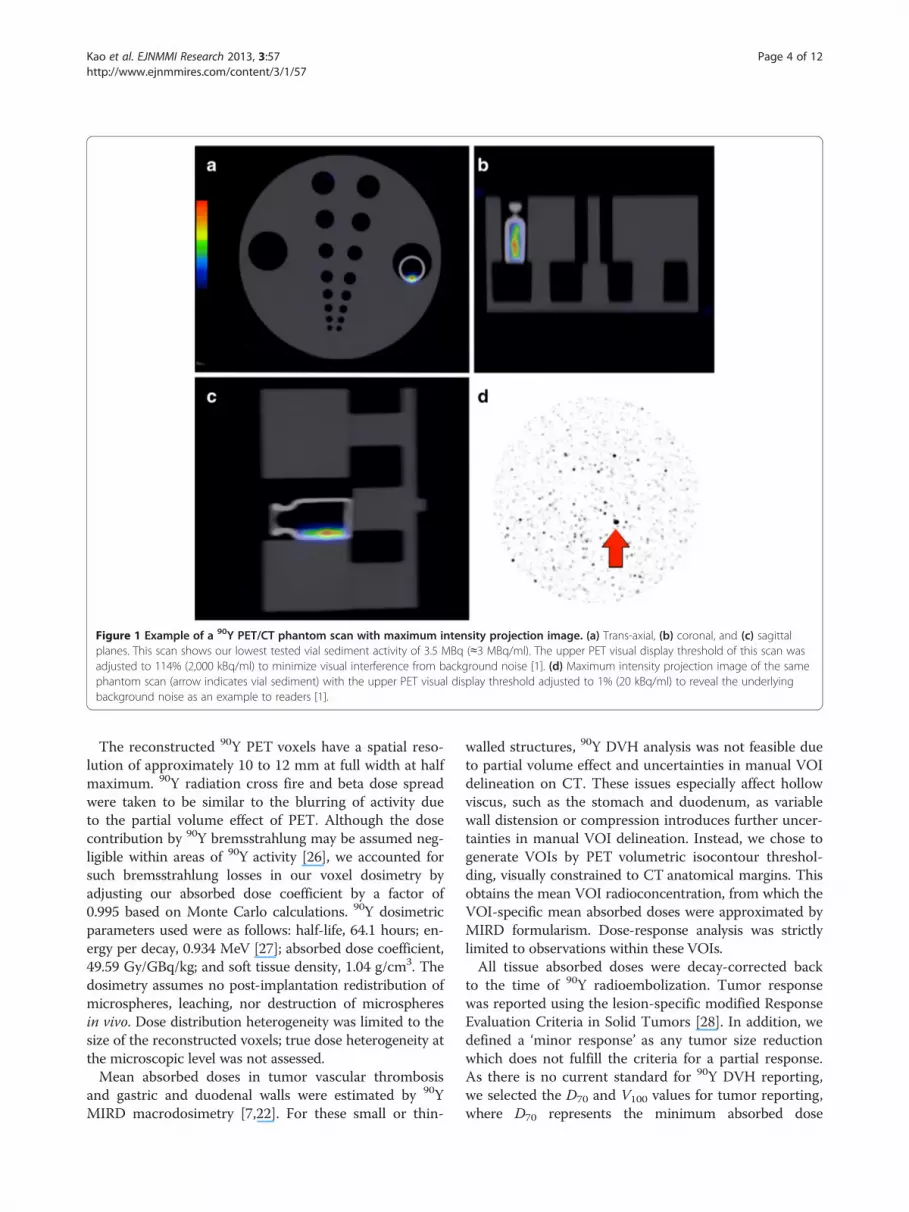
Figure 1 Example of a 90Y PET/CT phantom scan with maximum intensity projection image. (a) Trans-axial, (b) coronal, and (c) sagittalplanes. This scan shows our lowest tested vial sediment activity of 3.5 MBq (≈3 MBq/ml). The upper PET visual display threshold of this scan wasadjusted to 114% (2,000 kBq/ml) to minimize visual interference from background noise [1]. (d) Maximum intensity projection image of the samephantom scan (arrow indicates vial sediment) with the upper PET visual display threshold adjusted to 1% (20 kBq/ml) to reveal the underlyingbackground noise as an example to readers [1].
Kao et al. EJNMMI Research 2013, 3:57 Page 4 of 12http://www.ejnmmires.com/content/3/1/57
The reconstructed 90Y PET voxels have a spatial reso-lution of approximately 10 to 12 mm at full width at halfmaximum. 90Y radiation cross fire and beta dose spreadwere taken to be similar to the blurring of activity dueto the partial volume effect of PET. Although the dosecontribution by 90Y bremsstrahlung may be assumed neg-ligible within areas of 90Y activity [26], we accounted forsuch bremsstrahlung losses in our voxel dosimetry byadjusting our absorbed dose coefficient by a factor of0.995 based on Monte Carlo calculations. 90Y dosimetricparameters used were as follows: half-life, 64.1 hours; en-ergy per decay, 0.934 MeV [27]; absorbed dose coefficient,49.59 Gy/GBq/kg; and soft tissue density, 1.04 g/cm3. Thedosimetry assumes no post-implantation redistribution ofmicrospheres, leaching, nor destruction of microspheresin vivo. Dose distribution heterogeneity was limited to thesize of the reconstructed voxels; true dose heterogeneity atthe microscopic level was not assessed.Mean absorbed doses in tumor vascular thrombosis
and gastric and duodenal walls were estimated by 90YMIRD macrodosimetry [7,22]. For these small or thin-
walled structures, 90Y DVH analysis was not feasible dueto partial volume effect and uncertainties in manual VOIdelineation on CT. These issues especially affect hollowviscus, such as the stomach and duodenum, as variablewall distension or compression introduces further uncer-tainties in manual VOI delineation. Instead, we chose togenerate VOIs by PET volumetric isocontour threshol-ding, visually constrained to CT anatomical margins. Thisobtains the mean VOI radioconcentration, from which theVOI-specific mean absorbed doses were approximated byMIRD formularism. Dose-response analysis was strictlylimited to observations within these VOIs.All tissue absorbed doses were decay-corrected back
to the time of 90Y radioembolization. Tumor responsewas reported using the lesion-specific modified ResponseEvaluation Criteria in Solid Tumors [28]. In addition, wedefined a ‘minor response’ as any tumor size reductionwhich does not fulfill the criteria for a partial response.As there is no current standard for 90Y DVH reporting,we selected the D70 and V100 values for tumor reporting,where D70 represents the minimum absorbed dose
Kao et al. EJNMMI Research 2013, 3:57 Page 5 of 12http://www.ejnmmires.com/content/3/1/57
delivered to 70% of the tumor volume, and V100 repre-sents the percentage tumor volume receiving ≥100 Gy.Non-target absorbed doses were expressed in accordanceto external beam radiotherapy convention as far as pos-sible [29,30]. Cases of non-target activity were followed upby retrospective review of clinical records, and relevantclinical toxicities were graded according to the CommonTerminology Criteria for Adverse Events version 4.03(CTCAE; National Institutes of Health, Bethesda, MD,USA).The second objective of retrospective absorbed dose
quantification was to evaluate the accuracy of tumorpredictive dosimetry simulated by 99mTc MAA SPECT/CT. This was performed by a direct, i.e., Gy-to-Gy, com-parison between intended tumor mean doses and post-radioembolization tumor mean doses by 90Y PET,expressed as relative percentage errors. Our protocol forpredictive dosimetry was previously described [6], whichinvolves catheter-directed CT angiography for arterialterritory delineation, quantitative 99mTc MAA SPECT/CT of the target organ, and MIRD macrodosimetry toachieve artery-specific personalized radiation planningfor 90Y resin microspheres.
Statistical analysisData are presented as median, mean, range, and 95%confidence intervals (CI), where applicable. Linear re-gression with Pearson’s correlation coefficient (r) and
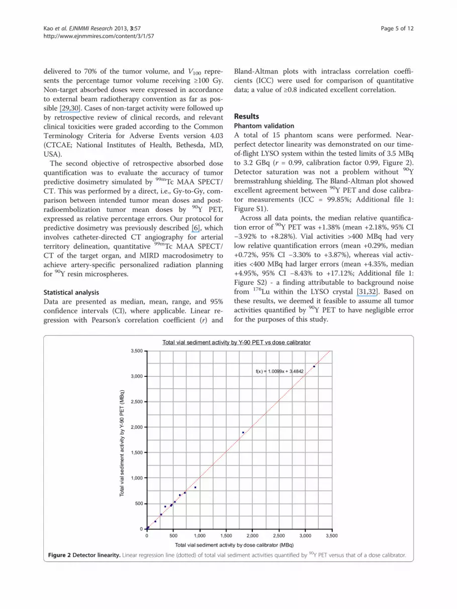
Figure 2 Detector linearity. Linear regression line (dotted) of total vial se
Bland-Altman plots with intraclass correlation coeffi-cients (ICC) were used for comparison of quantitativedata; a value of ≥0.8 indicated excellent correlation.
ResultsPhantom validationA total of 15 phantom scans were performed. Near-perfect detector linearity was demonstrated on our time-of-flight LYSO system within the tested limits of 3.5 MBqto 3.2 GBq (r = 0.99, calibration factor 0.99, Figure 2).Detector saturation was not a problem without 90Ybremsstrahlung shielding. The Bland-Altman plot showedexcellent agreement between 90Y PET and dose calibra-tor measurements (ICC = 99.85%; Additional file 1:Figure S1).Across all data points, the median relative quantifica-
tion error of 90Y PET was +1.38% (mean +2.18%, 95% CI−3.92% to +8.28%). Vial activities >400 MBq had verylow relative quantification errors (mean +0.29%, median+0.72%, 95% CI −3.30% to +3.87%), whereas vial activ-ities <400 MBq had larger errors (mean +4.35%, median+4.95%, 95% CI −8.43% to +17.12%; Additional file 1:Figure S2) - a finding attributable to background noisefrom 176Lu within the LYSO crystal [31,32]. Based onthese results, we deemed it feasible to assume all tumoractivities quantified by 90Y PET to have negligible errorfor the purposes of this study.
diment activities quantified by 90Y PET versus that of a dose calibrator.
Kao et al. EJNMMI Research 2013, 3:57 Page 6 of 12http://www.ejnmmires.com/content/3/1/57
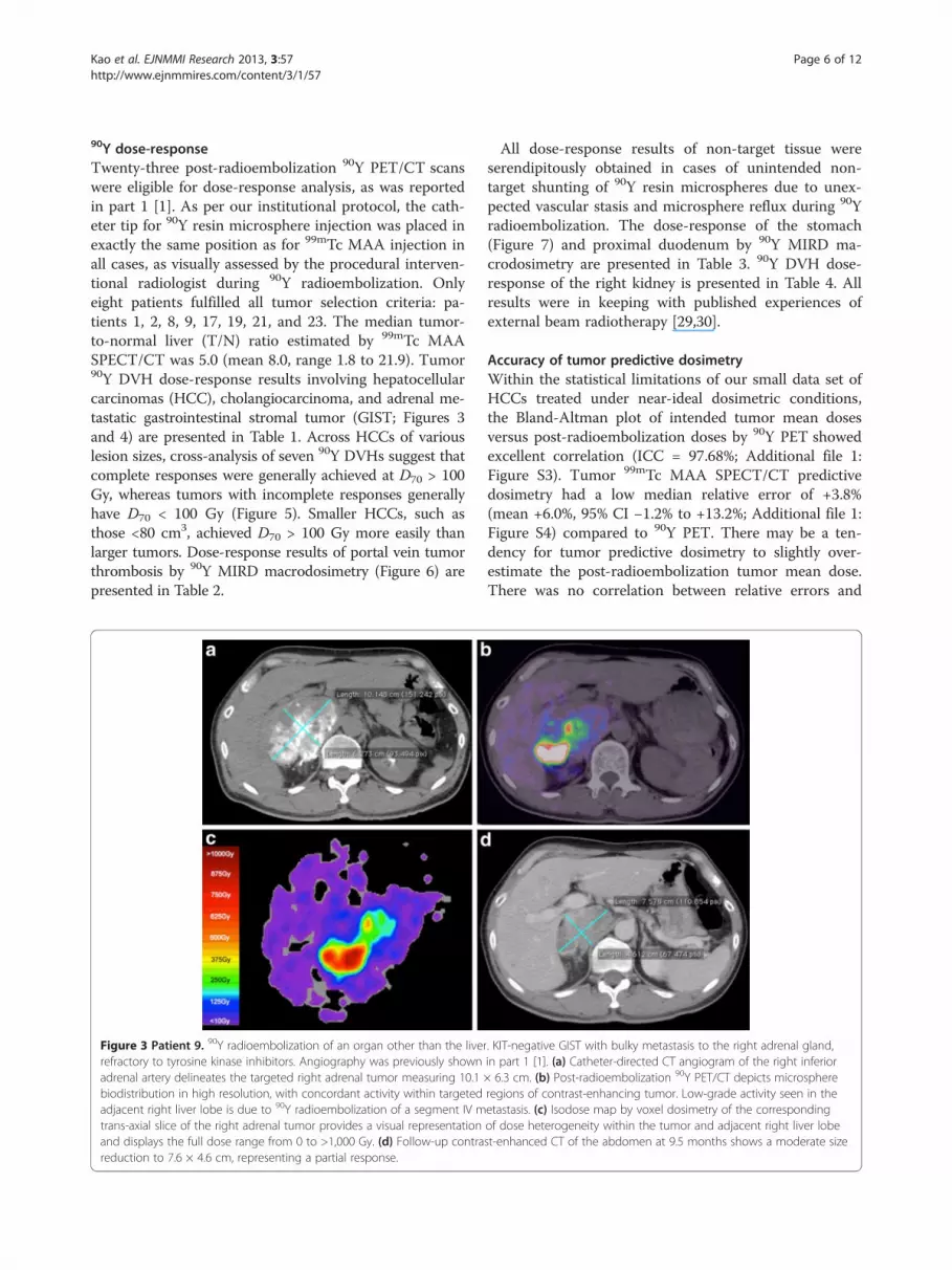
90Y dose-responseTwenty-three post-radioembolization 90Y PET/CT scanswere eligible for dose-response analysis, as was reportedin part 1 [1]. As per our institutional protocol, the cath-eter tip for 90Y resin microsphere injection was placed inexactly the same position as for 99mTc MAA injection inall cases, as visually assessed by the procedural interven-tional radiologist during 90Y radioembolization. Onlyeight patients fulfilled all tumor selection criteria: pa-tients 1, 2, 8, 9, 17, 19, 21, and 23. The median tumor-to-normal liver (T/N) ratio estimated by 99mTc MAASPECT/CT was 5.0 (mean 8.0, range 1.8 to 21.9). Tumor90Y DVH dose-response results involving hepatocellularcarcinomas (HCC), cholangiocarcinoma, and adrenal me-tastatic gastrointestinal stromal tumor (GIST; Figures 3and 4) are presented in Table 1. Across HCCs of variouslesion sizes, cross-analysis of seven 90Y DVHs suggest thatcomplete responses were generally achieved at D70 > 100Gy, whereas tumors with incomplete responses generallyhave D70 < 100 Gy (Figure 5). Smaller HCCs, such asthose <80 cm3, achieved D70 > 100 Gy more easily thanlarger tumors. Dose-response results of portal vein tumorthrombosis by 90Y MIRD macrodosimetry (Figure 6) arepresented in Table 2.
Figure 3 Patient 9. 90Y radioembolization of an organ other than the liverrefractory to tyrosine kinase inhibitors. Angiography was previously shownadrenal artery delineates the targeted right adrenal tumor measuring 10.1biodistribution in high resolution, with concordant activity within targetedadjacent right liver lobe is due to 90Y radioembolization of a segment IV mtrans-axial slice of the right adrenal tumor provides a visual representationand displays the full dose range from 0 to >1,000 Gy. (d) Follow-up contrareduction to 7.6 × 4.6 cm, representing a partial response.
All dose-response results of non-target tissue wereserendipitously obtained in cases of unintended non-target shunting of 90Y resin microspheres due to unex-pected vascular stasis and microsphere reflux during 90Yradioembolization. The dose-response of the stomach(Figure 7) and proximal duodenum by 90Y MIRD ma-crodosimetry are presented in Table 3. 90Y DVH dose-response of the right kidney is presented in Table 4. Allresults were in keeping with published experiences ofexternal beam radiotherapy [29,30].
Accuracy of tumor predictive dosimetryWithin the statistical limitations of our small data set ofHCCs treated under near-ideal dosimetric conditions,the Bland-Altman plot of intended tumor mean dosesversus post-radioembolization doses by 90Y PET showedexcellent correlation (ICC = 97.68%; Additional file 1:Figure S3). Tumor 99mTc MAA SPECT/CT predictivedosimetry had a low median relative error of +3.8%(mean +6.0%, 95% CI −1.2% to +13.2%; Additional file 1:Figure S4) compared to 90Y PET. There may be a ten-dency for tumor predictive dosimetry to slightly over-estimate the post-radioembolization tumor mean dose.There was no correlation between relative errors and
. KIT-negative GIST with bulky metastasis to the right adrenal gland,in part 1 [1]. (a) Catheter-directed CT angiogram of the right inferior× 6.3 cm. (b) Post-radioembolization 90Y PET/CT depicts microsphereregions of contrast-enhancing tumor. Low-grade activity seen in theetastasis. (c) Isodose map by voxel dosimetry of the correspondingof dose heterogeneity within the tumor and adjacent right liver lobest-enhanced CT of the abdomen at 9.5 months shows a moderate size
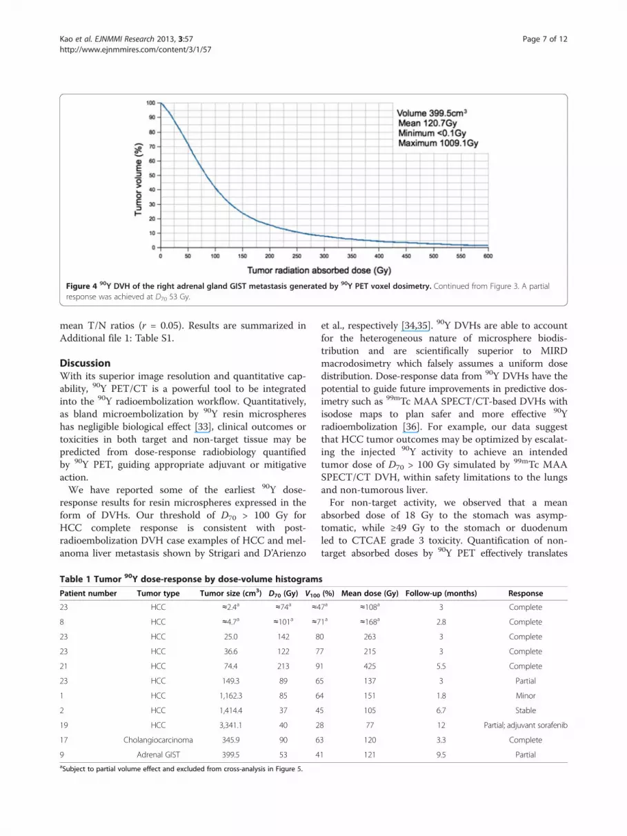
Figure 4 90Y DVH of the right adrenal gland GIST metastasis generated by 90Y PET voxel dosimetry. Continued from Figure 3. A partialresponse was achieved at D70 53 Gy.
Kao et al. EJNMMI Research 2013, 3:57 Page 7 of 12http://www.ejnmmires.com/content/3/1/57
mean T/N ratios (r = 0.05). Results are summarized inAdditional file 1: Table S1.
DiscussionWith its superior image resolution and quantitative cap-ability, 90Y PET/CT is a powerful tool to be integratedinto the 90Y radioembolization workflow. Quantitatively,as bland microembolization by 90Y resin microsphereshas negligible biological effect [33], clinical outcomes ortoxicities in both target and non-target tissue may bepredicted from dose-response radiobiology quantifiedby 90Y PET, guiding appropriate adjuvant or mitigativeaction.We have reported some of the earliest 90Y dose-
response results for resin microspheres expressed in theform of DVHs. Our threshold of D70 > 100 Gy forHCC complete response is consistent with post-radioembolization DVH case examples of HCC and mel-anoma liver metastasis shown by Strigari and D’Arienzo
Table 1 Tumor 90Y dose-response by dose-volume histogram
Patient number Tumor type Tumor size (cm3) D70 (Gy) V100
23 HCC ≈2.4a ≈74a ≈4
8 HCC ≈4.7a ≈101a ≈7
23 HCC 25.0 142 8
23 HCC 36.6 122 7
21 HCC 74.4 213 9
23 HCC 149.3 89 6
1 HCC 1,162.3 85 6
2 HCC 1,414.4 37 4
19 HCC 3,341.1 40 2
17 Cholangiocarcinoma 345.9 90 6
9 Adrenal GIST 399.5 53 4aSubject to partial volume effect and excluded from cross-analysis in Figure 5.
et al., respectively [34,35]. 90Y DVHs are able to accountfor the heterogeneous nature of microsphere biodis-tribution and are scientifically superior to MIRDmacrodosimetry which falsely assumes a uniform dosedistribution. Dose-response data from 90Y DVHs have thepotential to guide future improvements in predictive dos-imetry such as 99mTc MAA SPECT/CT-based DVHs withisodose maps to plan safer and more effective 90Yradioembolization [36]. For example, our data suggestthat HCC tumor outcomes may be optimized by escalat-ing the injected 90Y activity to achieve an intendedtumor dose of D70 > 100 Gy simulated by 99mTc MAASPECT/CT DVH, within safety limitations to the lungsand non-tumorous liver.For non-target activity, we observed that a mean
absorbed dose of 18 Gy to the stomach was asymp-tomatic, while ≥49 Gy to the stomach or duodenumled to CTCAE grade 3 toxicity. Quantification of non-target absorbed doses by 90Y PET effectively translates
s
(%) Mean dose (Gy) Follow-up (months) Response
7a ≈108a 3 Complete
1a ≈168a 2.8 Complete
0 263 3 Complete
7 215 3 Complete
1 425 5.5 Complete
5 137 3 Partial
4 151 1.8 Minor
5 105 6.7 Stable
8 77 12 Partial; adjuvant sorafenib
3 120 3.3 Complete
1 121 9.5 Partial
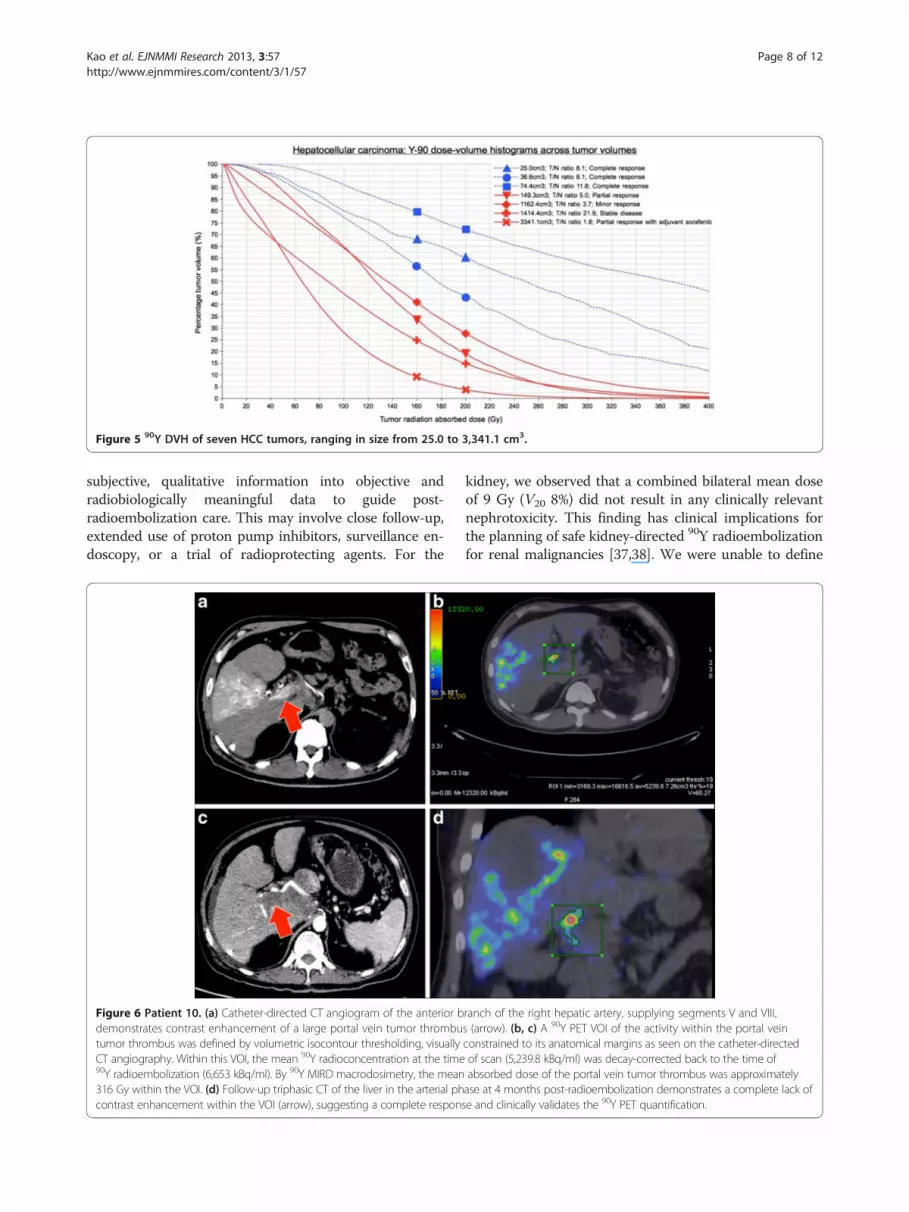
Figure 5 90Y DVH of seven HCC tumors, ranging in size from 25.0 to 3,341.1 cm3.
Kao et al. EJNMMI Research 2013, 3:57 Page 8 of 12http://www.ejnmmires.com/content/3/1/57
subjective, qualitative information into objective andradiobiologically meaningful data to guide post-radioembolization care. This may involve close follow-up,extended use of proton pump inhibitors, surveillance en-doscopy, or a trial of radioprotecting agents. For the
Figure 6 Patient 10. (a) Catheter-directed CT angiogram of the anterior bdemonstrates contrast enhancement of a large portal vein tumor thrombutumor thrombus was defined by volumetric isocontour thresholding, visuallyCT angiography. Within this VOI, the mean 90Y radioconcentration at the time90Y radioembolization (6,653 kBq/ml). By 90Y MIRD macrodosimetry, the mean316 Gy within the VOI. (d) Follow-up triphasic CT of the liver in the arterial phcontrast enhancement within the VOI (arrow), suggesting a complete respon
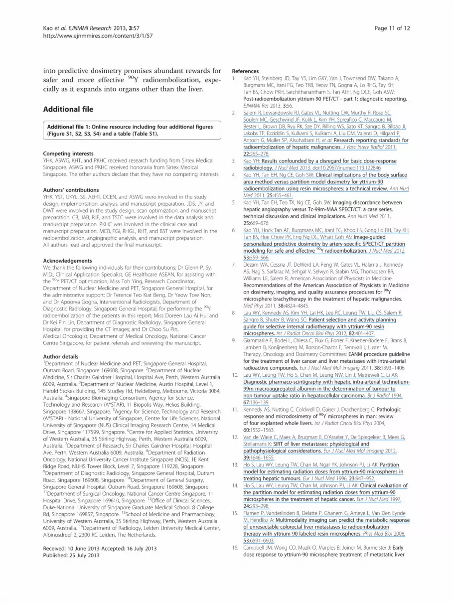
kidney, we observed that a combined bilateral mean doseof 9 Gy (V20 8%) did not result in any clinically relevantnephrotoxicity. This finding has clinical implications forthe planning of safe kidney-directed 90Y radioembolizationfor renal malignancies [37,38]. We were unable to define
ranch of the right hepatic artery, supplying segments V and VIII,s (arrow). (b, c) A 90Y PET VOI of the activity within the portal veinconstrained to its anatomical margins as seen on the catheter-directedof scan (5,239.8 kBq/ml) was decay-corrected back to the time ofabsorbed dose of the portal vein tumor thrombus was approximatelyase at 4 months post-radioembolization demonstrates a complete lack ofse and clinically validates the 90Y PET quantification.
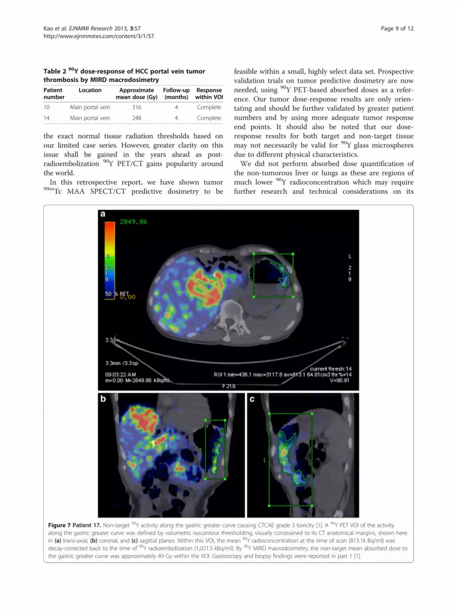
Table 2 90Y dose-response of HCC portal vein tumorthrombosis by MIRD macrodosimetry
Patientnumber
Location Approximatemean dose (Gy)
Follow-up(months)
Responsewithin VOI
10 Main portal vein 316 4 Complete
14 Main portal vein 248 4 Complete
Kao et al. EJNMMI Research 2013, 3:57 Page 9 of 12http://www.ejnmmires.com/content/3/1/57
the exact normal tissue radiation thresholds based onour limited case series. However, greater clarity on thisissue shall be gained in the years ahead as post-radioembolization 90Y PET/CT gains popularity aroundthe world.In this retrospective report, we have shown tumor
99mTc MAA SPECT/CT predictive dosimetry to be
Figure 7 Patient 17. Non-target 90Y activity along the gastric greater curvalong the gastric greater curve was defined by volumetric isocontour thresin (a) trans-axial, (b) coronal, and (c) sagittal planes. Within this VOI, the medecay-corrected back to the time of 90Y radioembolization (1,021.5 kBq/ml)the gastric greater curve was approximately 49 Gy within the VOI. Gastrosc
feasible within a small, highly select data set. Prospectivevalidation trials on tumor predictive dosimetry are nowneeded, using 90Y PET-based absorbed doses as a refer-ence. Our tumor dose-response results are only orien-tating and should be further validated by greater patientnumbers and by using more adequate tumor responseend points. It should also be noted that our dose-response results for both target and non-target tissuemay not necessarily be valid for 90Y glass microspheresdue to different physical characteristics.We did not perform absorbed dose quantification of
the non-tumorous liver or lungs as these are regions ofmuch lower 90Y radioconcentration which may requirefurther research and technical considerations on its
e causing CTCAE grade 3 toxicity [1]. A 90Y PET VOI of the activityholding, visually constrained to its CT anatomical margins, shown herean 90Y radioconcentration at the time of scan (813.1k Bq/ml) was. By 90Y MIRD macrodosimetry, the non-target mean absorbed dose toopy and biopsy findings were reported in part 1 [1].
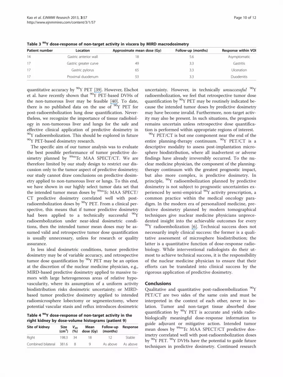
Table 3 90Y dose-response of non-target activity in viscera by MIRD macrodosimetry
Patient number Location Approximate mean dose (Gy) Follow-up (months) Response within VOI
14 Gastric anterior wall 18 5.6 Asymptomatic
17 Gastric greater curve 49 3.3 Gastritis
17 Gastric pylorus 65 3.3 Ulceration
17 Proximal duodenum 53 3.3 Duodenitis
Kao et al. EJNMMI Research 2013, 3:57 Page 10 of 12http://www.ejnmmires.com/content/3/1/57
quantitative accuracy by 90Y PET [39]. However, Elschotet al. have recently shown that 90Y PET-based DVHs ofthe non-tumorous liver may be feasible [40]. To date,there is no published data on the use of 90Y PET forpost-radioembolization lung dose quantification. Never-theless, we recognize the importance of tissue radiobiol-ogy in non-tumorous liver and lungs for the safe andeffective clinical application of predictive dosimetry in90Y radioembolization. This should be explored in future90Y PET-based dosimetry research.The specific aim of our tumor analysis was to evaluate
the best possible performance of tumor predictive do-simetry planned by 99mTc MAA SPECT/CT. We aretherefore limited by our study design to restrict our dis-cussion only to the tumor aspect of predictive dosimetry;our study cannot draw conclusions on predictive dosim-etry applied to non-tumorous liver or lungs. To this end,we have shown in our highly select tumor data set thatthe intended tumor mean doses by 99mTc MAA SPECT/CT predictive dosimetry correlated well with post-radioembolization doses by 90Y PET. From a clinical per-spective, this means that if tumor predictive dosimetryhad been applied to a technically successful 90Yradioembolization under near-ideal dosimetric condi-tions, then the intended tumor mean doses may be as-sumed valid and retrospective tumor dose quantificationis usually unnecessary, unless for research or qualityassurance.In less ideal dosimetric conditions, tumor predictive
dosimetry may be of variable accuracy, and retrospectivetumor dose quantification by 90Y PET may be an optionat the discretion of the nuclear medicine physician, e.g.,MIRD-based predictive dosimetry applied to massive tu-mors with large heterogeneous areas of relative hypo-vascularity, where its assumption of a uniform activitybiodistribution risks dosimetric uncertainty; or MIRD-based tumor predictive dosimetry applied to intendedradiomicrosphere lobectomy or segmentectomy, wherepotential vascular stasis and reflux introduces dosimetric
Table 4 90Y dose-response of non-target activity in theright kidney by dose-volume histograms (patient 9)
Site of kidney Size(cm3)
V20(%)
Meandose (Gy)
Follow-up(months)
Response
Right 198.3 34 18 12 Stable
Combined bilateral 381.6 8 9 As above As above
uncertainty. However, in technically unsuccessful 90Yradioembolization, we feel that retrospective tumor dosequantification by 90Y PET may be routinely indicated be-cause the intended tumor doses by predictive dosimetrymay have become invalid. Furthermore, non-target activ-ity may also be present. In such situations, the prognosisremains uncertain unless retrospective dose quantifica-tion is performed within appropriate regions of interest.
90Y PET/CT is but one component near the end of theentire planning-therapy continuum. 90Y PET/CT is adescriptive modality to assess post-implantation micro-sphere biodistribution, where all inadvertent or adversefindings have already irreversibly occurred. To the nu-clear medicine physician, the component of the planning-therapy continuum with the greatest prognostic impact,but also more complex, is predictive dosimetry. Inprinciple, 90Y radioembolization planned by predictivedosimetry is not subject to prognostic uncertainties ex-perienced by semi-empirical 90Y activity prescription, acommon practice within the medical oncology para-digm. In the modern era of personalized medicine, pre-dictive dosimetry planned by modern tomographicaltechniques give nuclear medicine physicians unprece-dented insight into the achievable outcomes for every90Y radioembolization [6]. Technical success does notnecessarily imply clinical success: the former is a quali-tative assessment of microsphere biodistribution; thelatter is a quantitative function of dose-response radio-biology. While interventional radiologists do their ut-most to achieve technical success, it is the responsibilityof the nuclear medicine physician to ensure that theirefforts can be translated into clinical success by therigorous application of predictive dosimetry.
ConclusionsQualitative and quantitative post-radioembolization 90YPET/CT are two sides of the same coin and must beinterpreted in the context of each other, never in iso-lation. Tumor and non-target tissue absorbed dosequantification by 90Y PET is accurate and yields radio-biologically meaningful dose-response information toguide adjuvant or mitigative action. Intended tumormean doses by 99mTc MAA SPECT/CT predictive dos-imetry correlated well with post-radioembolization dosesby 90Y PET. 90Y DVHs have the potential to guide futuretechniques in predictive dosimetry. Continued research
Kao et al. EJNMMI Research 2013, 3:57 Page 11 of 12http://www.ejnmmires.com/content/3/1/57
into predictive dosimetry promises abundant rewards forsafer and more effective 90Y radioembolization, espe-cially as it expands into organs other than the liver.
Additional file
Additional file 1: Online resource including four additional figures(Figure S1, S2, S3, S4) and a table (Table S1).
Competing interestsYHK, ASWG, KHT, and PKHC received research funding from Sirtex MedicalSingapore. ASWG and PKHC received honoraria from Sirtex MedicalSingapore. The other authors declare that they have no competing interests.
Authors’ contributionsYHK, YST, GKYL, SS, AEHT, DCEN, and ASWG were involved in the studydesign, implementation, analysis, and manuscript preparation. JDS, JY, andDWT were involved in the study design, scan optimization, and manuscriptpreparation. CB, JAB, RJF, and TSTC were involved in the data analysis andmanuscript preparation. PKHC was involved in the clinical care andmanuscript preparation. MCB, FGI, RHGL, KHT, and BST were involved in theradioembolization, angiographic analysis, and manuscript preparation.All authors read and approved the final manuscript.
AcknowledgementsWe thank the following individuals for their contributions: Dr Glenn P. Sy,M.D., Clinical Application Specialist, GE Healthcare ASEAN, for assisting withthe 90Y PET/CT optimization; Miss Toh Ying, Research Coordinator,Department of Nuclear Medicine and PET, Singapore General Hospital, forthe administrative support; Dr Terence Teo Kiat Beng, Dr Yeow Tow Non,and Dr Apoorva Gogna, Interventional Radiologists, Department ofDiagnostic Radiology, Singapore General Hospital, for performing the 90Yradioembolization of the patients in this report; Miss Doreen Lau Ai Hui andDr Kei Pin Lin, Department of Diagnostic Radiology, Singapore GeneralHospital, for providing the CT images; and Dr Choo Su Pin,Medical Oncologist, Department of Medical Oncology, National CancerCentre Singapore, for patient referrals and reviewing the manuscript.
Author details1Department of Nuclear Medicine and PET, Singapore General Hospital,Outram Road, Singapore 169608, Singapore. 2Department of NuclearMedicine, Sir Charles Gairdner Hospital, Hospital Ave, Perth, Western Australia6009, Australia. 3Department of Nuclear Medicine, Austin Hospital, Level 1,Harold Stokes Building, 145 Studley Rd, Heidelberg, Melbourne, Victoria 3084,Australia. 4Singapore Bioimaging Consortium, Agency for Science,Technology and Research (A*STAR), 11 Biopolis Way, Helios Building,Singapore 138667, Singapore. 5Agency for Science, Technology and Research(A*STAR) - National University of Singapore, Centre for Life Sciences, NationalUniversity of Singapore (NUS) Clinical Imaging Research Centre, 14 MedicalDrive, Singapore 117599, Singapore. 6Centre for Applied Statistics, Universityof Western Australia, 35 Stirling Highway, Perth, Western Australia 6009,Australia. 7Department of Research, Sir Charles Gairdner Hospital, HospitalAve, Perth, Western Australia 6009, Australia. 8Department of RadiationOncology, National University Cancer Institute Singapore (NCIS), 1E KentRidge Road, NUHS Tower Block, Level 7, Singapore 119228, Singapore.9Department of Diagnostic Radiology, Singapore General Hospital, OutramRoad, Singapore 169608, Singapore. 10Department of General Surgery,Singapore General Hospital, Outram Road, Singapore 169608, Singapore.11Department of Surgical Oncology, National Cancer Centre Singapore, 11Hospital Drive, Singapore 169610, Singapore. 12Office of Clinical Sciences,Duke-National University of Singapore Graduate Medical School, 8 CollegeRd, Singapore 169857, Singapore. 13School of Medicine and Pharmacology,University of Western Australia, 35 Stirling Highway, Perth, Western Australia6009, Australia. 14Department of Radiology, Leiden University Medical Center,Albinusdreef 2, 2300 RC Leiden, The Netherlands.
Received: 10 June 2013 Accepted: 16 July 2013Published: 25 July 2013
References1. Kao YH, Steinberg JD, Tay YS, Lim GKY, Yan J, Townsend DW, Takano A,
Burgmans MC, Irani FG, Teo TKB, Yeow TN, Gogna A, Lo RHG, Tay KH,Tan BS, Chow PKH, Satchithanantham S, Tan AEH, Ng DCE, Goh ASW:Post-radioembolization yttrium-90 PET/CT - part 1: diagnostic reporting.EJNMMI Res 2013, 3:56.
2. Salem R, Lewandowski RJ, Gates VL, Nutting CW, Murthy R, Rose SC,Soulen MC, Geschwind JF, Kulik L, Kim YH, Spreafico C, Maccauro M,Bester L, Brown DB, Ryu RK, Sze DY, Rilling WS, Sato KT, Sangro B, Bilbao JI,Jakobs TF, Ezziddin S, Kulkarni S, Kulkarni A, Liu DM, Valenti D, Hilgard P,Antoch G, Muller SP, Alsuhaibani H, et al: Research reporting standards forradioembolization of hepatic malignancies. J Vasc Interv Radiol 2011,22:265–278.
3. Kao YH: Results confounded by a disregard for basic dose-responseradiobiology. J Nucl Med 2013. doi:10.2967/jnumed.113.122846
4. Kao YH, Tan EH, Ng CE, Goh SW: Clinical implications of the body surfacearea method versus partition model dosimetry for yttrium-90radioembolization using resin microspheres: a technical review. Ann NuclMed 2011, 25:455–461.
5. Kao YH, Tan EH, Teo TK, Ng CE, Goh SW: Imaging discordance betweenhepatic angiography versus Tc-99m-MAA SPECT/CT: a case series,technical discussion and clinical implications. Ann Nucl Med 2011,25:669–676.
6. Kao YH, Hock Tan AE, Burgmans MC, Irani FG, Khoo LS, Gong Lo RH, Tay KH,Tan BS, Hoe Chow PK, Eng Ng DC, Whatt Goh AS: Image-guidedpersonalized predictive dosimetry by artery-specific SPECT/CT partitionmodeling for safe and effective 90Y radioembolization. J Nucl Med 2012,53:559–566.
7. Dezarn WA, Cessna JT, DeWerd LA, Feng W, Gates VL, Halama J, KennedyAS, Nag S, Sarfaraz M, Sehgal V, Selwyn R, Stabin MG, Thomadsen BR,Williams LE, Salem R, American Association of Physicists in Medicine:Recommendations of the American Association of Physicists in Medicineon dosimetry, imaging, and quality assurance procedures for 90Ymicrosphere brachytherapy in the treatment of hepatic malignancies.Med Phys 2011, 38:4824–4845.
8. Lau WY, Kennedy AS, Kim YH, Lai HK, Lee RC, Leung TW, Liu CS, Salem R,Sangro B, Shuter B, Wang SC: Patient selection and activity planningguide for selective internal radiotherapy with yttrium-90 resinmicrospheres. Int J Radiat Oncol Biol Phys 2012, 82:401–407.
9. Giammarile F, Bodei L, Chiesa C, Flux G, Forrer F, Kraeber-Bodere F, Brans B,Lambert B, Konijnenberg M, Borson-Chazot F, Tennvall J, Luster M,Therapy, Oncology and Dosimetry Committees: EANM procedure guidelinefor the treatment of liver cancer and liver metastases with intra-arterialradioactive compounds. Eur J Nucl Med Mol Imaging 2011, 38:1393–1406.
10. Lau WY, Leung TW, Ho S, Chan M, Leung NW, Lin J, Metreweli C, Li AK:Diagnostic pharmaco-scintigraphy with hepatic intra-arterial technetium-99m macroaggregated albumin in the determination of tumour tonon-tumour uptake ratio in hepatocellular carcinoma. Br J Radiol 1994,67:136–139.
11. Kennedy AS, Nutting C, Coldwell D, Gaiser J, Drachenberg C: Pathologicresponse and microdosimetry of 90Y microspheres in man: reviewof four explanted whole livers. Int J Radiat Oncol Biol Phys 2004,60:1552–1563.
12. Van de Wiele C, Maes A, Brugman E, D’Asseler Y, De Spiegeleer B, Mees G,Stellamans K: SIRT of liver metastases: physiological andpathophysiological considerations. Eur J Nucl Med Mol Imaging 2012,39:1646–1655.
13. Ho S, Lau WY, Leung TW, Chan M, Ngar YK, Johnson PJ, Li AK: Partitionmodel for estimating radiation doses from yttrium-90 microspheres intreating hepatic tumours. Eur J Nucl Med 1996, 23:947–952.
14. Ho S, Lau WY, Leung TW, Chan M, Johnson PJ, Li AK: Clinical evaluation ofthe partition model for estimating radiation doses from yttrium-90microspheres in the treatment of hepatic cancer. Eur J Nucl Med 1997,24:293–298.
15. Flamen P, Vanderlinden B, Delatte P, Ghanem G, Ameye L, Van Den EyndeM, Hendlisz A: Multimodality imaging can predict the metabolic responseof unresectable colorectal liver metastases to radioembolizationtherapy with yttrium-90 labeled resin microspheres. Phys Med Biol 2008,53:6591–6603.
16. Campbell JM, Wong CO, Muzik O, Marples B, Joiner M, Burmeister J: Earlydose response to yttrium-90 microsphere treatment of metastatic liver
Kao et al. EJNMMI Research 2013, 3:57 Page 12 of 12http://www.ejnmmires.com/content/3/1/57
cancer by a patient-specific method using single photon emissioncomputed tomography and positron emission tomography. Int J RadiatOncol Biol Phys 2009, 74:313–320.
17. Jiang M, Fischman A, Nowakowski FS, Heiba S, Zhuangyu Z, Knesaurek K,Weintraub J, Machac J: Segmental perfusion differences on paired Tc-99m macroaggregated albumin (MAA) hepatic perfusion imaging andyttrium-90 (Y-90) bremsstrahlung imaging studies in SIR-Sphereradioembolization: associations with angiography. J Nucl Med Radiat Ther2012, 3:122.
18. Chiesa C, Maccauro M, Romito R, Spreafico C, Pellizzari S, Negri A, Sposito C,Morosi C, Civelli E, Lanocita R, Camerini T, Bampo C, Bhoori S, Seregni E,Marchianò A, Mazzaferro V, Bombardieri E: Need, feasibility andconvenience of dosimetric treatment planning in liver selective internalradiation therapy with (90)Y microspheres: the experience of theNational Tumor Institute of Milan. Q J Nucl Med Mol Imaging 2011,55:168–197.
19. Garin E, Lenoir L, Rolland Y, Edeline J, Mesbah H, Laffont S, Porée P, Clément B,Raoul JL, Boucher E: Dosimetry based on 99mTc-macroaggregated albuminSPECT/CT accurately predicts tumor response and survival in hepatocellularcarcinoma patients treated with 90Y-loaded glass microspheres: preliminaryresults. J Nucl Med 2012, 53:255–263.
20. Lhommel R, van Elmbt L, Goffette P, Van den Eynde M, Jamar F, Pauwels S,Walrand S: Feasibility of 90Y TOF PET-based dosimetry in livermetastasis therapy using SIR-Spheres. Eur J Nucl Med Mol Imaging 2010,37:1654–1662.
21. Walrand S, Lhommel R, Goffette P, Van den Eynde M, Pauwels S, Jamar F:Hemoglobin level significantly impacts the tumor cell survival fraction inhumans after internal radiotherapy. EJNMMI Res 2012, 2:20.
22. Gulec SA, Mesoloras G, Stabin M: Dosimetric techniques in 90Y-microspheretherapy of liver cancer: the MIRD equations for dose calculations. J NuclMed 2006, 47:1209–1211.
23. Selwyn R, Micka J, DeWerd L, Nickles R, Thomadsen B: Technical note: thecalibration of 90Y-labeled SIR-Spheres using a nondestructivespectroscopic assay. Med Phys 2008, 35:1278–1279.
24. Boucek JA, Kao YH, Francis RJ: Yttrium-90 PET/CT voxel dosimetry. WesternAustralia, Australia: Department of Nuclear Medicine, Sir Charles GairdnerHospital; 2012.
25. Boucek JA, Francis RJ, Jones CG, Khan N, Turlach BA, Green AJ: Assessmentof tumour response with (18)F-fluorodeoxyglucose positron emissiontomography using three-dimensional measures compared to SUVmax -a phantom study. Phys Med Biol 2008, 53:4213–4230.
26. Stabin MG, Eckerman KF, Ryman JC, Williams LE: Bremsstrahlung radiationdose in yttrium-90 therapy applications. J Nucl Med 1994, 35:1377–1380.
27. Browne E, Firestone RB: Table of Radioactive Isotopes. New York: Wiley; 1986.28. Lencioni R, Llovet JM: Modified RECIST (mRECIST) assessment for
hepatocellular carcinoma. Semin Liver Dis 2010, 30:52–60.29. Marks LB, Yorke ED, Jackson A, Ten Haken RK, Constine LS, Eisbruch A,
Bentzen SM, Nam J, Deasy JO: Use of normal tissue complicationprobability models in the clinic. Int J Radiat Oncol Biol Phys 2010,76(Suppl):S10–S19.
30. Kavanagh BD, Pan CC, Dawson LA, Das SK, Li XA, Ten Haken RK, Miften M:Radiation dose-volume effects in the stomach and small bowel. Int JRadiat Oncol Biol Phys 2010, 76(Suppl):S101–S107.
31. Carlier T, Eugène T, Bodet-Milin C, Garin E, Ansquer C, Rousseau C, Ferrer L,Barbet J, Schoenahl F, Kraeber-Bodéré F: Assessment of acquisitionprotocols for routine imaging of Y-90 using PET/CT. EJNMMI Res. 2013,3:11.
32. Walrand S, Jamar F, van Elmbt L, Lhommel R, Bekonde EB, Pauwels S:4-Step renal dosimetry dependent on cortex geometry applied to 90Ypeptide receptor radiotherapy: evaluation using a fillable kidneyphantom imaged by 90Y PET. J Nucl Med 2010, 51:1969–1973.
33. Bilbao JI, de Martino A, de Luis E, Díaz-Dorronsoro L, Alonso-Burgos A,Martínez de la Cuesta A, Sangr B, García de Jalón JA: Biocompatibility,inflammatory response, and recannalization characteristics ofnonradioactive resin microspheres: histological findings. CardiovascIntervent Radiol 2009, 32:727–736.
34. Strigari L, Sciuto R, Rea S, Carpanese L, Pizzi G, Soriani A, Iaccarino G,Benassi M, Ettorre GM, Maini CL: Efficacy and toxicity related to treatmentof hepatocellular carcinoma with 90Y-SIR spheres: radiobiologicconsiderations. J Nucl Med 2010, 51:1377–1385.
35. D’Arienzo M, Chiaramida P, Chiacchiararelli L, Coniglio A, Cianni R, SalvatoriR, Ruzza A, Scopinaro F, Bagni O: 90Y PET-based dosimetry after selectiveinternal radiotherapy treatments. Nucl Med Commun 2012, 33:633–640.
36. Dieudonné A, Garin E, Laffont S, Rolland Y, Lebtahi R, Leguludec D, Gardin I:Clinical feasibility of fast 3-dimensional dosimetry of the liver fortreatment planning of hepatocellular carcinoma with 90Y-microspheres.J Nucl Med 2011, 52:1930–1937.
37. Sirtex Technology Pty Ltd: Pilot study of selective internal radiationtherapy (SIRT) with yttrium-90 resin microspheres (SIR-Spheresmicrospheres) in patients with renal cell carcinoma (STX0110, “RESIRT”).Trial ID ACTRN1261000069005. http://australiancancertrials.gov.au/
38. Hamoui N, Gates VL, Gonzalez J, Lewandowski RJ, Salem R:Radioembolization of renal cell carcinoma using yttrium-90microspheres. J Vasc Interv Radiol 2013, 24:298–300.
39. Goedicke A, Berker Y, Verburg F, Behrendt F, Winz O, Mottaghy F:Study-parameter impact in quantitative 90-yttrium PET imaging forradioembolization treatment monitoring and dosimetry. IEEE Trans MedImaging 2013, 32:485–492.
40. Elschot M, Vermolen BJ, Lam MG, de Keizer B, van den Bosch MA,de Jong HW: Quantitative comparison of PET and bremsstrahlung SPECTfor imaging the in vivo yttrium-90 microsphere distribution after liverradioembolization. PLoS One 2013, 8:e55742.
doi:10.1186/2191-219X-3-57Cite this article as: Kao et al.: Post-radioembolization yttrium-90PET/CT - part 2: dose-response and tumor predictive dosimetry for resinmicrospheres. EJNMMI Research 2013 3:57.
Submit your manuscript to a journal and benefi t from:
7 Convenient online submission
7 Rigorous peer review
7 Immediate publication on acceptance
7 Open access: articles freely available online
7 High visibility within the fi eld
7 Retaining the copyright to your article
Submit your next manuscript at 7 springeropen.com


