Embed Size (px)
Citation preview
LABORATORY INVESTIGATION
Pilot Study of Angiogenic Response to Yttrium-90Radioembolization with Resin Microspheres
Darren R. Carpizo, MD, PhD, Rebekah H. Gensure, BS, Xin Yu, PhD,Vyacheslav M. Gendel, MD, Samuel J. Greene, MD, Dirk F. Moore, PhD,
Salma K. Jabbour, MD, and John L. Nosher, MD
ABSTRACT
Purpose: To investigate the impact of radioembolization with yttrium-90 resin microspheres on the regulation of angiogenesisthrough observation of serial changes in a spectrum of angiogenic markers and other cytokines after therapy.
Materials and Methods: This prospective pilot study enrolled 22 patients with liver-dominant disease deriving from biopsy-proven hepatocellular carcinoma (HCC) (n ¼ 7) or metastatic colorectal carcinoma (mCRC) (n ¼ 15). Circulating angiogenicmarkers were measured from serum samples drawn at baseline and at time points after therapy ranging from 6 hours to 120days. Using multiplex enzyme-linked immunosorbent assay, several classic angiogenesis factors (vascular endothelial growthfactor [VEGF], angiopoietin-2 [Ang-2], basic fibroblast growth factor [bFGF], platelet-derived growth factor subunit BB[PDGF-BB], thrombospondin-1 [Tsp-1]) and nonclassic factors (follistatin, leptin, interleukin [IL]-8) were evaluated.
Results: Increases in cytokine levels Z 50% over baseline were observed in more than half of all patients studied for manycytokines, including classic angiogenic factors such as VEGF, Ang-2, and Tsp-1 as well as nonclassic factors IL-8 and follistatin(range, 36%–82% for all cytokines). Baseline cytokine levels in patients with overall survival (OS) o 6 months differedsignificantly from patients with longer survival for Ang-2 (P ¼ .033) and IL-8 (P ¼ .041). Patients with OS r 6 monthsexhibited transient increases in VEGF and PDGF-BB after therapy compared with patients with OS 4 6 months.
Conclusions: Radioembolization is associated with early transient increases in many angiogenic cytokines. In this small samplesize, some of these changes were associated with worse OS. This research has important implications for future studies ofradioembolization with antiangiogenic therapy performed during and after the procedure.
ABBREVIATIONS
aFGF = acidic fibroblast growth factor, Ang-2 = angiopoietin-2, bFGF = basic fibroblast growth factor, Conc = concentration, FGFR
= fibroblast growth factor receptor, G-CSF = granulocyte colony-stimulating factor, GM-CSF = granulocyte-macrophage colony-
stimulating factor, HCC = hepatocellular carcinoma, HGF = hepatocyte growth factor, IFN = interferon, IL = interleukin, IP =interferon-inducible protein, MCAF = macrophage chemotactic and activating factor, mCRC = metastatic colorectal carcinoma, MIP
= macrophage inflammatory protein, OS = overall survival, PDGF = platelet-derived growth factor, PDGF-BB = platelet-derived
growth factor subunit BB, PDGFR = platelet-derived growth factor receptor, PECAM-1 = platelet endothelial cell adhesion molecule,
RANTES = regulated on activation, normal T-cell expressed and secreted, TNF = tumor necrosis factor, Tsp-1 = thrombospondin 1,
Tx = treatment, VEGF = vascular endothelial growth factor, VEGFR = vascular endothelial growth factor receptor, 90Y = yttrium-90
Despite advances in chemotherapy for unresectablehepatocellular carcinoma (HCC) and hepatic colorectalmetastases, most patients experience progression ofdisease, and alternative regional hepatic therapies areconsidered (1,2). Yttrium-90 (90Y) radioembolizationhas emerged as an important locoregional therapy for
From the Division of Surgical Oncology (D.R.C., X.Y.), Center for BiomedicalImaging & Informatics (R.H.G.), and Department of Radiation Oncology (S.K.J.),Rutgers Cancer Institute of New Jersey, Rutgers Biomedical and HealthSciences; Department of Biostatistics (D.F.M.), School of Public Health, RutgersBiomedical and Health Sciences; and Department of Radiology (V.M.G., S.J.G.,J.L.N.), Rutgers Robert Wood Johnson Medical School, One RobertWood Johnson Place MEB 404, PO Box 19, New Brunswick, NJ 08903-0019. Received May 23, 2013; final revision received October 12, 2013;accepted October 18, 2013. Address correspondence to J.L.N.; E-mail:[email protected]
primary and metastatic cancers of the liver and isapproved in the United States for the treatment ofHCC and metastatic colorectal carcinoma (mCRC) (3).90Y radioembolization takes advantage of the dominantblood supply to hepatic neoplasms, which derives fromthe hepatic artery, whereas normal liver tissue receives
This study was supported by a research grant from SIRTeX Medical, Inc.(Wilmington, Massachusetts), under study principal investigator J.L.N.
None of the other authors have identified a conflict of interest.
Figure E1 and Table E1 are available online at www.jvir.org.
& SIR, 2014
J Vasc Interv Radiol 2014; 25:297–306
http://dx.doi.org/10.1016/j.jvir.2013.10.030

Carpizo et al ’ JVIR298 ’ 90Y Resin Microsphere Radioembolization and Angiogenesis
its dominant supply from the portal vein. This diffe-rential blood supply promotes the delivery of relativelylarge radiation doses (Z 300 Gy) to the tumor withrelative sparing of normal liver parenchyma (4,5). 90Yradioembolization is being rapidly adopted for bothHCC and mCRC worldwide and is being prospectivelyevaluated in combination with first-line systemic chemo-therapy in mCRC (6).Although there have been encouraging reports of
tumor response in many patients, some patients experi-ence early failure, either locally or at distant metastaticsites (7). Because both radiation and embolization areknown to promote angiogenesis (8,9), we hypothesizedthat upregulation of proangiogenic cytokines or decreasein cytokines inhibiting angiogenesis may influence dis-ease response after radioembolization. This hypothesis issupported in previous observations of cytokine responseafter transcatheter arterial chemoembolization of HCC(10). In this pilot study, we investigated the relationshipbetween 90Y radioembolization resin microsphere the-rapy and serum angiogenic cytokine profiles in patientswith HCC and mCRC.
MATERIALS AND METHODS
This prospective study was performed according to aprotocol approved by the institutional review board. Thestudy design included recruitment of all patients witheither HCC or mCRC with liver-limited disease whowere eligible for 90Y radioembolization and who gavewritten informed consent to evaluation during the 3-yearstudy period beginning in 2009. Radioembolization wasperformed using 90Y resin microspheres (SIR-Spheres;SIRTeX Medical Limited, Lane Cove, Australia).
Evaluation before and after the ProcedurePatients were considered eligible for treatment of meta-static disease to the liver if there was liver-dominantdisease deriving from primary tumors outside of theliver, adequate hepatic reserve, Eastern CooperativeOncology Group performance status of 0–2, ineligibilityfor surgical resection, and failure or complications withfirst-line and second-line chemotherapy. Patients withHCC who were not candidates for surgical resection orablative therapy (Barcelona Clinic Liver Cancer classi-fication stage B and C) were also eligible for treatmentwith radioembolization. Consensus evaluation by physi-cians from medical oncology, surgical oncology, radia-tion oncology, and interventional radiology was requiredbefore deciding if the patient qualified for 90Y radio-embolization. Patients were included in the presentinvestigation if they received whole-liver, sequentialbilobar, or unilobar 90Y radioembolization treatment.There were 46 patients screened for enrollment, and 22
patients were entered into the protocol (Table 1).Patients screened but not enrolled were either unwilling
or unable to return at designated follow-up intervals.The seven patients with HCC were either BarcelonaClinic Liver Cancer stage B (n ¼ 5) or C (n ¼ 2, eachhaving single-site bone metastasis). All patients werechemotherapy-naïve before treatment, and no patientreceived chemotherapy within 30 days after completionof treatment. All patients with colorectal carcinoma (15of 15) had liver-dominant disease with only 1 patientwith extrahepatic disease (single-site bone metastasis).All patients had failed at least two lines of chemo-therapy, and some had failed five lines. Of the patientswith mCRC, 73% (11 of 15) received bevacizumab, 33%(5 of 15) received cetuximab, and 6.7% (1 of 15) receivedsorafenib before, but not within 60 days of, treatment.No patient received antiangiogenic agents within 60 daysbefore or after treatment.The type of therapy administered to each patient
(unilobar, sequential bilobar, or whole-liver) was deter-mined based on tumor location or arterial anatomy on aper-case basis (Table 1). The median number oftreatment sessions per patient was two, with a mediandelivered dose of 36.6 Gy per treatment session. Medianbaseline bilirubin level was 0.7 mg/dL (95% confidenceinterval, 0.6–0.8 mg/dL).All patients underwent standard of care imaging and
laboratory testing before and after radioembolization,as described previously (11). Patients underwent base-line triphasic computed tomography (CT) imaging andpositron emission tomography/CT imaging (patientswith mCRC only) within 3 weeks of treatment, follo-wed by imaging after treatment at 3-month intervals forthe first 12–24 months. All imaging studies were assessedunblinded by a vascular interventional radiologist with4 20 years of experience in oncology imaging and 6years of experience in radioembolization. Laboratoryevaluation, including serum bilirubin, aspartate amino-transferase, alanine aminotransferase, alkaline phospha-tase, white blood cell and platelet counts, and tumormarkers as appropriate (ie, alpha fetoprotein for HCCand carcinoembryonic antigen for mCRC), was obtainedat baseline; 6 hours; 3, 14, 30, 60, and 90 days; andsubsequently at intervals similar to the imaging protocol.All blood draws after baseline, 6 hours, and day 3 timepoints were scheduled � 3 days around the specified timeinterval to account for avoidance of weekends orholidays or for patient convenience.Additional evaluation before the procedure included
angiographic optimization of the hepatic vasculaturewith coil embolization of arterial branches providingextrahepatic perfusion. All patients underwent infusionof 99m-technetium-labeled macroaggregated albumininto the hepatic artery to determine arteriovenous lungshunt fraction and for appropriate dose adjustment.Detailed descriptions of indications and contrain-dications for radioembolization treatment and evalua-tion before the procedure have been reported previously(12–14).

Table 1 . Patient Characteristics
Number Percent
Gender
Male 15 68%
Female 7 32%
Age (median)
All 62.5 y
mCRC 63 y (range, 36–83 y)
HCC 57 y (range, 51–80 y)
Primary diagnosis
mCRC 15 68%
HCC 7 32%
Survival
4 6 mo 14 64%
r 6 mo 8 36%
Radioembolization treatment protocol
Sequential bilobar 14 total (10 CRC, 4 HCC) 64% (71% CRC, 29% HCC)
Unilobar 4 total (3 CRC, 1 HCC) 18% (75% CRC, 25% HCC)
Whole-liver 1 total (1 CRC, 0 HCC) 5% (100% CRC, 0% HCC)
Retreatment 3 total (1 CRC, 2 HCC) 14% (33% CRC, 67% HCC)
Percent of liver occupied by tumor (median)
All 31%
mCRC 32%
HCC 30%
Estimated tumor volume (median)
All 580 mL
mCRC 589 mL
HCC 520 mL
CRC ¼ colorectal carcinoma, HCC ¼ hepatocellular carcinoma, mCRC ¼ metastatic colorectal carcinoma.
Volume 25 ’ Number 2 ’ February ’ 2014 299
Radiation Treatment PlanningTriphasic contrast-enhanced CT images reconstructed at1.25-mm and 5-mm slice thickness were used for treat-ment planning. Both liver and tumor volumes werecontoured manually by the radiation oncology teambased on contrast-enhanced CT and positron emissiontomography/CT scans obtained before the procedure,were reviewed with the interventional radiologist, andwere calculated for each patient on the Eclipse treatmentplanning system (Varian Medical Systems, Inc, PaloAlto, California). The body surface area method wasused for dose calculation (12). The dose delivered to theliver was calculated with the following equation:
Dose Gyð Þ½ �¼50 measured activity GBqð Þ � 1�LSFð Þð½� 1�Rð Þ�= Liver mass kgð Þ�½
where LSF is the lung shunt fraction, and R is theresidual activity in the dose vial connecting tubes anddelivery catheter as measured by the ionization chamber(12).Besides dose modifications based on arteriovenous
lung shunt fraction, dose was also modified based onprior chemotherapy administration (12). At the time oftreatment, the determined dose was delivered into theproper hepatic (whole-liver treatment) or left hepatic or
right hepatic (sequential bilobar treatment) artery basedon anatomic considerations.
Serologic Testing of Angiogenic CytokinesAt the time of routine follow-up evaluation and labo-ratory testing, additional blood was drawn for testing ofangiogenic markers according to the schedule illustratedin timeline format in Figures 1 and 2. At the specifiedtime points, 4 mL of blood was collected into a serumtube; serum samples were prepared and stored at �801Cuntil time of analysis.Human angiogenesis markers and cytokine levels were
measured from serum samples according to the manu-facturer’s instructions using the magnetic bead-based im-munoassay multiplex arrays Human Angiogenesis 9-Plexand Human Cytokine 27-Plex (BioPlex; Bio-Rad, Her-cules, California). Human acidic fibroblast growth factor(aFGF) and human thrombospondin-1 (Tsp-1) levelswere measured separately by individual enzyme-linkedimmunosorbent assay kits (R&D Systems, Minneapolis,Minnesota). Bio-Rad reports the intraassay coefficient ofvariance as 15% and the interassay coefficient of var-iance as 25%. There were 109 samples analyzed in threeindependent experiments. In all three experiments, thesamples were assayed in duplicates and averaged.

Figure 1. Blood draw schematic for cytokine assessments in single lobar or whole-liver radioembolization treatment plan. Tx = treatment.
(Available in color online at www.jvir.org.)
Figure 2. Blood draw schematic for cytokine assessments in sequential bilobar radioembolization treatment plan. Tx = treatment.
(Available in color online www.jvir.org.)
Carpizo et al ’ JVIR300 ’ 90Y Resin Microsphere Radioembolization and Angiogenesis
In all patients analyzed, the patients served as theirown control. The baseline (day 0), 6-hour, day 3, andday 14 data were compared in the same experimentusing the same standard curves. For patients treatedwith sequential bilobar treatment, the new baseline (day0 from the second treatment), 6-hour, day 3, day 14, andday 30 samples were run in the same experiment usingthe same standard curve. We also compared the data forthree proteins that overlapped between the 27-Plex and9-Plex, and there was relatively good correlation (TableE1 [available online at www.jvir.org]).Human Angiogenesis 9-Plex contained the following
markers: angiopoietin-2 (Ang-2), follistatin, granulocytecolony-stimulating factor (G-CSF), hepatocyte growthfactor (HGF), interleukin (IL)-8, leptin, platelet-derivedgrowth factor subunit BB (PDGF-BB), platelet endo-thelial cell adhesion molecule (PECAM-1), and vascularendothelial growth factor (VEGF). Human Cytokine
27-Plex contained the following markers: IL-1β, IL-1ra,IL-2, IL-4, IL-5, IL-6, IL-7, IL-8, IL-9, IL-10, IL-12(p70), IL-13, IL-15, IL-17, basic fibroblast growth factor(bFGF), eotaxin, G-CSF, granulocyte-macrophage col-ony-stimulating factor (GM-CSF), interferon (IFN)-γ,interferon-inducible protein (IP)-10, monocyte chemo-tactic protein-1, macrophage chemotactic and activatingfactor (MCAF), macrophage inflammatory protein(MIP)-1α, MIP-1β, PDGF-BB, regulated on activation,normal T-cell expressed and secreted (RANTES), tumornecrosis factor (TNF)-α, and VEGF. Additional indi-vidual enzyme-linked immunosorbent assay kits includeaFGF and Tsp-1.
Outcome Measures and StatisticsOverall survival (OS) was computed as the number ofdays from initial radioembolization treatment to date of
Volume 25 ’ Number 2 ’ February ’ 2014 301
death. For patients still surviving at the time of analysis(n ¼ 6), a common close date was used to assess OS.Statistical analysis was performed using the statisticalsoftware packages SAS (SAS Institute, Cary, NorthCarolina) and SPSS (SPSS, Inc, Chicago, Illinois). Coxproportional hazards survival analyses of survival timesof these patients for each of several biomarkers (VEGF,PDGF-BB, Ang-2, and Tsp-1) were performed. For eachbiomarker, three analyses were conducted, using aspredictors the biomarker level at 6 hours, day 3, andday 12. In each analysis, only a single predictor wasused. In addition, we stratified the patients by mediansurvival of 6 months into short survival (n ¼ 8) versuslong survival (n ¼ 14). Comparative median statisticsusing nonparametric Mann-Whitney U tests wereemployed to assess differences between the two groups,with a minimum of three in-range samples in eachgroup. All tests used a P value of .05 as the cutoff forstatistical significance.
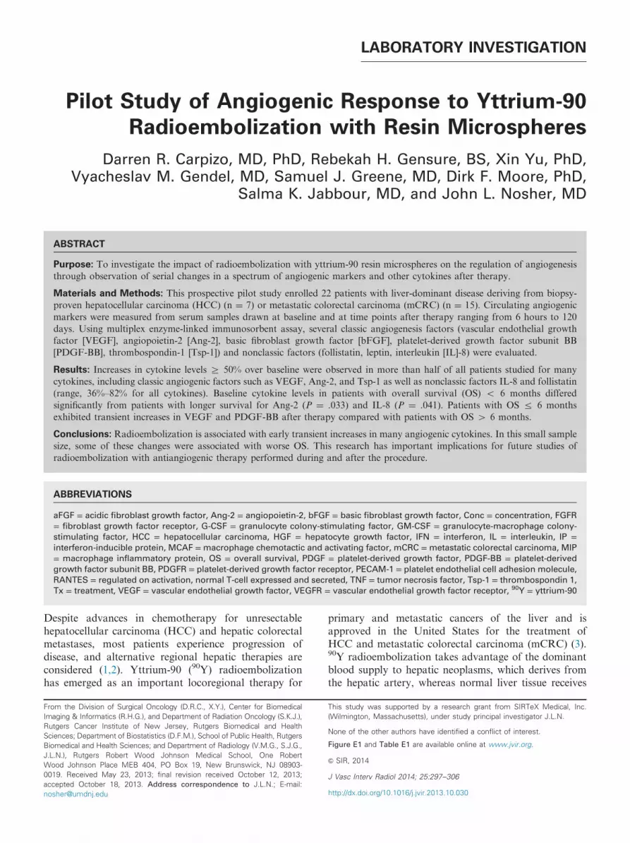
Figure 3. Examples of spikes in circulating angiogenic cytokines afte
changes in VEGF from three patients after treatment, second row
correspond to samples drawn at specific time points after first and
patterns after therapy for the first and third patients. Also note the stro
Conc = concentration, Tx = treatment. (Available in color online at w
RESULTS
Changes in Angiogenic Cytokines after
TherapyWe observed an increase in angiogenic cytokine levels,defined as an increase (spike) of Z 50% from the baselinevalue, that coincided with radioembolization therapy forboth classic and nonclassic angiogenic cytokines. Exam-ples of these observed spikes are illustrated in Figure 3.Not all patients experienced such spikes; some patientsdemonstrated progressive declines in cytokine levels aftertreatment. Comparison with curves for patients withminimal changes in cytokine levels is demonstrated inFigure E1 (available online at www.jvir.org). Amongpatients who underwent sequential bilobar treatmentsand exhibited perturbations or spikes after therapy, somewere apparent after the first treatment (eg, Fig 3, VEGF[first row], first and third plots), some were apparent afterbimodal treatment (after both first and second treatments;
r radioembolization therapy. First row illustrates representative
represents Ang-2, and third row represents PDGF-BB. Points
second radioembolization treatments. Note the bimodal Ang-2
ng spike in PDGF-BB after the first treatment for the third patient.
ww.jvir.org.)

Carpizo et al ’ JVIR302 ’ 90Y Resin Microsphere Radioembolization and Angiogenesis
eg, Fig 3, Ang-2 [second row], first and third plots), andothers were apparent only after the second treatment (eg,Fig 3, second plots shown for VEGF and Ang-2). Obser-ved patterns are presented in detail in Table 2. In morethan half of all patients studied, cytokines including bothclassic angiogenic factors (eg, VEGF, Ang-2, and Tsp-1)and nonclassic factors (eg, IL-8 and follistatin) demon-strated a spike in response to therapy (range, 36%–82%).
Survival Comparisons over TimeWe next examined the relationship between the increasesin angiogenic cytokine levels after radioembolizationand OS. Median OS for all patients was 8 months, witha 1-year survival rate of 41.25%. Patients were stratifiedinto two survival groups according to 6-month survival(n ¼ 8 surviving r 6 mo; n ¼ 14 surviving 4 6 mo).Baseline (before treatment) cytokine levels were signifi-cantly greater in the shorter survival group (r 6 mo) forAng-2 (P ¼ .033) and IL-8 (P ¼ .041) but not for VEGF(P ¼ .43) or PDGF-BB (P ¼ .91) (Fig 4).We next compared the variations in angiogenic cytokine
levels over time stratified by OS for VEGF, Ang-2, PDGF-BB, and Tsp-1 using the Mann-Whitney test. We foundthat patients within the shorter survival category experi-enced statistically significant increases in VEGF, Ang-2,
Table 2 . Distribution of Observed Spikes in Circulating Cytokine Lev
Cytokine Group Cytokine
Percent wit
Spike after
Classic angiogenic factors VEGF 59
Ang-2 59
bFGF 64
PDGF-BB 55
Tsp-1 77
Nonclassic angiogenic factors IL-8 77
Leptin 50
Follistatin 82
HGF 55
PECAM-1 36
Note. Spikes were identified by at least a 50% increase over baselin
Ang-2 ¼ angiopoietin-2, bFGF ¼ basic fibroblast growth factor, HG
platelet-derived growth factor subunit BB, PECAM-1 ¼ platelet en
VEGF = vascular endothelial growth factor.
Figure 4. Boxplots showing baseline comparisons across survival group
concentration. (Available in color online at www.jvir.org.)
and PDGF-BB at various early time points (6 h, day 3,and day 14) before decreasing in the later time pointsessentially to merge with the curves of the patients in thelonger survival category (Fig 5). We also performed Coxproportional hazard survival analyses of survival times ofthese patients for each angiogenic cytokine focusing on thetime points of 6 hours, day 3, and day 14. These resultsyielded complementary results (to the Mann-Whitney test)in that for VEGF, Ang-2, and PDGF-BB several earlytime points yielded significantly different survival times.VEGF was found to be significant at day 14 only, whereasAng-2 was significant for the 6-hour, day 3, and day 14time points. PDGF-BB was significant at the day 3 andday 14 time points. Tsp-1 level fluctuations in patientsappeared to be unrelated to the timing of treatment. Nodifference was found between the shorter and longersurvival groups (as shown in Fig 5, bottom row). We con-clude that for patients who experienced transient increasesin VEGF, Ang-2, and PDGF-BB after radioembolization,the transient increases correlated with shorter OS.
DISCUSSION90Y hepatic radioembolization, a regional form of brac-hytherapy, has been approved for the treatment of both
els after Treatment
h Z 50%
Treatment
Percent with No Major
Spike after Treatment
Percent
Undetermined
32 9
23 18
9 27
41 5
23 0
23 0
45 5
14 5
41 5
59 5
e levels at any time point after treatment.
F ¼ hepatocyte growth factor, IL-8 ¼ interleukin-8, PDGF-BB ¼dothelial cell adhesion molecule, Tsp-1 ¼ thrombospondin-1,
s for several angiogenic cytokines. *Po .05. Obs Conc = observed

Figure 5. Variations in circulating cytokine levels after radioembolization therapy for classic angiogenesis factors VEGF, Ang-2, PDGF-
BB, and Tsp-1 after therapy for two survival groups. Short survival (r 6 mo) is shown in green with square symbols; long survival (4 6
mo) is shown in blue with triangular symbols. Time points marked with an asterisk differed significantly between survival groups on
Mann-Whitney U comparisons; time points marked with a plus sign demonstrated significant differences on Cox proportional hazards
analysis. Conc = concentration, Tx = treatment. (Available in color online at www.jvir.org.)
Volume 25 ’ Number 2 ’ February ’ 2014 303
HCC and mCRC and is used off-label for various othercancers, either primary or metastatic to the liver (3). The90Y-bearing microspheres used in this study have diam-eters ranging from 20–60 μm and activity of 50 Bq persphere, with a 3-Gbq vial (81 mCi) of SIR-Spheres con-taining 40–80 million microspheres. 90Y radioemboliza-tion with resin microspheres delivers both a radiation andan embolic effect (12). Our observations with respect toangiogenic cytokine levels may be specific to resin micro-sphere radioembolization.Although many patients with both primary and meta-
static cancer to the liver experience prolonged local control
after hepatic radioembolization, others experience earlyfailure, either locally or at distant metastatic sites (7).Angiogenesis is an integral component in tumor growthand spread that is driven by a complex interplay betweenproangiogenic and antiangiogenic cytokines (15). Circu-lating VEGF, the most frequently studied angiogeniccytokine, strongly correlates with tumor expression ofVEGF (16). VEGF levels are reported to be associatedwith poor outcomes in both HCC and mCRC. In HCC,circulating VEGF further correlates with stage of disease,tumor burden, presence of intrahepatic and extrahepaticmetastasis, vascular invasion, postoperative recurrence,
Carpizo et al ’ JVIR304 ’ 90Y Resin Microsphere Radioembolization and Angiogenesis
response to treatment, progression-free survival, and OS(17,18). Previous work has demonstrated the potential forboth radiation and embolization to upregulate angio-genesis in vitro and in vivo (19,20). It is plausible that anangiogenic response to radioembolization may influencetherapeutic outcomes.In support of this notion, several studies have docu-
mented transient increases in angiogenic cytokine levelsshortly after transarterial chemoembolization for pa-tients with HCC and have correlated these changes withworse oncologic outcomes (10,18). These reports werelimited to either one (VEGF (10)) or three (VEGF,bFGF, and urokinase-type plasminogen activator (18))cytokines, whereas in this study we took a more com-prehensive strategy to studying this relationship throughthe investigation of classic, nonclassic, and nonangio-genic cytokines.After radioembolization, changes from baseline were
noted in serum levels of classic angiogenic cytokines,including Ang-2, PDGF-BB, VEGF, and IL-8. How-ever, the magnitude and temporal sequence of changes incytokine expression varied among patients. These obser-vations support appreciation that radioembolization isassociated with significant increases in many angiogeniccytokines. The variety of cytokines in which we observedchanges is more reflective of the complex biologicprocess of angiogenesis that has emerged since theidentification of the dominant genes, such as VEGFand FGF, in the 1990s (21,22).We found that Ang-2 levels were higher at baseline
and increased after treatment in a large number ofpatients in this cohort. Ang-2 is a ligand for the Tie-2receptor on endothelial cells and serves as a positiveregulator of tumor angiogenesis (23). Circulating Ang-2levels have been shown to serve as a surrogate markerfor tumor progression and metastasis, and blockade ofAng-2 represses metastasis (24). Mechanistic research intumor angiogenesis indicates that the Ang/Tie systemworks together with the VEGF system to break downand remodel tumor blood vessels (25). For these reasons,Tie-2 receptor inhibitors have been investigated; somereports indicate increased efficacy compared with VEGFinhibition alone (26). Our results suggest that con-sideration needs to be given to Ang-2/Tie-2 inhibitionin patients undergoing radioembolization. One possi-bility might be the multi–tyrosine kinase inhibitor rego-rafenib. Regorafenib and its active metabolites inhibitmultiple membrane-bound and intracellular kinases thatare involved in normal cellular functions and pathologicprocesses, including kinases in the RET, vascular endo-thelial growth factor receptor (VEGFR) 1, VEGFR2,VEGFR3, KIT, platelet-derived growth factor receptor(PDGFR)-α, PDGFR-β, fibroblast growth factor recep-tor (FGFR) 1, FGFR2, Tie-2, DDR2, Trk2A, Eph2A,RAF-1, BRAF, BRAFV600E, SAPK2, PTK5, and Ablpathways. Regorafenib was reported more recently in theCORRECT trial to show activity as monotherapy in
patients with chemotherapy-refractory mCRC (27). Thepatients in this trial were very similar to the typesof patients undergoing radioembolization in the pres-ent study. Another possibility would be cabozantinib(XL184), an agent approved by the U.S. Food and DrugAdministration that is another multi–tyrosine kinaseinhibitor that targets c-Met, VEGFR2, and Tie-2 recep-tors. Specific Tie-2 inhibitors are currently in clinicaltrials, such as AMG-386, which is an angiopoietinpeptibody (peptide [Fc]fragment of an angiopoietinantibody) currently in a phase III randomized trial ofpaclitaxel plus AMG-386 in metastatic ovarian cancer(NCT01204749) (28).An essential part of tumor angiogenesis is the recruit-
ment of pericytes to nascent endothelial tubes to allowfor their maturation into functional blood vessels. This isthe role of PDGF (29). PDGF-BB is an isoform ofPDGF that binds heparan sulfate proteoglycans in theextracellular matrix and helps localize it at the surface ofendothelial cells. This localization enhances its ability torecruit pericytes (30). We found that PDGF-BB levelstransiently increased on 90Y radioembolization to a gre-ater degree in patients surviving o 6 months comparedwith patients who survived 4 6 months. Although thistrend in this cohort was not significant, it may be with alarger sample size.IL-8 belongs to the chemokine family and has been
shown to be a macrophage-derived mediator of angio-genesis (31). IL-8 has been evaluated as a biomarker ofangiogenesis, and high levels of IL-8 have demonstrateda correlation with increased progression risk in a studyof patients with non–small cell lung cancer who weretreated with systemic cytotoxic chemotherapy and aVEGFR inhibitor (32). In this study, IL-8 levels atbaseline were significantly greater in the group of pati-ents with shorter survival; this suggests that IL-8 mayalso be useful as a biomarker to predict outcome in anytype of hepatic embolization therapy.As in the studies of Shim et al (10) and Sergio et al
(18), we found a potential correlation between increasesin VEGF, Ang-2, and PDGF-BB and worse oncologicoutcomes, suggesting not only that these cytokines maybe useful as biomarkers of prognosis after treatment butalso that these changes may be clinically relevant. Whenpatients were separated into long and short survivalgroups, there were trends for separation of groups bylevel of cytokine expression, and similar to previousstudies, the significant increases were transient. Collec-tively, these cytokines may influence survival in patientsundergoing radioembolization either by promoting an-giogenesis or by decreasing the effectiveness of radiation.Our study may have important therapeutic implica-
tions because it is has been suggested that patients mightbenefit from antiangiogenic therapy as an adjunct totransarterial chemoembolization in HCC (10,18,33). Ourresults in patients receiving radioembolization supportthis concept, but the broader spectrum of angiogenic
Volume 25 ’ Number 2 ’ February ’ 2014 305
cytokines we have reported complicate the choice ofantiangiogenic agent and make the case for using multi–tyrosine kinase inhibitors that would inhibit not only theVEGFR but also Tie-2 receptor, FGFR, and PDGFR.The temporal nature of the changes we observed shouldbe considered in the timing of administration of anti-angiogenic strategies used as adjuvant therapy in chemo-embolization or radioembolization. Our results supportgiving consideration to administering antiangiogenictherapy before regional embolic therapy.There are several limitations to our study. The major
limitation is sample size, which compromises the like-lihood of reaching statistical significance. Care must beexercised in interpreting the importance of our observa-tions given the limitations of using cytokine levels aspredictors of response to therapy (34). However, there isexperimental evidence that both radiation andembolization efficacy can be enhanced by the additionof antiangiogenic agents to chemoradiation protocols(8,20). Identification of specific proangiogenic pathwaysprovides the opportunity for targeted selection of appro-priate antiangiogenic agents from an ever-increasingarmamentarium—the goal of precision medicine.
ACKNOWLEDGMENTS
The authors wish to acknowledge the support of theBiospecimen Repository Service at the Rutgers CancerInstitute of New Jersey, under the direction of JulieFriedman.
REFERENCES
1. Llovet JM, Ricci S, Mazzaferro V, et al. Sorafenib in advanced hepato-cellular carcinoma. N Engl J Med 2008; 359:378–390.
2. Goldberg RM, Sargent DJ, Morton RF, et al. A randomized controlledtrial of fluorouracil plus leucovorin, irinotecan, and oxaliplatin combina-tions in patients with previously untreated metastatic colorectal cancer.J Clin Oncol 2004; 22:23–30.
3. Kennedy AS, Salem R. Radioembolization (yttrium-90 microspheres) forprimary and metastatic hepatic malignancies. Cancer J 2010; 16:163–175.
4. Welsh JS, Kennedy AS, Thomadsen B. Selective internal radiationtherapy (SIRT) for liver metastases secondary to colorectal adenocarci-noma. Int J Radiat Oncol Biol Phys 2006; 66:S62–S73.
5. Kennedy AS, Nutting C, Coldwell D, Gaiser J, Drachenberg C. Patho-logic response and microdosimetry of (90)Y microspheres in man: reviewof four explanted whole livers. Int J Radiat Oncol Biol Phys 2004; 60:1552–1563.
6. Gibbs P, van Hazel G. FOLFOX plus SIR-SPHERES MICROSPHERESversus FOLFOX alone in patients with liver mets from primary colorectalcancer (SIRFLOX). Bethesda, MD: National Library of Medicine; 2008–2013 ([cited 2013 Oct 7]. http://clinicaltrials.gov/show/NCT00724503.NLM Identifier: NCT00724503.).
7. Schonewolf CA, Patel B, Gensure RH, et al. Patterns of failure incolorectal patients with liver metastases after yttrium-90 radioemboliza-tion. Am J Clin Oncol. In press; available online December 27, 2012.
8. Gorski DH, Beckett MA, Jaskowiak NT, et al. Blockage of the vascularendothelial growth factor stress response increases the antitumor effectsof ionizing radiation. Cancer Res 1999; 59:3374–3378.
9. Korse CM, Bonfrer JM, Prevoo W, Baas P, Taal BG. Increase ofangiogenic growth factors after hepatic artery embolization in patientswith neuroendocrine tumours. Tumour Biol 2011; 32:647–652.
10. Shim JH, Park J-W, Kim JH, et al. Association between increment ofserum VEGF level and prognosis after transcatheter arterial chemo-embolization in hepatocellular carcinoma patients. Cancer Sci 2008; 99:2037–2044.
11. Nosher JL, Ohman-Strickland PA, Jabbour S, Narra V, Nosher B.Changes in liver and spleen volumes and liver function after radio-embolization with yttrium-90 resin microspheres. J Vasc Interv Radiol2011; 22:1706–1713.
12. Salem R, Thurston KG. Radioembolization with 90yttrium micro-spheres: a state-of-the-art brachytherapy treatment for primary andsecondary liver malignancies. Part 1: technical and methodologic consid-erations. J Vasc Interv Radiol 2006; 17:1251–1278.
13. Salem R, Thurston KG. Radioembolization with 90yttrium micro-spheres: a state-of-the-art brachytherapy treatment for primary andsecondary liver malignancies. Part 2: special topics. J Vasc Interv Radiol2006; 17:1425–1439.
14. Salem R, Lewandowski RJ, Gates VL, et al. Research reporting stand-ards for radioembolization of hepatic malignancies. J Vasc Interv Radiol2011; 22:265–278.
15. Bergers G, Benjamin LE. Tumorigenesis and the angiogenic switch.Nat Rev Cancer 2003; 3:401–410.
16. Poon RT, Lau CP, Cheung ST, Yu WC, Fan ST. Quantitative correlationof serum levels and tumor expression of vascular endothelial growthfactor in patients with hepatocellular carcinoma. Cancer Res 2003; 63:3121–3126.
17. Xiong ZP, Yang SR, Liang ZY, et al. Association between vascularendothelial growth factor and metastasis after transcatheter arterialchemoembolization in patients with hepatocellular carcinoma. Hepato-biliary Pancreat Dis Int 2004; 3:386–390.
18. Sergio A, Cristofori C, Cardin R, et al. Transcatheter arterialchemoembolization (TACE) in hepatocellular carcinoma (HCC): the role ofangiogenesis and invasiveness. Am J Gastroenterol 2008; 103:914–921.
19. Poon RT, Ng IO, Lau C, et al. Serum vascular endothelial growth factorpredicts venous invasion in hepatocellular carcinoma: a prospectivestudy. Ann Surg 2001; 233:227–235.
20. Gadaleta CD, Ranieri G. Trans-arterial chemoembolization as a therapyfor liver tumours: new clinical developments and suggestions forcombination with angiogenesis inhibitors. Crit Rev Oncol Hematol2011; 80:40–53.
21. Chung AS, Lee J, Ferrara N. Targeting the tumour vasculature: insightsfrom physiological angiogenesis. Nat Rev Cancer 2010; 10:505–514.
22. Weis SM, Cheresh DA. Tumor angiogenesis: molecular pathways andtherapeutic targets. Nat Med 2011; 17:1359–1370.
23. Fagiani E, Christofori G. Angiopoietins in angiogenesis. Cancer Lett2013; 328:18–26.
24. Holopainen T, Saharinen P, D’Amico G, et al. Effects of angiopoietin-2-blocking antibody on endothelial cell-cell junctions and lung metastasis. JNatl Cancer Inst 2012; 104:461–475.
25. Lobov IB, Brooks PC, Lang RA. Angiopoietin-2 displays VEGF-dependent modulation of capillary structure and endothelial cell survivalin vivo. Proc Natl Acad Sci U S A 2002; 99:11205–11210.
26. Koh YJ, Kim H-Z, Hwang S-I, et al. Double antiangiogenic protein,DAAP, targeting VEGF-A and angiopoietins in tumor angiogenesis,metastasis, and vascular leakage. Cancer Cell 2010; 18:171–184.
27. Grothey A, Van Cutsem E, Sobrero A, et al. Regorafenib monotherapyfor previously treated metastatic colorectal cancer (CORRECT): aninternational, multicentre, randomised, placebo-controlled, phase 3 trial.Lancet 2013; 381:303–312.
28. Amgen. A phase 3, randomized, double-blind trial of weekly paclitaxelplus AMG 386 or placebo in women with recurrent partially platinumsensitive or resistant epithelial ovarian, primary peritoneal or fallopiantube cancers. Bethesda, MD: National Library of Medicine; 2013 [cited2013 Oct 7]. Available at: http://clinicaltrials.gov/show/NCT01204749.NLM Identifier: NCT01204749.
29. Hellström M, Gerhardt H, Kalén M, et al. Lack of pericytes leads toendothelial hyperplasia and abnormal vascular morphogenesis. J Cell Biol2001; 153:543–553.
30. Lindblom P, Gerhardt H, Liebner S, et al. Endothelial PDGF-B retentionis required for proper investment of pericytes in the microvessel wall.Genes Dev 2003; 17:1835–1840.

Carpizo et al ’ JVIR306 ’ 90Y Resin Microsphere Radioembolization and Angiogenesis
31. Koch AE, Polverini PJ, Kunkel SL, et al. Interleukin-8 as a macrophage-derived mediator of angiogenesis. Science 1992; 258:1798–1801.
32. Hanrahan EO, Lin HY, Kim ES, et al. Distinct patterns of cytokine andangiogenic factor modulation and markers of benefit for vandetanib and/or chemotherapy in patients with non-small-cell lung cancer. J Clin Oncol2010; 28:193–201.
33. Weintraub JL, Salem R. Treatment of hepatocellular carcinoma combin-ing sorafenib and transarterial locoregional therapy: state of the science.J Vasc Interv Radiol 2013; 24:1123–1134.
34. Hegde PS, Jubb AM, Chen D, et al. Predictive impact of circulatingvascular endothelial growth factor in four phase III trials evaluatingbevacizumab. Clin Cancer Res 2013; 19:929–937.
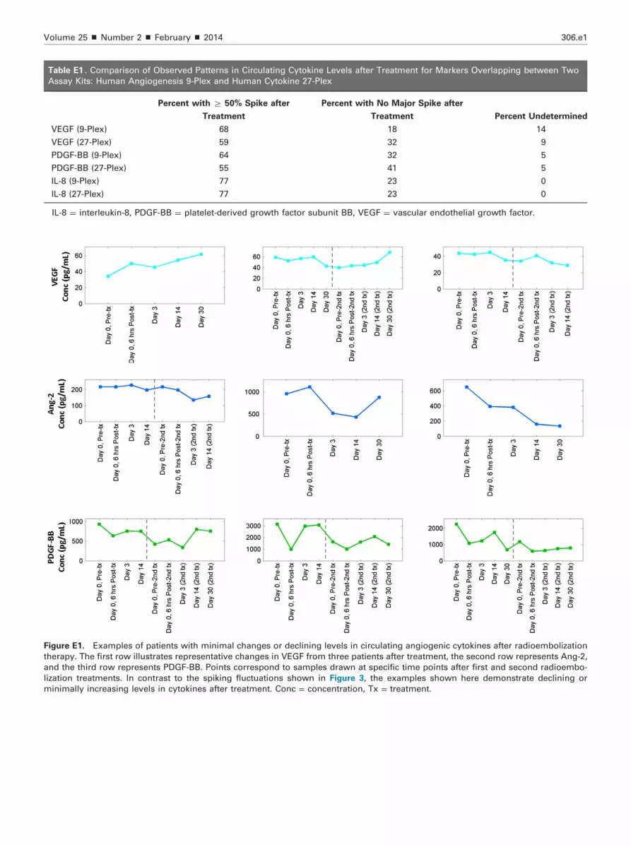
Figure E1. Examples of patients with minimal changes or declining levels in circulating angiogenic cytokines after radioembolization
therapy. The first row illustrates representative changes in VEGF from three patients after treatment, the second row represents Ang-2,
and the third row represents PDGF-BB. Points correspond to samples drawn at specific time points after first and second radioembo-
lization treatments. In contrast to the spiking fluctuations shown in Figure 3, the examples shown here demonstrate declining or
minimally increasing levels in cytokines after treatment. Conc = concentration, Tx = treatment.
Table E1 . Comparison of Observed Patterns in Circulating Cytokine Levels after Treatment for Markers Overlapping between Two
Assay Kits: Human Angiogenesis 9-Plex and Human Cytokine 27-Plex
Percent with Z 50% Spike after
Treatment
Percent with No Major Spike after
Treatment Percent Undetermined
VEGF (9-Plex) 68 18 14
VEGF (27-Plex) 59 32 9
PDGF-BB (9-Plex) 64 32 5
PDGF-BB (27-Plex) 55 41 5
IL-8 (9-Plex) 77 23 0
IL-8 (27-Plex) 77 23 0
IL-8 ¼ interleukin-8, PDGF-BB ¼ platelet-derived growth factor subunit BB, VEGF ¼ vascular endothelial growth factor.
Volume 25 ’ Number 2 ’ February ’ 2014 306.e1