Embed Size (px)
Citation preview

Melucci et al. Journal of Experimental & Clinical Cancer Research 2013, 32:13http://www.jeccr.com/content/32/1/13
RESEARCH Open Access
Decrease of survivin, p53 and Bcl-2 expression inchemorefractory colorectal liver metastases maybe predictive of radiosensivity afterradioembolization with yttrium-90 resinmicrospheresElisa Melucci1†, Maurizio Cosimelli2†, Livio Carpanese3, Giuseppe Pizzi3, Francesco Izzo4, Francesco Fiore5,Rita Golfieri6, Emanuela Giampalma6, Isabella Sperduti7, Cristiana Ercolani1, Rosa Sciuto8, Raffaello Mancini1,Carlo Garufi9, Maria Grazia Diodoro2, Marcella Mottolese1* and Italian Society of Locoregional Therapiesin Oncology (S.I.T.I.L.O.)
Abstract
In a prospective multicenter phase II trial of radioembolization with yttrium-90 (90Y-RE) in chemorefractory liver-dominant metastatic colorectal cancer (mCRC), we showed that median survival was 12.6 months (95% CI 7.0–18.3)with 48% of 50 patients achieving disease control. In this extension retrospective study, we analyzed whether apanel of biomarkers, known to be associated to an adverse clinical outcome, underwent variations in CRC livermetastases pre and post 90Y-RE.Of the 50 patients included in the study, 29 pre-90Y-RE therapy and 15 post-90Y-RE had liver biopsy specimensavailable. In these series we investigated survivin, p53, Bcl-2 and Ki-67 expression pre- and post-90Y-RE byimmuhistochemistry (IHC). Our findings evidenced a decrease of survivin (77% vs 33%), p53 (93% vs 73%), Bcl-2(37% vs 26%) expression as well as of Ki-67 proliferation index (62.5% vs 40%) on liver biopsies collected post-90Y-REas compared to pre-90Y-RE. In the subset of 13 matched liver metastases we further confirmed the reduction ofsurvivin (92.3% vs 53.8%; p = 0.06), p53 (100% vs 69.2%; p = 0.05) and Bcl-2 (69.2% vs 53.8%; p = 0.05) expressionpost-90Y-RE. This biomarker modulation was accompanied by morphological changes as steatohepatitis, hepatocytenecrosis, collagen deposition, proliferating and/or bile duct ectasia, focal sinusoidal dilatation and fibrosis.Although our analysis was conducted in a very limited number cases, these changes appear strictly related to theresponse to 90Y-RE therapy and may deserve further investigation on a larger series of patients.
Keywords: Colorectal cancer, Liver metastases, Radioembolization, Yttrium-90-resin microspheres, Survivin, p53,Bcl-2, Ki-67
* Correspondence: [email protected]†Equal contributors1Department of Pathology, Regina Elena National Cancer Institute, Rome,ItalyFull list of author information is available at the end of the article
© 2013 Melucci et al.; licensee BioMed Central Ltd. This is an Open Access article distributed under the terms of the CreativeCommons Attribution License (http://creativecommons.org/licenses/by/2.0), which permits unrestricted use, distribution, andreproduction in any medium, provided the original work is properly cited.

Melucci et al. Journal of Experimental & Clinical Cancer Research 2013, 32:13 Page 2 of 7http://www.jeccr.com/content/32/1/13
IntroductionLiver metastases are a significant cause of morbidity andmortality for more than 45% of patients who present withcolorectal cancer (CRC) [1]. Although chemotherapy regi-mens combined with biologic agents have improved thecontrol of liver metastases, the occurrence of hepatic me-tastases continues to present a life-limiting prognosis formost patients with advanced CRC [2] being 5 year survivalapproximately 11%. In the setting of clinical trials, medianoverall survival for unresectable metastases have been ex-tended beyond two years using combinations includingoxaliplatin, irinotecan, capecitabine and biologic agents(bevacizumab, cetuximab, panitumumab) [3,4]. In parallelwith these developments, the application of locally ablativeprocedures, such as radiofrequency ablation, are increas-ingly considered beneficial for patients with unresectableliver-only disease who present with tumors ≤ 3–4 cm indiameter. These regional treatments for liver metastasescan also be used to consolidate the treatment response withchemotherapy, in order to further increase the number ofpatients eligible for resection [5,6]. Despite these gains, oneof the major challenges in advanced CRC are the growingproportion of patients who continue to present with pro-gressive liver involvement having exhausted all other thera-peutic options.Radioembolization with yttrium-90 (90Y-RE) and, as re-
cently described, with holmium-166 poly (L-lactic acid) la-beled microspheres (166Ho-PLLA-MS) [7], are therapeuticprocedures applied to the liver that allow direct delivery ofhigh-dose radiation to liver tumors (both primary andmetastatic) by means of endovascular catheters, selectivelyplaced within the hepatic arterial vasculature. 90Y and166Ho-PLLA-MS (resin or glass) microspheres lodgewithin the neovascular rim of the tumor(s) [8,9].In a multicenter phase II trial conducted in highly
chemorefractory liver-dominant metastatic CRC (mCRC),we showed that 48% (24 of 50) of patients achieved dis-ease control with a median overall survival of 12.6months following RE with 90Y-radiolabelled resin micro-spheres [10]. This finding is consistent with the resultsfrom other multicenter evaluations using 90Y-RE in thechemorefractory setting [11]. Up to date, there are nostudies which have investigated biomarker expressionand response to 90Y-RE therapy.It is largely described that the ability to avoid apoptosis
is one of the major oncogenic switches contributing totumor progression. Among the gene coding apoptosisand cell proliferation protein regulators, Bcl-2, an antia-popototic protein, survivin, one of the member of theinhibitor of apoptosis (IAP) protein family and p53 mayidentify CRC patients at a higher risk of tumor progres-sion [12-14].In the present retrospective study which is an exten-
sion of our previous one [10], we evaluated whether the
expression of these biomarkers may undergo to signifi-cant changes before and after 90Y-RE thus providing pre-dictive information of clinical value.
MethodsPatients and treatmentBetween May 2005 and August 2007, 50 patients withunresectable, histologically proven CRC liver metastasesand limited extra-hepatic disease (≤ 3 nodules in the sameextra-hepatic organ each < 3 mm), in progression follow-ing standard systemic chemotherapy, were recruited fromfour Italian centers in a phase II prospective clinical trialconducted by the Italian Society of Locoregional Therapyin Oncology (SITILO). Further details of the treatmentplanning and patient selection have been outlined inour previous paper [10]. In brief, patients were requiredto be between 18 and 75 years of age, have livermetastases measurable by Response Evaluation Criteriain Solid Tumours (RECIST), adequate renal function(creatinine < 1.5 7 × normal values or creatinine clear-ance > 50 mL/minute), hemopoietic function, WHO orECOG performance status ≤ 2 and were able to give in-formed consent. To be eligible for 90Y-RE, patients wererequired to have: sufficient liver function; hepatic arterialanatomy that would enable safe delivery of microspheresto the liver only; liver to lung shunting of < 20% on a pre-treatment technetium-99m labeled macro-aggregated-al-bumin (99mTc-MAA) nuclear scan; and a patent mainportal vein. Patients were excluded if they were pregnant,had evidence of local recurrence of primary disease, in-flammatory gastrointestinal disease or had received priortreatment with hepatic arterial chemotherapy or externalbeam radiotherapy to the liver. The median interval be-tween diagnosis of mCRC and 90Y-RE was 17 months(range, 6–71 months). To investigate biomarkers expres-sion and response to 90Y-RE therapy, liver metastasesbiopsies were taken 8–21 days prior to 90Y-RE and2 months post-90Y-RE. Tissue specimens were availablefrom 29 patients pre therapy and 15 patients post ther-apy. Samples pre- and post-90Y-RE were concomitantlyavailable in 13 patients.The study was approved by the Ethical Committee at
the Regina Elena Cancer Institute (N°534; 22/03/05) anda written informed consent was obtained by all patients.
ImmunohistochemistryFormalin-fixed paraffin-embedded liver biopsies were cuton SuperFrost Plus slides (Menzel-Gläser, Braunschweig,Germany). Antigen retrieval was performed at 96°C(10 mM/L citrate buffer, pH 6) for 40 minutes in a thermo-static bath. Sections were incubated with the polyclonalantibody (PAb) anti-survivin (1:100, Novus Biological,DBA, Milan, Italy); with the anti-Ki-67 monoclonal anti-body (MoAb) MIB-1 (5 μg/ml; Dako, Milan, Italy), the
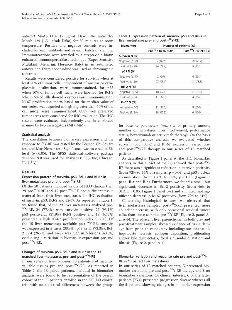
Table 1 Expression pattern of survivin, p53 and Bcl-2 inliver metastases pre- and post -90Y-RE
Biomarkers Number of patients (%)
Pre-90Y-RE (N = 29) Post-90Y-RE (N = 15)
Survivin N (%)
Negative (≤ 20) 5 (16.2) 10 (66.7)
Positive (> 20) 24 (77.4) 5 (33.3)
p53 N (%)
Negative (≤ 10) 2 (6.9) 4 (26.7)
Positive (> 10) 27 (93.1) 11 (73.3)
Bcl-2 N (%)
Negative (≤ 5) 18 (62.1) 11 (73.3)
Positive (> 5) 11 (37.9) 4 (26.7)
Ki-67 N (%)
Negative (<50) 11 (37.5) 9 (60.0)
Positive (≥ 50) 18 (62.5) 6 (40.0)
Melucci et al. Journal of Experimental & Clinical Cancer Research 2013, 32:13 Page 3 of 7http://www.jeccr.com/content/32/1/13
anti-p53 MoAb DO7 (5 μg/ml, Dako), the anti-Bcl-2MoAb 124 (1,5 μg/ml; Dako) for 30 minutes at roomtemperature. Positive and negative controls were in-cluded for each antibody and in each batch of staining.Immunoreactions were revealed by a streptavidin-biotinenhanced immunoperoxidase technique (Super SensitiveMultiLink Menarini, Florence, Italy) in an automatedautostainer. Diaminobenzidine was used as chromogenicsubstrate.Results were considered positive for survivin when at
least 20% of tumor cells, independent of nuclear or cyto-plasmic localization, were immunostained, for p53when 10% of tumor cell nuclei were labelled, for Bcl-2when > 5% of cells showed a cytoplasmic immunoreaction.Ki-67 proliferation index, based on the median value ofour series, was regarded as high if greater than 50% of thecell nuclei were immunostained. Only well preservedtumor areas were considered for IHC evaluation. The IHCresults were evaluated independently and in a blindedmanner by two investigators (MD, MM).
Statistical analysisThe correlation between biomarkers expression and theresponse to 90Y-RE was tested by the Pearson Chi-Squaretest and Mac Nemar test. Significance was assessed at 5%level (p < 0.05). The SPSS statistical software packageversion 19.0 was used for analyses (SPSS, Inc, Chicago,IL, USA).
ResultsExpression pattern of survivin, p53, Bcl-2 and Ki-67 inliver metastases pre- and post-90Y-REOf the 50 patients included in the SITILO clinical trial,29 pre-90Y-RE and 15 post-90Y-RE had sufficient tissuematerial from their liver metastases for IHC evaluationof survivin, p53, Bcl-2 and Ki-67. As reported in Table 1,we found that, of the 29 liver metastases analyzed pre-90Y-RE, 24 (77.4%) were survivin positive, 27 (93.1%)p53 positive,11 (37.9%) Bcl-2 positive and 18 (62.5%)presented a high Ki-67 proliferation index (>50%). Ofthe 15 liver metastases available post-90Y-RE, survivinwas expressed in 5 cases (33.3%), p53 in 11 (73.3%), Bcl-2 in 4 (26.7%) and Ki-67 was high in 6 lesions (40.0%)evidencing a variation in biomarker expression pre andpost-90Y-RE.
Changes of survivin, p53, Bcl-2 and Ki-67 in the 13matched liver metastases pre- and post-90Y-REIn our series of liver biopsies, 13 patients had matchedvaluable tissues pre and post-90Y-RE. As reported inTable 2, the 13 paired patients, included in biomarkeranalysis, were found to be representative of the overallcohort of the 50 patients enrolled in the SITILO clinicaltrial with no statistical differences between the groups
for baseline parameters (sex, site of primary tumors,number of metastases, liver involvement, performancestatus, bevacizumab or cetuximab therapy). On the basisof this comparative analysis, we evaluated whethersurvivin, p53, Bcl-2 and Ki-67 expression varied pre-and post-90Y-RE therapy in our series of 13 matchedpatients.As described in Figure 1 panel A, the IHC biomarker
analysis in this subset of mCRC showed that post-90Y-RE there was a significant reduction in survivin positivity(from 92% to 54% of samples; p = 0.06) and p53 nuclearaccumulation (from 100% to 69%; p = 0.05) (Figure 1panel B-a and B-b). Furthermore, we found a small, butsignificant, decrease in Bcl-2 positivity (from 46% to31%; p = 0.05; Figure 1 panel B-c) and a limited, not sig-nificant, decrease in Ki-67 positivity (from 77% to 61%).Concerning histological features, we observed that
liver metastases sampled post-90Y-RE presented moreabundant necrosis, with only occasional residual cancercells, than those sampled pre-90Y-RE (Figure 2, panel A-a, A-b). The adjacent liver parenchyma, in both pre- andpost-treatment samples, showed evidence of tissue dam-age from prior chemotherapy including: steatohepatitis,hepatocyte necrosis, collagen deposition, proliferatingand/or bile duct ectasia, focal sinusoidal dilatation andfibrosis (Figure 2, panel A-c).
Biomarker variation and response rate pre and post-90Y-RE in 13 paired liver metastasesIn our series of 13 matched patients, 5 presented bio-marker variations pre and post-90Y-RE therapy and 8 nobiomarker variations. Of clinical interest, 6 of the latterpatients (75%) presented progression disease whereas allthe 5 patients showing changes in biomarker expression

Table 2 Comparison of clinical variables between the overall series of patients and the series with liver biopsiespre- and post-90Y-RE
Baseline Characteristics
Patients Age (years)* Time to RE** FU months*** Sex N° (%) PT site N° (%) Met N° (%) Liver involvement N° (%) PS N° (%) Pre BV N° (%) Pre CTX N° (%)
M F Colon Rectum ≤ 4 > 4 <25% > 25% 0 ≥ 1 No Yes No Yes
Overall Series (N = 50) 64 19 14 37 13 41 9 21 29 20 7 35 15 39 11 45 5
(34–38) (6–71) (2–49) (74) (26) (82) (18) (42) (58) (40) (54) (70) (30) (78) (22) (90) (10)
Pre/Post RE series (N = 13) 58 21 15 9 4 11 2 4 9 30 6 9 4 9 4 12 1
(40–75) (9–53) (3–49) (69) (31) (85) (15) (31) (69) (60) (46) (69) (31) (69) (31) (92) (8)
P value 0.11 0.50 0.99 0.49 0.99 0.54 0.54 0.99 0.49 0.99
* mean (range); ** Months from diagnosis to 90Y-RE; ***Follow up post-90Y-RE;M, male; F, female; PT, Primary Tumor; Met, Metastases; PS, Performance Status; BV, bevacizumab; CTX, cetuximab.
Melucciet
al.JournalofExperim
ental&ClinicalCancer
Research2013,32:13
Page4of
7http://w
ww.jeccr.com
/content/32/1/13

Figure 1 Changes of survivin, p53, Bcl-2 and Ki-67 in livermetastases pre- and post-90Y-RE. A. The histogram shows thesignificant reduction of the positivity of survivin (from 92% to 54%;p = 0.06), p53 (from 100% to 69%; p = 0.05) and Bcl-2 (from 46% to31%; p = 0.05) expression in liver metastases pre- and post-90Y-REtherapy. A limited, not significant decrease in proliferation index byKi-67 (from 77% to 61%) is also evident. B. Immunohistochemicalstaining of 3 autologous liver metastases sampled pre- and post-therapy showing a strong decrease in survivin (a) p53 (b), and Bcl-2(c) immunoreactions.
Melucci et al. Journal of Experimental & Clinical Cancer Research 2013, 32:13 Page 5 of 7http://www.jeccr.com/content/32/1/13
had partial response or stable disease (Figure 2, panel B).Nevertheless, the limited number of patients did notallow us to determine whether these changes may reallyaffect survival.
DiscussionPatients included in the present study were from a mul-ticenter phase II clinical trial which is the first prospect-ive evaluation of 90Y-RE in CRC patients with livermetastases who failed previous oxaliplatinum and iri-notecan based chemotherapy regimen [10]. It has been
widely reported that alterations in genes, as survivin,p53 and Bcl-2, which regulate cell growth and apoptoticprocesses, are significantly associated to an unfavourableclinical outcome in CRC patients [15]. In our series of29 liver mCRC patients, we found that most tumorssampled prior to 90Y-RE were p53, survivin, and Bcl-2highly positive and presented a high Ki-67 proliferationindex. In contrast, we found a significant reduction inp53, survivin and Bcl-2 positive expression in liver me-tastasis sampled two months post-90Y-RE. There wasalso a trend towards a reduction in cells with a high pro-liferative index as measured by Ki-67. We have previ-ously shown that colon cancers harboring p53 nuclearaccumulation, as assessed by the DO7 anti-p53 antibody,represent a subset of tumors with a more aggressiveclinical behaviour in patients with stage II tumors as wellas in young patients [13,16]. Furthermore, several studieshave shown an increased incidence of p53 nuclear accu-mulation in liver metastases in comparison to the pri-mary tumor, hypothesizing a role for p53 in CRC livermetastatization. In particular, the presence of ≥ 3 livermetastases identified a subset of patients with a verypoor prognosis mainly when associated to p53 mutations[17]. A number of studies have also shown that tumorsthat do not express detectable levels of Bcl-2, but whichexhibited nuclear accumulation of p53, were associatedwith the shortest patient survival, while Bcl-2-positiveand p53-negative tumors had the best prognosis [12,17].Studies conducted at our Institute showed that p53 posi-tivity combined with Bcl-2 negativity and elevated Ki-67score correlated with advanced tumor stage, poorly dif-ferentiated tumors and increased probability of relapse.Also elevated survivin expression levels in primary CRCare related to decreased survival [14,15]. In resected livertumors, altered expression of survivin, p53, Ki-67 and,more recently, KRAS mutations, have been shown to beindependently predictive of hepatic recurrence and poorsurvival [13,16,18]. It is recently reported that defectivemismatch repair predicts resistance to 5-fluorouracil(5FU) and KRAS mutation resistance to anti-EGFR anti-body therapy [19]. Nevertheless, no predictive markersof RE efficacy in mCRC have been identified up to now.In terms of the predictive response to radiotherapy, sev-eral studies have linked epidermal growth factor re-ceptor (EGFR) and vascular endothelial growth factor(VEGF) expression to a lack of response to pre-operativeradiotherapy in locally advanced rectal cancer [19-21].Neither p53, Ki-67 and survivin expression appear to becorrelated to pre-operative chemo-radiotherapy responseand prognosis in locally advanced rectal cancer [22,23]. Todate, however, no study has evaluated the predictive valueof molecular markers on radiosensitivity of CRC liver me-tastasis. In this context, our findings, although in a verylimited number of patients, may be clinically relevant.

Figure 2 Morphological and phenotypic changes in paired liver metastases pre- and post- 90Y-RE. A. Example of histological features in apre-90Y-RE CRC liver metastasis with focal areas of necrosis (a), in a post-90Y-RE CRC liver metastasis with evident increase of tumor necrosis(b) and, within uninvolved peritumoral liver parenchyma, showing dysplastic hepatocytes, sinusoidal dilatation, leukocyte infiltration and bile-ductproliferation (c). B. Histogram summarizing Sirtex response in the 13 autologous liver biopsies according to biomarker changes pre- and post-therapy. Two patients (25%) not showing biomarker changes suffered PD whereas 6 patients (100%) showing biomarker changes had PR or SD.
Melucci et al. Journal of Experimental & Clinical Cancer Research 2013, 32:13 Page 6 of 7http://www.jeccr.com/content/32/1/13
The rapid changes of biomarkers observed in ourseries post-90Y-RE may be due to clonal selection or toepigenetic changes, not previously recorded in this con-text. Such mechanisms are usually discussed in the con-text of cell adaption to chemotherapy and evolvingresistance. Radio-sensitivity of colorectal cancer cells maybe determined by p53 mutation [23,24], whereas there isno evidence that chemotherapy per se cause changes inthe cellular expression of p53 [25]. This is the first timethat we have recorded a down-staging in p53 protein ex-pression after 90Y-RE.It is likely that both disease progression and a prolonged
prior chemotherapy affected the efficacy and tolerability of90Y-RE in the liver. In fact, mild manifestations of non-alcoholic fatty liver disease (NAFLD) after 5FU [26], moreserious non-alcoholic steatohepatitis after irinotecan andsinusoidal obstruction syndrome (SOS) after oxaliplatin-based treatment [27] have been recorded. Using the samebiomarkers as in our study, Panasiuk and colleagues [28]showed that the intensification of inflammation in NAFLDmay also impact on biomarker expression in human hepa-tocytes with the induction of pro-apoptotic protein p53and the inhibition of anti-apoptotic Bcl-2.There are clear limitations to our study, not least of
which was the small patient numbers and limited tissuesampling. Nevertheless, we believe that our findingsmerit further investigation in prospective clinical trials.We are planning to evaluate this biomarker panel in aphase II randomized trial on 2nd line treatment. KRASmutated CRC patients with unresectable liver metastasis
will be randomized to receive systemic therapy vs sys-temic therapy plus 90Y-RE. The combined assessment ofsurvivin, p53 and Bcl-2 pre and post-90Y-RE therapymay improve our ability to predict outcomes in thetreatment paradigm of metastatic KRAS mutated CRCpatients.
Competing interestsThe authors declared that they have no competing interest.
Authors’ contributionsEM and CE carried out immunohistochemical staining and contributed indata acquirement and interpretation. MC contributed to the study design,data interpretation and manuscript drafting. LC, GP, FF, RG, EG performedliver biopsies pre and post radioembolization in all the patients included inthis study. IS was responsible for the database set up and for the statisticalanalyses. RS was involved in the patient treatment with ytttium-90microspheres. MD evaluated the morphological features of liver biopsies andrevised all the slides submitted to immunohistochemical staining. CG and FI,RM provided clinical and surgical data of the patients including treatmentschedule and follow up. MM were responsible for the study concept anddesign and for the interpretation of results, helped in data discussion,critically revised the manuscript for important intellectual content, andobtained funding for the study. All authors have read and approved themanuscript.
AcknowledgementsWe would to thank the patients for agreeing to participate in this study,which was a collaboration of the Italian Society of Locoregional Therapies inOncology (SITILO). We would also like to thank Paolo Avetrani, PhD, and RaeHobbs and Maria Assunta Fonsi for their helpful contribution to this work.The yttrium-90 resin microspheres were provided by Sirtex Medical Limited.The study was partially supported by Associazione Italiana per la Ricerca sulCancro (AIRC 11770 CG).
Author details1Department of Pathology, Regina Elena National Cancer Institute, Rome,Italy. 2Department of Surgery, Regina Elena National Cancer Institute, Rome,

Melucci et al. Journal of Experimental & Clinical Cancer Research 2013, 32:13 Page 7 of 7http://www.jeccr.com/content/32/1/13
Italy. 3Department of Interventional Radiology, Regina Elena National CancerInstitute, Rome, Italy. 4Department of Surgery, Pascale Cancer Institute,Naples, Italy. 5Department of Interventional Radiology, Pascale CancerInstitute, Naples, Italy. 6Department of Interventional Radiology, MalpighiHospital, Bologna, Italy. 7Biostatistics, Scientific Direction, Regina ElenaNational Cancer Institute, Rome, Italy. 8Department of Nuclear Medicine,Regina Elena National Cancer Institute, Rome, Italy. 9Medical oncology,Regina Elena National Cancer Institute, Rome, Italy.
Received: 4 February 2013 Accepted: 27 February 2013Published: 6 March 2013
References1. Manfredi S, Lepage C, Hatem C, Coatmeur O, Faivre J, Bouvier AM:
Epidemiology and management of liver metastases from colorectalcancer. Ann Surg 2006, 244(2):254–259.
2. Nordlinger B, Sorbye H, Glimelius B, Poston GJ, Schlag PM, Rougier P,Bechstein WO, Primrose JN, Walpole ET, Finch-Jones M, et al: Perioperativechemotherapy with FOLFOX4 and surgery versus surgery alone forresectable liver metastases from colorectal cancer (EORTC Intergrouptrial 40983): a randomised controlled trial. Lancet 2008,371(9617):1007–1016.
3. Van Cutsem E, Kohne CH, Hitre E, Zaluski J, Chang Chien CR, Makhson A,D’Haens G, Pinter T, Lim R, Bodoky G, et al: Cetuximab and chemotherapyas initial treatment for metastatic colorectal cancer. N Engl J Med 2009,360(14):1408–1417.
4. Tol J, Koopman M, Cats A, Rodenburg CJ, Creemers GJ, Schrama JG,Erdkamp FL, Vos AH, Van Groeningen CJ, Sinnige HA, et al: Chemotherapy,bevacizumab, and cetuximab in metastatic colorectal cancer. N Engl JMed 2009, 360(6):563–572.
5. Popescu I, Alexandrescu S, Croitoru A, Boros M: Strategies to convert toresectability the initially unresectable colorectal liver metastases.Hepatogastroenterology 2009, 56(91–92):739–744.
6. Cianni R, Pelle G, Notarianni E, Saltarelli A, Rabuffi P, Bagni O, Filippi L,Cortesi E: Radioembolisation with (90)Y-labelled resin microspheres inthe treatment of liver metastasis from breast cancer. Eur Radiol 2013,23(1):182–189.
7. Smits ML, Nijsen JF, van den Bosch MA, Lam MG, Vente MA, Huijbregts JE,van het Schip AD, Elschot M, Bult W, De Jong HW, et al: Holmium-166radioembolization for the treatment of patients with liver metastases:design of the phase I HEPAR trial. J Exp Clin Cancer Res 2010, 29:70.
8. Kennedy AS, Nutting C, Coldwell D, Gaiser J, Drachenberg C: Pathologicresponse and microdosimetry of (90)Y microspheres in man: review offour explanted whole livers. Int J Radiat Oncol Biol Phys 2004,60(5):1552–1563.
9. Campbell AM, Bailey IH, Burton MA: Tumour dosimetry in human liverfollowing hepatic yttrium-90 microsphere therapy. Phys Med Biol 2001,46(2):487–498.
10. Cosimelli M, Golfieri R, Cagol PP, Carpanese L, Sciuto R, Maini CL, Mancini R,Sperduti I, Pizzi G, Diodoro MG, et al: Multi-centre phase II clinical trial ofyttrium-90 resin microspheres alone in unresectable, chemotherapyrefractory colorectal liver metastases. Br J Cancer 2010, 103(3):324–331.
11. Hendlisz A, Van den Eynde M, Peeters M, Maleux G, Lambert B, Vannoote J,De Keukeleire K, Verslype C, Defreyne L, Van Cutsem E, et al: Phase III trialcomparing protracted intravenous fluorouracil infusion alone or withyttrium-90 resin microspheres radioembolization for liver-limitedmetastatic colorectal cancer refractory to standard chemotherapy. J ClinOncol 2010, 28(23):3687–3694.
12. Buglioni S, D’Agnano I, Cosimelli M, Vasselli S, D’Angelo C, Tedesco M, ZupiG, Mottolese M: Evaluation of multiple bio-pathological factors incolorectal adenocarcinomas: independent prognostic role of p53 andbcl-2. Int J Cancer 1999, 84(6):545–552.
13. Buglioni S, D’Agnano I, Vasselli S, Perrone Donnorso R, D’Angelo C, BrennaA, Benevolo M, Cosimelli M, Zupi G, Mottolese M: p53 nuclearaccumulation and multiploidy are adverse prognostic factors insurgically resected stage II colorectal cancers independent offluorouracil-based adjuvant therapy. Am J Clin Pathol 2001,116(3):360–368.
14. Hernandez JM, Farma JM, Coppola D, Hakam A, Fulp WJ, Chen DT, SiegelEM, Yeatman TJ, Shibata D: Expression of the antiapoptotic proteinsurvivin in colon cancer. Clin Colorectal Cancer 2010, 10(3):188–193.
15. Sarela AI, Macadam RC, Farmery SM, Markham AF, Guillou PJ: Expression ofthe antiapoptosis gene, survivin, predicts death from recurrentcolorectal carcinoma. Gut 2000, 46(5):645–650.
16. Torsello A, Garufi C, Cosimelli M, Diodoro MG, Zeuli M, Vanni B, CampanellaC, D’Angelo C, Sperduti I, Perrone Donnorso R, et al: P53 and bcl-2 incolorectal cancer arising in patients under 40 years of age: distributionand prognostic relevance. Eur J Cancer 2008, 44(9):1217–1222.
17. Mollevi DG, Serrano T, Ginesta MM, Valls J, Torras J, Navarro M, Ramos E,Germa JR, Jaurrieta E, Moreno V, et al: Mutations in TP53 are a prognosticfactor in colorectal hepatic metastases undergoing surgical resection.Carcinogenesis 2007, 28(6):1241–1246.
18. Nash GM, Gimbel M, Shia J, Nathanson DR, Ndubuisi MI, Zeng ZS, KemenyN, Paty PB: KRAS mutation correlates with accelerated metastaticprogression in patients with colorectal liver metastases. Ann Surg Oncol2010, 17(2):572–578.
19. Sobrero A: Molecular markers of chemotherapy in advanced colorectalcancer: back to square one. Eur J Cancer 2009, 45(11):1902–1903.
20. Koopman M, Venderbosch S, Nagtegaal ID, Van Krieken JH, Punt CJ:A review on the use of molecular markers of cytotoxic therapy forcolorectal cancer, what have we learned? Eur J Cancer 2009,45(11):1935–1949.
21. Bertolini F, Bengala C, Losi L, Pagano M, Iachetta F, Dealis C, Jovic G,Depenni R, Zironi S, Falchi AM, et al: Prognostic and predictive value ofbaseline and posttreatment molecular marker expression in locallyadvanced rectal cancer treated with neoadjuvant chemoradiotherapy. IntJ Radiat Oncol Biol Phys 2007, 68(5):1455–1461.
22. Terzi C, Canda AE, Sagol O, Atila K, Sonmez D, Fuzun M, Gorken IB, Oztop I,Obuz F: Survivin, p53, and Ki-67 as predictors of histopathologicresponse in locally advanced rectal cancer treated with preoperativechemoradiotherapy. Int J Colorectal Dis 2008, 23(1):37–45.
23. Zlobec I, Vuong T, Compton CC, Lugli A, Michel RP, Hayashi S, Jass JR:Combined analysis of VEGF and EGFR predicts complete tumourresponse in rectal cancer treated with preoperative radiotherapy.Br J Cancer 2008, 98(2):450–456.
24. Albanese I, Scibetta AG, Migliavacca M, Russo A, Bazan V, Tomasino RM,Colomba P, Tagliavia M, La Farina M: Heterogeneity within and betweenprimary colorectal carcinomas and matched metastases as revealed byanalysis of Ki-ras and p53 mutations. Biochem Biophys Res Commun 2004,325(3):784–791.
25. Di Nicolantonio F, Mercer SJ, Knight LA, Gabriel FG, Whitehouse PA, SharmaS, Fernando A, Glaysher S, Di Palma S, Johnson P, et al: Cancer celladaptation to chemotherapy. BMC Cancer 2005, 5:78.
26. Tominaga T, Iwahashi M, Takifuji K, Hotta T, Yokoyama S, Matsuda K,Higashiguchi T, Oku Y, Nasu T, Yamaue H: Combination of p53 codon 72polymorphism and inactive p53 mutation predicts chemosensitivity to5-fluorouracil in colorectal cancer. Int J Cancer 2010, 126(7):1691–1701.
27. Zorzi D, Laurent A, Pawlik TM, Lauwers GY, Vauthey JN, Abdalla EK:Chemotherapy-associated hepatotoxicity and surgery for colorectal livermetastases. Br J Surg 2007, 94(3):274–286.
28. Panasiuk A, Dzieciol J, Panasiuk B, Prokopowicz D: Expression of p53, Baxand Bcl-2 proteins in hepatocytes in non-alcoholic fatty liver disease.World J Gastroenterol 2006, 12(38):6198–6202.
doi:10.1186/1756-9966-32-13Cite this article as: Melucci et al.: Decrease of survivin, p53 and Bcl-2expression in chemorefractory colorectal liver metastases may bepredictive of radiosensivity after radioembolization with yttrium-90resin microspheres. Journal of Experimental & Clinical Cancer Research 201332:13.


