Embed Size (px)
Citation preview

FULL ARTICLE
Plasmonic laser treatment for Morpholino oligomerdelivery in antisense applications
Stefan Kalies*; **; 1, Dag Heinemann**; 1, Markus Schomaker1, Hugo Murua Escobar2; 3,Alexander Heisterkamp4, Tammo Ripken1, and Heiko Meyer1; 5
1 Laser Zentrum Hannover e.V., Hollerithallee 8, 30419 Hannover, Germany2 University of Veterinary Medicine, Buenteweg 9, 30559 Hannover, Germany3 University of Rostock, Ernst-Heydemann-Str. 6, 18057 Rostock, Germany4 Friedrich-Schiller-University Jena, Froebelstieg 1, 07743 Jena, Germany5 Hannover Medical School, Carl-Neuberg-Str. 1, 30625 Hannover, Germany
Received 3 April 2013, revised 7 May 2013, accepted 12 May 2013Published online 10 Juny 2013
Key words: laser transfection, gold nanoparticle, plasmon, Morpholino, knockdown
1. Introduction
A key application in regenerative and therapeutic ap-proaches is the development of cell based therapies al-lowing reliable, effective and fine-tunable regulationof gene expression [1]. The constant treatment of sev-eral diseases, as severe combined immunodeficiencyor Parkinson’s disease, by gene therapeutic ap-
proaches were already widely discussed and evalu-ated. Several novel study concepts are objectives ofcurrent research [2, 3]. Today, the use of siRNAs andother antisense molecules to regulate gene expression,for example in the therapy of Huntington’s disease, isone of the major tools in regenerative science [4–7].
A popular alternative to siRNAs are so called“Morpholino oligomers” which can block gene trans-
# 2013 by WILEY-VCH Verlag GmbH & Co. KGaA, Weinheim
Journal of
BIOPHOTONICS
Early View publication onwww.wileyonlinelibrary.com(issue and page numbers not yet assigned;citable using Digital Object Identifier – DOI)
Several cell transfection techniques have been developedin the last decades for specific applications and for var-ious types of molecules. In this context, laser based ap-proaches are of great interest due to their minimal inva-siveness and spatial selectivity. In particular, laserinduced plasmon based delivery of exogenous moleculesinto cells can have great impact on future applications.This approach allows high-throughput laser transfectionby excitation of plasmon resonances at gold nanoparti-cles non-specifically attached to the cell membrane. Inthis study, we demonstrate specific gene-knockdown bytransfection of Morpholino oligos using this techniquewith optimized particle size. Furthermore, we evaluatedthe cytotoxicity of plasmonic laser treatment by variousassays, including LDH activity and ROS formation. Insummary, this study gives important insights into thisnew approach and clearly demonstrates its relevance forpossible biological applications. to GNOME laser transfection
* Corresponding author: e-mail: [email protected], Phone: +49 511 2788 352, Fax: +49 511 2788 100** These authors contributed equally to this publication and should be considered co-first authors.
J. Biophotonics 1–9 (2013) / DOI 10.1002/jbio.201300056

lation or modify the splicing of pre-mRNA [8, 9].Furthermore, Morpholinos were described to be sui-table tools to block the activity of microRNAs [9,10]. The use of Morpholinos for targeted knockdownof gene expression is of great interest in severalmodel organisms in developmental biology, like inthe analysis of Danio rerio embryonic development.They provide several benefits compared to otherantisense structures, such as being virtually free ofoff-target effects, high sequence specificity and highstability in biological systems [9–11]. Moreover, thedevelopment of Morpholinos as pharmaceutical ther-apeutics is a growing research field [12, 13].
Morpholino oligomers consist of a chain of ap-proximately 25 modified nucleotides. Compared toRNA, a ribose to morpholine conversion is one ofthe main properties of these subunits [8]. The consti-tuents of each subunit are a nucleic acid base, a mor-pholine ring and a non-ionic phosphorodiamidate in-tersubunit linkage [8]. A detailed description of allMorpholino properties and a comparison to siRNAis given by Summerton et al. [8, 9].
In general, several delivery strategies for anti-sense and knockdown applications, such as cationicdelivery reagents or electroporation, have been de-veloped in the last decades [14]. Furthermore, laserbased techniques for the delivery of various biologi-cally relevant molecules are under current research[15–19]. Many studies have successfully shown DNAor fluorophore cellular delivery by femtosecond la-ser treatment applying direct targeting of the mem-brane or using gold nanoparticles as a laser activatedmediator for the transfection process [17–19]. Thedelivery of siRNA by gold nanoparticle mediated la-ser transfection for antisense applications has alsobeen described [20]. However, up to date and to thebest of our knowledge, the delivery and functionalevaluation of other common antisense tools, likeMorpholinos, by laser based techniques in cell cul-ture has not been demonstrated. We focus on a pico-second laser based technique which utilizes laser in-duced effects on gold nanoparticles attached to thecell surface, to introduce Morpholino oligomers intocells for knockdown applications. This technique en-ables the delivery of Morpholinos to a large numberof cells on a short time span of approximately oneminute for a million cells.
Compared to single cell photoporation, the cellmembrane is not targeted directly by a focused laserbeam. Instead, laser induced plasmon resonances atthe surface of gold nanoparticles, which are in closevicinity to the cell membrane, evoke the permeabili-zation of the cell membrane. A more detailed de-scription of this process, the properties and the inter-actions of the laser beam with the gold nanoparticleshas been previously described by our group [20, 21].In short, the near field enhancement around the goldnanoparticle can lead to nonlinear effects connected
to the generation of a plasma of free electrons. Inthe case of off-resonant gold nanoparticle mediatedlaser transfection using femtosecond laser pulses, thisinvolves a nanocavitation process [19, 22]. Com-pared to the femtosecond laser sources, the main ad-vantages of the applied picosecond laser system arethe reduced costs, smaller dimensions and its flexibil-ity for the routine lab usage.
A problem of laser based techniques utilizingshort laser pulses in the range from femto- to nano-seconds in biologically relevant applications mightbe connected to the generation of free electrons inthe focal volume [23]. On the one hand, these elec-trons could be involved in the desired effect like insingle-cell femtosecond laser transfection where theymight contribute to permeabilization of the cellmembrane in the low-density plasma regime [17].On the other hand, low-energy free electrons mightcontribute to the formation of DNA strand breaksand elicit the generation of reactive oxygen species(ROS) having different impact and damage on var-ious cell components [24, 25]. Due to this reason, adetailed investigation of these effects during lasertreatment needs to be conducted.
We analyzed the formation of reactive oxygenspecies by 20,70-dichlorofluorescein diacetate, which isan indicator for oxidative stress. Furthermore, a clo-nogenic assay was performed to analyze the cell sur-vival and proliferation after laser treatment and na-noparticle application over a period of 10 days [26].Measurements of extracellular lactate dehydrogenase(LDH) activity provided information about the plas-ma membrane integrity in plasmonic laser treatment.
In summary, essential points concerning cytotoxi-city in plasmonic laser transfection are analyzed forthe first time. For the further development of manytechniques in this field, including off-resonance plas-monic enhanced molecule delivery or single celllaser transfection, these investigations are of greatinterest and especially important in regenerativemedicine. As one possible application we demon-strate gene-knockdown of an enhanced green fluo-rescent protein (EGFP) derivative by using this tech-nique for Morpholino oligo injection.
2. Materials and methods
2.1 Laser system
The laser system used in this study is a 532 nmNd : YAG Microchip laser (Horus Laser) with a max-imum output power of 115 mW generating pulses of850 ps at a repetition rate of 20.25 kHz. To adjustthe beam diameter, the beam was guided through atelescope consisting of two lenses with focal lengths
S. Kalies et al.: Plasmonic Morpholino oligomer delivery2
Journal of
BIOPHOTONICS
# 2013 by WILEY-VCH Verlag GmbH & Co. KGaA, Weinheim www.biophotonics-journal.org

f1 ¼ 30 mm and f2 ¼ �50 mm. An attenuator consist-ing of a half-wave plate, a polarizing beam-splittercube and a powermeter (Ophir) was utilized to setthe applied radiant exposure. The beam passedthrough a mechanical shutter (Thorlabs SC10) andwas deflected by two galvanometer scanning-mirrors(Litrack). It was focused onto the sample, which wasplaced on a positioning table (PALM Microbeam),by a lens with focal length f3 ¼ 75 mm. A meander-ing scanning pattern with a line-to-line distance of25 mm and a spot diameter of 88 mm was used. Theapplied radiant exposure as well as scanning velocitywere selected independently for every well of a well-plate. A detailed description of the parameter re-gime of this setup can be found in [20, 21]. The ex-perimental conditions were chosen accordingly.
2.2 Cell culture and fluorescencemeasurements
Canine pleomorphic adenoma ZMTH3 cells [26]were cultured in RPMI 1640 medium (Biochrom)supplemented with 10% fetal calf serum (FCS), theantibiotic Zellshield (Biochrom) and phenol red at37 �C and 5% humidified CO2 atmosphere. ZMTH3cells were transfected with pd2EGFP-N1 plasmid(Clonetech) using GeneJuice� Transfection Reagent(Merck Millipore) according to the manufacturers re-commendations. The d2EGFP-N1 plasmid encodes adestabilized form of EGFP, called d2EGFP [28].Transfected cells were selected with 400 mg/mL of theantibiotic G418 (Geneticin, Biochrom) and main-tained in medium containing 200 mg/mL G418 andtermed ZMTH3-d2EGFP cells. All transfection ex-periments were performed in black 96-well clear-bot-tom plates (BD Bioscience), which were suitable forfluorescence measurements in the plate reader infi-nite 200 Pro (Tecan). For EGFP fluorescence me-asurements, the build-in monochromator was set to475 � 9 nm as excitation wavelength and 511 � 20 nmas emission wavelength. The standardized efficiency
was calculated by subtracting the fluorescent back-ground from each well and normalizing the valuescorresponding to the highest value of the dataset [21].The viability indicator QBlue (BioChain) was addedto the cells at a concentration of 10% for 1 h at var-ious time points. In metabolically active cells, thefluorescent product resorufin is yielded by the re-duction of the redox dye resazurin. Q-Blue fluores-cence was measured at an excitation wavelength of570 � 9 nm and emission wavelength of 600 � 20 nm.In some experiments, Calcein Blue AM (1 mg/mL, In-vitrogen) served as a viability probe. In metabolicallyactive cells, it yields a fluorescent product with exci-tation maximum of 360 nm and emission maximumof 449 nm. Furthermore, a fluorescence microscope(Zeiss Axiovert 200, Carl Zeiss) equipped with anEMCCD-camera (Andor Luca R, Andor) was usedto assess d2EGFP, Calcein-Blue AM or fluoresceinlabeled Morpholino fluorescence. Results of Stu-dent’s t-test were considered significant different atp � 0.05, whereas only distinct wells were treated asindependent samples.
2.3 Particle size-dependence of dextranuptake
Unconjugated 80 nm, 150 nm, 200 nm or 250 nmspherical gold nanoparticles (Kisker Biotech) at aconcentration of 0.5 mg/cm2 were added to the cells.An incubation period of 3 h was applied to ensurethat the gold nanoparticles sedimented and attachedunspecifically to the cells. A concentration of 2 mg/mLfluorescein isothiocyanate (FITC) labeled 10 kDadextrans (Sigma-Aldrich) dissolved in phosphatebuffered saline (PBS) was added and cells were laserperforated. After five washing steps, FITC fluores-cence was measured at an excitation wavelengthof 488 � 9 nm and emission wavelength of 520 �20 nm.
2.4 Nanoparticle and Morpholinopreparation
200 nm gold nanoparticles were added as describedabove. Morpholino oligomers were obtained fromGene Tools and dissolved to a stock solution of 1 mMin distilled water. Directly before laser treatment, thecell medium was replaced with RPMI 1640 (withoutFCS) containing the appropriate Morpholino at a fi-nal concentration of 30 mM. After laser treatment, themedium was replaced with cell culture medium. Astandard control Morpholino oligo (antisense se-quence: 50-CCTCTTACCTCAGTTACAATTTATA-
Figure 1 Schematic drawing of the perforation setup.
J. Biophotonics (2013) 3
FULLFULLARTICLEARTICLE
# 2013 by WILEY-VCH Verlag GmbH & Co. KGaA, Weinheimwww.biophotonics-journal.org

30) with 30 carboxy fluorescein labeling was used toassess the delivery efficiencies in ZMTH3 cells. Agreen fluorescent protein knockdown Morpholino(50-ACAGCTCCTCGCCCTTGCTCACCAT-30) wasused to block the translation of destabilized EGFPprotein in ZMTH3-d2EGFP cells, such that EGFPfluorescence disappears after some time due to de-gradation of pre-existing d2EGFP protein. Decreaseof EGFP fluorescence was confirmed by fluo-rescence measurements and validated by WesternBlot analysis. Fluorescence decrease was calculated aspercent fluorescence of an untreated control group.
2.5 Western blot analysis
24 h after morpholino injection by plasmonic lasertreatment, all samples and controls were lysed in40 mL lysis buffer (150 mM NaCl, 1% Triton X-100,50 mM Tris, pH 8.0, Roche cOmplete ultra proteaseinhibitor) per well. After electrophoresis and blot-ting, EGFP was detected by 250 ng/mL Living Col-ors A.v. Monoclonal Antibody (Clontech Labora-tories) and 1000 ng/mL anti-mouse-HRP conjugate(dianova, Hamburg, Germany). The housekeepinggene b-Actin was detected by a dilution of 50 ng/mLdirectly HRP linked antibody (dianova). The bandintensities were determined using ImageJ. The val-ues were normalized to the respective housekeepingband and expressed in percent of the untreated con-trol.
2.6 ROS, Clonogenic and LDH assay
To assess the amount of ROS production duringplasmonic treatment, the cells were washed withphenol red free RPMI 1640 supplemented with 1%FCS and then kept in the same medium with 20 mMof 5-(and-6)-chloromethyl-20,70-dichlorodihydrofluo-rescein diacetate (CM-H2DCFDA, Invitrogen) of a1 mM stock solution in DMSO for 30 min at 37 �C.CM-H2DCFDA is non-fluorescent and cell-per-meant. Cellular esterases cleave off the acetategroups yielding non-fluorescent 20,70-dichlorodihy-drofluorescein, which can be oxidized to the fluores-cent 20,70-dichlorofluorescein in the presence of oxi-dative stress, e.g. by hydrogen peroxide (H2O2) [29].Afterwards, the cells were washed with Hepes buf-fered saline (25 mM Hepes, 120 mM NaCl, 5.4 mMKCl, 1.8 mM CaCl2, 25 mM NaHCO3 and 15 mMglucose, pH 7.4). Then, laser treatment was appliedor H2O2 was added in Hepes buffered saline. Afterlaser treatment or 15 min incubation with hydrogenperoxide, the fluorescence of 20,70-dichlorofluores-cein was measured in the plate reader. The excita-
tion and emission wavelengths were set to 485 nmand 525 nm.
The clonogenic assay was conducted as follows.After laser treatment cells were counted and 500cells of each sample were seeded in 10 cm cell cul-ture plates for 10 days. After this period, cell colo-nies were fixed with 6% glutaraldehyde and stainedwith 0.5% crystal violet [26]. All culture plates werescanned with 1200 dpi (Epson Stylus Scanner) andcell colonies counted using cell profiler [30].
Measurements of lactate dehydrogenase activitywere performed directly and 2 h after laser treat-ment according to the manufacturer’s recommenda-tions using a commercially available test kit (LDHCytotoxicity Assay Kit, Cayman Chemical).
3. Results and discussion
3.1 Particle size-dependence of dextranuptake
Our results show that with different gold nanoparti-cle sizes the uptake of 10 kDa dextrans in ZMTH3cells varied significantly (see Figures 2 and 3). Thedelivery efficiency was expressed as fluorescence perviability, both measured with the plate reader as de-scribed above. The highest uptake was achievedusing 200 nm gold nanoparticles.
With 250 nm gold nanoparticles, the viability wasabout 20% higher accompanied by loss in efficiency ofapproximately 18%. However, compared to 150 nmand especially 80 nm gold nanoparticles, both particlesizes, 200 nm and 250 nm, provided a more than 90%higher uptake for the given parameters. Based onthese results, 200 nm gold nanoparticles were used inall further experiments due to the highest efficiency.
Figure 2 Standardized efficiencies of 10 kDa dextran deliv-ery (dark) and cell viability (white) for different gold na-noparticle sizes in plasmonic laser perforation. 200 nmgold nanoparticles yielded the highest uptake. Each datapoint represents the mean � standard error of n ¼ 18 ex-periments. Laser parameters: 250 mm/s and 64 mJ/cm2.
S. Kalies et al.: Plasmonic Morpholino oligomer delivery4
Journal of
BIOPHOTONICS
# 2013 by WILEY-VCH Verlag GmbH & Co. KGaA, Weinheim www.biophotonics-journal.org

The delivery of molecules into the cells by plas-monic laser treatment is probably caused by a com-bination of nonlinear interactions like multiphotonionization and thermal effects [20, 21, 31, 32]. Theseeffects are spatially highly localized. A larger parti-cle size implies a larger spatial interaction zone be-tween cells and nanoparticles. Therefore, the rele-vant pore size for molecule inflow could be larger as
well. The near-field enhancement in the parameterregime of the experimental setup decreases with theincreasing particle size as indicated by Mie calcula-tions [33]. Consequently, the absolute value of thenear-field enhancement appears to be less importantthan other parameters like the spatial interactionzone.
Furthermore, we used unconjugated particles. Forthis reason it is possible that the unspecific attach-ment of larger particles, which show a more rapidsedimentation than the smaller particles, is morelikely than for smaller particles [34]. Accordingly,the incubation period could be a crucial factor influ-encing the uptake efficiency. This could be a furtherreason for higher delivery efficiencies with largerparticles. However, due to the constant mass concen-tration applied, the particle number is much higherfor smaller particles. Thus, the exact amount of at-tached particles needs to be evaluated for the differ-ent sizes in future studies.
3.2 Fluorescently labeled Morpholinodelivery
To evaluate the possibility of Morpholino delivery, afluorescently labeled Morpholino was used. The de-livery efficiency was calculated by dividing the num-ber of fluorescent cells by the total number of cellswithin one field of view. Per condition, more than1300 cells were analyzed. Successful delivery is indi-cated by a diffuse fluorescence signal within the cy-toplasm. The delivery efficiency varied between 69%and 89% for two different radiant exposures (seeFigure 4). The viability was assessed by Calcein AMBlue staining directly after laser treatment and wasabout 97% in both cases (see Figure 5).
Figure 3 Merged fluorescence and brightfield images vi-sualizing the dependence of 10 kDa dextran uptake onparticle size in plasmonic laser perforation. Scale bar100 mm. Laser parameters: 250 mm/s scanning velocity anda radiant exposure of 64 mJ/cm2.
Figure 4 Delivery efficiency (dark) and cell viability(white) for the injection of a fluorescently labeled Mor-pholino in plasmonic laser perforation. The efficiency var-ied from 69% to 89%. Each data point represents themean � standard deviation of n ¼ 10 experiments. Laserparameters: 50 mm/s scanning velocity and radiant expo-sure as indicated.
Figure 5 Merged fluorescence and brightfield images vi-sualizing the uptake of a fluorescently labeled Morpholinoand cell viability in plasmonic laser perforation. Scale bar100 mm. Laser parameters: 50 mm/s scanning velocity anda radiant exposure of 26 mJ/cm2.
J. Biophotonics (2013) 5
FULLFULLARTICLEARTICLE
# 2013 by WILEY-VCH Verlag GmbH & Co. KGaA, Weinheimwww.biophotonics-journal.org
3.3 Gene-knockdown by Morpholinodelivery
The delivery efficiencies of the fluorescently labeledMorpholino were promising for gene-knockdown ap-plications. Therefore, we applied the anti-GFP Mor-pholino in the ZMTH3-d2EGFP cells with plasmoniclaser treatment. The remaining d2EGFP fluorescencewas measured at various time points with the platereader.
For comparison purposes, three controls, one ofthem untreated, one only laser treated containingMorpholinos (laser control) and the third incubatedwith nanoparticles and containing Morpholinos butnot laser treated (nanoparticle control), were used.The viability was measured with QBlue at the sametime points as the fluorescence measurements.
A decrease of d2EGFP fluorescence to morethan half of its initial value was observed 24 h after
laser treatment in the sample wells (see Figures 6and 7). After 48 h the remaining fluorescencedropped even further to 34% (see Figure 6).
Incubating ZMTH3-d2EGFP cells with 10 mg/mLcycloheximid, which is inhibitory to protein synth-esis, revealed a d2EGFP half-life time of about 11 hin the ZMTH3-d2EGFP cells, which is in goodagreement with our knockdown measurements. Inthe control groups, a similar decrease of d2EGFPfluorescence could not be observed. For this reason,we conclude that the knockdown was solely inducedby the delivery of Morpholinos via plasmonic lasertreatment. The knockdown was furthermore con-firmed by Western Blot analysis (see Figure 8).
The overall viability of the knockdown samplesand the three controls was more than 88% at the re-spective time points (see Figure 6). The viability val-ues 24 h after laser treatment in the knockdownsamples or laser and nanoparticle controls were notsignificantly lower compared to the untreated con-trol group (p � 0.086, t-test).
The delivery of the fluorescently labeled Mor-pholino and the results obtained in the gene-knock-down experiments clearly demonstrate that goldnanoparticle mediated laser transfection is a veryvaluable tool for antisense applications, especially
Figure 6 Decrease of d2EGFP fluorescence (A) inZMTH3-d2EGFP cells and the corresponding viability (B)after plasmonic laser treatment normalized to the un-treated control. Each data point represents the mean �standard deviation of n � 4 experiments. Laser param-eters: 200 mm/s scanning velocity and a radiant exposureof 42 mJ/cm2.
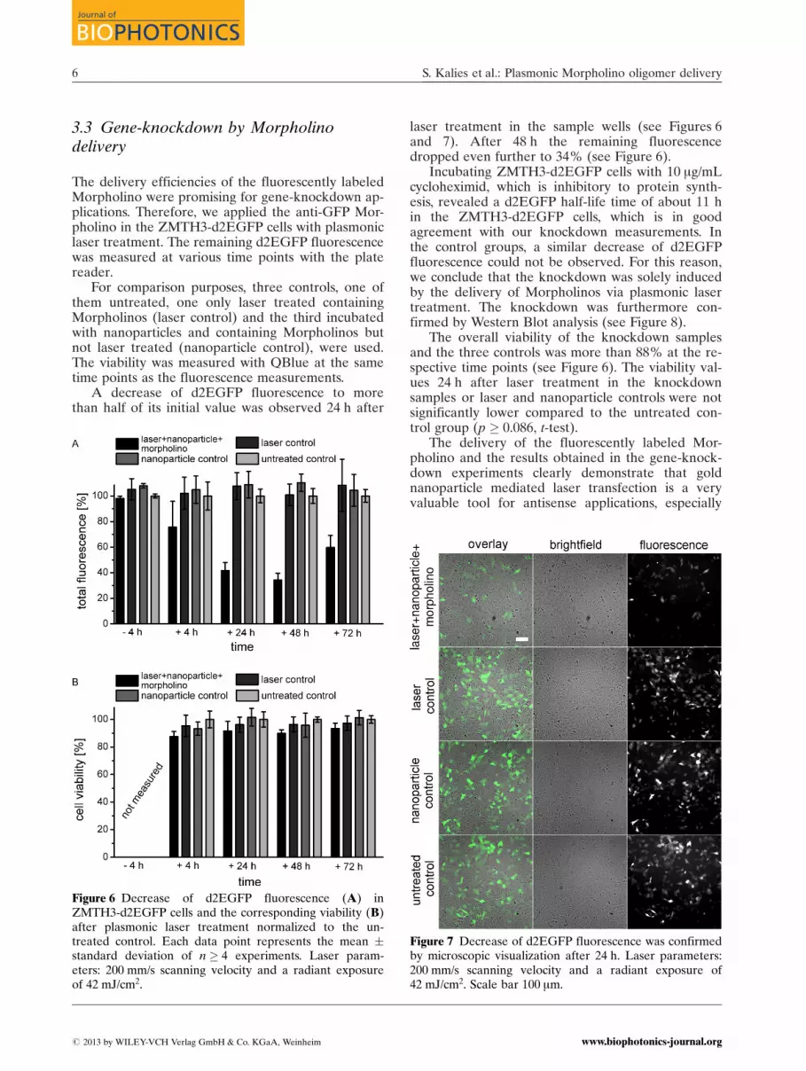
Figure 7 Decrease of d2EGFP fluorescence was confirmedby microscopic visualization after 24 h. Laser parameters:200 mm/s scanning velocity and a radiant exposure of42 mJ/cm2. Scale bar 100 mm.
S. Kalies et al.: Plasmonic Morpholino oligomer delivery6
Journal of
BIOPHOTONICS
# 2013 by WILEY-VCH Verlag GmbH & Co. KGaA, Weinheim www.biophotonics-journal.org

Morpholino injection. High transfection rates andsuccessful gene-knockdown can possibly be reachedbecause of the small size of antisense oligonucleo-tides (~10 kDa). Moreover, it can be assumed, thatas a physical method gold nanoparticle mediated la-ser transfection bears the main advantage of beingapplicable to a large variety of cell types. Addition-ally, the herein described approach enables thetransfection of cells with spatial selectivity by utiliz-ing conjugated gold nanoparticles or by the exposureof a defined cell monolayer region.
3.4 ROS production during plasmoniclaser treatment
The conditions used for molecular delivery by plas-monic laser treatment might evoke the creation ofreactive oxygen species (ROS) in the particle’s nearfield [23]. These could contribute to the perforationeffect, but also impact cell viability [20, 21]. There-fore, we analyzed the creation of ROS during plas-monic laser treatment using a CM-H2DCFDA basedassay.
Control groups showed that the assay can reliablydetect the oxidative effect of H2O2 in concentrationsas low as 30 mM (see Figure 9). No increase in fluo-rescence compared to an untreated control could bemeasured in the control groups being solely treated
with gold nanoparticles or laser irradiation. This in-dicates, that no oxidative stress occurred by the usedlaser intensities and that the applied particles haveno pronounced oxidative properties on the cells. Asample treated with both, gold nanoparticles and la-ser irradiation, yielded a very low but significant in-crease in the measured fluorescence level (p ¼ 0.026,Figure 9). This implies, that ROS are created in theenhanced near field around irradiated gold particles.These can enter the cell in low concentrations, pro-mote membrane perforation or impact cell viability.A study involving the threshold dependence of reac-tive oxygen species formation comparing differentlaser setups, in particular off-resonance plasmonicenhanced laser femtosecond transfection [19] andthe approach used herein, would be very valuablefor the future.
3.5 Viability measurements
As described above, only low acute impairment ofthe cell viability by plasmonic laser treatment wasobserved 1 h after laser treatment (see Figures 2–6).However, a transition of this technique to more so-phisticated or in vivo applications needs more de-tailed investigations on toxic effects. The lactate de-hydrogenase (LDH) activity assay is a standardmethod to determine the membrane integrity. No in-crease of extracellular LDH activity was determineddirectly after plasmonic laser treatment (Figure 10).In contrast, 2 h post manipulation, a significant ele-vated LDH level was detected (p < 0.01, Figure 10).
Figure 8 Western blot of the Morpholino induced knock-down of d2EGFP after plasmonic laser treatment. Semiquantitative analysis of the band intensities revealed aknockdown efficiency of about 60% compared to the un-treated control. Laser parameters: 200 mm/s scanning velo-city and a radiant exposure of 42 mJ/cm2.
Figure 9 Generation of ROS during plasmonic laser treat-ment and by H2O2. A slight increase of fluorescence wasobserved in the sample group, but not for the laser or goldnanoparticle controls. Sensitivity of the assay was con-firmed by incubation of cells with H2O2 as positive control.Each data point represents the mean � standard deviationof n ¼ 18 experiments. Laser parameters: 200 mm/s scan-ning velocity and a radiant exposure of 42 mJ/cm2.
J. Biophotonics (2013) 7
FULLFULLARTICLEARTICLE
# 2013 by WILEY-VCH Verlag GmbH & Co. KGaA, Weinheimwww.biophotonics-journal.org

This is connected to lysis of necrotic cells. The ob-served toxicity of 12% is in good agreement with theabove measured viability of about 90% 1 h after la-ser treatment (see Figures 4 and 6b).
In order to investigate the effect of plasmonic la-ser treatment on an expanded time span, we per-formed a clonogenic assay [26]. This assay followscell survival and proliferation ability over 10 d, giv-ing indications on subsequent impairment of the cellviability. Table 1 summarizes the colony count ofsample and control groups. No significant differencebetween the groups was observed (p > 0.073), indi-cating that plasmonic laser treatment has no lastingeffect on the cell proliferation.
4. Conclusion
In this study, we demonstrated the efficient deliveryof fluorescently labeled and biologically functional
Morpholino oligomers by plasmonic laser treatment.This represents a highly relevant application withinbasic and applied science. Moreover, we assessed indetail fundamental points regarding the toxic impactof this method and related applications of laser ex-cited gold nanoparticles. For the first time, cell survi-val and proliferation capacity over a longer time per-iod were investigated. Only minor acute toxic effectscould be observed, probably related to the formationof reactive oxygen species, whereas no long term im-pairment was found. To conclude, we provide impor-tant insights and understanding of plasmonic lasertreatment with high impact on further research andapplications.
Acknowledgements This work is supported by fundingfrom the Deutsche Forschungsgemeinschaft (DFG, Ger-man Research Foundation) within the Cluster of Excel-lence “REBIRTH” (From Regenerative Biology to Recon-structive Therapy). We highly appreciated technical helpby A. Deiwick, Dr. S. Schlie-Wolter and Dr. L. Koch (La-ser Zentrum Hannover e.V.) and helpful discussion withS. Willenbrock (University of Veterinary Medicine Hann-over) and T. Bohnenpoll (Hannover Medical School).
Author biographies Please see Supporting Informationonline.
References
[1] C. Mason and P. Dunnill, Regen Med. 3(1), 1–5(2008).
[2] A. S. Correia, S. V. Anisimov, J.-Y. Li, and P. BrundinAnnals of Medicine 37(7), 487–498 (2005).
[3] M. Cavazzana-Calvo, S. Hacein-Bey, G. de Saint Ba-sile, F. Gross, E. Yvon, P. Nusbaum, F. Selz, C. Hue,S. Certain, J.-L. Casanova, P. Bousso, F. Le Deist, andA. Fischer, Science 288(5466), 669–672 (2000).
[4] K. Gavrilov and W. M. Saltzman, Yale J Biol Med.85(2), 187–200 (2012).
[5] S. Q. Harper, P. D. Staber, X. He, S. L. Eliason, I. H.Martins, Q. Mao, L. Yang, R. M. Kotin, H. L. Paulsonand B. L. Davidson, Proc Natl Acad Sci 102, 5820–5825 (2005).
[6] P. D. Staber, C. Vargeese, I. H. Martins, B. Polisky,A. Mas Monteys, S. Q. Harper, and B. L. Davidson,Mol Ther 13, 36–S37 (2006).
[7] Y. Yao, C. Wang, P. R. Varshney, and D. A. Wang,Pharm Res. 26(2), 263–275 (2009).
[8] J. Summerton and D. Weller, Antisense Nucleic AcidDrug Dev 7, 187–195 (1997).
[9] J. Summerton, Med Chem. 7(7), 651–660 (2007).[10] A. S. Flynt, N. Li, E. J. Thatcher, L. Solnica-Krezel,
and J. G. Patton, Nature Genetics 39, 259–263 (2007).[11] J. Heasman, Dev. Biol. 243(2), 209–214 (2002).[12] G. Mc. Clorey, A. M. Fall, H. M. Moulton, P. L.
Iversen, J. E. Rasko, M. Ryan, S. Fletcher, and S. D.
Figure 10 Extracellular LDH activity after plasmonic lasertreatment. No increased activity was observed directlyafter the treatment, whereas elevated LDH activity wasmeasured 2 h later. Each data point represents the mean� standard deviation of n ¼ 9 experiments. Laser param-eters: 200 mm/s and 42 mJ/cm2.
Table 1 Clonogenic assay: Colony count after 10 d with atotal number of 500 seeded cells. Values represent themean � standard deviation of n ¼ 10 experiments. No sig-nificant differences were found. Laser parameters:200 mm/s and 42 mJ/cm2.
group number ofcolonies
standarddeviation
laser þ nanoparticle 98.4 28.2laser control 105.9 20.2nanoparticle control 110.0 26.5untreated control 120.2 22.7
S. Kalies et al.: Plasmonic Morpholino oligomer delivery8
Journal of
BIOPHOTONICS
# 2013 by WILEY-VCH Verlag GmbH & Co. KGaA, Weinheim www.biophotonics-journal.org

Wilton, Neuromuscul Disord. 16(9–10), 583–590(2006).
[13] B. L. Geller, Curr. Opin. Mol. Ther. 7(2), 109–113(2006).
[14] T. K. Kim and J. H. Eberwine, Anal Bioanal Chem397, 3173–3178 (2010).
[15] M. Tsukakoshi, S. Kurata, Y. Nomiya, Y. Ikawa, andT. Kasuya, Appl. Phys. B 35, 135–140 (1984).
[16] U. K. Tirlapur and K. Koenig, Nature 418, 290–291(2002).
[17] D. J. Stevenson, F. J. Gunn-Moore, P. Campbell, andK. Dholakia, Transfection by optical injection, in:Handbook of Photonics for Medical Science, V. V. Tu-chin (ed.) (Taylor and Francis Group, 2010).
[18] M. Schomaker, J. Baumgart, A. Ngezahayo, J. Buller-diek, I. Nolte, H. M. Escobar, H. Lubatschowski, andA. Heisterkamp, Proc. SPIE Int. Soc. Opt. Eng. 7192(2009).
[19] J. Baumgart, L. Humbert, E. Boulais, R. Lachaine,J.-J. Lebrun, and M. L. Meunier, Biomaterials 33,2345–2350 (2012).
[20] D. Heinemann, M. Schomaker, S. Kalies, M. Schieck,R. Carlson, H. Murua Escobar, T. Ripken, H. Meyer,and A. Heisterkamp, PLOS ONE 8(3), e58604 (2013).
[21] S. Kalies, T. Birr, D. Heinemann, M. Schomaker,A. Heisterkamp, T. Ripken, and H. Meyer, J Biopho-tonics, DOI: 10.1002/jbio.201200200 (2013).
[22] E. Boulais, R. Lachaine, and M. Meunier, Nano Lett,12(9), 4763–4769 (2012).
[23] A. Vogel, J. Noack, G. Huettman, and G. Paltauf,Appl. Phys. B 81, 1015–1047 (2005).
[24] B. P. Yu, Cellular defenses against damage from reac-tive oxygen species, Physiol Rev. 74(1), 139–62 (1994).
[25] B. Boudaiffa, P. Cloutier, D. Hunting, M. A. Huels,and L. Sanche, Science 287, 1658–1660 (2000).
[26] N. A. P. Franken, H. M. Rodermond, J. Stap, J. Have-man, and C. van Bree, Nat Protoc 1, 2315–2319(2006).
[27] H. Murua Escobar, B. Meyer, A. Richter, K. Becker,A. Flohr, J. Bullerdiek, and I. Nolte, Cytogenet Gen-ome Res 101, 33–38 (2003).
[28] X. Li, X. Zhao, Y. Fang, X. Jiang, T. Duong, C. Fan,C. C. Huang, and S. R. Kain, J Biol Chem. 273(52),34970–34975 (1998).
[29] W. O. Carter, P. K. Narayanan, and J. P. Robinson, JLeukoc Biol. 55(2), 253–258 (1994).
[30] A. E. Carpenter, T. R. Jones, M. R. Lamprecht,C. Clarke, I. H. Kang, O. Friman, D. A. Guertin,J. H. Chang, R. A. Lindquist, J. Moffat, P. Golland,and D. M. Sabatini, CellProfiler: image analysis soft-ware for identifying and quantifying cell phenotypes.Genome Biology 7, 7(10): R100 (2006).
[31] E. Y. Lukianova-Hleb and D. O. Lapotko, Nano Lett.9, 2160–2166 (2009).
[32] E. Y. Lukianova-Hleb, Y. Hu, L. Latterini, L. Tarpani,S. Lee, R. A. Drezek, J. H. Hafner, and D. O. Lapot-ko, ACS Nano 4, 2109–2123 (2010).
[33] B. J. Messinger, K. U. Raben, R. K. Chang, and P. W.Barber, Phys. Rev. B 24, 649–657 (1981).
[34] E. C. Cho, Q. Zhang, and Y. Xia, Nat Nanotechnol 6,385–391 (2011)
J. Biophotonics (2013) 9
FULLFULLARTICLEARTICLE
# 2013 by WILEY-VCH Verlag GmbH & Co. KGaA, Weinheimwww.biophotonics-journal.org


