Embed Size (px)
Citation preview

Molecular Biology of the CellVol. 13, 3696–3705, October 2002
An Endogenous RNA Transcript Antisense to CNG�1Cation Channel mRNAChin-Hung Cheng,* David Tai-Wai Yew,† Hiu-Yee Kwan,* Qing Zhou,* YuHuang,* Yong Liu,‡ Wing-Yee Chan,‡ Xiaoqiang Yao*§
*Departments of Physiology, †Anatomy, and ‡Anatomical and Cellular Pathology, Faculty ofMedicine, The Chinese University of Hong Kong, Hong Kong, China
Submitted March 6, 2002; Revised June 27, 2002; Accepted July 18, 2002Monitoring Editor: Keith R. Yamamoto
CNG channels are cyclic nucleotide-gated Ca2�-permeable channels that are suggested to beinvolved in the activity-dependent alterations of synaptic strength that are thought to underlieinformation storage in the CNS. In this study, we isolated an endogenous RNA transcriptantisense to CNG�1 mRNA. This transcript was capable of down-regulating the expression ofsense CNG�1 in the Xenopus oocyte expression system. RT-PCR, Northern blot, and in situhybridization analyses showed that the transcript was coexpressed with CNG�1 mRNA in manyregions of human brain, notably in those regions that were involved in long-term potentiation andlong-term depression, such as hippocampal CA1 and CA3, dentate gyrus, and cerebellar Purkinjelayer. Comparison of expression patterns between adult and fetal cerebral cortex revealed thatthere were concurrent developmental changes in the expression levels of anti-CNG1 and CNG�1.Treatment of human glioma cell T98 with thyroid hormone T3 caused a significant increase inanti-CNG1 expression and a parallel decrease in sense CNG�1 expression. These data suggest thatthe suppression of CNG�1 expression by anti-CNG1 may play an important role in neuronalfunctions, especially in synaptic plasticity and cortical development. Endogenous antisense RNA-mediated regulation may represent a new mechanism through which the activity of ion channelscan be regulated in the human CNS.
INTRODUCTION
Cyclic nucleotide-gated (CNG) channels are Ca2�-perme-able nonselective cation channels that exist as heteromericcomplexes consisting of � and � subunits. Four distinct �subunits and two � subunits have been identified. �1–3 mayform functional channels on their own. �4 and two � sub-units do not exhibit channel properties themselves but areable to modify the channel properties (Kaupp, 1995; Finn etal., 1996; Zagotta and Siegelbaum, 1996). CNG channels havefairly widespread tissue distribution, including sensory neu-rons, CNS, heart, kidney, blood vessels, and spleen. In theCNS, electrophysiological and molecular biological evidencehas demonstrated that CNG channels are present in manyregions of rat brain, including hippocampus, cerebral cortex,and cerebellum (Ahmad et al., 1994; Kingston et al., 1996;Bradley et al., 1997; Samanta Roy and Barnstable, 1999; Stri-jbos et al., 1999). Research has linked the nitric oxide (NO)–cGMP pathway to the CNG channel activity in neurons
(Ahmad et al., 1994). CNG channels may act as one of thedownstream effectors for the NO–cGMP pathway, modulat-ing neurotransmitter release and causing the activity-depen-dent alterations of synaptic strength that are thought tounderlie information storage, such as long-term potentiation(LTP) and long-term depression (LTD). In addition to theirproposed role in synaptic plasticity, CNG channels may alsoplay an important role in brain development. Evidence sug-gests that CNG channels may control axon guidance(Coburn and Bargmann, 1996) and influence cortical den-dritic outgrowth in the development of the CNS (SamantaRoy and Barnstable, 1999).
An important feature of CNG channels is their Ca2�-permeability (Finn et al., 1996; Dzeja et al., 1999). CNG chan-nels open in response to cyclic nucleotides and link cGMP/cAMP signaling to Ca2� homeostasis. Activation of CNGchannels would raise cytosolic Ca2� levels, and this couldtrigger secondary pathways that contribute to short-termand long-term alterations in neural functions (Zufall et al.,1997). Substantial amounts of data are available regardingthe modulation of CNG channel activity by cellular factors,including phosphorylation enzymes, Ca2�/calmodulin, anddiacylglycerol (Hsu and Molday, 1993; Molokanova et al.,1997; Crary et al., 2000). However, the regulation of CNG
Article published online ahead of print. Mol. Biol. Cell 10.1091/mbc.E02–03–0127. Article and publication date are at www.molbi-olcell.org/cgi/doi/10.1091/mbc.E02–03–0127.
§ Corresponding author. E-mail address: [email protected].
3696 © 2002 by The American Society for Cell Biology

channels at the gene transcription and/or protein translationlevels is largely unknown. In this study, we report theisolation of a cDNA clone representing an endogenous an-tisense transcript against the mRNA of human CNG�1 chan-nels. This endogenous antisense transcript is expressed inmany regions of human brain, and it may down-regulate thesense CNG�1 channels by suppressing the amount of chan-nel proteins.
MATERIALS AND METHODS
Library ScreeningA cDNA fragment for CNG�1 (underlined by dots in Figure 1) wasamplified by RT-PCR using total RNA isolated from the humanepithelial cell line ECV304. The primers used for PCR were (�)TT-GGTCCACAGGTAGTC and (�) TCATCATTATCCACTGGAA.The fragment was labeled by random primer extension with �-32P-labeled dCTP (Amersham), and it was then used to screen a com-mercial human cDNA library that was primed with oligo-dT andunidirectionally cloned in pCMV-SPORT1 (Life Technologies-BRL).The hybridizations were performed on Hybond nylon membranesin Rapid-Hyb buffer (Amersham) at 55°C overnight. The mem-branes were washed in 2� SSC/0.1% SDS at room temperature for1 h, followed by 0.2� SSC/0.1% SDS at 42°C for 45 min as describedpreviously (Yao et al., 1995). Positive clones were verified withSouthern blot using the same CNG�1-specific probe. A 1.66-kilobase
(kb) clone was isolated, and the nucleotide sequence was analyzedby Sanger sequencing with Sequenase (US Biochemicals).
Reverse Transcription–Polymerase Chain ReactionTotal RNAs were isolated at autopsy from adult human brain tissueand human glioma cell lines by the acid guanidinium thiocyanatemethod (Chomczynski and Sacchi, 1987). One of the human braintotal RNA samples was a DNase-treated sample purchased fromInvitrogen. RNAs were transcribed into first-strand cDNAs usingSuperscript reverse transcriptase (Life Technologies-BRL). Anti-CNG1–specific primers [(�)GATGACGATATACATAACAAGGand (�)CTCAGCAGAATATTTTCTACAGCC] were used to am-plify a 450-base pair (bp) PCR fragment. The primer sites are shownin Figure 1. PCR reactions of 100 �l contained 1 �l of the first-strandcDNA, 20 mM Tris-HCl (pH 8.4), 50 mM KCl, 1.5 mM MgCl2, 0.2mM dNTP, 1.0 �M primers, and 2.5 U Taq DNA polymerase (LifeTechnologies-BRL). Thirty cycles (94°C for 1 min, 50°C for 1 min,72°C for 1 min) were performed with a Robocycler (Stratagene).Negative controls applied the same samples and experimental pro-cedures except that the step of reverse transcription was omitted.The amplified PCR products were sequenced by ABI 310 autose-quencer (Perkin Elmer-Cetus). Human kidney and liver cDNA li-braries were purchased from Life Technologies-BRL.
Hormone EffectT98 cells were treated with 1 �M T3 (Calbiochem) or 100 ng/mlhuman growth hormone (Calbiochem) for 4 d in a culture medium
Figure 1. Nucleotide sequence of human anti-CNG1 RNA transcript. mRNA sequence is deduced from the DNA sequence of a cDNA cloneisolated from adult human brain cDNA library. The probe used for library screening was derived from the cDNA fragment underlined bydots. Amino acid sequence of a putative translation product is illustrated. Bold, the 1311-bp overlap between the sense and antisensetranscripts; double underlines, potential poly(A) signals; thick underlines, primer sites for RT-PCR of Figure 3A.
A Natural Antisense RNA against CNG�1
Vol. 13, October 2002 3697

consisting of 90% Ham’s F-12 and 10% fetal calf serum (Life Tech-nologies). Total RNAs were isolated thereafter for semiquantitativeRT-PCR assays. The primers used for anti-CNG1 detection were(�)GGTATCAGTGACAGAACATCAA and (�)TACAGCCATAG-GTTTATTAGTAT. The primers used for sense CNG�1 detectionwere (�)GAATTTGGCCGTTTGGCTAG and (�)CGTTGATG-GCAATTTCTGCT. PCR reactions of 50 �l contained 1 �l of thefirst-strand cDNA, 20 mM Tris-HCl (pH 8.4), 50 mM KCl, 1.5 mMMgCl2, 0.2 mM dNTP, 1.0 �M primers, and 2.5 U Taq DNA poly-merase (Life Technologies-BRL). Thirty-five cycles (94°C for 1 min,55°C for 1 min, 72°C for 1 min) were performed with a Robocycler(Stratagene). For anti-CNG1, a single PCR product of 271 bp wasamplified. For sense CNG�1, a single amplified product of 386 bpwas detected. The authenticity of the amplified products was con-firmed by ABI 310 autosequencer. Equal volumes of the PCR prod-ucts were loaded onto a 1%-agarose gel and stained with ethidiumbromide. The intensity of the bands was analyzed by FluorChem8000 imaging system (Alpha Innotech). As a control for analysis, weused the expression levels of glyceraldehyde-3-phosphate dehydro-genase (GAPDH) as a normalizing basis for comparison. The prim-ers for GAPDH were (�)ACCACAGTCCATGCCATCAC and(�)TCCACCACCCTGTTGCTGTA.
Northern BlotA 211-bp DNA fragment near the 3� end of anti-CNG1 (from posi-tion 1308 to 1518 as in Figure 1, or P2 as in Figure 2) was subclonedinto pCMVsport1 and then in vitro transcribed into a 32P-labeledriboprobe antisense to anti-CNG1 with the T7 MAXIscript transcrip-tion kit (Ambion). The molecular size of synthesized riboprobe wasconfirmed by gel electrophoresis. This strand-specific riboprobe wasused to hybridize with a Human Brain Multiple Tissue mRNA Blot(ClonTech) at 60°C overnight with ExpressHyb. The blot was thenwashed twice in 2� SSC/0.5% SDS for 45 min, followed by 2�
20-min washes in 0.1� SSC/0.1% SDS at room temperature, andthen exposed to x-ray film overnight. The Multiple Tissue mRNABlot was reprobed with human �-actin gene (Clontech) to demon-strate that equal amounts of poly(A) RNA was loaded onto eachlane.
Cloning of Sense CNG�1 GeneSense CNG�1 was amplified by RT-PCR using total RNA isolatedfrom the human epithelial cell line ECV304. The PCR primers[(�)TCCATGAACAATATTATCAAT and (�)TCAAAAGGAT-CATGAGGCAT] were designed on the basis of the published nu-cleotide sequence of human CNG�1 mRNA (GenBank AccessionNumber NM 000087) (Dhallan et al., 1992). The amplified PCRproduct of 2112 bp was cloned into pPCR-ScriptAmp cloning vector(Stratagene). The amplified PCR products were sequenced by ABI310 autosequencer (Perkin Elmer-Cetus). The DNA sequencing con-firmed that the clone represented authentic human CNG�1.
Oocyte ExpressionAnti-CNG1, CNG�1, and Kv1.5 were subcloned into pgh21 vectorand then expressed in Xenopus oocytes by microinjecting in vitrotranscribed cRNAs as previously described (Yao et al., 1995). ForCNG�1, oocyte membrane was clamped at �100 mV. For anti-CNG1 suppression studies, 25 ng of anti-CNG1 cRNA was injectedinto oocytes 2 h before the injection of sense CNG�1 cRNA. ForKv1.5, the outward currents were elicited by two-microelectrodevoltage clamp using 800-ms pulses of �80 mV from a holdingpotential of �80 mV. The experimental bath contained 88 mM NaCl,2 mM KCl, 1 mM CaCl2, 1 mM MgCl2, 2.5 mM NaH2CO3, and 5 mMHEPES, pH 7.4. Expressed currents were measured with OC-725oocyte clamp 2 days after cRNA injection as described previously(Yao et al., 1995). Measured currents were analyzed using Pulse andPulse-fit software (Heka Lambretch, Germany).
In Situ HybridizationThe same 211-bp DNA fragment as used in Northern blot (illustrat-ed as P2 in Figure 2) was in vitro transcribed into DIG-labeled RNAprobes with a DIG-labeling kit (Roche Biochemicals). The strandcomplementary to anti-CNG1 was used to detect anti-CNG1, andthe sense strand was used as control. For the detection of senseCNG�1 transcript, a 259-bp DNA fragment illustrated in Figure 2 asP1 was subcloned into pPCR-ScriptAmp cloning vector (Stratagene)and then in vitro transcribed into DIG-labeled riboprobes. Thestrand complementary to CNG�1 was used to detect CNG�1mRNA, and the sense strand was used as control. These probeswere used to hybridize with the sections cut from the brain tissuesembedded in paraffin. The brain tissues of human adult and 15-wk-old fetus were from autopsy cases with the consent of family mem-bers and the approval of the university clinical research ethicscommittee. Tissues were fixed overnight with 4% paraformalde-hyde in phosphate-buffered saline (PBS). The postmortem delaywas �7 h. The tissues were dehydrated through graded ethanol,cleared with xylene, and embedded in Parafilm, and 6-�m-thicksections were prepared. After dewaxing and hydration, the sectionswere washed briefly with diethylpyrocarbonate-treated water fol-lowed by PBS for 10 min. They were then digested with ProteinaseK (10 �g/ml) at 37°C for 15 min. Hybridization was performed at48°C in a hybridization buffer containing 4� SSC, 10% dextransulfate, 1� Denhardt’s solution, 5 mM EDTA, 0.1% CHAPS, 50%deionized formamide, 200 �l/ml herring sperm DNA, and 200ng/ml DIG-labeled probe (Yew et al., 1999). The slides were thenwashed four times for 15 min each in 2� SSC/0.1% SDS and thentwice for 15 min each in 0.2� SSC/0.1% SDS at 42°C. Colorimetricdetections were performed using an anti-DIG antibody conjugatedto alkaline phosphatase followed by incubation with NBT/BCIPcolor substrates using a digoxygenin-nucleic acid detection kit
Figure 2. Gene structure of anti-CNG1. Top, schematic illustrationof the nucleotide complementarity of anti-CNG1 to sense CNG�1.Black bar, sense CNG�1; gray bar, region of anti-CNG1 complemen-tary to sense CNG�1; hatched bar, region of anti-CNG1 lackingcomplementarity to sense CNG�1. P1, the region in which theCNG�1-specific riboprobe for in situ hybridization (Figures 5 and 6)was derived; P2, the region in which anti-CNG1–specific riboprobefor Northern blot (Figure 3B) and in situ hybridization (Figures 5and 6) was derived. Bottom, intron-exon structure of CNG�1 chan-nel gene. Black bars, coding regions; white bars, 5� and 3� untrans-lated regions; lines, introns. Anti-CNG1 is transcribed from theregion of intron 9 and exon 10 of CNG�1 channel gene but inreverse orientation.
C.-H. Cheng et al.
Molecular Biology of the Cell3698

(Roche, Germany) as described previously (Yew et al., 1999). Foranti-CNG1, positive signals appeared �5 min after incubation inNBT/BCIP color substrate solution. For the detection of senseCNG�1, �30 min was needed for the color development in NBT/BCIP color substrate solution.
RESULTS
A CNG�1-related cDNA clone was isolated by screening anadult human brain cDNA library with a DNA probe specificfor CNG�1. Nucleotide sequence of the isolated cDNA clonewas analyzed by Sanger’s sequencing. Because the librarywas a commercial cDNA library that was primed witholigo-dT and unidirectionally cloned in pCMVsport1, wewere able to determine the 5�-3� orientation of the clone onthe basis of the location of the 18-base oligo(A) (Figure 1).Comparison of nucleotide sequence of this clone with that ofhuman CNG�1 mRNA (GenBank Accession NumberNM 000087) revealed that a 1311-bp region at the 5� por-tion of this clone was complementary to CNG�1 mRNA,whereas the rest of the clone (345 bp at the 3� end) had nosimilarity to CNG�1 mRNA (Figure 2). Therefore, this tran-script represented a natural antisense transcript complemen-tary to CNG�1 mRNA. We named this transcript anti-CNG1. Comparison of the nucleotide sequence of anti-CNG1 with that of human chromosome 4 showed that the345 bp at the 3� end of the clone was actually transcribedfrom an immediate downstream region following theCNG�1-coding 1311-bp upstream region in chromosome 4between 4p12 and the centromere (Figure 2). It should benoted that anti-CNG1 and CNG�1 were transcribed in thesame locus but in reverse orientation. On the basis of thepublished intron-exon structure of human CNG�1 gene(Dhallan et al., 1992), the transcription of anti-CNG1 startedfrom exon 10 of CNG�1 and extended into intron 9. Theisolated clone might contain a genuine 3� end, because oli-go(A) was located at the end and multiple AAUAAA motifs,the signal for processing of mRNA at the 3� end (Nevins,1983), could be found near the A-rich region. It was not clearwhether this transcript might encode any protein, but the
longest possible open reading frame was only 243 bp long,which is equivalent to 81 amino acids. The putative aminoacid sequence from this open reading frame is illustrated inFigure 1. Blast GenBank search with this putative proteinshowed no significant similarity to any known protein.
A suspicion could be raised that the isolated clone mightrepresent a cloning artifact caused by a piece of cDNAinserted in the wrong direction. But this was unlikely, be-cause a detailed sequence analysis of the clone showed thatthe 3� end of the clones contained an intact oligo(dT) thatwas flanked by a NotI restriction site specially designed forunidirectional cloning, and this NotI restriction site wasfollowed immediately by a pCMV-SPORT1 vector sequence.In addition, the 5� end of the clone contained a completeEcoRI adaptor specially designed for unidirectional cloning,and this EcoRI site was followed immediately by a pCMV-SPORT1 vector sequence. The correct insertion pattern ar-gued against false insertion. The clone was certainly not aresult of the false fusion of cDNA for CNG1 with anothercDNA encoding different protein, because the analyses ofgenomic DNA showed that the cDNA was continuouslytranscribed from a single locus (Figure 2).
RT-PCR was used to examine the expression pattern ofthis endogenous anti-CNG1 transcript. PCR primers werecarefully designed so that the targeted 450-bp amplificationproducts extended across the boundary of intron 9 and exon10 of CNG�1 (Figure 1). In this way, only anti-CNG1 but notCNG�1 could be amplified. RT-PCR experiments revealedthe expression of anti-CNG1 in adult human brain and twohuman glioma cell lines, T98 and D247 (Figure 3A). Noexpression could be detected in the total RNA samples iso-lated from human adult stomach tissue and rat pheochro-mocytoma cell line PC12 (data not shown). DNA sequencingconfirmed that the amplified PCR products represented theauthentic anti-CNG1. The amplified anti-CNG1 did not orig-inate from the residual genomic DNA contaminations intotal RNA samples, because PCR reactions alone withoutreverse transcription did not produce any detectable prod-uct (Figure 3A). Furthermore, the same anti-CNG1 productof �450 bp could be amplified by RT-PCR from a commer-
Figure 3. Detection of anti-CNG1transcripts by RT-PCR and Northernblot. (A) RT-PCR detected the ex-pression of anti-CNG1 in total RNAsprepared from brain tissue, gliomacell lines D247 and T98, and a com-mercial DNase-treated human braintotal RNA (Invitrogen). Reverse tran-scription step was omitted in nega-tive controls. (B) Multiple TissueNorthern blot analysis detected threeanti-CNG1–related transcripts withmolecular size of 5.5, 3.5, and 1.7 kbin many human brain regions. Eachlane contained 2 �g poly(A) RNA.The probe for Northern blot was astrand-specific riboprobe derivedfrom P2 region as labeled in Figure 2.The blot was reprobed with human�-actin gene to demonstrate thatequal amounts of poly(A) RNA wereloaded onto each lane.
A Natural Antisense RNA against CNG�1
Vol. 13, October 2002 3699

cially available DNase-treated total RNA sample (Figure3A). In addition to brain, anti-CNG1 transcript was alsoexpressed in kidney and liver, because the same 450-bpproduct could be amplified by PCR from commerciallyavailable human cDNA libraries generated from kidney andliver (data not shown).
Northern blot analysis was used to further examine theexpression of anti-CNG1 in brain. A human Multiple TissuemRNA Blot was hybridized with a 32P-labeled riboprobespecific for anti-CNG1. Three different transcripts with mo-lecular sizes of 5.5, 3.5, and 1.7 kb were detected in poly(A)RNAs from different brain regions (Figure 3B). The 1.7-kbtranscript agreed well with the size of anti-CNG1 we iso-lated, and this transcript was expressed in amygdala, cau-date nucleus, hippocampus, and thalamus. Weak hybridiza-tion signals could also be observed in substantia nigra andwhole brain tissue. Large transcripts of 5.5 and 3.5 kb wereexpressed more abundantly in amygdala, caudate nucleus,and hippocampus. These large transcripts might representalternatively spliced forms of anti-CNG1. It should be notedthat the 211-bp riboprobe used for hybridization was de-rived from a region close to the 3� end of anti-CNG1 tran-script (Figure 2). This probe was highly specific for anti-CNG1, because BLAST GenBank search with anti-CNG1 didnot reveal significant similarity to any known gene. A pointworth mentioning was that the riboprobe was strand-spe-cific. It could recognize only the transcripts containing anti-CNG1 but not those containing the complementary strand.
To examine whether the isolated anti-CNG1 was capableof down-regulating sense CNG�1, in vitro transcribed anti-CNG1 cRNA was microinjected into Xenopus oocytes todetermine whether it could suppress the expression of senseCNG�1 currents. We used membrane-permeable 8-Br-
cGMP to activate sense CNG�1 channels in the oocytesinjected with CNG�1 cRNA. In Xenopus oocytes injectedwith 5 ng of CNG�1 cRNA, activation of CNG�1 channelsby 8-Br-cGMP initiated inward currents (Figure 4, A and B).Conversely, 8-Br-cGMP had no effect in the control oocytes,which did not receive the injection of CNG�1 cRNA. Impor-tantly, cGMP-activated inward currents in CNG�1-injectedoocytes were abolished if the oocytes were preinjected with25 ng anti-CNG1 cRNA 2 h before the injection of CNG�1(Figure 4, A and B). This suppression was not caused by anynonspecific effect associated with the injection of anti-CNG1transcript, because in control experiments, the expression ofa voltage-gated potassium channel, Kv1.5, was not affectedby anti-CNG1 transcript (Figure 4, C and D).
In situ hybridizations were performed to examine theexpression of anti-CNG1 and sense CNG�1 mRNA in dif-ferent human brain regions. DIG-labeled anti-CNG1 ribo-probe for in situ hybridization contained the same anti-CNG1–specific 211-base nucleotides as that used inNorthern blot. The 259-base riboprobe for CNG�1 was de-rived from a region of CNG�1 with no complementarity toanti-CNG1 (Figure 2). Our data showed that the expressionof anti-CNG1 was widespread in adult human CNS. Thisexpression could be observed in pyramidal neurons of hip-pocampal CA1 and CA3 of Ammon’s horn (Figure 5, A andC), granule neurons in dentate gyrus (Figure 5, A and G),and Purkinje cells and granular cells in cerebellum (Figure 5,H and I). Similar expression patterns were observed forsense CNG�1 (Figure 5, D and F–M). Very little hybridiza-tion signal could be seen in the experiments with controlriboprobes (Figure 5, B, E, J, and N). It was notable thatvirtually every visible hippocampal pyramidal, dentategranule, and cerebellar Purkinje neuron was labeled with
Figure 4. The suppression ofCNG�1-mediated inward cur-rents by anti-CNG1 cRNA. (A)cGMP stimulated the Ca2�-de-pendent Cl� inward currents inXenopus oocytes that were in-jected with 5 ng of CNG�1cRNA. The currents were abol-ished by prior injection of 25 nganti-CNG1 cRNA. Each trace is arepresentative experiment. Mem-brane was clamped at �100 mV.(B) Average amplitude of cGMP-activated Cl� currents from dif-ferent experiments. (C) Oocytesinjected with 0.5 ng of Kv1.5cRNA expressed voltage-depen-dent outward currents. The Kv1.5currents were not affected byprior injection of 25 ng anti-CNG1 cRNA. (D) Average ampli-tude of Kv1.5 currents from dif-ferent experiments. *p�0.01compared with the water-in-jected control; **p�0.01 com-pared with the CNG�1 injectionalone.
C.-H. Cheng et al.
Molecular Biology of the Cell3700

both anti-CNG1 and sense CNG�1 probes. Two additionalsets of DIG-labeled riboprobes, one covering the region fromposition 1–667 and the other covering the region from po-sition 567 to 952 of anti-CNG1 (the positions were as labeledin Figure 1), were used for in situ hybridization. Becausethese two probes covered the regions in which both anti-CNG1 and sense CNG�1 were transcribed, as expected, wewere able to detect the hybridization signals with the probesgenerated from both sense and antisense strands (data notshown).
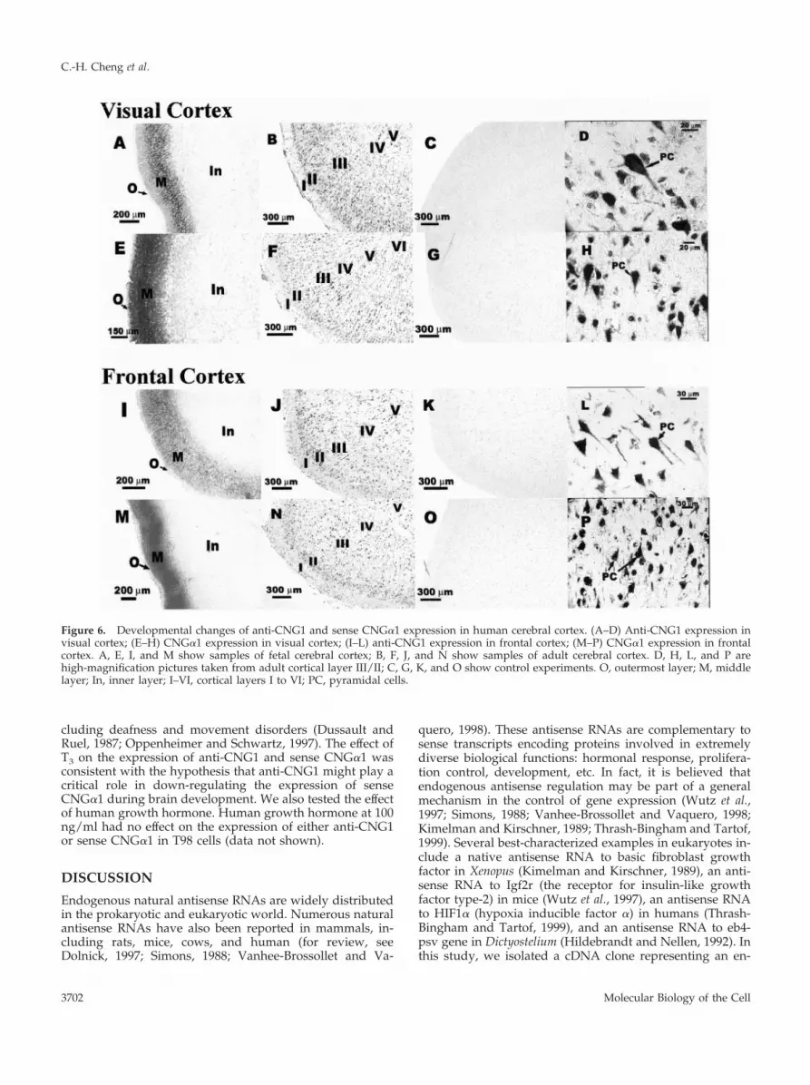
Because CNG channels were suggested to play a crucialrole in cortical development (Samanta Roy and Barnstable,1999), attempts were made to study the developmentalchanges of anti-CNG1 and sense CNG�1 in cerebral cortex.We found that in fetal cortex, the expression of anti-CNG1was observed predominantly in a cortical layer adjacent tothe outermost layer (Figure 6, A and I). Fetal cortex at 15 wkold was still not well differentiated and could not be dividedinto classic six-layered cortical structures. But on the basis ofthe anatomical location, these anti-CNG1–expressing neu-rons should correspond to the layer II and layer III cells ofthe adult cortex. Unlike fetal cortex, the expression of senseanti-CNG1 in adult cortex was more widespread and couldbe found in almost all cortical layers except layer I (Figure 6,
B and J). High-magnification pictures showed that anti-CNG1 transcript was expressed in the soma as well as in theprimary dendrite of pyramidal and granule neurons (Figure6, D and L). Very little stain was observed in cortical layer I,which contained primarily dendrites and axons of corticalneurons. Similar expression patterns were observed forsense CNG�1. The expression was located predominantly ina narrow layer in fetal cortex (Figure 6, E and M), but it waswidespread and existed in almost all cortical layers exceptlayer I in adult cortex (Figure 6, F and N). High-magnifica-tion pictures showed that both soma and primary dendriteof cortical neurons were stained (Figure 6, H and P). Nohybridization signal could be seen for the experiments withcontrol riboprobes (Figure 6, C, G, K, and O).
We also explored the possible endogenous factor(s) thatcould regulate the expression level of anti-CNG1. As shownin Figure 7, treatment of human glioma cell line T98 withthyroid hormone T3 (1 �M) caused a significant increase inthe expression level of anti-CNG1. The same treatment re-duced the expression of sense CNG�1. Thyroid hormone isa well-recognized agent involved in brain development andmaturation. The deficiency of thyroid hormone during crit-ical periods of development is associated with irreversiblemental retardation and profound neurological defects, in-
Figure 5. Detection of anti-CNG1 and sense CNG�1 transcripts in the hippocampus and cerebellum of adult human brain by in situhybridizations. Anti-CNG1 expression was observed in hippocampus (A, C, G) and cerebellum (H, I). B and J are the control experimentsfor anti-CNG1 in hippocampus and cerebellum, respectively. Sense CNG�1 expression was detected in hippocampus (D, F, K) andcerebellum (L, M). E and N are the control experiments for CNG�1 in hippocampus and cerebellum, respectively. (C and F) High-magnification pictures for hippocampal CA1 region; G and K, high-magnification pictures for dentate gyrus; I and M, high-magnificationpictures for cerebellar Purkinje/granular layer. DG, dentate gyrus; PC, pyramidal cells; GC, granule cells; WM, white matter; PJC, Purkinjecells.
A Natural Antisense RNA against CNG�1
Vol. 13, October 2002 3701
cluding deafness and movement disorders (Dussault andRuel, 1987; Oppenheimer and Schwartz, 1997). The effect ofT3 on the expression of anti-CNG1 and sense CNG�1 wasconsistent with the hypothesis that anti-CNG1 might play acritical role in down-regulating the expression of senseCNG�1 during brain development. We also tested the effectof human growth hormone. Human growth hormone at 100ng/ml had no effect on the expression of either anti-CNG1or sense CNG�1 in T98 cells (data not shown).
DISCUSSION
Endogenous natural antisense RNAs are widely distributedin the prokaryotic and eukaryotic world. Numerous naturalantisense RNAs have also been reported in mammals, in-cluding rats, mice, cows, and human (for review, seeDolnick, 1997; Simons, 1988; Vanhee-Brossollet and Va-
quero, 1998). These antisense RNAs are complementary tosense transcripts encoding proteins involved in extremelydiverse biological functions: hormonal response, prolifera-tion control, development, etc. In fact, it is believed thatendogenous antisense regulation may be part of a generalmechanism in the control of gene expression (Wutz et al.,1997; Simons, 1988; Vanhee-Brossollet and Vaquero, 1998;Kimelman and Kirschner, 1989; Thrash-Bingham and Tartof,1999). Several best-characterized examples in eukaryotes in-clude a native antisense RNA to basic fibroblast growthfactor in Xenopus (Kimelman and Kirschner, 1989), an anti-sense RNA to Igf2r (the receptor for insulin-like growthfactor type-2) in mice (Wutz et al., 1997), an antisense RNAto HIF1� (hypoxia inducible factor �) in humans (Thrash-Bingham and Tartof, 1999), and an antisense RNA to eb4-psv gene in Dictyostelium (Hildebrandt and Nellen, 1992). Inthis study, we isolated a cDNA clone representing an en-
Figure 6. Developmental changes of anti-CNG1 and sense CNG�1 expression in human cerebral cortex. (A–D) Anti-CNG1 expression invisual cortex; (E–H) CNG�1 expression in visual cortex; (I–L) anti-CNG1 expression in frontal cortex; (M–P) CNG�1 expression in frontalcortex. A, E, I, and M show samples of fetal cerebral cortex; B, F, J, and N show samples of adult cerebral cortex. D, H, L, and P arehigh-magnification pictures taken from adult cortical layer III/II; C, G, K, and O show control experiments. O, outermost layer; M, middlelayer; In, inner layer; I–VI, cortical layers I to VI; PC, pyramidal cells.
C.-H. Cheng et al.
Molecular Biology of the Cell3702

dogenous antisense transcript against the mRNA of humanCNG�1 channels. The presence of anti-CNG1 transcript(s)was verified by RT-PCR, Northern blot hybridization, and insitu hybridization. The expression of the transcript(s) couldbe found in many regions of human brain (Figures 3 and 5)as well as in human glioma cell lines T98 and D247 (Figure3A). Interestingly, the molecular size of the isolated anti-CNG1 cDNA clone matched that of one of the transcriptsdetected on the Northern blot (1.7 kb), suggesting that theisolated anti-CNG1 clone might represent a complete tran-script. This study represents the first demonstration of anendogenous antisense RNA transcript against any ion chan-nel.
A unique function of antisense transcripts is to regulatethe expression of their sense counterparts. It has been rec-ognized that antisense transcripts may hybridize with theircomplementary sense mRNAs to form RNA–RNA duplexes.
These duplexes can be digested by the RNases that arespecific for double-stranded RNA. Alternatively, because ofthe structural similarity between the sense and antisensetranscripts, antisense transcripts may down-regulate thesense gene by depriving sense mRNAs from the proteinsnecessary for their functions (Vanhee-Brossollet and Va-quero, 1998). To test whether the isolated 1.66-kb anti-CNG1transcript was capable of down-regulating the expression ofsense CNG�1, we used the Xenopus oocyte expression sys-tem. Xenopus oocytes have endogenous Ca2�-dependent Cl�channels that are sensitive to the Ca2� concentration beneaththe plasma membrane. Therefore, this expression system canbe used as an amplification system to detect Ca2� influx(Petersen and Berridge, 1994). We chose to use this Ca2�-activated Cl� channel reporter system instead of conven-tional inside-out patch clamp for the detection of CNG chan-nel expression, because the effect of antisense RNA shouldpresumably decrease the overall density (or number) ofCNG�1 channel protein. A decrease in the overall density ofthe channel protein could be better resolved in the whole-cell recording mode, which represented the overall expres-sion of CNG�1, rather than in the patch recording mode,which would not reflect the overall channel density. Wemicroinjected the in vitro transcribed cRNA for senseCNG�1 into Xenopus oocytes. The injected oocytes exhibitedcGMP-activated inward currents (Figure 4, A and B). Theinward currents were caused by the activation of Ca2�-permeable CNG�1 channels by 8-Br-cGMP. The opening ofCNG�1 channels promoted Ca2� influx, which in turn stim-ulated Ca2�-dependent Cl� channels. A critical piece ofevidence that supported the functional role of anti-CNG1was provided by anti-CNG1 preinjection study. The priorinjection of oocytes with anti-CNG1 cRNA before the injec-tion of sense CNG�1 effectively “knocked out” the cGMP-activated inward currents, indicating that the isolated anti-CNG1 transcript possessed the function of down-regulatingCNG�1 channels.
CNG channels may play a general role in a number ofactivity-dependent modulatory and adaptive changes insynaptic strength, such as LTP and LTD (Kingston et al.,1996; Savchenko et al., 1997; Zufall et al., 1997). A growingbody of evidence suggests that CNG channels are importantdownstream mediators for the effects of the diffusible mes-sengers NO and carbon monoxide (CO) (Shiells and Falk,1990; Ahmad et al., 1994; Leinders-Zufall et al., 1995), agentsknown to stimulate the activity of soluble guanylyl cyclaseand then cGMP level. The resultant activation of CNG chan-nels may subsequently increase the release of neurotrans-mitter(s) in presynaptic terminals through Ca2� influx–me-diated exocytosis (Zufall et al., 1997). This mechanism maybe widely used in brain as a retrograde signaling pathway tomodulate synaptic transmission (Reike and Schwartz, 1994;Savchenko et al., 1997), and it may represent an importantfeature of a number of forms of activity-dependent synapticplasticity (Arancio et al., 1995; Zufall et al., 1997). In agree-ment with the above notion, our data showed that CNG�1transcript was expressed in many brain regions that wereknown to be important for LTP and LTD, such as hippocam-pal CA1 and CA3, dentate gyrus, and the cerebellum Pur-kinje layer. If the function of anti-CNG1 is to regulate theexpression of sense CNG�1, it is likely that they will becoexpressed in the same type of tissues or cells (Laabi et al.,
Figure 7. The effect of thyroid hormone (T3) on the expression ofanti-CNG1 and sense CNG�1 in human glioma cell T98 as detectedby RT-PCR. (A–C) Three independent experiments. Top row, theexpression of anti-CNG1; middle row, the expression of senseCNG�1; bottom row, the expression of GAPDH as control (Ctrl). T3treatment increased the expression of anti-CNG1 but suppressed theexpression of sense CNG�1. D, Relative expression. The intensity ofthe bands for anti-CNG1 and sense CNG�1 was divided by theintensity of the bands for the internal marker GAPDH (anti-CNG1/GAPDH or sense CNG�1/GAPDH). Values are means � SEM (n �8). *p�0.01 compared with control. **p�0.05 compared with con-trol.
A Natural Antisense RNA against CNG�1
Vol. 13, October 2002 3703

1994; Knee et al., 1994; Thrash-Bingham and Tartof, 1999).Our experiments demonstrated that anti-CNG1 and CNG�1transcripts were indeed coexpressed in many different brainregions. It was also noticed in our experiments that a longcolor development time for in situ hybridization was neededfor the detection of sense CNG�1. This was in agreementwith several previous in situ hybridization studies ofCNG�1 in rat hippocampus and cerebral cortex (Kingston etal., 1996; Samanta Roy and Barnstable, 1999), and it sug-gested that the level of CNG�1 mRNA in neurons was low.It was possible that the suppressive effect of anti-CNG1might have contributed to the low levels of sense CNG�1mRNA.
CNG channels play a role in the development of CNS. Inrat brain, the CNG channels are highly expressed in devel-oping visual cortex during dendritic outgrowth, and theexpression level changes in an age-dependent manner (Sa-manta Roy and Barnstable, 1999). In Caenorhabditis elegans,mutation of CNG channels has been shown to cause defectsin axon outgrowth (Coburn and Bargmann, 1996). BecauseCNG channels are calcium-permeable channels and becausecalcium levels in growth cones of neurons are known to beimportant in regulating growth cone motility, it is conceiv-able that CNG channels, by influencing [Ca2�]i levels, mayexert a great effect on neuronal growth in cortical develop-ment. In agreement with what was reported by Samanta Royand Barnstable (1999) for rat visual cortex, we found that theexpression of CNG�1 in human cerebral cortex was devel-opmentally regulated. In fetal visual and frontal cortex, theexpression was concentrated primarily on a narrow neuro-nal layer that corresponded to the layer II and layer III ofadult cortex. In adult cortex, CNG�1 was universally ex-pressed in almost all areas of cortical structure except layerI, which contained primarily dendrites and axons of corticalneurons. Interestingly, the expression pattern of anti-CNG1was remarkably similar to that of CNG�1 (Figure 6). It wasexpressed predominantly in a narrow layer in fetal cortex,but it was universally expressed in all cortical layers exceptlayer I in adult cortex. The expression of anti-CNG1 in thedeveloping fetal cortex suggests that anti-CNG1 may down-regulate CNG channel level, thereby influencing neuronalgrowth during cortical development. Concurrent expressionof sense CNG�1 and anti-CNG1 together with the paralleldevelopmental changes in the expression levels of these twotranscripts supports the notion that anti-CNG1–mediatedregulation might be a general mechanism for the control ofCNG�1 expression in the CNS. The regulatory role of T3 onthe expression of anti-CNG1 and sense CNG�1 in humanglioma cell line T98 further substantiates the argument thatanti-CNG1 may play a critical role in down-regulating theexpression of sense CNG�1 during brain development.
In conclusion, we have isolated a 1.66-kb endogenoustranscript (anti-CNG1) that is antisense to CNG�1 mRNA.This transcript is coexpressed with sense CNG�1 mRNA inmany different brain regions, noticeably in those regionsinvolved in LTP and LTD, and there are parallel changes ofanti-CNG1 and CNG�1 transcripts during brain develop-ment. It is likely that the suppression of CNG�1 expressionby anti-CNG1 may play an important role in neuronal func-tions, especially in LTP/LTD and cortical development. En-dogenous antisense RNA–mediated regulation may repre-
sent a new mechanism through which the activity of ionchannels can be regulated in human CNS.
ACKNOWLEDGMENTS
We thank Dr. K.H. Lee for advice, F. Tang and M.W. Leung fortechnical support, and P.C. Leung for manuscript correction. Thisstudy was supported by Hong Kong Research Grant CouncilCUHK4259/99 M and the Chinese University Research Committee.
REFERENCES
Ahmad, I., Leinders-Zufall, T., Kocsis, J.D., Shepherd, G.M., Zufall,F., and Barnstable, C.J. (1994). Retinal ganglion cells express acGMP-gated cation conductance activatable by nitric oxide donors.Neuron 12, 155–165.
Arancio, O., Kandel, E.R., and Hawkins, R.D. (1995). Activity-de-pendent long-term enhancement of transmitter release by presyn-aptic 3�,5�-cyclic GMP in cultured hippocampal neurons. Nature376, 74–80.
Bradley, J., Zhang, Y., Bakin, R., Lester, H.A., Ronnett, G.V., andZinn, K. (1997). Functional expression of the heteromeric “olfactory”cyclic nucleotide-gated channel in the hippocampus: a potentialeffector of synaptic plasticity in brain neurons. J. Neurosci. 17,1993–2005.
Chomczynski, P., and Sacchi, N. (1987). Single-step method of RNAisolation by acid guanidinium thiocyanate-phenol-chloroform ex-traction. Anal. Biochem. 162, 156–159.
Coburn, C.M., and Bargmann, C.I. (1996). A putative cyclic nucle-otide-gated channel is required for sensory development and func-tion in C. elegans. Neuron 17, 695–706.
Crary, J.I., Dean, D.M., Nguitragool, W., Furshan, P.T., and Zim-merman, A.L. (2000). Mechanism of inhibition of cyclic nucleotide-gated ion channels by diacylglycerol. J. Gen. Physiol. 116, 755–768.
Dhallan, R.S., Macke, J.P., Eddy, R.L., Shows, T.B., Reed, R.R., Yau,K.W., and Nathans, J. (1992). Human rod photoreceptor cGMP-gated channel: amino acid sequence, gene structure, and functionalexpression. J. Neurosci. 12, 3248–3256.
Dolnick, B.J. (1997). Naturally occurring antisense RNA. Pharmacol.Ther. 75, 179–184.
Dussault, J.H., and Ruel, J. (1987). Thyroid hormones and braindevelopment. Annu. Rev. Physiol. 49, 321–334.
Dzeja, C., Hagen, V., Kaupp, U.B., and Frings, S. (1999). Ca2�
permeation in cyclic nucleotide-gated channels. EMBO J. 18, 131–144.
Finn, J.T., Grunward, M.E., and Yau, K.W. (1996). Cyclic nucleotide-gated ion channels: an extended family with diverse functions.Annu. Rev. Physiol. 58, 395–426.
Hildebrandt, M., and Nellen, W. (1992). Differential antisense tran-scription from the dictyostelium EB4 gene locus: implications onantisense-mediated regulation of mRNA stability. Cell 69, 197–204.
Hsu, Y.T., and Molday, R.S. (1993). Modulation of the cGMP-gatedchannel of rod photoreceptor cells by calmodulin. Nature 361, 76–79.
Kaupp, U.B. (1995). Family of cyclic nucleotide gated ion channels.Curr. Opin. Neurobiol. 5, 434–442.
Kimelman, D., and Kirschner, M.W. (1989). An antisense mRNAdirects the covalent modification of the transcript encoding fibro-blast growth factor in Xenopus oocytes. Cell 59, 687–696.
Kingston, P.A., Zufall, F., and Barnstable, C.J. (1996). Rat hippocam-pal neurons express genes for both rod retinal and olfactory cyclic
C.-H. Cheng et al.
Molecular Biology of the Cell3704

nucleotide-gated channels: novel targets for cAMP/cGMP function.Proc. Natl. Acad. Sci. USA 93, 10440–10445.Knee, R.S., Pitcher, S.E., and Murphy, P.R. (1994). Basic fibroblastgrowth factor sense (bFGF) and antisense (gfg) RNA transcript areexpressed in unfertilized human oocytes and in differentiated adulttissues. Biochem. Biophys. Res. Commun. 205, 577–583.Laabi, Y., Gras, M.P., Brouet, J.C., Berger, R., Larsen, C.J., andTsapis, A. (1994). The BCMA gene, preferentially expressed duringB lymphoid maturation, is bidirectionally transcribed. Nucleic Ac-ids Res. 22, 1147–1154.Leinders-Zufall, T., Shepherd, G.M., and Zufall, F. (1995). Regula-tion of cyclic nucleotide-gated channels and membrane excitabilityin olfactory receptor cells by carbon monoxide. J. Neurophysiol. 74,1498–1508.Molokanova, E., Trivedi, B., Savchenko, A., and Kramer, R.H.(1997). Modulation of rod photoreceptor cyclic nucleotide-gatedchannels by tyrosine phosphorylation. J. Neurosci. 17, 9068–9076.Nevins, J.R. (1983). The pathway of eukaryotic mRNA formation.Annu. Rev. Biochem. 52, 441–466.Oppenheimer, J.H., and Schwartz, H.L. (1997). Molecular basis ofthyroid hormone-dependent brain development. Endocr. Rev. 18,462–475.Petersen, C.C.H., and Berridge, M.J. (1994). The regulation of capac-itative calcium entry by calcium and protein kinase C in Xenopusoocytes. J. Biol. Chem. 269, 32246–32253.Reike, F., and Schwartz, E.A. (1994). A cGMP-gated current cancontrol exocytosis at cone synapses. Neuron 13, 863–873.Samanta Roy, D.R., and Barnstable, C.J. (1999). Temporal and spatialpattern of expression of cyclic nucleotide-gated channels in devel-oping rat visual cortex. Cereb Cortex 9, 340–347.Savchenko, A., Barnes, S., and Kramer, R.H. (1997). Cyclic-nucleotide-gated channels mediate synaptic feedback by nitric oxide. Nature 390,694–698.
Shiells, R.A., and Falk, G. (1990). Glutamate receptors of rod bipolarcells are linked to a cyclic GMP cascade via a G-protein. Proc. R. Soc.Lond. B 242, 91–94.
Simons, R.W. (1988). Naturally occurring antisense RNA control: abrief review. Gene 72, 35–44.
Strijbos, P.J.L., Pratt, G.D., Khan, S., Charles, I.G., and Garthwaite, J.(1999). Molecular characterization and in situ localization of a full-length cyclic nucleotide-gated channel in rat brain. Eur. J. Neurosci.11, 4463–4467.
Thrash-Bingham, C.A., and Tartof, K.D. (1999). aHIF: a naturalantisense transcript overexpressed in human renal cancer and dur-ing hypoxia. J. Natl. Cancer. Inst. 91, 143–151.
Vanhee-Brossollet, C., and Vaquero, C. (1998). Do natural antisensetranscripts make sense in eukaryotes? Gene 211, 1–9.
Wutz, A., Smrzka, O.W., Schweifer, N., Schellander, K., Wagner,E.F., and Barlow, D.P. (1997). Imprinted expression of the Igf2r genedepends on an intronic CpG island. Nature 389, 745–749.
Yao, X., Segal, A.S., Welling, P., Zhang, X., McNicholas, C.M., Engel,D., Boulpaep, E.L., and Desir, G.V. (1995). Primary structure andfunctional expression of a cGMP-gated potassium channel. Proc.Natl. Acad. Sci. USA 92, 11711–11715.
Yew, D.T., Wong, H.W., Li, W.P., Lai, H.W.L., and Yu, W.H.A.(1999). Nitric oxide synthase neurons in different areas of normalaged and Alzheimer’s brains. Neuroscience 89, 675–686.
Zagotta, W.N., and Siegelbaum, S.A. (1996). Structure and functionof cyclic nucleotide-gated channels. Annu. Rev. Neurosci. 19, 235–263.
Zufall, F., Shepherd, G.M., and Barnstable, C.J. (1997). Cyclic nucle-otide gated channels as regulators of CNS development and plas-ticity. Curr. Opin. Neurobiol. 7, 404–412.
A Natural Antisense RNA against CNG�1
Vol. 13, October 2002 3705


![Over 18 (75-minute version) [Transcript]](https://img.dokumen.tips/doc/110x75/6318740fcf65c6358f01fd1f/over-18-75-minute-version-transcript.jpg)


