Embed Size (px)
Citation preview
Vaccine 25 (2007) 4203–4212
Phase I dose escalation safety and immunogenicity trial of Plasmodiumfalciparum apical membrane protein (AMA-1) FMP2.1, adjuvanted
with AS02A, in malaria-naıve adults at theWalter Reed Army Institute of Research�
Mark E. Polhemus a,d,∗, Alan J. Magill a, James F. Cummings a, Kent E. Kester a,Chris F. Ockenhouse a, David E. Lanar a, Sheetij Dutta a, Arnoldo Barbosa a,1, Lorraine Soisson b,
Carter L. Diggs b, Sally A. Robinson a,2, John D. Haynes a, V. Ann Stewart a, Lisa A. Ware a,Clara Brando a, Urszula Krzych a, Robert A. Bowden a,3, Joe D. Cohen c, Marie-Claude Dubois c,
Opokua Ofori-Anyinam c, Els De-Kock c, W. Ripley Ballou c, D. Gray Heppner Jr. a
a Walter Reed Army Institute of Research (WRAIR), Silver Spring, MD, United Statesb U.S. Agency for International Development, Washington, DC, United States
c GlaxoSmithKline Biologicals, Rixensart, Belgiumd United States Army Medical Research Unit, Nairobi, Kenya
Received 11 August 2006; received in revised form 21 February 2007; accepted 2 March 2007Available online 26 March 2007
Abstract
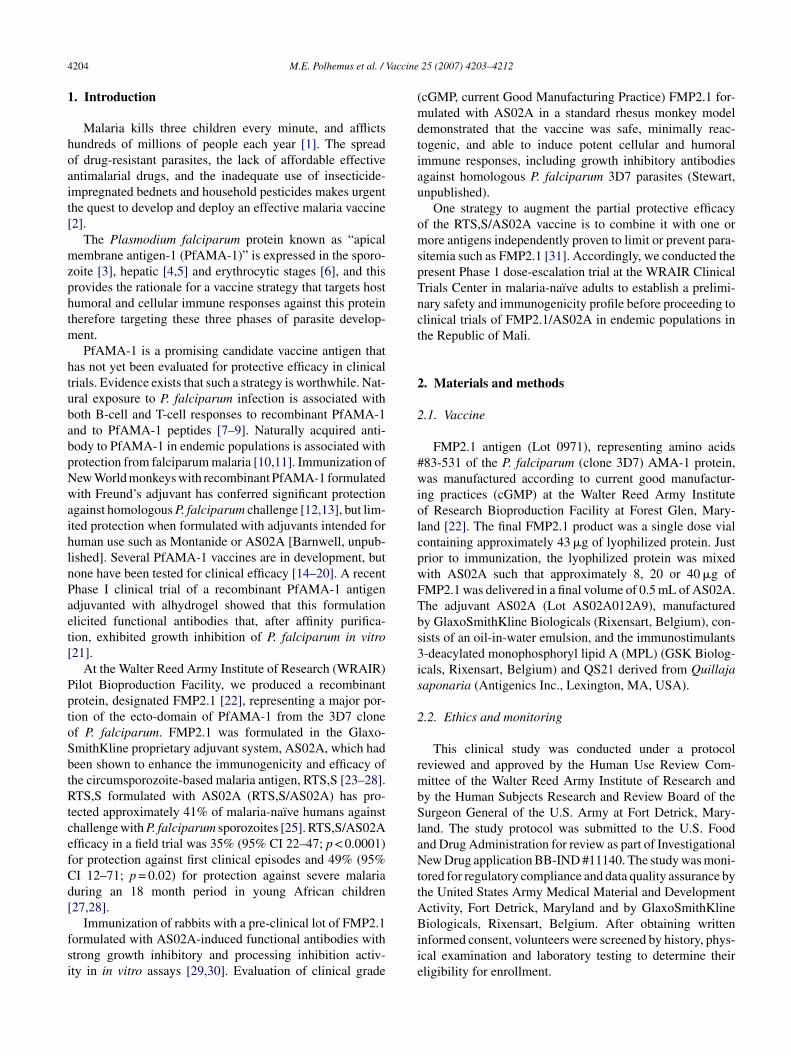
We report the first safety and immunogenicity trial of the Plasmodium falciparum vaccine candidate FMP2.1/AS02A, a recombinant E.coli-expressed protein based upon the apical membrane antigen-1 (AMA-1) of the 3D7 clone formulated with the AS02A adjuvant. Weconducted an open-label, staggered-start, dose-escalating Phase I trial in 23 malaria-naıve volunteers who received 8, 20 or 40 �g of FMP2.1in a fixed volume of 0.5 mL of AS02A on a 0, 1, and 2 month schedule. Nineteen of 23 volunteers received all three scheduled immunizations.The most frequent solicited local and systemic adverse events associated with immunization were injection site pain (68%) and headache(29%). There were no significant laboratory abnormalities or vaccine-related serious adverse events. All volunteers seroconverted aftersecond immunization as determined by ELISA. Immune sera recognized sporozoites and merozoites by immunofluorescence assay (IFA),and exhibited both growth inhibition and processing inhibition activity against homologous (3D7) asexual stage parasites. Post-immunization,peripheral blood mononuculear cells exhibited FMP2.1-specific lymphoproliferation and IFN-� and IL-5 ELISPOT assay responses. Thisis the first PfAMA-1-based vaccine shown to elicit both potent humoral and cellular immunity in humans. Encouraged by the potential ofFMP1/AS02A to target host immunity against PfAMA-1 that is known to be expressed by sporozoite, hepatic and erythrocytic stages, wehave initiated field trials of FMP2.1/AS02A in an endemic population in the Republic of Mali.© 2007 Elsevier Ltd. All rights reserved.
Keywords: Apical membrane antigen; Processing inhibition assay; FMP2.1; AS02A; Malaria; Vaccine; Falciparum; Recombinant protein; Growth inhibitionassay
� Disclaimer: The opinions expressed in this article are personal and are not to be construed as official views of the Departments of the Army or Defense.∗ Corresponding author at: Unit 64109 (MRU), APO AE 09831-4109, Kenya. Tel.: +254 733 616 550.
E-mail address: [email protected] (M.E. Polhemus).1 Present address: Centro de Investigacao em Saude da Manhica, Manhica, Mozambique.2 Present address: The National Academies, Washington, DC, United States.3 Present address: Bartlett Hall, Rm #404, Department of Chemistry and Life Sciences, United States Military Academy, West Point, NY 10996,
United States. Tel.: +1 845 938 3871.
0264-410X/$ – see front matter © 2007 Elsevier Ltd. All rights reserved.doi:10.1016/j.vaccine.2007.03.012
4 Vaccine
1
hoait[
mzphtm
htubabpNwaihlnPaet[
PptoSbtRtcefCd[
fsi
(mdtiau
omspTnct
2
2
#wiolcpwFTbs3is
2
rmbSlaNttA
204 M.E. Polhemus et al. /
. Introduction
Malaria kills three children every minute, and afflictsundreds of millions of people each year [1]. The spreadf drug-resistant parasites, the lack of affordable effectiventimalarial drugs, and the inadequate use of insecticide-mpregnated bednets and household pesticides makes urgenthe quest to develop and deploy an effective malaria vaccine2].
The Plasmodium falciparum protein known as “apicalembrane antigen-1 (PfAMA-1)” is expressed in the sporo-
oite [3], hepatic [4,5] and erythrocytic stages [6], and thisrovides the rationale for a vaccine strategy that targets hostumoral and cellular immune responses against this proteinherefore targeting these three phases of parasite develop-
ent.PfAMA-1 is a promising candidate vaccine antigen that
as not yet been evaluated for protective efficacy in clinicalrials. Evidence exists that such a strategy is worthwhile. Nat-ral exposure to P. falciparum infection is associated withoth B-cell and T-cell responses to recombinant PfAMA-1nd to PfAMA-1 peptides [7–9]. Naturally acquired anti-ody to PfAMA-1 in endemic populations is associated withrotection from falciparum malaria [10,11]. Immunization ofew World monkeys with recombinant PfAMA-1 formulatedith Freund’s adjuvant has conferred significant protection
gainst homologous P. falciparum challenge [12,13], but lim-ted protection when formulated with adjuvants intended foruman use such as Montanide or AS02A [Barnwell, unpub-ished]. Several PfAMA-1 vaccines are in development, butone have been tested for clinical efficacy [14–20]. A recenthase I clinical trial of a recombinant PfAMA-1 antigendjuvanted with alhydrogel showed that this formulationlicited functional antibodies that, after affinity purifica-ion, exhibited growth inhibition of P. falciparum in vitro21].
At the Walter Reed Army Institute of Research (WRAIR)ilot Bioproduction Facility, we produced a recombinantrotein, designated FMP2.1 [22], representing a major por-ion of the ecto-domain of PfAMA-1 from the 3D7 clonef P. falciparum. FMP2.1 was formulated in the Glaxo-mithKline proprietary adjuvant system, AS02A, which hadeen shown to enhance the immunogenicity and efficacy ofhe circumsporozoite-based malaria antigen, RTS,S [23–28].TS,S formulated with AS02A (RTS,S/AS02A) has pro-
ected approximately 41% of malaria-naıve humans againsthallenge with P. falciparum sporozoites [25]. RTS,S/AS02Afficacy in a field trial was 35% (95% CI 22–47; p < 0.0001)or protection against first clinical episodes and 49% (95%I 12–71; p = 0.02) for protection against severe malariauring an 18 month period in young African children27,28].
Immunization of rabbits with a pre-clinical lot of FMP2.1ormulated with AS02A-induced functional antibodies withtrong growth inhibitory and processing inhibition activ-ty in in vitro assays [29,30]. Evaluation of clinical grade
Biie
25 (2007) 4203–4212
cGMP, current Good Manufacturing Practice) FMP2.1 for-ulated with AS02A in a standard rhesus monkey model
emonstrated that the vaccine was safe, minimally reac-ogenic, and able to induce potent cellular and humoralmmune responses, including growth inhibitory antibodiesgainst homologous P. falciparum 3D7 parasites (Stewart,npublished).
One strategy to augment the partial protective efficacyf the RTS,S/AS02A vaccine is to combine it with one orore antigens independently proven to limit or prevent para-
itemia such as FMP2.1 [31]. Accordingly, we conducted theresent Phase 1 dose-escalation trial at the WRAIR Clinicalrials Center in malaria-naıve adults to establish a prelimi-ary safety and immunogenicity profile before proceeding tolinical trials of FMP2.1/AS02A in endemic populations inhe Republic of Mali.
. Materials and methods
.1. Vaccine
FMP2.1 antigen (Lot 0971), representing amino acids83-531 of the P. falciparum (clone 3D7) AMA-1 protein,as manufactured according to current good manufactur-
ng practices (cGMP) at the Walter Reed Army Institutef Research Bioproduction Facility at Forest Glen, Mary-and [22]. The final FMP2.1 product was a single dose vialontaining approximately 43 �g of lyophilized protein. Justrior to immunization, the lyophilized protein was mixedith AS02A such that approximately 8, 20 or 40 �g ofMP2.1 was delivered in a final volume of 0.5 mL of AS02A.he adjuvant AS02A (Lot AS02A012A9), manufacturedy GlaxoSmithKline Biologicals (Rixensart, Belgium), con-ists of an oil-in-water emulsion, and the immunostimulants-deacylated monophosphoryl lipid A (MPL) (GSK Biolog-cals, Rixensart, Belgium) and QS21 derived from Quillajaaponaria (Antigenics Inc., Lexington, MA, USA).
.2. Ethics and monitoring
This clinical study was conducted under a protocoleviewed and approved by the Human Use Review Com-ittee of the Walter Reed Army Institute of Research and
y the Human Subjects Research and Review Board of theurgeon General of the U.S. Army at Fort Detrick, Mary-
and. The study protocol was submitted to the U.S. Foodnd Drug Administration for review as part of Investigationalew Drug application BB-IND #11140. The study was moni-
ored for regulatory compliance and data quality assurance byhe United States Army Medical Material and Developmentctivity, Fort Detrick, Maryland and by GlaxoSmithKline
iologicals, Rixensart, Belgium. After obtaining writtennformed consent, volunteers were screened by history, phys-cal examination and laboratory testing to determine theirligibility for enrollment.
accine
2
eran
daaC
2
arprcrwrabaoc
2
onmauei
2
ismiTnfmaeoaItp
1aita1
eiops
haei
2
2
lb6ptr
2a
adcssiipSzpmmmao
2
h
M.E. Polhemus et al. / V
.3. Protocol
The study was an open-label, staggered-start, dose-scalating Phase 1 trial intended to determine the safety,eactogenicity and immunogenicity of three intramuscularlydministered doses of FMP2.1/AS02A in healthy malaria-aıve adults at the WRAIR Clinical Trials Center.
Volunteers were sequentially assigned to one of threeosage groups (Groups A, B, and C). Group A receivedpproximately 8 �g FMP2.1 in 0.5 mL AS02A; Group B,pproximately 20 �g FMP2.1 in 0.5 mL AS02A; and Group, approximately 40 �g FMP2.1 in 0.5 mL AS02A.
.3.1. ParticipantsParticipants were adult males and females 18–45 years of
ge. Exclusion criteria included history of malaria, previouseceipt of a malaria vaccine, splenectomy, known or sus-ected immunosuppression, use of systemic steroids, recenteceipt of any investigational or non-registered drug or vac-ine, simultaneous participation in any other clinical trial,eceipt of immunoglobulin or any blood product transfusionithin 3 months of study start, abnormal screening labo-
atories (CBC, alanine aminotransferase (ALT), aspartateminotransferase (AST), creatinine (Cr), or positive serumeta-human chorionic gonadotrophin), serologic evidence ofctive hepatitis B or C infection, antibody to HIV, or anyther clinically significant acute or chronic disease that mightonfound the interpretation of study results.
.3.2. Immunization proceduresVaccine was administered by injection into the deltoid
f the non-dominant arm on a 0, 1, and 2 month immu-ization schedule beginning with Group A. There was ainimum 14-day interval between group immunizations to
llow for safety evaluation of the previous group. Follow-p evaluations occurred on days 1, 2, 3, 7, and 14 afterach immunization and at 2, 4, and 6 months after the thirdmmunization.
.3.3. Adverse eventsThe volunteers were observed for 30 min after each
mmunization for evidence of anaphylaxis. The presence ofolicited local and general signs and symptoms, includingeasurement of oral temperature, were assessed after each
mmunization and 1, 2, 3 and 7 days post-immunization.he solicited injection site adverse events were pain, red-ess and swelling. Solicited general adverse events wereever, gastrointestinal complaints, fatigue, headache, malaise,yalgia and joint pain. In addition to the solicited signs
nd symptoms, investigators recorded any other adversevents occurring within a 28-day follow-up period (dayf immunization and 27 subsequent days) as unsolicited
dverse events. Adverse events were assessed for intensity.njection site pain was graded as 0 = absent, 1 = painful onouch, 2 = painful when limb is moved, and 3 = spontaneouslyainful. Solicited symptoms were graded as 0 = normal,aeop
25 (2007) 4203–4212 4205
= easily tolerated, 2 = interferes with normal activity,nd 3 = prevents normal daily activity. Additional grad-ng scales were applied to visible swelling or redness athe injection site; 0 = none, 1 = 0–20 mm, 2 = 20–50 mm,nd 3 = >50 mm, and to oral temperature; 0 = <37.5 ◦C,= 37.5–38 ◦C, 2 = 38–39 ◦C, and 3 = >39 ◦C.
Serious adverse events (SAEs) were reported fromnrollment until study completion 6 months after finalmmunization. SAEs were defined as any untoward medicalccurrence that resulted in death, significant disability, hos-italization, incapacity, or required intervention to preventuch outcomes.
Biochemical (ALT, AST, and Cr) and hematological (Hb,ct, WBC, and PLTs) laboratory parameters were measuredt screening, and on days of immunization, 2 weeks afterach immunization and at 2, 4 and 6 months after the thirdmmunization.
.4. Serology
.4.1. Anti-FMP2.1 ELISAIgG antibody to the test antigen was measured by enzyme-
inked immunosorbent assay (ELISA) in all volunteers ataseline, 2 weeks after each immunization and 2, 4, andmonths after the third immunization. IgG ELISAs were
erformed, using FMP2.1 as the capture antigen, in serialwo-fold dilution, and the titer defined as the serum dilutionequired to yield an optical density of 1.0 in our assay.
.4.2. Sporozoite and merozoite immunofluorescencessay (IFA)
Blood stage late schizonts were fixed with methanolnd immunofluorescence assays (IFA) were performed asescribed previously [22]. Briefly, 2% fetal bovine serumontaining PBS was used as a diluent and serial dilutionstarting from 1:2 to 1:25,600 were tested. FITC-conjugatedecondary antibodies diluted 1:500 (Southern Biotech, Birm-ngham, AL, USA) were used and slides were mountedn SlowFade® Antifade Kit with DAPI (4′,6′-diamidino-2-henylindole) (Molecular Probes, Inc. Eugene, OR, USA).porozoite IFA utilized an acetone fixed preparation of sporo-oites and was performed with two pre-immunization andost-third immunization serum samples from Group C. Theethodology was similar to the blood stage IFA except theounting solution did not contain DAPI. An Olympus BX50icroscope equipped with a mercury epifluorescence lamp,100× (oil) objective and a multi filter cube was used to
bserve the fluorescence.
.4.3. Growth invasion/inhibition assay (GIA)Sera were tested for growth inhibitory effects against
omologous 3D7 and heterologous FVO P. falciparum par-
sites in a one-cycle static assay [32]. Three time points forach volunteer were evaluated: baseline, 2 weeks after sec-nd immunization, and 2 weeks after third immunization. Thearasites were cultured for 2 days in heat inactivated, dialyzed
4 Vaccine
saiHaefws
2
aaPatpo1wraatwmrk[sobaS
2
msmi5artdvwe(
S
E
(c1wiMCPlp1ppToLaCo
2
bossM
3
3
CRua(FFcApBdTiwia
206 M.E. Polhemus et al. /
era (20%, v/v) at a 0.2% initial parasitemia (trophozoites)nd 4% hematocrit in triplicate 150 �L static culture volumesn 48-well plates. Cultures were harvested and stained withoechst dye 33342 (Molecular Probes, Eugene, OR, USA)
nd the number of new trophozoites were counted in 40,000rythrocytes by flow cytometry. Inhibition was calculatedrom final parasitemias as inhibition = (control-test)/control,here control was the final parasitemia with pre-immune
erum, and was expressed as a percent.
.4.4. Processing inhibition assay (PIA)We have described an immuno-chemical correlate of
nti-AMA-1 antibodies that measures the ability of thesentibodies to inhibit the natural proteolytic processing offAMA-1 on the merozoite surface [29,30]. We conductedparallel GIA and PIA on sera collected 2 weeks post
hird immunization. The PIA was performed in a 48-welllate format contained 80 �L of purified late-stage schizontsf the 3D7 strain of P. falciparum at a concentration of× 107 mL−1 and 20 �L test serum. The plate was gassedith 5% CO2 and incubated at 37 ◦C until > 90% of schizonts
uptured. The parasites were then collected by centrifugationnd analyzed by Western blot with biotin labeled polyclonalnti-AMA-1 and biotin labeled monoclonal antibody againsthe C-terminus of AMA-1, mAb 28G2dc1. This antibodyas a kind gift of Dr. Alan W. Thomas, Biomedical Pri-ate Research Center, Rijswijk, The Netherlands. The PIA
atio was calculated as band intensities of the 10-kDa/(10-Da + 20-kDa) AMA-1 specific bands on the Western blot29]. The GIA plate contained 80 �L of 0.5% parasitemiachizonts + normal human RBC at 4% hematocrit and 20 �Lf test serum in triplicates. Plates were gassed and incu-ated overnight at 37 ◦C; the percent invasion was determineds described above, except the ring stages were stained byYBR® Green (Molecular Probes, Eugene, OR) dye for 1 h.
.4.5. Cell-mediated immunityFor cellular reactivity of FMP2.1-immune cells we
easured lymphoproliferation as well as IFN-� and IL-5ecretion. Briefly, cryopreserved and thawed peripheral bloodononuclear cells (PBMC) obtained at pre- and post-third
mmunization were re-suspended in RPMI plus additives and% human AB serum. For proliferation, cells were cultured atconcentration of 2 × 10⁵ cells/well in triplicates in 96-well
ound bottom microtiter plates in the presence of FMP2.1 pro-ein (10 �g/mL), PHA (2 �g/mL), or medium control. After 5ays cultures were pulsed with 3H-TdR (1 �Ci/well) and har-ested after an additional 16 h. Incorporation of radioactivityas measured by scintillation spectrometry and results are
xpressed as counts per minute (cpm). Stimulation indicesSI) were determined according to the following formula:
I = cpm in experimental culture
cpm in control cultures.
IFN-� and IL-5 responses were evaluated by theLISPOT assay. One hundred microlitres of cell suspensions
Caic
25 (2007) 4203–4212
2 × 10⁶ mL−1 for IFN-� and 4 × 10⁶ mL−1 for IL-5) wereultured in duplicates or triplicates in the presence of 0, 0.1,.0 or 10 �g/mL FMP2.1 or 0.4 �g/mL PHA in ELISPOTells coated previously with 100 �L/well PBS contain-
ng 10 �g/mL anti-IFN-� or 15 �g/mL anti-IL-5 (Mabtech,airemont, OH, USA). After 18 h of culture in a 37 ◦C, 5%O2 humidified atmosphere, the plates were washed withBS, and 100 �L of 1:100 dilution of appropriate biotiny-
ated detecting antibody (Mabtech) were added per well, andlates were left for 2 h at room temperature. After washing,00 �L of a 1:1000 dilution of streptavidin–alkaline phos-hatase conjugate (Mabtech) were added per well and thelates were left for an additional 1.5 h at room temperature.he plates were then washed and the ELISPOTs were devel-ped by the addition of alkaline phosphatase substrate (Mossaboratories). The number of spots was counted with theid of an Immunospot Image Analyzer (Cellular Technology,leveland, OH). Results are expressed as the mean ± S.E.M.f triplicate wells for IFN-� and duplicate wells for IL-5.
.5. Statistics and data management
Data were entered into a Microsoft Excel spreadsheet dataase and analyzed using Excel statistical tools. In the casef GIA analyses, data were compared using the Wilcoxonigned-rank test (non-directional) with paired pre-immuneera. Differences between groups were compared using the
ann–Whitney-Wilcoxon rank-sum test (non-directional).
. Results
.1. Participant flow
The clinical portion of the study was conducted at thelinical Trials Center at the Walter Reed Army Institute ofesearch from September 2003 to July 2004. Fifty-two vol-nteers were screened. Twenty-three were eligible, enrollednd allocated to one of the three dosage groups: Group A8 �g FMP2.1 in 0.5 mL AS02A, n = 8), Group B (20 �gMP2.1 in 0.5 mL AS02A, n = 8), and Group C (40 �gMP2.1 in 0.5 mL AS02A, n = 7). Four volunteers did notomplete the immunization series. One volunteer from Group
was withdrawn by the PI after first immunization due tooor compliance with follow-up. One volunteer from Group
was withdrawn by the PI after the first immunizationue to a severe adverse event not-related to immunization.wo volunteers from Group C withdrew after the second
mmunization; one withdrew without explanation, the otherithdrew due to Grade 3 adverse events associated with
mmunization. Actual versus scheduled immunizations weres follows: Group A; 22 of 24, Group B; 22 of 24, and Group
; 19 of 21. During the 6 month post-immunization period, andditional three volunteers did not complete follow-up; onen Group A and two in group C. Thus, 16 of 23 volunteersompleted the study according to protocol (Fig. 1).
M.E. Polhemus et al. / Vaccine 25 (2007) 4203–4212 4207
nteer flo
3
Avoi3aiw
nm(ttbw
TI
PRSFGHMMFA
T
F
Fig. 1. Volu
.2. Safety and reactogenicity
Adverse events (AEs) by group are summarized in Table 1.ll solicited vaccine-related AEs occurred within 72 h afteraccination. Sixteen Grade 3 reactions were reported with 12f 16 occurring in the 40 �g dosage group and the remain-ng 4 occurred in the 20 �g dosage group. Five of the Grade
reactions occurred in one individual in the 40 �g groupfter the second vaccination and were self-reported, as thendividual did not return for follow-up during the time heas symptomatic. Local pain (43 incidents over 63 vacci-
3
I
able 1nstances of local and systemic solicited adverse events recorded during first 7 days
Group A 8 �g FMP2.1 in 0.5 mL AS02A Group B 20 �g FM
Grade1 Grade2 Grade3 Grade1 Grade
ain 6 4 0 6 13edness 0 0 0 0 0welling 1 1 0 2 0ever 0 0 0 0 1I 0 0 0 3 1A 2 0 0 3 5alaise 0 0 0 1 2yalgia 2 1 0 3 2
atigue 0 0 0 2 2rthralgia 0 0 0 0 1
otal 11 6 0 20 27
ive Grade 3 events in Group C occurred in one volunteer after the second immuniz
w diagram.
ations), local swelling (10 incidents over 63 vaccinations),yalgia (14 incidents over 63 vaccinations), and headache
18 incidents over 63 vaccinations) accounted for most ofhe solicited AEs. Almost all adverse events resolved withinhe first 72 h after immunization. No clinically significantiochemical or hematological abnormalities were associatedith immunization.
.2.1. Serious adverse events (SAEs)There was one SAE, and it was judged by the Principal
nvestigator and the Medical Monitor to be not related to
after immunization summarized by group and by grade
P2.1 in 0.5 mL AS02A Group C 40 �g FMP2.1 in 0.5 mL AS02A
2 Grade3 Grade1 Grade2 Grade3
1 6 7 01 4 0 21 4 1 01 2 0 20 3 2 00 3 2 30 1 1 20 3 2 10 2 4 10 1 1 1
4 29 20 12
ation. All solicited adverse events occurred within 72 h after immunization.
4208 M.E. Polhemus et al. / Vaccine 25 (2007) 4203–4212
Fada
ivpdhcd
3
3
(giwapsmtilotg
3
ipofGFna
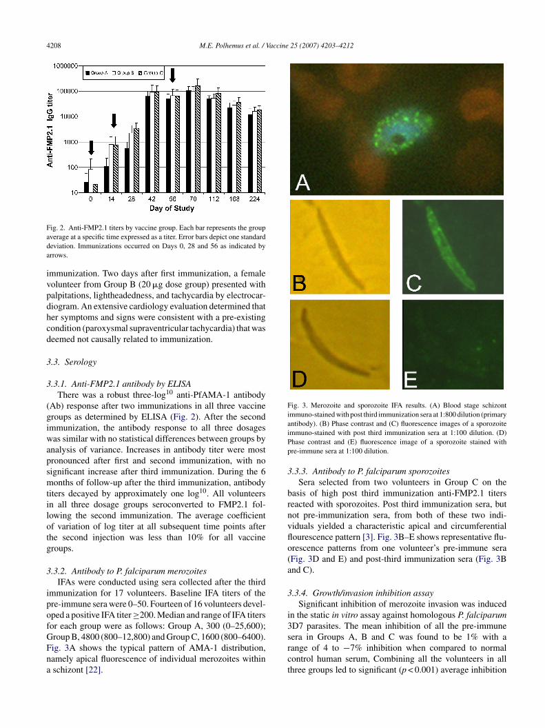
Fig. 3. Merozoite and sporozoite IFA results. (A) Blood stage schizontimmuno-stained with post third immunization sera at 1:800 dilution (primaryantibody). (B) Phase contrast and (C) fluorescence images of a sporozoiteiPp
3
brnvflo(a
3
i3
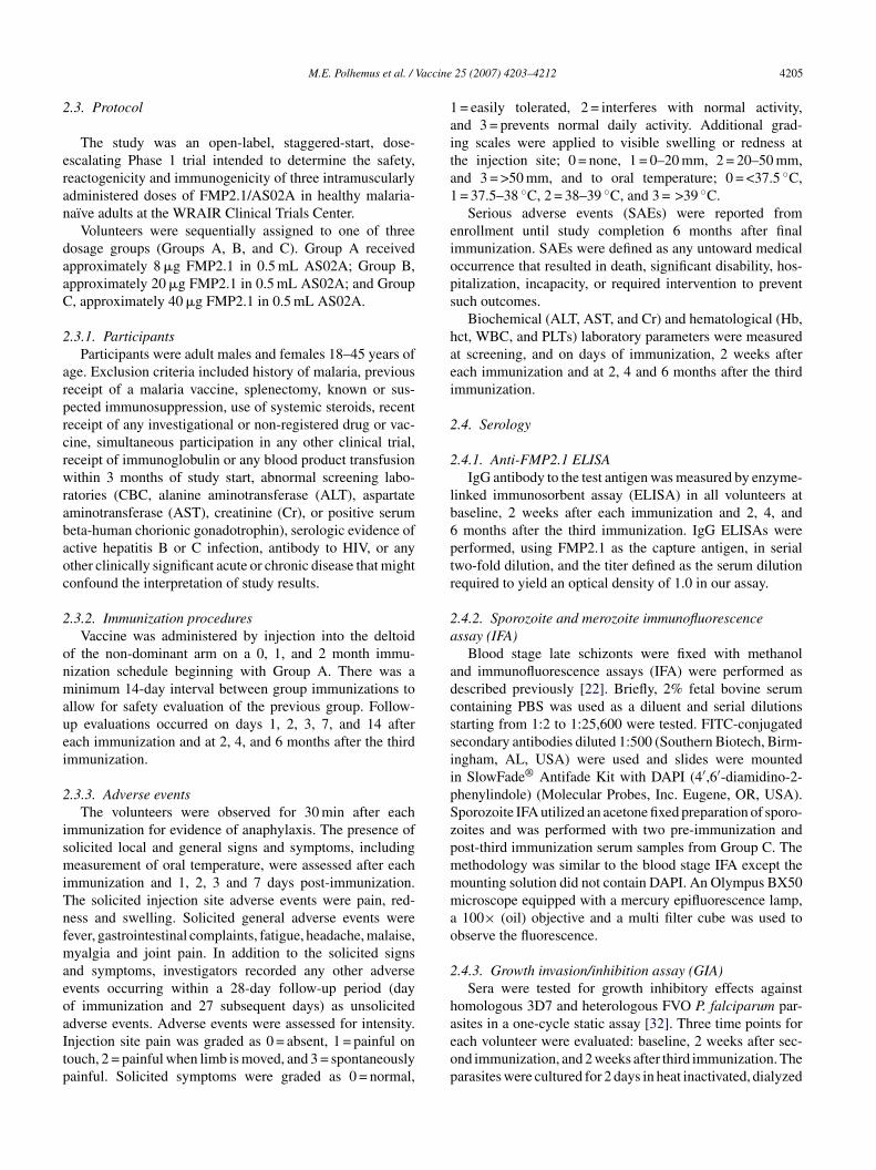
ig. 2. Anti-FMP2.1 titers by vaccine group. Each bar represents the groupverage at a specific time expressed as a titer. Error bars depict one standardeviation. Immunizations occurred on Days 0, 28 and 56 as indicated byrrows.
mmunization. Two days after first immunization, a femaleolunteer from Group B (20 �g dose group) presented withalpitations, lightheadedness, and tachycardia by electrocar-iogram. An extensive cardiology evaluation determined thater symptoms and signs were consistent with a pre-existingondition (paroxysmal supraventricular tachycardia) that waseemed not causally related to immunization.
.3. Serology
.3.1. Anti-FMP2.1 antibody by ELISAThere was a robust three-log10 anti-PfAMA-1 antibody
Ab) response after two immunizations in all three vaccineroups as determined by ELISA (Fig. 2). After the secondmmunization, the antibody response to all three dosagesas similar with no statistical differences between groups by
nalysis of variance. Increases in antibody titer were mostronounced after first and second immunization, with noignificant increase after third immunization. During the 6onths of follow-up after the third immunization, antibody
iters decayed by approximately one log10. All volunteersn all three dosage groups seroconverted to FMP2.1 fol-owing the second immunization. The average coefficientf variation of log titer at all subsequent time points afterhe second injection was less than 10% for all vaccineroups.
.3.2. Antibody to P. falciparum merozoitesIFAs were conducted using sera collected after the third
mmunization for 17 volunteers. Baseline IFA titers of there-immune sera were 0–50. Fourteen of 16 volunteers devel-ped a positive IFA titer ≥200. Median and range of IFA titersor each group were as follows: Group A, 300 (0–25,600);
roup B, 4800 (800–12,800) and Group C, 1600 (800–6400).ig. 3A shows the typical pattern of AMA-1 distribution,amely apical fluorescence of individual merozoites withinschizont [22].srct
mmuno-stained with post third immunization sera at 1:100 dilution. (D)hase contrast and (E) fluorescence image of a sporozoite stained withre-immune sera at 1:100 dilution.
.3.3. Antibody to P. falciparum sporozoitesSera selected from two volunteers in Group C on the
asis of high post third immunization anti-FMP2.1 titerseacted with sporozoites. Post third immunization sera, butot pre-immunization sera, from both of these two indi-iduals yielded a characteristic apical and circumferentialourescence pattern [3]. Fig. 3B–E shows representative flu-rescence patterns from one volunteer’s pre-immune seraFig. 3D and E) and post-third immunization sera (Fig. 3Bnd C).
.3.4. Growth/invasion inhibition assaySignificant inhibition of merozoite invasion was induced
n the static in vitro assay against homologous P. falciparumD7 parasites. The mean inhibition of all the pre-immune
era in Groups A, B and C was found to be 1% with aange of 4 to −7% inhibition when compared to normalontrol human serum, Combining all the volunteers in allhree groups led to significant (p < 0.001) average inhibition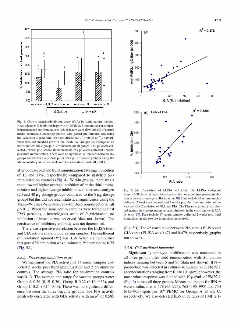
M.E. Polhemus et al. / Vaccine 25 (2007) 4203–4212 4209
Fig. 4. Growth invasion/inhibition assay (GIA) by static culture method.y-Axis denotes % inhibition in growth by 1:5 diluted immune sera in compar-ison to matched pre-immune sera (which in turn were all within 6% of normalmedia controls). Comparing growth with paired pre-immune sera usingthe Wilcoxon signed-rank test (non-directional), *p < 0.05 or **p < 0.001.Error bars are standard error of the mean. Av Group = the average of allindividuals within a group (6–7 volunteers) or all groups. 2wk p2 = sera col-lpgM
aoitn(gMpFip
aot(
3
lcwGGep
Fig. 5. (A) Correlation of ELISA and GIA. The ELISA end-pointtiters × 1000 (y-axis) were plotted against the corresponding percent inhibi-tion in the static one-cycle GIA (x-axis) [30]. Data include 37 serum samplescollected 2 weeks post second and 2 weeks post third immunization of thevaccine. (B) Correlation of GIA and PIA. The PIA ratio (x-axis) was plot-t(i
(Gn
3
aipam
ected 2 weeks post second immunization; 2wk p3 = sera collected 2 weeksost third immunization. There were no significant differences between anyroups (or between any 2wk p2 or 2wk p3 or pooled groups) using theann–Whitney-Wilcoxon rank-sum test (non-directional, all p > 0.1).
fter both second and third immunization (average inhibitionf 13 and 17%, respectively) compared to matched pre-mmunization controls (Fig. 4). Within groups, there was arend toward higher average inhibition after the third immu-ization and higher average inhibition with increased antigen20 and 40 �g dosage groups compared to the 8 �g dosageroup) but this did not reach statistical significance using theann–Whitney-Wilcoxon rank-sum test (non-directional, all> 0.1). When the same serum samples were tested againstVO parasites, a heterologous strain of P. falciparum, no
nhibition of invasion was observed (data not shown). Theersistence of inhibitory antibody was not determined.
There was a positive correlation between the ELISA titersnd GIA activity of individual serum samples. The coefficientf correlation squared (R2) was 0.38. When a single outlierhat gave 82% inhibition was eliminated, R2 increased to 0.75Fig. 5A).
.3.5. Processing inhibition assayWe measured the PIA activity of 17 serum samples col-
ected 2 weeks post third immunization and 2 pre-immuneontrols. The average PIA ratio for pre-immune controlsas 0.13. The average and range for vaccine groups were;
roup A 0.26 (0.19–0.36), Group B 0.22 (0.16–0.32), androup C 0.21 (0.14–0.43). There was no significant differ-nce between the three vaccine groups. The PIA activityositively correlated with GIA activity with an R2 of 0.585
(w(r
ed against the corresponding percent inhibition in the static one-cycle GIAy-axis) [27]. Data include 17 serum samples collected 2 weeks post thirdmmunization and two pre-immunization controls.
Fig. 5B). The R2 correlation between PIA versus ELISA andIA versus ELISA was 0.471 and 0.479, respectively (graphsot shown).
.3.6. Cell-mediated immunitySignificant lymphocyte proliferation was measured in
ll three groups after third immunization with stimulationndices ranging between 5 and 96 (data not shown). IFN-�roduction was detected in cultures stimulated with FMP2.1t concentrations ranging from 0.1 to 10 �g/mL; however, theost robust response was elicited with 10 �g/mL of FMP2.1
Fig. 6) across all three groups. Means and ranges for IFN-�ere similar, that is 578 (65–995), 765 (195–995) and 750
615–905) spots per 106 PBMC for Groups A, B and C,espectively. We also detected IL-5 in cultures of FMP 2.1-

4210 M.E. Polhemus et al. / Vaccine
Fig. 6. IFN-� response by ELISPOT. IFN-� response by ELISPOTat baseline (pre-immunization) or post third dose immunization (post-immunization) by vaccine group in response to in vitro stimulation with0.1, 1.0, or 10 �g of FMP2.1. Error bars represent standard error of themeans (mean + S.E.M.).
Fig. 7. IL-5 response by ELISPOT. IL-5 response by ELISPOT at baseline(vF
iccmoP
4
Fae
itpc
4
isafotraebwt[a
4
dlaltiu
pbapwitotvawFapolh
pre-immunization) or post third dose immunization (post-immunization) byaccine group in response to in vitro stimulation with 0.1, 1.0, or 10 �g ofMP2.1. Error bars represent standard error of the means (mean + S.E.M.).
mmune PBMC, and the responses were likewise FMP2.1oncentration-dependent with 10 �g/mL eliciting maximumytokine production (Fig. 7). IL-5 responses were of lesseragnitude than that seen for IFN-�, with means and ranges
f 34 (14–50), 84 (11–154) and 47 (15–113) spots per 106
BMC for Groups A, B and C, respectively.
. Discussion
This is the first immunization of humans withMP2.1/AS02A. The vaccine was reactogenic but gener-lly well-tolerated with no vaccine-related serious adversevents. The vaccine proved to be highly immunogenic
i1sa
25 (2007) 4203–4212
nducing both humoral and cellular responses. In addi-ion, the vaccine induced invasion and PfAMA-1 proteolyticrocessing-inhibitory antibodies against homologous P. fal-iparum parasites.
.1. Safety and reactogenicity
There were apparent differences among the dosage groupsn numbers of vaccine-associated adverse events in this smalltudy, with the higher dose groups having more soliciteddverse events. The duration of adverse events was similaror all the groups with the majority resolving within 72 hf immunization. However, because all the groups receivedhe same dose of adjuvant in their vaccine formulation, anyeal differences in reactogenicity by dosage group would bettributable to the amount of antigen. It should be noted how-ver, that the pattern of the 16 Grade 3 adverse events haseen well documented in a small percentage of volunteersho have received other AS02A vaccine formulations, so
he finding is not unique to the FMP2.1/AS02A formulation24,25]. There were no vaccine-related clinical laboratorybnormalities.
.2. Antibody responses
After the second immunization, there was no significantifference in antibody response between dose groups. Fol-owing three immunizations, all three dosage groups had highntibody levels by ELISA that persisted for 6 months after theast immunization. Two immunizations appear to be enougho induce a maximal antibody response, but since a thirdmmunization was given, its impact on antibody kinetics isnclear.
The potential for FMP2.1-specific antibodies to inhibitarasite invasion of erythrocytes in vivo was demonstratedy the finding of functionally active antibody by both GIAnd PIA in all dose groups against homologous P. falci-arum 3D7 parasites. It is important to note that the GIAas done at 1:5 dilution, hence anti-FMP2.1 antibody levels
n vivo would be higher and presumably more active. Therend toward greater inhibition in the GIA with higher dosesf antigen did not reach statistical significance, perhaps dueo small group sizes and variability in inhibition among indi-iduals in each group. The percent growth invasion inhibitionssay (GIA) also positively correlated with the ELISA andith the processing inhibitory assay (PIA) activity of anti-MP2.1, consistent with our hypothesis that antibodies aren important component of PfAMA-1 immunity and that therocessing inhibition may be causally related to inhibitionf invasion [29,30]. Although GIA and PIA are not estab-ished as a correlate of protection in humans, these functionalumoral response assays are likely to play an important role
n the evaluation of future trials of this and other PfAMA-vaccines, as well as other vaccines exhibiting these orimilar activities. The relevance of the absence of activitygainst heterologous P. falciparum FVO parasites in a GIA
accine
apah
irw
4
FwmtfmTPccIei[ie
4
aaGcaaeWawaFth
4
taTaic
qi
A
cCAa
MeAadhbfMM#TF
R
M.E. Polhemus et al. / V
ssay remains to be determined in field trials. The P. falci-arum FVO and 3D7 PfAMA-1 ecto-domains differ at 24mino-acid positions which represents the two most distantaplotypes reported [33].
The demonstration that FMP2.1/AS02A-induced antibod-es recognized sporozoites by IFA further suggests a potentialole of this vaccine in inducing antibodies that might interfereith the invasion of sporozoites into liver hepatocytes.
.3. Cellular immunity
The cell-mediated immune (CMI) results indicate thatMP2.1/AS02A produces a strong Th1 and a somewhateaker Th2 response. Given the expectation that antibodyay play a dominant role in controlling blood stage infec-
ion, these CMI responses suggest adequate T cell helpor the B cell response. Furthermore, the CMI responseay have implications beyond support of antibody response.lymphocytes from volunteers immunized with irradiated
. falciparum sporozoites recognize PfAMA-1, and mayontribute to sterile immunity against virulent sporozoitehallenge by targeting falciparum-infected hepatocytes [4].n AMA-1 vaccinated mice challenged with P. chabaudi, Xut al. demonstrated that AMA-1-specific T-cells played a rolen controlling parasitemia independent of antibody activity34]. Taken together, these observations suggest that T-cellsnduced by FMP2.1/AS02A might act against PfAMA-1xpressed in pre-erythrocytic as well as on blood stages [4,5].
.4. PfAMA-1 vaccine design
Blood stage malaria antigens, including PfAMA-1, exists diverse alleles that elicit varying degrees of cross-reactiventibody. Sero-epidemiologic evidence [35], and the limitedIA data presented here, suggest a PfAMA-1-based vac-
ine may elicit allele-specific antibodies. Consequently, inddition to monovalent PfAMA-1 vaccines [14–17,21] therere also bivalent PfAMA-1 vaccines in development in anffort to broaden protective immune responses [18,19,21].ith FMP2.1/AS02A, we intend to determine if high titer
ntibodies elicited against sporozoite and asexual stages, asell as potent anti-PfAMA-1 cellular responses, might act
gainst diverse alleles. For this reason, initial field trials ofMP2.1/AS02A will include allelic genotyping endpoints
o determine its ability to protect against parasites bearingomologous and heterologous alleles of PfAMA-1 [36].
.5. Conclusion
This pilot study has established the initial safety, reac-ogenicity and immunogenicity profile for FMP2.1/AS02A,PfAMA-1-based vaccine that elicited potent humoral and
h1-biased cellular immune responses. Further studies arelready underway to evaluate this vaccine in volunteers liv-ng in malaria-endemic regions. Thus, a Phase 1B trial inhildren is now ongoing in Bandiagara, Mali, where subse-25 (2007) 4203–4212 4211
uent trials are planned to determine its safety and efficacyn children [37].
cknowledgements
The authors acknowledge the valuable time and the effortontributed to this clinical trial by the Medical Monitor,olonel Donald R. Skillman, MD, and by the United Statesrmy Medical Materiel Development Activity Product Man-
ger, Commander (ret.) Charles K. English, PhD.Conflict of interest: W.R. Ballou, J.D. Cohen, E. De-Kock,
arie-Claude Dubois and O. Ofori-Anyinam are employ-es of GlaxoSmithKline Biologicals, the manufacturer of theS02A adjuvant described in this report. S. Dutta, D.E. Lanar
nd L.A. Ware hold patents for the FMP2.1 vaccine antigenescribed in this report. The other authors declare that theyave no conflict of interests. Funding: This study was fundedy the Malaria Vaccine Development Program, US Agencyor International Development, Washington, DC and by the
ilitary Infectious Diseases Research Program, Fort Detrick,D. Previous disclosure: Presentation in part as Abstract
924 at the 53rd Annual Meeting of the American Society ofropical Medicine and Hygiene held October 2004 in Miami,lorida.
eferences
[1] Breman JG. The ears of the hippopotamus: manifestations, determi-nants, and estimates of the malaria burden. Am J Trop Med Hyg2001;64:1–11.
[2] Malaria Vaccine Initiative, <http://www.malariavaccine.org/>[accessed 21 February 2007].
[3] Silvie O, Franetich JF, Charrin S, Mueller MS, Siau A, BodescotM, et al. A role for apical membrane antigen 1 during invasion ofhepatocytes by Plasmodium falciparum sporozoites. J Biol Chem2004;279:9490–6.
[4] Krzych U, Lyon JA, Jareed T, Schneider I, Hollingdale MR, GordonDM, et al. T lymphocytes from volunteers immunized with irradiatedPlasmodium falciparum sporozoites recognize liver and blood stagemalaria antigens. J Immunol 1995;155:4072–7.
[5] Bodescot M, Silvie O, Siau A, Refour P, Pino P, Franetich JF, et al. Tran-scription status of vaccine candidate genes of Plasmodium falciparumduring the hepatic phase of its life cycle. Parasitol Res 2004;92:449–52.
[6] Peterson MG, Marshall VM, Smythe JA, Crewther PE, Lew A, SilvaA, et al. Integral membrane protein located in the apical complex ofPlasmodium falciparum. Mol Cell Biol 1989;9:3151–4.
[7] Udhayakumar V, Kariuki S, Kolczack M, Girma M, Roberts JM,Oloo AJ, et al. Longitudinal study of natural immune response to thePlasmodium falciparum apical membrane antigen (AMA-1) in a holen-demic region in Western Kenya: Asembo Bay Cohort Project VIII. AmJ Trop Med Hyg 2001;65:100–7.
[8] Thomas AW, Trape JF, Rogier C, Goncalves A, Rosario VE, NarumDL. High prevalence of natural antibodies against Plasmodium fal-ciparum 83-kilodalton apical membrane antigen (PF83/AMA-1) as
detected by capture-enzyme-linked immunosorbent assay using full-length baculovirus recombinant PF83/AMA-1. Am J Trop Med Hyg1994;51:730–40.[9] Lal AA, Hughes MA, Oliveira DA, Nelson C, Bloland PB, Oloo AJ, etal. Identification of T-cell determinants in natural immune responses to
4 Vaccine
[
[
[
[
[
[
[
[
[
[[
[
[
[
[
[
[
[
[
[
[
[
[
[
[
[
212 M.E. Polhemus et al. /
the Plasmodium falciparum apical membrane antigen (AMA-1) in anadult population exposed to malaria. Infect Immun 1996;64:1054–9.
10] Polley SD, Mwangi T, Kocken CH, Thomas AW, Dutta S, Lanar DE, etal. Human antibodies to recombinant protein constructs of Plasmodiumfalciparum apical membrane antigen 1 (AMA1) and their associationswith protection from malaria. Vaccine 2004;23:718–28.
11] Polley SD, Conway DJ, Cavanagh DR, McBride JS, Lowe BS, WilliamsTN, et al. High levels of serum antibodies to merozoite surface protein 2of Plasmodium falciparum are associated with reduced risk of clinicalmalaria in coastal Kenya. Vaccine 2006;24:4233–46.
12] Cubillos M, Salazar LM, Torres L, Patarroyo ME. Protection againstexperimental P. falciparum malaria is associated with short AMA-1peptide analogue alpha-helical structures. Biochimie 2002;84:1181–8.
13] Stowers AW, Kennedy MC, Keegan BP, Saul A, Long CA, Miller LH.Vaccination of monkeys with recombinant Plasmodium falciparum api-cal membrane antigen 1 confers protection against blood stage malaria.Infect Immun 2002;70:6961–7.
14] Saul A, Lawrence G, Allworth A, Elliott S, Anderson K, RzepczykC, et al. A human phase 1 vaccine clinical trial of the Plasmodiumfalciparum malaria vaccine candidate apical membrane antigen 1 inMontanide ISA720 adjuvant. Vaccine 2005;23:3076–83.
15] Kocken CH, Withers-Martinez C, Dubbeld MA, van der Wel A, HackettF, Valderrama A, et al. High-level expression of the malaria blood-stagevaccine candidate Plasmodium falciparum apical membrane antigen 1and induction of antibodies that inhibit erythrocyte invasion. InfectImmun 2002;70:5901.
16] Mueller MS, Renard A, Boato F, Vogel D, Naegeli M, Zurbriggen R,et al. Induction of parasite growth-inhibitory antibodies by a viroso-mal formulation of a peptidomimetic of loop I from domain III ofPlasmodium falciparum apical membrane antigen 1. Infect Immun2003;71:4749–58.
17] Pan W, Huang D, Zhang Q, Qu L, Zhang D, Zhang X, et al. Fusion of twomalaria vaccine candidate antigens enhances product yield, immuno-genicity, and antibody-mediated inhibition of parasite growth in vitro.J Immunol 2004;172:6167–74.
18] Mullen GE, Giersing BK, Ajose-Popoola O, Davis HL, Kothe C, ZhouH, et al. Enhancement of functional antibody responses to AMA1-C1/Alhydrogel, a Plasmodium falciparum malaria vaccine, with CpGoligodeoxynucleotide. Vaccine 2006;24:2497–505.
19] http://www.clinicaltrials.gov/ct/show/NCT00414336.20] Ballou WR. Malaria vaccines in development. Expert Opin Emerg
Drugs 2005;10:489–93.21] Malkin EM, Diemert DJ, McArthur JH, Perreault JR, Miles AP, Giers-
ing BK, et al. Phase 1 clinical trial of apical membrane antigen 1: anasexual blood-stage vaccine for Plasmodium falciparum malaria. InfectImmun 2005;73:3677–85.
22] Dutta S, Lalitha PV, Ware LA, Barbosa A, Moch JK, Vassell MA, etal. Purification, characterization, and immunogenicity of the refolded
ectodomain of the Plasmodium falciparum apical membrane antigen 1expressed in Escherichia coli. Infect Immun 2002;70:3101–10.23] Garcon N, Heppner DG, Cohen J. Development of RTS, S/AS02: apurified subunit-based malaria vaccine candidate formulated with anovel adjuvant. Expert Rev Vaccines 2003;2:231–8.
[
[
25 (2007) 4203–4212
24] Stoute JA, Slaoui M, Heppner DG, Momin P, Kester KE, DesmonsP, et al. A preliminary evaluation of a recombinant circumsporozoiteprotein vaccine against Plasmodium falciparum malaria. N Engl J Med1997;336:86–91.
25] Kester KE, McKinney DA, Tornieporth N, Ockenhouse CF, HeppnerDG, Hall T, et al. Efficacy of recombinant circumsporozoite protein vac-cine regimens against experimental Plasmodium falciparum malaria. JInfect Dis 2001;183:640–7.
26] Bojang KA, Milligan PJ, Pinder M, Vigneron L, Alloueche A, KesterKE, et al. Efficacy of RTS, S/AS02 malaria vaccine against Plasmod-ium falciparum infection in semi-immune adult men in The Gambia: arandomised trial. Lancet 2001;358:1927–34.
27] Alonso PL, Sacarlal J, Aponte JJ, Leach A, Macete E, Milman J, et al.Efficacy of the RTS, S/AS02A vaccine against Plasmodium falciparuminfection and disease in young African children: randomised controlledtrial. Lancet 2004;364:1411–20.
28] Alonso PL, Sacarlal J, Aponte JJ, Leach A, Macete E, Aide P, etal. Duration of protection with RTS, S/AS02A malaria vaccine inprevention of Plasmodium falciparum disease in Mozambican chil-dren: single-blind extended follow-up of a randomised controlled trial.Lancet 2005;366:2012–8.
29] Dutta S, Haynes JD, Barbosa A, Ware LA, Snavely JD, Moch JK, etal. Mode of action of invasion-inhibitory antibodies directed againstapical membrane antigen 1 of Plasmodium falciparum. Infect Immun2005;73:2116–22.
30] Dutta S, Haynes JD, Moch JK, Barbosa A, Lanar DE. Invasion-inhibitory antibodies inhibit proteolytic processing of apical membraneantigen 1 of Plasmodium falciparum merozoites. Proc Natl Acad SciUSA 2003;100:12295–300.
31] Heppner Jr DG, Kester KE, Ockenhouse CF, Tornieporth N, Ofori O,Lyon JA, et al. Towards an RTS,S-based, multi-stage, multi-antigenvaccine against falciparum malaria: progress at the Walter Reed ArmyInstitute of Research. Vaccine 2005;23:2243–50.
32] Haynes JD, Moch JK, Smoot DS. Erythrocytic growth or invasion inhi-bition assays with emphasis on suspension culture GIA. Methods MolMed 2002;72:535–54.
33] Kennedy MC, Wang J, Zhang Y, Miles AP, Chitsaz F, Saul A, etal. In vitro studies with recombinant Plasmodium falciparum api-cal membrane antigen 1 (AMA1): production and activity of anAMA1 vaccine and generation of a multiallelic response. Infect Immun2002;70:6948–60.
34] Xu H, Hodder AN, Yan H, Crewther PE, Anders RF, Good MF. CD4+
T cells acting independently of antibody contribute to protective immu-nity to Plasmodium chabaudi infection after apical membrane antigen1 immunization. J Immunol 2000;165:389–96.
35] Cortes A, Mellombo M, Masciantonio R, Murphy VJ, Reeder JC,Anders RF. Allele specificity of naturally acquired antibody responsesagainst Plasmodium falciparum apical membrane antigen 1. Infect
Immun 2005;73:422–30.36] Takala SL, Coulibaly D, Thera MA, Dicko A, Smith DL, Guindo AB,et al. Dynamics of polymorphism in a malaria vaccine antigen at avaccine-testing site in Mali. PLoS Med 2007;4(3):e93.
37] http://clinicaltrials.gov/ct/show/NCT00358332.