Embed Size (px)
Citation preview

Identification and Characterization of Eimeria tenellaApical Membrane Antigen-1 (AMA1)Lianlian Jiang, Jiaojiao Lin, Hongyu Han, Hui Dong, Qiping Zhao, Shunhai Zhu, Bing Huang*
Key Laboratory for Animal Parasitology, Ministry of Agriculture, Shanghai Veterinary Research Institute, Chinese Academy of Agricultural Sciences, Minhang District,
Shanghai, China
Abstract
Apical membrane antigen-1 (AMA1) is a micronemal protein of apicomplexan parasites that appears to be essential duringthe invasion of host cells. In this study, a full-length cDNA of AMA1 was identified from Eimeria tenella (Et) using expressedsequence tag and the rapid amplification of cDNA ends technique. EtAMA1 had an open reading frame of 1608 bpencoding a protein of 535 amino acids. Quantitative real-time PCR analysis revealed that EtAMA1 was expressed at higherlevels in sporozoites than in the other developmental stages (unsporulated oocysts, sporulated oocysts and second-generation merozoites). The ectodomain sequence was expressed as recombinant EtAMA1 (rEtAMA1) and rabbit polyclonalantibodies raised against the rEtAMA1 recognized a 58-kDa native parasite protein by Western Blotting and had a potentinhibitory effect on parasite invasion, decreasing it by approximately 70%. Immunofluorescence analysis andimmunohistochemistry analysis showed EtAMA1 might play an important role in sporozoite invasion and development.
Citation: Jiang L, Lin J, Han H, Dong H, Zhao Q, et al. (2012) Identification and Characterization of Eimeria tenella Apical Membrane Antigen-1 (AMA1). PLoSONE 7(7): e41115. doi:10.1371/journal.pone.0041115
Editor: Ross Frederick Waller, University of Melbourne, Australia
Received January 18, 2012; Accepted June 19, 2012; Published July 19, 2012
Copyright: � 2012 Jiang et al. This is an open-access article distributed under the terms of the Creative Commons Attribution License, which permitsunrestricted use, distribution, and reproduction in any medium, provided the original author and source are credited.
Funding: This work was supported by the Shanghai Science Foundation of China (Grant No. 09ZR1438700) and by the National Nonprofit Institute ResearchGrant of CATAS-ITBB (Grant No. 2010JB02). The funders had no role in study design, data collection and analysis, decision to publish, or preparation of themanuscript.
Competing Interests: The authors have declared that no competing interests exist.
* E-mail: [email protected]
Introduction
Avian coccidiosis is the major disease of poultry infected by
parasitic Eimeria spp. and inflicts severe economic losses on the
poultry industry [1]. At present, conventional disease control
strategies rely on prophylactic medication and immunization
with live vaccines [2,3]. Owing to the emergence of drug-
resistant parasites and the difficulties associated with the use live
vaccines, novel approaches to control coccidiosis are urgently
needed [4–6]. Recent efforts to clone genes from Eimeria spp.
for potential recombinant vaccines are directed toward devel-
oping an alternative strategy for the parasite control [7].
Although several recombinant vaccine candidate proteins have
been proposed, effective recombinant subunit vaccines have not
yet been formulated [8–10]. To develop effective recombinant
vaccines, it is crucial to characterize various components of the
parasite and to understand the nature of the host–parasite
interaction.
The invasion of host cells by Eimeria spp. is a complex, multi-
step process that begins with the apical attachment of the parasite
to the host cell. This is followed by rapid internalization to form an
intracellular, parasitophorous vacuole (PV) in which the newly
invaded parasite becomes enclosed, enabling its survival within the
host [11]. During the invasion process, specialized secretory
organelles known as micronemes, rhoptries and dense granules
deliver cargo proteins in a coordinated fashion. The secreted
proteins are thought to have a central role in invasion and the
establishment of infection [12,13].
Apical membrane antigen 1 (AMA1), which is secreted by
micronemes, was first identified as a conserved antigenic protein in
the malaria parasite Plasmodium knowlesi [14]. In addition to rhoptry
neck proteins (RONs), AMA1 is involved in the formation of the
moving junction complex, which is a circumferential zone that
moves backward and eventually pinches the PV from the host cell
membrane [15–19]. Plasmodium falciparum AMA1 (PfAMA1) has
been demonstrated to induce protective immunity against the
parasite challenge in animal models [20]. Recently, AMA1 has
been identified as immunoprotective proteins from other apicom-
plexan parasites, such as Babesia, Toxoplasma, Neospora and Theileria
[21–24].
Recent developments in genomic and proteomic research
have led to further insights into AMA1 from Eimeria spp.
Expressed sequence tags (ESTs) of Eimeria tenella were analyzed
and some EST sequences showed homology with AMA1
[25,26]. The AMA1 protein was also detected in sporoziotes
by proteomic comparison of four E. tenella life-cycle stages [27].
In 2011, one sequence of Eimeria maxima AMA1 was reported as
a potential vaccine candidate [28]. However, no information
has been available regarding the full-length cDNA and further
characterization of E. tenella AMA1 (EtAMA1). E. tenella is one
of the most virulent of seven Eimeria species that infect chickens.
It develops in the intestinal ceca, provoking hemorrhage and, in
severe cases, anemia and death owing to blood loss and shock
[29].
Here, we report the cloning, sequencing and characterization of
EtAMA1 and provide novel insights into the parasite invasion and
development resulting from a detailed study of the expression of
EtAMA1.
PLoS ONE | www.plosone.org 1 July 2012 | Volume 7 | Issue 7 | e41115
Materials and Methods
Parasite Propagation and PurificationThe Shanghai strain of E. tenella was isolated from a sample
collected in a chicken farm in Shanghai, China in 1985 and has
been maintained in our laboratory. Parasites were propagated as
previously described [30], by passage through 2-week-old coccidia-
free chickens. Unsporulated oocysts were obtained from the cecal
contents of chickens at day 8 post-infection (p.i.). A portion of the
unsporulated oocysts was purified and stored in liquid nitrogen,
and another portion was incubated in 2.5% potassium dichromate
solution to induce sporulation. After sporulation, the sporulated
oocysts were collected and purified. Sporozoites were prepared
from cleaned sporulated oocysts by in vitro excystation and were
purified by chromatography over columns packed with nylon wool
and DE-52 cellulose [31]. Second-generation merozoites were
collected from ceca at 110 h p.i. from chickens that had been
inoculated with 16105 sporulated oocysts per bird. Isolation was
carried out as previously described [32]. Isolated sporozoites and
merozoites were stored in liquid nitrogen until required.
Isolation of RNA and Amplification of Full-length cDNAA single EST homologous to the AMA1 (GenBank accession
number: BG929589) was selected for full-length amplification
[25]. Total RNA was extracted from sporozoites of E. tenella by
using Trizol reagent (Invitrogen, USA), according to the
manufacturer’s protocol. The resultant RNA quality was analyzed
by 1% agarose gel electrophoresis and visualization with ethidium
bromide (EtBr) staining. Total RNA concentration was quantified
by UV spectrophotometry (Eppendorf, Germany). Rapid ampli-
fication of the cDNA ends (RACE) was carried out with the
GeneRacer TM kit (Invitrogen) to obtain the full-length 59- and 39-
termini sequences. Approximately 2 mg of total RNA was used to
synthesize the 59- and 39-RACE-Ready cDNA. Sequences were
amplified by Touchdown PCR with either the EtA1 or EtA2 gene-
specific primers (Table 1) and with the GeneRacer 39- or 59-
primers. Nested PCRs were performed with nested gene-specific
primers and 39- or 59- nested GeneRacer primers (Table 1).
Amplification products were electrophoresed through 1% agarose
gels and single bands were extracted, purified, cloned into the
pGEM-T Easy vector (Promega, USA), and propagated in
Escherichia coli TOP10 (Invitrogen). The clones were sequenced
using an ABI 3730 sequencer (Applied Biosystems, USA). The
sequences of 59RACE and 39RACE were compared against the
original EST sequence by using DNAstar software (Promega) to
determine overlap. The full-length cDNA was then spliced
accordingly. The entire sequence of AMA1 was PCR-amplified
from the sporozoite cDNA library using primers SA1 and SA2 to
ensure the correct sequence had been obtained. The sequence
analysis was carried out as previously described [33].
EtAMA1 Transcript Expression in Four Life Stages of E.tenella
Total isolated RNA from four life stages of E. tenella
(unsporulated oocysts, sporulated oocysts, sporozoites and sec-
ond-generation merozoites) was treated with DNase I (Invitrogen)
to remove all DNA contamination. The quality and quantity of
total RNA were assessed as described above. cDNA was generated
by SuperScript II reverse transcriptase (Invitrogen) using random
primers. Quantitative real-time PCR (qRT-PCR) was performed
on a Rotor-Gene 3000 (Corbett Robotics, USA) using the SYBR1
green I dye method. Negative (no template) controls were included
in each PCR run. Five positive controls of known concentration
were included in every run to confirm consistent amplification.
Finally, quantitation of relative differences in expression was
performed using Rotor-Gene version 6.0.38 software (Corbett
Research, Australia). Expression of the gene encoding 18S rRNA
was used as a control. The relative mRNA expression was
determined as the ratio of EtAMA1 to 18S rRNA. Primers for
EtAMA1 and the 18S rRNA were designed by the Beacon
Designer program (Corbett Robotics) (Table 1). Each reaction was
carried out in triplicate, and the experiment was performed three
times.
Expression and Purification of Recombinant ProteinsThe sequence-encoding ectodomain of EtAMA1 (amino acids
24 Val to 446 Glu) was amplified with A1 and A2 primers, which
incorporated BamH I and EcoR I restriction sites (Table 1). The
DNA product was cloned into the expression vector pGEX-6P-1
(Pharmacia Biotech, USA). The recombinant EtAMA1 (rE-
tAMA1) fused with a glutathione S-transferase (GST) tag was
expressed in the E. coli BL21 strain, according to the manufac-
turer’s instructions. The denaturing and refolding of insoluble
rEtAMA1 with urea and subsequent purification were performed
as described previously [34].
Production of Anti-rEtAMA1 SerumTwo 2-month-old rabbits were immunized by intraperitoneal
(i.p.) injection with 0.2 mg of purified rEtAMA1 emulsified in
Freund complete adjuvant (Sigma, USA). The rabbits were
boosted three times at 2-week intervals with proteins emulsified
in Freund incomplete adjuvant (Sigma). Eight days after the final
immunization, serum was separated from the rabbits’ blood.
EtAMA1 Expression Analysis in ParasitesTo investigate the developmental expression of EtAMA1 in
different stages, cytosol proteins and membrane proteins of
unsporulated oocysts, sporulated oocysts, sporozoites and sec-
ond-generation merozoites were, respectively, prepared by a
Membrane and Cytosol Protein Extraction Kit (Beyotime, China).
Proteins concentrations were determined with a BCA Protein
Assay Kit (Beyotime). Then parasite protein extracts of each stage
(150 mg) were subsequently separated on 12% SDS-PAGE.
Western Blotting assays were performed according to standard
procedures [29]. Anti-rEtAMA1 antibodies were used 1:100
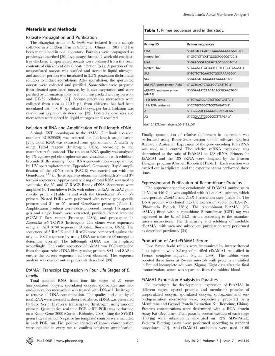
Table 1. Primer sequences used in this study.
Primer ID Primer sequences
EtA1 59-AAGTGCGAGTCTAAAGGGGGCGGTGT-39
Nested-EtA1 59-GTGTCTTCATTGGGCTGGCCGTCG-39
EtA2 59-GAAGGGAAGTAGTAGCCGGGACT-39
Nested-EtA2 59-GGGACTTGTTGCTGCTTCGTCTTGAGAT-39
SA1 59-TCTTCTTCAACTCTGGCAAAAGC-39
SA2 59-GAAGTGAAAAGGCGAAAACT-39
qRT-PCR sense primer (AMA1) 59-GCTGACTCTGCTGCTCATTTG-39
qRT-PCR antisense primer(AMA1)
59-GGAATATCAAGAGACCGCAACTG-39
18Sr RNA sense 59-TGTAGTGGAGTCTTGGTGATTC-39
18Sr RNA antisense 59-CCTGCTGCCTTCCTTAGATG-39
A1 59-CGGGATCCGGGGTGCAGCACAA-39
A2 59-CGGAATTCGCCCCCTTTAGA-39
doi:10.1371/journal.pone.0041115.t001
Eimeria tenella Apical Membrane Antigen-1
PLoS ONE | www.plosone.org 2 July 2012 | Volume 7 | Issue 7 | e41115

Figure 1. Characterization of the EtAMA1 gene. (A) Multiple-sequence alignment of AMA1 proteins from E. tenella (Et), E. maxima (Em), N.caninum (Nc), T. gondii (Tg) and P. falciparum (Pf). Strictly conserved residues are indicated with a black background, and five different cysteineresidues with species of Plasmodium in DIII are indicated with a gray arrowhead. The cleavage site of the EtAMA1 putative signal peptide is indicatedby an arrowhead, and the transmembrane region is indicated by ( ); cysteine residues that formed disulfide bonds in EtAMA1 are indicated by domain(I, II and III) and bond (a, b and c) designations according to TgAMA1. (B) Phylogenetic tree of AMA1 proteins from E. maxima (Em), E. tenella (Et), N.caninum (Nc), T. gondii (Tg), P. falciparum (Pf), P. vivax (Pv), B. bovis (Bbo), B. gibsoni (Bg),and P. bigemina (Pbi).doi:10.1371/journal.pone.0041115.g001
Eimeria tenella Apical Membrane Antigen-1
PLoS ONE | www.plosone.org 3 July 2012 | Volume 7 | Issue 7 | e41115

diluted and mouse monoclonal anti-tubulin antibodies (Beyotime)
diluted 1:2000 were used as controls. Secondary antibodies
conjugated to horseradish peroxidase conjugated (HRP) (Sigma)
were added 1:5000 diluted.
Inhibition of Host-cell Invasion in VitroThe chicken embryo fibroblast cell line DF-1 (ATCC) was
used for the inhibition assays [35]. The antibodies were purified
with Protein A Agarose (Beyotime). Sporozoites were labeled
using the dye carboxyfluorescein diacetate, succinimidyl ester
(CFDA SE, Beyotime) according to the manufacturer’s instruc-
tions. The labeled sporozoites (16105) were resuspended in
1 mL of Dulbecco’s modified Eagle’s medium (DMEM; Gibco,
USA) containing 50, 100, 200 and 400 mg/mL of anti-
rEtAMA1 antibodies, and incubated 2 h at 37uC. Anti-GST
IgG antibodies and parallel wells without the antibodies were
used as controls. The sporozoites were then washed twice in
sterile phosphate buffered saline (PBS) by centrifugation at
20006g for 5 min and infected 16105 DF-1 cells in a 24-well
plate (Corning, USA). After 12 h of culture at 41uC, cells were
collected and analyzed by flow cytometry (Beckman Coulter,
USA). The infected cells, non-infected cells and free sporozoites
were gated using the software RXP for subsequent delineation
and counting of the infected (containing the labeled sporozoites)
and non-infected (fluorescence-free) cells. All assays were
performed in triplicate. The deduced percentages of infected
cells in the presence or absence of inhibitory antibodies were
used for the calculation of the inhibition rates, as previously
described [36].
Immunofluorescence Analysis of EtAMA1 during the FirstSchizogony
Freshly excysted sporozoites were incubated for 1 h at 41uCin either PBS or complete DMEM and air-dried on a glass slide
before fixation. Infected DF-1 cells were maintained on glass
slides in a 6-well plate (Corning) and collected at different time
points for fixation. Sporozoites or cells were fixed and
permeabilized with 2% paraformaldehyde and 1% Triton X-
100 for 10 min at room temperature and washed three times.
Antibodies against rEtAMA1 were incubated with the purified
recombinant protein GST at 37uC for 1 h to remove anti-GST
antibodies and then incubated (1:100 dilution) with the cells or
sporozoites for 2 h at 37uC. After being washed three times,
goat anti-rabbit IgG (H+L) fluorescein isothiocyanate (FITC)-
conjugated antibody (1:1000 dilution, Sigma) was added to the
glass slides. Nuclei were labeled with 10 mg/mL of 49,6-
diamidino-2-phenylindole (DAPI) (Beyotime) for 5 min and
washed five times. All dilutions and washes were performed in
0.05% Tween-20 PBS. Before observations under £uorescence
microscopy (Nikon, Japan), 10 mL of 1,4-diazabicyclo[2.2.2]oc-
tane (DABCO; Sigma) were added.
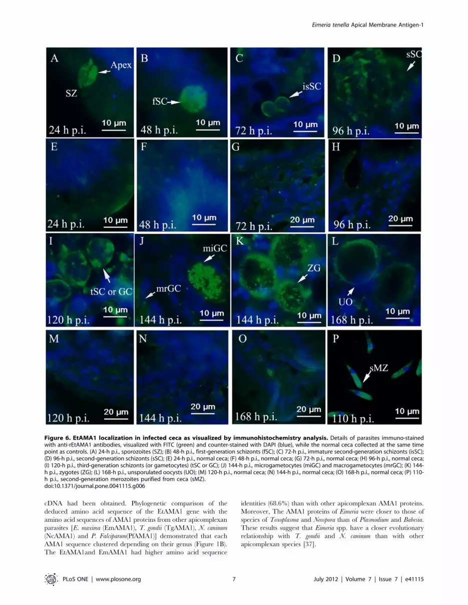
Immunohistochemistry Analysis of EtAMA1 during theParasitic Life-cycle
Three-day-old chickens were infected with 16104 sporulated
oocysts. Their ceca were collected at 24-h intervals and fixed with
4% paraformaldehyde in PBS at room temperature. As controls,
the ceca of uninfected chickens were also collected at the same
time points. The ceca were embedded within optimal cutting
temperature (OCT) compound mounted in a cryostat for
production of a rapid (frozen) section 10-mm thick for immuno-
histochemistry analysis. Second-generation merozoites purified
from the ceca of chickens were also prepared for observation. The
samples were dyed using the immunofluorescence analysis
procedure described above.
Results
Cloning and Molecular Characterization of EtAMA1The full-length cDNA of EtAMA1 was 2349 bp (GenBank
accession number: JN032081), including a single open reading
frame (ORF) of 1608 bp, encoding a polypeptide of 535 amino
acid residues. SignalP program analysis revealed that the N-
terminus of EtAMA1 contained a signal peptide of 23 amino acids,
with a hypothetical cleavage site located between glycine and
valine. The EtAMA1 composed an ectodomain (from 24 to 446
aa), a transmembrane domain (from 447 to 469 aa) and a
cytoplasmic domain (from 470 to 535 aa), as predicted using
TMHMM2.0 (Figure 1A).
To determine the phylogenetically conserved functional regions
of EtAMA1, its deduced amino acid sequence was aligned with the
AMA1 amino acid sequences from eight apicomplexan parasites
[E. maxima (CBL80633.1), Toxoplasma gondii (XP_002364854),
Neospora caninum (CBZ53079), P. falciparum (AAC15773.1), P. vivax
(ACY68740), B. bovis (ACM44019), B. bigemina (BAH22706) and B.
Figure 2. EtAMA1 mRNA expression levels in differentdevelopmental stages of E. tenella as revealed by SYBR Greenquantitative real-time PCR. EtAMA1 mRNA levels in sporulatedoocysts (SO), unsporulated oocysts (UO), sporozoites (SZ) and second-generation merozoites (MZ) were normalized to the 18S rRNA level ofthe corresponding stage. *P,0.05, n = 3.doi:10.1371/journal.pone.0041115.g002
Figure 3. Western Blotting analysis of EtAMA1 in different lifestages of E. tenella probed with anti-rEtAMA1 antibodies. (A)The cytosol proteins of parasites. (B) The membrane proteins ofparasites. Lanes: UO, unsporulated oocysts; SO, sporulated oocysts; SZ,sporozoites; MZ, second-generation merozoites.doi:10.1371/journal.pone.0041115.g003
Eimeria tenella Apical Membrane Antigen-1
PLoS ONE | www.plosone.org 4 July 2012 | Volume 7 | Issue 7 | e41115

gibsoni (ABD04040)]. The phylogenetic tree was constructed by
MEGA 4.0 software (Figure 1B). As calculated using Needleman-
Wunsch global alignment, the highest amino acid sequence
homology between EtAMA1 and EmAMA1 was 68.6% and the
lowest homology between EtAMA1 and B. bovis AMA1 was
36.1%. Sixteen cysteine residues were conserved in E. maxima and
E. tenella, whereas eight cysteine residues in the putative domains I
and II were conserved in E. tenella, E. maxima, T. gondii, N. caninum
and P. falciparum, as calculated using Genetyx software (Develop-
ment Co., Ltd, Japan) (Figure 1A).
EtAMA1 Transcript Expression in Four Life Stages of E.tenella
The expression of EtAMA1 showed high variation stages among
the four life stages of E. tenella. EtAMA1 transcripts were abundant
in sporozoites (30-fold higher than in unsporulated oocysts)
(P,0.05) and only moderately expressed in sporulated oocysts
and second-generation merozoites (no more than three-fold higher
than in unsporulated oocysts) (Figure 2).
EtAMA1 Protein Expression in Different Life Stages of E.tenella
The presence of EtAMA1 in unsporulated oocysts, sporulated
oocysts, sporozoites and second-generation merozoites was deter-
mined by immunoblotting using antibodies obtained from rabbits
immunized with rEtAMA1 as described above. Anti-tubulin
monoclonal antibodies were used as controls to reveal a similar
quantity of total proteins in all samples. Western Blotting of the
cytosol proteins revealed that anti-rEtAMA1 antibodies strongly
labeled the same 58-kDa bands in sporulated oocysts and
sporozoites, but weaker reactivity in unsporulated oocysts and
second-generation merozoites (Figure 3A). The membrance
proteins probed with anti-rEtAMA1 antibodies showed EtAMA1
had high expression in sporulated oocysts, sporozoites and second-
generation merozoites, while little expression in unsporulated
oocysts.
Anti-rEtAMA1 Antibodies Inhibited Host-cell Invasion ofE. tenella Sporozoites
The polyclonal antibodies derived from rEtAMA1-vaccinated
rabbits were tested for the ability to inhibit the invasion of
cultured DF-1 cells with E. tenella sporozoites. Representative
flow cytometry charts used to determine the inhibitory activity of
the anti-rEtAMA1 antibodies on labeled sporozoites were
presented in Figure 4A. There was an obvious distinction
between cells infected with sporozoites pre-incubated without
antibodies (as a negative control) and those infected with
sporozoites with inhibitory anti-rEtAMA1 antibodies at a
concentration of 200 mg/mL. Pretreatment with the anti-
rEtAMA1 antibodies significantly decreased the invasion capacity
of sporozoites, and the observed inhibition effect was dose
dependent. Under the experimental conditions, the inhibition
plateau of 6962.55% was reached at an antibody concentration
Figure 4. Inhibition of sporozoite invasion in vitro by antibodies specific to rEtAMA1. (A) Representative flow cytometry charts used todetermine the inhibitory activity of the antibody on labeled sporozoites of E. tenella. Left panel, non-infected cells; Middle panel, infected cells as anegative control; right panel: infection with sporozoites preincubated with inhibitory antibodies of rEtAMA1 at a concentration of 400 mg/mL. (B)Dose-dependent inhibition of sporozoite invasion by antibodies specific to rEtAMA1 and GST. The results are representative of three individualexperiments, and the error bars indicate standard deviations. (*) According to the Student’s t-test, the differences between the treatment with anti-GST antibodies and anti-rEtAMA1 antibodies at the same IgG concentration were significant (P,0.05).doi:10.1371/journal.pone.0041115.g004
Eimeria tenella Apical Membrane Antigen-1
PLoS ONE | www.plosone.org 5 July 2012 | Volume 7 | Issue 7 | e41115

of 400 mg/mL. By comparative analysis, anti-GST antibodies did
not have a significant effect on invasion (8.561.23%) (Figure 4B).
EtAMA1 Localization during in Vitro Infection byImmunofluorescence Analysis
Anti-rEtAMA1 antibodies were applied to detect the localiza-
tion of EtAMA1 in sporozoites and during the first schizogony.
EtAMA1 exhibited a homogenous distribution pattern throughout
the membrane of sporozoites incubated in PBS (Figure 5A). By
contrast, when sporozoites were incubated in culture medium,
EtAMA1 expression increased (Figure 5B). After sporozoites
invaded host cells, the localization of EtAMA1 was mainly on
the apical end of the parasites (Figure 5C). Observations 24 h p.i.
showed that EtAMA1 protein expression increased and was
distributed in trophozoites (Figure 5D, E). The labeled EtAMA1
eventually became uniformly dispersed in immature and mature
schizonts (Figure 5F, G). After merozoites were released from host
cells, EtAMA1 was translocated to the merozoite surface, although
some remained in the PV membrane (Figure 5H).
EtAMA1 Localization during in Vivo Infection byImmunohistochemistry Analysis
When sporozoites adhered to cecal epithelial cells, EtAMA1 was
expressed on the apical end of the parasites (Figure 6A). As
sporozoites developed into first-generation schizonts, EtAMA1 was
expressed in first-generation merozoites (Figure 6B). After the
parasites developed into immature second-generation schizonts,
the expression of EtAMA1 was mainly around the schizont
membrane (Figure 6C). When second-generation schizonts were
mature, EtAMA1 was located in merozoites (Figure 6D). Once the
parasites had developed into third-generation schizonts (or
gametocytes), EtAMA1 expression was increased and mainly
around the schizont/gametocyte membrane (Figure 6I). Interest-
ingly, when the parasites entered into the stage of sexual
reproduction, the expression of EtAMA1 was significantly
increased in microgametocytes and decreased in macrogameto-
cytes (Figure 6J). Simultaneously, EtAMA1 became concentrated
in the cytoplasm of fertilized macrogametes as small defined
regions and was also highly expressed around the membrane of the
parasites in the contents of ceca (Figure 6K). However, as the
parasites developed into unsporulated oocysts, EtAMA1 expres-
sion was reduced and was only detected around some oocysts wall
(Figure 6L). In free second-generation merozoites, EtAMA1 was
expressed mainly at the apical end of the parasites (Figure 6P).
Discussion
In the present work, we described, for the first time, the
molecular characterization of AMA1 in E. tenella. The full-length
cDNA of EtAMA1 was 2349 bp, with a 1608 bp ORF encoding a
protein of 535 amino acids. The 59 untranslated region was
located 469 bp upstream of the putative start codon (ATG). The
39 untranslated region of 267 nucleotides ended at a poly (A) tail.
These sequence characteristics all indicated that a full-length
Figure 5. EtAMA1 localization in DF-1 cell infection as visualized by immunofluorescence analysis. Details of parasites immuno-stainedwith anti-rEtAMA1 antibodies, visualized with FITC (green) and counter-stained with DAPI (blue). (A) Sporozoites (SZ) were incubated in PBS or (B)complete medium (CM) at 41uC. Infected DF-1 cells were collected at different time points p.i. (C) 2-h p.i., intracellular sporozoites (iSZ); (D) 24-h p.i.,intracellular trophozoite (Tropho); (E) 36-h p. i., immature schizont (iSc); (F) 48-h p.i., immature schizont (iSc); (G) 60-h p.i., mature schizont (mSc); (H)72-h p.i., merozoites (MZ).doi:10.1371/journal.pone.0041115.g005
Eimeria tenella Apical Membrane Antigen-1
PLoS ONE | www.plosone.org 6 July 2012 | Volume 7 | Issue 7 | e41115
cDNA had been obtained. Phylogenetic comparison of the
deduced amino acid sequence of the EtAMA1 gene with the
amino acid sequences of AMA1 proteins from other apicomplexan
parasites [E. maxima (EmAMA1), T. gondii (TgAMA1), N. caninum
(NcAMA1) and P. Falciparum(PfAMA1)] demonstrated that each
AMA1 sequence clustered depending on their genus (Figure 1B).
The EtAMA1and EmAMA1 had higher amino acid sequence
identities (68.6%) than with other apicomplexan AMA1 proteins.
Moreover, The AMA1 proteins of Eimeria were closer to those of
species of Toxoplasma and Neospora than of Plasmodium and Babesia.
These results suggest that Eimeria spp. have a closer evolutionary
relationship with T. gondii and N. caninum than with other
apicomplexan species [37].
Figure 6. EtAMA1 localization in infected ceca as visualized by immunohistochemistry analysis. Details of parasites immuno-stainedwith anti-rEtAMA1 antibodies, visualized with FITC (green) and counter-stained with DAPI (blue), while the normal ceca collected at the same timepoint as controls. (A) 24-h p.i., sporozoites (SZ); (B) 48-h p.i., first-generation schizonts (fSC); (C) 72-h p.i., immature second-generation schizonts (isSC);(D) 96-h p.i., second-generation schizonts (sSC); (E) 24-h p.i., normal ceca; (F) 48-h p.i., normal ceca; (G) 72-h p.i., normal ceca; (H) 96-h p.i., normal ceca;(I) 120-h p.i., third-generation schizonts (or gametocytes) (tSC or GC); (J) 144-h p.i., microgametocytes (miGC) and macrogametocytes (mrGC); (K) 144-h p.i., zygotes (ZG); (L) 168-h p.i., unsporulated oocysts (UO); (M) 120-h p.i., normal ceca; (N) 144-h p.i., normal ceca; (O) 168-h p.i., normal ceca; (P) 110-h p.i., second-generation merozoites purified from ceca (sMZ).doi:10.1371/journal.pone.0041115.g006
Eimeria tenella Apical Membrane Antigen-1
PLoS ONE | www.plosone.org 7 July 2012 | Volume 7 | Issue 7 | e41115

AMA1 proteins are type I integral transmembrane proteins that
are highly conserved and present in all Plasmodium [14], Toxoplasma
[21], Babesia [22] and Neospora [23] species. Analysis of the amino
acid sequences revealed that full-length EtAMA1 contained a
predicted transmembrane region (from 479 to 501 bp) near the C
terminus, as seen in other AMA1 proteins. The predicted signal
peptide of EtAMA1 contained 23 amino acids, which were
associated with the secretory pathway. Many of the conserved and
polymorphic residues in AMA1 proteins have been reported.
EtAMA1 also contained 16 cysteine amino acid residues, which
contribute to disulfide bonding, the pattern of which prompted the
suggestion that the mature ectodomain folds as an N-terminal pro-
sequence and three domains (DI, DII and DIII) [38]. Although
there were many differences between the protein sequence of
EtAMA1 and EmAMA1, both contained 16 highly conserved
cysteines and exhibited identical localization. TgAMA1 and
NcAMA1 included 13 cysteine residues with EtAMA1. The
AMA1 proteins of Plasmodium spp. were more distantly related to
those of E. tenella, E. maxima, T. gondii and N. caninum, but all the
AMA1 proteins contained highly conserved eight cysteine amino
acid residues in DI and DII which belong to the PAN module
superfamily, which is commonly associated with carbohydrate or
protein receptor-binding functions [38]. These results revealed
that the number and localization of cysteine residues on the
sequences of the AMA1 proteins might be conserved depending
on their genus [23]; moreover, the differences in localization of the
cysteine residues usually occurred in the DIII domain.
The correct folding of AMA1, as in case of Plasmodium antigens,
has been shown to be crucial for its immunological activity [39]. In
addition, the PfAMA1 ectodomain expressed in E. coli can be
correctly folded and highly immunogenic [40]. In this study, the
cDNA fragment of the EtAMA1 ectodomain was expressed in E.
coli. The rEtAMA1 was initially insoluble; however, after refolding
and purification, the rEtAMA1 became more soluble. The
antibodies raised against sporozoites were reactive with rEtA-
MA1(date not show); Moreover, anti-rEtAMA1 antibodies inhib-
ited host-cell invasion by the parasites in vitro and recognized the
parasitic AMA1, as shown by Western Blotting, immunofluores-
cence analysis and immunohistochemistry analysis. These obser-
vations suggest that the refolded rEtAMA1 has immunological
activity.
In our study, both qRT-PCR and Western Blotting demon-
strated that the newly identified EtAMA1 was constitutively
expressed in all four developmental stages. Proteomic comparison
of E. tenella showed EtAMA1 was found in sporozoites and
EtAMA2 was found in merozoites [27]. As Western Blotting
analysis of second-generation merozoites showed EtAMA1
expression was little in soluble proteins, high in membrance
proteins (Figure 3), soluble proteomic analysis of merozoites might
miss the menbrance EtAMA1. There was one conflicting data for
RNA versus protein levels of EtAMA1 expression in sporulated
oocysts. As we know, oocysts of Eimeria spp. are able to persist in
the environment for years by oxidation of lipids supporting the
metabolism in sporulated oocysts during dormancy [41,42]. So we
thought even EtAMA1 transcripts were moderately expressed,
EtAMA1 protein could remain a high expression in sporulated
oocysts.
AMA1 has been an essential protein in apicomplexan invasion,
which can be identified in invasive zoites [43]. qRT-PCR and
Western Blotting analysis showed EtAMA1 was high expressed in
sporozoites. Invasion inhibition assays revealed that rabbit
antiserum against recombinant EtAMA1 blocked invasion of host
cells by approximately 70%. Localization of EtAMA1 in DF-1
cells or in chicken ceca showed that the expression of EtAMA1 on
the sporozoite surface increased when the parasites invaded the
cells. These data therefore supported a more direct role for
EtAMA1 in host invasion.
In the localization of EtAMA1, we observed the protein was not
only found at the apical end, but also in the entire surface of the
parasite even during invasion, which could also observed in
Toxoplasma parasites [43]. Later during the parasite development
in DF-1 cells, EtAMA1 was found on the merozoite surface and in
the PV membrane. The PV is a crucial structure that protects the
parasite against the antagonistic environment of the host cell [12].
When merozoites escape from mature schizonts to invade new
host cells, they must pass through the PV membrane. Thus, we
suggest that EtAMA1 has an important role in merozoite release.
In the sexual reproduction stage of E. tenella, EtAMA1 appeared
to be expressed in gametocytes. However, not all the gametocytes
examined expressed EtAMA1 and the protein was expressed in the
form of numerous granules. In E. tenella, sexual reproduction
involves a microgamete initiatively entering a macrogamete to
form a zygote, which then develops into unsporulated oocyst.
Therefore, the microgamete also requires an ‘invasion’ process.
Many parasites, appearing to be zygotes in form, were observed in
the ceca; therefore, we came to a conclusion that these
gametocytes were microgametocytes with a high EtAMA1
expression, which needed further replication experiments. It may
lead to the interesting observation that EtAMA1 is important not
only in host invasion, but also in ‘self’ invasion.
In conclusion, we cloned and characterized the AMA1 of E.
tenella and, as a result, have added significantly to current
understanding of its role during parasite invasion. Given the
importance of EtAMA1 in invasion and parasite development, this
study is likely to have implications for both novel chemo- and
immuno-therapeutic approaches to interfering with EtAMA1
function.
Acknowledgments
We sincerely appreciate Chan Ding (CAAS) for generously providing the
DF-1 cell line used in this study. We also thank Zhixin Zhao and Beimin
Zhang for their excellent technical assistance.
Author Contributions
Conceived and designed the experiments: JL BH LJ. Performed the
experiments: LJ HH HD QZ SZ. Analyzed the data: LJ. Contributed
reagents/materials/analysis tools: HH QZ. Wrote the paper: LJ.
References
1. Shirley MW, Ivens A, Gruber A, Madeira AM, Wan KL, et al. (2004) The
Eimeria genome projects: a sequence of events. Trends Parasitol 20(5): 199–201.
2. Williams RB (1998) Epidemiological aspects of the use of live anticoccidial
vaccines for chickens. Int J Parasitol 28(7): 1089–1098.
3. Allen PC, Fetterer RH (2002) Recent advances in biology and immunobiology of
Eimeria species and in diagnosis and control of infection with these coccidian
parasites of poultry. Clin Microbiol Rev 15(1): 58–65.
4. Williams RB (2006) Tracing the emergence of drug-resistance in coccidian
(Eimeria spp.) of commercial broiler £ocks medicated with decoquinate for the
first time in the United Kingdom. Vet Parasitol 135(1): 1–14.
5. Morris GM, Woods WG, Richard DG, Gasser RB (2007) Investigating a
persistent coccidiosis problem on a commercial broiler-breeder farm utilising
PCR-coupled capillary electrophoresis. Parasitol Res 101(3): 583–589.
6. Innes EA, Vermeulen AN (2006) Vaccination as a control strategy against the
coccidial parasites Eimeria, Toxoplasma and Neospora. Parasitology 133: 145–168.
7. Dalloul RA, Lillehoj HS (2006) Poultry coccidiosis: recent advancements in
control measures and vaccine development. Expert Rev Vaccines 5(1): 143–163.
8. Vermeulen AN, Schaap DC, Schetters TP (2001) Control of coccidiosis in
chickens by vaccination. Vet Parasitol 100(1–2): 13–20.
Eimeria tenella Apical Membrane Antigen-1
PLoS ONE | www.plosone.org 8 July 2012 | Volume 7 | Issue 7 | e41115
9. Shirley MW, Smith AL, Blake DP (2007) Challenges in the successful control of
the avian coccidia. Vaccine 25(30): 5540–5547.
10. Han HY, Lin JJ, Zhao QP, Dong H, Jiang LL, et al. (2010) Identification of
differentially expressed genes in early stages of Eimeria tenella by suppression
subtractive hybridization and cDNA microarray. J Parasitol 96(1): 95–102.
11. Tabares E, Ferguson D, Clark J, Soon PE, Wan KL, et al. (2004) Eimeria tenella
sporozoites and merozoites differentially express glycosylphosphatidylinositol-
anchored variant surface proteins. Mol Biochem Parasitol 135(1): 123–132.
12. Daszak P (1999) Zoite migration during Eimeria tenella infection: parasite
adaptation to host defences. Parasitol Today 15(2): 67–72.
13. Sasai K, Fetterer RH, Lillehoj H, Matusra S, Constantinoiu CC, et al. (2008)
Characterization of monoclonal antibodies that recognize the Eimeria tenella
microneme protein MIC2. J Parasitol 94(6): 1432–1434.
14. Thomas AW, Deans JA, Mitchell GH, Alderson T, Cohen S (1984) The Fab
fragments of monoclonal IgG to a merozoite surface antigen inhibit Plasmodium
knowlesi invasion of erythrocytes. Mol Biochem Parasitol 13(2): 187–199.
15. Alexander DL, Mital J, Ward GE, Bradley P, Boothroyd JC (2005) Identification
of the moving junction complex of Toxoplasma gondii: a collaboration between
distinct secretory organelles. PLoS Pathog 1(2): e17.
16. Santos JM, Ferguson DJP, Blackman MJ, Soldati-Favre D (2011) Intramem-
brane cleavage of AMA1 triggers Toxoplasma to switch from an invasive to a
replicative mode. Science 331(6016): 473–477.
17. Besteiro S, Michelin A, Poncet J, Dubremetz JF, Lebrun M (2009) Export of a
Toxoplasma gondii rhoptry neck protein complex at the host cell membrane to
form the moving junction during invasion. PLoS Pathog 5(2): e1000309.
18. Lamarque M, Besteiro S, Papoin J, Roques M, Vulliez-Le Normand B, et al.
(2011) The RON2-AMA1 interaction is a critical step in moving junction-
dependent invasion by apicomplexan parasites. PLos Pathog 7(2): e1001276.
19. Curtidor H, Patino LC, Arevalo-Pinzon G, Patarroyo ME, Patarroyo MA (2011)
Identification of the Plasmodium falciparum rhoptry neck protein 5 (PfRON5).
Gene 474(1–2): 22–28.
20. Remarque EJ, Faber BW, Kocken CH, Thomas AW (2008) Apical membrane
antigen 1: a malaria vaccine candidate in review. Trends Parasitol 24(2): 74–84.
21. Donahue CG, Carruthers VB, Gilk SD, Ward GE (2000) The Toxoplasma
homolog of Plasmodium apical membrane antigen-1 (AMA-1) is a microneme
protein secreted in response to elevated intracellular calcium levels. Mol
Biochem Parasitol 111(1): 15–30.
22. Gaffar FR, Yatsuda AP, Franssen FFJ, Vries E (2004) Erythrocyte invasion by
Babesia bovis merozoites is inhibited by polyclonal antisera directed against
peptides derived from a homologue of Plasmodium falciparum apical membrane
antigen 1. Infect Immun 72(5): 2947–2955.
23. Zhang H, Compaore MK, Lee EG, Liao M, Zhang G, et al. (2007) Apical
membrane antigen 1 is a cross-reactive antigen between Neospora caninum and
Toxoplasma gondii, and the anti-NcAMA1 antibody inhibits host cell invasion by
both parasites. Mol Biochem Parasitol 151(2): 205–212.
24. Tonukari NJ (2010) Theileria parva apical membrane antigen-1 (AMA-1) shares
conserved sequences with apicomplexan homologs. Int J Biotech Mol Biol Res
1(3): 36–41.
25. Ng ST, Sanusi Jangi M, Shirley MW, Tomley FM, Wan KL (2002)
Comparative EST analyses provide insights into gene expression in two asexual
developmental stages of Eimeria tenella. Exp Parasitol 101(2–3): 168–173.
26. Klotz C, Marhofer RJ, Selzer PM, Lucius R, Pogonka T (2005) Eimeria tenella:
Identification of secretory and surface proteins from expressed sequence tags.Exp Parasitol 111(1): 14–23.
27. Lal K, Bromley E, Oakes R, Prieto JH, Sanderson SJ, et al. (2009) Proteomic
comparison of four Eimeria tenella life-cycle stages: unsporulated oocyst,sporulated oocyst, sporozoite and second-generation merozoite. Proteomics
9(19): 4566–4576.28. Blake DP, Billington KJ, Copestake SL, Oakes RD, Quail MA, et al. (2011)
Genetic mapping identifies novel highly protective antigens for an apicomplexan
parasite. PLoS Pathog 7(2): e1001279.29. Peroval M, Pery P, Labbe M (2006) The heat shock protein 90 of Eimeria tenella is
essential for invasion of host cell and schizont growth. Int J Parasitol 36(10–11):1205–1215.
30. Tomley F (1997) Techniques for isolation and characterization of apicalorganelles from Eimeria tenella sporozoites. Methods 13(2): 171–176.
31. Shirley MW (1995) Eimeria species and strains of chickens. In Biotechnology
Guidelines on Techniques in Coccidiosis Research. The European CommissionDGXII, Luxembourg City, Luxembourg, 24.
32. Xie MQ, Gilbert JM, Fuller AL, McDougald LR (1990) A new method forpurification of Eimeria tenella merozoites. Parasitol Res 76(7): 566–569.
33. Jiang LL, Lin JJ, Han HY, Dong H, Zhao QP, et al. (2011) Identification and
partial characterization of a serine protease inhibitor (serpin) of Eimeria tenella.Parasitol Res 110(2): 865–874.
34. Kimbitaa EN, Xuan X, Huang X, Miyazawa T, Fukumoto S, et al. (2001)Serodiagnosis of Toxoplasma gondii infection in cats by enzyme-linked immuno-
sorbent assay using recombinant SAG1. Vet Parasitol 102(1–2): 35–44.35. Jiang LL, Lin JJ, Han HY, Dong H, Zhao QP, et al. (2011) Establishment and
application of DF-1 cell culture for the sporozoites of Eimeria tenella. Chin Vet Sci
41: 551–556.36. Jahn D, Matros A, Bakulina AY, Tiedemann J, Schubert U, et al. (2009) Model
structure of the immunodominant surface antigen of Eimeria tenella identified as atarget for sporozoite-neutralizing monoclonal antibody. Parasitol Res 105(3):
655–668.
37. Li L, Brunk BP, Kissinger JC, Pape D, Tang K, et al. (2003) Gene discovery inthe apicomplexa as revealed by EST sequencing and assembly of a comparative
gene database. Genome Res 13(3): 443–454.38. Pizarro JC, Vulliez-Le Normand B, Chesne-Seck ML, Collins CR, Withers-
Martinez C, et al. (2005) Crystal structure of the malaria vaccine candidateapical membrane antigen 1. Science 308(5720): 408–411.
39. Hodder AN, Crewther PE, Anders RF (2001) Specificity of the protective
antibody response to apical membrane antigen 1. Infect Immun 69(5): 3286–3294.
40. Dutta S, Lalitha PV, Ware LA, Barbosa A, Moch JK, et al. (2002) Purification,characterization, and immunogenicity of the refolded ectodomain of the
Plasmodium falciparum apical membrane antigen 1 expressed in Escherichia coli.
Infect Immun 70(6): 3101–3110.41. Belli SI, Walker RA, Flowers SA (2005) Global protein expression analysis in
apicomplexan parasites: Current status. Proteomics 5(4): 918–924.42. Wilson PAG, Fairbairnz D (1961) Biochemistry of sporulation in oocysts of
Eimeria acervulina. J Eukaryot Microbiol 8(4): 410–416.43. Tyler JS, Treeck M, Boothroyd JC (2011) Focus on the ringleader: the role of
AMA1 in apicomplexan invasion and replication. Trends Parasitol 27(9): 410–
420.
Eimeria tenella Apical Membrane Antigen-1
PLoS ONE | www.plosone.org 9 July 2012 | Volume 7 | Issue 7 | e41115


