Embed Size (px)
Citation preview

Phagocytosis is inhibited by autophagic induction in murine macrophages
José Geraldo Bomfim Lima a, Christiana de Freitas Vinhas a,b, Ivana Nunes Gomes a,Carine Machado Azevedo c, Ricardo Ribeiro dos Santos c, Marcos André Vannier-Santos d,Patrícia Sampaio Tavares Veras a,⇑
a Laboratório de Patologia e Biointervenção, IGM-FIOCRUZ, Bahia, Brazilb Escola Bahiana de Medicina e Saúde Pública, Bahia, Brazilc Laboratório de Engenharia Tecidual e Imunofarmacologia, IGM-FIOCRUZ, Bahia, Brazild Laboratório de Biologia Parasitária, IGM-FIOCRUZ, Bahia, Brazil
a r t i c l e i n f o
Article history:
Received 13 January 2011
Available online 24 January 2011
Keywords:
Autophagy
Phagocytosis
Large particles
Starvation
Rapamycin
Macrophage
a b s t r a c t
Recent studies have demonstrated that communication takes place between the autophagic and phago-
cytic pathways, indicating that the convergence of these two pathways plays an important role in the
innate immune response against intracellular microbes. The present study investigated the effect of auto-
phagic induction on the phagocytic capacity of murine macrophages. Autophagy induced by physiological
and pharmacological means was shown to reduce the phagocytic capacity of murine macrophages,
regardless of cell origin or the nature of the phagocytosed particles themselves. This autophagic inhibi-
tory effect on phagocytosis was shown to be an early and reversible event that results in no loss of cell
viability. Furthermore, the data presented herein demonstrate that the induction of autophagy does not
affect a macrophage’s capacity to recognize and bind to particles, indicating that autophagy does not inhi-
bit the particle recognition process, even though particle internalization is suppressed. The findings
herein support the notion that phagocytosis and autophagy may be interdependent and complementary
processes.
Ó 2011 Elsevier Inc. All rights reserved.
1. Introduction
Originally, autophagy was described as a mechanism that main-tains cellular homeostasis [1] by way of a natural physiologicalprocess that leads to the acquisition of energy in response to stress,as well as being the process responsible for the renewal of cellularorganelles [2]. Autophagy leads to the degradation of proteins, sub-cellular domains and organelles, which is an evolutionarily con-served process occurring naturally in eukaryotic cells and isinducible in response to a variety of stress situations [3]. To date,the known autophagic processes are subdivided into differentpathways, including chaperone-mediated autophagy, microauto-phagy and macroautophagy. Both micro- and macroautophagyare intracellular processes involving macromolecular degradationin eukaryotic cells. In the microautophagic process, the transferof cytosolic components into the lysosomal compartment occursby direct invagination of the lysosomal membrane, while in themacroautophagic process, cytosolic components are sequesteredinto an autophagosome prior to fusion with lysosomes [4].
Macroautophagy, also known as the autophagic process, is trig-gered in response to starvation conditions and regulated by theactivity of the serine–threonine kinase, the mammalian target ofrapamycin (mTOR). This enzyme is activated under nutrient-richconditions. By contrast, mTOR activity is blocked by rapamycin,triggering the formation of autophagosomes under nutrient-richconditions [5]. Furthermore, the autophagic pathway occurs in anorderly manner. Initially, a portion of the cytoplasm and organ-elles, such as endoplasmic reticulum (ER), mitochondria and per-oxisomes, are sequestered by a double-layered membrane whichleads to the formation of an autophagosome. Autophagosomesthen fuse with lysosomes, losing their inner membranes, and thesevesicles become acidic due to the acquisition of proton ATPases inthe outer membranes. Subsequently, autophagosomes acquireother proteins, such as lysosomal enzymes, and this organelle isthen called an autophagolysosome, or autolysosome, which iswhere the degradation of sequestered material occurs [6,7]. Theorigin of the autophagosome double membrane is unknown. Evi-dence indicates that this structure originates from ribosome-devoid regions of the ER [6]. However, it has been recentlysuggested that the plasma membrane may contribute to theformation of the autophagosome membrane [8].
The autophagic process has also been described as a mechanismthat plays a role in the innate and adaptive immune systems
0006-291X/$ - see front matter Ó 2011 Elsevier Inc. All rights reserved.
doi:10.1016/j.bbrc.2011.01.076
⇑ Corresponding author. Address: Laboratório de Patologia e Biointervenção,
CPqGM, FIOCRUZ, RuaWaldemar Falcão, 121, Candeal, Salvador/BA, CEP 40296-710,
Brazil. Fax: +55 71 3176 2290.
E-mail address: [email protected] (P.S.T. Veras).
Biochemical and Biophysical Research Communications 405 (2011) 604–609
Contents lists available at ScienceDirect
Biochemical and Biophysical Research Communications
journal homepage: www.elsevier .com/locate /ybbrc
involved in intracellular infection by microorganisms. This cellularmechanism may protect host cells and promote the establishmentof infection, depending on the nature of the pathogen [9–11]. Thesedata suggest that the course of infection depends not only on theinitial steps of host cell–pathogen interaction, but also on subse-quent events in which pathogen-induced vacuoles interact withautophagic vesicles.
Phagocytosis is the primary mechanism by which unicellulareukaryotes obtain nutrients [12]. In metazoans, phagocytosis isimportant not only for cell nutrition, but is also involved in inter-nalization, killing and removal of foreign particles. In mammals,it is a mechanism involved in tissue remodeling during organismdevelopment, senescent cell removal and inflammation. Addition-ally, phagocytosis is one of the most important processes involvedin the mammalian defense against infection by intracellular micro-organisms [12,13].
Recent studies have observed crosstalk between the autophagicand phagocytic pathways [14,15]. Fusion between nascent phago-somes and autophagosomes has been recently described [16]. Thisphenomenon suggests that autophagy may play a complementaryrole during phagocytosis. Furthermore, it has recently been dem-onstrated that in the macrophage cell line J774, the induction ofautophagy enhances bacterial uptake prior to phagocytosis [17].
The present study aimed to evaluate possible connections be-tween autophagic and phagocytic pathways by determining the ef-fect of autophagic induction on the phagocytic capacity of murinemacrophages. We hypothesized that the induction of autophagyprior to phagocytosis would alter the phagocytic capacity ofmacrophages.
2. Materials and methods
2.1. Macrophage obtainment and cultivation
All experiments were performed according to the standards ofthe Ethics Committee on Animal Experimentation at the OswaldoCruz Foundation – CPqGM/FIOCRUZ. Thioglycolate-elicited inflam-matory peritoneal macrophages from CBA mice were obtained andcultivated in 24-well plates containing 12-mm glass slides at aconcentration of 2 � 105 macrophages per well as previously de-scribed [18].
2.2. Parasites
The Leishmania amazonensis (strain MHOM/Br88/Ba-125) prom-astigotes used in this study were maintained in axenic culture forup to seven passages, suspended in Schneider’s (Gibco) completemedium supplemented with 10% inactivated fetal calf serum and50 lg/mL of gentamicin (Sigma). All parasites were washed,counted and added to macrophage cultures at a ratio of 10:1.
2.3. Induction of autophagy
To induce physiological autophagy (Starvation), macrophageswere incubated in EBSS nutrient-poor medium (Earl’s Balanced SaltSolution, Sigma) as previously described [19]. To induce pharmaco-logical autophagy, macrophages were incubated in nutrient-richmedium (complete DMEM) in the presence of 50 lg/mL of rapamy-cin (Sigma), a drug that inhibits mTOR kinase activity [5].
2.4. Autophagic effect on phagocytosis
To assess the in vitro effects of autophagic induction on thephagocytic capacity of murine macrophages, the cells were incu-bated under autophagic conditions as described above. Next, heat
inactivated yeast particles (Saccharomyces cerevisiae, Sigma) or L.
amazonensis stationary phase promastigotes were added to macro-phage cultures at a ratio of 10:1 particles or parasites per cell. Thenthe macrophage cultures were fixed in ethanol at 99% for 15 min,stained with H&E and a minimum of 400 cells were counted usinga bright-field light microscope. The percentage of macrophagesthat phagocytosed particles or parasites was calculated as the per-centage of phagocytosis and the number of particles or parasitesper macrophage was quantified. Cell viability was tested usingthe trypan blue exclusion technique which showed that 95–99%of cells incubated under both physiological and pharmacologicalautophagic conditions suffered no loss of cell viability.
2.5. Transmission electron microscopy
To evaluate the ultrastructural aspects of the cells incubatedunder physiological and pharmacological autophagic conditions,macrophage suspensions were distributed in 6-well plates at aconcentration of 2 � 106 per well. At the end of the incubation per-iod, the cells were processed as previously described [20]. Thesamples were embedded in Polybed epoxy resin (Polysciences,Warrington, PA, USA) and after polymerization, ultrathin sectionswere obtained. The sections were contrasted and then observedusing a transmission electron microscope (JEOL JEM 1230 at80 kV).
2.6. Fluorescence microscopy
Macrophages were incubated in physiological and pharmaco-logical autophagic conditions for 2 h at 37 °C 5% CO2 and fluoresceinisothiocyanate-labeled zymosan particles (FITC-zymosan, Sigma)were added at a ratio of 10 particles per macrophage (10:1). Theculture plates were centrifuged (500g) at 4 °C for 5 min and thenincubated for 10 min at 4 °C, a temperature at which cells recognizeand bind to particles, but under which internalization is infeasible[21] and differentiation between particle binding and particle inter-nalization is observable. After this time period, cultures werewashed to remove non-adherent particles and fixed in 4% parafor-maldehyde (Sigma) for 20 min. Themacrophage cultures were thenstained with DAPI (Vectashield, Burlingame, CA, USA), whichmarksnuclei in blue, and 0.16 lg/mL of rhodamine–phalloidin (Sigma) toobserve polymerized actin in fixed cells. Particle binding was iden-tified by colocalization of zymosan-FITC stained in green with actincups labeled in red, a phenomenon that is quantifiable due to thepresence of yellow-colored assemblies in plasma membrane. Thepercentage of cells displaying colocalization of actin cups withzymosan-FITC was determined by counting at least 600 cells in fivefields using Image-Pro Plus 6.0.
2.7. Statistical analysis
The graphs and statistical analyses were done using the pro-gram GraphPad Prism, version 5.00 – Incorporate GraphPad Soft-ware. The bar graphs represent the means ± SE (standard error)of a set of experiments done at least in triplicate and repeated fourtimes. The one-way ANOVA and Tukey post-test were used forcomparison between three or more groups. Differences were con-sidered statistically significant when p 6 0.05.
3. Results and discussion
In order to evaluate the effect of autophagy on the phagocyticcapacity of murine macrophages, cell cultures were incubated un-der physiological autophagic conditions as described in Section 2.Transmission electron microscopy was performed to evaluate
J.G.B. Lima et al. / Biochemical and Biophysical Research Communications 405 (2011) 604–609 605
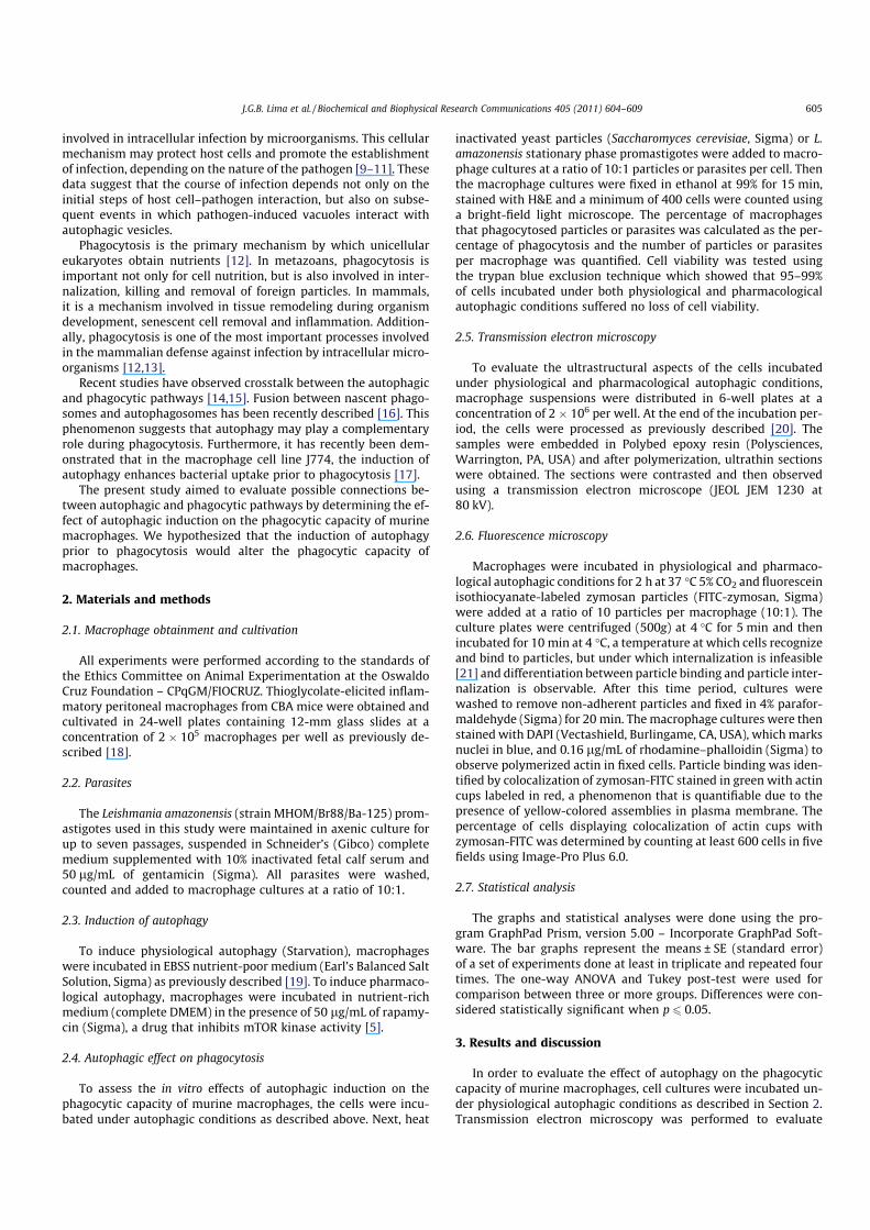
whether morphological alterations were present in macrophagessubjected to starvation conditions. In addition to less electron-dense cytoplasm, numerous double membrane vacuoles character-istic of mature autophagosomes [22] were observed in more than90% of the cells (Fig. 1A and B), confirming that autophagy was in-duced in peritoneal inflammatory CBA macrophages.
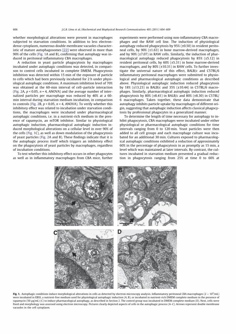
A reduction in yeast particle phagocytosis by macrophagesincubated under autophagic conditions was detected, in compari-son to control cells incubated in complete DMEM. Phagocytosisinhibition was detected within 15 min of the exposure of particleto cells which had been previously incubated for 2 h under physi-ological autophagic conditions. A maximum inhibition level of 70%was obtained at the 60-min interval of cell–particle interaction(Fig. 2A, p < 0.05, n = 4, ANOVA) and the average number of inter-nalized particles per macrophage was reduced by 40% at a 60-min interval during starvation medium incubation, in comparisonto controls (Fig. 2B, p < 0.05, n = 4, ANOVA). To verify whether thisinhibitory effect was related to incubation under starvation condi-tions, the macrophages were incubated under pharmacologicalautophagic conditions, i.e. in a nutrient-rich medium in the pres-ence of rapamycin, an mTOR inhibitor. Similar to physiologicalautophagic induction, pharmacological autophagic induction in-duced morphological alterations on a cellular level in over 90% ofthe cells (Fig. 1C), as well as down modulation of the phagocytosisof yeast particles (Fig. 2A and B). These findings indicate that it isthe autophagic process itself which triggers an inhibitory effecton the phagocytosis of yeast particles by macrophages, regardlessof incubation conditions.
To test whether this inhibitory effect occurs in other phagocytesas well as in inflammatory macrophages from CBA mice, further
experiments were performed using non-inflammatory CBA macro-phages and the RAW cell line. The induction of physiologicalautophagy reduced phagocytosis by 95% (±0.50) in resident perito-neal cells, by 90% (±1.63) in bone marrow-derived macrophages,and by 90% (±7.07) in RAW cells. Similarly, the induction of phar-macological autophagy reduced phagocytosis by 85% (±5.12) inresident peritoneal cells, by 60% (±5.31) in bone marrow-derivedmacrophages, and by 80% (±10.31) in RAW cells. To further inves-tigate the universal nature of this effect, BALB/c and C57BL/6inflammatory peritoneal macrophages were submitted to physio-logical and pharmacological autophagic conditions as describedabove. Physiological autophagic induction reduced phagocytosisby 18% (±13.23) in BALB/c and 35% (±10.44) in C57BL/6 macro-phages. Similarly, pharmacological autophagic induction reducedphagocytosis by 80% (±8.41) in BALB/c and 80% (±8.30) in C57BL/6 macrophages. Taken together, these data demonstrate thatautophagy inhibits particle uptake by macrophages of different ori-gin, suggesting that autophagic induction affects classical phagocy-tosis by professional phagocytes in a generalized manner.
To determine the length of time necessary for autophagy to in-hibit phagocytosis, CBA macrophages were incubated under eitherphysiological or pharmacological autophagic conditions for timeintervals ranging from 0 to 120 min. Yeast particles were thenadded to all cell groups and each macrophage culture was incu-bated for an additional 30 min. Cultures exposed to pharmacolog-ical autophagic conditions exhibited a reduction of approximately60% in the percentage of phagocytosis in as promptly as 15 min, alevel which was maintained at later intervals. By contrast, the cul-tures incubated in starvation medium presented a gradual reduc-tion in phagocytosis ranging from 25% at time 0 to 60% at
Fig. 1. Autophagic conditions induce morphological alterations in cells as detected by electron microscopy analysis. Inflammatory peritoneal CBA macrophages (2 � 105/mL)
were incubated in EBSS, a nutrient-free medium used for physiological autophagic induction (A, B), or incubated in nutrient-rich DMEM complete medium in the presence of
rapamycin (50 lg/mL) (C) to induce pharmacological autophagy, as described in Section 2. The control group was incubated in DMEM complete medium (D). Next, cells were
fixed and morphology was assessed using electron microscopy. Pictures clearly depicted aspects of cells in the autophagic process (A–C). Arrows represent double membrane
vacuoles in the cell cytoplasm.
606 J.G.B. Lima et al. / Biochemical and Biophysical Research Communications 405 (2011) 604–609
120 min, which was similar to the levels obtained in pharmacolog-ical autophagic induction (Fig. 2C, p < 0.05, n = 4, ANOVA). Thenumber of internalized particles per cell was also reduced by 20–30% under both autophagic conditions at all intervals (Fig. 2D,p < 0.05, n = 4, ANOVA). These data demonstrate that the inhibitoryeffect on phagocytic capacity occurs at relatively early stages ofboth types of autophagic induction.
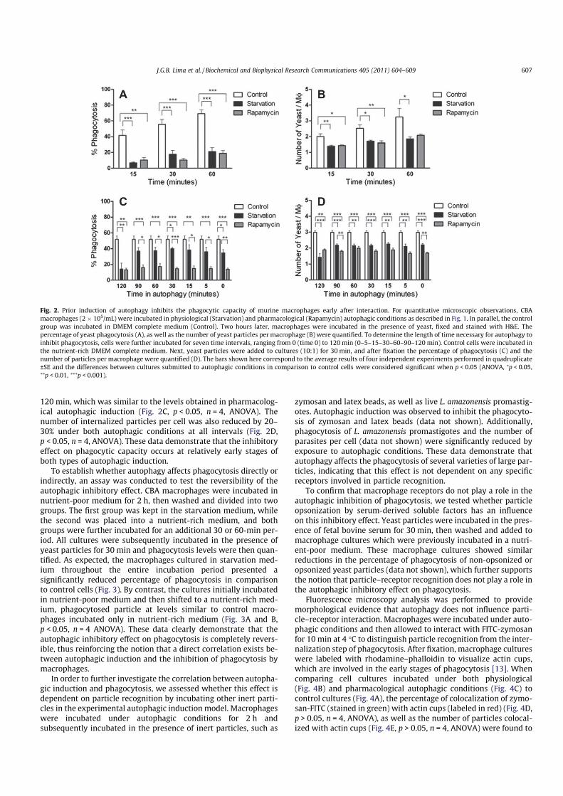
To establish whether autophagy affects phagocytosis directly orindirectly, an assay was conducted to test the reversibility of theautophagic inhibitory effect. CBA macrophages were incubated innutrient-poor medium for 2 h, then washed and divided into twogroups. The first group was kept in the starvation medium, whilethe second was placed into a nutrient-rich medium, and bothgroups were further incubated for an additional 30 or 60-min per-iod. All cultures were subsequently incubated in the presence ofyeast particles for 30 min and phagocytosis levels were then quan-tified. As expected, the macrophages cultured in starvation med-ium throughout the entire incubation period presented asignificantly reduced percentage of phagocytosis in comparisonto control cells (Fig. 3). By contrast, the cultures initially incubatedin nutrient-poor medium and then shifted to a nutrient-rich med-ium, phagocytosed particle at levels similar to control macro-phages incubated only in nutrient-rich medium (Fig. 3A and B,p < 0.05, n = 4 ANOVA). These data clearly demonstrate that theautophagic inhibitory effect on phagocytosis is completely revers-ible, thus reinforcing the notion that a direct correlation exists be-tween autophagic induction and the inhibition of phagocytosis bymacrophages.
In order to further investigate the correlation between autopha-gic induction and phagocytosis, we assessed whether this effect isdependent on particle recognition by incubating other inert parti-cles in the experimental autophagic induction model. Macrophageswere incubated under autophagic conditions for 2 h andsubsequently incubated in the presence of inert particles, such as
zymosan and latex beads, as well as live L. amazonensis promastig-otes. Autophagic induction was observed to inhibit the phagocyto-sis of zymosan and latex beads (data not shown). Additionally,phagocytosis of L. amazonensis promastigotes and the number ofparasites per cell (data not shown) were significantly reduced byexposure to autophagic conditions. These data demonstrate thatautophagy affects the phagocytosis of several varieties of large par-ticles, indicating that this effect is not dependent on any specificreceptors involved in particle recognition.
To confirm that macrophage receptors do not play a role in theautophagic inhibition of phagocytosis, we tested whether particleopsonization by serum-derived soluble factors has an influenceon this inhibitory effect. Yeast particles were incubated in the pres-ence of fetal bovine serum for 30 min, then washed and added tomacrophage cultures which were previously incubated in a nutri-ent-poor medium. These macrophage cultures showed similarreductions in the percentage of phagocytosis of non-opsonized oropsonized yeast particles (data not shown), which further supportsthe notion that particle–receptor recognition does not play a role inthe autophagic inhibitory effect on phagocytosis.
Fluorescence microscopy analysis was performed to providemorphological evidence that autophagy does not influence parti-cle–receptor interaction. Macrophages were incubated under auto-phagic conditions and then allowed to interact with FITC-zymosanfor 10 min at 4 °C to distinguish particle recognition from the inter-nalization step of phagocytosis. After fixation, macrophage cultureswere labeled with rhodamine–phalloidin to visualize actin cups,which are involved in the early stages of phagocytosis [13]. Whencomparing cell cultures incubated under both physiological(Fig. 4B) and pharmacological autophagic conditions (Fig. 4C) tocontrol cultures (Fig. 4A), the percentage of colocalization of zymo-san-FITC (stained in green) with actin cups (labeled in red) (Fig. 4D,p > 0.05, n = 4, ANOVA), as well as the number of particles colocal-ized with actin cups (Fig. 4E, p > 0.05, n = 4, ANOVA) were found to
Fig. 2. Prior induction of autophagy inhibits the phagocytic capacity of murine macrophages early after interaction. For quantitative microscopic observations, CBA
macrophages (2 � 105/mL) were incubated in physiological (Starvation) and pharmacological (Rapamycin) autophagic conditions as described in Fig. 1. In parallel, the control
group was incubated in DMEM complete medium (Control). Two hours later, macrophages were incubated in the presence of yeast, fixed and stained with H&E. The
percentage of yeast phagocytosis (A), as well as the number of yeast particles per macrophage (B) were quantified. To determine the length of time necessary for autophagy to
inhibit phagocytosis, cells were further incubated for seven time intervals, ranging from 0 (time 0) to 120 min (0–5–15–30–60–90–120 min). Control cells were incubated in
the nutrient-rich DMEM complete medium. Next, yeast particles were added to cultures (10:1) for 30 min, and after fixation the percentage of phagocytosis (C) and the
number of particles per macrophage were quantified (D). The bars shown here correspond to the average results of four independent experiments performed in quadruplicate
±SE and the differences between cultures submitted to autophagic conditions in comparison to control cells were considered significant when p < 0.05 (ANOVA, ⁄p < 0.05,⁄⁄p < 0.01, ⁄⁄⁄p < 0.001).
J.G.B. Lima et al. / Biochemical and Biophysical Research Communications 405 (2011) 604–609 607
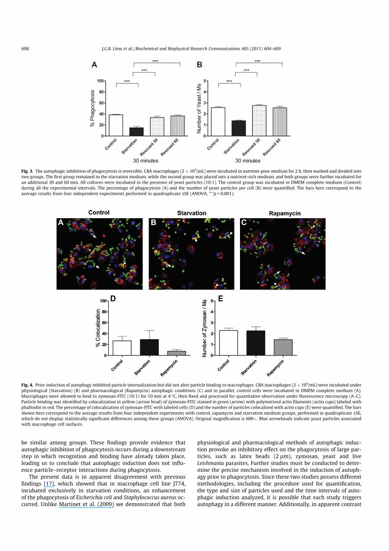
be similar among groups. These findings provide evidence thatautophagic inhibition of phagocytosis occurs during a downstreamstep in which recognition and binding have already taken place,leading us to conclude that autophagic induction does not influ-ence particle–receptor interactions during phagocytosis.
The present data is in apparent disagreement with previousfindings [17], which showed that in macrophage cell line J774,incubated exclusively in starvation conditions, an enhancementof the phagocytosis of Escherichia coli and Staphylococcus aureus oc-curred. Unlike Martinet et al. (2009) we demonstrated that both
physiological and pharmacological methods of autophagic induc-tion provoke an inhibitory effect on the phagocytosis of large par-ticles, such as latex beads (2 lm), zymosan, yeast and liveLeishmania parasites. Further studies must be conducted to deter-mine the precise mechanism involved in the induction of autoph-agy prior to phagocytosis. Since these two studies possess differentmethodologies, including the procedure used for quantification,the type and size of particles used and the time intervals of auto-phagic induction analyzed, it is possible that each study triggersautophagy in a different manner. Additionally, in apparent contrast
Fig. 3. The autophagic inhibition of phagocytosis is reversible. CBA macrophages (2 � 105/mL) were incubated in nutrient-poor medium for 2 h, then washed and divided into
two groups. The first group remained in the starvation medium, while the second group was placed into a nutrient-rich medium, and both groups were further incubated for
an additional 30 and 60 min. All cultures were incubated in the presence of yeast particles (10:1). The control group was incubated in DMEM complete medium (Control)
during all the experimental intervals. The percentage of phagocytosis (A) and the number of yeast particles per cell (B) were quantified. The bars here correspond to the
average results from four independent experiments performed in quadruplicate ±SE (ANOVA, ⁄⁄⁄p < 0.001).
Fig. 4. Prior induction of autophagy inhibited particle internalization but did not alter particle binding to macrophages. CBA macrophages (2 � 105/mL) were incubated under
physiological (Starvation) (B) and pharmacological (Rapamycin) autophagic conditions (C) and in parallel, control cells were incubated in DMEM complete medium (A).
Macrophages were allowed to bind to zymosan-FITC (10:1) for 10 min at 4 °C, then fixed and processed for quantitative observation under fluorescence microscopy (A–C).
Particle binding was identified by colocalization in yellow (arrow head) of zymosan-FITC stained in green (arrow) with polymerized actin filaments (actin cups) labeled with
phalloidin in red. The percentage of colocalization of zymosan-FITC with labeled cells (D) and the number of particles colocalized with actin cups (E) were quantified. The bars
shown here correspond to the average results from four independent experiments with control, rapamycin and starvation medium groups, performed in quadruplicate ±SE,
which do not display statistically significant differences among these groups (ANOVA). Original magnification is 600�. Blue arrowheads indicate yeast particles associated
with macrophage cell surfaces.
608 J.G.B. Lima et al. / Biochemical and Biophysical Research Communications 405 (2011) 604–609
with the present results, the prior induction of autophagy wasshown to not affect the phagocytosis of live Coxiella burnetti [10]or killed S. aureus [23]. However, both of these studies employedChinese hamster ovary (CHO) cells, a non-macrophage cell line,which may indicate that the inhibitory autophagic effect on phago-cytosis could be restricted to professional phagocytes.
Different lines of evidence describe the existence of interactionsbetween autophagy and phagocytosis [16]. Using proteomic analy-sis, the autophagic protein LC3-II was identified in the membranesof latex bead-containing phagosomes [14]. In addition, LC3 recruit-ment to zymosan-containing phagosomes, as well as IgG bead-con-taining phagosomes, has been previously described [24]. Recentstudies have demonstrated plasma membrane involvement as amembrane donor for the formation of autophagosomes [25,26].These authors observed the formation of structures suggestive ofpre-forming autophagosomes derived from the plasma membrane,requiring the recruitment of proteins from the endocytic pathway[8]. In the present study, one possible explanation for the loss ofphagocytic ability in macrophages incubated under autophagicconditions may be the lack of available plasma membrane neededto perform particle internalization. The induction of autophagymay recruit and mobilize part of the plasma membrane for the for-mation of autophagosomes, thereby preventing the internalizationof particles via the phagocytic pathway. Since the autophagic pro-cess has been demonstrated to be reversible, it is possible thatwhen an autophagic stimulus is removed, the plasma membranequickly become available again for the internalization process.
Alternatively, since the ER membrane has reportedly taken partin the biogenesis of both the autophagic [7] and phagocytic [27]pathways, another possible explanation of the inhibitory effect ofautophagy on phagocytosis may originate from the insufficientamount of ER cisternal membrane available for both processessimultaneously.
Taken together, the present data clearly show that the inductionof autophagy inhibits classical phagocytosis inmurinemacrophagesvia a mechanism that does not interfere with particle–receptorinteraction. The results support the notion that phagocytosis andautophagy can be interdependent and complementary processes.
Acknowledgments
The authors thank Claudio Pereira Filgueira and Dra. AdrianaLanfredi Rangel for technical support in transmission electronmicroscopy studies. This work was supported by grants and fellow-ships from FAPESB (Fundação de Amparo a Pesquisa no estado daBahia), CAPES (Coordenação de Aperfeiçoamento de Pessoal deNível Superior) and CNPq (Conselho Nacional de Pesquisa e Desen-volvimento). Veras, P.S.T. holds a CNPq productivity in researchGrant (306672/2008-1).
References
[1] G.E. Mortimore, C.M. Schworer, Induction of autophagy by amino-aciddeprivation in perfused rat liver, Nature 270 (1977) 174–176.
[2] D.J. Klionsky, S.D. Emr, Autophagy as a regulated pathway of cellulardegradation, Science 290 (2000) 1717–1721.
[3] A.J. Meijer, P. Codogno, Regulation and role of autophagy in mammalian cells,Int. J. Biochem. Cell Biol. 36 (2004) 2445–2462.
[4] V. Todde, M. Veenhuis, I.J. van der Klei, Autophagy: principles and significancein health and disease, Biochim. Biophys. Acta 1792 (2009) 3–13.
[5] T. Noda, Y. Ohsumi, Tor, a phosphatidylinositol kinase homologue, controlsautophagy in yeast, J. Biol. Chem. 273 (1998) 3963–3966.
[6] W.A. Dunn Jr., Studies on the mechanisms of autophagy: maturation of theautophagic vacuole, J. Cell Biol. 110 (1990) 1935–1945.
[7] W.A. Dunn Jr., Studies on the mechanisms of autophagy: formation of theautophagic vacuole, J. Cell Biol. 110 (1990) 1923–1933.
[8] B. Ravikumar, K. Moreau, L. Jahreiss, C. Puri, D.C. Rubinsztein, Plasmamembrane contributes to the formation of pre-autophagosomal structures,Nat. Cell Biol. 12 (2010) 747–757.
[9] M.G. Gutierrez, S.S. Master, S.B. Singh, G.A. Taylor, M.I. Colombo, V. Deretic,Autophagy is a defense mechanism inhibiting BCG and Mycobacteriumtuberculosis survival in infected macrophages, Cell 119 (2004) 753–766.
[10] M.G. Gutierrez, C.L. Vazquez, D.B. Munafo, F.C. Zoppino, W. Beron, M.Rabinovitch, M.I. Colombo, Autophagy induction favours the generation andmaturation of the Coxiella-replicative vacuoles, Cell. Microbiol. 7 (2005) 981–993.
[11] B. Levine, V. Deretic, Unveiling the roles of autophagy in innate and adaptiveimmunity, Nat. Rev. Immunol. 7 (2007) 767–777.
[12] E.J. Brown, H.D. Gresham, Phagocytosis, in: W. Paul (Ed.), FundamentalImmunology, Raven Press, Lippincott, New York, 2003, pp. 1105–1126.
[13] L.M. Stuart, R.A. Ezekowitz, Phagocytosis: elegant complexity, Immunity 22(2005) 539–550.
[14] W. Shui, L. Sheu, J. Liu, B. Smart, C.J. Petzold, T.Y. Hsieh, A. Pitcher, J.D. Keasling,C.R. Bertozzi, Membrane proteomics of phagosomes suggests a connection toautophagy, Proc. Natl. Acad. Sci. USA 105 (2008) 16952–16957.
[15] Y. Xu, C. Jagannath, X.D. Liu, A. Sharafkhaneh, K.E. Kolodziejska, N.T. Eissa, Toll-like receptor 4 is a sensor for autophagy associated with innate immunity,Immunity 27 (2007) 135–144.
[16] M.A. Sanjuan, C.P. Dillon, S.W. Tait, S. Moshiach, F. Dorsey, S. Connell, M.Komatsu, K. Tanaka, J.L. Cleveland, S. Withoff, D.R. Green, Toll-like receptorsignalling in macrophages links the autophagy pathway to phagocytosis,Nature 450 (2007) 1253–1257.
[17] W. Martinet, D.M. Schrijvers, J.P. Timmermans, A.G. Herman, G.R. De Meyer,Phagocytosis of bacteria is enhanced in macrophages undergoing nutrientdeprivation, FEBS J. 276 (2009) 2227–2240.
[18] I.N. Gomes, A.F. Calabrich, S. Tavares Rda, J. Wietzerbin, L.A. De Freitas, P.S.Veras, Differential properties of CBA/J mononuclear phagocytes recoveredfrom an inflammatory site and probed with two different species ofLeishmania, Microbes Infect. 5 (2003) 251–260.
[19] D.B. Munafo, M.I. Colombo, A novel assay to study autophagy: regulation ofautophagosome vacuole size by amino acid deprivation, J. Cell Sci. 114 (2001)3619–3629.
[20] P.F. Pimenta, M.A. Dos Santos, W. De Souza, Fine structure and cytochemistryof the interaction between Leishmania mexicana amazonensis and ratneutrophils and eosinophils, J. Submicrosc. Cytol. 19 (1987) 387–395.
[21] M. Rabinovitch, The dissociation of the attachment and ingestion phases ofphagocytosis by macrophages, Exp. Cell Res. 46 (1967) 19–28.
[22] E.L. Eskelinen, Maturation of autophagic vacuoles in mammalian cells,Autophagy 1 (2005) 1–10.
[23] P.S. Romano, M.A. Arboit, C.L. Vazquez, M.I. Colombo, The autophagic pathwayis a key component in the lysosomal dependent entry of Trypanosoma cruziinto the host cell, Autophagy 5 (2009) 6–18.
[24] J. Huang, V. Canadien, G.Y. Lam, B.E. Steinberg, M.C. Dinauer, M.A. Magalhaes,M. Glogauer, S. Grinstein, J.H. Brumell, Activation of antibacterial autophagy byNADPH oxidases, Proc. Natl. Acad. Sci. USA 106 (2009) 6226–6231.
[25] A.M. Cuervo, The plasma membrane brings autophagosomes to life, Nat. CellBiol. 12 (2010) 735–737.
[26] B. Ravikumar, K. Moreau, D.C. Rubinsztein, Plasma membrane helpsautophagosomes grow, Autophagy 6 (2010) 1184–1186.
[27] E. Gagnon, S. Duclos, C. Rondeau, E. Chevet, P.H. Cameron, O. Steele-Mortimer,J. Paiement, J.J. Bergeron, M. Desjardins, Endoplasmic reticulum-mediatedphagocytosis is a mechanism of entry into macrophages, Cell 110 (2002) 119–131.
J.G.B. Lima et al. / Biochemical and Biophysical Research Communications 405 (2011) 604–609 609


