Embed Size (px)
Citation preview
Cargill , 2014 ANT 342
OBTAINING POSTCRANIAL AGE & SEX
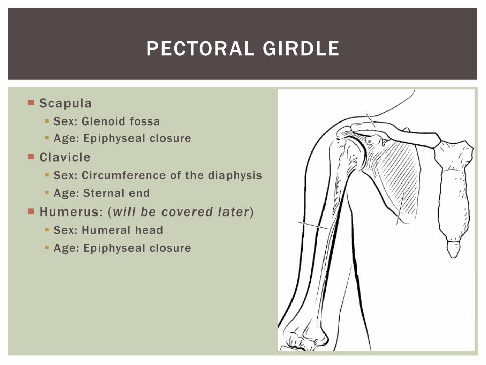
¡ Scapula § Sex: Glenoid fossa § Age: Epiphyseal closure
¡ Clavicle § Sex: Circumference of the diaphysis § Age: Sternal end
¡ Humerus: (will be covered later) § Sex: Humeral head § Age: Epiphyseal closure
PECTORAL GIRDLE
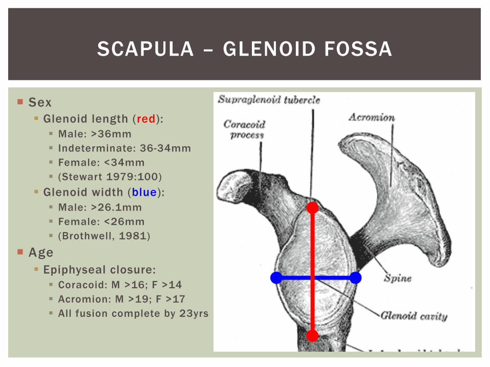
¡ Sex § Glenoid length (red):
§ Male: >36mm § Indeterminate: 36-34mm § Female: <34mm § (Stewart 1979:100)
§ Glenoid width (blue): § Male: >26.1mm § Female: <26mm § (Brothwell, 1981)
¡ Age § Epiphyseal closure:
§ Coracoid: M >16; F >14 § Acromion: M >19; F >17 § All fusion complete by 23yrs
SCAPULA – GLENOID FOSSA
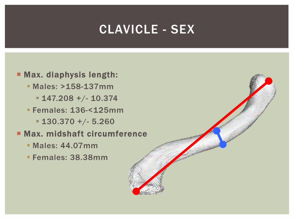
¡ Max. diaphysis length: § Males: >158-137mm
§ 147.208 +/- 10.374 § Females: 136-<125mm
§ 130.370 +/- 5.260 ¡ Max. midshaft circumference
§ Males: 44.07mm § Females: 38.38mm
CLAVICLE - SEX
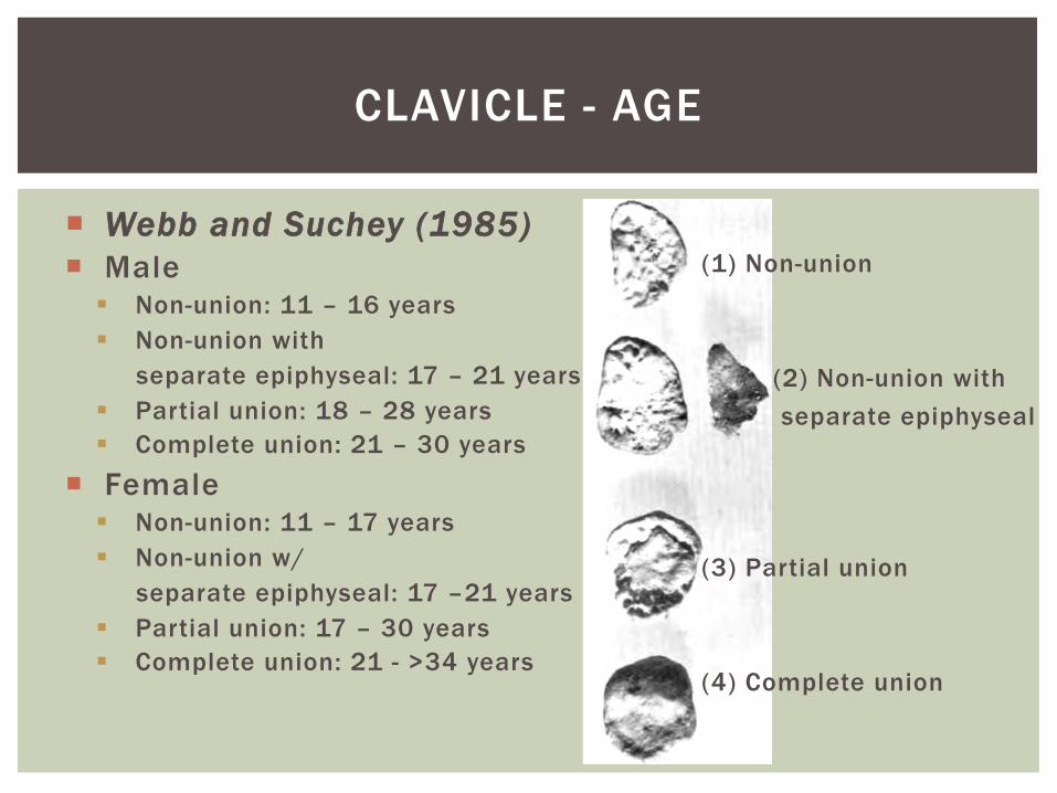
¡ Webb and Suchey (1985) ¡ Male
§ Non-union: 11 – 16 years § Non-union with separate epiphyseal: 17 – 21 years § Partial union: 18 – 28 years § Complete union: 21 – 30 years
¡ Female § Non-union: 11 – 17 years § Non-union w/ separate epiphyseal: 17 –21 years § Partial union: 17 – 30 years § Complete union: 21 - >34 years
CLAVICLE - AGE
(2) Non-union with separate epiphyseal
(1) Non-union (3) Partial union (4) Complete union
RIBS
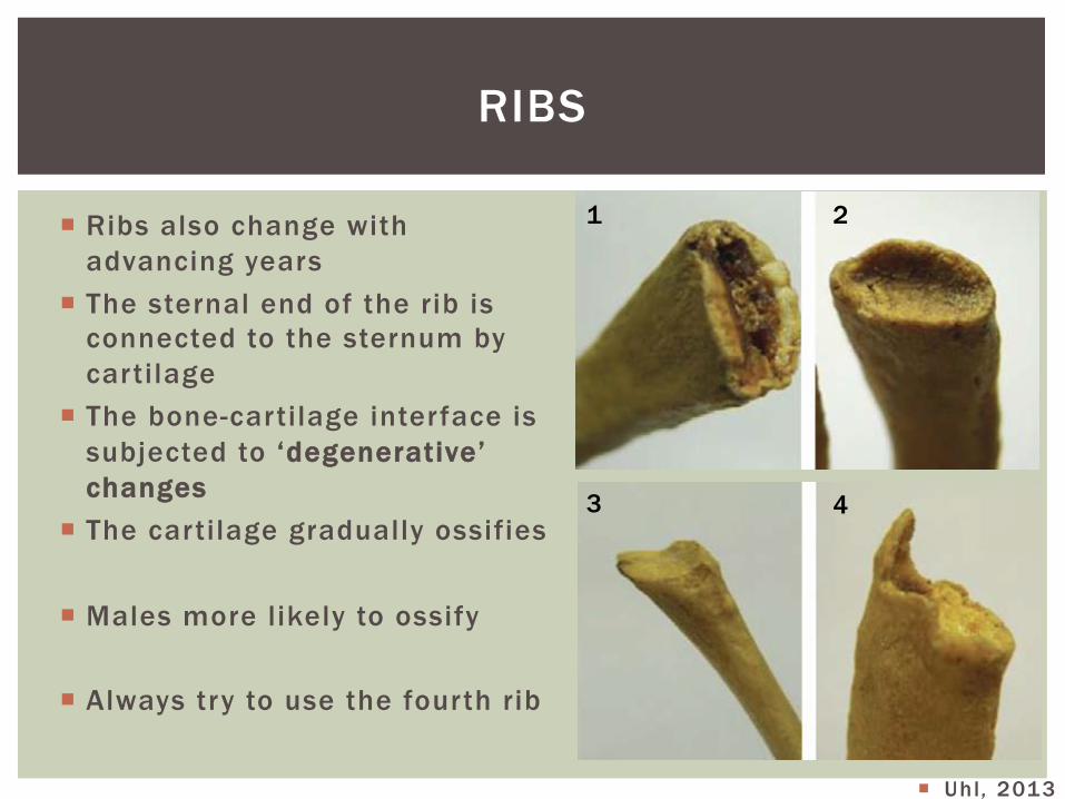
¡ Ribs also change with advancing years
¡ The sternal end of the rib is connected to the sternum by cartilage
¡ The bone-cartilage interface is subjected to ‘degenerative’ changes
¡ The cartilage gradually ossifies
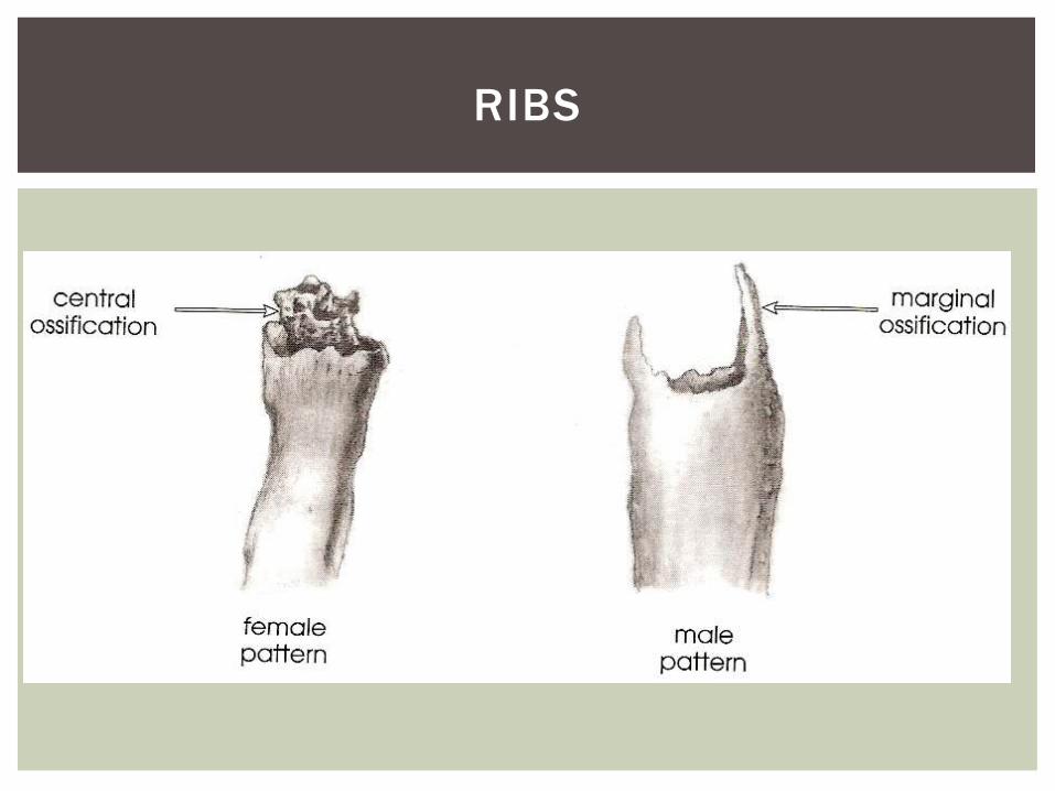
¡ Males more likely to ossify
¡ Always try to use the fourth rib
¡ Uhl , 2013
1 2
3 4
RIBS
¡ Pelvis is the most reliable indicator of sex
¡ Generally, the pelvis is more strongly dimorphic at an earlier age than cranial features
¡ Estimation may be undertaken on individual for whom tripartite pelvic fusion is complete (~15 years)
¡ Compared to cranial development of sexual dimorphic traits occurs over a period of time from puberty to mid 20s’
¡ And throughout your life
THE PELVIS
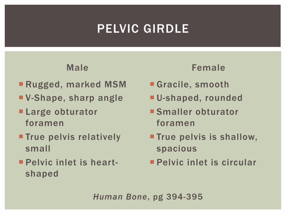
Male
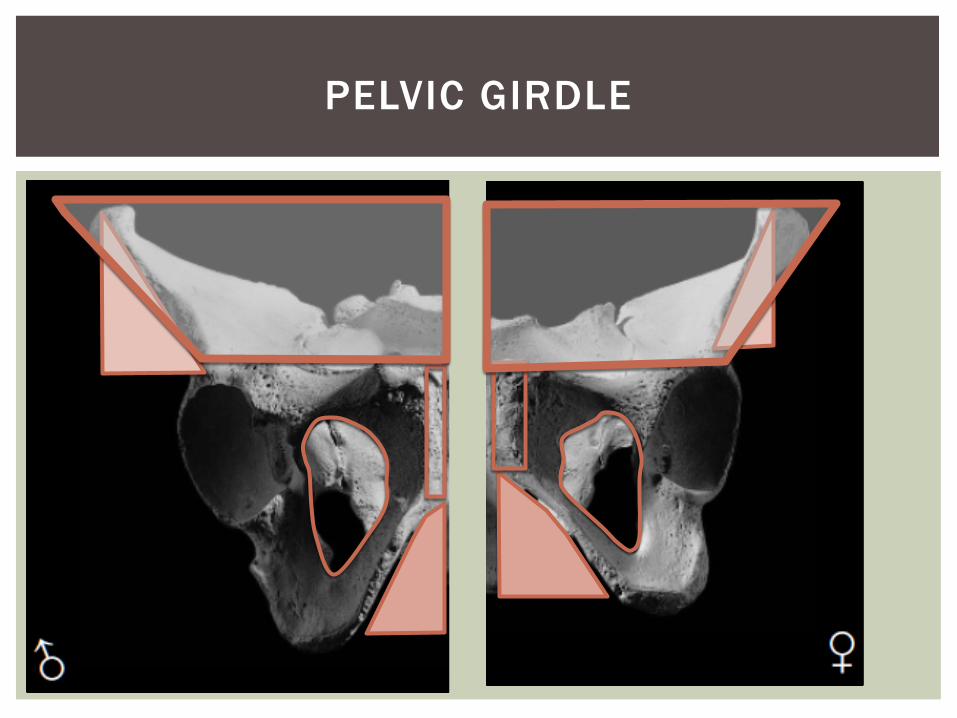
¡ Rugged, marked MSM ¡ V-Shape, sharp angle ¡ Large obturator
foramen ¡ True pelvis relatively
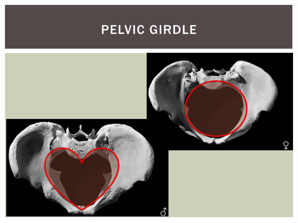
small ¡ Pelvic inlet is heart-
shaped
Female
¡ Gracile, smooth ¡ U-shaped, rounded ¡ Smaller obturator
foramen ¡ True pelvis is shallow,
spacious ¡ Pelvic inlet is circular
PELVIC GIRDLE
Human Bone, pg 394-395
PELVIC GIRDLE
PELVIC GIRDLE
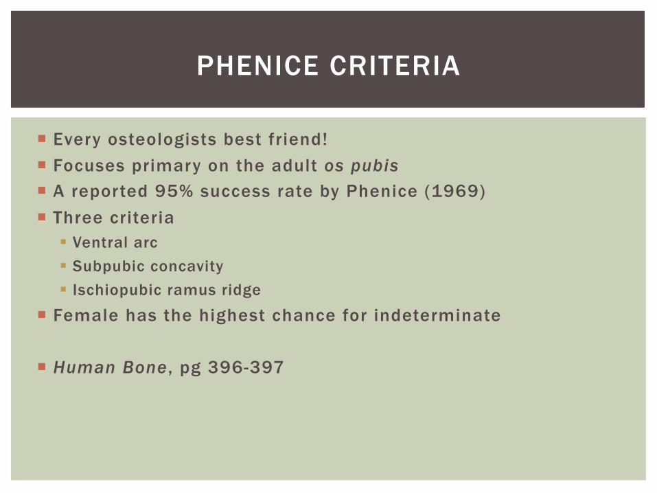
¡ Every osteologists best friend! ¡ Focuses primary on the adult os pubis ¡ A reported 95% success rate by Phenice (1969) ¡ Three criteria
§ Ventral arc § Subpubic concavity § Ischiopubic ramus ridge
¡ Female has the highest chance for indeterminate
¡ Human Bone , pg 396-397
PHENICE CRITERIA
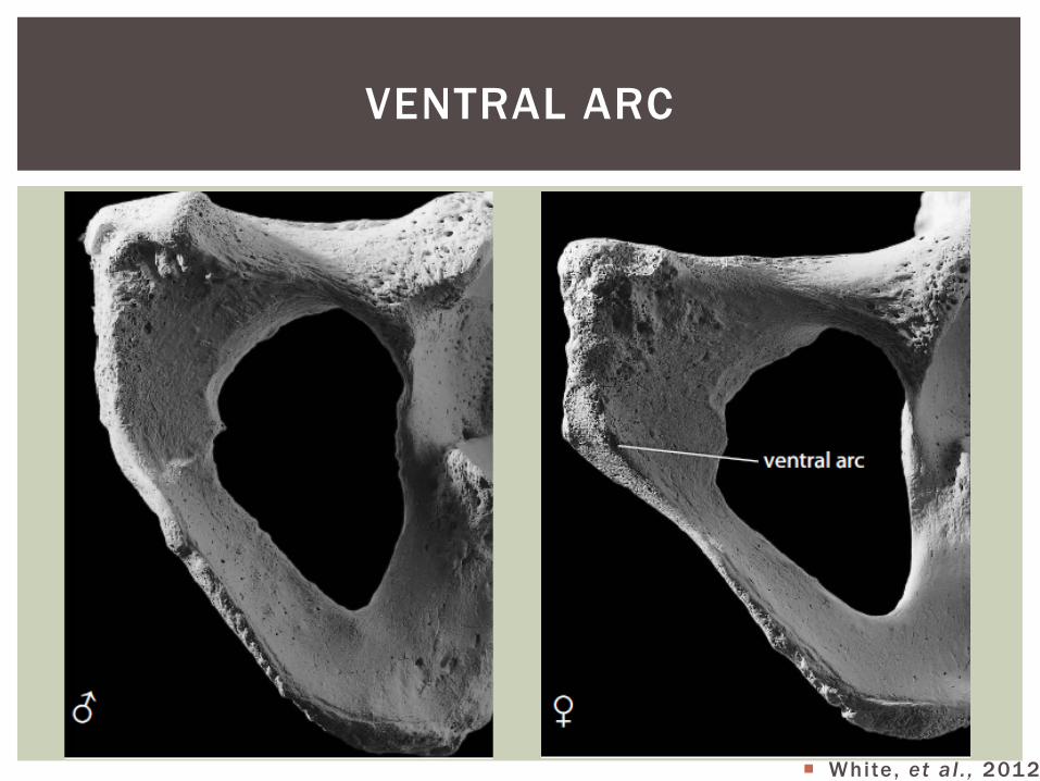
¡ White, et al . , 2012
VENTRAL ARC
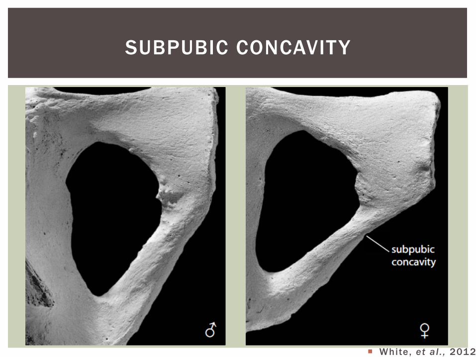
¡ White, et al . , 2012
SUBPUBIC CONCAVITY
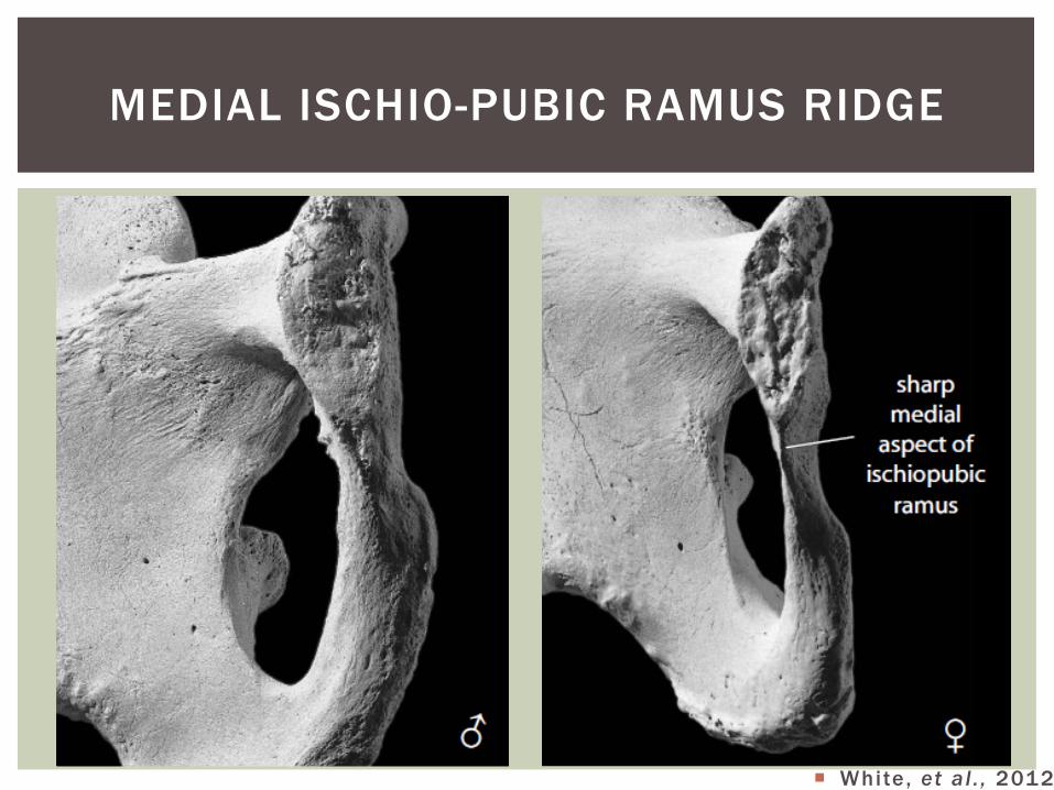
¡ White, et al . , 2012
MEDIAL ISCHIO-PUBIC RAMUS RIDGE
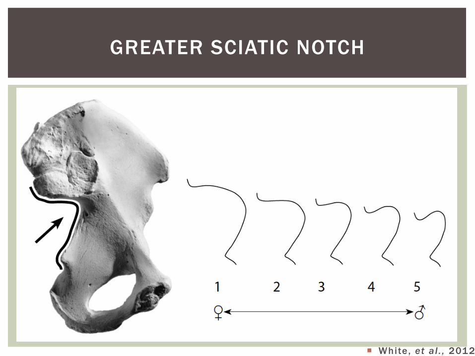
GREATER SCIATIC NOTCH
¡ White, et al . , 2012
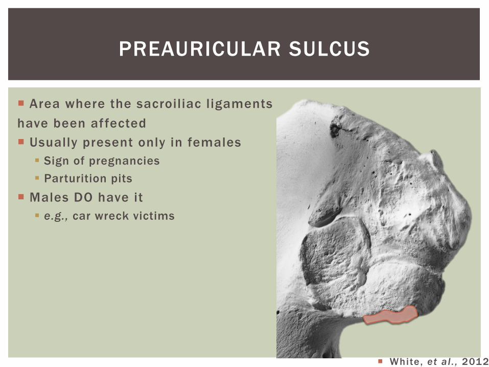
¡ Area where the sacroiliac ligaments have been affected ¡ Usually present only in females
§ Sign of pregnancies § Parturition pits
¡ Males DO have it § e.g., car wreck victims
PREAURICULAR SULCUS
¡ White, et al . , 2012
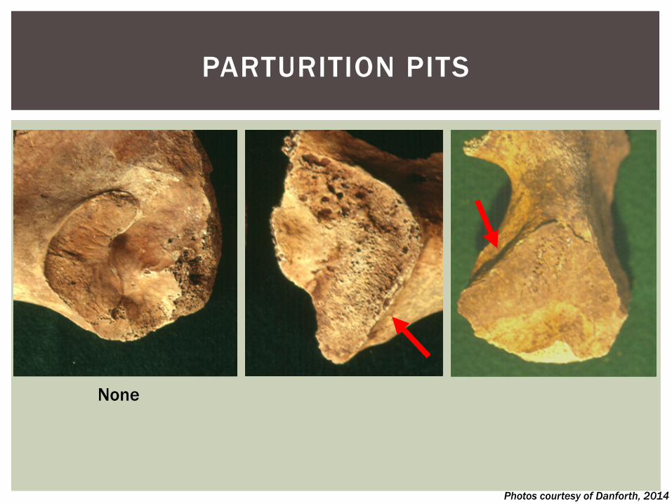
PARTURITION PITS
Photos courtesy of Danforth, 2014
None
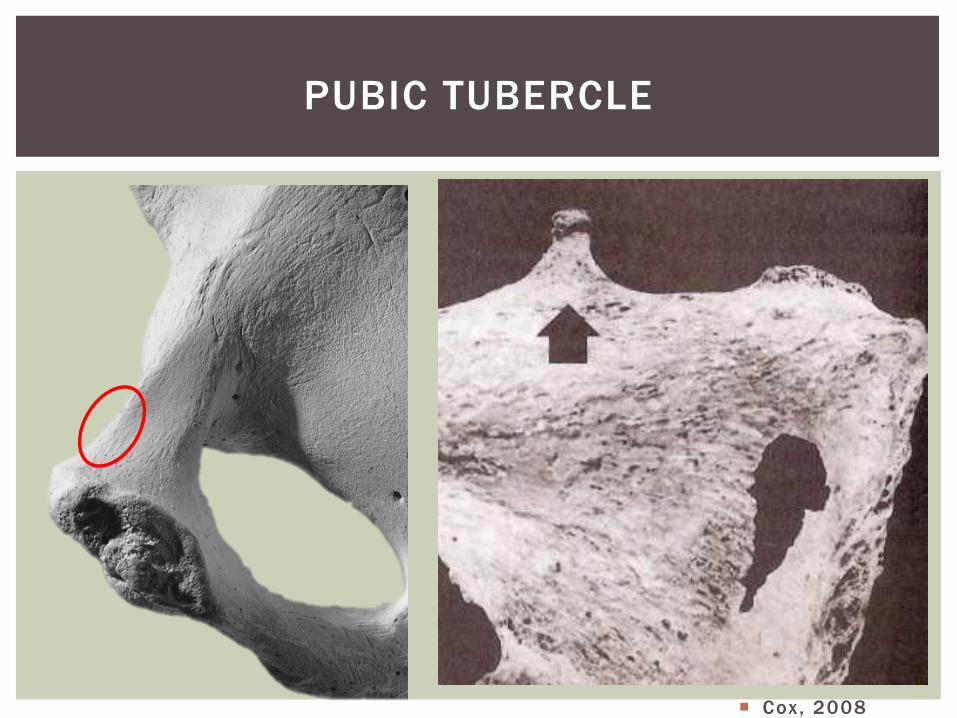
PUBIC TUBERCLE
¡ Cox, 2008
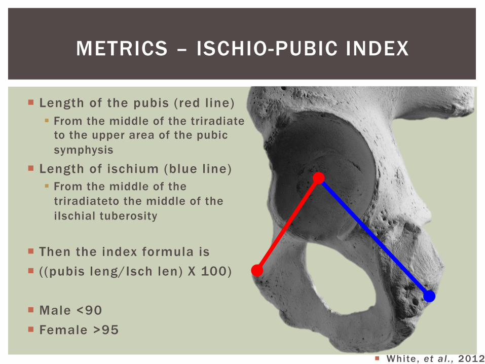
METRICS – ISCHIO-PUBIC INDEX
¡ Length of the pubis (red line) § From the middle of the triradiate
to the upper area of the pubic symphysis
¡ Length of ischium (blue line) § From the middle of the
triradiateto the middle of the iIschial tuberosity
¡ Then the index formula is ¡ ((pubis leng/Isch len) X 100)
¡ Male <90 ¡ Female >95
¡ White, et al . , 2012
¡ Epiphyseal closure
¡ Pubic symphyseal surface § Todd scoring system (1920)
§ Phases were more reliable indicators between 20 and 40, then after 40 § Young soldiers
§ Suchey-Brooks scoring system (1990) § Primary one used
¡ Auricular surface § Lovejoy et al. (1985) § Buckberry and Chamberlain (2002) § Mulhern and Jones (2005)
USING THE OS COXAE TO AGE
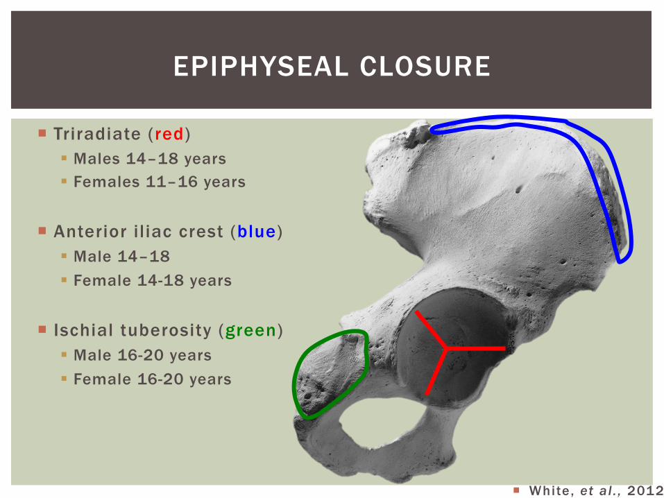
¡ Triradiate (red) § Males 14–18 years § Females 11–16 years
¡ Anterior iliac crest (blue) § Male 14–18 § Female 14-18 years
¡ Ischial tuberosity (green) § Male 16-20 years § Female 16-20 years
EPIPHYSEAL CLOSURE
¡ White, et al . , 2012
PUBIC SYMPHYSIS
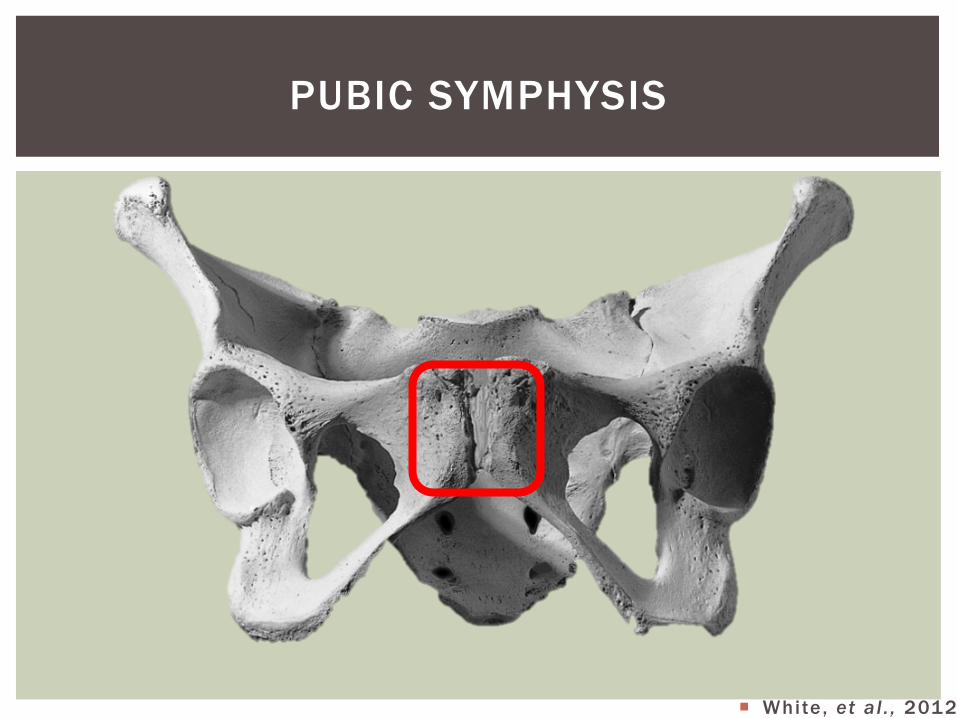
¡ White, et al . , 2012
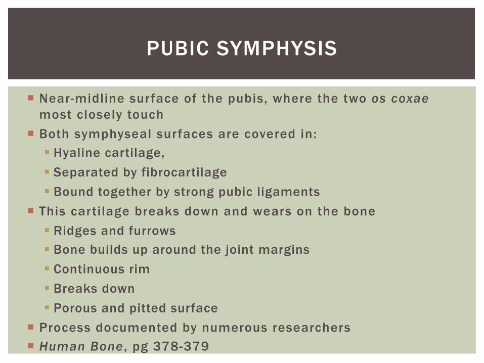
¡ Near-midline surface of the pubis, where the two os coxae most closely touch
¡ Both symphyseal surfaces are covered in: § Hyaline cartilage, § Separated by fibrocartilage § Bound together by strong pubic ligaments
¡ This cartilage breaks down and wears on the bone § Ridges and furrows § Bone builds up around the joint margins § Continuous rim § Breaks down § Porous and pitted surface
¡ Process documented by numerous researchers ¡ Human Bone , pg 378-379
PUBIC SYMPHYSIS
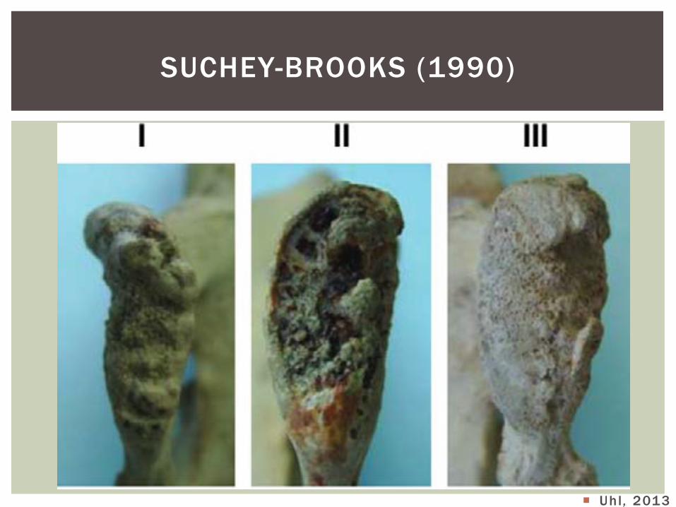
SUCHEY-BROOKS (1990)
¡ Uhl , 2013
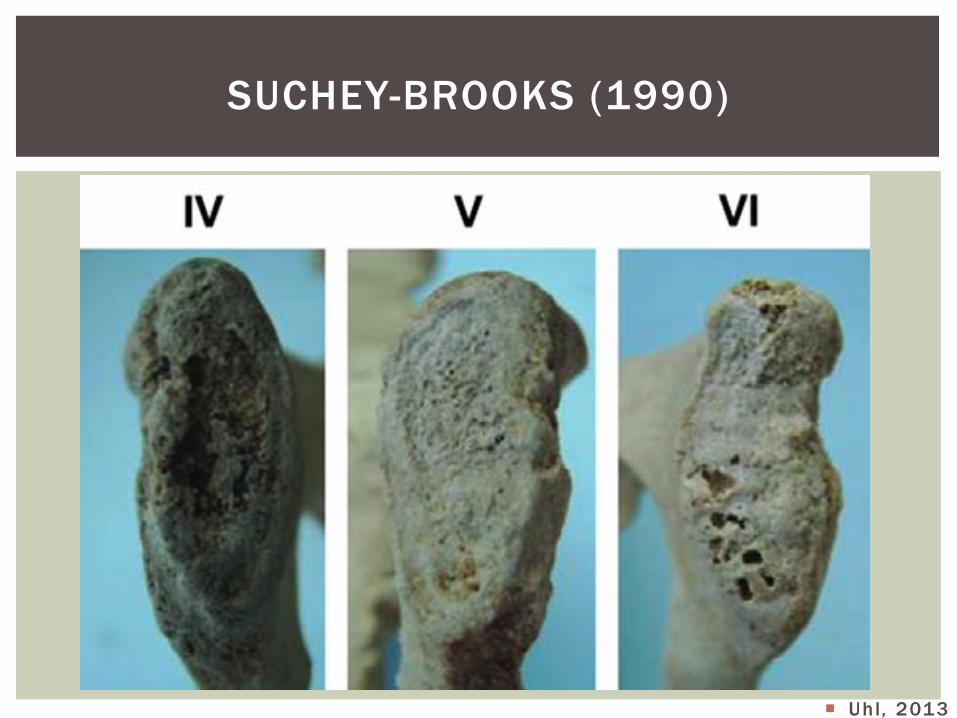
SUCHEY-BROOKS (1990)
¡ Uhl , 2013
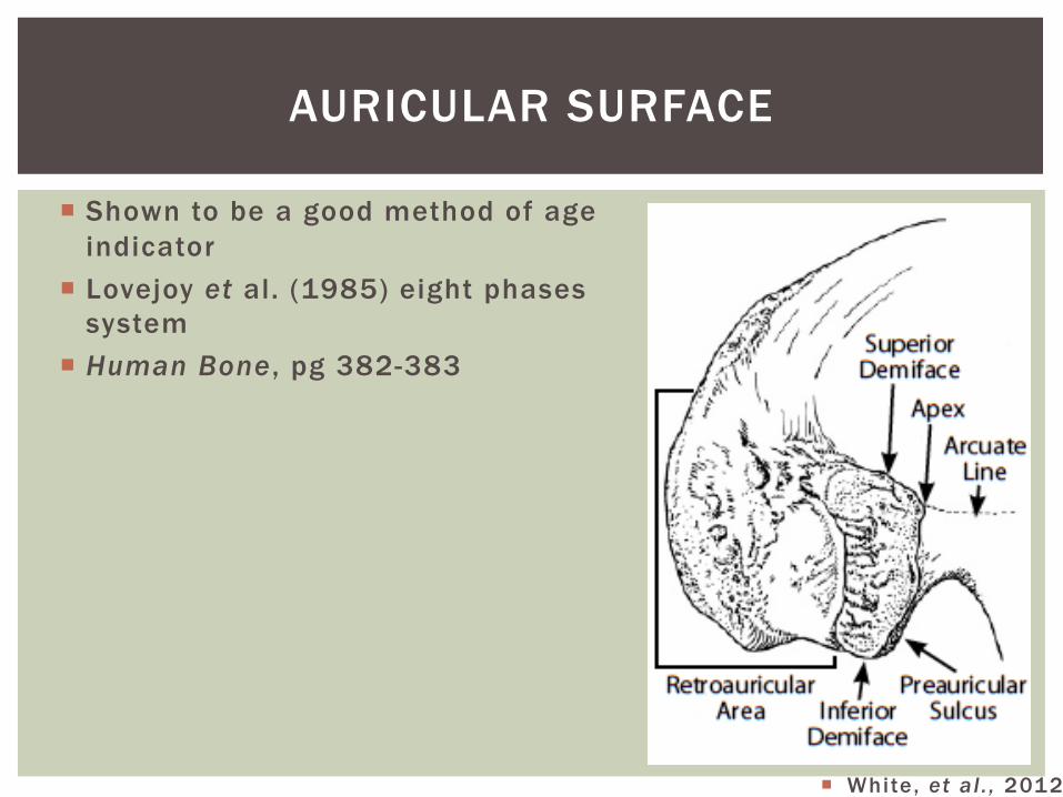
AURICULAR SURFACE
¡ Shown to be a good method of age indicator
¡ Lovejoy et al. (1985) eight phases system
¡ Human Bone , pg 382-383
¡ White, et al . , 2012
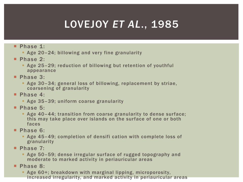
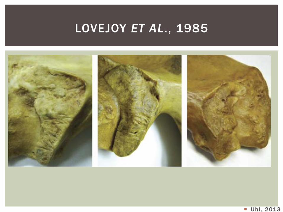
LOVEJOY ET AL., 1985
¡ Phase 1: § Age 20–24; billowing and very fine granularity
¡ Phase 2: § Age 25–29; reduction of billowing but retention of youthful
appearance ¡ Phase 3:
§ Age 30–34; general loss of billowing, replacement by striae, coarsening of granularity
¡ Phase 4: § Age 35–39; uniform coarse granularity
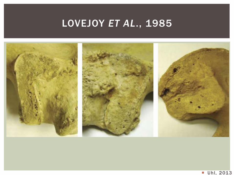
¡ Phase 5: § Age 40–44; transition from coarse granularity to dense surface;
this may take place over islands on the surface of one or both faces
¡ Phase 6: § Age 45–49; completion of densifi cation with complete loss of
granularity ¡ Phase 7:
§ Age 50–59; dense irregular surface of rugged topography and moderate to marked activity in periauricular areas
¡ Phase 8: § Age 60+; breakdown with marginal lipping, microporosity,
increased irregularity, and marked activity in periauricular areas
LOVEJOY ET AL., 1985
¡ Uhl , 2013
LOVEJOY ET AL., 1985
¡ Uhl , 2013
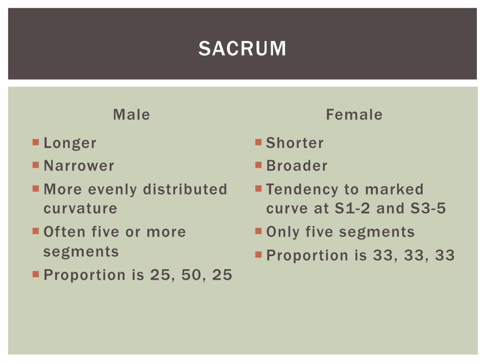
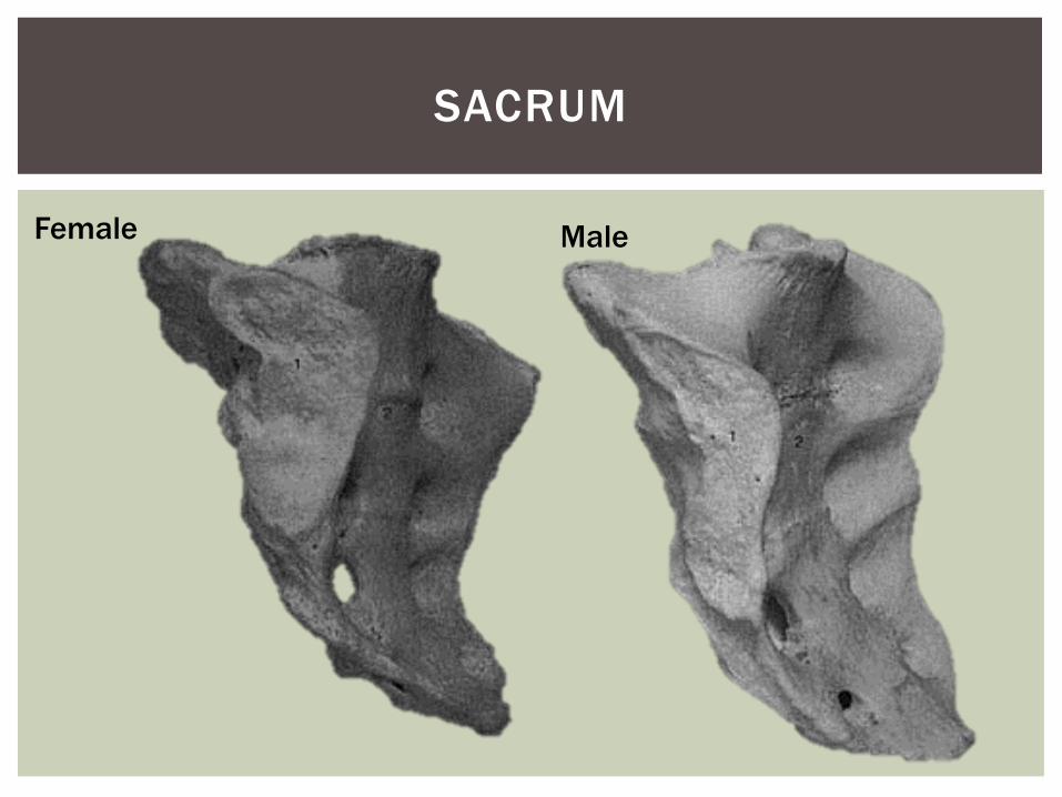
Male
¡ Longer ¡ Narrower ¡ More evenly distributed
curvature ¡ Often five or more
segments ¡ Proportion is 25, 50, 25
Female
¡ Shorter ¡ Broader ¡ Tendency to marked
curve at S1-2 and S3-5 ¡ Only five segments ¡ Proportion is 33, 33, 33
SACRUM
SACRUM
Female Male
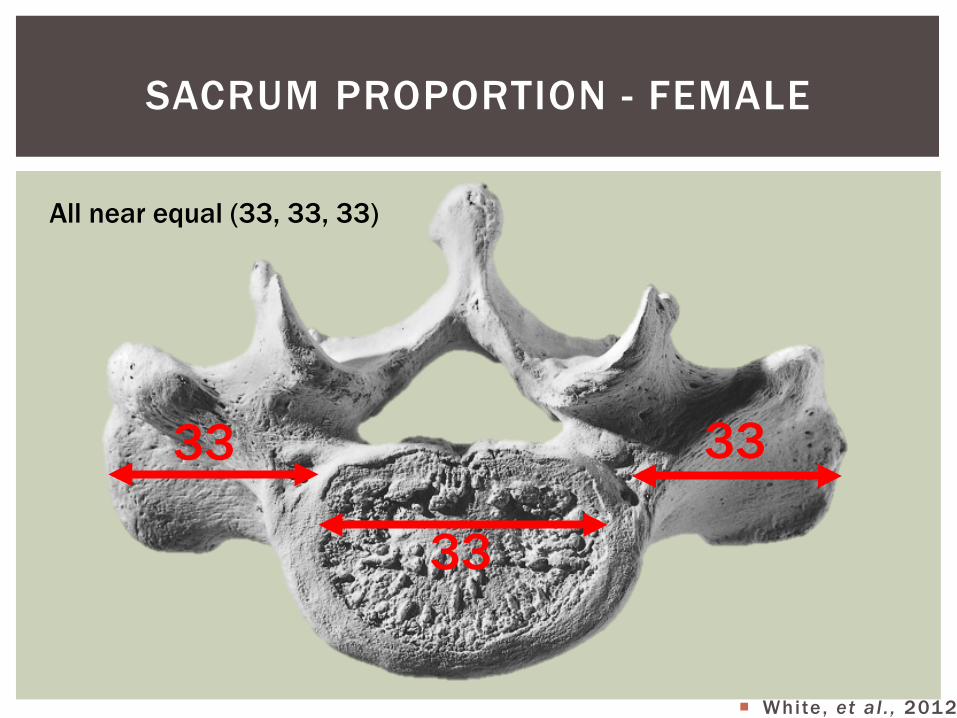
SACRUM PROPORTION - FEMALE
All near equal (33, 33, 33)
33
33
33
¡ White, et al . , 2012
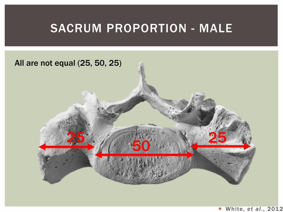
SACRUM PROPORTION - MALE
All are not equal (25, 50, 25)
25 25 50
¡ White, et al . , 2012
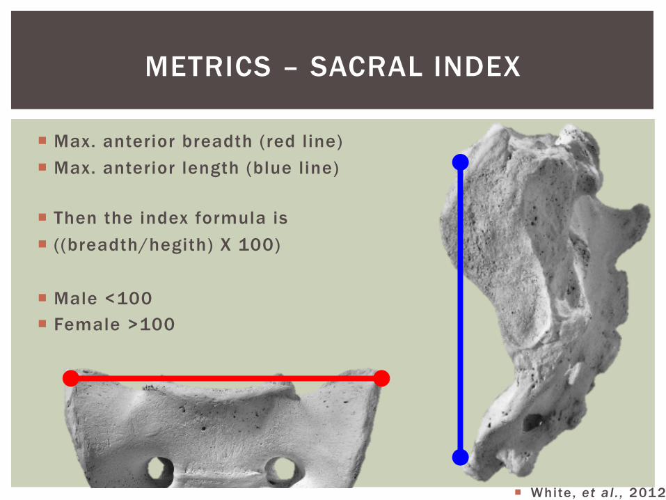
METRICS – SACRAL INDEX
¡ Max. anterior breadth (red line) ¡ Max. anterior length (blue line)
¡ Then the index formula is ¡ ((breadth/hegith) X 100)
¡ Male <100 ¡ Female >100
¡ White, et al . , 2012
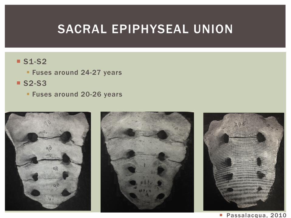
¡ S1-S2 § Fuses around 24-27 years
¡ S2-S3 § Fuses around 20-26 years
SACRAL EPIPHYSEAL UNION
¡ Passalacqua, 2010
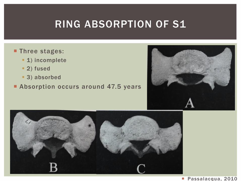
¡ Three stages: § 1) incomplete § 2) fused § 3) absorbed
¡ Absorption occurs around 47.5 years
RING ABSORPTION OF S1
¡ Passalacqua, 2010
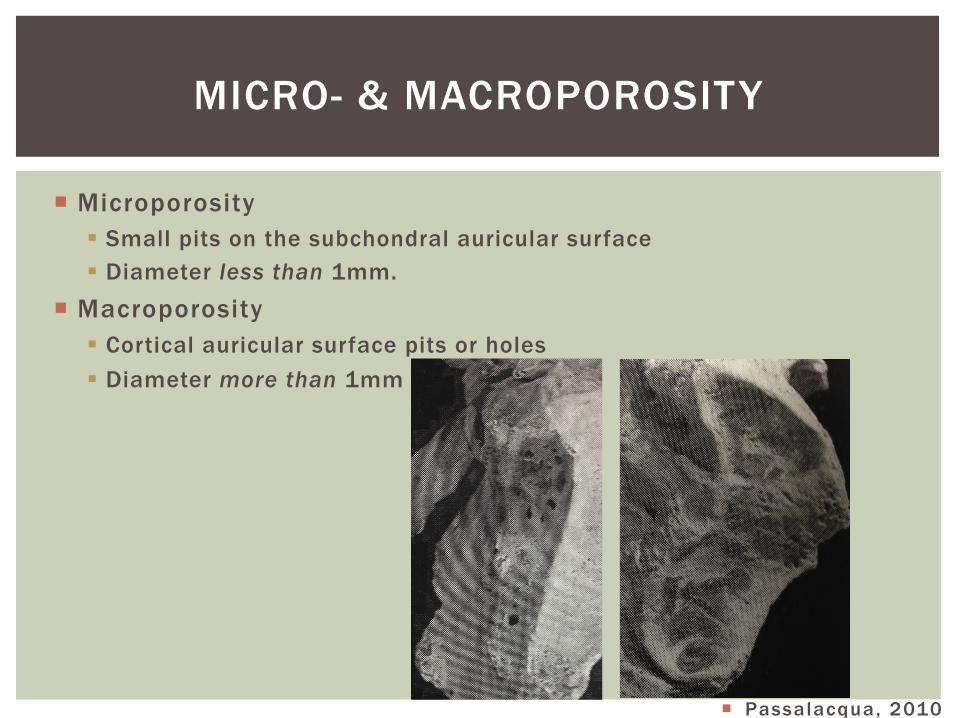
¡ Microporosity § Small pits on the subchondral auricular surface § Diameter less than 1mm.
¡ Macroporosity § Cortical auricular surface pits or holes § Diameter more than 1mm
MICRO- & MACROPOROSITY
¡ Passalacqua, 2010
PSEDUO- AGE INDICATORS
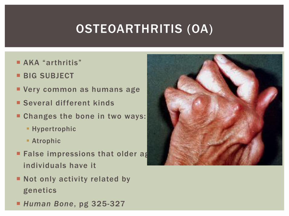
¡ AKA “arthritis”
¡ BIG SUBJECT
¡ Very common as humans age
¡ Several dif ferent kinds
¡ Changes the bone in two ways: § Hypertrophic
§ Atrophic
¡ False impressions that older aged individuals have it
¡ Not only activity related by genetics
¡ Human Bone , pg 325-327
OSTEOARTHRITIS (OA)
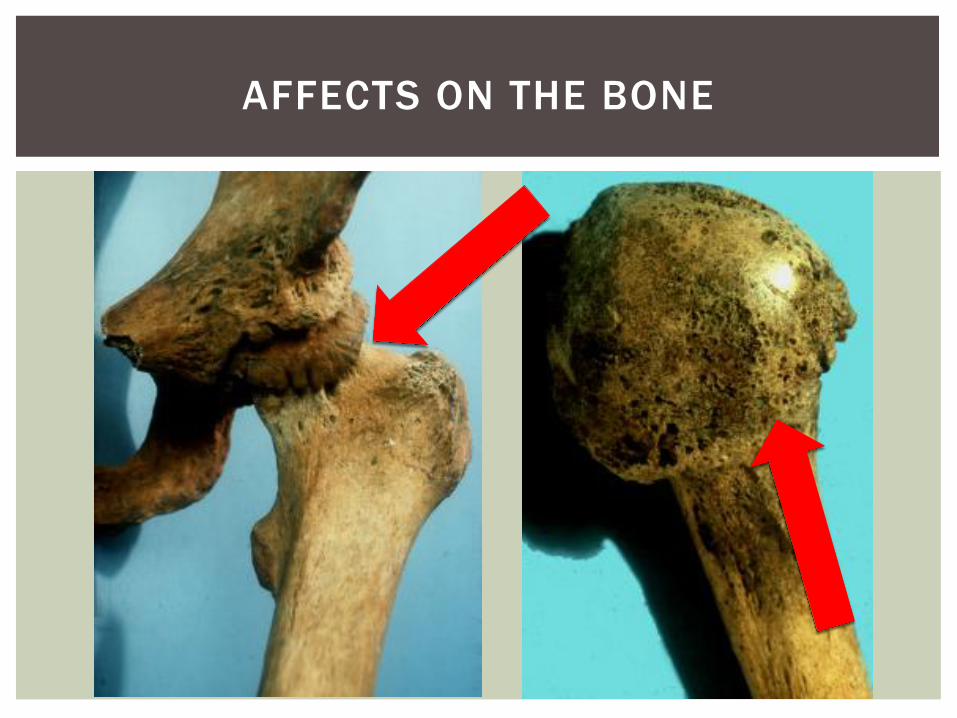
AFFECTS ON THE BONE
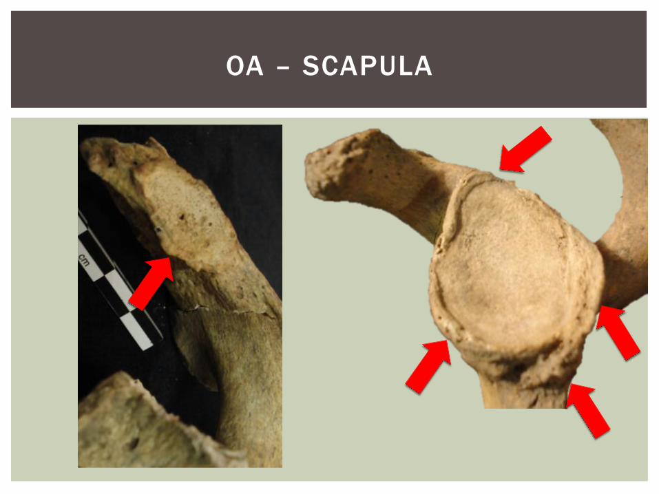
OA – SCAPULA

22 year old College pitcher? 55 year old farmer
WHO MOST LIKELY HAS IT?
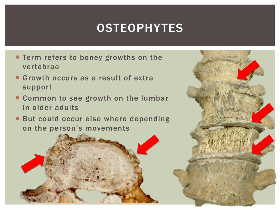
OSTEOPHYTES
¡ Term refers to boney growths on the vertebrae
¡ Growth occurs as a result of extra support
¡ Common to see growth on the lumbar in older adults
¡ But could occur else where depending on the person’s movements
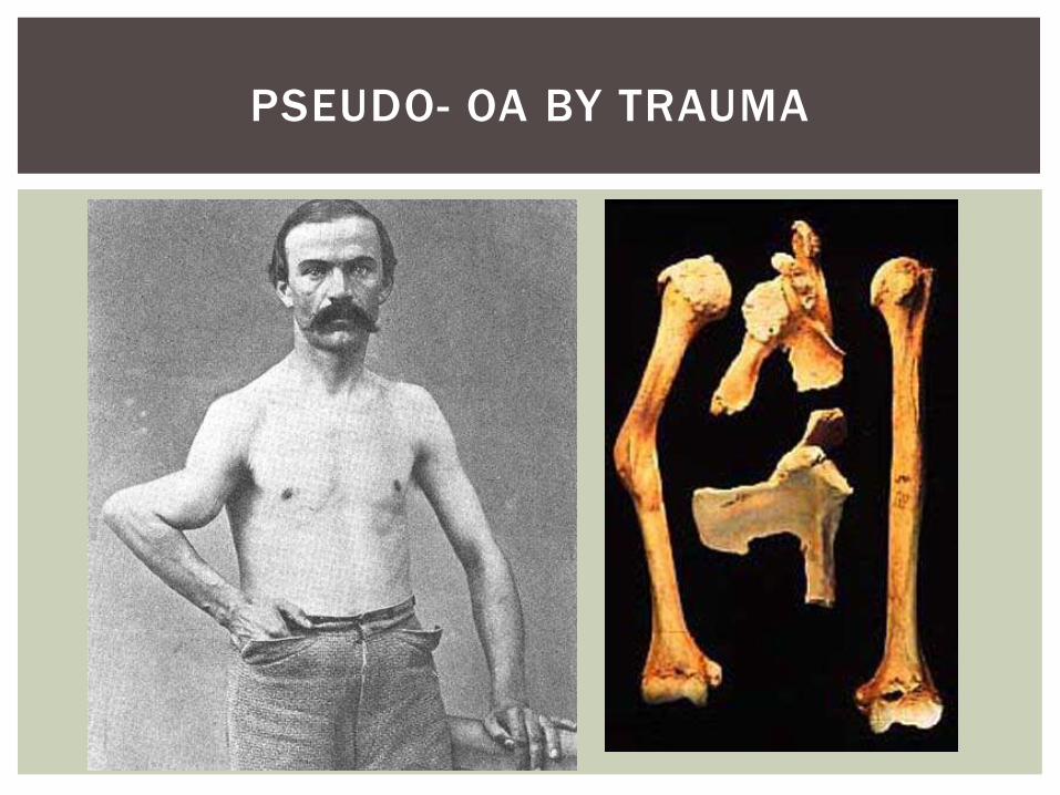
PSEUDO- OA BY TRAUMA
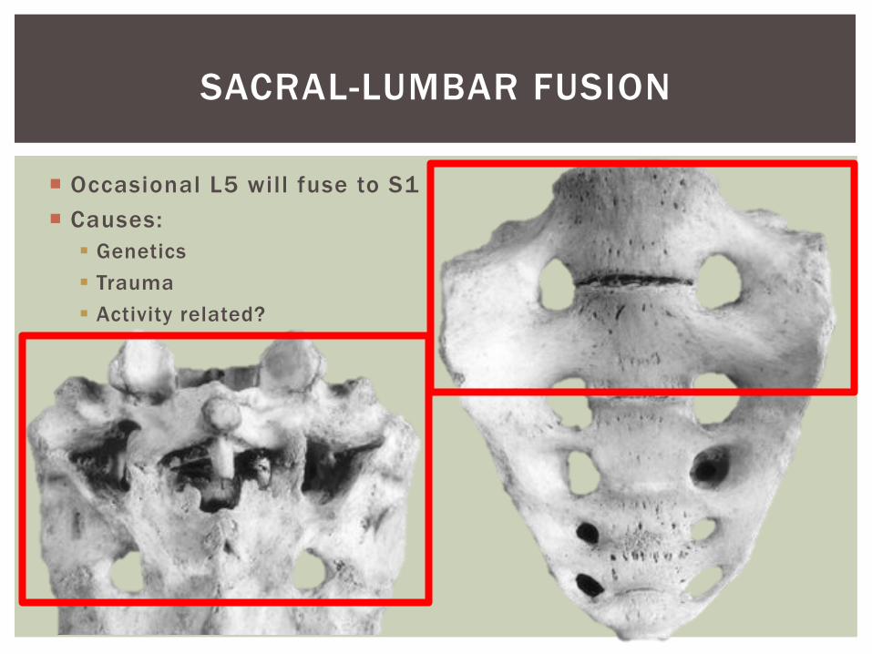
¡ Occasional L5 will fuse to S1 ¡ Causes:
§ Genetics § Trauma § Activity related?
SACRAL-LUMBAR FUSION
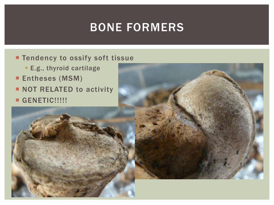
¡ Tendency to ossify soft tissue § E.g., thyroid cartilage
¡ Entheses (MSM) ¡ NOT RELATED to activity ¡ GENETIC!!!!!
BONE FORMERS
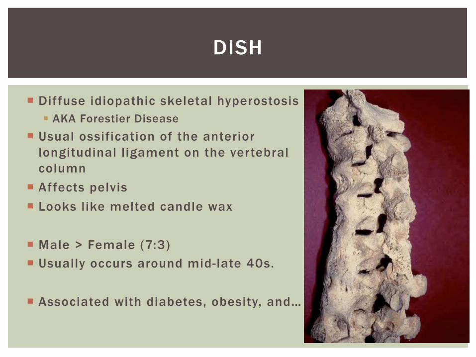
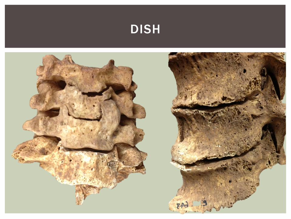
¡ Diffuse idiopathic skeletal hyperostosis § AKA Forestier Disease
¡ Usual ossification of the anterior longitudinal ligament on the vertebral column
¡ Affects pelvis ¡ Looks like melted candle wax ¡ Male > Female (7:3) ¡ Usually occurs around mid-late 40s. ¡ Associated with diabetes, obesity, and…
DISH

DRINKING, LOTS AND LOTS OF DRINKING
DISH

¡ See me
REFERENCES


