Embed Size (px)
Citation preview
HAL Id: hal-03199944https://hal.archives-ouvertes.fr/hal-03199944
Submitted on 16 Apr 2021
HAL is a multi-disciplinary open accessarchive for the deposit and dissemination of sci-entific research documents, whether they are pub-lished or not. The documents may come fromteaching and research institutions in France orabroad, or from public or private research centers.
L’archive ouverte pluridisciplinaire HAL, estdestinée au dépôt et à la diffusion de documentsscientifiques de niveau recherche, publiés ou non,émanant des établissements d’enseignement et derecherche français ou étrangers, des laboratoirespublics ou privés.
Obtaining nonspherical poly(alkylcyanoacrylate)nanoparticles by the stretching method applied with a
marketed water-soluble filmClaudio Palazzo, Gilles Ponchel, Jean Vachon, Sarah Villebrun, Florence
Agnely, Christine Vauthier
To cite this version:Claudio Palazzo, Gilles Ponchel, Jean Vachon, Sarah Villebrun, Florence Agnely, et al.. Obtainingnonspherical poly(alkylcyanoacrylate) nanoparticles by the stretching method applied with a marketedwater-soluble film. International Journal of Polymeric Materials and Polymeric Biomaterials, Taylor& Francis, 2017, 66 (8), pp.416 - 424. �10.1080/00914037.2016.1233420�. �hal-03199944�
Author manuscript from Int J Polym Mater 2017;66:416‐424.
1
Obtaining non‐spherical poly(alkylcyanoacrylate)
nanoparticles by the stretching method applied with a
marketed water‐soluble film.
Claudio Palazzo,1,2+ Gilles Ponchel, 2 Jean Jacques Vachon,2 Sarah Villebrun,2 Florence Agnely,2 Christine Vauthier 2*
1 Institut Galien Paris Sud, CNRS UMR 8612, Univ. Paris-Sud, Université Paris-Saclay, Chatenay‐Malabry, France 2Dipartimento di Farmacia‐Scienze del Farmaco, Università degli Studi di Bari “Aldo Moro”, Via Orabona, 4 – 70125 Bari, Italy
Short title: Making elongated PIBCA nanoparticles by stretching
Published in: Int J Polym Mater 2017;66:416‐424. https://doi.org/10.1080/00914037.2016.1233420 *Corresponding author Christine Vauthier, Institut Galien Paris Sud, CNRS UMR 8612, Univ. Paris-Sud, Université Paris-Saclay, 5 Rue J.B. Clément, 92296 Chatenay‐Malabry Cedex, France E‐mail address: christine.vauthier@u‐psud.fr, Phone 33 1 4683 5603 / Fax : 33 1 4683 5946
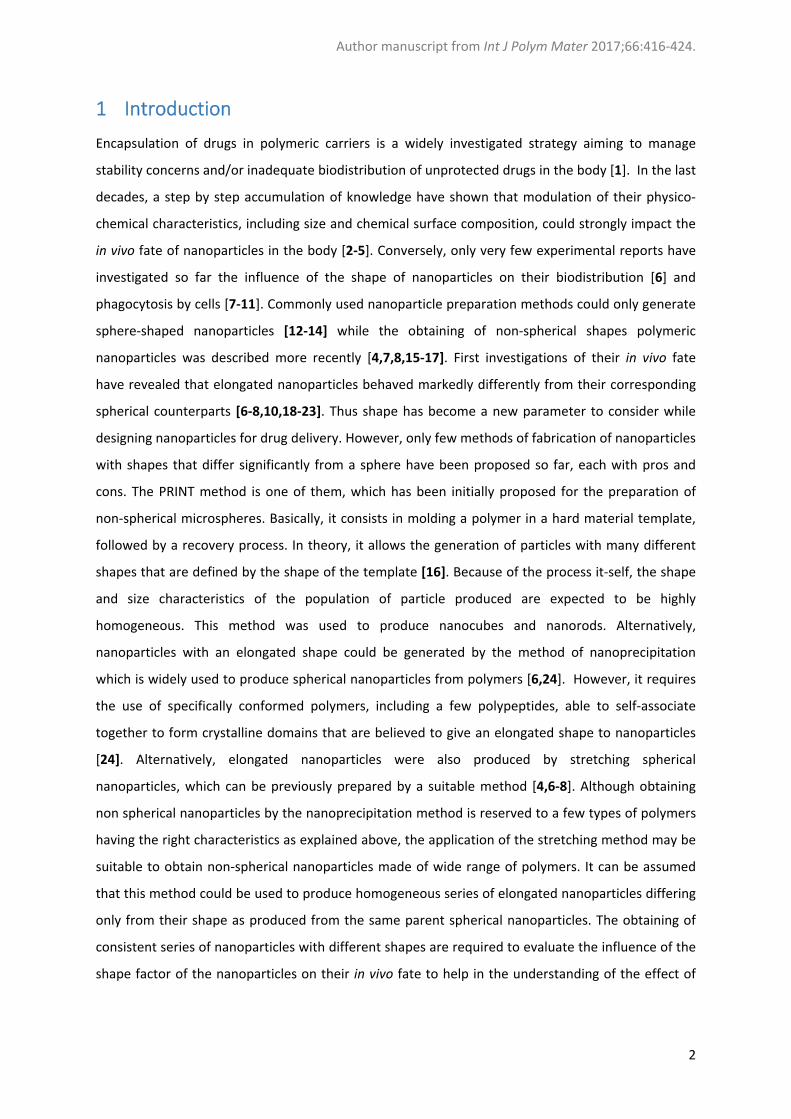
Abstract: This work was aimed to produce elongated nanoparticles of clinical interest by the stretching method. Polymer films prepared from material recovered from the dissolution of a marketed stretchable water‐soluble film were loaded with 10% of poly(isobutylcyanoacrylate) nanoparticles. The films were elongated at 50°C to 230% of their initial lengths. The shape factor of the nanoparticles recovered after elongation was consistent with stretching conditions applied on the film. This work has demonstrated the feasibility of the preparation of non‐spherical poly(isobutylcyanoacrylate) nanoparticles by the stretching method and described conditions to apply the method.
Key words: stretching, nanoparticles, shape factor, polymer film, poly(isobutylcyanoacrylate)
Author manuscript from Int J Polym Mater 2017;66:416‐424.
2
1 Introduction
Encapsulation of drugs in polymeric carriers is a widely investigated strategy aiming to manage
stability concerns and/or inadequate biodistribution of unprotected drugs in the body [1]. In the last
decades, a step by step accumulation of knowledge have shown that modulation of their physico‐
chemical characteristics, including size and chemical surface composition, could strongly impact the
in vivo fate of nanoparticles in the body [2‐5]. Conversely, only very few experimental reports have
investigated so far the influence of the shape of nanoparticles on their biodistribution [6] and
phagocytosis by cells [7‐11]. Commonly used nanoparticle preparation methods could only generate
sphere‐shaped nanoparticles [12‐14] while the obtaining of non‐spherical shapes polymeric
nanoparticles was described more recently [4,7,8,15‐17]. First investigations of their in vivo fate
have revealed that elongated nanoparticles behaved markedly differently from their corresponding
spherical counterparts [6‐8,10,18‐23]. Thus shape has become a new parameter to consider while
designing nanoparticles for drug delivery. However, only few methods of fabrication of nanoparticles
with shapes that differ significantly from a sphere have been proposed so far, each with pros and
cons. The PRINT method is one of them, which has been initially proposed for the preparation of
non‐spherical microspheres. Basically, it consists in molding a polymer in a hard material template,
followed by a recovery process. In theory, it allows the generation of particles with many different
shapes that are defined by the shape of the template [16]. Because of the process it‐self, the shape
and size characteristics of the population of particle produced are expected to be highly
homogeneous. This method was used to produce nanocubes and nanorods. Alternatively,
nanoparticles with an elongated shape could be generated by the method of nanoprecipitation
which is widely used to produce spherical nanoparticles from polymers [6,24]. However, it requires
the use of specifically conformed polymers, including a few polypeptides, able to self‐associate
together to form crystalline domains that are believed to give an elongated shape to nanoparticles
[24]. Alternatively, elongated nanoparticles were also produced by stretching spherical
nanoparticles, which can be previously prepared by a suitable method [4,6‐8]. Although obtaining
non spherical nanoparticles by the nanoprecipitation method is reserved to a few types of polymers
having the right characteristics as explained above, the application of the stretching method may be
suitable to obtain non‐spherical nanoparticles made of wide range of polymers. It can be assumed
that this method could be used to produce homogeneous series of elongated nanoparticles differing
only from their shape as produced from the same parent spherical nanoparticles. The obtaining of
consistent series of nanoparticles with different shapes are required to evaluate the influence of the
shape factor of the nanoparticles on their in vivo fate to help in the understanding of the effect of
Author manuscript from Int J Polym Mater 2017;66:416‐424.
3
the shape of nanoparticles on their interactions with cells and their capacity to go across different
biological barriers.
The aim of the present work was to set up experimental conditions to prepare elongated
nanoparticles of poly(alkylcyanoacrylate) nanoparticles from parent spherical nanoparticles applying
the stretching method. The parent nanoparticles used in the present study were selected for several
reasons. They can be prepared with different characteristics in a well‐controlled manner by highly
reproducible methods of emulsion polymerization [25‐30]. Their interest to improve in vivo delivery
of drug is well documented considering a wide range of drug and therapeutic applications [31‐38].
They are one of the very few nanomedicines made of polymer nanoparticles that are considered in
an ongoing clinical trial phase II/III [34‐36]. Additionally, poly(alkylcyanoacrylate) nanoparticles were
developed as model nanoparticles to investigate the influence of the size and surface properties on
interactions with biological systems and in turn on their in vivo fate [27,39‐42]. Having the
corresponding elongated species of this series would provide with the required model nanoparticles
to also investigate the influence of the nanoparticle shape. The chitosan‐coated
poly(isobutylcyanoacrylate) (PIBCA) nanoparticles were selected to carry on the present work. These
nanoparticles were designed as mucoadhesive drug delivery systems to promote oral administration
of drugs [28]. They were also tested as an antitumor treatment based on the delivery of siRNA by
intravenous injection [37,38,43].
The stretching method was applied to produce elongated micro‐ and nanoparticles from their
spherical parent particles [6‐8,11,44]. It can be applied to any type of particles that are made of a
material that can be soften at the operating temperature of the stretching machine. Elongated
microparticles made of both non‐biodegradable and biodegradable polymers have been produced.
In contrast, to the best of our knowledge, the method was only applied with nanoparticles made of
non‐biodegradable polymers (i.e. polystyrene) [11]. The method was not applied yet to the
production of elongated biodegradable particles in the nano‐size range. Interestingly, the stretching
method can be scaled up to produce large amount of elongated particles thanks to a possible
automatization [44]. As described above, the method implies basically: (i) the dispersion of pre‐
formed spherical nanoparticles (supposed to eventually contain a drug if any pharmaceutical
application are foreseen) into a polymeric material film, followed by the stretching of the
nanoparticles containing material at a high temperature (generally above 120°C). At the end of the
stretching process, the stretched particles need to be extracted from the polymeric material film
[6,7,11]. In the present work, our aim was to set conditions for the application of the method to the
elaboration of elongated nanoparticles made of biodegradable polymer. It was also aimed to apply
the method at a much lower temperature (50°C) as those used in previous work (>120°C) [6,7,11].
Author manuscript from Int J Polym Mater 2017;66:416‐424.
4
Although in previous work, the polymer films in which the nanoparticles were incorporated to be
stretched were formulated in home from solution of poly(vinyl alcohol), for this work, we have
selected a water soluble and stretchable film available on the market as the material to prepare the
film incorporating the spherical nanoparticles to be stretched. The paper reports the different steps
of the preparation of the film loaded with nanoparticles from the material of the marketed film and
results on the application of the stretching method to prepare elongated nanoparticles of chitosan
coated poly(isobutylcyanoacrylate) nanoparticles.
2 Materials and methods
2.1 Materials
Chitosan (CS, MW 20 kDa, deacetylation degree 92% was purchased from Amicogen.Inc. (Jinju, South
Korea), isobutylcyanoacrylate (IBCA) was gift from Henkel Biomedical (Loctite, Dublin, Ireland); a
poly(vinylalcohol) (PVA) based water soluble polymer film (STBT® 30µm) (STBT®) was purchased from
Soluble Technology Ltd. (Chechire, United Kingdom) for the elongation experiments. Milli‐Q® water
was produced using the Milli‐Q® direct 8 water purification system from Merck Millipore (Millipore
SAS, Guyancourt, France).
2.2 Methods
2.2.1 Preparation of the parent spherical nanoparticles.
Poly(isobutylcyanoacrylate) nanoparticles coated with chitosan (CSPIBCA) were prepared by redox
radical polymerization according to a previously described protocol [26,30,43]. Briefly, chitosan (130
mg) was dissolved in HNO3 0.2 M (8 mL) at 42°C under vigorous stirring and argon atmosphere. Once
the dissolution was completed, a solution of ammonium cerium (IV) nitrate (8x10‐2 M) in HNO3 0.2 M
(2 mL) and IBCA (0.5 mL) were added successively, the argon flux was stopped and the reaction was
allowed to continue at 42°C for 50 min. Then, the nanoparticles were purified by dialysis (cut‐off
100,000 g/mol, Spectra/Por membranes) three times against Milli‐Q® water for 1 h and once
overnight. The obtained CSPIBCA nanoparticle dispersion was stored at 4°C until use.
The concentration in nanoparticles of the dispersion was evaluated by gravimetry performed by
measuring the mass of the dried residue obtained after lyophilisation of 0.5 mL of sample. The
experiment was done in triplicate.
The mean hydrodynamic diameter of the nanoparticles was determined by dynamic light scattering
using a Zetasizer® ZS90 (Malvern Instrument, Orsay, France). Measurements were performed at 25°C
at a scattered angle of 90°. Samples were diluted in Milli‐Q® water to a concentration of 0.35 mg/mL.
Author manuscript from Int J Polym Mater 2017;66:416‐424.
5
Results were expressed as the mean hydrodynamic diameter deduced from size measurements
performed on 3 different preparations of nanoparticles.
The ζ potential was deduced from the electrophoretic mobility of the particles measured by Laser
Doppler Electrophoresis after dilution of CSPIBCA suspension in NaCl solution (1 mM, 1/100 (v/v)).
Determination was achieved using a Zetasizer® ZS90 (Malvern Instrument, Orsay, France).
2.2.2 Preparation of the polymer film incorporating the nanoparticles
At first, STBT® film (100 mg) was dissolved in 1.5 mL of Milli‐Q® water, under magnetic stirring for 4
hours at room temperature. The concentration of the solutions was 67 mg.mL‐1. Then, a dispersion
of CSPIBCA nanoparticles at the concentration in nanoparticles of 31 mg.mL‐1 was added to this
solution to a weight percentage of nanoparticles and STBT® of 10 % (323 μL), 13% (419 μL) and 15%
(484 μL). The resulting mixture was gently stirred for 30 min. The mixture STBT®‐CSPIBCA was spread
on a glass slide (25x75 mm²) and dried in an incubator (Memmert, Grosseron, France) at different
temperatures, 25, 37 and 50 °C for 24 h, to allow the evaporation of the water and the formation of
a new film of STBT® incorporating the CSPIBCA nanoparticles. For control experiments films were
prepared by the same procedure but without nanoparticles. The films were used immediately after
preparation
2.2.3 Characterization of film properties
Films containing various amounts of nanoparticles (0, 10, 13, 17, 20 % w/w) and prepared at
different temperatures (25°C, 37°C, 50°C,) were obtained according to the procedure described in
2.2.2. At first, the homogeneity of the dispersion of nanoparticles in the polymer solution was
appreciated by visual inspection and under light microscopy using the contrast phase mode
(Olympus BH2). Pictures were recorded using a Mightex USB2.0 color camera. After the film was
formed, the homogeneity of the films was also appreciated by the same method. The purpose of
these observations was to check for the occurrence of phase separation. All systems that appeared
heterogenous were discarded.
Only homogenous films were selected to determine the percent elongation at break using a TA.XT2
texture analyser (Stable Micro Systems, London, United Kingdom). Films with same dimensions as
the ones used to prepare elongated nanoparticles (25 x 75 mm, 30 µm thick) were tightly attached
to grips on the TA.XT2 machine. The initial grip separation was checked prior to the start of each
experiment. It corresponded to the initial length of the sample (L0). The cross head was moving at a
rate of 0.1 mm.s‐1 while the stretching experiment was performed at room temperature (20°C) until
breakage of the film. The curves gave the tensile strength (TS) as the function of the strain. The TS
Author manuscript from Int J Polym Mater 2017;66:416‐424.
6
was calculated dividing the measured force, F (in newton), by the section of the film, s (in m²)
(Equation 1).
𝑇𝑆 Equation 1
The strain was deduced from the ration between the extension length, ΔL, and the initial length (L0)
(Equation 2).
𝑆𝑡𝑟𝑎𝑖𝑛∆
Equation 2
The elongation at breakage (EAB) was calculated following equation (3).
𝐸𝐴𝐵 %∆
100 Equation 3
Where ΔLAB was the extension length at breakage. Measurements were performed at least 3 times
on each film.
The maximal elongation (𝐸 capacity of the film could be calculated from the equation (4).
𝐸 Equation 4
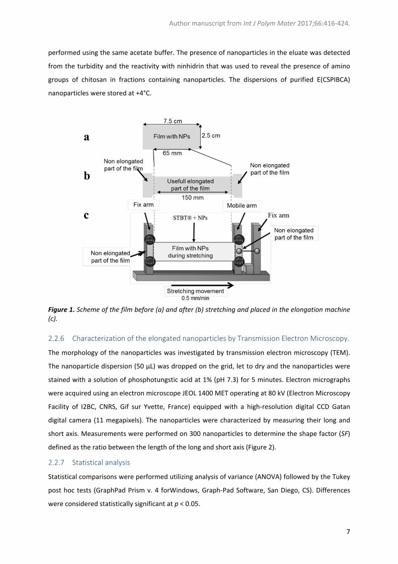
2.2.4 Elongation of the film producing elongated nanoparticles
To achieve preparation of elongated nanoparticles (E(CDPIBCA) nanoparticles), films were prepared
with a concentration of 13% nanoparticles as explained in section 2.2.3. Second generation films
recovered from the glass slide were placed in a home‐made stretching machine as described in
Figure 1. The initial grip separation length (𝐿 ) was 65 mm. The stretching machine was placed in an
incubator (Memmert, Grosseron France) at 50°C and let to equilibrate for 30 min. The film was
stretched at a temperature of 50°C and at a speed of 0.5 mm.min‐1 to a final length, L, of 150 mm
giving an elongation capacity, Ec, of 231% as calculated from equation 5.
𝐸 Equation 5
Immediately after the stop of the stretching, the film was rapidly cooled down to 20 °C using a flux
of cold air (temperature ‐16.5 °C) while it was maintained on the maximum stretched position. Then,
the parts of films used to secure the film in the elongation machine were discarded (Figure 1).
Effectively elongated parts were kept to recover nanoparticles embedded in the stretch film.
2.2.5 Recovery of E(CSPIBCA) nanoparticles from the films after stretching
The stretch part of the film was dissolved in 500 μL of water or nitric acid 0.2 M (pH 1) for 30
minutes at room temperature under gentle stirring. After complete dissolution of the film, E(CSPIBA)
nanoparticles were purified by Size Exclusion Chromatography (SEC) using a column of Sephacryl® S‐
1000 gel (5 cm x 1.4 cm). The column was equilibrated in acetate buffer at pH 3.2. Elution was
Author manuscript from Int J Polym Mater 2017;66:416‐424.
7
performed using the same acetate buffer. The presence of nanoparticles in the eluate was detected
from the turbidity and the reactivity with ninhidrin that was used to reveal the presence of amino
groups of chitosan in fractions containing nanoparticles. The dispersions of purified E(CSPIBCA)
nanoparticles were stored at +4°C.
Figure 1. Scheme of the film before (a) and after (b) stretching and placed in the elongation machine (c).
2.2.6 Characterization of the elongated nanoparticles by Transmission Electron Microscopy.
The morphology of the nanoparticles was investigated by transmission electron microscopy (TEM).
The nanoparticle dispersion (50 µL) was dropped on the grid, let to dry and the nanoparticles were
stained with a solution of phosphotungstic acid at 1% (pH 7.3) for 5 minutes. Electron micrographs
were acquired using an electron microscope JEOL 1400 MET operating at 80 kV (Electron Microscopy
Facility of I2BC, CNRS, Gif sur Yvette, France) equipped with a high‐resolution digital CCD Gatan
digital camera (11 megapixels). The nanoparticles were characterized by measuring their long and
short axis. Measurements were performed on 300 nanoparticles to determine the shape factor (SF)
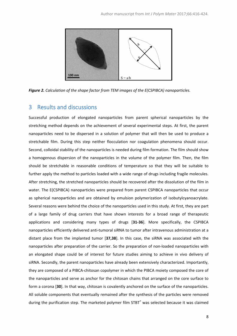
defined as the ratio between the length of the long and short axis (Figure 2).
2.2.7 Statistical analysis
Statistical comparisons were performed utilizing analysis of variance (ANOVA) followed by the Tukey
post hoc tests (GraphPad Prism v. 4 forWindows, Graph‐Pad Software, San Diego, CS). Differences
were considered statistically significant at p < 0.05.
Author manuscript from Int J Polym Mater 2017;66:416‐424.
8
Figure 2. Calculation of the shape factor from TEM images of the E(CSPIBCA) nanoparticles.
3 Results and discussions
Successful production of elongated nanoparticles from parent spherical nanoparticles by the
stretching method depends on the achievement of several experimental steps. At first, the parent
nanoparticles need to be dispersed in a solution of polymer that will then be used to produce a
stretchable film. During this step neither flocculation nor coagulation phenomena should occur.
Second, colloidal stability of the nanoparticles is needed during film formation. The film should show
a homogenous dispersion of the nanoparticles in the volume of the polymer film. Then, the film
should be stretchable in reasonable conditions of temperature so that they will be suitable to
further apply the method to particles loaded with a wide range of drugs including fragile molecules.
After stretching, the stretched nanoparticles should be recovered after the dissolution of the film in
water. The E(CSPIBCA) nanoparticles were prepared from parent CSPIBCA nanoparticles that occur
as spherical nanoparticles and are obtained by emulsion polymerization of isobutylcyanoacrylate.
Several reasons were behind the choice of the nanoparticles used in this study. At first, they are part
of a large family of drug carriers that have shown interests for a broad range of therapeutic
applications and considering many types of drugs [31‐36]. More specifically, the CSPIBCA
nanoparticles efficiently delivered anti‐tumoral siRNA to tumor after intravenous administration at a
distant place from the implanted tumor [37,38]. In this case, the siRNA was associated with the
nanoparticles after preparation of the carrier. So the preparation of non‐loaded nanoparticles with
an elongated shape could be of interest for future studies aiming to achieve in vivo delivery of
siRNA. Secondly, the parent nanoparticles have already been extensively characterized. Importantly,
they are composed of a PIBCA‐chitosan copolymer in which the PIBCA moiety composed the core of
the nanoparticles and serve as anchor for the chitosan chains that arranged on the core surface to
form a corona [30]. In that way, chitosan is covalently anchored on the surface of the nanoparticles.
All soluble components that eventually remained after the synthesis of the particles were removed
during the purification step. The marketed polymer film STBT® was selected because it was claimed
Author manuscript from Int J Polym Mater 2017;66:416‐424.
9
to dissolve rapidly in cold water and to be stretchable. Its composition based on PVA would be of
good compatibility for a use in a pharmaceutical process. Water solubility and fast dissolution were
mandatory characteristics. It was intended to incorporate nanoparticles within a film of polymer
prepared from the material recovered from the dissolution of the marketed STBT® film to which the
nanoparticles were added. So, these characteristics were needed to incorporate the nanoparticles in
the film and also to be able to recover the nanoparticles embedded in the film after stretching.
Viscoelastic properties of the marketed film were requested to have a suitable matrix to stretch the
embedded nanoparticles. According to the supplier, the STBT® film has elongation capacities of 185%
at room temperature and 250% at 50°C. Besides, the marketed film showed another interesting
property for the intended application. Its transparency was found useful to observe eventual
occurrence of aggregation of nanoparticles introduced in the film made from the marketed film
material.
The work included the following 3 steps:
‐ Determination of the maximal load in nanoparticles that can be incorporated in the film.
‐ Production of elongated nanoparticles
‐ Determination of the shape factor of the nanoparticles recovered from the film after elongation.
3.1 Characteristics of the parent nanoparticles
The parent CSPIBCA nanoparticles used in the present study consisted of core‐corona type
nanoparticles. The core was composed of PIBCA that was coated by a layer of chitosan covalently
anchored to the nanoparticle core by PIBCA chains [30,38]. Characteristics of the nanoparticles
synthesized for this work were consistent with those of our previous works [28,30,38] with a mean
hydrodynamic dynamic diameter of 394 ± 1 nm and a zeta potential of + 22.7 ± 0.6 mV evaluated at
neutral pH. The concentration of the parent dispersion was 31 mg.mL‐1. The number of
nanoparticles, Np, found in 1 mg can be calculated from the hydrodynamic diameter of the
nanoparticles converted in cm, dH, and the volumetric mass, ρ that is 1140 mg.mL‐1 for
poly(isobutylcyanoacrylate) nanoparticles [45] (Equation 6).
𝑁
Equation 6
By applying equation 1, it could be determined that one mg of nanoparticles contained 27.4 109
nanoparticles.
Author manuscript from Int J Polym Mater 2017;66:416‐424.
10
3.2 Determination of the maximal concentration in nanoparticles that can be
incorporated in the film.
It is known that colloids can flocculate or coagulate in the presence of polymers [46]. Thus, the first
step of the work was dedicated to the preparation of films incorporating nanoparticles in which the
nanoparticles would be homogenously dispersed while the new film would show interesting
stretching properties for the intended application. As indicated by the supplier, the STBT® film
dissolved easily in water. A solution of the polymer obtained from the dissolution of the film could
be prepared at a concentration of 67 mg.mL‐1. Addition of nanoparticles to the polymer solution led
to homogenous dispersions from both visual and microscopic observations up to a load in
nanoparticles of 15% expressed as the percentage of nanoparticles in weight added from the weight
of polymer dissolved. Above this load, a phase separation was observed. Films were prepared only
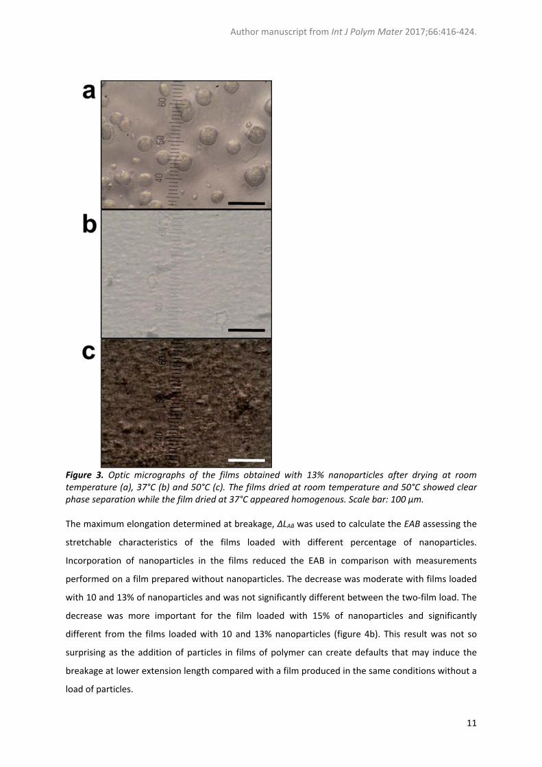
with nanoparticle containing dispersions that appeared homogenous. Films prepared at room
temperature and at 50°C showed the occurrence of a phase separation (Figure 3). Conversely, films
obtained from a working procedure carried out at 37°C were homogenous at both macroscopic and
microscopic scales up to a load in nanoparticles of 13% (Figure 3). For a film obtained from a solution
composed of STBT® at a concentration of 67 mg.mL‐1 and nanoparticles at a concentration of 6.8
mg.mL‐1 (10% load), the calculated number of nanoparticles per unit surface area dispersed in the
film, n, was 14.5 x 10⁹ nanoparticles.cm‐². This number could be calculated from equation 7 with Np,
the number of nanoparticles in 1 mg deduced from equation 6, v, the volume of dispersion of
nanoparticles added during the preparation of the film (0.323 mL), C the concentration in
nanoparticle of the dispersion (31 mg.mL‐1) and the area of the film, A (2.5 x 7.5 cm²).
𝑛
Equation 7
Only films that appeared homogeneous under the microscope were further used. Obviously,
embedding nanoparticles into the films could considerably influence their mechanical and visco‐
elastic characteristics, compared to a nanoparticle‐empty STBT® films. The stretching characteristics
of the films, both unloaded and embedding nanoparticles, could be a critical determinant of the
elongation efficacy. Therefore, the maximal elongation at breakage, EAB, of the films prepared with
various loads of nanoparticles was determined using a universal mechanical testing machine
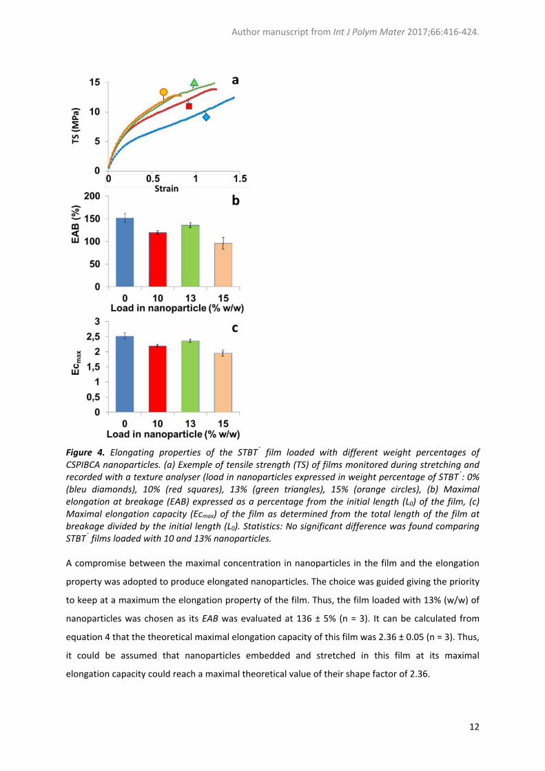
(Texture analyser). The figure 4a presents tensile stress of the films monitored during the elongation
process. Consistently to what was expected, the tensile stress increased up to a maximum where the
curves showed a sudden break that corresponded to the breakage of the film. The general shape of
the curves suggested that the films behaved like a ductile material. The breakage occurred in the
plastic domain of the film.
Author manuscript from Int J Polym Mater 2017;66:416‐424.
11
Figure 3. Optic micrographs of the films obtained with 13% nanoparticles after drying at room temperature (a), 37°C (b) and 50°C (c). The films dried at room temperature and 50°C showed clear phase separation while the film dried at 37°C appeared homogenous. Scale bar: 100 µm. The maximum elongation determined at breakage, ΔLAB was used to calculate the EAB assessing the
stretchable characteristics of the films loaded with different percentage of nanoparticles.
Incorporation of nanoparticles in the films reduced the EAB in comparison with measurements
performed on a film prepared without nanoparticles. The decrease was moderate with films loaded
with 10 and 13% of nanoparticles and was not significantly different between the two‐film load. The
decrease was more important for the film loaded with 15% of nanoparticles and significantly
different from the films loaded with 10 and 13% nanoparticles (figure 4b). This result was not so
surprising as the addition of particles in films of polymer can create defaults that may induce the
breakage at lower extension length compared with a film produced in the same conditions without a
load of particles.
Author manuscript from Int J Polym Mater 2017;66:416‐424.
12
Figure 4. Elongating properties of the STBT® film loaded with different weight percentages of CSPIBCA nanoparticles. (a) Exemple of tensile strength (TS) of films monitored during stretching and recorded with a texture analyser (load in nanoparticles expressed in weight percentage of STBT®: 0% (bleu diamonds), 10% (red squares), 13% (green triangles), 15% (orange circles), (b) Maximal elongation at breakage (EAB) expressed as a percentage from the initial length (L0) of the film, (c) Maximal elongation capacity (Ecmax) of the film as determined from the total length of the film at breakage divided by the initial length (L0). Statistics: No significant difference was found comparing STBT® films loaded with 10 and 13% nanoparticles. A compromise between the maximal concentration in nanoparticles in the film and the elongation
property was adopted to produce elongated nanoparticles. The choice was guided giving the priority
to keep at a maximum the elongation property of the film. Thus, the film loaded with 13% (w/w) of
nanoparticles was chosen as its EAB was evaluated at 136 ± 5% (n = 3). It can be calculated from
equation 4 that the theoretical maximal elongation capacity of this film was 2.36 ± 0.05 (n = 3). Thus,
it could be assumed that nanoparticles embedded and stretched in this film at its maximal
elongation capacity could reach a maximal theoretical value of their shape factor of 2.36.
Author manuscript from Int J Polym Mater 2017;66:416‐424.
13
3.3 3.2. Production of elongated nanoparticles.
The selected film loaded with 13% nanoparticles was placed in the stretching machine to be
stretched at 50°C. The machine was set to perform an elongation of the film giving more than twice
its original length (230%) but remaining slighly below the EAB predetermined for this film to avoid
film breakage. At the end of the experiment, it was assumed that a tempering of the film would be
necessary to preserve the new shape acquired by the nanoparticles that were submitted to the
elongation stress. Thus, films were cooled down immediately after the elongation was stopped by
blowing cold air (‐16°C) and allowing the temperature of the film to drop down to the ambient
temperature in less than 30 sec. The effective experimental elongation that was calculated from the
measurement of the length of the film before and after recovery from the elongating machine was
2.3 consistently with the applied operating conditions.
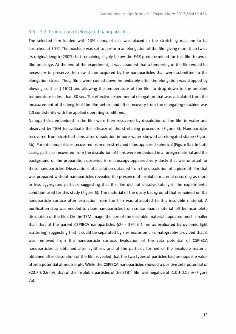
Nanoparticles embedded in the film were then recovered by dissolution of the film in water and
observed by TEM to evaluate the efficacy of the stretching procedure (Figure 5). Nanoparticles
recovered from stretched films after dissolution in pure water showed an elongated shape (Figure
5b). Parent nanoparticles recovered from non‐stretched films appeared spherical (Figure 5a). In both
cases, particles recovered from the dissolution of films were embedded in a foreign material and the
background of the preparation observed in microscopy appeared very dusty that was unusual for
these nanoparticles. Observations of a solution obtained from the dissolution of a piece of film that
was prepared without nanoparticles revealed the presence of insoluble material occurring as more
or less aggregated particles suggesting that the film did not dissolve totally in the experimental
condition used for this study (Figure 6). The material of the dusty background that remained on the
nanoparticle surface after extraction from the film was attributed to this insoluble material. A
purification step was needed to clean nanoparticles from contaminant material left by incomplete
dissolution of the film. On the TEM image, the size of the insoluble material appeared much smaller
than that of the parent CSPIBCA nanoparticles (DH = 394 ± 1 nm as evaluated by dynamic light
scattering) suggesting that it could be separated by size exclusion chromatography provided that it
was removed from the nanoparticle surface. Evaluation of the zeta potential of CSPIBCA
nanoparticles as obtained after synthesis and of the particles formed of the insoluble material
obtained after dissolution of the film revealed that the two types of particles had an opposite value
of zeta potential at neutral pH. While the CSPIBCA nanoparticles showed a positive zeta potential of
+22.7 ± 0.6 mV, that of the insoluble particles of the STBT® film was negative at ‐1.0 ± 0.1 mV (Figure
7a).
Author manuscript from Int J Polym Mater 2017;66:416‐424.
14
Figure 5. Transmission Electron Microscographs (TEM) of the parent CSPIBCA nanoparticles (a) and of the E(CSPIBCA) nanoparticles recovered after dissolution of the film in pure water (b). E(CSPIBCA) nanoparticles recovered from the film after dissolution at pH 1 and purification by SEC performed at pH 3.5 (c).
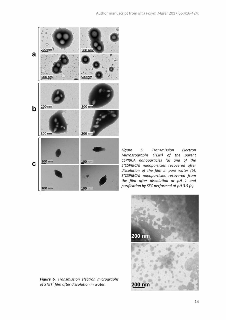
Figure 6. Transmission electron micrographs of STBT® film after dissolution in water.
Author manuscript from Int J Polym Mater 2017;66:416‐424.
15
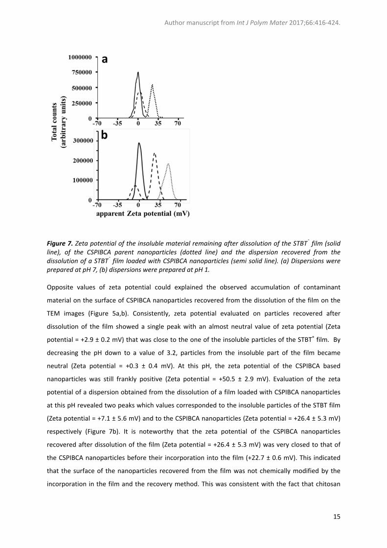
Figure 7. Zeta potential of the insoluble material remaining after dissolution of the STBT® film (solid line), of the CSPIBCA parent nanoparticles (dotted line) and the dispersion recovered from the dissolution of a STBT® film loaded with CSPIBCA nanoparticles (semi solid line). (a) Dispersions were prepared at pH 7, (b) dispersions were prepared at pH 1. Opposite values of zeta potential could explained the observed accumulation of contaminant
material on the surface of CSPIBCA nanoparticles recovered from the dissolution of the film on the
TEM images (Figure 5a,b). Consistently, zeta potential evaluated on particles recovered after
dissolution of the film showed a single peak with an almost neutral value of zeta potential (Zeta
potential = +2.9 ± 0.2 mV) that was close to the one of the insoluble particles of the STBT® film. By
decreasing the pH down to a value of 3.2, particles from the insoluble part of the film became
neutral (Zeta potential = +0.3 ± 0.4 mV). At this pH, the zeta potential of the CSPIBCA based
nanoparticles was still frankly positive (Zeta potential = +50.5 ± 2.9 mV). Evaluation of the zeta
potential of a dispersion obtained from the dissolution of a film loaded with CSPIBCA nanoparticles
at this pH revealed two peaks which values corresponded to the insoluble particles of the STBT film
(Zeta potential = +7.1 ± 5.6 mV) and to the CSPIBCA nanoparticles (Zeta potential = +26.4 ± 5.3 mV)
respectively (Figure 7b). It is noteworthy that the zeta potential of the CSPIBCA nanoparticles
recovered after dissolution of the film (Zeta potential = +26.4 ± 5.3 mV) was very closed to that of
the CSPIBCA nanoparticles before their incorporation into the film (+22.7 ± 0.6 mV). This indicated
that the surface of the nanoparticles recovered from the film was not chemically modified by the
incorporation in the film and the recovery method. This was consistent with the fact that chitosan
Author manuscript from Int J Polym Mater 2017;66:416‐424.
16
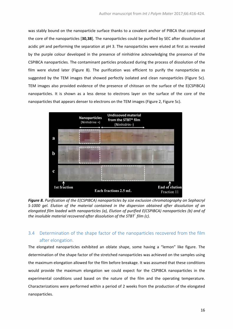
was stably bound on the nanoparticle surface thanks to a covalent anchor of PIBCA that composed
the core of the nanoparticles [30,38]. The nanoparticles could be purified by SEC after dissolution at
acidic pH and performing the separation at pH 3. The nanoparticles were eluted at first as revealed
by the purple colour developed in the presence of ninhidrine acknowledging the presence of the
CSPIBCA nanoparticles. The contaminant particles produced during the process of dissolution of the
film were eluted later (Figure 8). The purification was efficient to purify the nanoparticles as
suggested by the TEM images that showed perfectly isolated and clean nanoparticles (Figure 5c).
TEM images also provided evidence of the presence of chitosan on the surface of the E(CSPIBCA)
nanoparticles. It is shown as a less dense to electrons layer on the surface of the core of the
nanoparticles that appears denser to electrons on the TEM images (Figure 2, Figure 5c).
Figure 8. Purification of the E(CSPIBCA) nanoparticles by size exclusion chromatography on Sephacryl S‐1000 gel. Elution of the material contained in the dispersion obtained after dissolution of an elongated film loaded with nanoparticles (a), Elution of purified E(CSPIBCA) nanoparticles (b) and of the insoluble material recovered after dissolution of the STBT® film (c).
3.4 Determination of the shape factor of the nanoparticles recovered from the film
after elongation.
The elongated nanoparticles exhibited an oblate shape, some having a “lemon” like figure. The
determination of the shape factor of the stretched nanoparticles was achieved on the samples using
the maximum elongation allowed for the film before breakage. It was assumed that these conditions
would provide the maximum elongation we could expect for the CSPIBCA nanoparticles in the
experimental conditions used based on the nature of the film and the operating temperature.
Characterizations were performed within a period of 2 weeks from the production of the elongated
nanoparticles.
Author manuscript from Int J Polym Mater 2017;66:416‐424.
17
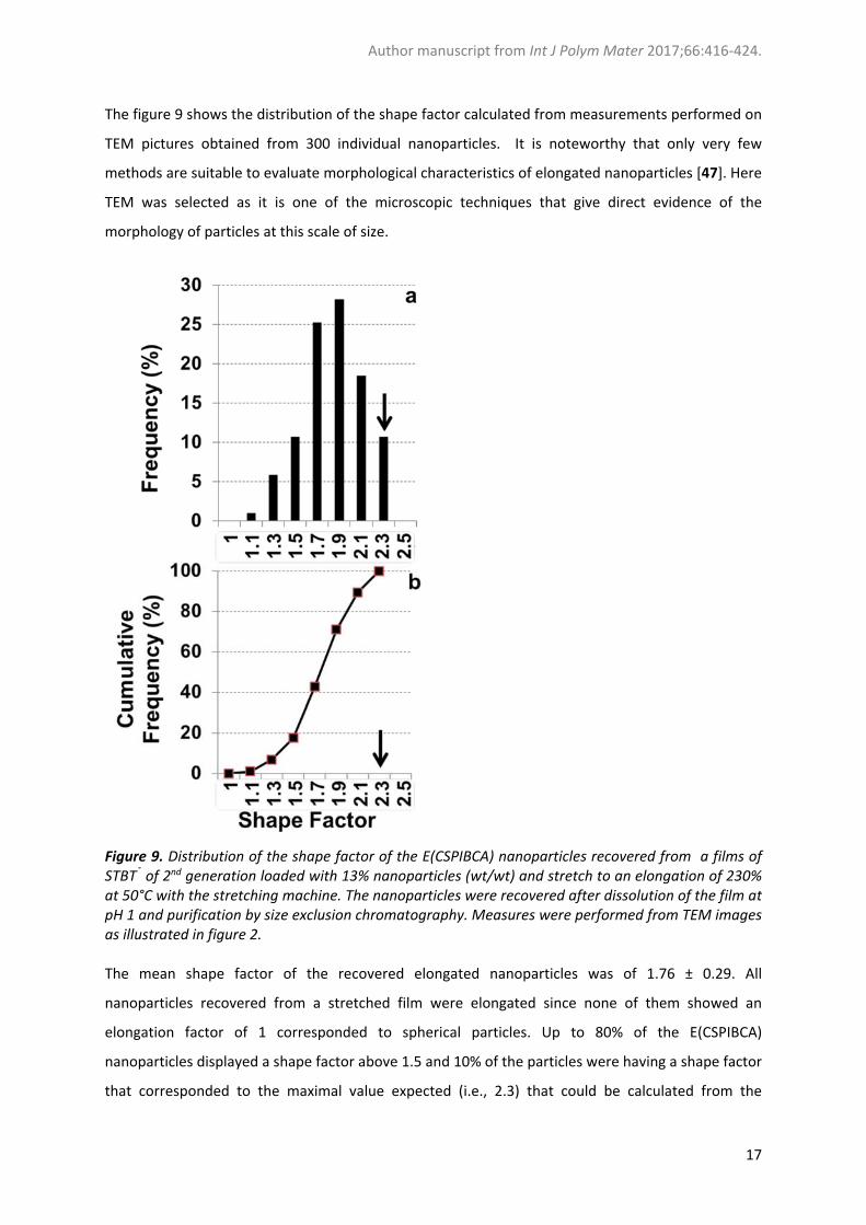
The figure 9 shows the distribution of the shape factor calculated from measurements performed on
TEM pictures obtained from 300 individual nanoparticles. It is noteworthy that only very few
methods are suitable to evaluate morphological characteristics of elongated nanoparticles [47]. Here
TEM was selected as it is one of the microscopic techniques that give direct evidence of the
morphology of particles at this scale of size.
Figure 9. Distribution of the shape factor of the E(CSPIBCA) nanoparticles recovered from a films of STBT® of 2nd generation loaded with 13% nanoparticles (wt/wt) and stretch to an elongation of 230% at 50°C with the stretching machine. The nanoparticles were recovered after dissolution of the film at pH 1 and purification by size exclusion chromatography. Measures were performed from TEM images as illustrated in figure 2. The mean shape factor of the recovered elongated nanoparticles was of 1.76 ± 0.29. All
nanoparticles recovered from a stretched film were elongated since none of them showed an
elongation factor of 1 corresponded to spherical particles. Up to 80% of the E(CSPIBCA)
nanoparticles displayed a shape factor above 1.5 and 10% of the particles were having a shape factor
that corresponded to the maximal value expected (i.e., 2.3) that could be calculated from the

Author manuscript from Int J Polym Mater 2017;66:416‐424.
18
elongation capacity of the film. The nanoparticles embedded in the film were significantly distorted
during the stretching procedure. However, the discrepancy between the film elongation and the one
of the particles suggest that micro‐rheological behaviour of the CSPIBCA nanoparticles dispersed in
the PVA matrix of the film, as well as the temperature and the imposed macroscopic rate of
deformation could be important determinants of the attainable deformation, which would deserve
further investigations.
4 Conclusion
The procedure proposed in the present work was suitable to prepare E(CSPIBCA) nanoparticles with
a shape factor up to 2.3 working at a reasonable temperature at 50°C with marketed water‐soluble
film forming materials. Many drugs are stable at this temperature that makes this procedure suitable
to be applied to produce elongated nanoparticles being loaded with a wide range of drugs. Further,
film loads in nanoparticles was high (up to 13% expressed in dry weight) and the method allows the
recovery of an aqueous suspension of elongated nanoparticles simply by dissolution of the water‐
soluble PVA film. Besides, the method was applied to prepare elongated species of
poly(alkylcyanoacrylate) nanoparticles from their spherical counterparts. The elongated species will
constitute a new model of nanoparticles complemental to the already available series of spherical
species. They will be used in studies considering the influence of the shape of nanoparticles on their
in vivo fate under various situations for their application as drug delivery systems. They will also be
used to investigate the influence of the nanoparticle shape with biological systems by in vitro
methods. For instance, they are of interest to investigate the influence of the nanoparticle shape on
(i) nanoparticle interactions with biomacromolecules, particularly with proteins present in biological
fluids, (ii) the capacity of the nanoparticles to go through biological barriers, including epithelia and
endothelia as well as (iii) their propensity to be endocytosed by cells. This new model of
nanoparticles will also be interested to investigate the influence of the shape of the nanoparticles in
drug delivery methods that consider a mucosal route of administration.
Acknowledgments
C. Palazzo received a fellowship from “Ministero dell’Istruzione dell’Università e della Ricerca”,
MIUR, Italy. The present work has benefited from the facilities and expertise of the Electron
Microscopy facilities of I2BC, CNRS, Gif sur Yvette, France (http://www.cgm.cnrs‐
gif.fr/spip.php?article282&lang=fr).
Author manuscript from Int J Polym Mater 2017;66:416‐424.
19
References
1 Brown, P.D. and Patel, P.R., WIREs Nanomed. Nanobiotechnol., 7, 125–130(2015). 2 Alexis, F., Pridgen, E., Molnar, L.K. and Farokhzad, O.C., Mol. Pharm., 5, 505‐15 (2008).
3 Clogston, J.D. and Patri, A.K. In: Dobrovolskaia, M., McNeil S.E. (2013). Frontiers in
nanobiomaterial research Vol 1: Handbook of immunological properties of engineered
nanomaterials, (World Scientific Publishing Co. Pte. Ltd.), pp. 25‐52.
4 Wang, J., Byrne, J.D., Napier, M.E. and DeSimone, J.M., Small, 7 (2011) 1919‐1931.
5 Moghimi, S.M., Hunter, A.C. and Andresen, T.L., Ann. Rev. Pharmacol. Tox., 52 (2012) 481‐503.
6 Cauchois, O., Conception, Preparation & Characterization of nanoparticles of complex shapes:
Study of their in vivo fate, PhD Thesis of the Université Paris Sud ‐ Paris XI, 2011, HAL Id: tel‐
00659379. https://tel.archives‐ouvertes.fr/tel‐00659379 .
7 Champion, J.A. and Mitragotri, S., Proc. Natl. Acad. Sci. USA, 103 (2006) 4930–4934.
8 Champion, J.A., Katare, Y.K. and Mitragotri, S., Proc. Natl. Acad. Sci. USA, 104 (2007) 11901‐
11904.
9 Barua, S., Yoo, J.W., Kolhar, P., Wakankar, A., Gokarn, Y.R. and Mitragotri, S., Proc. Natl. Acad. Sci.
USA, 110 (2013) 3270‐3275.
10 Mathaes, R., Winter, G., Besheer, A. and Engert, J., Int. J. Pharm., 465 (2014) 159‐164.
11 Kumar, S., Anselmo, A.C., Banerjee, A., Zakrewsky, M., Mitragotri, S., J. Control. Release, 28
(2015) 141‐148.
12 Vauthier, C. and Bouchemal, K., Pharm. Res., 26 (2009) 1025‐1058.
13 Prasad Rao, J. and Geckeler, K.E., Prog. Polym. Sci., 36 (2011) 887‐913.
14 Meyer, R.A. and Green, J.J., WIREs Nanomed. Nanobiotechnol., 8 (2016) 191‐207.
15 Cauchois, O., Segura‐Sanchez, F. and Ponchel, G., Int. J. Pharm., 452 (2013) 292‐299.
16 Landwehr, J., Fader, R., Rumler, M., Rommel, M., Bauer, A.J., Frey, L., Simon, B., Fodor, B., Petrik,
P., Schiener, A., Winter, B. and Spiecker, E., Nanotechnology, 25 (2014) 505301.
17 Beck‐Broichsitter, M., Nicolas, J. and Couvreur, P., Eur. J. Pharm. Biopharm., 97 (2015) 304‐17.
18 Mitragotri, S., Pharm. Res., 26 (2009) 232‐234.
19 Liu, Y., Tan, J., Thomas, A., Ou‐Yang, D and, Muzykantov, V.R., Ther. Deliv., 3 (2012) 181‐194.
20 Chu, K.S., Hasan, W., Rawal, S., Walsh, M.D., Enlow, E.M., Luft, J.C., Bridges, A.S., Kuijer, J.L.,
Napier, M.E., Zamboni, W.C. and DeSimone, J.M., Nanomedicine, 9 (2013) 686‐693.
21 Kolhar, P., Anselmo, A.C., Gupta, V., Pant, K., Prabhakarpandian, B., Ruoslahti, E. and Mitragotri,
S., Proc. Natl. Acad. Sci. U S A, 110 (2013) 10753‐10758.
22 Setyawati, M.L., Tay, C.Y., Docter, D., Stauberb, R.H. and Leong, D.T., Chem. Soc. Rev., 44 (2015),
8174‐8199.
23 Truong, N.P., Whittaker, M.R., Mak, C.W. and Davis, T.P., The importance of nanoparticle shape in
cancer drug delivery. Expert. Opin. Drug. Deliv. 12 (2015) 129‐142.
24 Barbosa, M.E., Bouteiller, L., Cammas‐Marion, S., Montembault, V., Fontaine, L. and Ponchel, G.,
J. Mol. Recognit., 21 (2008) 169‐178.
25 Chauvierre, C., Labarre, D., Couvreur, P. and Vauthier, C., Pharm. Res., 20 (2003) 1786‐1793.
26 Bertholon I., Vauthier, C., Labarre D. Pharm. Res., Pharm. Res., 23, (2006) 1313‐1323.
27 Lira, M.C., Santos‐Magalhães, N.S., Nicolas, V., Marsaud, V., Silva, M.P., Ponchel, G. and Vauthier,
C., Eur. J. Pharm. Biopharm., 79 (2011) 162‐170.
28 Bravo‐Osuna I., Ponchel G. and Vauthier C., Eur. J. Pharm. Sci., 30 (2007) 143‐154.
29 Nicolas, J. and Couvreur, P., Wiley Interdiscip. Rev. Nanomed. Nanobiotechnol., 1 (2009) 111‐127.
Author manuscript from Int J Polym Mater 2017;66:416‐424.
20
30 Zandanel, C. and Vauthier, C., J. Coll. Sci. Biotechnol., 1 (2012) 68‐81.
31 Barraud, L., Merle, P., Soma, E., Lefrancois, L., Guerret, S., Chevallier, M., Dubernet, C., Couvreur,
P., Trepo, C. and Vitvitski L., J. Hepatol., 42 (2005) 736‐743.
32 Couvreur, P. and Vauthier, C., Pharm. Res., 23 (2006) 1417‐1450.
33 Vauthier, C., Labarre, D. and Ponchel G., J. Drug Target., 15 (2007) 641‐663.
34 Soma, E., Attali, P. and Merle, P., In: Alonso, M.J. and Csaba, N.S. (2012). RSC Drug Discovery
Series No. 22: Nanostructured Biomaterials for Overcoming Biological Barriers, (The Royal Society
of Chemistry, Cambridge, UK), pp. 591‐600.
35 http://www.onxeo.com/fr/nos-produits/portefeuilles-produits/orphelins-oncologie/ Accessed 12 May 2016.
36 Zhou, Q., Sun, X., Zeng, L., Liu, J. and Zhang, Z., Nanomedicine. 5 (2009) 419‐423.
37 Ramon, A.L., Bertrand, J.R., de Martimprey, H., Bernard, G., Ponchel, G., Malvy, C. and Vauthier,
C., J. Mol. Recognit., 26 (2013) 318‐329.
38 de Martimprey, H., Bertrand, J.R., Malvy, C., Couvreur, P., Vauthier, C., Pharm. Res., 27 (2010)
498‐509.
39 Labarre, D., Vauthier, C., Chauvierre, C., Petri, B., Muller, R. and Chehimi, M.M., Biomaterials, 26
(2005) 5075‐5084.
40 Vauthier, C., Lindner, P. and Cabane, B., Colloids Surf. B Biointerfaces, 69 (2009) 207‐215.
41 Vauthier, C., Persson, B., Lindner, P., Cabane, B., Biomaterials, 32 (2011) 1646‐1656.
42 Alhareth, K., Vauthier, C., Bourasset, F., Gueutin, C., Ponchel, G. and Moussa, F., Eur. J. Pharm.
Biopharm., 81 (2012) 453‐457.
43 de Martimprey, H., Vauthier, C., Malvy, C. and Couvreur, P., Eur. J. Pharm. Biopharm., 71 (2009)
490‐504.
44 Meyer, R.A., Meyer, R.S., Green, J.J., J. Biomed. Mater. Res. A., 103 (2015) 2747‐2757.
45 Vauthier, C., Schmidt, C., Couvreur P., J. Nanoparticle Res., 1 (1999) 411‐418.
46 Kamibayashi, M., Ogura, H. and Otsubo, Y., J. Colloid Interface Sci., 321 (2008) 294‐301.
47 Mathaes, R., Winter, G., Engert, J., Besheer, A., Int. J. Pharm., 10 (2013) 620‐629.


