Embed Size (px)
Citation preview
University of South FloridaScholar Commons
Graduate Theses and Dissertations Graduate School
4-7-2017
Non-classical regulators in Staphylococcus aureusAndy WeissUniversity of South Florida, [email protected]
Follow this and additional works at: http://scholarcommons.usf.edu/etd
Part of the Microbiology Commons
This Dissertation is brought to you for free and open access by the Graduate School at Scholar Commons. It has been accepted for inclusion inGraduate Theses and Dissertations by an authorized administrator of Scholar Commons. For more information, please [email protected].
Scholar Commons CitationWeiss, Andy, "Non-classical regulators in Staphylococcus aureus" (2017). Graduate Theses and Dissertations.http://scholarcommons.usf.edu/etd/6779
Non-classical regulators in Staphylococcus aureus
by
Andy Weiss
A dissertation submitted in partial fulfillment of the requirements for the degree of
Doctor of Philosophy Department of Cell Biology, Microbiology, and Molecular Biology
College of Arts and Sciences University of South Florida
Major Professor: Lindsey Shaw, Ph.D. Kathleen Scott, Ph.D. James Riordan, Ph.D. Burt Anderson, Ph.D.
Date of Approval: March 8, 2017
Keywords: small RNA polymerase subunits, RpoE, δ subunit, RpoZ, ω subunit, small regulatory RNA, sRNA
Copyright © 2017, Andy Weiss

ACKNOWLEDGEMENTS
The road to my Ph.D. was at no point a solitary one. At any given time of my academic
journey, I have been surrounded by mentors and friends who have supported me in a
myriad of ways and without whom I may never have achieved this goal.
First, I would like to thank Dr. Lindsey Shaw, my mentor and PI. He brought me into his
lab, gave me freedom to develop my own ideas, and handed me the intellectual tools
necessary for success in the scientific world. He has not only offered professional but
also personal advice, and in this light, the German expression “Doktorvater” is highly
appropriate and reflects the paternal guidance he has shown me. Furthermore, I would
like to thank the members of my committee, Dr. Kathleen Scott, Dr. James Riordan and
Dr. Burt Anderson, all of whom have challenged and pushed me wherever necessary
and whose support has helped me to become a better scientist.
I would also like to thank one of my mentors who did not formally appear on my
committee nor any of my paperwork. Dr. Ronan Carroll, as a postdoc in Dr. Shaw’s lab
and later an independent researcher after his departure, has always supported me both
as a colleague and a friend. I am grateful for his patience and for each beer shared over
(mostly) scientific conversations.
Although a lab community is a transient thing, with new students constantly coming and
going, I was fortunate to cross paths and develop friendships with many outstanding
individuals. Past and present Shaw lab members have helped foster my joy for science

as well as preserve my sanity during late hours and weekends in the lab. I am thankful
to consider each of them a close friend and our time thus far as only the first step in
future endeavors together.
Not all of the help I received was professional, but more often than not, emotional in
nature. This special kind of support came from my family. It wasn’t always easy for me
to pursue a degree almost 5,000 miles from home, but it must have been even harder to
let a son, brother or grandson venture so far away. Despite this, I never once heard a
complaint or felt pressure to live closer, which was not due to a lack of wish on their
behalf, but instead testimony to their wholehearted support. Though each of my family
members has added their piece to the puzzle of who I am today, I want to thank in
particular my mom and dad. Both have given me more than anyone could expect from
another person and now that I have seen more of the world, I can fully appreciate how
uniquely loving my past was and how lucky I am to have been raised by such
outstanding parents.
Lastly, and most importantly, I want to thank my wonderful wife, Amy. Of all the sections
and paragraphs written in this dissertation, this might be the hardest. Not because of the
lack of things to say, of memorable anecdotes, adventures or challenges that we have
overcome together, but because of the shortage of sufficient language that would allow
to express how thankful I am for everything that she has done for me. When Amy
stepped into my life, everything became a little brighter and clearer. She made all of the
seemingly impossible challenges appear miniscule, and her support allowed me to work
a little bit harder in order to achieve not my, but our goals. Without her, many of the
pages in this dissertation and indeed my life would have remained empty. ILD
i
TABLE OF CONTENTS
Abstract ........................................................................................................................... iii Chapter 1: Introduction ..................................................................................................... 1
A case for the complexity of the Staphylococcus aureus lifestyle and the need for constant adaptation ............................................................................ 1
From the hospital to the community: The adaptation of S. aureus to the healthy population ...................................................................... 2 Not a one-trick pony: The broad spectrum of diseases caused by S. aureus ............................................................................................... 8 Adjustment to highly variable environments: From nasal colonization to invasive infection ............................................................................. 10
Colonization with S. aureus ........................................................... 11 Microbial interaction during nose colonization ............................... 12 Adaptation to the nasal environment ............................................. 13 After entering the body ................................................................... 16 Regulatory circuits guiding S. aureus ............................................ 20
S. aureus plays its hand ........................................................................... 25 Small things considered: The small accessory subunits of RNA polymerase in Gram-positive bacteria ............................................................................... 27
Note to reader ........................................................................................... 27 Chapter 2: The δ subunit of RNA polymerase guides promoter selectivity and virulence in Staphylococcus aureus .......................................................................... 28
Note to reader ...................................................................................................... 28 Chapter 3: The ω subunit governs RNA polymerase stability and transcriptional specificity in Staphylococcus aureus ........................................................................ 29
Note to reader ...................................................................................................... 29 Chapter 4: Genome-wide annotation, identification, and global transcriptomic analysis of regulatory or small RNA gene expression in Staphylococcus aureus ....................................................................................................................... 30
Note to reader ...................................................................................................... 30 Chapter 5: Concluding remarks and future directions .................................................... 31
Checks… ............................................................................................................. 31 …And Balances ................................................................................................... 35 The importance of non-classical regulators ......................................................... 39 Future directions .................................................................................................. 50
ii
Literature ........................................................................................................................ 54 Appendix I ...................................................................................................................... 80 Appendix II ................................................................................................................... 101 Appendix III .................................................................................................................. 129 Appendix IV .................................................................................................................. 164
iii
ABSTRACT
Staphylococcus aureus is a highly problematic human pathogen due to its ability to
cause devastating infections, paired with a capacity to withstand the action of a large
fraction of available antibiotics. Both pathogenicity and antibiotic resistance are encoded
by numerous genomic elements, though the expression of these factors is energetically
costly and not always beneficial for cellular survival. Therefore, S. aureus has
developed sophisticated regulatory networks to integrate a multitude of signals,
enabling it to navigate the delicate balance between its pathogenic lifestyle and baseline
needs for cellular energy homeostasis. It is thus imperative to study S. aureus behavior
and its underlying regulatory circuits not as isolated entities, but rather holistically as
part of an optimized, highly interconnected system. To do so, we must seek to achieve
a comprehensive understanding of all encoded regulators, that is, not only historically
well defined elements like transcription factors, two-component systems and σ factors,
but also the lesser studied ’non-classical’ regulators like small regulatory RNAs and
regulatory subunits of RNA-dependent RNA polymerase (RNAP). To this end, we
describe here the identification of numerous, previously unidentified sRNAs and their
incorporation into a new standardized cataloging and annotation system. The
identification and annotation procedures are based on a number of RNAseq
experiments performed in three different S. aureus backgrounds (MRSA252, NCTC
8325, and USA300). We then apply RNAseq to evaluate the expression patterns of
iv
these elements when grown in human serum, thus probing for possible connections
between sRNAs and S. aureus pathogenicity. In addition, we characterize the role of
two small RNAP subunits, δ and ω, for S. aureus RNAP function. δ is of particular
interest, as it is unique to Gram-positive bacteria; deletion of the subunit results in a loss
of transcriptional stringency and decreased expression of numerous virulence
determinants. These alterations are accompanied by impaired survival of the δ mutant
in whole human blood, increased phagocytosis by human leukocytes, and decreased
survival in a murine model septicemia when compared to its parental strain. In contrast,
there is no indication of direct and gene-specific transcriptional functions for ω. Rather,
we describe a role for ω in the structural integrity of the RNAP complex, where its loss
leads to a structural disturbance in the RNAP complex that causes altered affinities for
(alternative) σ factors and the δ subunit. Overall, the findings presented here contribute
to a better understanding of the intricate regulatory systems that guide the lifestyle of an
organism that presents an immense burden to patients and our health care system
alike.

1
CHAPTER 1: INTRODUCTION
A CASE FOR THE COMPLEXITY OF THE STAPHYLOCOCCUS AUREUS
LIFESTYLE AND THE NEED FOR CONSTANT ADAPTATION
“Life is not always a matter of holding good cards, but sometimes, playing a poor hand well.” - Jack London
Staphylococcus aureus is a Gram-positive, facultative anaerobe. Phylogenetically, this
coccal bacterium belongs to the phylum Firmicutes, whose members are characterized
by a low G-C content. This bacterium can be found as a commensal asymptomatically
colonizing the human body, but can alternatively present itself as a formidable
pathogen. It is thus characterized as an opportunistic pathogen. Since its first isolation
in 1880 [1], the bacterium has garnered widespread attention driven by several key
events, the most notable of which being the development of resistance to a number of
widely used antibiotics [2-5] and large-scale outbreaks in nosocomial settings, as well
as in healthy communities in Northern America, Europe, and the Asia-Pacific region. A
recent study reported ~500,000 S. aureus-related hospitalizations in the USA in 2005,
associated with annual costs of an estimated $830 million to $9.6 billion [6], highlighting
the immense socio-economic burden this organism presents.
2
From the hospital to the community: The adaptation of S. aureus to the healthy
population
Historically, S. aureus infections have occurred in high rates within the nosocomial
setting. These trends have escalated due to the appearance of numerous resistance
mechanisms against most available antibiotics [7, 8]. These “waves of resistance’ [8]
have been the result of the acquisition of genetic elements that render the bacterium,
which is ‘naturally susceptible to virtually every antibiotic that has ever been developed’
[8], resistant to precisely these antimicrobial agents. Soon after the introduction of the
very first antibiotic, penicillin, in 1941 the first resistant strains were reported in 1942 [2]
and soon spread through the hospital system. As of 1959, it was possible to combat
these strains for a limited time through the use of methicillin. Nevertheless, the
appearance of methicillin-resistant S. aureus (MRSA) [3], which acquired the mecA
locus in the early 1960’s, caused a second large wave of infections (reviewed by [9]).
These infections were largely nosocomial, so the causative strains were referred to as
hospital acquired-MRSA (HA-MRSA). Subsequently, a third large wave occurred in the
late 70’s and mid 80’s [8] and led to high usage of vancomycin, a last resort antibiotic
against MRSA infection. The bacterium quickly adapted, and intermediate (vancomycin
intermediate S. aureus, VISA) [4] and full vancomycin resistance (vancomycin
resistance S. aureus, VRSA) [5] appeared. This development seriously endangered the
treatment of complicated infections, but the concerning strains were still largely refined
to hospitals. However, novel strains appeared concurrently and were able to infect the
otherwise healthy population (community acquired MRSA, CA-MRSA) in Western
Australia [10] as well as in the US [11]. Research into the unique features and
3
differences between HA-MRSA and CA-MRSA is an active field of research and has
been reviewed in detail on several occasions [12-15]. In order to delineate between S.
aureus subgroups in context of the health care setting, the Center for Disease Control
and Prevention (CDC) has defined a case as CA-MRSA-related when MRSA isolates
from outpatients were obtained within 48 h after admission to the hospital and
underlying risk factors like a history of hospitalizations, surgery, dialysis, or residence in
a long-term care facility within one year prior to hospitalization are absent. Disqualifying
criteria for CA-MRSA also include the presence of an indwelling catheter or
percutaneous device and previous isolation of MRSA from the patient [16].
From an epidemiological standpoint, CA-MRSA strains in the US belong to the USA300
and USA400 pulsed-field gel electrophoresis (PFGE) pulsotypes, while HA-MRSA are
commonly USA100 and USA200 [17]. As mentioned before, outbreaks with CA-MRSA
strains usually affect parts of the population that did not have any previous
hospitalization or underlying risk factors. Nevertheless, certain groups with elevated
risks for infection are present in the population, and David and Daum [12] have
produced a comprehensive overview of outbreaks in a variety of community settings.
Briefly, parts of the population that are proposed to be at particular risk for CA-MRSA
infections are, amongst others, children of different ages, athletes of various sports
(usually those involving skin to skin contact), underserved urban communities,
indigenous populations, inmates in correctional facilities, military members, and
individuals that are in close contact with animals (e.g. livestock handlers, pet owners,
veterinarians) [12]. The newly acquired ability to infect young and otherwise healthy
4
individuals has attracted a lot of attention from the scientific community, and we have
consequently developed a clearer understanding of the genomic changes that allow
transition from the hospital to the community. Several hallmarks of CA-MRSA strains,
when compared to their nosocomial counterparts, were identified [14]:
i) A major finding was that there are obvious differences in the genomic content
concerning the presence of distinct mobile genetic elements in HA- and CA-MRSA
strains. As mentioned above, MRSA strains carry the mecA gene, which encodes for
penicillin binding protein 2a, conferring resistance to methicillin [18]. Together with two
regulators of mecA, mecI and mecR, this locus is encoded on the staphylococcal
cassette chromosome mec (SCCmec) [19]. At least 11 different SCCmec types with
varying structural organization and content have been found ([20]; an updated list is
available under http://www.sccmec.org/). While HA-MRSA strains carry the larger
SCCmec types I-III, the smaller types IV or V are found in CA-MRSA. Though the size
of the latter two is connected to a lower fitness cost [21] and a higher mobility of the
elements themselves [22], types I-III carry additional antibiotic resistance genes [23],
explaining the multi-drug resistance (MDR) phenotypes of HA-MRSA. In contrast, CA-
MRSA strains are, with exceptions [24], sensitive to non-β lactam antibiotics. Combined,
these findings concerning the distribution of SCCmec elements aided our understanding
of the differences between HA- and CA-MRSA strains, ultimately leading Lee and
colleagues [21] to propose that the presence of type IV and V elements in CA-MRSA is
driven by ‘the selection for factors that contribute to ecological fitness [and] may
outweigh the need for multiple resistance determinants’.
5
ii) As CA-MRSA strains are able to infect otherwise healthy individuals, it was long
suspected that community and hospital strains differ in the expression of virulence
determinants (S. aureus encodes for a large number of proteins that allow for
colonization and survival in or on the host, termed virulence factors, and collectively
referred to as the ‘virulome’ [25]). Such a notion has now been confirmed for several
proteins that directly contribute to S. aureus pathogenicity. These factors include the
Panton-Valentine Leukocidin (PVL), several Phenol Soluble Modulins (PSMs), and α-
toxin, all of which are cytolytic (though acting against different cell types). The two-
component leukocidin PVL was first described and connected to infection in 1894 [26]
and 1932 [27], respectively. The factor was the first to be thoroughly researched for a
connection to the appearance of CA-MRSA strains, as it was observed early that the
encoding genes (lukS-PV and lukF-PV) are found in nearly all CA-MRSA isolates [28-
31] and are often absent in HA-MRSA strains [28]. Although this β-pore-forming toxin is
well known to lyse human neutrophils [32] and is therefore considered a bona fide
virulence factor, there is debate as to how much the factor actually contributes to the
CA-MRSA specific pathophysiology (discussed by [12, 14, 33]). This doubt is derived
mainly from the finding, amongst others, that PVL action is model-dependent [32] (e.g.
mouse [34] vs. rabbit [35]) and therefore studies have produced partially contradicting
results.
Two additional (groups of) toxins that have been connected to the appearance of CA-
MRSA strains are PSMs [36] and the α-toxin [37], both of which are effective cytolysins
(reviewed by [38] and [39, 40], respectively). The seven PSMs (PSMα1-PSMα4,
6
PSMβ1, PSMβ2 and δ-toxin) encoded by S. aureus are small amphipathic α-helical
peptides that have been linked to S. aureus pathogenicity [36, 41], largely due to their
ability to lyse neutrophils [36], erythrocytes [42] and osteoblasts [43]. Alpha-toxin, also
known as α-hemolysin, due to its ability to lyse erythrocytes (amongst a variety of other
cell types [40]), has also been shown to contribute to the ability of S. aureus to cause
disease [41, 44]. In contrast to PVL, there is no obvious difference in the presence of α-
toxin and PSM genes between HA- and CA-MRSA isolates. Nevertheless, differences in
their corresponding expression patterns (and subsequently protein abundance) when
comparing HA- and CA-MRSA have been described [36, 45]. Since both PSM [46] and
α-toxin [47] loci are controlled by the major regulatory system Agr (discussed in more
detail below), the discovery of increased Agr activity in CA-MRSA [48] ultimately
explained the higher abundance of such toxins in these strains.
iii) Another factor that was connected to the success of S. aureus as a pathogen within
the healthy community was the acquisition of an additional mobile genetic element,
ACME (arginine catabolic mobile element). This element, acquired originally from
Staphylococcus epidermidis [49], is omnipresent in the USA300 pulsotype and has
been linked to its ability to cause disease in a rabbit model [49]. At least two avenues for
how the ACME-encoded genes aid colonization and infection by S. aureus have been
reported. First, the gene cluster encodes for an arginine deiminase system (Arc), which
enables S. aureus to persist in acidic environments [50], such as that found on human
skin [51]. A second gene, speG, encodes for a spermine/spermidine-acetyltransferase,
which confers resistance to polyamines (e.g. spermine and spermidine) [52].
7
Polyamines are naturally produced by the human body as important factors in wound
healing [50, 53]. Interestingly, it was previously thought that all forms of life were able to
synthesize and therefore resist possible inhibitory effects of polyamines. Contrary to this
assumption, it was recently shown that S. aureus inherently is incapable of producing
polyamines and is highly sensitive to their presence [52]. By the acquisition of ACME,
and in particular speG, CA-MRSA strains are able to overcome these effects. Thus, the
presence of ACME allows S. aureus to effectively colonize and infect the skin, which
explains the high incident rate of skin infections with CA-MRSA strains (as discussed in
the following section). These findings extend our epidemiological understanding of S.
aureus in other contexts, too: since ACME was described to be solely present in the
USA300 lineage [54], it has been suggested that the element aided in the replacement
of other CA-MRSA lineages, e.g. USA400 [50].
Overall, the differences between HA- and CA-MRSA are multifactorial, but can be
summarized as a fine balance between virulence and fitness. While certain virulence
genes are upregulated (PSMs and α-toxin) or omnipresent (PVL) in CA-MRSA strains,
these lineages sacrificed larger SCCmec elements that carry higher number of
resistance genes for smaller and more mobile elements with a smaller fitness burden on
the cell. Simultaneously, the acquisition of novel elements, such as ACME, presents a
fitness advantage on the skin, therefore making parts of the human body more
accessible to CA-MRSA strains. These alterations have been the result of both
horizontal gene transfer (e.g. in case of PVL or ACME) as well as modifications of
existing genomic content (e.g. increased activity of Agr and a concomitant increase in
8
expression of virulence determinants). Ultimately, the findings summarized in this
section highlight the extraordinary ability of this pathogen to adjust to novel conditions
and challenges, e.g. the increased usage of antibiotics, and explain our difficulties in
overcoming the negative impact of HA- as well as CA-MRSA strains; despite all of the
advancements made in our health care system.
Not a one-trick pony: The broad spectrum of diseases caused by S. aureus
The increasing attention cast towards S. aureus can be explained by its versatility as a
pathogen, allowing the bacterium to infect almost every site within the human body
(comprehensively reviewed by [55]). Amongst the different disease manifestations,
perhaps the most concerning is the occurrence of invasive infections, and the
development of acute Staphylococcus aureus bacteremia (SAB) [56-60]. In this regard,
several studies found S. aureus to be the leading cause of bacteremia in Europe [61]
and the Americas [62, 63]. Although advances in health care and in particular the
introduction of antibiotics (i.e. penicillin [64]), have rapidly decreased mortality rates
from untreated S. aureus infections (75%-82%) [65, 66], mortality within 30 days of the
onset of infection at the end of the 20th and beginning of the 21st century is still ~20%
[67-69]. These numbers represent the immense progress modern medicine has made
to date, but also highlight existing limitations for treatment of SAB and the threat that an
impending postantibiotic era due to increasing (multi-) drug resistance amongst
bacterial pathogens presents [70]. In addition to general and systemic implications of
SAB, infection of the endocardium (invasive endocarditis, IE) is especially problematic
and commonly seen during invasive infection with S. aureus. At this point, S. aureus is
9
still a leading cause of IE and was found to be the most prevalent pathogen when
investigating ~1800 cases of IE in 16 countries [71]. Even more concerning, the number
of patients in the US affected by IE as well as overall mortality rates have been
increasing during the first decade of the 21st century [72]. Beyond these directly life-
threatening invasive infections, S. aureus is the leading cause of osteoarticular
infections, including osteomyelitis, septic arthritis, and prosthetic joint infection [55]. In
particular, the latter presents a large burden in a steadily aging population, where
arthroplasty is and will continue to be a common procedure [73]; with ~2% of patients
affected by S. aureus [74, 75].
Although invasive infections present a major concern, not all diseases caused by S.
aureus require deep penetration of the bacterium into the human body: the clinical
picture of S. aureus also commonly comprises skin and soft tissue infections (SSTIs),
including cutaneous abscesses as well as purulent and non-purulent cellulitis, impetigo,
and necrotizing fasciitis [12]. Although these infections have been prevalent and
historically connected to S. aureus, the number of such diseases has drastically
increased since the appearance of CA-MRSA strains, and is particularly driven by the
USA300 lineage [76-78]. Severity of these infections can vary widely, but increasing
numbers of hospitalizations have been observed [78], adding to the challenges that S.
aureus presents for society and the healthcare system. Lastly, along with the disease
manifestations above, S. aureus is a common cause for a variety of pleuropulmonary-,
gastrointestinal-, and urinary tract infections, as well as for bacterial meningitis, which
won’t be discussed in detail here, but have been reviewed elsewhere [55].
10
These high numbers of infections within the hospital and in the community ultimately
raise the question of the natural reservoir and/or path of infection. In a nosocomial
environment, transmissions have been well tracked and it is widely acknowledged that
improved hygienic standards can effectively prevent S. aureus outbreaks and cross-
contamination between patients [79-83]. In contrast, the mode of transmission within the
community is more complex, though we now have a better understanding of the survival
of the bacterium on different fomites [84], as well as its transmission dynamics
(reviewed by [12]). Such findings ultimately let the CDC to release a list of risk factors
for MRSA transmission (‘5 C’s’): i) Crowding ii) frequent skin-to-skin Contact iii)
Compromised skin iv) Contaminated items and surfaces and v) lack of Cleanliness
(http://www.cdc.gov/niosh/topics/mrsa/). Furthermore, nose picking was related to
higher chances of S. aureus nasal colonization and therefore should be avoided where
possible [85].
Despite these hygiene and safety measures, we now appreciate that large numbers of
individuals are asymptomatically colonized with S. aureus at a variety of sites of the
human body [86]. Most notably, S. aureus has a natural reservoir in vestibulum nasi
within the squamous epithelium [86]. Additionally, extra-nasal colonization sites include
skin, throat, perineum, vagina, and gastrointestinal tract [86-89].
Adjustment to highly variable environments: From nasal colonization to invasive
infection
As discussed in the previous sections, S. aureus strains have evolved to interact with
11
healthy and predisposed individuals alike, causing a variety of clinical manifestations.
Nevertheless, S. aureus can also reside on the human host without causing any
symptoms. This asymptomatic colonization with a highly virulent pathogen raises the
question: how does the bacterium employ its genetic content to foster a balance that
allows for niche survival without triggering a strong immune response, such that it can
survive in close proximity to its host? Therefore, this section will consider the adaptation
of S. aureus to various host environments, with a focus on the nose as the primary
reservoir for S. aureus colonization.
Colonization with S. aureus
Numerous studies have investigated the extent of colonization with S. aureus within the
healthy population, i.e. asymptomatic colonization. This is of particular interest, as nasal
colonization is associated with risk of infection by the pathogen [86, 90, 91]. Historically,
three colonization patterns have been identified to describe S. aureus carrier
subpopulations. These include i) persistent carriers who always carry the bacterium in
their nose (~20% of the population), ii) intermittent carriers who carry the bacterium for
short times (~30%), and iii) non-carriers who are never colonized (~50%) (reviewed by
[86]). These categories, however, have been brought into question recently, and it has
been recommended to distinguish only between carriers and ‘others’ [92], as
intermittent and non-carriers show similarly contrasting features from persistent carriers
when comparing i) immune response to colonization [92], ii) bacterial loads during
colonization [92], iii) survival rates of the bacterium in the nose [92], and iv) risk of
infection [93]. This latter point is additionally supported by evidence that the
12
endogenous strain is usually that found during invasive infection [91]. Nevertheless,
persistent colonization has been associated with lower mortality rates in the event of an
invasive infection [94].
Microbial interaction during nose colonization
Once S. aureus is transferred and begins to colonize the nose, the bacterium
encounters a number of unfavorable factors, from nutrient limitation, to mechanical
forces, to the action of the immune system. Moreover, due to its constant stream of
airflow, the nose is an organ that is heavily colonized with a number of other
microorganisms [95], and we are only starting to understand the complex interplay of S.
aureus with other members of the nasal microbiome [96]. Antagonistic interactions have
been reported for many of these co-colonizers, including Lactobacillus [90, 97],
Corynebacterium sp. [98, 99], Streptococcus pneumoniae [100, 101], Streptococcus
mitis [90] and S. epidermidis [99, 102, 103]. Particular focus has been given to
competition with Staphylococcus ludgenensis, as this bacterium produces a
thiazolidine-containing cyclic peptide antibiotic, Lugdunin, which inhibits S. aureus
growth and could be used as a novel therapeutic [104]. Although information concerning
the interaction of S. aureus with the nasal microbiota is steadily growing, it is noteworthy
that some studies reported partially conflicting results [105] (here in the case of S.
epidermidis and Corynebacterium sp.), which may be attributable to strain- and species-
specific differences and highlight the need to further investigate polybacterial
interactions in this niche.
13
Adaptation to the nasal environment
Adhesion to (biotic or abiotic) surfaces is the first and most important step for
colonization, and ultimate success, of a pathogen (with the exception of toxin-mediated
diseases). Although the colonization and formation of biofilms on abiotic medical
devices, e.g. catheters or artificial heart valves, is an immense problem in the hospital
setting [55], this section will primarily focus on the attachment to biotic surfaces.
Attachment is typically differentiated into i) primary attachment through binding to a
(host) surface, and ii) the formation of a biofilm, which necessitates proliferation and
establishment of contact between dividing bacteria (intercellular adherence) [106].
During initial attachment to surfaces, bacterial proteins interact with host extracellular
matrix components via both proteinaceous and non-proteinaceous factors. Components
that are attached to the cell wall, usually by action of the protein sortase [107], are
termed microbial surface components recognizing adhesive matrix molecules
(MSCRAMMs) [108]. In addition, secreted proteins that are not anchored to the cell wall
but still affect adhesion (and in some cases immune response) are referred to as
secretable expanded repertoire adhesive molecules (SERAMs) [109]. Lastly, non-
specific interactions have also been suggested to play a role, e.g. in the form of
hydrophobic interactions [110]. Numerous host factors are bound by MSCRAMMs and
SERAMs, including fibronectin, fibrinogen, fibrin, collagen, elastin, vitronectin,
thrombospondin, bone sialoprotein, elastin, collagen, prothrombin, and von Willebrand
factor (reviewed by [111]).
14
Although there are numerous MSCRAMMs and SERAMs, only a few have been shown
to significantly affect colonization of the nose [112, 113]. Two MSCRAMMs, clumping
factor B (ClfB) [114-117] and iron-regulated surface determinant A (IsdA) [118-120], as
well as wall teichoic acids (WTA) [121, 122], have been identified as the main
contributors to nasal attachment. Additionally, the MSCRAMMs S. aureus surface
protein G (SasG) [123] and serine-aspartic acid repeat proteins C and D (SdrC/D) [124]
allow binding to human desquamated nasal epithelial cells, while fibronectin binding
proteins (FnBPs) can mediate internalization into a number of (non-phagocytic) cell
types [125]. Some other factors (summarized by [126]) have also been linked to nose
colonization, although to a lesser extent than those listed above.
Several of these adhesion factors are seemingly redundant as they are able to bind to
the same cell types. Burian et al. [127] shed light on this apparent redundancy with their
investigation into temporal expression patterns over 10 days of nose colonization using
a cotton rat model. The study revealed that WTA biosynthesis genes are highly
expressed during early stages of attachment, while later phase attachment is driven
primarily by high expression of clfB and isdA. This finding is consistent with a study
investigating the effects of loss of adhesion factors on the adhesion process [128], in
which, as expected, a WTA-deficient strain had a severely impaired ability to colonize
rat noses. Inversely, a sortase mutant deficient in MSCRAMM display on the cell
surface (ClfB and IsdA, amongst others) was able to undergo early colonization, but
was prematurely cleared some time between 6 and 14 days post infection, indicating a
15
role in colonization persistence, which is also in line with other group’s findings [116,
117].
In addition to investigating the expression patterns of adhesion factors, numerous
studies (including those referred to above) have sought to understand how cellular
physiology is shaped by exposure to the environmental conditions found in the nose,
using in vitro (employing artificial nasal medium) [129] and in vivo transcriptomics [127,
130-132], as well as proteomic [133] approaches. All of these studies have consistently
shown that the environment in the nose not only strongly favors attachment but also
induces the expression of immune evasion proteins [130, 131] and simultaneous
decreases virulence factor and toxin production [129, 130], pointing towards a
commensal state for the bacterium. Correspondingly, known regulators of virulence,
including agr and sae, sigB, sarR, graR, have each been reported by one or more
studies to be lowly expressed [127, 129-131]. Interestingly, gene expression patterns
can differ from host to host, indicating that a host-specific response is present, further
complicating the host-pathogen interaction [130, 131]. This notion is made evident by a
recent study using a mouse model, which showed that the transcriptional profiles of
invading S. aureus are strongly dependent on (the level of) the immune response of the
host [132]. Such a notion was furthermore supported by the finding that persistent
carriers decontaminated for S. aureus tend be re-colonized with their previously
endogenous strain when exposed to a mixture of strains, ultimately pointing towards the
existence of optimal host-pathogen pairings [92].
16
All of these adaptation processes are guided by complex regulatory networks that
determine the balance between commensalism and invasive disease. In this regard,
Edwards et al. [112] postulate that an absence of evolutionary pressure for virulence
expression in the nose is consistent with low expression of the agr system, and the
presence of agr- genotypes in nasal and blood isolates [134-136]. Nevertheless,
virulence factor expression is determinative for S. aureus infection, and cues must
therefore be present that switch the balance towards virulence, and thus favor invasive
behavior, rather than adhesion and asymptomatic colonization.
After entering the body
Although the human skin is a formidable physical barrier with various additional defense
mechanisms [137, 138], S. aureus is still able to penetrate and cause invasive infection.
For a long time, it was believed that a breach in the skin is required for infection, e.g.
due to contact in sports (such as football or wrestling) [139, 140], intravenous drug use
[141], or shaving and subsequent skin-to-skin contact during sex [142]. Nevertheless,
recent studies investigating the invasion of healthy skin showed that S. aureus is able to
provoke programmed cell death (pyroptosis) and thereby penetrate through the
keratinocyte barrier without necessitating previous structural damage [143, 144].
Once inside the body, information about site-specific in vivo interactions with the host is
less complete, due to the complexity of the system, and the inaccessibility of sites
compared to the nose. We (chapter 4 of this dissertation) and others have attempted to
perform transcriptomics either under conditions that are thought to mimic the conditions
17
found within the host [145] or from specimens collected directly from infection sites
[146]. Nevertheless, this picture is far from complete, as a multitude of factors influence
the behavior of the pathogen when entering the human body. This section thus provides
only a brief overview of this complex field of investigation.
A comprehensive review of the adjustment of S. aureus to various sites and conditions
was recently published by Balasubramanian and colleagues [147]. The authors describe
in great detail the response of S. aureus to different environments and conditions within
the host as well as the bacterial regulatory response, with a focus on the expression of
virulence determinants. In general, four main categories of stimuli are delineated, all of
which can differ significantly between different organs and organ systems: i) oxygen
content, ii) nutrient (e.g. carbohydrates or amino acids) availability, iii) iron availability,
and iv) organ-specific immune responses:
i) The human body varies widely in local oxygen levels, ranging from high oxygen levels
in the blood to nearly anaerobic conditions in the intestines. Since bacteria as well as
recruited immune cells consume oxygen, levels have been shown to further decrease at
sites of infection ([148, 149] and reviewed by [150]). In the context of hypoxia (a state of
oxygen deprivation), osteomyelitis has been studied intensively. Osseous tissue is
considered hypoxic [151], particularly during infection [152]. This environment triggers a
complex response in S. aureus involving multiple regulatory systems (e.g. SrrAB),
leading to an increased cytotoxicity towards both murine and human cell types [152].
NreBC [153, 154] and AirSR/YhcSR [155, 156] regulatory networks, too, have been
18
connected to adaptation to hypoxic conditions, underscoring the complex response of
the bacterium to oxygen levels as an environmental stimulus.
ii) Similar to oxygen sensitivity, cells are able to react to fluctuating nutrient availability,
including carbon sources and amino acids, which can vary greatly between distinct sites
in the human body. Furthermore, the amount of available compounds depends on the
individual and any comorbid conditions, e.g. increased glucose in patients suffering from
diabetes mellitus triggers an alternative reaction by the pathogen. In line with these
findings, high glucose levels following surgery were connected to increased occurrence
of infection (not limited to S. aureus) in diabetic patients [157]. These patients have a
increased risk for pneumonia caused by S. aureus and are often affected by infections
of foot ulcers [158], leading to further complications and increased mortality [159].
Similarly, poorer outcomes after infection have been reported during animal studies
using a diabetic mouse model compared to healthy counterparts [160].
The effects of high glucose levels on S. aureus were investigated to elucidate how
comorbid conditions affect the pathogen. A connection between glucose availability and
disease manifestation was established when mutants deficient in glucose uptake were
shown to display attenuated virulence in a murine SSTI model [161]. Likewise, high
glucose levels are connected to elevated expression of virulence factors dependent on
the regulator CcpA [162]. CcpA is known to be involved in carbon catabolite repression,
a common theme in Gram-positive bacteria [163, 164]. Other regulators that connect
the physiological state of the cell with the expression of virulence determinants are the
19
regulators RpiRc [165, 166] and CodY [167, 168]. While RpiRc repression of virulence
is induced during metabolic shifts, CodY repression of virulence genes is induced during
shortage of branched chain amino acids. These examples serve to highlight the
response of S. aureus to changing environments, linking metabolism and virulence by
connecting nutritional status to the invasive and pathogenic behavior of S. aureus.
iii) Another response by the human host to infection is driven by the iron requirement
(as well as other transition metals) for all forms of life. Due to its vital importance, host
and invading pathogen constantly compete for this valuable resource. This interplay has
been extensively researched and reviewed on several occasions [169-171]. Briefly, the
host is able to efficiently bind and store iron, creating an extremely iron-deprived
environment with extracellular concentrations of free iron at attomolar (10-18 M) levels
[172]. These levels are actively decreased further in response to infection, leaving
bacteria extremely iron-starved, effectively supporting the activity of the immune system
in a process described as ‘nutritional immunity’ [173]. In response to these low iron
levels, S. aureus has developed sophisticated mechanisms to free iron from the host,
including scavenging iron from host glycoproteins (siderophore-mediated), or
internalization of host heme and recovery of bound iron. The important survival function
of these mechanisms is demonstrated by studies showing that depletion of involved
proteins leaves the bacterium significantly impaired in its ability to cause disease in a
mouse model [174-176]. As would be expected for an important and limited resource,
several regulatory circuits within the bacterium are in place to ensure sufficient iron
pools. These are directed by the main regulator Fur, which represses iron-
20
transport/import in the presence of iron [177] to prevent toxic effects caused by excess
iron accumulation. Thus, this regulator, just like CodY, RpiRc and CcpA, connects
nutritional status to virulence by repressing virulence determinants in response to iron
starvation [178].
iv) Although our understanding of the human immune system has dramatically
improved over the last few decades, only limited information is available about organ-
specific (innate) immune responses to S. aureus. Nonetheless, this is currently a highly
active area of investigation whose work will be indispensable to our understanding of S.
aureus infection (discussed in detail elsewhere [179, 180]). For now, a model has been
posited to guide efforts in this direction with emphasis on the influence of the unique
interaction that each organ maintains with the environment (i.e. varying exposure to
microorganisms). In this model, each organ has a colonization threshold depending on
its proximal environment and function (among other factors) ranging from ‘sterile’ to
heavily colonized organs [180]. Therefore, S. aureus likely has to exploit various
mechanisms to avoid eradication by the immune system with location-dependent
specificity, and a consideration for macro- and micronutrient levels.
Regulatory circuits guiding S. aureus
S. aureus encodes an exceptionally large arsenal of virulence determinants that aid in
its ability to infect various sites in the human body [181-183]. As the expression of these
virulence determinants requires precise spatio-temporal control at each colonization
site, complex regulatory networks must be in place to secure a precise orchestration of
21
these factors [184]. These ‘classical’ regulatory proteins include 115 transcriptional
regulators [185] and 16 two-component systems (TCSs) [185, 186]. Furthermore, S.
aureus encodes four σ factors, including σA [187], the house keeping factor; σB [188],
the primary alternative σ factor; and two additional alternative factors, σH [189] and σS
[190] with more elusive roles.
Due to their importance for S. aureus pathogenicity, regulatory proteins have been
identified as possible targets for the development of antimicrobials against S. aureus
[191-194]. Accordingly, these regulatory networks have long been the focus of S.
aureus research. Initial investigation into these systems was vastly advanced with the
discovery of the Agr (accessory gene regulator) system, still widely regarded as the
most important regulatory system of the bacterium ([195-197] and reviewed by [198]).
The agr locus (agrBDCA) was found to encode a quorum sensing system [199] that
allows the activation of virulence once the population reaches a critical mass [200]. In
this system, agrD encodes for a propeptide that is processed and secreted by the
membrane protein AgrB in concert with the peptidase SpsB [201]. Processing of the
propeptide results in the mature autoinducing peptide (AIP), which, once a threshold
extracellular concentration is reached, is bound by the receptor AgrC. This sensory
protein coordinates with the response regulator AgrA in a TCS. Upon AIP-dependent
activation of the system, AgrA induces expression of the agrBDCA promoter (P2),
initiating a positive feedback loop. In addition, AgrA induces expression from the
promoter P3 (downstream and divergent to P2) as well as other target promoters (e.g.
controlling PSM genes) [46]. It is thought that AgrA’s primary contribution to S. aureus
22
pathogenicity is mediated via the effector transcript RNAIII, which is under control of
promoter P3 [47, 202]. RNAIII itself is a ‘pleiotropic effector’ [203] that not only encodes
for δ-hemolysin [204] but also acts as a regulatory RNA either directly, or indirectly
through its action on the regulator Rot. For its direct regulatory roles, RNAIII binds its
target mRNAs - virulence factors like protein A [205], coagulase [206], SA1000 and
SA2353 [207] - to prevent translation and induce degradation of the transcripts
(reviewed by [208]). Perhaps the most thoroughly studied RNAIII interaction is with the
rot transcript, which was first identified by McNamara and colleagues [209] during a
transposon screen for compensatory mutations in an agr-null strain. The mechanistic
significance of this interaction is that RNAIII-mediated translational repression of the rot
transcript, via binding and degradation, prevents Rot-dependent repression of virulence
factors (Rot: repressor of toxins) [207, 210]. This clarified why i) an agr null strain is
severely impaired in virulence factor expression [47] and ii) an agr rot double mutant
displays increased production of these factors when compared to an agr single mutant
[209, 211]. However, it is also noteworthy that Rot can also act as a positive regulator.
Its overall regulon includes 146 genes, of which 60 are negatively and 86 positively
regulated [212]. Therefore, the name Rot, though historically justified, is partially
misleading in the context of its overall function.
In addition to this well-characterized system, there are myriad other systems involved in
the regulation of S. aureus virulence. The Rot protein, for example, belongs to the family
of SarA proteins, which encompasses at least eleven members in S. aureus and was
defined based on homology of the proteins to the S. aureus staphylococcal accessory
23
regulator A (SarA) regulator [213, 214]. The SarA protein was first described by Cheung
and colleagues [215] in connection to agr [216]. Since these early investigations, this
regulator has been the topic of extensive research (reviewed by [203]) describing both,
the direct and indirect, effects of the protein. SarA has been shown to i) induce
expression of genes controlled by promoters P2 and P3, thereby feeding into the action
of the Agr system [217-219]; ii) facilitate the binding of AgrA to P2 and P3 [220]; and iii)
directly bind to sites upstream of its target genes to induce their expression [219, 221,
222]. Each of these actions directly affects the expression of virulence determinants,
establishing SarA as one of the master regulators of S. aureus pathogenicity [213]. The
control of sarA itself is multifactorial (reviewed by [203]), but peak expression has been
reported during later growth phases [223] and thus approximately coincides with Agr-
dependent activation of virulence determinants. Additional post-translational regulatory
mechanisms have also been proposed [224, 225], adding further nuance to this already
complex system.
Another protein with homology to SarA is MgrA (formerly referred to as Rat, [226]),
which has a tripartite effect on virulence factor expression, by either i) direct interaction
with Agr, ii) interaction with the transcription factor SarS or iii) direct induction of its
target transcripts [227]. In these ways, the regulator positively and negatively controls a
large regulon, which, amongst others, includes numerous virulence factors and surface
proteins [228]. Other systems in turn control MgrA, e.g. the TCS ArlRS [229] as well as
the small regulatory RNA RsaA [230]. RsaA is of particular interest, as (translational)
regulation by RNA-RNA interaction has been found to be a ubiquitous mechanism of
24
gene regulation (reviewed by [208, 231]), as discussed in chapter 4 of this dissertation.
RsaA thus interacts with the mgrA transcript and represses translation of the transcript
[230]. Notably, RsaA itself is controlled by the alternative σ factor σB [230, 232], which
has been previously shown to modulate virulence of S. aureus [233-237]. This creates a
rather interesting situation where an alternative σ factor (σB) controls a regulatory RNA
(RsaA), which in turn controls a transcription factor (MgrA), that interacts with the major
regulatory system (AgrBDCA), that activates another regulatory transcript (RNAIII),
which suppresses the (negative) regulator (Rot), allowing the expression of proteins that
promote invasive behavior and pathogenicity of S. aureus. Furthermore, even this
presentation belies the true complexity of these systems: at each junction, secondary
(direct or indirect) effects on other genes or regulators are also present, producing an
even more involved regulatory network. Several other transcription factors (e.g. other
members of the Sar family [203]), two-component systems (e.g. SaeRS [238-240]) and
regulatory RNAs (e.g. SSR42 [241], SprD [242] and SprC [243]) have been found to
influence pathogenic behavior, but will be not discussed in further detail here.
The intricacy, redundancy, and interconnectivity of these regulatory networks are no
mere coincidence but nothing less than an absolute requisite for survival. The delicate
modulation of energy-consuming virulence factor production under different micro- and
macronutrient conditions is exemplary of the economic allocation of energy resources in
this bacterium. Therefore, all indications support the case that no encoded regulatory
factor presents an unnecessary burden, but that every convergence and divergence,
every layer of regulation, and every additional regulator integrated in the network that
25
guides S. aureus lifestyle serves an integral purpose for fine-tuning the conditions that
allow the bacterium to successfully colonize or invade its host.
S. aureus plays its hand
The environments where S. aureus must survive differ significantly on every possible
level, from an undisturbed colony in the human nose to the peril of engulfment by a
macrophage. Moreover, the organism must account for host-dependent, large-scale
variations (e.g. the immune response of carriers vs. non-carriers) as well as specific
interactions with other microorganisms in the polymicrobial communities where it
resides. In simpler terms, the living conditions of S. aureus are exceedingly complex;
the bacterium’s ability to quickly adjust to new conditions is thus the cornerstone of its
success as a pathogen. These changes are governed by elaborate regulatory networks
that dictate whether to hide or attack, to lie dormant or proliferate, to save or expend
energy. This complexity may well be the explanation for the failure of research across
several decades to find ‘the holy grail’ of S. aureus research, the single master regulator
that controls virulence; it is likely there is no such regulator, as the intricacy of
environmental cues and stimuli cannot be accommodated by one major switch, but
rather a finely adjusted system of interconnected relays. The shortcomings of this
pursuit are perhaps best exemplified in the fact that the most significant regulator of
virulence, the Agr quorum sensing system in concert with RNAIII, is inactive in
numerous clinical isolates, not only during invasive infection, but also prior to colonizing
the nose [134-136]. Therefore, it is imperative to consider the regulatory networks in
their entirety - every switch and gear that they encompass - for minor changes in these
26
systems can propagate to major alterations in the final outcome (i.e. severe bacterial
infection). To do so, we must take into account not only ‘classical’ transcription factors
(TFs), two-component systems (TCSs) and σ factors, but other proteins and molecules
that have historically been underappreciated in terms of their regulatory capabilities.
These would include, for example, both regulatory small RNAs (sRNAs) and active
components of the transcription machinery, and will be referred to as ‘non-classical’
regulators. Both groups of alternative regulators will be discussed in this dissertation.
Specifically, the research described here aims to i) understand how under-investigated
components of the transcription machinery can have both global as well as gene-
specific effects on the transcription process (chapter 2 and 3), and ii) identify and
catalogue undiscovered regulatory elements (sRNAs) in S. aureus (chapter 4). This
latter approach is of particular interest, as horizontal gene transfer and strain-specific
differences have been shown to play an integral role in the evolution of this bacterium.
It is imperative to understand the precise role of these regulatory systems in the
adjustment of S. aureus to ever-changing environments, be it in a battle with the
immune system or other bacteria in its natural niche, in a quest for nutrients during
infection, or during exposure to antibiotics in the hospital. When performed in
meaningful backgrounds (e.g. the USA300), research directed to fill in the blanks of our
existing regulatory maps will produce invaluable information about the adaptive
mechanisms of S. aureus, an organism that must be constantly ‘running to stand still’
[112] in order to secure its success as a pathogen.
27
SMALL THINGS CONSIDERED: THE SMALL ACCESSORY SUBUNITS OF RNA
POLYMERASE IN GRAM-POSITIVE BACTERIA
Note to reader
This chapter was previously published as a manuscript [244], and has been included
with permission from the publisher (Appendix I).
28
CHAPTER 2: THE δ SUBUNIT OF RNA POLYMERASE GUIDES PROMOTER
SELECTIVITY AND VIRULENCE IN STAPHYLOCOCCUS AUREUS
NOTE TO READER
This chapter was previously published as a manuscript [245], and has been included
with permission from the publisher (Appendix II).
29
CHAPTER 3: THE ω SUBUNIT GOVERNS RNA POLYMERASE STABILITY AND
TRANSCRIPTIONAL SPECIFICITY IN STAPHYLOCOCCUS AUREUS
NOTE TO READER
This chapter was previously published as a manuscript [246], and has been included
with permission from the publisher (Appendix III).
30
CHAPTER 4: GENOME-WIDE ANNOTATION, IDENTIFICATION, AND GLOBAL
TRANSCRIPTOMIC ANALYSIS OF REGULATORY OR SMALL RNA GENE
EXPRESSION IN STAPHYLOCOCCUS AUREUS
NOTE TO READER
This chapter was previously published as a manuscript [247], and has been included
with permission from the publisher (Appendix IV).
31
CHAPTER 5: CONCLUDING REMARKS AND FUTURE DIRECTIONS
CHECKS…
The variation and hostility of the environments where S. aureus resides have demanded
an unparalleled versatility to secure bacterial survival. This adaptation involves the
ability to quickly adjust physiological processes via differential expression of regulatory
components governing these pathways. As discussed in this dissertation, a multitude of
individual regulatory elements (including the ones identified in chapters 2-4) guide
alterations in gene expression patterns. Nevertheless, our knowledge of regulators
individually must be complemented by an understanding of these factors in their native
context within global regulatory networks. Only this will allow us to ultimately develop an
understanding of how S. aureus (and bacteria in general) makes complex decisions in
an ever-changing environment [248].
Evidence suggests that at least some aspects of S. aureus gene expression follow
temporal patterns, perhaps driven by proliferation and subsequent increase in bacterial
culture density over time. The importance of these factors is reinforced by the central
role of the Agr quorum sensing system in the lifestyle of S. aureus (as discussed in
chapter 1). With regards to temporally resolved expression patterns, it has been
repeatedly demonstrated that different adhesins are expressed during the consecutive
32
stages of nasal colonization [116, 117, 128]. Similarly, temporal patterns have long
been known to be present during the ‘normal’ bacterial growth curve under laboratory
conditions, wherein gene expression is regulated depending on culture density,
amongst other factors (as discussed by us recently [249]). Nevertheless, growth in a
laboratory setting presents a rather crude representation of the situation in vivo, since
(external) stimuli like nutrient alterations/limitations, sudden exposure to antibacterial
host molecules, and/or onset of an immune response, are entirely absent (although
nutrient and/or oxygen deprivation are present at later phases of growth). Furthermore,
bacteria in the nose fail to reach densities found in laboratory culture (though permanent
carriers have been connected to higher bacterial loads [92]), therefore limiting the
potential influence of quorum sensing signals under these conditions. In line with these
findings, Burian and colleagues [127, 130] showed that the agr locus (as well as saeRS)
is transcriptionally inactive during nasal colonization, while the essential walKR TCS is
highly transcribed, supporting the conclusion that the latter controls nasal adhesion.
Nevertheless, the precise stimulus for activation of the WalKR system is not known, and
it is therefore unresolved which signals guide temporal expression patterns during nasal
colonization. Such a shortcoming in understanding the adaptation to an important and,
at the same time, easily accessible, niche highlights the need to further investigate the
decision making process in S. aureus.
Regardless of these obvious limitations, the scientific community has made significant
advancements in unraveling the function as well as the signals governing numerous
regulators in S. aureus. Despite this progress, most studies that aim to understand a
33
particular regulatory factor are restricted to only characterizing specific in- and outputs,
e.g. CodY (indirectly) senses the absence of branched chain amino acids and in turn
reduces expression of genes involved in central metabolism as well as virulence factor
production [167, 168, 250]. However, as indicated previously, no circumstance exists in
which only one input determines the cell’s fate. Rather, multiple signals are sensed at
any given time and the receiving factors/pathways intersect and interact synergistically
or antagonistically in turn. Therefore, bacterial gene regulation cannot be framed as a
linear series of events but rather as a three-dimensional network with a multitude of
nodes and switches that integrate these inputs to fine-tune major processes of the cell,
e.g. the Agr system’s role as a ‘nexus’ [25] for virulence.
Seshasayee and colleagues eloquently discuss these regulatory networks on the basis
of results from the model organism Escherichia coli [248], though their conclusions are
nonetheless valid for other bacteria, including S. aureus. In order to model how various
signals influence cellular behavior, three primary classes of transcription factors are
outlined: i) Exogenous regulators are often TCSs, which consist of two distinct
functional units: a membrane-bound sensor (histidine kinase) that responds to an
external stimulus by transmitting a signal to an intracellular TF, which then affects the
transcription of the system’s target genes [251]. External stimuli can also be sensed
when extracellular molecules are imported and bound by regulatory enzymes within the
cell. A primary example of this type of TF is the Fur regulator, which reacts to cellular
levels of the essential transition metal iron in S. aureus and other (pathogenic) bacteria
[252]. Another system able to sense external signals is the TCS SaeRS (described
34
above) that adjusts virulence factor expression in response to salt stress, low pH,
subinhibitory clindamycin concentrations and exposure to proteins/peptides produced
by human neutrophils [253-255]. In contrast to exogenous TFs that connect cellular
physiology to the environment, ii) Endogenous TFs only bind molecules within the cell.
These are commonly key molecules of major metabolic pathways that indicate the
general energy and physiological state of the cell. iii) The last category, hybrid
regulators, can sense signals (e.g. amino acids) that can be of exo- or endogenous in
origin. This bifunctionality of such regulators prevents a definitive assignment to one of
the two previous groups.
Information about the abundance of each class of regulators can give general
information about the extent of interaction of a bacterium with its environment. In E. coli,
it was shown that ~24% of the 120 regulators with known targets are endogenous,
48.5% exogenous and 27.5% hybrid, meaning that ~76 % have the ability to respond to
external stimuli [256]. Although only speculative at this point, a similar relationship could
also be described for S. aureus, supported by the comparable numbers of TCSs and
TFs between S. aureus and E. coli (TCSs: 16 vs. 28, TFs: 115 vs. 120) [185, 186, 248,
257]. These findings corroborate the extensive interplay observed between S. aureus
and its immediate surroundings. Here, the bacterium must be able to integrate external
and internal signals that provide information about the activity (e.g. antibacterial) of the
host and the cell’s own physiological status, respectively. These inputs have to be
simultaneously processed so that the subsequent cellular responses can be
35
orchestrated in a coordinated and logical fashion, securing survival of the single cell as
well as the population.
Although significant advances have been made in our understanding of regulatory
networks in general (with E. coli as the primary model) and TFs in S. aureus specifically,
our knowledge of the minutiae involved in global regulation is still rudimentary. This is
due to a variety of shortcomings, though a defining bottleneck is the identification and
characterization of large numbers of yet undescribed or uncharacterized regulators. As
discussed, regulatory active components of RNAP as well as regulatory RNAs are
prime examples of this shortcoming, which we attempted to resolve in this dissertation.
…AND BALANCES
In our effort to understand how and when a bacterium infects its host, we are inevitably
confronted with the questions ‘What defines virulence?’ and ‘Which bacterial properties
unlock its pathogenicity?’. Historically, we have characterized a bacterium’s pathogenic
capability as the sum of its encoded proteins that injure the host or avoid its immune
responses. Only recently has the scientific community started to appreciate that
virulence is not solely defined by encoded virulence determinants, but that virulence is
largely dependent on the susceptibility of the host and the nature of general host-
pathogen interactions [258]. Opportunistic pathogens like Acinetobacter baumannii or S.
epidermidis exemplify the advantage of this perspective. While A. baumannii is
responsible for devastating infections [259, 260], it is only sparsely equipped with
36
pathogenic factors, and it has even been proposed that the bacterium does not encode
a single bona fide virulence factor [261]. Similarly, S. epidermidis has been referred to
as an ‘accidental’ pathogen [262], as it is ubiquitously found as a commensal on the
human skin and lacks the vast majority of virulence factors found in related pathogens
(e.g. S. aureus) [263, 264]. Nevertheless, this commensalism can quickly escalate to
life-threatening invasive infections (e.g. blood stream infections in ICUs), particularly in
connection to indwelling implants [265, 266]. These examples highlight how largely
benign bacteria can, under certain conditions, cause disease and emphasize that a
bacteria-centric view, focusing only on encoded virulence markers, is indeed too
simplistic. Instead, the complex interplay at the host-pathogen interface must be
considered to understand a bacterium’s pathogenic potential.
For S. aureus, several lines of research have shown that the interaction of host and
bacterium is particularly host-specific. A prime example is the process of nasal
colonization, where a large number of studies identified, amongst other aspects, genetic
predisposition and gender to be risk factors for S. aureus colonization (comprehensively
reviewed in [267]). We now appreciate that the intricate relationship of S. aureus with its
human host is a result of co-evolution [268-270]. Both the host immune system and
bacterial virulence have evolved to survive at their interface in what has been described
as an ‘arms race’ by Dawkins and Krebs in 1979 [271]. Although a plethora of
battlegrounds for pathogen and host have been described, perhaps the best
characterized is the fight for iron (chapter 1). Here, the host has adapted to sequester
most available iron and even reduce iron levels upon recognition of an infection [172,
37
173]. In response, S. aureus has developed sophisticated means to recover these
hidden treasures by ‘cheating, thievery, and piracy’ [170, 272]. Another important
example is the fine balance between commensalism and infection at the skin barrier,
where the (innate) immune system and bacterial factors (e.g. ACME-encoded SpeG
[52]) are engaged in a constant, counteractive effort [273]. Although this balance has
developed over millions of years, the equilibrium is frequently disturbed. An example of
this relationship is the start of the antibiotic era when the host prevailed and, for a very
brief window in the 60’s/70’s, we had the hubris to believe that we would be able to
eradicate bacterial infections altogether [274]. In response to the ‘many millions of
metric tons of antibiotic compounds… [that] have been released into the biosphere’
[275] since the beginning of the antibiotic era, bacteria have rapidly developed
resistance mechanisms to circumvent their negative actions [275]. Through these
antibiotic resistance patterns, in combination with the spread of newly emerging CA-
MRSA strains in the healthy population, the bacterium has once again gained significant
momentum. As these examples show, the host-pathogen balance is fragile, and small
changes, e.g. host-specific differences or evolutionary adaptation of the pathogen, can
have a strong influence on the overall outcome of any given interaction.
The delicacy of this interplay is now appreciated, and antimicrobial strategies, rather
than attempting to eradicate the pathogen, aim to disarm the organism or boost host
defense to increase the immune system’s chances of gaining the upper hand [276].
Historically, bacteriostatic antibiotics have followed this model, in which bacteria are
hindered in their plan of action and the eradication itself is driven by the immune system
38
(reviewed by [277]). In this vein, interference with the host-pathogen balance can be far
subtler than our current approaches and still move the needle in favor of the host, in
turn promoting clearance of the infection. To date, significant process has been made in
this area, and numerous immunomodulatory candidate molecules have been identified
[278-285].
During the ‘arms race’ between bacteria and host, S. aureus strongly relies on virulence
factors that it utilizes in hiding from the immune system (e.g. various surface factors of
S. aureus [286]) and/or interference with the bodies defense mechanisms (e.g. cytolytic
toxins [287]). However, virulence factors expression is thought to be energetically costly
[288]; similar to antibiotic resistance expression (e.g. from larger SCCmec elements in
HA-MRSA strains [21]), this expression program is considered a fitness burden that
forces the bacterium to balance toxicity and energy homeostasis [289]. Ultimately, the
cell spends only i) as much as it can afford and ii) as much as is necessary [132]. In
terms of pathogenic organisms, this temporal thriftiness is closely related to the idea
that a given (virulence) gene can be beneficial under certain conditions, e.g. during
invasive disease, while being disadvantageous during others, e.g. colonization of the
host (a concept referred to as ‘antagonistic pleiotropy’ [290]). Bliven and Maurelli [269]
have thus speculated that pathogens circumvent this dilemma by ‘evolv[ing]
mechanisms to neutralize the deleterious effects arising from antagonistic pleiotropy,
while at the same time conserving the beneficial ones’. One way, for example, to
prevent the deleterious effects while maintaining the advantages is to use regulatory
systems that only use genes under certain conditions.
39
In order to understand the interplay between host and pathogen and, more importantly,
to intervene with infection in a targeted manner, we must investigate those factors that
guide bacterial (virulence factor) gene expression. Bacteria not only gain new weapons
during their ‘arms race’ with the body, but also learn how to use them in the most
efficient way without disturbing the (energy) homeostasis of the cell. In the context of S.
aureus, it is necessary to elucidate how and why virulence is induced and how certain
genes (not only regulators, but effectors as well) influence the host-pathogen balance.
Once we have a better understanding of the regulatory processes and the signals that
influence the interaction between host and bacterium, we can utilize this information to
revise and extend treatment therapies.
THE IMPORTANCE OF NON-CLASSICAL REGULATORS
As discussed, it is of utmost importance to characterize regulatory circuits in their
entirety in order to understand and, in turn, predict bacterial pathophysiology. Although
significant progress has been made in the field of S. aureus gene regulation, our picture
is far from complete. These shortcomings are particularly salient in our current
understanding of ‘non-classical’ gene regulators.
For ‘classical’ proteinaceous regulatory molecules, including TFs, TCSs and σ factors,
an abundance of information is available, facilitating the identification [185] and
prediction of their function based on protein domain conservation [291] and homology to
proteins in other bacteria, e.g. ones deposited to the Protein Data Bank [292]. Similarly,
putative targets of TFs, TCSs or σ factors can be identified based on the known
40
consensus sequences for their binding sites on promoter regions (e.g. Fur box [293] or
the consensus sequence for the alternative σ factor σB [294]). For other regulatory
molecules, like small regulatory RNAs or proteins that exert their regulatory behavior via
alternative mechanisms (e.g. small accessory RNAP subunits), such endeavors are
significantly more challenging. Ultimately, the current limitations in the integration of
‘non-classical’ regulators into existing networks can be attributed to: i) not yet having
identified all such elements, ii) a lack of information about the signals that trigger a
specific response in these regulators, and iii) an unavailability of experimental data
identifying possible transcriptional targets of these regulators. In the following, we will
further discuss each of these challenges and the contribution of this dissertation in
overcoming them:
i) RNAP is one of the best-studied enzyme complexes due to its central role in
organismal function. Consequently, it has long been known that RNAP contains several
small subunits in addition to its four main subunits α2ββ’, and closely associated σ
factors [295-298] (though the presence of these subunits can differ between Gram-
positives and Gram-negatives [244]). Within the complex, several proteins have already
been assigned regulatory roles. It is generally appreciated that the α subunits are able
to make contact to DNA-sequences upstream of a promoter or bind additional
regulatory factors, while σ factors guide transcription machinery by recognizing specific
promoter sequences. In contrast, other subunits, i.e. δ, have been less appreciated for
their regulatory properties: while δ was initially studied in the context of phage-infection
[296], it was not until several decades later that the protein was established as a
41
permanent RNAP component [299], which plays a role in the maintenance of stringency
in the transcriptional process (demonstrated in a number of studies and summarized by
us [244]). Although this general regulatory role was known, no gene-specific effects had
been assigned until recently, leading to its exclusion from ‘classical’ regulatory
networks. Through the research described in this dissertation, we have extended the
existing knowledge base and suggested potential selective effects for δ. Our data show
that abrogation of the subunit’s activity results in broadly altered virulence factor
expression. As we detected no large-scale alterations in expression of known S. aureus
virulence regulators, it is likely that δ interacts directly with its targets. Though precise
mechanisms for this phenomenon will need to be elucidated, these findings, together
with various observations from others ([300-303] and discussed by us [244]), imply a
potentially important role for this subunit within the regulatory circuits of S. aureus and
other bacteria. This is particular intriguing as δ resides ‘at the core’ of the transcriptional
process, pointing towards a very basic role in regulating intrinsically important
processes, e.g. as described for σ factors.
With respect to sRNAs, these molecules have been known to function as global
regulators of bacterial physiology [304], and have already been demonstrated to play
central roles in controlling virulence [305-308], stress response [309] and metabolism
[310]. They exert their roles by acting as important sensors, connectors or effectors
(transcriptional, post-transcriptional and at the protein level) in the regulatory network of
the cell, and it is thus crucial to include these elements in our future regulatory
roadmaps [311]. Unfortunately, the available information about encoded sRNAs was
42
rudimentary for a long time, mainly due to technical limitations (to be discussed further).
Nevertheless, driven by the establishment of next generation sequencing (NGS) as a
standard lab technology, this picture is changing quickly [312, 313]. As a result of this
extensive use of NGS, the number of sequenced genomes in publicly available
databases steadily increases: at the time of this dissertation, >13k complete bacterial
genome sequences were available in the NCBI Microbial Genomes database
(https://www.ncbi.nlm.nih.gov/genome/). In order to transfer these raw sequences into a
usable format, adequate tools for reliable data processing and genome annotation must
be developed alongside these advances. Although automated annotation has
significantly improved [314], the sheer amount of available data still 'makes
bioinformatics the bottleneck' [315]. While prediction software can not only identify the
presence of protein-coding sequences but also certain conserved noncoding RNA
(ncRNA) species (i.e. tRNA and rRNA), the identification of genes (e.g. sRNAs) that are
not widely conserved, missing ribosomal binding sites, and/or with unclear open reading
frames commonly presents itself as a hurdle. Despite these challenges, automated
sRNA prediction is now a valuable tool to supplement proteinaceous as well as tRNA
and rRNA annotations [316-318]. As with all computational approaches, these
predictions must be complemented by experimental data to prevent erroneous
identification and allow for the confident integration of sRNAs into existing genome files
(and subsequently into regulatory networks). Likewise, a unified nomenclature system
for identified transcripts should be implemented, as discrepancies can lead to the
redundant identification of sRNAs.
43
In the study described in chapter 4, we addressed each of these problems. For the first
time, we introduced an annotation system for S. aureus that supports the classification
and organization of all previously identified sRNAs. This was used as a platform for the
identification of 39 as yet undescribed transcripts, increasing the combined number of
currently known sRNAs in the USA300 background to 303 transcripts (~10% of all
encoded genes). This large number highlights the importance of this relatively
unexplored group of regulatory factors in S. aureus: while the bacterium encodes for
135 known proteinaceous regulatory factors (TFs, TCSs and σ factors combined), there
are at least twice as many sRNAs transcribed. Our optimized RNAseq [319] and sRNA
identification/annotation procedure can now be used as a blueprint for future studies in
other organisms. Consequently, we performed similar studies for three additional
organisms, S. epidermidis, S. carnosus [320] and A. baumannii [321]. The latter
bacterium was investigated due to its ability to cause devastating infections, including
ventilator-associated pneumonia, blood stream infections or SSTIs [322]. In our study of
this organism, we identified 78 uncharacterized sRNA transcripts [321], including a
group of conserved and highly expressed phage-derived sRNAs, as well as
conservation patterns amongst these sRNAs that suggest a modular arrangement
similar to protein domain architecture.
In order to further demonstrate the utility of our sRNA identification and annotation
approach, we employed our genome annotation files, now including currently known
sRNAs, to study the evolutionary implications of these transcripts [320]. Here, we cross-
compared the sRNA content between a pathogenic bacterium (e.g. S. aureus),
44
opportunistic pathogen (e.g. S. epidermidis) and non-pathogenic organism (e.g. S.
carnosus) [320]. In so doing, we were able to gain insight into orthologous elements that
pointed to both a set of core sRNAs, which are found in a wide number of strains, as
well as elements only present in virulent lineages, i.e. those that possibly contribute to
pathogenicity.
Though the identification and classification of these sRNAs is an important first step
toward a more complete understanding of bacterial gene regulation, the role of these
data is first and foremost the production of a solid foundation upon which further
experimental efforts can investigate the precise roles of these transcripts in the cell.
Indeed, these experiments have only further expanded the horizon of this field of
research, providing us with a greater appreciation for the magnitude of the task ahead.
ii) After initial identification and cataloguing, the next logical next step is exploration of
the precise conditions and signals that trigger a response by these elements.
Unfortunately, these factors are not yet known for many (if not most) sRNAs; largely as
a result of their absence in genome annotation files, and their consequent omission
from many high throughput transcriptomic analyses. Our unfamiliarity with such signals
is not only a problem for ‘non-classical’ regulators but also for known proteinaceous
elements, as discussed earlier in this dissertation. Only when we understand which
inputs govern and alter specific cellular processes can we create maps of networks and
predict the outcomes of host-pathogen interplay, which in turn will lead to more
successful attempts to interfere with the pathogen’s plan of action. As our ultimate goal
45
is to identify the contribution of these S. aureus transcripts to its regulatory network (and
ultimately to its ability to cause disease), we need to decipher which of the transcripts
are active (or inactive) under conditions that mirror those found in the host. Herein we
extend current knowledge by performing RNAseq in human serum (chapter 4), as well
as during adaptation to stationary phase growth [249]. Serum was chosen for
examination because blood stream infections present an increasing problem [56-60],
and serum contains (natural) antibodies [323], antimicrobial peptides [324, 325], and
low iron levels [326], presenting an appropriate model for conditions and challenges that
the bacterium might encounter during host invasion. After initial examination, we
recorded decreased expression of RNAIII in line with previous studies [327, 328],
therefore validating our approach. In addition to RNAIII, 83 other sRNAs (~27% of the
total sRNA content) displayed differential expression in serum vs. TSB, with the two
most strongly affected transcripts showing a ~600-fold and ~1200-fold increase and
decrease in abundance, respectively. The decrease of agr/RNAIII expression itself
could be a result of AIP sequestration by the serum component apolipoprotein B [325],
or via a more active mechanism, as high virulence factor expression could be a
disadvantage during growth in serum [288]. Independent of the ultimate cause of altered
RNAIII expression, it is interesting to speculate as to whether the other differentially
expressed sRNAs are controlled in an agr-dependent manner, influenced by other
serum factors or react to the cell’s overall physiological state. Regardless of the nature
of the stimulus, these transcripts are certainly highly responsive to the environments
they encounter in the human body. Further work should aim to deconvolute which
46
stimuli in human serum (i.e. as a system radically different from TSB) trigger the
activation of these transcripts and which other regulators mediate this response.
For any given sRNAs, the change observed in its expression pattern under certain
environmental conditions/stimuli is a good indicator for its role within the cell’s regulatory
networks. The role of proteinaceous factors, too, can often be evaluated for activity and
specificity of their response in this way (e.g. Agr system, σB). However, expression-
activity relationships are not always so straightforward; TCSs, for example, are often
constitutively expressed (although additional autoinduction can be present) [329].
Contrary to sRNAs and in line with TCS expression, we were able to show a relative,
stable and robust expression of small RNA polymerase subunits δ and ω throughout the
growth of S. aureus (chapter 2 and 3, as well as [249]). This finding is surprising given
the gene-specific regulatory roles of the δ subunit, and the likely non-constitutive
expression of most of its target genes (e.g. virulence determinants). We thus expect
alternative mechanisms to mediate activation/deactivation similarly to TCSs. Our
studies into the ω subunit of RNA polymerase (chapter 3) offered the first glimpse of
such an alternative control system for the δ subunit. The function of ω was found to be
mainly of a structural nature and deletion of the subunit induced structural stress within
the RNAP complex accompanied by release of δ. Since δ can only perform its function
when bound to the core RNAP subunits, this release would be an effective way to
regulate activity of δ itself. Although this hypothesis is rather novel and will require
further evaluation, this would indicate an additional layer of transcriptional regulation,
comparable to a previously described change in the affinity of alternative σ factors to ω-
47
less RNAP in E. coli [330] and cyanobacteria [331]. In this type of regulatory setup, no
specific biochemical signals would be required; rather, it would allow the cell to respond
to structural damage of the RNAP complex, e.g. due to heat stress of other physical
signals (in the context of chapter 3, due to depletion of ω). This mode of activation
would conceptually resemble that of RNA thermometers, in which temperature-sensitive
RNA structures, e.g. in the 5’ untranslated region, control mRNA translation [332]. For
both RNAP subunits as well as RNA thermometers, the ‘signal’ would therefore cause
structural changes, which in turn alter regulator activity and transcriptional outcomes.
In summary, our studies have identified conditions of high/low activity of alternative
regulators of interest, and therefore present an important first step towards the
integration of sRNAs and small RNAP subunits into ‘classical’ regulatory networks.
iii) The last of the initial steps towards the incorporation of newly identified regulatory
elements into established networks is the characterization of their physiological effects.
In the context of small RNA polymerase subunits, in particular δ, our transcriptomics
approaches showed not only a general loss of stringency in the transcriptional process,
but also a decreased expression of various virulence determinants, which was
additionally confirmed by extensive phenotyping. This strong shift in expression of one
particular set of genes in the absence of any notable changes in expression of typical
regulators of S. aureus virulence implies that δ may in fact act as a gene-specific
regulator itself. If so, the subunit must be able to distinguish between genes while bound
48
to RNAP via either specific DNA-sequences and/or DNA-modifications. Each of these
has been previously shown to be recognized by proteins carrying a specific (HARE)
helix-turn-helix (hth) domain [300] that we found to be highly conserved amongst δ
proteins across various Firmicutes [244]. During our investigation of possible δ-specific
promoter features, we were able to identify differences in abundance of the presence of
a consensus [HAATTWD] motif in promoter regions of putative δ-regulated genes
(unpublished observation). This sequence was found in the upstream regions of 76.5%
of genes that showed decreased expression in the δ-less strain, while only 24.5% of the
upregulated genes in this strain carried the motif in their extended promoter region.
These data are further supported by Prajapati et al, [303], who described that
replacement of certain AA repeats (as modeled in the consensus) by CC in AT-rich
regions diminished the positive transcriptional effects of δ on a model promoter.
Although somewhat preliminary at this point, these data combined suggest that the
selectivity of δ for its target genes is driven by the recognition of specific AT-rich
promoter features by a conserved hth domain of the subunit.
In contrast to target identification for RNAP subunits, where the search is limited to
particular DNA-sequences or modification, the pinpointing of sRNA targets is decidedly
more challenging. Accordingly, of the 303 sRNAs in S. aureus USA300, less than 10%
have been functionally characterized (often lacking the exact mechanism of action).
Although reverse genetic screens have been useful in the past in the identification of the
precise function of genes in S. aureus [333], this strategy proves to be recalcitrant for
sRNA study. The non-coding RNA SSR42, a strong effector of virulence in S. aureus
49
[241], exemplifies these difficulties. This sRNA has been shown to be required for
cytolytic activity, immune evasion and pathogenesis during infection of a murine SSTI
model. Though the cell’s transcriptome is dramatically altered in a ΔSSR42 strain, no
specific cellular targets could be identified [241]. This case is particularly notable, as
loss of the sRNA results in clear phenotype, affecting one of the best-studied aspects
(virulence factor production) of S. aureus. This author, too, has spent significant energy
on identifying a target for SSR42 interaction (data not shown) to no avail. These
examples are not meant to belittle such efforts, but rather indicate how easy it can be to
miss even the biggest target when the system is as challenging as for sRNAs.
Overall, our studies serve as a model for the three steps required toward integration of
‘non-classical’ regulators into the ‘classical’ regulatory landscape of S. aureus: i) We
were able to describe the potentially gene-specific activity of a resident component (δ)
of RNAP. In addition, we identified 39 unknown sRNAs in the S. aureus USA300
background and made information about previously known sRNAs more accessible for
further investigations by creating the first publicly available genome files to contain
sRNA annotations. ii) These files were used to catalogue differential sRNA expression
under pathophysiologically relevant conditions (human serum), thereby establishing
relevance of these transcripts to the infection process. Furthermore, we reported the
release of δ from the structurally disturbed transcription machinery, explaining how this
regulatory active component of the RNAP complex could be controlled itself. iii) With
regard to target identification, evaluation of the diminished virulence factor production in
50
δ-depleted strains led to the identification of putative targets and DNA features that may
mediate target recognition by this subunit.
In the future, each of these elements will need to be fully integrated into our existing
regulatory maps of the cell. Before then, there is significant amount of work ahead of us.
However, with the first steps behind us, further studies can evaluate the cross-talk of
these ‘non-classical’ regulators with known (and yet to be identified) elements and sub-
networks. Such an endeavor will be a herculean task for the coming decades, but is
nonetheless paramount in the construction of a holistic view of the cell’s complex
machinery that will pave the way for more effective development of antimicrobial
treatments.
FUTURE DIRECTIONS
The experiments in this dissertation have advanced our knowledge about regulation in
S. aureus, and we hope that our contributions will bolster future efforts to combat this
pathogen that has for too long plagued patients across the globe. Nevertheless, our
studies have to be seen as just a starting point, and follow-up experiments are required
to fully elucidate the role of the investigated regulatory elements in S. aureus disease
causation. Experiments that could supplement this knowledge base are described in the
following two sections, with regard to either i) small RNAP subunits or ii) sRNA-related
projects.
51
i) In the studies described in this dissertation, we were able to identify a role of δ in
gene-specific regulation of S. aureus and its effect on the production of numerous
virulence determinants. As this activity implies regulatory control by δ itself, we propose
that the physical state of the RNAP complex and δ release upon structural disturbance
may govern the activity of this subunit. Further characterization of promoter features
that result in δ subunit dependency will be required to adequately describe its effect on
target virulence factors. In order to characterize these features, DNA-sequences or
modifications of directly δ-dependent promoters need to be identified. In such
circumstances, where limited information is available about specific targets, chromatin
immunoprecipitation sequencing (ChIPseq) is often the method of choice. The method
was first introduced in 2007 [334] and is widely used today [335-337] in both prokaryotic
and eukaryotic studies. Briefly, ChIPseq is based on the use of tagged DNA-binding
proteins, which, after cross-linking, are enriched upon precipitation along with their
bound DNA-fragments. Sequencing of these fragments then provides information on the
target sequences of the regulator in question. Such an approach has previously been
successfully employed to study genomic RNAP distribution in E. coli [338]. In the
context of δ, a similar study could be conducted in the presence and absence of the
subunit, with precipitated promoters then correlated with existing RNAseq data to
generate a list of elements that are specific δ targets.
Ultimately, target identification for regulatory elements must be accompanied by
information concerning the conditions that prompt this regulation. Our experiments
suggested that, under certain conditions, i.e. due to loss of ω, δ can be released from
52
the RNAP, abolishing its influence on transcription. Nevertheless, it is unclear as to
whether this “stress-model” is an artifact of the depletion of ω or if the release of ω (and
subsequently δ) actually occurs in vivo. This uncertainty highlights the future need to
study RNAP complex composition under different stressors to establish an improved
structure-function relationship. In the end, only this information can contextualize our
data to relevancy in the sophisticated regulatory machinery of S. aureus.
ii) As discussed earlier, expression and activity of sRNAs, as with bacterial regulators in
general, has to be investigated under meaningful conditions, preferably during infection.
Three studies which are prime examples of such in vivo systems include RNAseq
experiments with RNA isolated from immunodeficient versus healthy mice during
systemic infection [132], from a murine model of osteomyelitis [339] as well as from
human nasal S. aureus carriers [131]. Studies like these have the potential to greatly
advance the field and the publicly available datasets should be thoroughly investigated
for sRNA activity. Although all three studies are pushing the boundaries of our field, they
are limited to either easily accessible niches (e.g. nose) or cohorts of high S. aureus
abundance during infection (e.g. kidney). In comparison, one important niche stands out
as unexplored territory: though heavily guarded by the immune system, blood is the
medium that allows dissemination to distal sites. Therefore, gaining further insight into
the host-pathogen interaction at this particular juncture could prove beneficial to future
efforts to intervene with SAB and infection in general. Unfortunately, technical limitations
severely complicate this progress. While kidneys commonly bear the highest bacterial
loads in murine systemic infection models of S. aureus, bacterial loads in the blood are
53
significantly lower, making RNA preparation from these samples challenging.
Nevertheless, these limitations may soon be overcome by recent advances in
sequencing technologies, of which a prime example is single cell RNAseq [340]. In this
technique, problems of low input are circumvented and additionally heterogeneity within
the sample appreciated. However, in order to reflect a bacterial community in its
entirety, information from several such experiemnts needs to be combined.
Despite a steadily increasing pool of information about S. aureus encoded sRNAs and
their expression patterns, identification of direct targets is indispensable and one of the
rate limiting steps in understanding the precise functions of these transcripts. Pulldown
procedures to pinpoint the interacting partners of sRNAs have been proposed and
tested, but reliable methods are still sparse. We are currently in the process of
examining an approach where an oligo d(A)25 tail is added to a plasmid-encoded version
of an sRNA in question along with its native promoter. As such long, homogenous
strings of a particular nucleotide are irregular in bacteria, the tagged sRNA is easily
distinguished from the remainder of the cell’s RNA content. Using a eukaryotic mRNA
cleanup kit (based on oligo d(A)25 coupled to magnetic beats), this RNA, as well as the
corresponding interacting elements (RNAs or proteins), can be identified via
transcriptomic or proteomic approaches.

54
LITERATURE
1. Ogston A. Report upon micro-organisms in surgical diseases. The British
medical journal 1881, 1(1054):369 b362-375. 2. Rammelkamp CH, Maxon T. Resistance of Staphylococcus aureus to the
action of Penicillin. Experimental Biology and Medicine 1942, 51(3):386-389. 3. Jevons MP. “Celbenin”-resistant staphylococci. The British medical journal
1961, 1(5219):124. 4. Centers for Disease C, Prevention. Reduced susceptibility of Staphylococcus
aureus to vancomycin--Japan, 1996. MMWR Morbidity and mortality weekly report 1997, 46(27):624-626.
5. Control CfD, Prevention. Staphylococcus aureus resistant to vancomycin--United States, 2002. MMWR Morbidity and mortality weekly report 2002, 51(26):565.
6. Klein E, Smith DL, Laxminarayan R. Hospitalizations and deaths caused by methicillin-resistant Staphylococcus aureus, United States, 1999-2005. Emerging infectious diseases 2007, 13(12):1840-1846.
7. Lowy FD. Antimicrobial resistance: the example of Staphylococcus aureus. The Journal of clinical investigation 2003, 111(9):1265-1273.
8. Chambers HF, Deleo FR. Waves of resistance: Staphylococcus aureus in the antibiotic era. Nature reviews Microbiology 2009, 7(9):629-641.
9. Chambers HF. Methicillin resistance in staphylococci: molecular and biochemical basis and clinical implications. Clinical microbiology reviews 1997, 10(4):781-791.
10. Udo EE, Pearman JW, Grubb WB. Genetic analysis of community isolates of methicillin-resistant Staphylococcus aureus in Western Australia. The Journal of hospital infection 1993, 25(2):97-108.
11. Centers for Disease Control and Prevention (CDC). Four pediatric deaths from community-acquired methicillin-resistant Staphylococcus aureus—Minnesota and North Dakota, 1997-1999. MMWR Morbidity and mortality weekly report 1999, 48(32):707.
12. David MZ, Daum RS. Community-associated methicillin-resistant Staphylococcus aureus: epidemiology and clinical consequences of an emerging epidemic. Clinical microbiology reviews 2010, 23(3):616-687.
13. DeLeo FR, Otto M, Kreiswirth BN, Chambers HF. Community-associated meticillin-resistant Staphylococcus aureus. Lancet 2010, 375(9725):1557-1568.
14. Otto M. Community-associated MRSA: what makes them special? International journal of medical microbiology 2013, 303(6-7):324-330.
55
15. Chambers HF. The changing epidemiology of Staphylococcus aureus? Emerging infectious diseases 2001, 7(2):178-182.
16. Morrison MA, Hageman JC, Klevens RM. Case definition for community-associated methicillin-resistant Staphylococcus aureus. The Journal of hospital infection 2006, 62(2):241.
17. McDougal LK, Steward CD, Killgore GE, Chaitram JM, McAllister SK, Tenover FC. Pulsed-field gel electrophoresis typing of oxacillin-resistant Staphylococcus aureus isolates from the United States: establishing a national database. Journal of clinical microbiology 2003, 41(11):5113-5120.
18. Ubukata K, Nonoguchi R, Matsuhashi M, Konno M. Expression and inducibility in Staphylococcus aureus of the mecA gene, which encodes a methicillin-resistant S. aureus-specific penicillin-binding protein. Journal of bacteriology 1989, 171(5):2882-2885.
19. Katayama Y, Ito T, Hiramatsu K. A new class of genetic element, staphylococcus cassette chromosome mec, encodes methicillin resistance in Staphylococcus aureus. Antimicrobial agents and chemotherapy 2000, 44(6):1549-1555.
20. International Working Group on the Classification of Staphylococcal Cassette Chromosome E. Classification of staphylococcal cassette chromosome mec (SCCmec): guidelines for reporting novel SCCmec elements. Antimicrobial agents and chemotherapy 2009, 53(12):4961-4967.
21. Lee SM, Ender M, Adhikari R, Smith JM, Berger-Bachi B, Cook GM. Fitness cost of staphylococcal cassette chromosome mec in methicillin-resistant Staphylococcus aureus by way of continuous culture. Antimicrobial agents and chemotherapy 2007, 51(4):1497-1499.
22. Berglund C, Soderquist B. The origin of a methicillin-resistant Staphylococcus aureus isolate at a neonatal ward in Sweden-possible horizontal transfer of a staphylococcal cassette chromosome mec between methicillin-resistant Staphylococcus haemolyticus and Staphylococcus aureus. Clinical microbiology and infection 2008, 14(11):1048-1056.
23. Hiramatsu K, Katayama Y, Yuzawa H, Ito T. Molecular genetics of methicillin-resistant Staphylococcus aureus. International journal of medical microbiology 2002, 292(2):67-74.
24. Diep BA, Chambers HF, Graber CJ, Szumowski JD, Miller LG, Han LL, Chen JH, Lin F, Lin J, Phan TH, Carleton HA, McDougal LK, Tenover FC, Cohen DE, Mayer KH, Sensabaugh GF, Perdreau-Remington F. Emergence of multidrug-resistant, community-associated, methicillin-resistant Staphylococcus aureus clone USA300 in men who have sex with men. Annals of internal medicine 2008, 148(4):249-257.
25. Thomas MS, Wigneshweraraj S. Regulation of virulence gene expression. Virulence 2014, 5(8):832-834.
26. Van de Velde H. Etude sur le mécanisme de la virulence du Staphylocoque pyogene. La Cellule 1894, 10:401-410.
27. Panton P, Valentine F. Staphylococcal toxin. The Lancet 1932, 219(5662):506-508.
56
28. Naimi TS, LeDell KH, Como-Sabetti K, Borchardt SM, Boxrud DJ, Etienne J, Johnson SK, Vandenesch F, Fridkin S, O'Boyle C, Danila RN, Lynfield R. Comparison of community- and health care-associated methicillin-resistant Staphylococcus aureus infection. JAMA 2003, 290(22):2976-2984.
29. Vandenesch F, Naimi T, Enright MC, Lina G, Nimmo GR, Heffernan H, Liassine N, Bes M, Greenland T, Reverdy ME, Etienne J. Community-acquired methicillin-resistant Staphylococcus aureus carrying Panton-Valentine leukocidin genes: worldwide emergence. Emerging infectious diseases 2003, 9(8):978-984.
30. Diep BA, Sensabaugh GF, Somboonna N, Carleton HA, Perdreau-Remington F. Widespread skin and soft-tissue infections due to two methicillin-resistant Staphylococcus aureus strains harboring the genes for Panton-Valentine leucocidin. Journal of clinical microbiology 2004, 42(5):2080-2084.
31. Dumitrescu O, Tristan A, Meugnier H, Bes M, Gouy M, Etienne J, Lina G, Vandenesch F. Polymorphism of the Staphylococcus aureus Panton-Valentine leukocidin genes and its possible link with the fitness of community-associated methicillin-resistant S. aureus. The Journal of infectious diseases 2008, 198(5):792-794.
32. Loffler B, Hussain M, Grundmeier M, Bruck M, Holzinger D, Varga G, Roth J, Kahl BC, Proctor RA, Peters G. Staphylococcus aureus panton-valentine leukocidin is a very potent cytotoxic factor for human neutrophils. PLoS pathogens 2010, 6(1):e1000715.
33. Yoong P, Torres VJ. The effects of Staphylococcus aureus leukotoxins on the host: cell lysis and beyond. Current opinion in microbiology 2013, 16(1):63-69.
34. Bubeck Wardenburg J, Palazzolo-Ballance AM, Otto M, Schneewind O, DeLeo FR. Panton-Valentine leukocidin is not a virulence determinant in murine models of community-associated methicillin-resistant Staphylococcus aureus disease. The Journal of infectious diseases 2008, 198(8):1166-1170.
35. Cremieux AC, Dumitrescu O, Lina G, Vallee C, Cote JF, Muffat-Joly M, Lilin T, Etienne J, Vandenesch F, Saleh-Mghir A. Panton-valentine leukocidin enhances the severity of community-associated methicillin-resistant Staphylococcus aureus rabbit osteomyelitis. PloS one 2009, 4(9):e7204.
36. Wang R, Braughton KR, Kretschmer D, Bach TH, Queck SY, Li M, Kennedy AD, Dorward DW, Klebanoff SJ, Peschel A, DeLeo FR, Otto M. Identification of novel cytolytic peptides as key virulence determinants for community-associated MRSA. Nature medicine 2007, 13(12):1510-1514.
37. Glenny AT. A note on the deterioration of diphtheria and tetanus antitoxins. Quarterly journal of pharmacy and pharmacology 1945, 18:383.
38. Peschel A, Otto M. Phenol-soluble modulins and staphylococcal infection. Nature reviews Microbiology 2013, 11(10):667-673.
39. Bhakdi S, Tranum-Jensen J. Alpha-toxin of Staphylococcus aureus. Microbiological reviews 1991, 55(4):733-751.
40. Berube BJ, Bubeck Wardenburg J. Staphylococcus aureus α-toxin: nearly a century of intrigue. Toxins 2013, 5(6):1140-1166.
57
41. Kobayashi SD, Malachowa N, Whitney AR, Braughton KR, Gardner DJ, Long D, Bubeck Wardenburg J, Schneewind O, Otto M, Deleo FR. Comparative analysis of USA300 virulence determinants in a rabbit model of skin and soft tissue infection. The Journal of infectious diseases 2011, 204(6):937-941.
42. Cheung GY, Duong AC, Otto M. Direct and synergistic hemolysis caused by Staphylococcus phenol-soluble modulins: implications for diagnosis and pathogenesis. Microbes and infection 2012, 14(4):380-386.
43. Rasigade JP, Trouillet-Assant S, Ferry T, Diep BA, Sapin A, Lhoste Y, Ranfaing J, Badiou C, Benito Y, Bes M, Couzon F, Tigaud S, Lina G, Etienne J, Vandenesch F, Laurent F. PSMs of hypervirulent Staphylococcus aureus act as intracellular toxins that kill infected osteoblasts. PloS one 2013, 8(5):e63176.
44. Bubeck Wardenburg J, Bae T, Otto M, Deleo FR, Schneewind O. Poring over pores: alpha-hemolysin and Panton-Valentine leukocidin in Staphylococcus aureus pneumonia. Nature medicine 2007, 13(12):1405-1406.
45. Li M, Cheung GY, Hu J, Wang D, Joo HS, Deleo FR, Otto M. Comparative analysis of virulence and toxin expression of global community-associated methicillin-resistant Staphylococcus aureus strains. The Journal of infectious diseases 2010, 202(12):1866-1876.
46. Queck SY, Jameson-Lee M, Villaruz AE, Bach TH, Khan BA, Sturdevant DE, Ricklefs SM, Li M, Otto M. RNAIII-independent target gene control by the agr quorum-sensing system: insight into the evolution of virulence regulation in Staphylococcus aureus. Molecular cell 2008, 32(1):150-158.
47. Novick RP, Ross HF, Projan SJ, Kornblum J, Kreiswirth B, Moghazeh S. Synthesis of staphylococcal virulence factors is controlled by a regulatory RNA molecule. The EMBO journal 1993, 12(10):3967-3975.
48. Cheung GY, Wang R, Khan BA, Sturdevant DE, Otto M. Role of the accessory gene regulator agr in community-associated methicillin-resistant Staphylococcus aureus pathogenesis. Infection and immunity 2011, 79(5):1927-1935.
49. Diep BA, Stone GG, Basuino L, Graber CJ, Miller A, des Etages SA, Jones A, Palazzolo-Ballance AM, Perdreau-Remington F, Sensabaugh GF, DeLeo FR, Chambers HF. The arginine catabolic mobile element and staphylococcal chromosomal cassette mec linkage: convergence of virulence and resistance in the USA300 clone of methicillin-resistant Staphylococcus aureus. The Journal of infectious diseases 2008, 197(11):1523-1530.
50. Thurlow LR, Joshi GS, Clark JR, Spontak JS, Neely CJ, Maile R, Richardson AR. Functional modularity of the arginine catabolic mobile element contributes to the success of USA300 methicillin-resistant Staphylococcus aureus. Cell host & microbe 2013, 13(1):100-107.
51. Di Meglio P, Perera GK, Nestle FO. The multitasking organ: recent insights into skin immune function. Immunity 2011, 35(6):857-869.
52. Joshi GS, Spontak JS, Klapper DG, Richardson AR. Arginine catabolic mobile element encoded speG abrogates the unique hypersensitivity of Staphylococcus aureus to exogenous polyamines. Molecular microbiology 2011, 82(1):9-20.
58
53. Curran JN, Winter DC, Bouchier-Hayes D. Biological fate and clinical implications of arginine metabolism in tissue healing. Wound repair and regeneration 2006, 14(4):376-386.
54. Diep BA, Gill SR, Chang RF, Phan TH, Chen JH, Davidson MG, Lin F, Lin J, Carleton HA, Mongodin EF, Sensabaugh GF, Perdreau-Remington F. Complete genome sequence of USA300, an epidemic clone of community-acquired meticillin-resistant Staphylococcus aureus. The Lancet 2006, 367(9512):731-739.
55. Tong SY, Davis JS, Eichenberger E, Holland TL, Fowler VG, Jr.. Staphylococcus aureus infections: epidemiology, pathophysiology, clinical manifestations, and management. Clinical microbiology reviews 2015, 28(3):603-661.
56. Laupland KB, Ross T, Gregson DB. Staphylococcus aureus bloodstream infections: risk factors, outcomes, and the influence of methicillin resistance in Calgary, Canada, 2000-2006. The Journal of infectious diseases 2008, 198(3):336-343.
57. Dantes R, Mu Y, Belflower R, Aragon D, Dumyati G, Harrison LH, Lessa FC, Lynfield R, Nadle J, Petit S, Ray SM, Schaffner W, Townes J, Fridkin S. National burden of invasive methicillin-resistant Staphylococcus aureus infections, United States, 2011. JAMA internal medicine 2013, 173(21):1970-1978.
58. Nguyen DB, Lessa FC, Belflower R, Mu Y, Wise M, Nadle J, Bamberg WM, Petit S, Ray SM, Harrison LH, Lynfield R, Dumyati G, Thompson J, Schaffner W, Patel PR. Invasive methicillin-resistant Staphylococcus aureus infections among patients on chronic dialysis in the United States, 2005-2011. Clinical infectious diseases 2013, 57(10):1393-1400.
59. Klevens RM, Morrison MA, Nadle J, Petit S, Gershman K, Ray S, Harrison LH, Lynfield R, Dumyati G, Townes JM, Craig AS, Zell ER, Fosheim GE, McDougal LK, Carey RB, Fridkin SK. Invasive methicillin-resistant Staphylococcus aureus infections in the United States. JAMA 2007, 298(15):1763-1771.
60. Centers for Disease C, Prevention. Invasive methicillin-resistant Staphylococcus aureus infections among dialysis patients--United States, 2005. MMWR Morbidity and mortality weekly report 2007, 56(9):197-199.
61. Fluit AC, Jones ME, Schmitz FJ, Acar J, Gupta R, Verhoef J. Antimicrobial susceptibility and frequency of occurrence of clinical blood isolates in Europe from the SENTRY antimicrobial surveillance program, 1997 and 1998. Clinical infectious diseases 2000, 30(3):454-460.
62. Diekema DJ, Pfaller MA, Schmitz FJ, Smayevsky J, Bell J, Jones RN, Beach M, Group SP. Survey of infections due to Staphylococcus species: frequency of occurrence and antimicrobial susceptibility of isolates collected in the United States, Canada, Latin America, Europe, and the Western Pacific region for the SENTRY Antimicrobial Surveillance Program, 1997-1999. Clinical Infectious Diseases 2001, 32 Suppl 2:S114-132.
63. Wisplinghoff H, Bischoff T, Tallent SM, Seifert H, Wenzel RP, Edmond MB. Nosocomial bloodstream infections in US hospitals: analysis of 24,179 cases from a prospective nationwide surveillance study. Clinical infectious diseases 2004, 39(3):309-317.
59
64. Spink WW, Hall WH. Penicillin therapy at the University of Minnesota Hospitals: 1942-1944. Annals of internal medicine 1945, 22(4):510-525.
65. Skinner D, Keefer CS. Significance of bacteremia caused by Staphylococcus aureus: a study of one hundred and twenty-two cases and a review of the literature concerned with experimental infection in animals. Archives of internal medicine 1941, 68(5):851-875.
66. MENDELL TH. Staphylococcic Septicemia: A review of thirty-five cases, with six recoveries, twenty-nine deaths and sixteen autopsies. Archives of internal medicine 1939, 63(6):1068-1083.
67. van Hal SJ, Jensen SO, Vaska VL, Espedido BA, Paterson DL, Gosbell IB. Predictors of mortality in Staphylococcus aureus bacteremia. Clinical microbiology reviews 2012, 25(2):362-386.
68. Melzer M, Welch C. Thirty-day mortality in UK patients with community-onset and hospital-acquired meticillin-susceptible Staphylococcus aureus bacteraemia. The Journal of hospital infection 2013, 84(2):143-150.
69. Turnidge JD, Kotsanas D, Munckhof W, Roberts S, Bennett CM, Nimmo GR, Coombs GW, Murray RJ, Howden B, Johnson PD, Dowling K. Staphylococcus aureus bacteraemia: a major cause of mortality in Australia and New Zealand. The Medical journal of Australia 2009, 191(7):368-373.
70. Alanis AJ. Resistance to antibiotics: are we in the post-antibiotic era? Archives of medical research 2005, 36(6):697-705.
71. Fowler VG, Jr., Miro JM, Hoen B, Cabell CH, Abrutyn E, Rubinstein E, Corey GR, Spelman D, Bradley SF, Barsic B, Pappas PA, Anstrom KJ, Wray D, Fortes CQ, Anguera I, Athan E, Jones P, van der Meer JT, Elliott TS, Levine DP, Bayer AS. Staphylococcus aureus endocarditis: a consequence of medical progress. JAMA 2005, 293(24):3012-3021.
72. Federspiel JJ, Stearns SC, Peppercorn AF, Chu VH, Fowler VG, Jr.. Increasing US rates of endocarditis with Staphylococcus aureus: 1999-2008. Archives of internal medicine 2012, 172(4):363-365.
73. Kurtz S, Ong K, Lau E, Mowat F, Halpern M. Projections of primary and revision hip and knee arthroplasty in the United States from 2005 to 2030. The journal of bone and joint surgery 2007, 89(4):780-785.
74. Kurtz SM, Ong KL, Lau E, Bozic KJ, Berry D, Parvizi J. Prosthetic joint infection risk after TKA in the Medicare population. Clinical orthopaedics and related research 2010, 468(1):52-56.
75. Ong KL, Kurtz SM, Lau E, Bozic KJ, Berry DJ, Parvizi J. Prosthetic joint infection risk after total hip arthroplasty in the Medicare population. The journal of arthroplasty 2009, 24(6 Suppl):105-109.
76. Pallin DJ, Egan DJ, Pelletier AJ, Espinola JA, Hooper DC, Camargo CA, Jr.. Increased US emergency department visits for skin and soft tissue infections, and changes in antibiotic choices, during the emergence of community-associated methicillin-resistant Staphylococcus aureus. Annals of emergency medicine 2008, 51(3):291-298.
77. Hersh AL, Chambers HF, Maselli JH, Gonzales R. National trends in ambulatory visits and antibiotic prescribing for skin and soft-tissue infections. Archives of internal medicine 2008, 168(14):1585-1591.
60
78. Edelsberg J, Taneja C, Zervos M, Haque N, Moore C, Reyes K, Spalding J, Jiang J, Oster G. Trends in US hospital admissions for skin and soft tissue infections. Emerging infectious diseases 2009, 15(9):1516-1518.
79. Han JH, Sullivan N, Leas BF, Pegues DA, Kaczmarek JL, Umscheid CA. Cleaning hospital room surfaces to prevent health care-associated infections: A technical brief. Annals of internal medicine 2015, 163(8):598-607.
80. Lei H, Jones RM, Li Y. Exploring surface cleaning strategies in hospital to prevent contact transmission of methicillin-resistant Staphylococcus aureus. BMC infectious iseases 2017, 17(1):85.
81. Dancer SJ. How do we assess hospital cleaning? A proposal for microbiological standards for surface hygiene in hospitals. The Journal of hospital infection 2004, 56(1):10-15.
82. Christensen TE, Jorgensen JS, Kolmos HJ. The importance of hygiene for hospital infections. Ugeskrift for laeger 2007, 169(49):4249-4251.
83. Calfee DP, Salgado CD, Milstone AM, Harris AD, Kuhar DT, Moody J, Aureden K, Huang SS, Maragakis LL, Yokoe DS. Strategies to prevent methicillin-resistant Staphylococcus aureus transmission and infection in acute care hospitals: 2014 update. Infection control and hospital epidemiology 2014, 35 Suppl 2:S108-132.
84. Desai R, Pannaraj PS, Agopian J, Sugar CA, Liu GY, Miller LG. Survival and transmission of community-associated methicillin-resistant Staphylococcus aureus from fomites. American journal of infection control 2011, 39(3):219-225.
85. Wertheim HF, van Kleef M, Vos MC, Ott A, Verbrugh HA, Fokkens W. Nose picking and nasal carriage of Staphylococcus aureus. Infection control and hospital epidemiology 2006, 27(8):863-867.
86. Wertheim HF, Melles DC, Vos MC, van Leeuwen W, van Belkum A, Verbrugh HA, Nouwen JL. The role of nasal carriage in Staphylococcus aureus infections. The Lancet infectious diseases 2005, 5(12):751-762.
87. Mertz D, Frei R, Jaussi B, Tietz A, Stebler C, Fluckiger U, Widmer AF. Throat swabs are necessary to reliably detect carriers of Staphylococcus aureus. Clinical infectious diseases 2007, 45(4):475-477.
88. Acton DS, Plat-Sinnige MJ, van Wamel W, de Groot N, van Belkum A. Intestinal carriage of Staphylococcus aureus: how does its frequency compare with that of nasal carriage and what is its clinical impact? European journal of clinical microbiology & infectious diseases 2009, 28(2):115-127.
89. Bourgeois-Nicolaos N, Lucet JC, Daubie C, Benchaba F, Rajguru M, Ruimy R, Andremont A, Armand-Lefevre L. Maternal vaginal colonisation by Staphylococcus aureus and newborn acquisition at delivery. Paediatric and perinatal epidemiology 2010, 24(5):488-491.
90. Bessesen MT, Kotter CV, Wagner BD, Adams JC, Kingery S, Benoit JB, Robertson CE, Janoff EN, Frank DN. MRSA colonization and the nasal microbiome in adults at high risk of colonization and infection. Journal of infection 2015, 71(6):649-657.
61
91. von Eiff C, Becker K, Machka K, Stammer H, Peters G. Nasal carriage as a source of Staphylococcus aureus bacteremia. Study Group. The New England journal of medicine 2001, 344(1):11-16.
92. van Belkum A, Verkaik NJ, de Vogel CP, Boelens HA, Verveer J, Nouwen JL, Verbrugh HA, Wertheim HF. Reclassification of Staphylococcus aureus nasal carriage types. The Journal of infectious diseases 2009, 199(12):1820-1826.
93. Nouwen JL, Fieren MW, Snijders S, Verbrugh HA, van Belkum A. Persistent (not intermittent) nasal carriage of Staphylococcus aureus is the determinant of CPD-related infections. Kidney international 2005, 67(3):1084-1092.
94. Wertheim HF, Vos MC, Ott A, van Belkum A, Voss A, Kluytmans JA, van Keulen PH, Vandenbroucke-Grauls CM, Meester MH, Verbrugh HA. Risk and outcome of nosocomial Staphylococcus aureus bacteraemia in nasal carriers versus non-carriers. The Lancet 2004, 364(9435):703-705.
95. Bassis CM, Tang AL, Young VB, Pynnonen MA. The nasal cavity microbiota of healthy adults. Microbiome 2014, 2:27.
96. Liu CM, Price LB, Hungate BA, Abraham AG, Larsen LA, Christensen K, Stegger M, Skov R, Andersen PS. Staphylococcus aureus and the ecology of the nasal microbiome. Science advances 2015, 1(5):e1400216.
97. Roos K, Simark-Mattsson C, Grahn Hakansson E, Larsson L, Sandberg T, Ahren C. Can probiotic lactobacilli eradicate persistent carriage of meticillin-resistant Staphylococcus aureus? The Journal of hospital infection 2011, 78(1):77-78.
98. Uehara Y, Nakama H, Agematsu K, Uchida M, Kawakami Y, Abdul Fattah AS, Maruchi N. Bacterial interference among nasal inhabitants: eradication of Staphylococcus aureus from nasal cavities by artificial implantation of Corynebacterium sp. The Journal of hospital infection 2000, 44(2):127-133.
99. Lina G, Boutite F, Tristan A, Bes M, Etienne J, Vandenesch F. Bacterial competition for human nasal cavity colonization: role of Staphylococcal agr alleles. Applied and environmental microbiology 2003, 69(1):18-23.
100. Regev-Yochay G, Dagan R, Raz M, Carmeli Y, Shainberg B, Derazne E, Rahav G, Rubinstein E. Association between carriage of Streptococcus pneumoniae and Staphylococcus aureus in children. JAMA 2004, 292(6):716-720.
101. Bogaert D, van Belkum A, Sluijter M, Luijendijk A, de Groot R, Rumke HC, Verbrugh HA, Hermans PW. Colonisation by Streptococcus pneumoniae and Staphylococcus aureus in healthy children. The Lancet 2004, 363(9424):1871-1872.
102. Frank DN, Feazel LM, Bessesen MT, Price CS, Janoff EN, Pace NR. The human nasal microbiota and Staphylococcus aureus carriage. PloS one 2010, 5(5):e10598.
103. Iwase T, Uehara Y, Shinji H, Tajima A, Seo H, Takada K, Agata T, Mizunoe Y. Staphylococcus epidermidis Esp inhibits Staphylococcus aureus biofilm formation and nasal colonization. Nature 2010, 465(7296):346-349.
104. Zipperer A, Konnerth MC, Laux C, Berscheid A, Janek D, Weidenmaier C, Burian M, Schilling NA, Slavetinsky C, Marschal M, Willmann M, Kalbacher H, Schittek
62
B, Brotz-Oesterhelt H, Grond S, Peschel A, Krismer B. Human commensals producing a novel antibiotic impair pathogen colonization. Nature 2016, 535(7613):511-516.
105. Yan M, Pamp SJ, Fukuyama J, Hwang PH, Cho DY, Holmes S, Relman DA. Nasal microenvironments and interspecific interactions influence nasal microbiota complexity and S. aureus carriage. Cell host & microbe 2013, 14(6):631-640.
106. Heilmann C. Adhesion mechanisms of staphylococci. Advances in experimental medicine and biology 2011, 715:105-123.
107. Mazmanian SK, Liu G, Ton-That H, Schneewind O. Staphylococcus aureus sortase, an enzyme that anchors surface proteins to the cell wall. Science 1999, 285(5428):760-763.
108. Patti JM, Allen BL, McGavin MJ, Hook M. MSCRAMM-mediated adherence of microorganisms to host tissues. Annual review of microbiology 1994, 48:585-617.
109. Chavakis T, Wiechmann K, Preissner KT, Herrmann M. Staphylococcus aureus interactions with the endothelium: the role of bacterial "secretable expanded repertoire adhesive molecules" (SERAM) in disturbing host defense systems. Thromb Haemost 2005, 94(2):278-285.
110. Cree RG, Aleljung P, Paulsson M, Witte W, Noble WC, Ljungh A, Wadstrom T. Cell surface hydrophobicity and adherence to extra-cellular matrix proteins in two collections of methicillin-resistant Staphylococcus aureus. Epidemiology and infection 1994, 112(2):307-314.
111. Hauck CR, Ohlsen K. Sticky connections: extracellular matrix protein recognition and integrin-mediated cellular invasion by Staphylococcus aureus. Current opinion in microbiology 2006, 9(1):5-11.
112. Edwards AM, Massey RC, Clarke SR. Molecular mechanisms of Staphylococcus aureus nasopharyngeal colonization. Molecular oral microbiology 2012, 27(1):1-10.
113. Weidenmaier C, Goerke C, Wolz C. Staphylococcus aureus determinants for nasal colonization. Trends in microbiology 2012, 20(5):243-250.
114. O'Brien LM, Walsh EJ, Massey RC, Peacock SJ, Foster TJ. Staphylococcus aureus clumping factor B (ClfB) promotes adherence to human type I cytokeratin 10: implications for nasal colonization. Cellular microbiology 2002, 4(11):759-770.
115. Mulcahy ME, Geoghegan JA, Monk IR, O'Keeffe KM, Walsh EJ, Foster TJ, McLoughlin RM. Nasal colonisation by Staphylococcus aureus depends upon clumping factor B binding to the squamous epithelial cell envelope protein loricrin. PLoS pathogens 2012, 8(12):e1003092.
116. Schaffer AC, Solinga RM, Cocchiaro J, Portoles M, Kiser KB, Risley A, Randall SM, Valtulina V, Speziale P, Walsh E, Foster T, Lee JC. Immunization with Staphylococcus aureus clumping factor B, a major determinant in nasal carriage, reduces nasal colonization in a murine model. Infection and immunity 2006, 74(4):2145-2153.
117. Wertheim HF, Walsh E, Choudhurry R, Melles DC, Boelens HA, Miajlovic H, Verbrugh HA, Foster T, van Belkum A. Key role for clumping factor B in
63
Staphylococcus aureus nasal colonization of humans. PLoS medicine 2008, 5(1):e17.
118. Clarke SR, Wiltshire MD, Foster SJ. IsdA of Staphylococcus aureus is a broad spectrum, iron-regulated adhesin. Molecular microbiology 2004, 51(5):1509-1519.
119. Clarke SR, Brummell KJ, Horsburgh MJ, McDowell PW, Mohamad SA, Stapleton MR, Acevedo J, Read RC, Day NP, Peacock SJ, Mond JJ, Kokai-Kun JF, Foster SJ. Identification of in vivo-expressed antigens of Staphylococcus aureus and their use in vaccinations for protection against nasal carriage. The Journal of infectious diseases 2006, 193(8):1098-1108.
120. Clarke SR, Andre G, Walsh EJ, Dufrene YF, Foster TJ, Foster SJ. Iron-regulated surface determinant protein A mediates adhesion of Staphylococcus aureus to human corneocyte envelope proteins. Infection and immunity 2009, 77(6):2408-2416.
121. Weidenmaier C, Kokai-Kun JF, Kristian SA, Chanturiya T, Kalbacher H, Gross M, Nicholson G, Neumeister B, Mond JJ, Peschel A. Role of teichoic acids in Staphylococcus aureus nasal colonization, a major risk factor in nosocomial infections. Nature medicine 2004, 10(3):243-245.
122. Aly R, Shinefield HR, Litz C, Maibach HI. Role of teichoic acid in the binding of Staphylococcus aureus to nasal epithelial cells. The Journal of infectious diseases 1980, 141(4):463-465.
123. Roche FM, Meehan M, Foster TJ. The Staphylococcus aureus surface protein SasG and its homologues promote bacterial adherence to human desquamated nasal epithelial cells. Microbiology 2003, 149(Pt 10):2759-2767.
124. Corrigan RM, Miajlovic H, Foster TJ. Surface proteins that promote adherence of Staphylococcus aureus to human desquamated nasal epithelial cells. BMC microbiology 2009, 9:22.
125. Sinha B, Francois PP, Nusse O, Foti M, Hartford OM, Vaudaux P, Foster TJ, Lew DP, Herrmann M, Krause KH. Fibronectin-binding protein acts as Staphylococcus aureus invasin via fibronectin bridging to integrin α5β1. Cellular microbiology 1999, 1(2):101-117.
126. Verhoeven PO, Gagnaire J, Botelho-Nevers E, Grattard F, Carricajo A, Lucht F, Pozzetto B, Berthelot P. Detection and clinical relevance of Staphylococcus aureus nasal carriage: an update. Expert review of anti-infective therapy 2014, 12(1):75-89.
127. Burian M, Rautenberg M, Kohler T, Fritz M, Krismer B, Unger C, Hoffmann WH, Peschel A, Wolz C, Goerke C. Temporal expression of adhesion factors and activity of global regulators during establishment of Staphylococcus aureus nasal colonization. The Journal of infectious diseases 2010, 201(9):1414-1421.
128. Weidenmaier C, Kokai-Kun JF, Kulauzovic E, Kohler T, Thumm G, Stoll H, Gotz F, Peschel A. Differential roles of sortase-anchored surface proteins and wall teichoic acid in Staphylococcus aureus nasal colonization. International journal of medical microbiology 2008, 298(5-6):505-513.
129. Krismer B, Liebeke M, Janek D, Nega M, Rautenberg M, Hornig G, Unger C, Weidenmaier C, Lalk M, Peschel A. Nutrient limitation governs
64
Staphylococcus aureus metabolism and niche adaptation in the human nose. PLoS pathogens 2014, 10(1):e1003862.
130. Burian M, Wolz C, Goerke C. Regulatory adaptation of Staphylococcus aureus during nasal colonization of humans. PloS one 2010, 5(4):e10040.
131. Chaves-Moreno D, Wos-Oxley ML, Jauregui R, Medina E, Oxley AP, Pieper DH. Exploring the transcriptome of Staphylococcus aureus in its natural niche. Scientific reports 2016, 6:33174.
132. Thanert R, Goldmann O, Beineke A, Medina E. Host-inherent variability influences the transcriptional response of Staphylococcus aureus during in vivo infection. Nature Communications 2017, 8:14268.
133. Muthukrishnan G, Quinn GA, Lamers RP, Diaz C, Cole AL, Chen S, Cole AM. Exoproteome of Staphylococcus aureus reveals putative determinants of nasal carriage. Journal of proteome research 2011, 10(4):2064-2078.
134. Smyth DS, Kafer JM, Wasserman GA, Velickovic L, Mathema B, Holzman RS, Knipe TA, Becker K, von Eiff C, Peters G, Chen L, Kreiswirth BN, Novick RP, Shopsin B. Nasal carriage as a source of agr-defective Staphylococcus aureus bacteremia. The Journal of infectious diseases 2012, 206(8):1168-1177.
135. Shopsin B, Drlica-Wagner A, Mathema B, Adhikari RP, Kreiswirth BN, Novick RP. Prevalence of agr dysfunction among colonizing Staphylococcus aureus strains. The Journal of infectious diseases 2008, 198(8):1171-1174.
136. Traber KE, Lee E, Benson S, Corrigan R, Cantera M, Shopsin B, Novick RP. agr function in clinical Staphylococcus aureus isolates. Microbiology 2008, 154(Pt 8):2265-2274.
137. Harder J, Schroder JM. Antimicrobial peptides in human skin. Chemical immunology and allergy 2005, 86:22-41.
138. Nizet V, Ohtake T, Lauth X, Trowbridge J, Rudisill J, Dorschner RA, Pestonjamasp V, Piraino J, Huttner K, Gallo RL. Innate antimicrobial peptide protects the skin from invasive bacterial infection. Nature 2001, 414(6862):454-457.
139. Begier EM, Frenette K, Barrett NL, Mshar P, Petit S, Boxrud DJ, Watkins-Colwell K, Wheeler S, Cebelinski EA, Glennen A, Nguyen D, Hadler JL, Connecticut Bioterrorism Field Epidemiology Response Team. A high-morbidity outbreak of methicillin-resistant Staphylococcus aureus among players on a college football team, facilitated by cosmetic body shaving and turf burns. Clinical infectious diseases 2004, 39(10):1446-1453.
140. Braun T, Kahanov L, Dannelly K, Lauber C. CA-MRSA infection incidence and care in high school and intercollegiate athletics. Medicine and science in sports and exercise 2016, 48(8):1530-1538.
141. Charlebois ED, Perdreau-Remington F, Kreiswirth B, Bangsberg DR, Ciccarone D, Diep BA, Ng VL, Chansky K, Edlin BR, Chambers HF. Origins of community strains of methicillin-resistant Staphylococcus aureus. Clinical infectious diseases 2004, 39(1):47-54.
142. Cook HA, Furuya EY, Larson E, Vasquez G, Lowy FD. Heterosexual transmission of community-associated methicillin-resistant Staphylococcus aureus. Clinical infectious diseases 2007, 44(3):410-413.
65
143. Soong G, Chun J, Parker D, Prince A. Staphylococcus aureus activation of caspase 1/calpain signaling mediates invasion through human keratinocytes. The Journal of infectious diseases 2012, 205(10):1571-1579.
144. Mempel M, Schnopp C, Hojka M, Fesq H, Weidinger S, Schaller M, Korting HC, Ring J, Abeck D. Invasion of human keratinocytes by Staphylococcus aureus and intracellular bacterial persistence represent haemolysin-independent virulence mechanisms that are followed by features of necrotic and apoptotic keratinocyte cell death. British journal of dermatology 2002, 146(6):943-951.
145. Mader U, Nicolas P, Depke M, Pane-Farre J, Debarbouille M, van der Kooi-Pol MM, Guerin C, Derozier S, Hiron A, Jarmer H, Leduc A, Michalik S, Reilman E, Schaffer M, Schmidt F, Bessieres P, Noirot P, Hecker M, Msadek T, Volker U, van Dijl JM. Staphylococcus aureus transcriptome architecture: From laboratory to infection-mimicking Conditions. PLoS genetics 2016, 12(4):e1005962.
146. Xu Y, Maltesen RG, Larsen LH, Schonheyder HC, Le VQ, Nielsen JL, Nielsen PH, Thomsen TR, Nielsen KL. In vivo gene expression in a Staphylococcus aureus prosthetic joint infection characterized by RNA sequencing and metabolomics. a pilot study. BMC microbiology 2016, 16:80.
147. Balasubramanian D, Harper L, Shopsin B, Torres VJ. Staphylococcus aureus pathogenesis in diverse host environments. Pathogens and disease 2017.
148. Colgan SP, Taylor CT. Hypoxia: an alarm signal during intestinal inflammation. Nature reviews gastroenterology & hepatology 2010, 7(5):281-287.
149. Campbell EL, Bruyninckx WJ, Kelly CJ, Glover LE, McNamee EN, Bowers BE, Bayless AJ, Scully M, Saeedi BJ, Golden-Mason L, Ehrentraut SF, Curtis VF, Burgess A, Garvey JF, Sorensen A, Nemenoff R, Jedlicka P, Taylor CT, Kominsky DJ, Colgan SP. Transmigrating neutrophils shape the mucosal microenvironment through localized oxygen depletion to influence resolution of inflammation. Immunity 2014, 40(1):66-77.
150. Zeitouni NE, Chotikatum S, von Kockritz-Blickwede M, Naim HY. The impact of hypoxia on intestinal epithelial cell functions: consequences for invasion by bacterial pathogens. Molecular and cellular pediatrics 2016, 3(1):14.
151. Spencer JA, Ferraro F, Roussakis E, Klein A, Wu J, Runnels JM, Zaher W, Mortensen LJ, Alt C, Turcotte R, Yusuf R, Cote D, Vinogradov SA, Scadden DT, Lin CP. Direct measurement of local oxygen concentration in the bone marrow of live animals. Nature 2014, 508(7495):269-273.
152. Wilde AD, Snyder DJ, Putnam NE, Valentino MD, Hammer ND, Lonergan ZR, Hinger SA, Aysanoa EE, Blanchard C, Dunman PM, Wasserman GA, Chen J, Shopsin B, Gilmore MS, Skaar EP, Cassat JE . Bacterial hypoxic responses revealed as critical determinants of the host-pathogen outcome by TnSeq analysis of Staphylococcus aureus invasive infection. PLoS pathogens 2015, 11(12):e1005341.
153. Kamps A, Achebach S, Fedtke I, Unden G, Gotz F. Staphylococcal NreB: an O2-sensing histidine protein kinase with an O2-labile iron-sulphur cluster of the FNR type. Molecular microbiology 2004, 52(3):713-723.
66
154. Schlag S, Fuchs S, Nerz C, Gaupp R, Engelmann S, Liebeke M, Lalk M, Hecker M, Gotz F. Characterization of the oxygen-responsive NreABC regulon of Staphylococcus aureus. Journal of bacteriology 2008, 190(23):7847-7858.
155. Sun J, Zheng L, Landwehr C, Yang J, Ji Y. Identification of a novel essential two-component signal transduction system, YhcSR, in Staphylococcus aureus. Journal of bacteriology 2005, 187(22):7876-7880.
156. Sun F, Ji Q, Jones MB, Deng X, Liang H, Frank B, Telser J, Peterson SN, Bae T, He C. AirSR, a [2Fe-2S] cluster-containing two-component system, mediates global oxygen sensing and redox signaling in Staphylococcus aureus. Journal of the American Chemical Society 2012, 134(1):305-314.
157. Pomposelli JJ, Baxter JK, 3rd, Babineau TJ, Pomfret EA, Driscoll DF, Forse RA, Bistrian BR. Early postoperative glucose control predicts nosocomial infection rate in diabetic patients. Journal of Parenteral and Enteral Nutrition 1998, 22(2):77-81.
158. Dang CN, Prasad YD, Boulton AJ, Jude EB. Methicillin-resistant Staphylococcus aureus in the diabetic foot clinic: a worsening problem. Diabetic medicine 2003, 20(2):159-161.
159. Mantey I, Hill RL, Foster AV, Wilson S, Wade JJ, Edmonds ME. Infection of foot ulcers with Staphylococcus aureus associated with increased mortality in diabetic patients. Communicable disease and public health 2000, 3(4):288-290.
160. Rich J, Lee JC. The pathogenesis of Staphylococcus aureus infection in the diabetic NOD mouse. Diabetes 2005, 54(10):2904-2910.
161. Vitko NP, Grosser MR, Khatri D, Lance TR, Richardson AR. Expanded glucose import capability affords Staphylococcus aureus optimized glycolytic flux during infection. mBio 2016, 7(3).
162. Seidl K, Stucki M, Ruegg M, Goerke C, Wolz C, Harris L, Berger-Bachi B, Bischoff M. Staphylococcus aureus CcpA affects virulence determinant production and antibiotic resistance. Antimicrobial agents and chemotherapy 2006, 50(4):1183-1194.
163. Titgemeyer F, Hillen W. Global control of sugar metabolism: a gram-positive solution. Lactic Acid Bacteria: Genetics, Metabolism and Applications 2002, 82(1-4):59-71.
164. Jankovic I, Bruckner R. Carbon catabolite repression by the catabolite control protein CcpA in Staphylococcus xylosus. Journal of molecular microbiology and biotechnology 2002, 4(3):309-314.
165. Balasubramanian D, Ohneck EA, Chapman J, Weiss A, Kim MK, Reyes-Robles T, Zhong J, Shaw LN, Lun DS, Ueberheide B, Shopsin B, Torres VJ. Staphylococcus aureus coordinates leukocidin expression and pathogenesis by sensing metabolic fluxes via RpiRc. mBio 2016, 7(3).
166. Gaupp R, Wirf J, Wonnenberg B, Biegel T, Eisenbeis J, Graham J, Herrmann M, Lee CY, Beisswenger C, Wolz C, Tschernig T, Bischoff M, Somerville GA. RpiRc Is a pleiotropic effector of virulence determinant synthesis and attenuates pathogenicity in Staphylococcus aureus. Infection and immunity 2016, 84(7):2031-2041.
167. Pohl K, Francois P, Stenz L, Schlink F, Geiger T, Herbert S, Goerke C, Schrenzel J, Wolz C. CodY in Staphylococcus aureus: a regulatory link
67
between metabolism and virulence gene expression. Journal of bacteriology 2009, 191(9):2953-2963.
168. Roux A, Todd DA, Velazquez JV, Cech NB, Sonenshein AL. CodY-mediated regulation of the Staphylococcus aureus Agr system integrates nutritional and population density signals. Journal of bacteriology 2014, 196(6):1184-1196.
169. Kehl-Fie TE, Skaar EP. Nutritional immunity beyond iron: a role for manganese and zinc. Current opinion in chemical biology 2010, 14(2):218-224.
170. Hood MI, Skaar EP. Nutritional immunity: transition metals at the pathogen-host interface. Nature reviews Microbiology 2012, 10(8):525-537.
171. Cassat JE, Skaar EP. Metal ion acquisition in Staphylococcus aureus: overcoming nutritional immunity. Seminars in immunopathology 2012, 34(2):215-235.
172. Bullen JJ, Rogers HJ, Griffiths E. Role of iron in bacterial infection. Current topics in microbiology and immunology 1978, 80:1-35.
173. Weinberg ED. Nutritional immunity. Host's attempt to withold iron from microbial invaders. JAMA 1975, 231(1):39-41.
174. Skaar EP, Humayun M, Bae T, DeBord KL, Schneewind O. Iron-source preference of Staphylococcus aureus infections. Science 2004, 305(5690):1626-1628.
175. Dale SE, Doherty-Kirby A, Lajoie G, Heinrichs DE. Role of siderophore biosynthesis in virulence of Staphylococcus aureus: identification and characterization of genes involved in production of a siderophore. Infection and immunity 2004, 72(1):29-37.
176. Torres VJ, Pishchany G, Humayun M, Schneewind O, Skaar EP. Staphylococcus aureus IsdB is a hemoglobin receptor required for heme iron utilization. Journal of bacteriology 2006, 188(24):8421-8429.
177. Xiong A, Singh VK, Cabrera G, Jayaswal RK. Molecular characterization of the ferric-uptake regulator, fur, from Staphylococcus aureus. Microbiology 2000, 146 ( Pt 3):659-668.
178. Torres VJ, Attia AS, Mason WJ, Hood MI, Corbin BD, Beasley FC, Anderson KL, Stauff DL, McDonald WH, Zimmerman LJ, Friedman DB, Heinrichs DE, Dunman PM, Skaar EP. Staphylococcus aureus fur regulates the expression of virulence factors that contribute to the pathogenesis of pneumonia. Infection and immunity 2010, 78(4):1618-1628.
179. Engwerda CR, Kaye PM. Organ-specific immune responses associated with infectious disease. Immunology today 2000, 21(2):73-78.
180. Raz E. Organ-specific regulation of innate immunity. Nature immunology 2007, 8(1):3-4.
181. Hogevik H, Soderquist B, Tung HS, Olaison L, Westberg A, Ryden C, Tarkowski A, Andersson R. Virulence factors of Staphylococcus aureus strains causing infective endocarditis--a comparison with strains from skin infections. APMIS : acta pathologica, microbiologica, et immunologica Scandinavica 1998, 106(9):901-908.
68
182. Powers ME, Bubeck Wardenburg J. Igniting the fire: Staphylococcus aureus virulence factors in the pathogenesis of sepsis. PLoS pathogens 2014, 10(2):e1003871.
183. Lacey KA, Geoghegan JA, McLoughlin RM. The Role of Staphylococcus aureus virulence factors in skin infection and their potential as vaccine antigens. Pathogens 2016, 5(1).
184. Geisinger E, Novick RP. Signal integration and virulence gene regulation in Staphylococcus aureus. Chemical communication among bacteria 2008:161-184.
185. Ibarra JA, Perez-Rueda E, Carroll RK, Shaw LN. Global analysis of transcriptional regulators in Staphylococcus aureus. BMC genomics 2013, 14:126.
186. Kuroda M, Ohta T, Uchiyama I, Baba T, Yuzawa H, Kobayashi I, Cui L, Oguchi A, Aoki K, Nagai Y, Lian J, Ito T, Kanamori M, Matsumaru H, Maruyama A, Murakami H, Hosoyama A, Mizutani-Ui Y, Takahashi NK, Sawano T, Inoue R, Kaito C, Sekimizu K, Hirakawa H, Kuhara S, Goto S, Yabuzaki J, Kanehisa M, Yamashita A, Oshima K, Furuya K, Yoshino C, Shiba T, Hattori M, Ogasawara N, Hayashi H, Hiramatsu K. Whole genome sequencing of meticillin-resistant Staphylococcus aureus. The Lancet 2001, 357(9264):1225-1240.
187. Deora R, Misra TK. Characterization of the primary σ factor of Staphylococcus aureus. The Journal of biological chemistry 1996, 271(36):21828-21834.
188. Kullik II, Giachino P. The alternative sigma factor σB in Staphylococcus aureus: regulation of the sigB operon in response to growth phase and heat shock. Archives of microbiology 1997, 167(2/3):151-159.
189. Tao L, Wu X, Sun B. Alternative sigma factor σH modulates prophage integration and excision in Staphylococcus aureus. PLoS pathogens 2010, 6(5):e1000888.
190. Shaw LN, Lindholm C, Prajsnar TK, Miller HK, Brown MC, Golonka E, Stewart GC, Tarkowski A, Potempa J. Identification and characterization of σS, a novel component of the Staphylococcus aureus stress and virulence responses. PloS one 2008, 3(12):e3844.
191. Park J, Jagasia R, Kaufmann GF, Mathison JC, Ruiz DI, Moss JA, Meijler MM, Ulevitch RJ, Janda KD. Infection control by antibody disruption of bacterial quorum sensing signaling. Chemistry & biology 2007, 14(10):1119-1127.
192. Kirchdoerfer RN, Garner AL, Flack CE, Mee JM, Horswill AR, Janda KD, Kaufmann GF, Wilson IA. Structural basis for ligand recognition and discrimination of a quorum-quenching antibody. The Journal of biological chemistry 2011, 286(19):17351-17358.
193. Khodaverdian V, Pesho M, Truitt B, Bollinger L, Patel P, Nithianantham S, Yu G, Delaney E, Jankowsky E, Shoham M. Discovery of antivirulence agents against methicillin-resistant Staphylococcus aureus. Antimicrobial agents and chemotherapy 2013, 57(8):3645-3652.
194. Hendrix AS, Spoonmore TJ, Wilde AD, Putnam NE, Hammer ND, Snyder DJ, Guelcher SA, Skaar EP, Cassat JE. Repurposing the nonsteroidal anti-inflammatory drug diflunisal as an osteoprotective, antivirulence therapy
69
for Staphylococcus aureus osteomyelitis. Antimicrobial agents and chemotherapy 2016, 60(9):5322-5330.
195. Recsei P, Kreiswirth B, O'Reilly M, Schlievert P, Gruss A, Novick RP. Regulation of exoprotein gene expression in Staphylococcus aureus by agr. Molecular and general genetics 1986, 202(1):58-61.
196. Morfeldt E, Janzon L, Arvidson S, Lofdahl S. Cloning of a chromosomal locus (exp) which regulates the expression of several exoprotein genes in Staphylococcus aureus. Molecular and general genetics 1988, 211(3):435-440.
197. Peng HL, Novick RP, Kreiswirth B, Kornblum J, Schlievert P. Cloning, characterization, and sequencing of an accessory gene regulator (agr) in Staphylococcus aureus. Journal of bacteriology 1988, 170(9):4365-4372.
198. Novick RP. Autoinduction and signal transduction in the regulation of staphylococcal virulence. Molecular microbiology 2003, 48(6):1429-1449.
199. Miller MB, Bassler BL. Quorum sensing in bacteria. Annual review of microbiology 2001, 55:165-199.
200. Ji G, Beavis RC, Novick RP. Cell density control of staphylococcal virulence mediated by an octapeptide pheromone. Proceedings of the National Academy of Sciences of the United States of America 1995, 92(26):12055-12059.
201. Kavanaugh JS, Thoendel M, Horswill AR. A role for type I signal peptidase in Staphylococcus aureus quorum sensing. Molecular microbiology 2007, 65(3):780-798.
202. Janzon L, Arvidson S. The role of the δ-lysin gene (hld) in the regulation of virulence genes by the accessory gene regulator (agr) in Staphylococcus aureus. The EMBO journal 1990, 9(5):1391-1399.
203. Bronner S, Monteil H, Prevost G. Regulation of virulence determinants in Staphylococcus aureus: complexity and applications. FEMS microbiology reviews 2004, 28(2):183-200.
204. Janzon L, Lofdahl S, Arvidson S. Identification and nucleotide sequence of the δ-lysin gene, hld, adjacent to the accessory gene regulator (agr) of Staphylococcus aureus. Molecular and general genetics 1989, 219(3):480-485.
205. Huntzinger E, Boisset S, Saveanu C, Benito Y, Geissmann T, Namane A, Lina G, Etienne J, Ehresmann B, Ehresmann C, Jacquier A, Vandenesch F, Romby P. Staphylococcus aureus RNAIII and the endoribonuclease III coordinately regulate spa gene expression. The EMBO journal 2005, 24(4):824-835.
206. Chevalier C, Boisset S, Romilly C, Masquida B, Fechter P, Geissmann T, Vandenesch F, Romby P. Staphylococcus aureus RNAIII binds to two distant regions of coa mRNA to arrest translation and promote mRNA degradation. PLoS pathogens 2010, 6(3):e1000809.
207. Boisset S, Geissmann T, Huntzinger E, Fechter P, Bendridi N, Possedko M, Chevalier C, Helfer AC, Benito Y, Jacquier A, Gaspin C, Vandenesch F, Romby P. Staphylococcus aureus RNAIII coordinately represses the synthesis of virulence factors and the transcription regulator Rot by an antisense mechanism. Genes & development 2007, 21(11):1353-1366.
208. Fechter P, Caldelari I, Lioliou E, Romby P. Novel aspects of RNA regulation in Staphylococcus aureus. FEBS letters 2014, 588(15):2523-2529.
70
209. McNamara PJ, Milligan-Monroe KC, Khalili S, Proctor RA. Identification, cloning, and initial characterization of rot, a locus encoding a regulator of virulence factor expression in Staphylococcus aureus. Journal of bacteriology 2000, 182(11):3197-3203.
210. Geisinger E, Adhikari RP, Jin R, Ross HF, Novick RP. Inhibition of rot translation by RNAIII, a key feature of agr function. Molecular microbiology 2006, 61(4):1038-1048.
211. McNamara PJ, Bayer AS. A rot mutation restores parental virulence to an agr-null Staphylococcus aureus strain in a rabbit model of endocarditis. Infection and immunity 2005, 73(6):3806-3809.
212. Said-Salim B, Dunman PM, McAleese FM, Macapagal D, Murphy E, McNamara PJ, Arvidson S, Foster TJ, Projan SJ, Kreiswirth BN. Global regulation of Staphylococcus aureus genes by Rot. Journal of bacteriology 2003, 185(2):610-619.
213. Cheung AL, Zhang G. Global regulation of virulence determinants in Staphylococcus aureus by the SarA protein family. Frontiers in Bioscience 2002, 7:d1825-1842.
214. Cheung AL, Nishina KA, Trotonda MP, Tamber S. The SarA protein family of Staphylococcus aureus. The international journal of biochemistry & cell biology 2008, 40(3):355-361.
215. Cheung AL, Koomey JM, Butler CA, Projan SJ, Fischetti VA. Regulation of exoprotein expression in Staphylococcus aureus by a locus (sar) distinct from agr. Proceedings of the National Academy of Sciences of the United States of America 1992, 89(14):6462-6466.
216. Cheung AL, Projan SJ. Cloning and sequencing of sarA of Staphylococcus aureus, a gene required for the expression of agr. Journal of bacteriology 1994, 176(13):4168-4172.
217. Morfeldt E, Tegmark K, Arvidson S. Transcriptional control of the agr-dependent virulence gene regulator, RNAIII, in Staphylococcus aureus. Molecular microbiology 1996, 21(6):1227-1237.
218. Cheung AL, Bayer MG, Heinrichs JH. sar genetic determinants necessary for transcription of RNAII and RNAIII in the agr locus of Staphylococcus aureus. Journal of bacteriology 1997, 179(12):3963-3971.
219. Chien Y, Manna AC, Projan SJ, Cheung AL. SarA, a global regulator of virulence determinants in Staphylococcus aureus, binds to a conserved motif essential for sar-dependent gene regulation. The Journal of biological chemistry 1999, 274(52):37169-37176.
220. Rechtin TM, Gillaspy AF, Schumacher MA, Brennan RG, Smeltzer MS, Hurlburt BK. Characterization of the SarA virulence gene regulator of Staphylococcus aureus. Molecular microbiology 1999, 33(2):307-316.
221. Dunman PM, Murphy E, Haney S, Palacios D, Tucker-Kellogg G, Wu S, Brown EL, Zagursky RJ, Shlaes D, Projan SJ. Transcription profiling-based identification of Staphylococcus aureus genes regulated by the agr and/or sarA loci. Journal of bacteriology 2001, 183(24):7341-7353.
71
222. Sterba KM, Mackintosh SG, Blevins JS, Hurlburt BK, Smeltzer MS. Characterization of Staphylococcus aureus SarA binding sites. Journal of bacteriology 2003, 185(15):4410-4417.
223. Manna A, Cheung AL. Characterization of sarR, a modulator of sar expression in Staphylococcus aureus. Infection and immunity 2001, 69(2):885-896.
224. Blevins JS, Gillaspy AF, Rechtin TM, Hurlburt BK, Smeltzer MS. The Staphylococcal accessory regulator (sar) represses transcription of the Staphylococcus aureus collagen adhesin gene (cna) in an agr-independent manner. Molecular microbiology 1999, 33(2):317-326.
225. Schumacher MA, Hurlburt BK, Brennan RG. Crystal structures of SarA, a pleiotropic regulator of virulence genes in S. aureus. Nature 2001, 409(6817):215-219.
226. Ingavale SS, Van Wamel W, Cheung AL. Characterization of RAT, an autolysis regulator in Staphylococcus aureus. Molecular microbiology 2003, 48(6):1451-1466.
227. Ingavale S, van Wamel W, Luong TT, Lee CY, Cheung AL. Rat/MgrA, a regulator of autolysis, is a regulator of virulence genes in Staphylococcus aureus. Infection and immunity 2005, 73(3):1423-1431.
228. Luong TT, Dunman PM, Murphy E, Projan SJ, Lee CY. Transcription profiling of the mgrA regulon in Staphylococcus aureus. Journal of bacteriology 2006, 188(5):1899-1910.
229. Luong TT, Lee CY. The arl locus positively regulates Staphylococcus aureus type 5 capsule via an mgrA-dependent pathway. Microbiology 2006, 152(Pt 10):3123-3131.
230. Romilly C, Lays C, Tomasini A, Caldelari I, Benito Y, Hammann P, Geissmann T, Boisset S, Romby P, Vandenesch F. A non-coding RNA promotes bacterial persistence and decreases virulence by regulating a regulator in Staphylococcus aureus. PLoS pathogens 2014, 10(3):e1003979.
231. Gottesman S, Storz G. Bacterial small RNA regulators: versatile roles and rapidly evolving variations. Cold Spring Harbor perspectives in biology 2011, 3(12).
232. Wu S, de Lencastre H, Tomasz A. Sigma-B, a putative operon encoding alternate sigma factor of Staphylococcus aureus RNA polymerase: molecular cloning and DNA sequencing. Journal of bacteriology 1996, 178(20):6036-6042.
233. Gertz S, Engelmann S, Schmid R, Ziebandt AK, Tischer K, Scharf C, Hacker J, Hecker M. Characterization of the σB regulon in Staphylococcus aureus. Journal of bacteriology 2000, 182(24):6983-6991.
234. Jonsson IM, Arvidson S, Foster S, Tarkowski A. Sigma factor B and RsbU are required for virulence in Staphylococcus aureus-induced arthritis and sepsis. Infection and immunity 2004, 72(10):6106-6111.
235. Ziebandt AK, Becher D, Ohlsen K, Hacker J, Hecker M, Engelmann S. The influence of agr and σB in growth phase dependent regulation of virulence factors in Staphylococcus aureus. Proteomics 2004, 4(10):3034-3047.
72
236. Depke M, Burian M, Schafer T, Broker BM, Ohlsen K, Volker U. The alternative sigma factor B modulates virulence gene expression in a murine Staphylococcus aureus infection model but does not influence kidney gene expression pattern of the host. International journal of medical microbiology 2012, 302(1):33-39.
237. Mitchell G, Fugere A, Pepin Gaudreau K, Brouillette E, Frost EH, Cantin AM, Malouin F. SigB is a dominant regulator of virulence in Staphylococcus aureus small-colony variants. PloS one 2013, 8(5):e65018.
238. Giraudo AT, Raspanti CG, Calzolari A, Nagel R. Characterization of a Tn551-mutant of Staphylococcus aureus defective in the production of several exoproteins. Canadian journal of microbiology 1994, 40(8):677-681.
239. Giraudo AT, Calzolari A, Cataldi AA, Bogni C, Nagel R. The sae locus of Staphylococcus aureus encodes a two-component regulatory system. FEMS microbiology letters 1999, 177(1):15-22.
240. Giraudo AT, Cheung AL, Nagel R. The sae locus of Staphylococcus aureus controls exoprotein synthesis at the transcriptional level. Archives of Microbiology 1997, 168(1):53-58.
241. Morrison JM, Miller EW, Benson MA, Alonzo F, 3rd, Yoong P, Torres VJ, Hinrichs SH, Dunman PM. Characterization of SSR42, a novel virulence factor regulatory RNA that contributes to the pathogenesis of a Staphylococcus aureus USA300 representative. Journal of bacteriology 2012, 194(11):2924-2938.
242. Chabelskaya S, Gaillot O, Felden B. A Staphylococcus aureus small RNA is required for bacterial virulence and regulates the expression of an immune-evasion molecule. PLoS pathogens 2010, 6(6):e1000927.
243. Le Pabic H, Germain-Amiot N, Bordeau V, Felden B. A bacterial regulatory RNA attenuates virulence, spread and human host cell phagocytosis. Nucleic acids research 2015, 43(19):9232-9248.
244. Weiss A, Shaw LN. Small things considered: the small accessory subunits of RNA polymerase in Gram-positive bacteria. FEMS microbiology reviews 2015, 39(4):541-554.
245. Weiss A, Ibarra JA, Paoletti J, Carroll RK, Shaw LN. The δ subunit of RNA polymerase guides promoter selectivity and virulence in Staphylococcus aureus. Infection and immunity 2014, 82(4):1424-1435.
246. Weiss A, Moore BD, Tremblay MH, Chaput D, Kremer A, Shaw LN. The ω subunit governs RNA Polymerase stability and transcriptional specificity in Staphylococcus aureus. Journal of bacteriology 2017, 199(2).
247. Carroll RK, Weiss A, Broach WH, Wiemels RE, Mogen AB, Rice KC, Shaw LN. Genome-wide annotation, identification, and global transcriptomic analysis of regulatory or small RNA gene expression in Staphylococcus aureus. mBio 2016, 7(1):e01990-01915.
248. Seshasayee AS, Bertone P, Fraser GM, Luscombe NM. Transcriptional regulatory networks in bacteria: from input signals to output responses. Current opinion in microbiology 2006, 9(5):511-519.
73
249. Weiss A, Broach WH, Shaw LN. Characterizing the transcriptional adaptation of Staphylococcus aureus to stationary phase growth. Pathogens and disease 2016, 74(5).
250. Majerczyk CD, Dunman PM, Luong TT, Lee CY, Sadykov MR, Somerville GA, Bodi K, Sonenshein AL. Direct targets of CodY in Staphylococcus aureus. Journal of bacteriology 2010, 192(11):2861-2877.
251. Stock AM, Robinson VL, Goudreau PN. Two-component signal transduction. Annual review of biochemistry 2000, 69:183-215.
252. Troxell B, Hassan HM. Transcriptional regulation by Ferric Uptake Regulator (Fur) in pathogenic bacteria. Frontiers in cellular and infection microbiology 2013, 3:59.
253. Novick RP, Jiang D. The staphylococcal saeRS system coordinates environmental signals with agr quorum sensing. Microbiology 2003, 149(Pt 10):2709-2717.
254. Zurek OW, Nygaard TK, Watkins RL, Pallister KB, Torres VJ, Horswill AR, Voyich JM. The role of innate immunity in promoting SaeR/S-mediated virulence in Staphylococcus aureus. Journal of innate immunity 2014, 6(1):21-30.
255. Cho H, Jeong DW, Liu Q, Yeo WS, Vogl T, Skaar EP, Chazin WJ, Bae T. Calprotectin increases the activity of the SaeRS two component system and murine mortality during Staphylococcus aureus infections. PLoS pathogens 2015, 11(7):e1005026.
256. Martinez-Antonio A, Janga SC, Salgado H, Collado-Vides J. Internal-sensing machinery directs the activity of the regulatory network in Escherichia coli. Trends in microbiology 2006, 14(1):22-27.
257. Kanehisa M, Goto S, Hattori M, Aoki-Kinoshita KF, Itoh M, Kawashima S, Katayama T, Araki M, Hirakawa M. From genomics to chemical genomics: new developments in KEGG. Nucleic acids research 2006, 34(Database issue):D354-357.
258. Casadevall A, Pirofski L. Host-pathogen interactions: the attributes of virulence. The Journal of infectious diseases 2001, 184(3):337-344.
259. McConnell MJ, Actis L, Pachon J. Acinetobacter baumannii: human infections, factors contributing to pathogenesis and animal models. FEMS microbiology reviews 2013, 37(2):130-155.
260. Davis KA, Moran KA, McAllister CK, Gray PJ. Multidrug-resistant Acinetobacter extremity infections in soldiers. Emerging infectious diseases 2005, 11(8):1218-1224.
261. Antunes LC, Visca P, Towner KJ. Acinetobacter baumannii: evolution of a global pathogen. Pathogens and disease 2014, 71(3):292-301.
262. Otto M. Staphylococcus epidermidis--the 'accidental' pathogen. Nature reviews Microbiology 2009, 7(8):555-567.
263. Kloos WE, Musselwhite MS. Distribution and persistence of Staphylococcus and Micrococcus species and other aerobic bacteria on human skin. Applied microbiology 1975, 30(3):381-385.
264. Schleifer KH, Kloos WE. Isolation and characterization of Staphylococci from human skin: I. Amended descriptions of Staphylococcus epidermidis and
74
Staphylococcus saprophyticus and descriptions of three new species: Staphylococcus cohnii, Staphylococcus haemolyticus, and Staphylococcus xylosus. International journal of systematic and evolutionary microbiology 1975, 25(1):50-61.
265. Rogers KL, Fey PD, Rupp ME. Coagulase-negative staphylococcal infections. Infectious disease clinics of North America 2009, 23(1):73-98.
266. Becker K, Heilmann C, Peters G. Coagulase-negative staphylococci. Clinical microbiology reviews 2014, 27(4):870-926.
267. Sollid JU, Furberg AS, Hanssen AM, Johannessen M. Staphylococcus aureus: determinants of human carriage. Infection, genetics and evolution 2014, 21:531-541.
268. van Belkum A, Melles DC, Nouwen J, van Leeuwen WB, van Wamel W, Vos MC, Wertheim HF, Verbrugh HA. Co-evolutionary aspects of human colonisation and infection by Staphylococcus aureus. Infection, genetics and evolution 2009, 9(1):32-47.
269. Bliven KA, Maurelli AT. Evolution of Bacterial Pathogens Within the Human Host. Microbiology spectrum 2016, 4(1).
270. Toft C, Andersson SG. Evolutionary microbial genomics: insights into bacterial host adaptation. Nature reviews Genetics 2010, 11(7):465-475.
271. Dawkins R, Krebs JR. Arms races between and within species. Proceedings of the royal society of London B: biological sciences 1979, 205(1161):489-511.
272. Palmer LD, Skaar EP. Transition metals and virulence in bacteria. Annual review of genetics 2016, 50:67-91.
273. Krishna S, Miller LS. Host-pathogen interactions between the skin and Staphylococcus aureus. Current opinion in microbiology 2012, 15(1):28-35.
274. Spellberg B. Dr. William H. Stewart: mistaken or maligned? Clinical infectious diseases 2008, 47(2):294.
275. Davies J, Davies D. Origins and evolution of antibiotic resistance. Microbiology and molecular biology reviews : MMBR 2010, 74(3):417-433.
276. Park B, Liu GY. Targeting the host-pathogen interface for treatment of Staphylococcus aureus infection. Seminars in immunopathology 2012, 34(2):299-315.
277. Pankey GA, Sabath LD. Clinical relevance of bacteriostatic versus bactericidal mechanisms of action in the treatment of Gram-positive bacterial infections. Clinical infectious diseases 2004, 38(6):864-870.
278. Mansour SC, de la Fuente-Nunez C, Hancock RE. Peptide IDR-1018: modulating the immune system and targeting bacterial biofilms to treat antibiotic-resistant bacterial infections. Journal of Peptide Science 2015, 21(5):323-329.
279. Napier RJ, Norris BA, Swimm A, Giver CR, Harris WA, Laval J, Napier BA, Patel G, Crump R, Peng Z, Bornmann W, Pulendran B, Buller RM, Weiss DS, Tirouvanziam R, Waller EK, Kalman D. Low doses of imatinib induce myelopoiesis and enhance host anti-microbial immunity. PLoS pathogens 2015, 11(3):e1004770.
280. Brouillette E, Hyodo M, Hayakawa Y, Karaolis DK, Malouin F. 3',5'-cyclic diguanylic acid reduces the virulence of biofilm-forming Staphylococcus
75
aureus strains in a mouse model of mastitis infection. Antimicrobial agents and chemotherapy 2005, 49(8):3109-3113.
281. Liappis AP, Kan VL, Rochester CG, Simon GL. The effect of statins on mortality in patients with bacteremia. Clinical infectious diseases 2001, 33(8):1352-1357.
282. Pruefer D, Makowski J, Schnell M, Buerke U, Dahm M, Oelert H, Sibelius U, Grandel U, Grimminger F, Seeger W, Meyer J, Darius H, Buerke M. Simvastatin inhibits inflammatory properties of Staphylococcus aureus α-toxin. Circulation 2002, 106(16):2104-2110.
283. Karaolis DK, Means TK, Yang D, Takahashi M, Yoshimura T, Muraille E, Philpott D, Schroeder JT, Hyodo M, Hayakawa Y, Talbot BG, Brouillette E, Malouin F. Bacterial c-di-GMP is an immunostimulatory molecule. Journal of immunology 2007, 178(4):2171-2181.
284. Scott MG, Dullaghan E, Mookherjee N, Glavas N, Waldbrook M, Thompson A, Wang A, Lee K, Doria S, Hamill P, Yu JJ, Li Y, Donini O, Guarna MM, Finlay BB, North JR, Hancock RE. An anti-infective peptide that selectively modulates the innate immune response. Nature biotechnology 2007, 25(4):465-472.
285. Zinkernagel AS, Peyssonnaux C, Johnson RS, Nizet V. Pharmacologic augmentation of hypoxia-inducible factor-1alpha with mimosine boosts the bactericidal capacity of phagocytes. The Journal of infectious diseases 2008, 197(2):214-217.
286. Foster TJ, Geoghegan JA, Ganesh VK, Hook M. Adhesion, invasion and evasion: the many functions of the surface proteins of Staphylococcus aureus. Nature reviews Microbiology 2014, 12(1):49-62.
287. DuMont AL, Torres VJ. Cell targeting by the Staphylococcus aureus pore-forming toxins: it's not just about lipids. Trends in microbiology 2014, 22(1):21-27.
288. Laabei M, Uhlemann AC, Lowy FD, Austin ED, Yokoyama M, Ouadi K, Feil E, Thorpe HA, Williams B, Perkins M, Peacock SJ, Clarke SR, Dordel J, Holden M, Votintseva AA, Bowden R, Crook DW, Young BC, Wilson DJ, Recker M, Massey RC. Evolutionary trade-offs underlie the multi-faceted virulence of Staphylococcus aureus. PLoS Biology 2015, 13(9):e1002229.
289. Choby JE, Mike LA, Mashruwala AA, Dutter BF, Dunman PM, Sulikowski GA, Boyd JM, Skaar EP. A Small-molecule inhibitor of iron-sulfur cluster assembly uncovers a link between virulence regulation and metabolism in Staphylococcus aureus. Cell chemical biology 2016, 23(11):1351-1361.
290. Williams GC. Pleiotropy, natural selection, and the evolution of senescence. Science's SAGE KE: Science of aging knowledge and environment 2001, 2001(1):13.
291. Finn RD, Bateman A, Clements J, Coggill P, Eberhardt RY, Eddy SR, Heger A, Hetherington K, Holm L, Mistry J, Sonnhammer EL, Tate J, Punta M. Pfam: the protein families database. Nucleic acids research 2014, 42(Database issue):D222-230.
292. Berman HM, Westbrook J, Feng Z, Gilliland G, Bhat TN, Weissig H, Shindyalov IN, Bourne PE. The Protein Data Bank. Nucleic acids research 2000, 28(1):235-242.
76
293. Baichoo N, Helmann JD. Recognition of DNA by Fur: a reinterpretation of the Fur box consensus sequence. Journal of bacteriology 2002, 184(21):5826-5832.
294. Miyazaki E, Chen JM, Ko C, Bishai WR. The Staphylococcus aureus rsbW (orf159) gene encodes an anti-sigma factor of SigB. Journal of bacteriology 1999, 181(9):2846-2851.
295. Burgess RR. Separation and characterization of the subunits of ribonucleic acid polymerase. The Journal of biological chemistry 1969, 244(22):6168-6176.
296. Pero J, Nelson J, Fox TD. Highly asymmetric transcription by RNA polymerase containing phage-SP01-induced polypeptides and a new host protein. Proceedings of the National Academy of Sciences of the United States of America 1975, 72(4):1589-1593.
297. Achberger EC, Tahara M, Whiteley HR. Interchangeability of delta subunits of RNA polymerase from different species of the genus Bacillus. Journal of bacteriology 1982, 150(2):977-980.
298. Keller AN, Yang X, Wiedermannová J, Delumeau O, Krásný L, Lewis PJ. ε, a New Subunit of RNA Polymerase Found in Gram-Positive Bacteria. Journal of bacteriology 2014, 196(20):3622-3632.
299. Doherty GP, Fogg MJ, Wilkinson AJ, Lewis PJ. Small subunits of RNA polymerase: localization, levels and implications for core enzyme composition. Microbiology 2010, 156(Pt 12):3532-3543.
300. Aravind L, Iyer LM. The HARE-HTH and associated domains: novel modules in the coordination of epigenetic DNA and protein modifications. Cell Cycle 2012, 11(1):119-131.
301. Xue X, Li J, Wang W, Sztajer H, Wagner-Dobler I. The global impact of the delta subunit RpoE of the RNA polymerase on the proteome of Streptococcus mutans. Microbiology 2012, 158(Pt 1):191-206.
302. Rabatinova A, Sanderova H, Jirat Matejckova J, Korelusova J, Sojka L, Barvik I, Papouskova V, Sklenar V, Zidek L, Krasny L. The δ subunit of RNA polymerase is required for rapid changes in gene expression and competitive fitness of the cell. Journal of bacteriology 2013, 195(11):2603-2611.
303. Prajapati RK, Sengupta S, Rudra P, Mukhopadhyay J. Bacillus subtilis δ factor functions as a transcriptional regulator by facilitating the open complex formation. The Journal of biological chemistry 2016, 291(3):1064-1075.
304. Waters LS, Storz G. Regulatory RNAs in bacteria. Cell 2009, 136(4):615-628. 305. Gripenland J, Netterling S, Loh E, Tiensuu T, Toledo-Arana A, Johansson J.
RNAs: regulators of bacterial virulence. Nature reviews Microbiology 2010, 8(12):857-866.
306. Romby P, Vandenesch F, Wagner EG. The role of RNAs in the regulation of virulence-gene expression. Current opinion in microbiology 2006, 9(2):229-236.
307. Geissmann T, Possedko M, Huntzinger E, Fechter P, Ehresmann C, Romby P. Regulatory RNAs as mediators of virulence gene expression in bacteria. RNA Towards Medicine, Springer Berlin Heidelberg 2006, 9-43.
77
308. Harris JF, Micheva-Viteva S, Li N, Hong-Geller E. Small RNA-mediated regulation of host-pathogen interactions. Virulence 2013, 4(8):785-795.
309. Hoe CH, Raabe CA, Rozhdestvensky TS, Tang TH. Bacterial sRNAs: regulation in stress. International journal of medical microbiology 2013, 303(5):217-229.
310. Oliva G, Sahr T, Buchrieser C. Small RNAs, 5' UTR elements and RNA-binding proteins in intracellular bacteria: impact on metabolism and virulence. FEMS microbiology reviews 2015, 39(3):331-349.
311. Beisel CL, Storz G. Base pairing small RNAs and their roles in global regulatory networks. FEMS microbiology reviews 2010, 34(5):866-882.
312. Zhang J, Chiodini R, Badr A, Zhang G. The impact of next-generation sequencing on genomics. Journal of genetics and genomics 2011, 38(3):95-109.
313. Salipante SJ, SenGupta DJ, Cummings LA, Land TA, Hoogestraat DR, Cookson BT. Application of whole-genome sequencing for bacterial strain typing in molecular epidemiology. Journal of clinical microbiology 2015, 53(4):1072-1079.
314. Richardson EJ, Watson M. The automatic annotation of bacterial genomes. Briefings in bioinformatics 2013, 14(1):1-12.
315. Dark MJ. Whole-genome sequencing in bacteriology: state of the art. Infection and drug resistance 2013, 6:115-123.
316. Pichon C, Felden B. Small RNA gene identification and mRNA target predictions in bacteria. Bioinformatics 2008, 24(24):2807-2813.
317. Backofen R, Hess WR. Computational prediction of sRNAs and their targets in bacteria. RNA biology 2010, 7(1):33-42.
318. Sridhar J, Gunasekaran P. Computational small RNA prediction in bacteria. Bioinform Biol Insights 2013, 7:83-95.
319. Carroll RK, Weiss A, Shaw LN. RNA-Sequencing of Staphylococcus aureus messenger RNA. Methods in molecular biology 2016, 1373:131-141.
320. Broach WH, Weiss A, Shaw LN. Transcriptomic analysis of Staphylococcal sRNAs: Insights into species specific adaption and the evolution of pathogenesis. Microbial Genomics 2016.
321. Weiss A, Broach WH, Lee MC, Shaw LN. Towards the complete small RNome of Acinetobacter baumannii. Microbial Genomics 2015.
322. Peleg AY, Seifert H, Paterson DL. Acinetobacter baumannii: emergence of a successful pathogen. Clinical microbiology reviews 2008, 21(3):538-582.
323. Skurnik D, Kropec A, Roux D, Theilacker C, Huebner J, Pier GB. Natural antibodies in normal human serum inhibit Staphylococcus aureus capsular polysaccharide vaccine efficacy. Clinical infectious diseases 2012, 55(9):1188-1197.
324. Surewaard BG, Nijland R, Spaan AN, Kruijtzer JA, de Haas CJ, van Strijp JA. Inactivation of staphylococcal phenol soluble modulins by serum lipoprotein particles. PLoS pathogens 2012, 8(3):e1002606.
325. Peterson MM, Mack JL, Hall PR, Alsup AA, Alexander SM, Sully EK, Sawires YS, Cheung AL, Otto M, Gresham HD. Apolipoprotein B Is an innate barrier
78
against invasive Staphylococcus aureus infection. Cell host & microbe 2008, 4(6):555-566.
326. Hammer ND, Skaar EP. Molecular mechanisms of Staphylococcus aureus iron acquisition. Annual review of microbiology 2011, 65:129-147.
327. Malachowa N, Whitney AR, Kobayashi SD, Sturdevant DE, Kennedy AD, Braughton KR, Shabb DW, Diep BA, Chambers HF, Otto M, DeLeo FR. Global changes in Staphylococcus aureus gene expression in human blood. PloS one 2011, 6(4):e18617.
328. Yarwood JM, McCormick JK, Paustian ML, Kapur V, Schlievert PM. Repression of the Staphylococcus aureus accessory gene regulator in serum and in vivo. Journal of bacteriology 2002, 184(4):1095-1101.
329. Bijlsma JJ, Groisman EA. Making informed decisions: regulatory interactions between two-component systems. Trends in microbiology 2003, 11(8):359-366.
330. Geertz M, Travers A, Mehandziska S, Sobetzko P, Chandra-Janga S, Shimamoto N, Muskhelishvili G. Structural coupling between RNA polymerase composition and DNA supercoiling in coordinating transcription: a global role for the omega subunit? mBio 2011, 2(4).
331. Gunnelius L, Hakkila K, Kurkela J, Wada H, Tyystjarvi E, Tyystjarvi T. The omega subunit of the RNA polymerase core directs transcription efficiency in cyanobacteria. Nucleic acids research 2014, 42(7):4606-4614.
332. Kortmann J, Narberhaus F. Bacterial RNA thermometers: molecular zippers and switches. Nature reviews Microbiology 2012, 10(4):255-265.
333. Fey PD, Endres JL, Yajjala VK, Widhelm TJ, Boissy RJ, Bose JL, Bayles KW. A genetic resource for rapid and comprehensive phenotype screening of nonessential Staphylococcus aureus genes. mBio 2013, 4(1):e00537-00512.
334. Barski A, Cuddapah S, Cui K, Roh TY, Schones DE, Wang Z, Wei G, Chepelev I, Zhao K. High-resolution profiling of histone methylations in the human genome. Cell 2007, 129(4):823-837.
335. Collas P. The current state of chromatin immunoprecipitation. Molecular biotechnology 2010, 45(1):87-100.
336. Furey TS. ChIP-seq and beyond: new and improved methodologies to detect and characterize protein-DNA interactions. Nature reviews Genetics 2012, 13(12):840-852.
337. Nakato R, Shirahige K. Recent advances in ChIP-seq analysis: from quality management to whole-genome annotation. Brief Bioinform 2016.
338. Grainger DC, Hurd D, Harrison M, Holdstock J, Busby SJ. Studies of the distribution of Escherichia coli cAMP-receptor protein and RNA polymerase along the E. coli chromosome. Proceedings of the National Academy of Sciences of the United States of America 2005, 102(49):17693-17698.
339. Szafranska AK, Oxley AP, Chaves-Moreno D, Horst SA, Rosslenbroich S, Peters G, Goldmann O, Rohde M, Sinha B, Pieper DH, Loffler B, Jauregui R, Wos-Oxley ML, Medina E. High-resolution transcriptomic analysis of the adaptive response of Staphylococcus aureus during acute and chronic phases of osteomyelitis. mBio 2014, 5(6).

79
340. Saliba AE, Westermann AJ, Gorski SA, Vogel J. Single-cell RNA-seq: advances and future challenges. Nucleic acids research 2014, 42(14):8845-8860.

80
APPENDIX I
OXFORD UNIVERSITY PRESS LICENSE
TERMS AND CONDITIONS
Apr 05, 2017
This Agreement between Andy Weiss ("You") and Oxford University Press ("Oxford
University Press") consists of your license details and the terms and conditions provided by
Oxford University Press and Copyright Clearance Center.
License Number 4052600493123
License date
Licensed content publisher Oxford University Press
Licensed content publication FEMS Microbiology Reviews
Licensed content title Small things considered: the small accessory subunits of RNA
polymerase in Grampositive bacteria
Licensed content author Weiss, Andy; Shaw, Lindsey N.
Licensed content date 20150415
Type of Use Thesis/Dissertation
Institution name
Title of your work Nonclassical regulators in Staphylococcus aureus
Publisher of your work n/a
Expected publication date May 2017
Permissions cost 0.00 USD
Value added tax 0.00 USD
Total 0.00 USD
Requestor Location Andy Weiss
University of South Florida
CMMB Department
4202 East Fowler Avenue, ISA2015
TAMPA, FL 33620
United States
Attn: Andy Weiss
Publisher Tax ID GB125506730
Billing Type Invoice
Billing Address Andy Weiss
University of South Florida
CMMB Department
4202 East Fowler Avenue, ISA2015
TAMPA, FL 33620
United States
Attn: Andy Weiss
Total 0.00 USD
Terms and Conditions
STANDARD TERMS AND CONDITIONS FOR REPRODUCTION OF MATERIAL
FROM AN OXFORD UNIVERSITY PRESS JOURNAL
1. Use of the material is restricted to the type of use specified in your order details.
2. This permission covers the use of the material in the English language in the following
territory: world. If you have requested additional permission to translate this material, the
terms and conditions of this reuse will be set out in clause 12.
3. This permission is limited to the particular use authorized in (1) above and does not allow
you to sanction its use elsewhere in any other format other than specified above, nor does it
apply to quotations, images, artistic works etc that have been reproduced from other sources
which may be part of the material to be used.
4. No alteration, omission or addition is made to the material without our written consent.
Permission must be re-cleared with Oxford University Press if/when you decide to reprint.
5. The following credit line appears wherever the material is used: author, title, journal, year,
volume, issue number, pagination, by permission of Oxford University Press or the
sponsoring society if the journal is a society journal. Where a journal is being published on
behalf of a learned society, the details of that society must be included in the credit line.
6. For the reproduction of a full article from an Oxford University Press journal for whatever
purpose, the corresponding author of the material concerned should be informed of the
proposed use. Contact details for the corresponding authors of all Oxford University Press
journal contact can be found alongside either the abstract or full text of the article concerned,
accessible from www.oxfordjournals.org Should there be a problem clearing these rights,
please contact [email protected]
7. If the credit line or acknowledgement in our publication indicates that any of the figures,
images or photos was reproduced, drawn or modified from an earlier source it will be
necessary for you to clear this permission with the original publisher as well. If this
permission has not been obtained, please note that this material cannot be included in your
publication/photocopies.
8. While you may exercise the rights licensed immediately upon issuance of the license at
the end of the licensing process for the transaction, provided that you have disclosed
complete and accurate details of your proposed use, no license is finally effective unless and
until full payment is received from you (either by Oxford University Press or by Copyright
Clearance Center (CCC)) as provided in CCC's Billing and Payment terms and conditions. If
full payment is not received on a timely basis, then any license preliminarily granted shall be
deemed automatically revoked and shall be void as if never granted. Further, in the event
that you breach any of these terms and conditions or any of CCC's Billing and Payment
terms and conditions, the license is automatically revoked and shall be void as if never
granted. Use of materials as described in a revoked license, as well as any use of the
materials beyond the scope of an unrevoked license, may constitute copyright infringement
and Oxford University Press reserves the right to take any and all action to protect its
copyright in the materials.
9. This license is personal to you and may not be sublicensed, assigned or transferred by you
to any other person without Oxford University Press’s written permission.
10. Oxford University Press reserves all rights not specifically granted in the combination of
(i) the license details provided by you and accepted in the course of this licensing
transaction, (ii) these terms and conditions and (iii) CCC’s Billing and Payment terms and
conditions.
11. You hereby indemnify and agree to hold harmless Oxford University Press and CCC, and
their respective officers, directors, employs and agents, from and against any and all claims
arising out of your use of the licensed material other than as specifically authorized pursuant
to this license.
12. Other Terms and Conditions:
v1.4
Questions? [email protected] or +18552393415 (toll free in the US) or
+19786462777.
FEMS Microbiology Reviews, fuv005, 39, 2015, 541–554
doi: 10.1093/femsre/fuv005Advance Access Publication Date: 15 April 2015Review Article
REVIEW ARTICLE
Small things considered: the small accessory subunitsof RNA polymerase in Gram-positive bacteriaAndy Weiss and Lindsey N. Shaw∗
Department of Cell Biology, Microbiology and Molecular Biology, University of South Florida, Tampa, FL 33620,USA∗Corresponding author: Department of Cell Biology, Microbiology and Molecular Biology, University of South Florida, 4202 East Fowler Avenue, ISA 2015,Tampa, FL 33620-5150, USA. Tel: 813-974-2087; Fax: 813-974-1614; E-mail: [email protected] sentence summary: This review details over four decades of research on the small, accessory subunits of the Gram-positive transcriptionalmachinery.Editor: Grzegorz Wegrzyn
ABSTRACT
The DNA-dependent RNA polymerase core enzyme in Gram-positive bacteria consists of seven subunits. Whilst four ofthem (α2ββ ′) are essential, three smaller subunits, δ, ε and ω (∼9–21.5 kDa), are considered accessory. Both δ and ω have beenviewed as integral components of RNAP for several decades; however, ε has only recently been described. Functionally thesethree small subunits carry out a variety of tasks, imparting important, supportive effects on the transcriptional process ofGram-positive bacteria. While ω is thought to have a wide range of roles, reaching from maintaining structural integrity ofRNAP to σ factor recruitment, the only suggested function for ε thus far is in protecting cells from phage infection. Thethird subunit, δ, has been shown to have distinct influences in maintaining transcriptional specificity, and thus has a keyrole in cellular fitness. Collectively, all three accessory subunits, although dispensable under laboratory conditions, areoften thought to be crucial for proper RNAP function. Herein we provide an overview of the available literature on eachsubunit, summarizing landmark findings that have deepened our understanding of these proteins and their function, andoutline future challenges in understanding the role of these small subunits in the transcriptional process.
Keywords: RNA polymerase; delta subunit; RpoE; omega subunit; RpoZ; epsilon subunit; RpoY; transcriptional regulation
INTRODUCTION
The ability of bacterial cells to precisely adjust and adapt totheir environment is crucial for survival. Accordingly, the ex-pression of genes, and their products that facilitate adapta-tion to changing conditions, is a highly controlled and orga-nized process. Transcription in all forms of life is performed byDNA-dependent RNA polymerase (RNAP), with enzymes fromthe different branches of life showing a high degree of similar-ity (Ebright 2000; Sekine, Tagami and Yokoyama 2012). With re-spect to eubacteria, core RNAP consists of two α subunits, one β
and one β ′ subunit, all of which are essential for a viable cell.Besides these well-studied and essential components, several
additional, smaller subunits, δ, ε and ω, have been described andintensively researched over a number of decades (Burgess 1969;Pero, Nelson and Fox 1975; Keller et al., 2014). Additional to this,bacteria possess σ factors (housekeeping or alternative) that as-sist RNAP with promoter recognition, and the initiation of tran-scription (Helmann and Chamberlin 1988; Feklistov et al., 2014).While α2ββ ′ RNAP and the σ factor together are able to performall of the required steps for transcription, the additional, smallersubunits support the complex by various means, reaching fromcoordinating RNAP folding and assembly to increased transcrip-tional specificity, influencing RNAP recycling and possibly pro-tecting the cell against phage infection.
Received: 19 December 2014; Accepted: 19 January 2015C© FEMS 2015. All rights reserved. For permissions, please e-mail: [email protected]
541
by guest on February 3, 2016http://fem
sre.oxfordjournals.org/D
ownloaded from
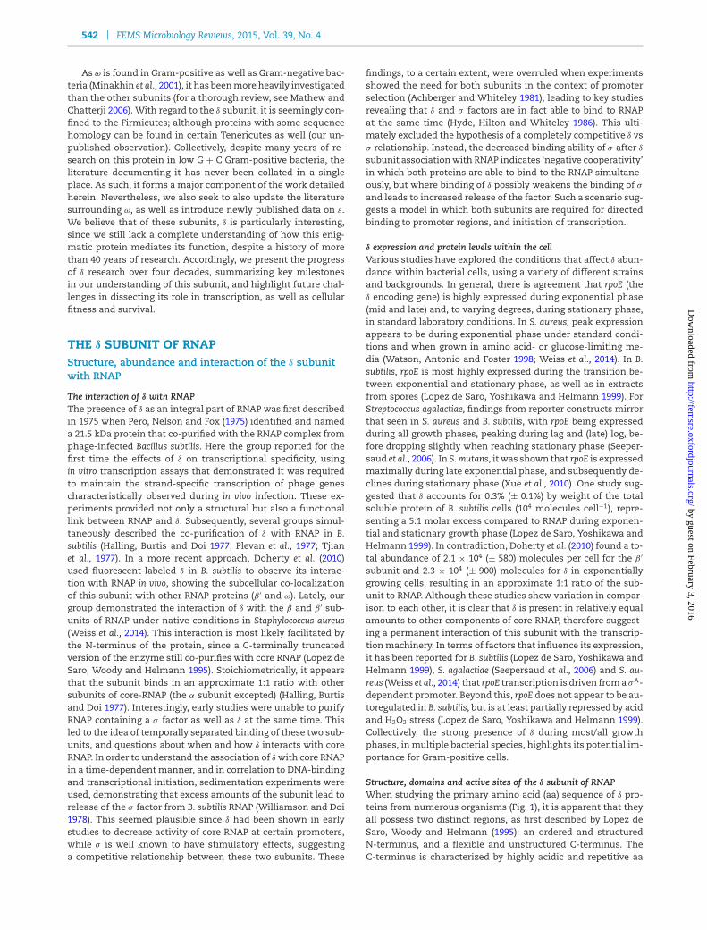
542 FEMS Microbiology Reviews, 2015, Vol. 39, No. 4
As ω is found in Gram-positive as well as Gram-negative bac-teria (Minakhin et al., 2001), it has beenmore heavily investigatedthan the other subunits (for a thorough review, see Mathew andChatterji 2006). With regard to the δ subunit, it is seemingly con-fined to the Firmicutes; although proteins with some sequencehomology can be found in certain Tenericutes as well (our un-published observation). Collectively, despite many years of re-search on this protein in low G + C Gram-positive bacteria, theliterature documenting it has never been collated in a singleplace. As such, it forms a major component of the work detailedherein. Nevertheless, we also seek to also update the literaturesurrounding ω, as well as introduce newly published data on ε.We believe that of these subunits, δ is particularly interesting,since we still lack a complete understanding of how this enig-matic protein mediates its function, despite a history of morethan 40 years of research. Accordingly, we present the progressof δ research over four decades, summarizing key milestonesin our understanding of this subunit, and highlight future chal-lenges in dissecting its role in transcription, as well as cellularfitness and survival.
THE δ SUBUNIT OF RNAPStructure, abundance and interaction of the δ subunitwith RNAP
The interaction of δ with RNAPThe presence of δ as an integral part of RNAP was first describedin 1975 when Pero, Nelson and Fox (1975) identified and nameda 21.5 kDa protein that co-purified with the RNAP complex fromphage-infected Bacillus subtilis. Here the group reported for thefirst time the effects of δ on transcriptional specificity, usingin vitro transcription assays that demonstrated it was requiredto maintain the strand-specific transcription of phage genescharacteristically observed during in vivo infection. These ex-periments provided not only a structural but also a functionallink between RNAP and δ. Subsequently, several groups simul-taneously described the co-purification of δ with RNAP in B.subtilis (Halling, Burtis and Doi 1977; Plevan et al., 1977; Tjianet al., 1977). In a more recent approach, Doherty et al. (2010)used fluorescent-labeled δ in B. subtilis to observe its interac-tion with RNAP in vivo, showing the subcellular co-localizationof this subunit with other RNAP proteins (β ′ and ω). Lately, ourgroup demonstrated the interaction of δ with the β and β ′ sub-units of RNAP under native conditions in Staphylococcus aureus(Weiss et al., 2014). This interaction is most likely facilitated bythe N-terminus of the protein, since a C-terminally truncatedversion of the enzyme still co-purifies with core RNAP (Lopez deSaro, Woody and Helmann 1995). Stoichiometrically, it appearsthat the subunit binds in an approximate 1:1 ratio with othersubunits of core-RNAP (the α subunit excepted) (Halling, Burtisand Doi 1977). Interestingly, early studies were unable to purifyRNAP containing a σ factor as well as δ at the same time. Thisled to the idea of temporally separated binding of these two sub-units, and questions about when and how δ interacts with coreRNAP. In order to understand the association of δ with core RNAPin a time-dependent manner, and in correlation to DNA-bindingand transcriptional initiation, sedimentation experiments wereused, demonstrating that excess amounts of the subunit lead torelease of the σ factor from B. subtilis RNAP (Williamson and Doi1978). This seemed plausible since δ had been shown in earlystudies to decrease activity of core RNAP at certain promoters,while σ is well known to have stimulatory effects, suggestinga competitive relationship between these two subunits. These
findings, to a certain extent, were overruled when experimentsshowed the need for both subunits in the context of promoterselection (Achberger and Whiteley 1981), leading to key studiesrevealing that δ and σ factors are in fact able to bind to RNAPat the same time (Hyde, Hilton and Whiteley 1986). This ulti-mately excluded the hypothesis of a completely competitive δ vsσ relationship. Instead, the decreased binding ability of σ after δ
subunit associationwith RNAP indicates ‘negative cooperativity’in which both proteins are able to bind to the RNAP simultane-ously, but where binding of δ possibly weakens the binding of σ
and leads to increased release of the factor. Such a scenario sug-gests a model in which both subunits are required for directedbinding to promoter regions, and initiation of transcription.
δ expression and protein levels within the cellVarious studies have explored the conditions that affect δ abun-dance within bacterial cells, using a variety of different strainsand backgrounds. In general, there is agreement that rpoE (theδ encoding gene) is highly expressed during exponential phase(mid and late) and, to varying degrees, during stationary phase,in standard laboratory conditions. In S. aureus, peak expressionappears to be during exponential phase under standard condi-tions and when grown in amino acid- or glucose-limiting me-dia (Watson, Antonio and Foster 1998; Weiss et al., 2014). In B.subtilis, rpoE is most highly expressed during the transition be-tween exponential and stationary phase, as well as in extractsfrom spores (Lopez de Saro, Yoshikawa and Helmann 1999). ForStreptococcus agalactiae, findings from reporter constructs mirrorthat seen in S. aureus and B. subtilis, with rpoE being expressedduring all growth phases, peaking during lag and (late) log, be-fore dropping slightly when reaching stationary phase (Seeper-saud et al., 2006). In S. mutans, it was shown that rpoE is expressedmaximally during late exponential phase, and subsequently de-clines during stationary phase (Xue et al., 2010). One study sug-gested that δ accounts for 0.3% (± 0.1%) by weight of the totalsoluble protein of B. subtilis cells (104 molecules cell−1), repre-senting a 5:1 molar excess compared to RNAP during exponen-tial and stationary growth phase (Lopez de Saro, Yoshikawa andHelmann 1999). In contradiction, Doherty et al. (2010) found a to-tal abundance of 2.1 × 10⁴ (± 580) molecules per cell for the β ′
subunit and 2.3 × 10⁴ (± 900) molecules for δ in exponentiallygrowing cells, resulting in an approximate 1:1 ratio of the sub-unit to RNAP. Although these studies show variation in compar-ison to each other, it is clear that δ is present in relatively equalamounts to other components of core RNAP, therefore suggest-ing a permanent interaction of this subunit with the transcrip-tionmachinery. In terms of factors that influence its expression,it has been reported for B. subtilis (Lopez de Saro, Yoshikawa andHelmann 1999), S. agalactiae (Seepersaud et al., 2006) and S. au-reus (Weiss et al., 2014) that rpoE transcription is driven froma σA-dependent promoter. Beyond this, rpoE does not appear to be au-toregulated in B. subtilis, but is at least partially repressed by acidand H2O2 stress (Lopez de Saro, Yoshikawa and Helmann 1999).Collectively, the strong presence of δ during most/all growthphases, in multiple bacterial species, highlights its potential im-portance for Gram-positive cells.
Structure, domains and active sites of the δ subunit of RNAPWhen studying the primary amino acid (aa) sequence of δ pro-teins from numerous organisms (Fig. 1), it is apparent that theyall possess two distinct regions, as first described by Lopez deSaro, Woody and Helmann (1995): an ordered and structuredN-terminus, and a flexible and unstructured C-terminus. TheC-terminus is characterized by highly acidic and repetitive aa
by guest on February 3, 2016http://fem
sre.oxfordjournals.org/D
ownloaded from
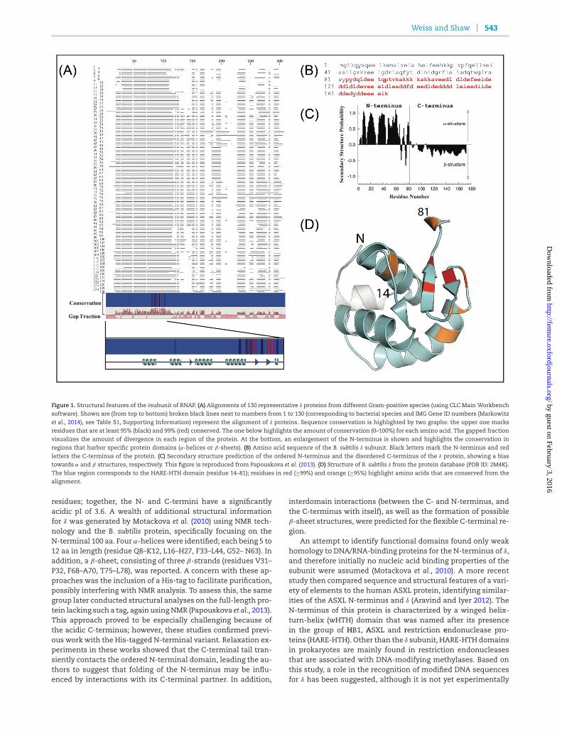
Weiss and Shaw 543
Figure 1. Structural features of the δsubunit of RNAP. (A) Alignments of 130 representative δ proteins from different Gram-positive species (using CLC MainWorkbenchsoftware). Shown are (from top to bottom) broken black lines next to numbers from 1 to 130 (corresponding to bacterial species and IMG Gene ID numbers (Markowitz
et al., 2014), see Table S1, Supporting Information) represent the alignment of δ proteins. Sequence conservation is highlighted by two graphs: the upper one marksresidues that are at least 95% (black) and 99% (red) conserved. The one below highlights the amount of conservation (0–100%) for each amino acid. The gapped fractionvisualizes the amount of divergence in each region of the protein. At the bottom, an enlargement of the N-terminus is shown and highlights the conservation inregions that harbor specific protein domains (α-helices or β-sheets). (B) Amino acid sequence of the B. subtilis δ subunit. Black letters mark the N-terminus and red
letters the C-terminus of the protein. (C) Secondary structure prediction of the ordered N-terminus and the disordered C-terminus of the δ protein, showing a biastowards α and β structures, respectively. This figure is reproduced from Papouskova et al. (2013). (D) Structure of B. subtilis δ from the protein database (PDB ID: 2M4K).The blue region corresponds to the HARE-HTH domain (residue 14–81); residues in red (≥99%) and orange (≥95%) highlight amino acids that are conserved from thealignment.
residues; together, the N- and C-termini have a significantlyacidic pI of 3.6. A wealth of additional structural informationfor δ was generated by Motackova et al. (2010) using NMR tech-nology and the B. subtilis protein, specifically focusing on theN-terminal 100 aa. Four α-helices were identified; each being 5 to12 aa in length (residue Q8–K12, L16–H27, F33–L44, G52– N63). Inaddition, a β-sheet, consisting of three β-strands (residues V31–P32, F68–A70, T75–L78), was reported. A concern with these ap-proaches was the inclusion of a His-tag to facilitate purification,possibly interfering with NMR analysis. To assess this, the samegroup later conducted structural analyses on the full-length pro-tein lacking such a tag, again usingNMR (Papouskova et al., 2013).This approach proved to be especially challenging because ofthe acidic C-terminus; however, these studies confirmed previ-ous work with the His-tagged N-terminal variant. Relaxation ex-periments in these works showed that the C-terminal tail tran-siently contacts the ordered N-terminal domain, leading the au-thors to suggest that folding of the N-terminus may be influ-enced by interactions with its C-terminal partner. In addition,
interdomain interactions (between the C- and N-terminus, andthe C-terminus with itself), as well as the formation of possibleβ-sheet structures, were predicted for the flexible C-terminal re-gion.
An attempt to identify functional domains found only weakhomology to DNA/RNA-binding proteins for the N-terminus of δ,and therefore initially no nucleic acid binding properties of thesubunit were assumed (Motackova et al., 2010). A more recentstudy then compared sequence and structural features of a vari-ety of elements to the human ASXL protein, identifying similar-ities of the ASXL N-terminus and δ (Aravind and Iyer 2012). TheN-terminus of this protein is characterized by a winged helix-turn-helix (wHTH) domain that was named after its presencein the group of HB1, ASXL and restriction endonuclease pro-teins (HARE-HTH). Other than the δ subunit, HARE-HTHdomainsin prokaryotes are mainly found in restriction endonucleasesthat are associated with DNA-modifying methylases. Based onthis study, a role in the recognition of modified DNA sequencesfor δ has been suggested, although it is not yet experimentally
by guest on February 3, 2016http://fem
sre.oxfordjournals.org/D
ownloaded from
544 FEMS Microbiology Reviews, 2015, Vol. 39, No. 4
proven. Nevertheless, the presence of a HARE-HTH motif in δ
presents the interesting possibility that not only does it interactwith RNAP but that it also contacts DNA itself, allowing it to dis-criminate between sequences that differ, for example, in theirdegrees of DNA modification. As such, it is tempting to specu-late that, similar to alternative σ -factors, δ potentially mediatesdirected transcriptional effects by recognizing specific promoterfeatures, rather than solely having global effects on a large num-ber of genes through modulation of RNAP activity.
To understand the importance and conservation of key δ re-gions, our group recently published alignments using variousdifferent species that carry a δ subunit protein (Weiss et al.,2014) (Fig. 1). We demonstrate that HARE-HTH domains show ahigher degree of conservation than other portions of the protein,emphasizing its likely importance for function. In contrast, theC-terminus of δ shows a large degree of variation. Further tothis, there is a clear degree of divergence in the context of pro-tein length for δ from various species (Table S1, Supporting In-formation), largely resulting from variations in the C-terminaldomain. This suggests that the C-terminus either plays speciesspecific roles or is at least partially dispensable for full functionof the subunit. In line with this latter idea, we have shown thata δ variant of S. aureus containing a truncated C-terminus (miss-ing ∼25% of the protein) is still able to successfully interact withcomponents of the transcription machinery, and complement,at least in part, the phenotypes of an rpoE-null strain (Weiss et al.,2014). This is in line with earlier findings in B. subtilis, also de-scribing that interaction of δ with RNAP is mediated by the N-terminus of the subunit (Lopez de Saro, Woody and Helmann1995).
The δ subunit and its impact on transcriptionalselectivity
Early work highlighting the influence of δ on transcriptionThe majority of early information regarding δ function was gen-erated by studies exploring transcriptional patterns of phagegenes in B. subtilis. Upon infection, phages go through a pre-dictable pattern of early, middle (4–5 min) and late (8–10 min)gene expression (Gage and Geiduschek 1971). Phage genes tran-scribed in this temporal pattern are located on different strands(heavy or light) of the phage genome. Whilst these specifictemporal and strand-specific transcriptional patterns are gov-erned by phage-encoded proteins (Gage and Geiduschek 1971;Fox 1976; Giacomoni 1981), it has been shown that δ, as an innatepart of RNAP, is also involved in maintaining their specificityand order of transcription (Pero, Nelson and Fox 1975; Achberger,Hilton and Whiteley 1982a; Dobinson and Spiegelman 1987).These experiments initially demonstrated that δ has a support-ive role for transcriptional selectivity, acting alongside and inconcert with other transcription factors (e.g. phage-encoded σ
factors and regulatory proteins). This was projected to not onlybe true for phage-related genes but for the transcriptional pro-cess in general.
The δ subunit enhances RNAP fidelity and selectivityOther early studies on the function of δ centered not only aroundphage genes but also on its effects on transcription from syn-thetic templates (poly(dA-dT)) (Tjian et al., 1977). It was re-ported that whilst δ had only a limited effect on the expres-sion of phage genes, it strongly decreased RNAP activity towardssynthetic templates, suggesting a general function in selectiv-ity. This led to studies focused on a wider spectrum of tran-scriptional targets, employing a variety of templates, including
different phages, B. subtilis chromosomal DNA, plasmids andsynthetic targets (Dickel, Burtis andDoi 1980). These in vitro tran-scription experiments revealed varying effects after the additionof purified δ to reaction mixtures. Specifically, in the case of onephage tested (e), δ had no negative effects on RNAP activity,whilst for B. subtilis chromosomal DNA, the synthesis of RNAwas decreased by up to 87% for some regions in the presence ofthis subunit. Moreover, transcription from DNA fragments thatdid not contain specific and known promoter sequences was al-most entirely repressed by the presence of δ. This suggests thatthe δ subunit affects different promoters in different ways, thussupporting promoter specificity of the transcriptional machin-ery, whilst at the same time suppressing transcription from locithat do not contain δ-specific promoter elements. Similar effectsfor δ on the transcription of non-coding-regions were also de-scribed by Achberger andWhiteley (1981). Their studies, using invitro transcription employing endonucleolytic fragmented DNAfrom B. subtilis bacteriophage SP82, revealed that in the absenceof δ, the RNAP holoenzyme binds non-specifically to fragmentsthat do not have promoters, resulting in transcription from thesesites. The addition of δ to in vitro transcription reactions restoredbinding of the complex exclusively to promoter regions and pro-duced transcription patterns (mid vs late phage genes) that arecharacteristic of those seen in vivo. Along the same lines, UVcross-linking experiments reveal that the addition of δ to core-RNAP or the RNAP holoenzyme leads to decreased binding atnon-promoter sequences (Hilton and Whiteley 1985).
In a study by Dobinson and Spiegelman (1987), features thatguide the variable effects of different promoters were exploredby assessing the expression of two early 29 promoters in run-off experiments (initiation complex assays). In these works, itwas shown that the influence of δ on transcription appearsto be dependent on the strength of the promoter in question.Specifically, varying effects on transcriptional repression wereobserved, with the stronger of the two promoters showing nodecrease in expression upon δ addition, whilst the weaker pro-moter resulted in a 50% decrease in transcription after supple-mentationwith the subunit. These results gave early insight intothe selective effects of δ, and suggested that promoter strengthmay be a driving force of its activity. This model has been sup-ported by the findings of Juang and Helmann (1994b) in theirworkwith the B. subtilis ilv-leu operon. In this study, it was shownthat δ negatively influences open complex formation and hasstronger effects towards the transcription of weaker promoters,and less of an influence on medium strength promoters. Con-sequently, it was suggested that δ decreases promoter melting,which leads to decreased transcription from non-promoter se-quences, as well as at weaker promoters. In contrast, promotersthat are considered closer to optimal are relatively independentof the effects of δ. Thus, a model describing three general typesof promoters and their susceptibility to δ was proposed as fol-lows (Juang and Helmann 1994b): (i) at weak promoters, δ pre-vents RNAP binding and therefore transcription; (ii) at mediumstrength promoters, δ allows the binding of RNAP, but decreasesopen complex formation; and (iii) at strong promoters, δ seem-ingly has little effect on gene expression.
δ interferes with open complex formationInitial Studies by Spiegelman, Hiatt and Whiteley (1978) andAchberger, Hilton and Whiteley (1982a) indicated that δ func-tions in the window between promoter recognition and initi-ation, rather than being directly involved in initiation itself.Subsequently, experiments by Chen and Helmann (1997) fur-thered this notion, showing that δ is important for open complex
by guest on February 3, 2016http://fem
sre.oxfordjournals.org/D
ownloaded from
Weiss and Shaw 545
formation. This is in support of Juang and Helmann, who used,amongst other approaches, footprinting analysis to describethe inhibitory effects of δ on open complex formation (Juangand Helmann 1994a, 1994b, 1995). Furthermore, they demon-strated that loss of δ specifically interfered with promoter melt-ing, rather than impacting the stability of the open promotercomplex, which is somewhat in contrast to Rabatinova et al.(2013), who suggested that the longevity of open complexes is di-minished by δ. Importantly, each of these studies again supportthe notion that δ influences transcription prior to initiation.
RNAP recyclingWhilst the majority of δ-related studies describe its negative ef-fects on transcription of specific promoters, several groups havedemonstrated that δ also has the capacity to increase overalltranscriptional activity (Spiegelman, Hiatt and Whiteley 1978;Achberger andWhiteley 1981; Juang and Helmann 1994b). Thesepositive effects are suggested to be a result of a decrease in non-specific transcription, as well as increased core-RNAP recyclingin the presence of δ. The specific mechanism by which this lat-ter process occurs is not fully understood; however, a more effi-cient release of RNAP after termination of transcription has beensuggested (Juang and Helmann 1994b). This effect is thought toparallel the manner by which δ prevents RNAP from binding tonon-promoter sequences, thereby leading to decreased productinhibition, and increasing the speed at which the transcriptioncomplex is available to initiate new rounds of gene expression.Structurally, it has been hypothesized that this process is facil-itated by the C-terminus of δ (Lopez de Saro, Woody and Hel-mann 1995), where the negatively charged part of the proteincompetes with DNA/RNA for RNAP binding, thereby causing in-creased release of the transcriptional machinery. In more recentexperiments investigating the function of HelD, another RNAP-associated protein, it was confirmed that δ is able to increasetranscriptional cycling by faster release of RNAP after termina-tion; and is also able to release stalled RNAP from DNA, increas-ing transcriptional activity (Wiedermannova et al., 2014). Both ef-fects have also been found to be true for HelD, and interestingly,both proteins together show a strong synergistic effect. Whilstthe precise reason for this synergy is incompletely understood,the results highlight the complex and multifactorial regulationof RNAP activity.
The role of the initiating nucleotide on δ-dependent regulationAlthough the contention that δ-dependent transcriptional ef-fects are influenced by individual promoter strength is well es-tablished, it fails to completely explain all changes in gene ex-pression mediated by this subunit. Therefore, a more recentstudy in B. subtilis has centered on delineating the exact roleof δ in promoter melting and open complex formation, with aspecific focus on the transcription initiating NTP (iNTP) (Rabati-nova et al., 2013). This idea was shaped by previous works, show-ing that promoter activity is not only determined by pure bind-ing efficiency of RNAP to promoter sequences but also by theability to initiate transcription. While high levels of iNTPs en-sure efficient transcription, lower amounts cause collapse of theopen promoter complex, and prevent transcriptional initiation.In Escherichia coli, transcriptional initiation from rRNA promot-ers, which have inherently unstable open promoter complexes,is dependent on the availability of transcript specific iNTPs (ATPor GTP) (Gaal et al., 1997; Murray, Schneider and Gourse 2003).A similar scenario was also shown for B. subtilis, where nutri-tional starvation and onset of the stringent response resultsin decreased levels of GTP and therefore negatively influences
transcription of iNTP-sensitive rRNA promoters (Ochi, Kandalaand Freese 1982; Krasny and Gourse 2004). Subsequently, it wasshown that such regulation is not only true of rRNA promot-ers but other genes as well, thus presenting a general regula-tory concept (Krasny et al., 2008). Accordingly, Rabatinova et al.(2013) explored the influence of iNTP on δ-mediated function inB. subtilis, revealing that the δ subunit influences transcription byenhancing the effects of iNTP on open complex formation. Pro-moters that possess relatively unstable open complexes requirehigher amounts of iNTP to initiate transcription. δ destabilizesopen complex formation, thus increasing the amount of iNTP re-quired for successful transcription initiation; supporting the no-tion of iNTP-dependent transcriptional regulation. This appearsto be particularly true for promoters that are iNTP sensitive (e.g.rRNA promoters), whilst other promoters, which display stableopen complexes, are less likely to be affected by the influence ofthe δ subunit.
The impact of abrogated δ activity on fitness andvirulence
Phenotypic effects of δ deletionAlthough the specific effects of δ on transcription have beenwelldocumented, uncovering the phenotypic effects of rpoE deletionwas, at least initially, more challenging. A B. subtilismutant lack-ing the δ subunit of RNAP was shown to be viable, able to formspores and did not display obvious growth defects under stan-dard laboratory conditions (Lampe et al., 1988). Although unex-pected at first, these results are not surprising for an accessorysubunit. They do, however, raise the question about underwhichconditions the transcriptional effects of δ result in altered be-havior of the cell, and, thus, under which stimuli the δ subunit isbeneficial for cellular survival. An early indication about possibleδ function came froma study in S. aureus investigating starvationsurvival. Watson, Antonio and Foster (1998) identified rpoE dur-ing a transposon screen targeted towards genes that are impor-tant for survival and recovery from prolonged stationary phasestarvation. A transposon insertion within the early portion ofrpoE caused impaired survival under amino acid-limiting condi-tions and acid stress. Similar rpoE-dependent stress phenotypeswere later shown in S. mutans, where a δ subunit mutant is im-paired in acid and H2O2 stress survival (Xue et al., 2010). Addi-tionally, the mutant was described as having a clumping pheno-type, and to reach lower final densities in liquid cultures whencompared to the wild type. Another common phenotype de-scribed for various bacterial species, including B. subtilis (Lopezde Saro, Yoshikawa and Helmann 1999), S. aureus (Weiss et al.,2014) and S. agalactiae (Jones, Needham and Rubens 2003), isthe extended lag phase of rpoE-depleted strains when subcul-tured into fresh media from stationary phase growth. Thus, itwould appear that δ is required for adaption to, and survivalduring, stress and changing environmental conditions, as wellas growth phase transition. It has also been documented thatB. subtilis rpoE mutants are characterized by elongated cell mor-phology, the propensity to clump during log phase and the ap-pearance of rough-edged colonies when grown on solid media(Lopez de Saro, Yoshikawa and Helmann 1999). Interestingly,and despite earlier studies that described normal sporulationof B. subtilis rpoE strains (Lampe et al., 1988; Lopez de Saro,Yoshikawa and Helmann 1999), it has also been shown that atransposon insertion in this gene suppresses the effects causedby mutations (pdhB or pdhC) that block sporulation (Gao andAronson 2004). The exactmechanism bywhich this occurs is un-clear; however, these findings emphasize the widespread effects
by guest on February 3, 2016http://fem
sre.oxfordjournals.org/D
ownloaded from
546 FEMS Microbiology Reviews, 2015, Vol. 39, No. 4
of δ on transcription, and its importance in maintaining cellu-lar homeostasis. Considering the widespread defects displayedby rpoE mutants in a wealth of species, it is perhaps expectedthat rpoE mutants of B. subtilis are readily outcompeted by thewild-type strain when cultured together over prolonged periods(Rabatinova et al., 2013).
The role of δ in pathogenic organismsSeveral groups have examined the effects of rpoE deletion onmore complex processes, such as during the interaction ofpathogenic bacteria with their hosts. Since disease causationis a process that involves the precise timing of expression fora variety of different genes, including those required for inter-action with the immune system, virulence factor expression,biofilm formation and nutrient acquisition, it is perhaps no sur-prise that δ has been shown to play and important role in viru-lence. In S. agalactiae, a study employing signature-tagged trans-poson mutagenesis in a neonatal rat sepsis model identified anrpoE mutant as displaying the most impaired virulence fromthe entire library (Jones, Knoll and Rubens 2000). In a subse-quent study by the same group, it was suggested that the vir-ulence defect was linked to increased killing by phagocytes, ex-plaining the lower survival rates observed for rpoE mutants inwhole human blood; despite not showing altered survival in hu-man plasma and chemically defined media (Jones, Needhamand Rubens 2003). An important consideration with these find-ings is that the rpoE transposonmutant is not a complete knock-out, but is in fact only a low rpoE expressing strain due to thelocation of the insertion. As such, the strain possesses arounda 10-fold decrease in rpoE transcript compared to the wild type,which is seemingly insufficient tomaintain full δ-function, lead-ing to the decrease in virulence described. Therefore, it appearsthat in S. agalactiae, and perhaps other pathogens (see below),there is a minimal concentration of δ required in the cell to en-sure transcriptional specificity and facilitate virulence.
The pathogenic role of δ has also been explored in anotherstreptococcus, namely S. mutans. Interestingly, and in contrast tothe negative effects of rpoE loss on virulence that were obtainedfor S. agalactiae, it was described that deletion of rpoE leads to in-creased expression of virulence-related traits (Xue et al., 2011).Specifically, increased co- and self-aggregation, and an alteredextracellularmatrix, have been reported for rpoEmutants, whichmay be caused by differentially expressed surface proteins andpolysaccharides. Furthermore, S. mutans rpoE strains also dis-play elevated attachment to human extracellular matrix com-ponents, such as collagen and fibronectin. Despite these seem-ingly enhanced aggregative properties, rpoE deletion causes aninability to attach to human epithelial cells (HEp-2) comparedto the wild-type strain; likely due to increased clumping of themutant. Beyond these findings, loss of rpoE in S. mutans results inthe derepression of enzymes that facilitate the metabolism of anumber of sugars, allowing themutant to utilize a larger array ofcarbon sources than the parental strain. Additionally, a panel ofantibiotics and compounds that are toxic to the wild type wereineffective against the mutant in phenotypic microarray stud-ies. This highlights the remarkably broad impact that δ has onbacterial physiology and metabolism, each of which influencecellular fitness, and thus pathogenesis.
In a more recent study by our group, we explored the effectsof δ in the major human pathogen, S. aureus (Weiss et al., 2014).Deletion of rpoE resulted in decreased expression and abun-dance of a variety of virulence factors, leading to decreased fit-ness of the strain and impaired virulence. This latter point ismanifested by a diminished ability to survive in whole human
blood, and resist phagocytosis by human leukocytes. These invitro/ex vivo phenotypes were shown to correlate with in vivofindings, where the mutant strain was found to be significantlyimpaired in its ability to cause disease in amurinemodel of sep-sis and dissemination.
Global transcriptional and translational effects of δ
Exploring the influence of δ on global gene expression has beenaided by cutting edge high-throughput methodologies, includ-ing transcriptomics and proteomics. These techniques have pro-vided insight into the role of δ in maintaining transcriptionalspecificity, and how this links to the various phenotypes ob-served. To explore why an rpoE mutant of S. mutans displaysalterations in virulence-related behaviors, as well as impairedability to survive during stress (Xue et al., 2010, 2011), gene ex-pression (Xue et al., 2010) and protein abundance (Xue et al.,2012) were assessed for the wild type and mutant under sev-eral conditions. Transcriptomic analyses conducted viamicroar-ray revealed that, independent of the growth condition, morethan 50% of the upregulated transcripts in an rpoE mutant werenon-coding regions (Xue et al., 2010). In comparison, only 20–30%of non-coding transcripts displayed increased expression in themutant strain. These findings corroborate previous in vitro stud-ies that demonstrate non-specific transcription of non-promoterregions when δ is removed from core RNAP (see above).
In addition to its effects on non-coding regions, coding se-quences were also shown to be influenced by δ in S. mutans,in a manner partially independent of growth phase or condi-tion (Xue et al., 2010). Specifically, 24 genes were identified asbeing downregulated in the rpoE mutant under all conditions.These genes were involved in malolactic fermentation, histi-dine metabolism, biofilm formation, adherence, virulence, andresistance to antibiotics and other inhibitory compounds. In ad-dition, during growth under standard laboratory conditions, alarge number of transcripts were differentially expressed whencomparing the mutant and parental strain, including those in-volved in the transport of a wealth of different compounds,metabolism and energy production/conversion. Following H2O2
or acid stress, a number of protective genes were not induced inthemutant strain, indicating that an incomplete stress responseexists upon rpoE loss; however, the general stress response stillappeared to be intact in the mutant strain. The authors ulti-mately concluded from these works that diminished survival ofthe mutant strain when exposed to stress results from a gen-eral deregulation of the S. mutans transcriptome, leading to animpaired, slower and less directed attempt to adapt to changingenvironments and stress conditions.
In follow-up studies, the same group compared previoustranscriptomic data with new proteomic data of the rpoE mu-tant and wild type, using identical growth conditions (Xueet al., 2012). It was noted that two proteins, phosphoglucomu-tase (PGM) and phosphopentomutase (DeoB), were decreased inabundance either during all (PGM) ormost (DeoB) growth phasesand conditions. Bothwere identified as displaying this same pat-tern in microarray analysis as well, suggesting distinct and verygene-specific regulatory effects of δ. In the context of growthphase, chaperones, stress-related factors and enzymes knownto be involved in protein turnover were altered in the mutantduring exponential phase; whilst multiple sugar transportersand metabolism (MSM) systems demonstrated fluctuations dur-ing stationary phase. It was suggested that these changes inprotein abundance could be an indicator of internal cell stress,rather than direct rpoE-guided effects. When comparing protein
by guest on February 3, 2016http://fem
sre.oxfordjournals.org/D
ownloaded from
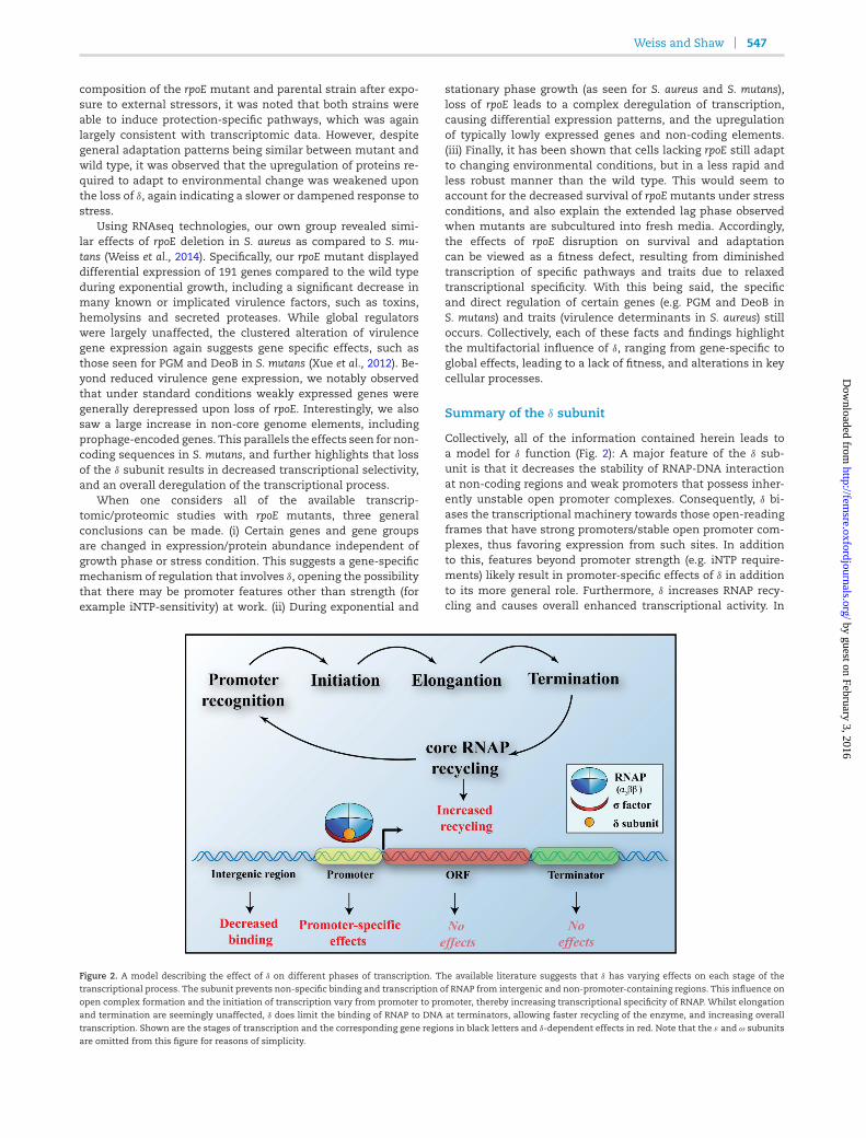
Weiss and Shaw 547
composition of the rpoE mutant and parental strain after expo-sure to external stressors, it was noted that both strains wereable to induce protection-specific pathways, which was againlargely consistent with transcriptomic data. However, despitegeneral adaptation patterns being similar between mutant andwild type, it was observed that the upregulation of proteins re-quired to adapt to environmental change was weakened uponthe loss of δ, again indicating a slower or dampened response tostress.
Using RNAseq technologies, our own group revealed simi-lar effects of rpoE deletion in S. aureus as compared to S. mu-tans (Weiss et al., 2014). Specifically, our rpoE mutant displayeddifferential expression of 191 genes compared to the wild typeduring exponential growth, including a significant decrease inmany known or implicated virulence factors, such as toxins,hemolysins and secreted proteases. While global regulatorswere largely unaffected, the clustered alteration of virulencegene expression again suggests gene specific effects, such asthose seen for PGM and DeoB in S. mutans (Xue et al., 2012). Be-yond reduced virulence gene expression, we notably observedthat under standard conditions weakly expressed genes weregenerally derepressed upon loss of rpoE. Interestingly, we alsosaw a large increase in non-core genome elements, includingprophage-encoded genes. This parallels the effects seen for non-coding sequences in S. mutans, and further highlights that lossof the δ subunit results in decreased transcriptional selectivity,and an overall deregulation of the transcriptional process.
When one considers all of the available transcrip-tomic/proteomic studies with rpoE mutants, three generalconclusions can be made. (i) Certain genes and gene groupsare changed in expression/protein abundance independent ofgrowth phase or stress condition. This suggests a gene-specificmechanism of regulation that involves δ, opening the possibilitythat there may be promoter features other than strength (forexample iNTP-sensitivity) at work. (ii) During exponential and
stationary phase growth (as seen for S. aureus and S. mutans),loss of rpoE leads to a complex deregulation of transcription,causing differential expression patterns, and the upregulationof typically lowly expressed genes and non-coding elements.(iii) Finally, it has been shown that cells lacking rpoE still adaptto changing environmental conditions, but in a less rapid andless robust manner than the wild type. This would seem toaccount for the decreased survival of rpoE mutants under stressconditions, and also explain the extended lag phase observedwhen mutants are subcultured into fresh media. Accordingly,the effects of rpoE disruption on survival and adaptationcan be viewed as a fitness defect, resulting from diminishedtranscription of specific pathways and traits due to relaxedtranscriptional specificity. With this being said, the specificand direct regulation of certain genes (e.g. PGM and DeoB inS. mutans) and traits (virulence determinants in S. aureus) stilloccurs. Collectively, each of these facts and findings highlightthe multifactorial influence of δ, ranging from gene-specific toglobal effects, leading to a lack of fitness, and alterations in keycellular processes.
Summary of the δ subunit
Collectively, all of the information contained herein leads toa model for δ function (Fig. 2): A major feature of the δ sub-unit is that it decreases the stability of RNAP-DNA interactionat non-coding regions and weak promoters that possess inher-ently unstable open promoter complexes. Consequently, δ bi-ases the transcriptional machinery towards those open-readingframes that have strong promoters/stable open promoter com-plexes, thus favoring expression from such sites. In additionto this, features beyond promoter strength (e.g. iNTP require-ments) likely result in promoter-specific effects of δ in additionto its more general role. Furthermore, δ increases RNAP recy-cling and causes overall enhanced transcriptional activity. In
Figure 2. A model describing the effect of δ on different phases of transcription. The available literature suggests that δ has varying effects on each stage of thetranscriptional process. The subunit prevents non-specific binding and transcription of RNAP from intergenic and non-promoter-containing regions. This influence on
open complex formation and the initiation of transcription vary from promoter to promoter, thereby increasing transcriptional specificity of RNAP. Whilst elongationand termination are seemingly unaffected, δ does limit the binding of RNAP to DNA at terminators, allowing faster recycling of the enzyme, and increasing overalltranscription. Shown are the stages of transcription and the corresponding gene regions in black letters and δ-dependent effects in red. Note that the ε and ω subunitsare omitted from this figure for reasons of simplicity.
by guest on February 3, 2016http://fem
sre.oxfordjournals.org/D
ownloaded from
548 FEMS Microbiology Reviews, 2015, Vol. 39, No. 4
general, δ weakens the binding of RNAP to DNA and negativelyinfluence open complex formation and stability; which appearsto be mediated by structural features of the δ protein. Specifi-cally, its N-terminusmediates interactionwith core RNAP,whilstthe negatively charged and intrinsically disordered C-terminusis thought to interfere with the RNAP-DNA interaction. Ulti-mately, δ acts as a guide for RNAP, increasing its transcriptionalspecificity. Loss of this factor causes deregulation, and results ina decreased ability to adjust to changing environments, generalfitness defects and the reduced virulence of pathogenic species.All of these consequences highlight the central importance of δ
as a regulatory factor directing gene expression in Gram-positivebacteria.
ADDITIONAL, SMALL ACCESSORY RNAPSUBUNITS IN GRAM-POSITIVE BACTERIA
In addition to the δ subunit, Gram-positive bacteria possess twoother accessory RNAP subunits, omega, ω (RpoZ) and epsilon, ε
(RpoY). Whilst ε is unique to Gram-positives, ω is found in RNAPcomplexes from archea to eukaryotes. Both of these subunitsand their roles within the RNAP complex is discussed in the fol-lowing sections.
The ω subunit
The ω subunit of RNAP is an intensively studied protein thathas been widely described in the literature. With this being said,the majority of studies have been performed using the Gram-negative model organism E. coli, and appear to be only partiallytranslatable to Gram-positive species, as discussed below. An ex-tensive and comprehensive review on the history of ω research,including Gram-positive as well as Gram-negative bacteria, hasbeen published by Mathew and Chatterji (2006) where the au-thors summarize structural and functional roles of the subunit.In the context of this review, we will offer an overview of datathat is available and valid for Gram-positive organisms, as wellas highlight functional differences compared to Gram-negativebacteria.
The ω protein is widely conserved across all forms of lifeWith an increasing number of bacterial, archaeal and eukary-otic genomes available, it has become clear that the ω subunitis conserved in all branches of life. In eukaryotes, a sequence,structural and functional homolog of ω (RPB6) exists that is as-sociated with RNAPI, II and III, whilst archaeal genomes harbora sequence homolog termed RpoK (Minakhin et al., 2001). RPB6has been shown to be essential for RNAP assembly in eukary-otes (Nouraini, Archambault and Friesen 1996); however, in bac-teria, deletion of the ω subunit in a variety of different speciesdoes not result in cell lethality (Gentry and Burgess 1989; Ko-jima et al., 2002; Mathew, Ramakanth and Chatterji 2005; Do-herty et al., 2010; Jie, JiLiang and DongJie 2010; Santos-Beneitet al., 2011; Gunnelius et al., 2014). This is particularly intriguing,since the subunit is conserved in all of the sequenced genomesof free-living bacteria (Minakhin et al., 2001), suggesting an im-portant evolutionary role for it in cellular survival
The binding of ω to RNAPThe ω protein, which ranges in size from ∼9 to 11.5 kDa depend-ing on the bacterial species in question, was first shown to be adedicated subunit of E. coli RNAP, rather than just a tightly boundfactor, when cross-linking experiments demonstrated its spe-cific binding to the β ′ subunit (Gentry and Burgess 1993). Sub-
sequently, it was found to be an integral part of the transcrip-tion machinery in vivo when the association of ω directly withRNAP was shown (Dove and Hochschild 1998). In these studies,ω was translationally fused to a DNA-binding protein (the cI re-pressor from bacteriophage λ) and was found to activate tran-scription of promoters adjacent to the corresponding λ opera-tor, demonstrating interaction of the chimeric λcI-ω fusion pro-tein with RNAP. Both of these studies have since been supportedby more recent work solving the structure of ω-containing RNAPfrom Thermus aquaticus and E. coli (Zhang et al., 1999; Murakami2013). Although no such crystallization studies for RNAP havebeen performed in Gram-positive bacteria, several experimentsin B. subtilis demonstrate co-purification of ω with RNAP, high-lighting its associationwith other subunits of the transcriptionalmachinery (Spiegelman, Hiatt and Whiteley 1978; Achberger,Tahara and Whiteley 1982b). These results were confirmed andextended in a recent approach in which Delumeau et al. (2011)were not only able to co-purify ω with the essential RNAP sub-units (α, β, β ′) but also determined a 2:1:1:1 ratio for α2ββ ′ωduring exponential and stationary growth. These results clearlyshow that, as for Gram-negative bacteria, the ω subunit is an in-tegral part of the Gram-positive transcriptional machinery.
The role of ω for structural integrity of RNAPExtensive research has been performed on the involvement of ω
in folding of the β ′ subunit, and RNAP assembly. Its importancefor the assembly process was first considered when RNAP puri-fied from Rhodobacter capsulatus and E. coli was shown to requireω, or GroEL and ω, respectively, to remain active in vitro (Mukher-jee and Chatterji 1997; Richard et al., 2003). This requirement forthe GroEL chaperone was considered puzzling, but this factorhad been shown to co-purify with RNAP, and was thus thoughtto be important for the folding of the transcriptional complex(Mukherjee and Chatterji 1997; Mukherjee et al., 1999). Interest-ingly, deletion of ω in E. coli results in amassive increase of GroELrecruitment to RNAP, suggesting that GroEL might be employedto overcome misfolding due to ω loss (Mukherjee et al., 1999).This was confirmed when in vivo GroEL substrates in E. coli weredetermined (Houry et al., 1999), showing that other than β ′, allof the essential RNAP subunits, as well as ω, require GroEL forproper folding. Surprisingly, these results were found to be onlypartially true in B. subtilis, where GroEL does not appear to beassociated with RNAP subunits (Endo and Kurusu 2007). Withthis being said, the Gram-positive study identified considerablyfewer GroEL substrates than the study performed in E. coli (28vs ∼300). Therefore, either there are indeed fewer substrates forthis chaperone in B. subtilis or experimentation in the Gram-positive organism had lower sensitivity in comparison with theE. coli study.
Interestingly, an earlier study in E. coli described in vitro cross-linking of ω and β ′ (Gentry and Burgess 1993), whilst at the sametime showing that β ′ was not a substrate of GroEL and that theω subunit increases (together with GroEL) the in vitro activity ofRNAP. As such, it was suggested that ω had a specific functionin supporting the folding of β ′. This hypothesis was reinforcedby a structural study that investigated the precise interplay of ω
and β ′, demonstrating interaction of conserved regions for thesetwo proteins (Minakhin et al., 2001). Consequently, it was pro-posed that ω acts as amolecular latch, guiding the interaction ofβ ′ with the α2β complex. Studies by Ghosh, Ishihama and Chat-terji (2001) supported this notion by demonstrating that the in-teraction of ω with β ′ was able to prevent aggregation of the lat-ter protein during in vitro renaturation. Furthermore, this samegroup was able to show that native folding of β ′ requires ω, and
by guest on February 3, 2016http://fem
sre.oxfordjournals.org/D
ownloaded from
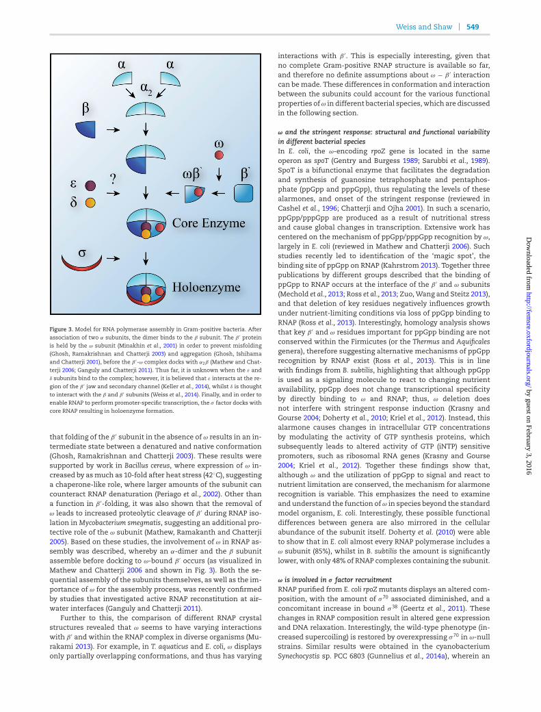
Weiss and Shaw 549
Figure 3. Model for RNA polymerase assembly in Gram-positive bacteria. After
association of two α subunits, the dimer binds to the β subunit. The β ′ proteinis held by the ω subunit (Minakhin et al., 2001) in order to prevent misfolding(Ghosh, Ramakrishnan and Chatterji 2003) and aggregation (Ghosh, Ishihamaand Chatterji 2001), before the β ′-ω complex docks with α2β (Mathew and Chat-
terji 2006; Ganguly and Chatterji 2011). Thus far, it is unknown when the ε andδ subunits bind to the complex; however, it is believed that ε interacts at the re-gion of the β ′ jaw and secondary channel (Keller et al., 2014), whilst δ is thoughtto interact with the β and β ′ subunits (Weiss et al., 2014). Finally, and in order to
enable RNAP to perform promoter-specific transcription, the σ factor docks withcore RNAP resulting in holoenzyme formation.
that folding of the β ′ subunit in the absence of ω results in an in-termediate state between a denatured and native conformation(Ghosh, Ramakrishnan and Chatterji 2003). These results weresupported by work in Bacillus cereus, where expression of ω in-creased by asmuch as 10-fold after heat stress (42C), suggestinga chaperone-like role, where larger amounts of the subunit cancounteract RNAP denaturation (Periago et al., 2002). Other thana function in β ′-folding, it was also shown that the removal ofω leads to increased proteolytic cleavage of β ′ during RNAP iso-lation inMycobacterium smegmatis, suggesting an additional pro-tective role of the ω subunit (Mathew, Ramakanth and Chatterji2005). Based on these studies, the involvement of ω in RNAP as-sembly was described, whereby an α-dimer and the β subunitassemble before docking to ω-bound β ′ occurs (as visualized inMathew and Chatterji 2006 and shown in Fig. 3). Both the se-quential assembly of the subunits themselves, as well as the im-portance of ω for the assembly process, was recently confirmedby studies that investigated active RNAP reconstitution at air–water interfaces (Ganguly and Chatterji 2011).
Further to this, the comparison of different RNAP crystalstructures revealed that ω seems to have varying interactionswith β ′ and within the RNAP complex in diverse organisms (Mu-rakami 2013). For example, in T. aquaticus and E. coli, ω displaysonly partially overlapping conformations, and thus has varying
interactions with β ′. This is especially interesting, given thatno complete Gram-positive RNAP structure is available so far,and therefore no definite assumptions about ω − β ′ interactioncan bemade. These differences in conformation and interactionbetween the subunits could account for the various functionalproperties ofω in different bacterial species, which are discussedin the following section.
ω and the stringent response: structural and functional variabilityin different bacterial speciesIn E. coli, the ω-encoding rpoZ gene is located in the sameoperon as spoT (Gentry and Burgess 1989; Sarubbi et al., 1989).SpoT is a bifunctional enzyme that facilitates the degradationand synthesis of guanosine tetraphosphate and pentaphos-phate (ppGpp and pppGpp), thus regulating the levels of thesealarmones, and onset of the stringent response (reviewed inCashel et al., 1996; Chatterji and Ojha 2001). In such a scenario,ppGpp/pppGpp are produced as a result of nutritional stressand cause global changes in transcription. Extensive work hascentered on the mechanism of ppGpp/pppGpp recognition by ω,largely in E. coli (reviewed in Mathew and Chatterji 2006). Suchstudies recently led to identification of the ‘magic spot’, thebinding site of ppGpp on RNAP (Kahrstrom 2013). Together threepublications by different groups described that the binding ofppGpp to RNAP occurs at the interface of the β ′ and ω subunits(Mechold et al., 2013; Ross et al., 2013; Zuo,Wang and Steitz 2013),and that deletion of key residues negatively influences growthunder nutrient-limiting conditions via loss of ppGpp binding toRNAP (Ross et al., 2013). Interestingly, homology analysis showsthat key β ′ and ω residues important for ppGpp binding are notconserved within the Firmicutes (or the Thermus and Aquificalesgenera), therefore suggesting alternative mechanisms of ppGpprecognition by RNAP exist (Ross et al., 2013). This is in linewith findings from B. subtilis, highlighting that although ppGppis used as a signaling molecule to react to changing nutrientavailability, ppGpp does not change transcriptional specificityby directly binding to ω and RNAP; thus, ω deletion doesnot interfere with stringent response induction (Krasny andGourse 2004; Doherty et al., 2010; Kriel et al., 2012). Instead, thisalarmone causes changes in intracellular GTP concentrationsby modulating the activity of GTP synthesis proteins, whichsubsequently leads to altered activity of GTP (iNTP) sensitivepromoters, such as ribosomal RNA genes (Krasny and Gourse2004; Kriel et al., 2012). Together these findings show that,although ω and the utilization of ppGpp to signal and react tonutrient limitation are conserved, the mechanism for alarmonerecognition is variable. This emphasizes the need to examineand understand the function of ω in species beyond the standardmodel organism, E. coli. Interestingly, these possible functionaldifferences between genera are also mirrored in the cellularabundance of the subunit itself. Doherty et al. (2010) were ableto show that in E. coli almost every RNAP polymerase includes aω subunit (85%), whilst in B. subtilis the amount is significantlylower, with only 48% of RNAP complexes containing the subunit.
ω is involved in σ factor recruitmentRNAP purified from E. coli rpoZmutants displays an altered com-position, with the amount of σ 70 associated diminished, and aconcomitant increase in bound σ 38 (Geertz et al., 2011). Thesechanges in RNAP composition result in altered gene expressionand DNA relaxation. Interestingly, the wild-type phenotype (in-creased supercoiling) is restored by overexpressing σ 70 in ω-nullstrains. Similar results were obtained in the cyanobacteriumSynechocystis sp. PCC 6803 (Gunnelius et al., 2014a), wherein an
by guest on February 3, 2016http://fem
sre.oxfordjournals.org/D
ownloaded from
550 FEMS Microbiology Reviews, 2015, Vol. 39, No. 4
rpoZ knockout had impaired recruitment of the primary σ fac-tor, resulting in decreased expression of genes under the con-trol of this element. Inversely, a large number of typically lowlyexpressed genes were upregulated, suggesting activity of one ormore alternative σ factors. These data indicate a role for ω intranscriptional specificity, and are supported by later publica-tions describing an altered response to heat stress upon dele-tion of rpoZ, leading to impaired survival at higher temperatures(Gunnelius et al., 2014b). Importantly, such findings correlatewith early studies showing that purification of ω-deficient RNAPin E. coli is depleted for σ 70, leading to the conclusion that ω isrequired for correct RNAP folding and σ -factor binding (Mukher-jee et al., 1999). Overall, this line of research describes how thestructural effects of ω are required for RNAP integrity and func-tionality, which leads to a role in fine-tuning transcription byadjusting affinity for central transcriptional regulators such asσ factors. These results were all obtained in Gram-negative or-ganisms, thus highlighting the need for further investigation inGram-positive species.
The ω subunit of Gram-positive bacteriaAs detailed above, in E. coli the ω subunit mediates folding ofthe β ′ subunit, influences assembly of RNAP, orchestrates geneexpression during the stringent response and plays a role in or-dering transcriptional specificity. In Gram-positive bacteria, thefunctional role of ω is much less clear. Since ppGpp does notbind RNAP in B. subtilis, the ω subunit is unlikely to be directlyinvolved in the stringent response of this organism. This raisesthe question of additional functions for the Gram-positiveω sub-unit outside of ppGpp binding and β ′ folding.Whilst studies havebeen conducted for various Gram-negative bacteria beyond E.coli, including Sinorhizoboium meliloti (Krol and Becker 2011), Xan-thomonas campestris (Jie, JiLiang and DongJie 2010) and cyanobac-teria species (Gunnelius et al., 2014a, 2014b), thus far the role ofω in Gram-positive bacteria has only been extensively examinedin two Streptomyces species and M. smegmatis.
When exploring the role of the ω subunit in Streptomyces ka-sugaensis, it was found that an rpoZmutant had diminished syn-thesis of the antibiotic kasugamycin, and that the strain wasalso deficient for aerial hyphae generation (Kojima et al., 2002).A genetic basis for this latter phenotype was not reported; how-ever, the kasugamycin biosynthetic genes were found to havedecreased expression in the mutant strain. This decrease inexpression appears to be mediated by reduced production ofthe KasT regulator, which is known to positively regulate ka-sugamycin production (Ikeno et al., 2002, 2006). Interestingly,when kasT was expressed under the control of a different pro-moter (ermEp), it was transcribed at wild-type levels in therpoZ mutant, and resulted in expression of the kasugamycin-producing operon (Kojima et al., 2002, 2006). Although the under-lyingmechanism for this regulation is not currently understood,these results do indicate that ω is capable of having promoter-specific transcriptional effects.
Subsequent to this, the role of ω was also analyzed in Strep-tomyces coelicolor. Interestingly, the ω proteins of S. kasugaensisand S. coelicolor display 98% sequence similarity, with the pro-tein from the latter being able to complement rpoZ loss in S. ka-sugaensis (Kojima et al., 2002). Santos-Beneit et al. (2011) have de-tailed pleiotropic phenotypes for a S. coelicolor rpoZ mutant, in-cluding slow growth, absent spore-pigmentation and increasedsensitivity to heat, with temperatures above 40C leading toan aerial hyphae-null phenotype. Additionally, the mutant dis-played altered expression of two antibiotics, actinorhodin (ACT)and undecylprodigiosin (RED). Whilst ACT was produced earlier
during growth in an rpoZ-depleted strain, the mutant failed toproduce the antibiotic in later growth phases, showing overallreduced levels after 70 h. For RED, it was reported that produc-tion was essentially abolished in the mutant strain, akin to ka-sugamycin synthesis in S. kasugaensis. While there is no clearexplanation for how deletion of rpoZ simulatiously modulatesexpression of these structurally unrelated metabolites, or leadsto pleiotropic phenotypic alterations in S. coelicolor, it was shownthat rpoZ has a PhoP-binding site (PHO box) (Sola-Landa et al.,2005) upstream of its transcriptional starts site (Santos-Beneitet al., 2011). The repressor PhoP is known to inversely regulatethe expression of secondarymetabolites in response to the avail-ability of phosphate (Sola-Landa, Moura and Martin 2003). ForrpoZ, it was shown that PhoP does indeed bind to its promoter re-gion, and that deletion of phoP results in an increased abundanceof the subunit. Whilst these experiments suggest that ω itselfcan be regulated in an environment-depenent manner, it is notcompletely understood how the subunit exerts its effects on aplethora of different genes and cellular processes. Furthermore,the notion that rpoZ expression is controlled by an environment-responsive regulator is challenged, at least in part, by findingsin B. subtilis. In this organism, Delumeau et al. (2011) describedthat during sporulation, or following stress in vegetative cells,no changes in RNAP subunit stoichiometry are observed. This isin line with findings from Nicolas et al. (2012), who report thatexpression of the subunit is stable under 104 different growthconditions in B. subtilis, suggesting constant and environment-independent abundance of ω. These opposing findings may rep-resent species-specific differences, highlighting not only theneed to investigate how ω mediates its effects on gene expres-sion but also how its production within the cell is controlled.
In M. smegmatis, it was first shown that rpoZ deletion causeschanges in colony morphology, and a slower growth phenotypesimilar to that reported for E. coli (Mukherjee et al., 1999; Mathew,Ramakanth and Chatterji 2005). Further studies described thatω-depleted strains present additional pleiotropic alterations, in-cluding a decrease in sliding motility, and diminished biofilmformation due to an altered biofilm matrix and cell surface re-sulting from shorter chain mycolic acids (Mathew et al., 2006).With regard to this latter point, ω-deficient strains ofM. smegma-tis are unable to manipulate their mycolic acid and glycopepti-dolipide profiles to that required for biofilmmaturation; indeed,even after the onset of biofilm formation, rpoZmutantsmaintainmycolates only found in the wild type during planktonic growth(Mathew et al., 2006; Mukherjee and Chatterji 2008). Two possibleexplanations for these findings have been suggested: firstly, theonset and maturation of biofilms requires significant changesin transcription (Sauer 2003), which are facilitated by variousregulatory mechanisms, including alternative σ factors (Rachidet al., 2000; Bateman et al., 2001; Knobloch et al., 2001, 2004). Sec-ondly, such events are governed by stress adaption through thestringent response and ppGpp (Balzer and McLean 2002; Tayloret al., 2002; Lemos, Brown and Burne 2004; Gjermansen, Ragasand Tolker-Nielsen 2006; He et al., 2012). Both of these processesare controlled by ω in Gram-negative species, although, as sug-gested above, there appears to be disparate function for this sub-unit between the two classes of bacteria. Indeed, an alignmentof the ω protein between M. smegmatis and E. coli reveals thatthe conserved residues required for ppGpp binding are, as withB. subtilis (Ross et al., 2013), not conserved (our unpublished ob-servation). As such, it seems more probable that, as with E. coliand cyanobacteria, the binding of ω to RNAP in M. smegmatisresults in an altered affinity for different σ factors, and thereforenegatively influences the ability to express genes required for
by guest on February 3, 2016http://fem
sre.oxfordjournals.org/D
ownloaded from
Weiss and Shaw 551
biofilm formation. This highlights the variable nature and, moreimportantly, the need to study ω in a wider range of Gram-positive bacteria, before drawing general conclusions about itsfunction.
The ε subunit
Besides the α2, β, β ′, δ and ω, an additional seventh RNAPsubunit was recently identified in Firmicutes. Early studiesdescribed a protein that co-purifies with RNAP in different Bacil-lus species (Spiegelman, Hiatt and Whiteley 1978; Achberger,Tahara and Whiteley 1982b), but not in E. coli (Achberger, Taharaand Whiteley 1982b). Since it had a similar size to the 9 kDAω subunit (first termed ω2), this additional 11 kDa protein wasoriginally named ω1. Subsequent studies in B. subtilis showedthat the protein was expressed (Nicolas et al., 2012) and asso-ciated with RNAP during different growth phases, and duringenvironmental stress (Delumeau et al., 2011). This protein wasalso shown to remain tightly bound when overexpressing theα2ββ ′ω RNAP complex (Yang and Lewis 2008), suggesting thatit is indeed a real subunit, rather than just an RNAP-associatedfactor. This hypothesis was further supported by the findingthat ω1, as well as ω, is present in the cell at equal molecularamounts to β (Delumeau et al., 2011).
Notably, since its initial description in the literature in 1978,no significant advances have been made to functionally andstructurally characterize this protein, until recently. Acknowl-edging this lack of focus, Keller et al. (2014) investigated ω1 in B.subtilis and confirmed its interaction with RNAP by examiningco-localization of the protein with RNAP, proving that it actu-ally functions as a true subunit. Furthermore, sequence analy-sis revealed that it is conserved only in the Firmicutes and showsno sequence similarity to the ω subunit. Therefore, the proteinwas recently renamed the epsilon subunit (ε), and its encod-ing gene termed rpoY. Interestingly, in Firmicute genomes se-quenced to date, rpoY is located in a bicistronic operon with thegene encoding RNase J1 (rnjA). This RNase has been extensivelydescribed as being required for post-transcriptional regulation,including RNA degradation and maturation (Britton et al., 2007;Deikus and Bechhofer 2011; Linder, Lemeille and Redder 2014).Although a connection for these two gene products has yet tobe established, it is tempting to suggest that a functional impor-tance may exist for both elements in posttranscriptional pro-cesses and/or transcript maturation.
When analyzing ε structure by X-ray crystallography, a ββαβ
motif that may putatively influence protein–protein interactionwas described (Keller et al., 2014). Interestingly, the structure ofε shows homology to Gp2 proteins, which are found in bacte-riophages that infect Gram-negative bacteria (Hesselbach andNakada 1977), and have been shown to block RNAP functionby preventing open complex formation (Nechaev and Severinov1999; Camara et al., 2010; James et al., 2012; Bae et al., 2013). In-deed, similar to Gp2, it was shown that ε binds to the β ′ jawand secondary channel of RNAP, potentially preventing accessof phage proteins to the transcriptional machinery (Keller et al.,2014). Although Gp2 is known to be present only in phages thatinfect Gram-negative bacteria, Keller and colleagues hypothe-size that the ε subunit could represent a protective factor againstthus far unidentified Gp2-like proteins of Gram-positive bacte-riophages. This suggestion is in line with the increasing numberof mechanisms that are being documented which protect bacte-ria against phage infection (Labrie, Samson and Moineau 2010).Even though the structural data derived by Keller et al. (2014)support such a hypothesis, no experimental evidence, including
phage infection assays, has yet been established. Additionally,no phenotypes or effects on transcription in vitro or in vivo werereported upon ε deletion. This makes the ε subunit of RNAP aninteresting target for future experimentation in B. subtilis andother Gram-positive bacteria in general.
CONCLUDING REMARKS
With the wealth of information available on small RNAP sub-units in Gram-positive bacteria, it is clear that these proteinspresent an important part of the transcriptional machinery.Studies describing the role of δ, ε and ω have been conductedover several decades, and considerable progress has beenmade.These subunits have been shown by countless groups to possessvital functions, ranging from guiding the assembly and struc-tural integrity of RNAP, σ factor selectivity, influencing promoterspecificity and potential roles in protecting cells against phageinfection. Their significance is mirrored by their presence andconservation within nearly all of the Gram-positive genomesthus far sequenced. With modern high-throughput transcrip-tomic and proteomic approaches, as well as structural studies,information has been brought to light that allows explanationof the numerous phenotypes observed for null mutants of thesubunits in various bacterial species. Nevertheless, the underly-ing mechanism by which these proteins mediate their functionis still incompletely understood. It will be fascinating to observehow new approaches and studies, combined with this existingbody of knowledge, will guide future work to reveal the preciserole of these subunits within bacterial cells.
SUPPLEMENTARY DATA
Supplementary data is available at FEMSRE online.
ACKNOWLEDGEMENTS
We would like to thank William Broach, Christina Krute andHalie Miller for their review of the manuscript, as well as GalenMadden for help designing the illustrations.
FUNDING
This study was supported in part by grant AI080626 (LNS) fromthe National Institute of Allergies and Infectious Diseases.
Conflict of interest. None declared.
REFERENCES
Achberger EC, Hilton MD, Whiteley HR. The effect of the deltasubunit on the interaction of Bacillus subtilis RNA polymerasewith bases in a SP82 early gene promoter. Nucleic Acids Res1982a;10:2893–910.
Achberger EC, Tahara M, Whiteley HR. Interchangeability ofdelta subunits of RNA polymerase from different species ofthe genus Bacillus. J Bacteriol 1982b;150:977–80.
Achberger EC, Whiteley HR. The role of the delta peptide of theBacillus subtilis RNA polymerase in promoter selection. J BiolChem 1981;256:7424–32.
Aravind L, Iyer LM. The HARE-HTH and associated domains:novel modules in the coordination of epigenetic DNA andprotein modifications. Cell Cycle 2012;11:119–31.
Bae B, Davis E, Brown D, et al. Phage T7 Gp2 inhibition of Es-cherichia coli RNA polymerase involves misappropriation ofsigma70 domain 1.1. P Natl Acad Sci USA 2013;110:19772–7.
by guest on February 3, 2016http://fem
sre.oxfordjournals.org/D
ownloaded from
552 FEMS Microbiology Reviews, 2015, Vol. 39, No. 4
Balzer GJ, McLean RJ. The stringent response genes relA andspoT are important for Escherichia coli biofilms under slow-growth conditions. Can J Microbiol 2002;48:675–80.
Bateman BT, Donegan NP, Jarry TM, et al. Evaluation of atetracycline-inducible promoter in Staphylococcus aureus invitro and in vivo and its application in demonstrating therole of sigB in microcolony formation. Infect Immun 2001;69:7851–7.
Britton RA, Wen T, Schaefer L, et al. Maturation of the 5′ endof Bacillus subtilis 16S rRNA by the essential ribonucleaseYkqC/RNase J1. Mol Microbiol 2007;63:127–38.
Burgess RR. Separation and characterization of the subunits ofribonucleic acid polymerase. J Biol Chem 1969;244:6168–76.
Camara B, Liu M, Reynolds J, et al. T7 phage protein Gp2 inhibitsthe Escherichia coli RNA polymerase by antagonizing stableDNA strand separationnear the transcription start site. PNatlAcad Sci USA 2010;107:2247–52.
Cashel M, Gentry D, Hernandez V, et al. The Stringent Response.Washington, DC: ASM Press, 1996.
Chatterji D, Ojha AK. Revisiting the stringent response, ppGppand starvation signaling. Curr Opin Microbiol 2001;4:160–5.
Chen YF, Helmann JD. DNA-melting at the Bacillus subtilis flag-ellin promoter nucleates near -10 and expands unidirection-ally. J Mol Biol 1997;267:47–59.
Deikus G, Bechhofer DH. 5′ End-independent RNase J1 endonu-clease cleavage of Bacillus subtilis model RNA. J Biol Chem2011;286:34932–40.
Delumeau O, Lecointe F, Muntel J, et al. The dynamic proteinpartnership of RNA polymerase in Bacillus subtilis. Proteomics2011;11:2992–3001.
Dickel CD, Burtis KC, Doi RH. Delta factor increases promoterselectivity of Bacillus subtilis vegetative cell RNA polymerase.Biochem Bioph Res Co 1980;95:1789–95.
Dobinson KF, Spiegelman GB. Effect of the delta subunit ofBacillus subtilis RNA polymerase on initiation of RNA syn-thesis at two bacteriophage phi 29 promoters. Biochemistry1987;26:8206–13.
Doherty GP, Fogg MJ, Wilkinson AJ, et al. Small subunits of RNApolymerase: localization, levels and implications for core en-zyme composition. Microbiology 2010;156:3532–43.
Dove SL, Hochschild A. Conversion of the omega subunit of Es-cherichia coli RNA polymerase into a transcriptional activatoror an activation target. Gene Dev 1998;12:745–54.
Ebright RH. RNA polymerase: structural similarities betweenbacterial RNA polymerase and eukaryotic RNA polymeraseII. J Mol Biol 2000;304:687–98.
Endo A, Kurusu Y. Identification of in vivo substrates of thechaperonin GroEL from Bacillus subtilis. Biosci Biotech Bioch2007;71:1073–7.
Feklistov A, Sharon BD, Darst SA, et al. Bacterial sigma factors:a historical, structural, and genomic perspective. Annu RevMicrobiol 2014;68:357–76.
Fox TD. Identification of phage SP01 proteins coded by regulatorygenes 33 and 34. Nature 1976;262:748–53.
Gaal T, Bartlett MS, RossW, et al. Transcription regulation by ini-tiatingNTP concentration: rRNA synthesis in bacteria. Science1997;278:2092–7.
Gage LP, Geiduschek EP. RNA synthesis during bacteriophageSPO1 development: six classes of SPO1 RNA. J Mol Biol1971;57:279–97.
Ganguly A, Chatterji D. Sequential assembly of an active RNApolymerase molecule at the air-water interface. Langmuir2011;27:3808–14.
Gao H, Aronson AI. The delta subunit of RNA polymerase func-tions in sporulation. Curr Microbiol 2004;48:401–4.
Geertz M, Travers A, Mehandziska S, et al. Structural couplingbetween RNA polymerase composition and DNA supercoil-ing in coordinating transcription: a global role for the omegasubunit? mBio 2011;2.
Gentry DR, Burgess RR. rpoZ, encoding the omega subunit of Es-cherichia coli RNA polymerase, is in the same operon as spoT.J Bacteriol 1989;171:1271–7.
Gentry DR, Burgess RR. Cross-linking of Escherichia coli RNApolymerase subunits: identification of beta’ as the bindingsite of omega. Biochemistry 1993;32:11224–7.
Ghosh P, IshihamaA, Chatterji D. Escherichia coli RNApolymerasesubunit omega and its N-terminal domain bind full-lengthbeta’ to facilitate incorporation into the alpha2beta sub-assembly. Eur J Biochem/ FEBS 2001;268:4621–7.
Ghosh P, Ramakrishnan C, Chatterji D. Inter-subunit recognitionand manifestation of segmental mobility in Escherichia coliRNA polymerase: a case study with omega-beta’ interaction.Biophys Chem 2003;103:223–37.
Giacomoni PU. Effects of the positively regulating product ofgene 28 of the B. subtilis phage SPO1 on in vitro transcrip-tion. Mol Gen Genet 1981;183:422–7.
Gjermansen M, Ragas P, Tolker-Nielsen T. Proteins with GGDEFand EAL domains regulate Pseudomonas putida biofilm for-mation and dispersal. FEMS Microbiol Lett 2006;265:215–24.
Gunnelius L, Hakkila K, Kurkela J, et al. The omega subunit ofthe RNA polymerase core directs transcription efficiency incyanobacteria. Nucleic Acids Res 2014a;42:4606–14.
Gunnelius L, Kurkela J, Hakkila K, et al. The omega subunitof RNA polymerase is essential for thermal acclimation ofthe Cyanobacterium Synechocystis Sp. PCC 6803. PloS One2014b;9:e112599.
Halling SM, Burtis KC, Doi RH. Reconstitution studies showthat rifampicin resistance is determined by the largestpolypeptide of Bacillus subtilis RNA polymerase. J Biol Chem1977;252:9024–31.
He H, Cooper JN, Mishra A, et al. Stringent response regulation ofbiofilm formation in Vibrio cholerae. J Bacteriol 2012;194:2962–72.
Helmann JD, Chamberlin MJ. Structure and function of bacterialsigma factors. Annu Rev Biochem 1988;57:839–72.
Hesselbach BA, Nakada D. I protein: bacteriophage T7-coded in-hibitor of Escherichia coli RNA polymerase. J Virol 1977;24:746–60.
Hilton MD, Whiteley HR. UV cross-linking of the Bacillus sub-tilis RNA polymerase to DNA in promoter and non-promotercomplexes. J Biol Chem 1985;260:8121–7.
Houry WA, Frishman D, Eckerskorn C, et al. Identificationof in vivo substrates of the chaperonin GroEL. Nature1999;402:147–54.
Hyde EI, HiltonMD,Whiteley HR. Interactions of Bacillus subtilisRNApolymerasewith subunits determining the specificity ofinitiation. Sigma and delta peptides can bind simultaneouslyto core. J Biol Chem 1986;261:16565–70.
Ikeno S, Aoki D, Hamada M, et al. DNA sequencing and tran-scriptional analysis of the kasugamycin biosynthetic genecluster from Streptomyces kasugaensis M338-M1. J Antibiot2006;59:18–28.
Ikeno S, Aoki D, Sato K, et al. kasT gene of Streptomyces kasug-aensisM338-M1 encodes a DNA-binding proteinwhich bindsto intergenic region of kasU-kasJ in the kasugamycin biosyn-thesis gene cluster. J Antibiot 2002;55:1053–62.
by guest on February 3, 2016http://fem
sre.oxfordjournals.org/D
ownloaded from
Weiss and Shaw 553
James E, Liu M, Sheppard C, et al. Structural and mechanistic ba-sis for the inhibition of Escherichia coli RNA polymerase by T7Gp2. Mol Cell 2012;47:755–66.
Jie Z, JiLiang T, DongJie T. Inactivation of rpoZXcc, a gene en-codes the ω subunit of RNAP, results in reduced growth rateof Xanthomonas campestris pv. campestris. Guangxi Agric Sci2010;41:749–54.
Jones AL, Knoll KM, Rubens CE. Identification of Streptococ-cus agalactiae virulence genes in the neonatal rat sep-sis model using signature-tagged mutagenesis. MolMicrobiol2000;37:1444–55.
Jones AL, Needham RH, Rubens CE. The Delta subunit of RNApolymerase is required for virulence of Streptococcus agalac-tiae. Infect Immun 2003;71:4011–7.
Juang YL, Helmann JD. A promotermelting region in the primarysigma factor of Bacillus subtilis. Identification of functionallyimportant aromatic amino acids. J Mol Biol 1994a;235:1470–88.
Juang YL, Helmann JD. The delta subunit of Bacillus subtilis RNApolymerase. An allosteric effector of the initiation and core-recycling phases of transcription. J Mol Biol 1994b;239:1–14.
Juang YL, Helmann JD. Pathway of promoter melting by Bacillussubtilis RNA polymerase at a stable RNA promoter: effects oftemperature, delta protein, and sigma factor mutations. Bio-chemistry 1995;34:8465–73.
Kahrstrom CT. Bacterial transcription: finding themagic spot onRNAP. Nat Rev Microbiol 2013;11:429.
Keller AN, Yang X, Wiedermannova J, et al. ε, a new subunit ofRNA polymerase found in Gram-positive bacteria. J Bacteriol2014;196:3622–32.
Knobloch JK, Bartscht K, Sabottke A, et al. Biofilm forma-tion by Staphylococcus epidermidis depends on functionalRsbU, an activator of the sigB operon: differential activa-tion mechanisms due to ethanol and salt stress. J Bacteriol2001;183:2624–33.
Knobloch JK, Jager S, Horstkotte MA, et al. RsbU-dependent regu-lation of Staphylococcus epidermidis biofilm formation ismedi-ated via the alternative sigma factor sigmaB by repression ofthe negative regulator gene icaR. Infect Immun 2004;72:3838–48.
Kojima I, Kasuga K, Kobayashi M, et al. The rpoZ gene, encodingthe RNApolymerase omega subunit, is required for antibioticproduction andmorphological differentiation in Streptomyceskasugaensis. J Bacteriol 2002;184:6417–23.
Kojima I, Kasuga K, Kobayashi M, et al. The biosynthesis of ka-sugamycin, an antibiotic against rice blast disease, with par-ticular reference to the involvement of rpoZ, a gene encodingRNA polymerase omega subunit, 2006;14:28–32.
Krasny L, Gourse RL. An alternative strategy for bacterial ribo-some synthesis: Bacillus subtilis rRNA transcription regula-tion. EMBO J 2004;23:4473–83.
Krasny L, Tiserova H, Jonak J, et al. The identity of the transcrip-tion +1 position is crucial for changes in gene expression inresponse to amino acid starvation in Bacillus subtilis. Mol Mi-crobiol 2008;69:42–54.
Kriel A, Bittner AN, Kim SH, et al. Direct regulation of GTP home-ostasis by (p)ppGpp: a critical component of viability andstress resistance. Mol Cell 2012;48:231–41.
Krol E, Becker A. ppGpp in Sinorhizobium meliloti: biosynthesis inresponse to sudden nutritional downshifts and modulationof the transcriptome. Mol Microbiol 2011;81:1233–54.
Labrie SJ, Samson JE,Moineau S. Bacteriophage resistancemech-anisms. Nat Rev Microbiol 2010;8:317–27.
Lampe M, Binnie C, Schmidt R, et al. Cloned gene encodingthe delta subunit of Bacillus subtilis RNA polymerase. Gene1988;67:13–9.
Lemos JA, Brown TA, Jr, Burne RA. Effects of RelA on key vir-ulence properties of planktonic and biofilm populations ofStreptococcus mutans. Infect Immun 2004;72:1431–40.
Linder P, Lemeille S, Redder P. Transcriptome-wide analyses of5′-ends in RNase J mutants of a gram-positive pathogen re-veal a role in RNA maturation, regulation and degradation.PLoS Genet 2014;10:e1004207.
Lopez de Saro FJ, Woody AY, Helmann JD. Structural analysis ofthe Bacillus subtilis delta factor: a protein polyanion whichdisplaces RNA from RNA polymerase. J Mol Biol 1995;252:189–202.
Lopez de Saro FJ, Yoshikawa N, Helmann JD. Expression, abun-dance, and RNA polymerase binding properties of the deltafactor of Bacillus subtilis. J Biol Chem 1999;274:15953–8.
Markowitz VM, Chen IM, Palaniappan K, et al. IMG 4 version ofthe integrated microbial genomes comparative analysis sys-tem. Nucleic Acids Res 2014;42:D560–7.
Mathew R, Chatterji D. The evolving story of the omega subunitof bacterial RNA polymerase. Trends Microbiol 2006;14:450–5.
Mathew R, Mukherjee R, Balachandar R, et al. Deletion of therpoZ gene, encoding the omega subunit of RNA polymerase,results in pleiotropic surface-related phenotypes inMycobac-terium smegmatis. Microbiology 2006;152:1741–50.
Mathew R, Ramakanth M, Chatterji D. Deletion of the gene rpoZ,encoding the omega subunit of RNA polymerase, inMycobac-terium smegmatis results in fragmentation of the beta’ sub-unit in the enzyme assembly. J Bacteriol 2005;187:6565–70.
Mechold U, Potrykus K, Murphy H, et al. Differential regulationby ppGpp versus pppGpp in Escherichia coli. Nucleic Acids Res2013;41:6175–89.
Minakhin L, Bhagat S, Brunning A, et al. Bacterial RNA poly-merase subunit omega and eukaryotic RNA polymerase sub-unit RPB6 are sequence, structural, and functional homologsand promote RNA polymerase assembly. P Natl Acad Sci USA2001;98:892–7.
Motackova V, Sanderova H, Zidek L, et al. Solution structure ofthe N-terminal domain of Bacillus subtilis delta subunit ofRNA polymerase and its classification based on structuralhomologs. Proteins 2010;78:1807–10.
Mukherjee K, Chatterji D. Studies on the omega subunit of Es-cherichia coli RNA polymerase–its role in the recovery of de-natured enzyme activity. Eur J Biochem/ FEBS 1997;247:884–9.
Mukherjee K, Nagai H, Shimamoto N, et al. GroEL is involved inactivation of Escherichia coli RNA polymerase devoid of theomega subunit in vivo. Eur J Biochem/ FEBS 1999;266:228–35.
Mukherjee R, Chatterji D. Proteomics and mass spectrometricstudies reveal planktonic growth of Mycobacterium smegma-tis in biofilm cultures in the absence of rpoZ. J Chromatogr B2008;861:196–202.
Murakami KS. X-ray crystal structure of Escherichia coli RNA poly-merase sigma70 holoenzyme. J Biol Chem 2013;288:9126–34.
Murray HD, Schneider DA, Gourse RL. Control of rRNA expres-sion by small molecules is dynamic and nonredundant. MolCell 2003;12:125–34.
Nechaev S, Severinov K. Inhibition of Escherichia coli RNA poly-merase by bacteriophage T7 gene 2 protein. J Mol Biol1999;289:815–26.
Nicolas P, Mader U, Dervyn E, et al. Condition-dependent tran-scriptome reveals high-level regulatory architecture in Bacil-lus subtilis. Science 2012;335:1103–6.
by guest on February 3, 2016http://fem
sre.oxfordjournals.org/D
ownloaded from
554 FEMS Microbiology Reviews, 2015, Vol. 39, No. 4
Nouraini S, Archambault J, Friesen JD. Rpo26p, a subunit com-mon to yeast RNA polymerases, is essential for the assemblyof RNA polymerases I and II and for the stability of the largestsubunits of these enzymes. Mol Cell Biol 1996;16:5985–96.
Ochi K, Kandala J, Freese E. Evidence that Bacillus subtilis sporu-lation induced by the stringent response is caused by the de-crease in GTP or GDP. J Bacteriol 1982;151:1062–5.
Papouskova V, Kaderavek P, Otrusinova O, et al. Structuralstudy of the partially disordered full-length delta subunitof RNA polymerase from Bacillus subtilis. Chembiochem 2013;14:1772–9.
Periago PM, van SchaikW, Abee T, et al. Identification of proteinsinvolved in the heat stress response of Bacillus cereus ATCC14579. Appl Environ Microb 2002;68:3486–95.
Pero J, Nelson J, Fox TD. Highly asymmetric transcription by RNApolymerase containing phage-SP01-induced polypeptidesand a new host protein. P Natl Acad Sci USA 1975;72:1589–93.
Plevan P, Albertini AM, Galizzi A, et al. RNA polymerase fromBacillus subtilis: isolation of core and holo enzyme by DNA-cellulose chromatography. Nucleic Acids Res 1977;4:603–23.
Rabatinova A, Sanderova H, Jirat Matejckova J, et al. The deltasubunit of RNA polymerase is required for rapid changes ingene expression and competitive fitness of the cell. J Bacteriol2013;195:2603–11.
Rachid S, Ohlsen K, Wallner U, et al. Alternative transcriptionfactor sigma(B) is involved in regulation of biofilm expres-sion in a Staphylococcus aureus mucosal isolate. J Bacteriol2000;182:6824–6.
Richard CL, Tandon A, Sloan NR, et al. RNA polymerase subunitrequirements for activation by the enhancer-binding proteinRhodobacter capsulatus NtrC. J Biol Chem 2003;278:31701–8.
Ross W, Vrentas CE, Sanchez-Vazquez P, et al. The magic spot: appGpp binding site on E. coli RNA polymerase responsible forregulation of transcription initiation. Mol Cell 2013;50:420–9.
Santos-Beneit F, Barriuso-Iglesias M, Fernandez-Martinez LT,et al. The RNA polymerase omega factor RpoZ is regulatedby PhoP and has an important role in antibiotic biosynthesisand morphological differentiation in Streptomyces coelicolor.Appl Environ Microb 2011;77:7586–94.
Sarubbi E, Rudd KE, Xiao H, et al. Characterization of the spoTgene of Escherichia coli. J Biol Chem 1989;264:15074–82.
Sauer K. The genomics and proteomics of biofilm formation.Genome Biol 2003;4:219.
Seepersaud R, Needham RH, Kim CS, et al. Abundance of thedelta subunit of RNA polymerase is linked to the virulenceof Streptococcus agalactiae. J Bacteriol 2006;188:2096–105.
Sekine S, Tagami S, Yokoyama S. Structural basis of transcriptionby bacterial and eukaryotic RNA polymerases. Curr Opin StrucBiol 2012;22:110–8.
Sola-Landa A, Moura RS, Martin JF. The two-component PhoR-PhoP system controls both primary metabolism and sec-ondary metabolite biosynthesis in Streptomyces lividans. PNatl Acad Sci USA 2003;100:6133–8.
Sola-Landa A, Rodriguez-Garcia A, Franco-Dominguez E, et al.Binding of PhoP to promoters of phosphate-regulated genesin Streptomyces coelicolor: identification of PHO boxes. Mol Mi-crobiol 2005;56:1373–85.
Spiegelman GB, HiattWR,Whiteley HR. Role of the 21 000molec-ular weight polypeptide of Bacillus subtilis RNA polymerase inRNA synthesis. J Biol Chem 1978;253:1756–65.
Taylor CM, Beresford M, Epton HA, et al. Listeria monocyto-genes relA andhptmutants are impaired in surface-attachedgrowth and virulence. J Bacteriol 2002;184:621–8.
Tjian R, Losick R, Pero J, et al. Purification and comparativeproperties of the delta and sigma subunits of RNA poly-merase from Bacillus subtilis. Eur J Biochem/ FEBS 1977;74:149–54.
Watson SP, Antonio M, Foster SJ. Isolation and characteriza-tion of Staphylococcus aureus starvation-induced, stationary-phase mutants defective in survival or recovery. Microbiology1998;144(Pt 11):3159–69.
Weiss A, Ibarra JA, Paoletti J, et al. The delta subunit of RNA poly-merase guides promoter selectivity and virulence in Staphy-lococcus aureus. Infect Immun 2014;82:1424–35.
Wiedermannova J, Sudzinova P, Koval T, et al.Characterization ofHelD, an interacting partner of RNA polymerase from Bacil-lus subtilis. Nucleic Acids Res 2014;42:5151–63.
Williamson VM, Doi RH. Delta factor can displace sigma factorfrom Bacillus subtilis RNA polymerase holoenzyme and regu-late its initiation activity. Mol Gen Genet 1978;161:135–41.
Xue X, Li J, Wang W, et al. The global impact of the delta subunitRpoE of the RNA polymerase on the proteome of Streptococ-cus mutans. Microbiology 2012;158:191–206.
Xue X, Sztajer H, Buddruhs N, et al. Lack of the delta subunit ofRNA polymerase increases virulence related traits of Strepto-coccus mutans. PloS One 2011;6:e20075.
Xue X, Tomasch J, Sztajer H, et al. The delta subunit of RNA poly-merase, RpoE, is a global modulator of Streptococcus mutansenvironmental adaptation. J Bacteriol 2010;192:5081–92.
Yang X, Lewis PJ. Overproduction and purification of recom-binant Bacillus subtilis RNA polymerase. Protein Expres Purif2008;59:86–93.
Zhang G, Campbell EA, Minakhin L, et al. Crystal structure ofThermus aquaticus core RNA polymerase at 3.3 A resolution.Cell 1999;98:811–24.
Zuo Y, Wang Y, Steitz TA. The mechanism of E. coli RNA poly-merase regulation by ppGpp is suggested by the structure oftheir complex. Mol Cell 2013;50:430–6.
by guest on February 3, 2016http://fem
sre.oxfordjournals.org/D
ownloaded from
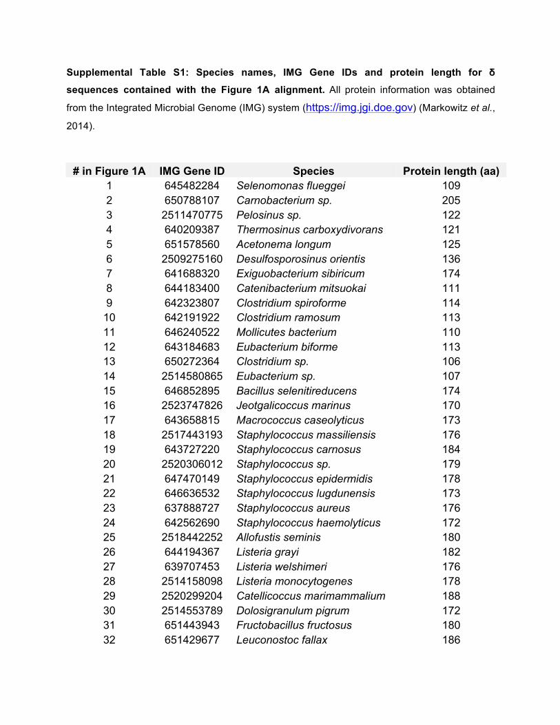
Supplemental Table S1: Species names, IMG Gene IDs and protein length for δ
sequences contained with the Figure 1A alignment. All protein information was obtained
from the Integrated Microbial Genome (IMG) system (https://img.jgi.doe.gov) (Markowitz et al.,
2014).
# in Figure 1A IMG Gene ID Species Protein length (aa) 1 645482284 Selenomonas flueggei 109 2 650788107 Carnobacterium sp. 205 3 2511470775 Pelosinus sp. 122 4 640209387 Thermosinus carboxydivorans 121 5 651578560 Acetonema longum 125 6 2509275160 Desulfosporosinus orientis 136 7 641688320 Exiguobacterium sibiricum 174 8 644183400 Catenibacterium mitsuokai 111 9 642323807 Clostridium spiroforme 114
10 642191922 Clostridium ramosum 113 11 646240522 Mollicutes bacterium 110 12 643184683 Eubacterium biforme 113 13 650272364 Clostridium sp. 106 14 2514580865 Eubacterium sp. 107 15 646852895 Bacillus selenitireducens 174 16 2523747826 Jeotgalicoccus marinus 170 17 643658815 Macrococcus caseolyticus 173 18 2517443193 Staphylococcus massiliensis 176 19 643727220 Staphylococcus carnosus 184 20 2520306012 Staphylococcus sp. 179 21 647470149 Staphylococcus epidermidis 178 22 646636532 Staphylococcus lugdunensis 173 23 637888727 Staphylococcus aureus 176 24 642562690 Staphylococcus haemolyticus 172 25 2518442252 Allofustis seminis 180 26 644194367 Listeria grayi 182 27 639707453 Listeria welshimeri 176 28 2514158098 Listeria monocytogenes 178 29 2520299204 Catellicoccus marimammalium 188 30 2514553789 Dolosigranulum pigrum 172 31 651443943 Fructobacillus fructosus 180 32 651429677 Leuconostoc fallax 186
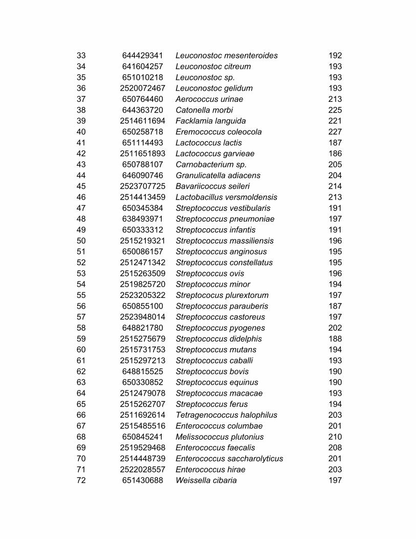
33 644429341 Leuconostoc mesenteroides 192 34 641604257 Leuconostoc citreum 193 35 651010218 Leuconostoc sp. 193 36 2520072467 Leuconostoc gelidum 193 37 650764460 Aerococcus urinae 213 38 644363720 Catonella morbi 225 39 2514611694 Facklamia languida 221 40 650258718 Eremococcus coleocola 227 41 651114493 Lactococcus lactis 187 42 2511651893 Lactococcus garvieae 186 43 650788107 Carnobacterium sp. 205 44 646090746 Granulicatella adiacens 204 45 2523707725 Bavariicoccus seileri 214 46 2514413459 Lactobacillus versmoldensis 213 47 650345384 Streptococcus vestibularis 191 48 638493971 Streptococcus pneumoniae 197 49 650333312 Streptococcus infantis 191 50 2515219321 Streptococcus massiliensis 196 51 650086157 Streptococcus anginosus 195 52 2512471342 Streptococcus constellatus 195 53 2515263509 Streptococcus ovis 196 54 2519825720 Streptococcus minor 194 55 2523205322 Streptococus plurextorum 197 56 650855100 Streptococcus parauberis 187 57 2523948014 Streptococcus castoreus 197 58 648821780 Streptococcus pyogenes 202 59 2515275679 Streptococcus didelphis 188 60 2515731753 Streptococcus mutans 194 61 2515297213 Streptococcus caballi 193 62 648815525 Streptococcus bovis 190 63 650330852 Streptococcus equinus 190 64 2512479078 Streptococcus macacae 193 65 2515262707 Streptococcus ferus 194 66 2511692614 Tetragenococcus halophilus 203 67 2515485516 Enterococcus columbae 201 68 650845241 Melissococcus plutonius 210 69 2519529468 Enterococcus faecalis 208 70 2514448739 Enterococcus saccharolyticus 201 71 2522028557 Enterococcus hirae 203 72 651430688 Weissella cibaria 197
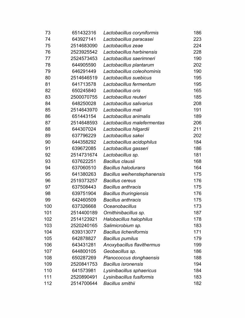
73 651432316 Lactobacillus coryniformis 186 74 643927141 Lactobacillus paracasei 223 75 2514683090 Lactobacillus zeae 224 76 2523925542 Lactobacillus harbinensis 228 77 2524573453 Lactobacillus saerimneri 190 78 644905590 Lactobacillus plantarum 202 79 646291449 Lactobacillus coleohominis 190 80 2514646519 Lactobacillus suebicus 195 81 641713578 Lactobacillus fermentum 195 82 650245840 Lactobacillus oris 165 83 2500070755 Lactobacillus reuteri 185 84 648250028 Lactobacillus salivarius 208 85 2514643970 Lactobacillus mali 191 86 651443154 Lactobacillus animalis 189 87 2514648593 Lactobacillus malefermentas 206 88 644307024 Lactobacillus hilgardii 211 89 637796229 Lactobacillus sakei 202 90 644358292 Lactobacillus acidophilus 184 91 639672085 Lactobacillus gasseri 186 92 2514731674 Lactobacillus sp. 181 93 637622251 Bacillus clausii 168 94 637060510 Bacillus halodurans 164 95 641380263 Bacillus weihenstephanensis 175 96 2519373257 Bacillus cereus 176 97 637508443 Bacillus anthracis 175 98 639751904 Bacillus thuringiensis 176 99 642460509 Bacillus anthracis 175
100 637326668 Oceanobacillus 173 101 2514400189 Ornithinibacillus sp. 187 102 2514123921 Halobacillus halophilus 178 103 2520240165 Salimicrobium sp. 183 104 639313077 Bacillus licheniformis 171 105 642878827 Bacillus pumilus 179 106 643431281 Anoxybacillus flavithermus 199 107 644800105 Geobacillus sp. 186 108 650287269 Planococcus donghaensis 188 109 2520841753 Bacillus isronensis 194 110 641573981 Lysinibacillus sphaericus 184 111 2520890491 Lysinibacillus fusiformis 183 112 2514700644 Bacillus smithii 182
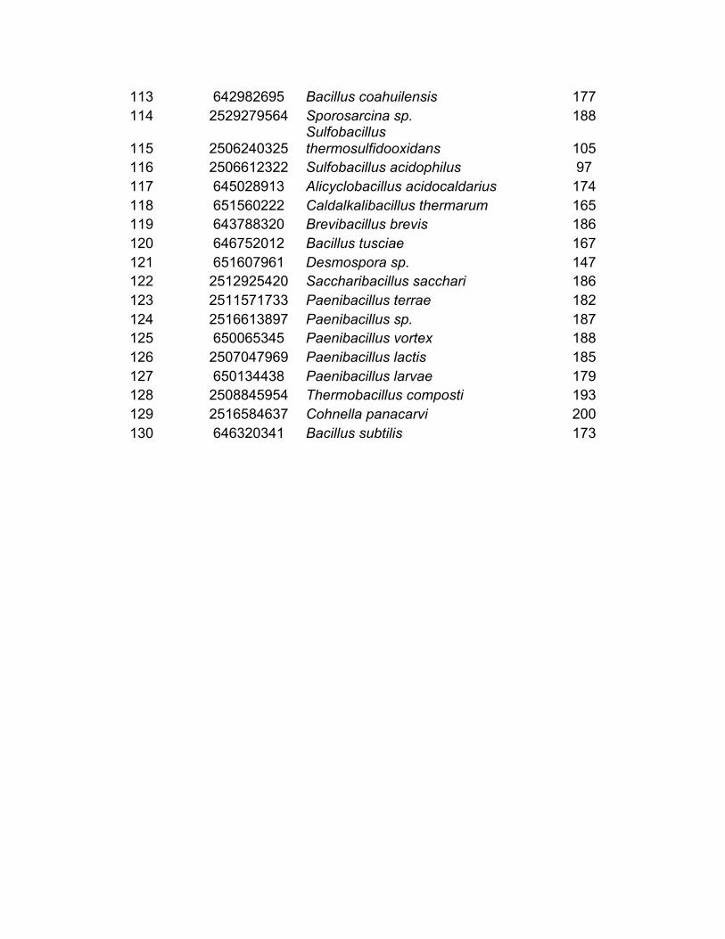
113 642982695 Bacillus coahuilensis 177 114 2529279564 Sporosarcina sp. 188
115 2506240325 Sulfobacillus thermosulfidooxidans 105
116 2506612322 Sulfobacillus acidophilus 97 117 645028913 Alicyclobacillus acidocaldarius 174 118 651560222 Caldalkalibacillus thermarum 165 119 643788320 Brevibacillus brevis 186 120 646752012 Bacillus tusciae 167 121 651607961 Desmospora sp. 147 122 2512925420 Saccharibacillus sacchari 186 123 2511571733 Paenibacillus terrae 182 124 2516613897 Paenibacillus sp. 187 125 650065345 Paenibacillus vortex 188 126 2507047969 Paenibacillus lactis 185 127 650134438 Paenibacillus larvae 179 128 2508845954 Thermobacillus composti 193 129 2516584637 Cohnella panacarvi 200 130 646320341 Bacillus subtilis 173

101
APPENDIX II

American Society for Microbiology Journals LICENSE
TERMS AND CONDITIONS
Apr 05, 2017
This is a License Agreement between Andy Weiss ("You") and American Society for
Microbiology - Journals ("American Society for Microbiology - Journals") provided by
Copyright Clearance Center ("CCC"). The license consists of your order details, the terms
and conditions provided by American Society for Microbiology - Journals, and the payment
terms and conditions.
All payments must be made in full to CCC. For payment instructions, please see
information listed at the bottom of this form.
License Number 4053651327049
License date Feb 19, 2017
Licensed content publisher American Society for Microbiology Journals
Licensed content title Infection and immunity
Licensed content date Jan 1, 1970
Type of Use Thesis/Dissertation
Requestor type Author of requested content
Format Print, Electronic
Portion chapter/article
Title or numeric reference of
the portion(s)
This article is intended to be used as a chapter in my dissertation.
The article, as well as the supplemental data, will be placed in the
appendix of the dissertation
Title of the article or chapter
the portion is from
The δ Subunit of RNA Polymerase Guides Promoter Selectivity and
Virulence in Staphylococcus aureus
Editor of portion(s) A. J. Bäumler
Author of portion(s) Andy Weiss, J. Antonio Ibarra, Jessica Paoletti, Ronan K. Carroll,
and Lindsey N. Shaw
Volume of serial or
monograph.
82
Issue, if republishing an
article from a serial
4
Page range of the portion
Publication date of portion 2014 April
Rights for Main product and any product related to main product
Duration of use Life of current edition
Creation of copies for the
disabled
no
With minor editing privileges yes
For distribution to Worldwide
In the following language(s) Original language of publication
With incidental promotional
use
no
The lifetime unit quantity of
new product
Up to 499
Made available in the
following markets
educational

Specified additional
information
The requesting
person/organization is:
Order reference number
Author/Editor
The standard identifier of
New Work
Title of New Work
Publisher of New Work
Expected publication date
Estimated size (pages)
Total (may include CCC user
fee)
Minor changes might required.
Andy Weiss
Andy Weiss, J. Antonio Ibarra, Jessica Paoletti, Ronan K. Carroll,
and Lindsey N. Shaw
University of South Florida
Nonclassical regulators in Staphylococcus aureus
University of South Florida
May 2017
130
0.00 USD
Terms and Conditions
TERMS AND CONDITIONS
The following terms are individual to this publisher:
We ask that you review the copyright information associated with the article that you are
interested in, as some articles in the ASM Journals are covered by creative commons
licenses that would permit free reuse with attribution.
Please note that ASM cannot grant permission to reuse figures or images that are credited to
publications other than ASM journals. For images credited to non-ASM journal publications,
you will need to obtain permission from the journal referenced in the figure or table legend
or credit line before making any use of the image(s) or table(s). ASM is unable to grant
permission to reuse supplemental material as copyright for the supplemental material
remains with the author.
Other Terms and Conditions:
STANDARD TERMS AND CONDITIONS
1. Description of Service; Defined Terms. This Republication License enables the User to
obtain licenses for republication of one or more copyrighted works as described in detail on
the relevant Order Confirmation (the “Work(s)”). Copyright Clearance Center, Inc. (“CCC”)
grants licenses through the Service on behalf of the rightsholder identified on the Order
Confirmation (the “Rightsholder”). “Republication”, as used herein, generally means the
inclusion of a Work, in whole or in part, in a new work or works, also as described on the
Order Confirmation. “User”, as used herein, means the person or entity making such
republication.
2. The terms set forth in the relevant Order Confirmation, and any terms set by the
Rightsholder with respect to a particular Work, govern the terms of use of Works in
connection with the Service. By using the Service, the person transacting for a republication
license on behalf of the User represents and warrants that he/she/it (a) has been duly
authorized by the User to accept, and hereby does accept, all such terms and conditions on
behalf of User, and (b) shall inform User of all such terms and conditions. In the event such
person is a “freelancer” or other third party independent of User and CCC, such party shall
be deemed jointly a “User” for purposes of these terms and conditions. In any event, User
shall be deemed to have accepted and agreed to all such terms and conditions if User
republishes the Work in any fashion.
3. Scope of License; Limitations and Obligations.
3.1 All Works and all rights therein, including copyright rights, remain the sole and
exclusive property of the Rightsholder. The license created by the exchange of an Order
Confirmation (and/or any invoice) and payment by User of the full amount set forth on that
document includes only those rights expressly set forth in the Order Confirmation and in
these terms and conditions, and conveys no other rights in the Work(s) to User. All rights not
expressly granted are hereby reserved.

3.2 General Payment Terms: You may pay by credit card or through an account with us
payable at the end of the month. If you and we agree that you may establish a standing
account with CCC, then the following terms apply: Remit Payment to: Copyright Clearance
Center, 29118 Network Place, Chicago, IL 60673-1291. Payments Due: Invoices are payable
upon their delivery to you (or upon our notice to you that they are available to you for
downloading). After 30 days, outstanding amounts will be subject to a service charge of 1-
1/2% per month or, if less, the maximum rate allowed by applicable law. Unless otherwise
specifically set forth in the Order Confirmation or in a separate written agreement signed by
CCC, invoices are due and payable on “net 30” terms. While User may exercise the rights
licensed immediately upon issuance of the Order Confirmation, the license is automatically
revoked and is null and void, as if it had never been issued, if complete payment for the
license is not received on a timely basis either from User directly or through a payment
agent, such as a credit card company.
3.3 Unless otherwise provided in the Order Confirmation, any grant of rights to User (i) is
“one-time” (including the editions and product family specified in the license), (ii) is non-
exclusive and non-transferable and (iii) is subject to any and all limitations and restrictions
(such as, but not limited to, limitations on duration of use or circulation) included in the
Order Confirmation or invoice and/or in these terms and conditions. Upon completion of the
licensed use, User shall either secure a new permission for further use of the Work(s) or
immediately cease any new use of the Work(s) and shall render inaccessible (such as by
deleting or by removing or severing links or other locators) any further copies of the Work
(except for copies printed on paper in accordance with this license and still in User's stock at
the end of such period).
3.4 In the event that the material for which a republication license is sought includes third
party materials (such as photographs, illustrations, graphs, inserts and similar materials)
which are identified in such material as having been used by permission, User is responsible
for identifying, and seeking separate licenses (under this Service or otherwise) for, any of
such third party materials; without a separate license, such third party materials may not be
used.
3.5 Use of proper copyright notice for a Work is required as a condition of any license
granted under the Service. Unless otherwise provided in the Order Confirmation, a proper
copyright notice will read substantially as follows: “Republished with permission of
[Rightsholder’s name], from [Work's title, author, volume, edition number and year of
copyright]; permission conveyed through Copyright Clearance Center, Inc. ” Such notice
must be provided in a reasonably legible font size and must be placed either immediately
adjacent to the Work as used (for example, as part of a by-line or footnote but not as a
separate electronic link) or in the place where substantially all other credits or notices for the
new work containing the republished Work are located. Failure to include the required notice
results in loss to the Rightsholder and CCC, and the User shall be liable to pay liquidated
damages for each such failure equal to twice the use fee specified in the Order Confirmation,
in addition to the use fee itself and any other fees and charges specified.
3.6 User may only make alterations to the Work if and as expressly set forth in the Order
Confirmation. No Work may be used in any way that is defamatory, violates the rights of
third parties (including such third parties' rights of copyright, privacy, publicity, or other
tangible or intangible property), or is otherwise illegal, sexually explicit or obscene. In
addition, User may not conjoin a Work with any other material that may result in damage to
the reputation of the Rightsholder. User agrees to inform CCC if it becomes aware of any
infringement of any rights in a Work and to cooperate with any reasonable request of CCC
or the Rightsholder in connection therewith.
4. Indemnity. User hereby indemnifies and agrees to defend the Rightsholder and CCC, and
their respective employees and directors, against all claims, liability, damages, costs and
expenses, including legal fees and expenses, arising out of any use of a Work beyond the
scope of the rights granted herein, or any use of a Work which has been altered in any
unauthorized way by User, including claims of defamation or infringement of rights of
copyright, publicity, privacy or other tangible or intangible property.
5. Limitation of Liability. UNDER NO CIRCUMSTANCES WILL CCC OR THE
RIGHTSHOLDER BE LIABLE FOR ANY DIRECT, INDIRECT, CONSEQUENTIAL OR
INCIDENTAL DAMAGES (INCLUDING WITHOUT LIMITATION DAMAGES FOR

LOSS OF BUSINESS PROFITS OR INFORMATION, OR FOR BUSINESS
INTERRUPTION) ARISING OUT OF THE USE OR INABILITY TO USE A WORK,
EVEN IF ONE OF THEM HAS BEEN ADVISED OF THE POSSIBILITY OF SUCH
DAMAGES. In any event, the total liability of the Rightsholder and CCC (including their
respective employees and directors) shall not exceed the total amount actually paid by User
for this license. User assumes full liability for the actions and omissions of its principals,
employees, agents, affiliates, successors and assigns.
6. Limited Warranties. THE WORK(S) AND RIGHT(S) ARE PROVIDED “AS IS”. CCC
HAS THE RIGHT TO GRANT TO USER THE RIGHTS GRANTED IN THE ORDER
CONFIRMATION DOCUMENT. CCC AND THE RIGHTSHOLDER DISCLAIM ALL
OTHER WARRANTIES RELATING TO THE WORK(S) AND RIGHT(S), EITHER
EXPRESS OR IMPLIED, INCLUDING WITHOUT LIMITATION IMPLIED
WARRANTIES OF MERCHANTABILITY OR FITNESS FOR A PARTICULAR
PURPOSE. ADDITIONAL RIGHTS MAY BE REQUIRED TO USE ILLUSTRATIONS,
GRAPHS, PHOTOGRAPHS, ABSTRACTS, INSERTS OR OTHER PORTIONS OF THE
WORK (AS OPPOSED TO THE ENTIRE WORK) IN A MANNER CONTEMPLATED
BY USER; USER UNDERSTANDS AND AGREES THAT NEITHER CCC NOR THE
RIGHTSHOLDER MAY HAVE SUCH ADDITIONAL RIGHTS TO GRANT.
7. Effect of Breach. Any failure by User to pay any amount when due, or any use by User of
a Work beyond the scope of the license set forth in the Order Confirmation and/or these
terms and conditions, shall be a material breach of the license created by the Order
Confirmation and these terms and conditions. Any breach not cured within 30 days of
written notice thereof shall result in immediate termination of such license without further
notice. Any unauthorized (but licensable) use of a Work that is terminated immediately upon
notice thereof may be liquidated by payment of the Rightsholder's ordinary license price
therefor; any unauthorized (and unlicensable) use that is not terminated immediately for any
reason (including, for example, because materials containing the Work cannot reasonably be
recalled) will be subject to all remedies available at law or in equity, but in no event to a
payment of less than three times the Rightsholder's ordinary license price for the most
closely analogous licensable use plus Rightsholder's and/or CCC's costs and expenses
incurred in collecting such payment.
8. Miscellaneous.
8.1 User acknowledges that CCC may, from time to time, make changes or additions to the
Service or to these terms and conditions, and CCC reserves the right to send notice to the
User by electronic mail or otherwise for the purposes of notifying User of such changes or
additions; provided that any such changes or additions shall not apply to permissions already
secured and paid for.
8.2 Use of User-related information collected through the Service is governed by CCC’s
privacy policy, available online here:
http://www.copyright.com/content/cc3/en/tools/footer/privacypolicy.html.
8.3 The licensing transaction described in the Order Confirmation is personal to User.
Therefore, User may not assign or transfer to any other person (whether a natural person or
an organization of any kind) the license created by the Order Confirmation and these terms
and conditions or any rights granted hereunder; provided, however, that User may assign
such license in its entirety on written notice to CCC in the event of a transfer of all or
substantially all of User’s rights in the new material which includes the Work(s) licensed
under this Service.
8.4 No amendment or waiver of any terms is binding unless set forth in writing and signed
by the parties. The Rightsholder and CCC hereby object to any terms contained in any
writing prepared by the User or its principals, employees, agents or affiliates and purporting
to govern or otherwise relate to the licensing transaction described in the Order
Confirmation, which terms are in any way inconsistent with any terms set forth in the Order
Confirmation and/or in these terms and conditions or CCC's standard operating procedures,
whether such writing is prepared prior to, simultaneously with or subsequent to the Order
Confirmation, and whether such writing appears on a copy of the Order Confirmation or in a
separate instrument.
8.5 The licensing transaction described in the Order Confirmation document shall be
governed by and construed under the law of the State of New York, USA, without regard to

the principles thereof of conflicts of law. Any case, controversy, suit, action, or proceeding
arising out of, in connection with, or related to such licensing transaction shall be brought, at
CCC's sole discretion, in any federal or state court located in the County of New York, State
of New York, USA, or in any federal or state court whose geographical jurisdiction covers
the location of the Rightsholder set forth in the Order Confirmation. The parties expressly
submit to the personal jurisdiction and venue of each such federal or state court.If you have
any comments or questions about the Service or Copyright Clearance Center, please contact
us at 978-750-8400 or send an e-mail to [email protected].
v 1.1
Questions? [email protected] or +18552393415 (toll free in the US) or
+19786462777.
Published Ahead of Print 3 February 2014. 2014, 82(4):1424. DOI: 10.1128/IAI.01508-14. Infect. Immun.
Carroll and Lindsey N. ShawAndy Weiss, J. Antonio Ibarra, Jessica Paoletti, Ronan K. Staphylococcus aureusPromoter Selectivity and Virulence in
Subunit of RNA Polymerase GuidesδThe
http://iai.asm.org/content/82/4/1424Updated information and services can be found at:
These include:
SUPPLEMENTAL MATERIAL Supplemental material
REFERENCEShttp://iai.asm.org/content/82/4/1424#ref-list-1at:
This article cites 50 articles, 25 of which can be accessed free
CONTENT ALERTS more»articles cite this article),
Receive: RSS Feeds, eTOCs, free email alerts (when new
http://journals.asm.org/site/misc/reprints.xhtmlInformation about commercial reprint orders: http://journals.asm.org/site/subscriptions/To subscribe to to another ASM Journal go to:
on March 20, 2014 by guest
http://iai.asm.org/
Dow
nloaded from
on March 20, 2014 by guest
http://iai.asm.org/
Dow
nloaded from
The Subunit of RNA Polymerase Guides Promoter Selectivity andVirulence in Staphylococcus aureus
Andy Weiss, J. Antonio Ibarra,* Jessica Paoletti, Ronan K. Carroll, Lindsey N. Shaw
Department of Cell Biology, Microbiology and Molecular Biology, University of South Florida, Tampa, Florida, USA
In Gram-positive bacteria, and particularly the Firmicutes, the DNA-dependent RNA polymerase (RNAP) complex contains anadditional subunit, termed the factor, or RpoE. This enigmatic protein has been studied for more than 30 years for various or-ganisms, but its function is still not well understood. In this study, we investigated its role in the major human pathogen Staphy-lococcus aureus. We showed conservation of important structural regions of RpoE in S. aureus and other species and demon-strated binding to core RNAP that is mediated by the and/or = subunits. To identify the impact of the subunit ontranscription, we performed transcriptome sequencing (RNA-seq) analysis and observed 191 differentially expressed genes inthe rpoE mutant. Ontological analysis revealed, quite strikingly, that many of the downregulated genes were known virulencefactors, while several mobile genetic elements (SaPI5 and prophage SA3usa) were strongly upregulated. Phenotypically, therpoE mutant had decreased accumulation and/or activity of a number of key virulence factors, including alpha toxin, secretedproteases, and Panton-Valentine leukocidin (PVL). We further observed significantly decreased survival of the mutant in wholehuman blood, increased phagocytosis by human leukocytes, and impaired virulence in a murine model of infection. Collectively,our results demonstrate that the subunit of RNAP is a critical component of the S. aureus transcription machinery and playsan important role during infection.
Bacterial gene transcription is a complex, multifactorial processthat involves several key enzymes and regulatory elements. It
is driven by the activity of DNA-dependent RNA polymerase(RNAP) and its associated proteins, which form a multisubunitenzyme consisting of one subunit, one = subunit, two identical subunits, and one subunit (reviewed in reference 1). Togetherthese form the RNAP apoenzyme, which is able to perform RNAelongation and termination; however, initiation requires the in-volvement of a factor. Typically, most bacterial species harborseveral different factors, a primary one (A or 70) that mediateshousekeeping gene transcription and a variety of alternative sigmafactors, which aid in the response to unfavorable environmentalconditions and stress.
In certain Gram-positive species and particularly the Firmic-utes (see Fig. S1 in the supplemental material), an additionalRNAP subunit is present, termed the factor, or RpoE (2). InBacillus subtilis, the 173-amino-acid delta subunit has been shownto reduce nonspecific binding of RNAP to DNA and lead to anelevated preference for DNA regions that include promoter se-quences (3, 4). In early experiments, it was shown that RpoE has arole specifically confined to promoter selection and influences theability of RNAP to form open promoter complexes (5, 6). Full-length RpoE has also been described as displacing nucleic acidsfrom RNAP-DNA or RNAP-RNA complexes in vitro, which mayexplain the decreased affinity of RNAP-RpoE for nonpromoterregions. This feature of RpoE activity has additionally been attrib-uted to its ability to stimulate RNAP recycling by enhancing therelease of the complex from terminator sequences (6).
The effects of RpoE are far from arbitrary, since it has beenshown that the subunit of RNAP not only distinguishes betweenpromoter and nonpromoter sequences but also is important in therecognition of individual promoter features. A comparison of two29 promoters by transcription runoff revealed selective, pro-moter-dependent effects of RpoE (7). While the stronger of thesetwo promoters was largely unaffected by RpoE, the weaker pro-
moter showed significantly decreased expression in the presenceof this factor. In order to explain the effect of RpoE on promoterselection, Rabatinova et al. (8) investigated the role of initiatingnucleotide triphosphate (iNTP) recognition by RNAP and RNAP-RpoE complexes. iNTP is required to stabilize open complex for-mation and therefore to initiate transcription. It is thought that bydestabilizing DNA-RNAP complexes, RpoE increases the amountof iNTP required to transcribe from certain promoters. Finally, inan approach aiming to identify functional regions of RpoE, Lopezde Saro et al. (9) created several truncated variants of the protein.In doing so, they showed that the N-terminal portion is requiredfor binding to RNAP and also guides the correct orientation of thedisordered and polyanionic C-terminal region. A limitation ofthese studies regarding RpoE function, however, is that they wereperformed in vitro, using only single promoters. Therefore, thetrue extent of -subunit influence on genome-wide transcrip-tional effects is still somewhat restricted at this point.
Phenotypically, rpoE mutants of B. subtilis have an extendedlag phase upon subculturing of stationary-phase cells in fresh me-dium, as well as a moderately changed cell morphology (10). Fur-thermore, competition experiments revealed that when cocul-
Received 24 January 2014 Accepted 26 January 2014
Published ahead of print 3 February 2014
Editor: A. J. Bäumler
Address correspondence to Lindsey N. Shaw, [email protected].
* Present address: J. Antonio Ibarra, Departamento de Microbiología, EscuelaNacional de Ciencias Biológicas, Instituto Politécnico Nacional, Mexico City, DF,Mexico.
Supplemental material for this article may be found at http://dx.doi.org/10.1128/IAI.01508-14.
Copyright © 2014, American Society for Microbiology. All Rights Reserved.
doi:10.1128/IAI.01508-14
1424 iai.asm.org Infection and Immunity p. 1424 –1435 April 2014 Volume 82 Number 4
on March 20, 2014 by guest
http://iai.asm.org/
Dow
nloaded from
tured with the wild type, -factor-lacking strains show decreasedfitness and are outcompeted during growth over several days (8).Finally, a role for RpoE in sporulation has been suggested, sincerpoE loss was shown to suppress a mutation (pdhC) that negativelyinfluences sporulation (11).
The role of RpoE has also been addressed for a small number ofpathogenic organisms, most notably in streptococcal species.RpoE in Streptococcus mutans is most abundant during exponen-tial and early stationary phases of growth, and rpoE deletion causesan extended lag phase. S. mutans rpoE mutants are also more sen-sitive to environmental stresses (H2O2 stress and acid stress) andshow alterations in biofilm formation and virulence (12). Inter-estingly, rpoE deletion was also reported to increase self-aggrega-tion, coaggregation with other oral microorganisms (13), and anelevated ability to bind human extracellular matrix proteins. ForStreptococcus agalactiae, there have been a limited number of stud-ies which show greatest rpoE expression during exponentialgrowth (14) and attenuated virulence of rpoE mutants in a ratsepsis model (15, 16) and human whole-blood survival assays.
In Staphylococcus aureus, the only information thus far regard-ing RpoE is that strains lacking this factor display defects in star-vation-induced stationary-phase survival/recovery and slightlyincreased acid sensitivity (17). Notably, however, no investigationof its role in cellular physiology and virulence has been performed.Therefore, in this study, we investigated the role of RpoE in pro-moter selectivity using transcriptome sequencing (RNA-seq)technology and determined that in S. aureus, the subunit specif-ically guides RNAP toward strongly expressed promoters. Dele-tion of this factor results in a normalizing of transcriptional activ-ity across genes, guiding RNAP away from key virulence-affectingloci. Such effects strongly impair the pathogenic potential of S.aureus, leading to diminished infection using a systemic model ofsepsis and increased phagocytosis by human leukocytes. Collec-tively, our results suggest that rpoE is involved in orchestrating theability of S. aureus to react and adapt to environmental changesand thus plays a critical role during virulence.
MATERIALS AND METHODSBacterial strains and growth conditions. Strains used for experimentalprocedures are listed in Table 1. An rpoE transposon mutant in S. aureusUSA300 strain JE2 was acquired from the Nebraska transposon library(NARSA). This mutation was transferred via 11 transduction to ourUSA300 Houston wild-type strain (18). Successful transduction of thismutation was confirmed by PCR, using gene-specific (OL1709 andOL1710) and transposon-specific (OL14721 and OL1472) primers. Cul-tivation of bacteria was performed in tryptic soy broth (TSB) at 37°C.Where required, erythromycin (5 mg/ml), lincomycin (10 mg/ml), orchloramphenicol (10 mg/ml) was added to the medium. Synchronouscultures were obtained as described by us previously (19).
Construction of an rpoE complemented strain. The rpoE gene and itspromoter were amplified via PCR, using primer pair OL1973 andOL1975, which are located 200 bp upstream from the rpoE translationalstart codon and at the 3= end of the rpoE coding region, respectively. Inaddition to the native rpoE sequence, a hexahistidine (His6) tag was in-cluded in the reverse primer (OL1975) to create a fusion protein thatcould be used for downstream purification. For the cloning of truncatedrpoE-His6 fragments, primers 2931 (half of the N terminus only, aminoacids 1 to 159), 2932 (the N-terminal half of the protein, amino acids 1 to264), and 2933 (the N-terminal half of the protein and half of the Cterminus, amino acids 1 to 396) were used instead of 1975. The PCRproducts were cloned into shuttle vector pMK4 and transformed intochemically competent Escherichia coli DH5. Clones were confirmed via
PCR using the same set of oligonucleotides used for cloning. Additionally,Sanger sequencing using primers for the pMK4 multiple cloning site(M13Fw and M13Rv) was performed to confirm fidelity of the construct.The plasmids were transformed into S. aureus RN4220 by electroporationand confirmed by PCR. Correct clones were used to generate a 11 lysatefor transduction into the S. aureus USA300 Houston rpoE transposonmutant, which was again confirmed as described above.
Mapping the rpoE promoter. Rapid amplification of cDNA ends(RACE) was used to identify the rpoE transcriptional start site. For thisapproach, we used the 5=RACE core set (TaKaRa) and primers OL2018,OL2019, OL2020, OL2021, and OL2022 (Table 1). PCR products were TAcloned using the StrataClone PCR cloning kit (Agilent). The transcrip-tional start site was identified by sequencing (MWG operon) 10 plasmidsthat originated with the TA cloning using the universal primers M13Fwand M13Rv.
qPCR. Quantitative real-time PCR (qPCR) analysis was conducted asdescribed previously (20) using the rpoE gene-specific primers OL1976and OL1978 and OL2981 and OL2982 and 16S rRNA primers OL1184 andOL1185. Three independent replicates were used to calculate final values.
Zymograms and Western blot analysis. S. aureus strains were grownas described above, with samples taken at the time points specified. Zy-mograms were performed with culture supernatants as described by uspreviously (21). Intracellular and secreted protein fractions were har-vested and subjected to SDS-PAGE and Western blot analysis as describedby us previously (21). Immunoblotting was performed with a mousemonoclonal anti-His6 (Covance) or anti-LukS (IBT) antibody at a 1:1,000or 1:20.000 dilution, respectively, overnight at 4°C. Secondary antibodieswere affinity-purified horseradish peroxidase (HRP)-conjugated goat an-ti-mouse IgG or goat anti-rabbit IgG (Cell Signaling Technology).
RpoE pulldown assay. A coprecipitation was performed to identifyproteins that interact with RpoE. Briefly, rpoE-negative strains carryingpMK4::rpoE-His6 or pMK4::rpoEtrunc1-His6 were harvested after 3 h ofgrowth, and intracellular fractions were isolated as described previously(22). Cellular extracts were mixed with nickel-nitrilotriacetic acid (Ni-NTA) agarose beads (Qiagen), and samples were incubated with agitationat 37°C for 1 h. Beads were then washed 5 times (50 mM NaH2PO4, 300mM NaCl, and 20 mM imidazole, pH 8.0) while rotating for 10 min at37°C. After each wash step, beads were allowed to settle for 10 min, thesupernatant was removed, and fresh washing buffer was added. RpoE andany interacting proteins were recovered by the addition of elution buffer(50 mM NaH2PO4, 300 mM NaCl, and 250 mM imidazole, pH 8.0).Isolated proteins were then used either to perform SDS-PAGE with sub-sequent silver staining or for mass-spectrometric analysis, as described byus previously (23).
Transcriptomic analysis via RNA-seq. For both the wild-type andmutant strains, three independent cultures were grown as describedabove. Synchronized cultures were harvested after 3 h, and RNA was iso-lated using a Qiagen RNeasy kit (Qiagen). To ensure complete removal ofgenomic DNA, RNA was treated with DNase I (Turbo DNA free; Am-bion). The concentration and purity of RNA were determined using anAgilent 2100 bioanalyzer. Following quantification, equimolar amountsof RNA, from each of the three replicate preparations, were pooled, andrRNA was removed using the MICROBExpress (Invitrogen) and Ribo-Zero (Epicentre) kits. Removal of rRNA was confirmed, again employingthe Agilent 2100 bioanalyzer. rRNA-depleted samples were then used forRNA-seq analysis using the IonTorrent Total RNAseq kit v2 according tothe manufacturer’s instructions (Life Technologies). Templated ionsphere particles were generated using the Ion OneTouch 200 template kit,v2. Sequencing was performed on an IonTorrent 318 chip using an IonPGM 200 sequencing kit. Data generated were exported to the CLCGenomics Workbench software package for analysis. Reads were alignedto the USA300 FPR genome, and expression values for each gene weredetermined as RPKM (reads per kilobase per million reads) values. AnRPKM threshold of detection value of 10 was imposed as a lower-levelcutoff, and data were normalized using the quantile normalization ap-
RpoE Controls Virulence in Staphylococcus aureus
April 2014 Volume 82 Number 4 iai.asm.org 1425
on March 20, 2014 by guest
http://iai.asm.org/
Dow
nloaded from
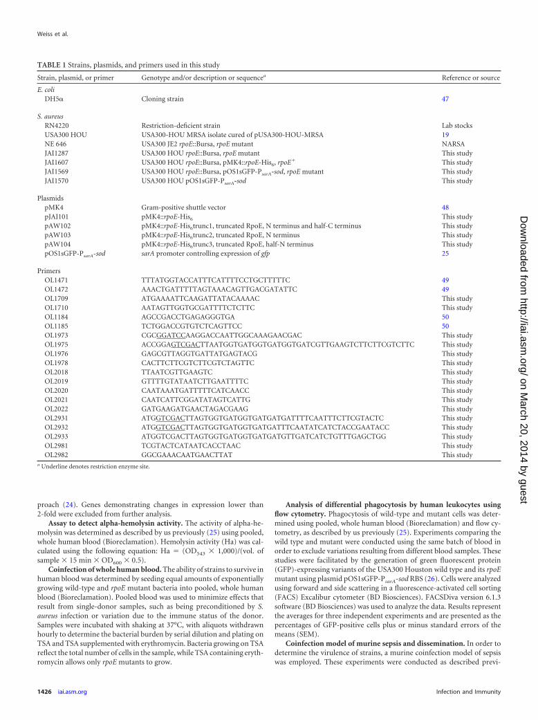
proach (24). Genes demonstrating changes in expression lower than2-fold were excluded from further analysis.
Assay to detect alpha-hemolysin activity. The activity of alpha-he-molysin was determined as described by us previously (25) using pooled,whole human blood (Bioreclamation). Hemolysin activity (Ha) was cal-culated using the following equation: Ha (OD543 1,000)/(vol. ofsample 15 min OD600 0.5).
Coinfection of whole human blood. The ability of strains to survive inhuman blood was determined by seeding equal amounts of exponentiallygrowing wild-type and rpoE mutant bacteria into pooled, whole humanblood (Bioreclamation). Pooled blood was used to minimize effects thatresult from single-donor samples, such as being preconditioned by S.aureus infection or variation due to the immune status of the donor.Samples were incubated with shaking at 37°C, with aliquots withdrawnhourly to determine the bacterial burden by serial dilution and plating onTSA and TSA supplemented with erythromycin. Bacteria growing on TSAreflect the total number of cells in the sample, while TSA containing eryth-romycin allows only rpoE mutants to grow.
Analysis of differential phagocytosis by human leukocytes usingflow cytometry. Phagocytosis of wild-type and mutant cells was deter-mined using pooled, whole human blood (Bioreclamation) and flow cy-tometry, as described by us previously (25). Experiments comparing thewild type and mutant were conducted using the same batch of blood inorder to exclude variations resulting from different blood samples. Thesestudies were facilitated by the generation of green fluorescent protein(GFP)-expressing variants of the USA300 Houston wild type and its rpoEmutant using plasmid pOS1sGFP-PsarA-sod RBS (26). Cells were analyzedusing forward and side scattering in a fluorescence-activated cell sorting(FACS) Excalibur cytometer (BD Biosciences). FACSDiva version 6.1.3software (BD Biosciences) was used to analyze the data. Results representthe averages for three independent experiments and are presented as thepercentages of GFP-positive cells plus or minus standard errors of themeans (SEM).
Coinfection model of murine sepsis and dissemination. In order todetermine the virulence of strains, a murine coinfection model of sepsiswas employed. These experiments were conducted as described previ-
TABLE 1 Strains, plasmids, and primers used in this study
Strain, plasmid, or primer Genotype and/or description or sequencea Reference or source
E. coliDH5 Cloning strain 47
S. aureusRN4220 Restriction-deficient strain Lab stocksUSA300 HOU USA300-HOU MRSA isolate cured of pUSA300-HOU-MRSA 19NE 646 USA300 JE2 rpoE::Bursa, rpoE mutant NARSAJAI1287 USA300 HOU rpoE::Bursa, rpoE mutant This studyJAI1607 USA300 HOU rpoE::Bursa, pMK4::rpoE-His6, rpoE This studyJAI1569 USA300 HOU rpoE::Bursa, pOS1sGFP-PsarA-sod, rpoE mutant This studyJAI1570 USA300 HOU pOS1sGFP-PsarA-sod This study
PlasmidspMK4 Gram-positive shuttle vector 48pJAI101 pMK4::rpoE-His6 This studypAW102 pMK4::rpoE-His6trunc1, truncated RpoE, N terminus and half-C terminus This studypAW103 pMK4::rpoE-His6trunc2, truncated RpoE, N terminus This studypAW104 pMK4::rpoE-His6trunc3, truncated RpoE, half-N terminus This studypOS1sGFP-PsarA-sod sarA promoter controlling expression of gfp 25
PrimersOL1471 TTTATGGTACCATTTCATTTTCCTGCTTTTTC 49OL1472 AAACTGATTTTTAGTAAACAGTTGACGATATTC 49OL1709 ATGAAAATTCAAGATTATACAAAAC This studyOL1710 AATAGTTGGTGCGATTTTCTCTTC This studyOL1184 AGCCGACCTGAGAGGGTGA 50OL1185 TCTGGACCGTGTCTCAGTTCC 50OL1973 CGCGGATCCAAGGACCAATTGGCAAAGAACGAC This studyOL1975 ACCGGAGTCGACTTAATGGTGATGGTGATGGTGATCGTTGAAGTCTTCTTCGTCTTC This studyOL1976 GAGCGTTAGGTGATTATGAGTACG This studyOL1978 CACTTCTTCGTCTTCGTCTAGTTC This studyOL2018 TTAATCGTTGAAGTC This studyOL2019 GTTTTGTATAATCTTGAATTTTC This studyOL2020 CAATAAATGATTTTTCATCAACC This studyOL2021 CAATCATTCGGATATAGTCATTG This studyOL2022 GATGAAGATGAACTAGACGAAG This studyOL2931 ATGGTCGACTTAGTGGTGATGGTGATGATGATTTTCAATTTCTTCGTACTC This studyOL2932 ATGGTCGACTTAGTGGTGATGGTGATGATTTCAATATCATCTACCGAATACC This studyOL2933 ATGGTCGACTTAGTGGTGATGGTGATGATGTTGATCATCTGTTTGAGCTGG This studyOL2981 TCGTACTCATAATCACCTAAC This studyOL2982 GGCGAAACAATGAACTTAT This study
a Underline denotes restriction enzyme site.
Weiss et al.
1426 iai.asm.org Infection and Immunity
on March 20, 2014 by guest
http://iai.asm.org/
Dow
nloaded from
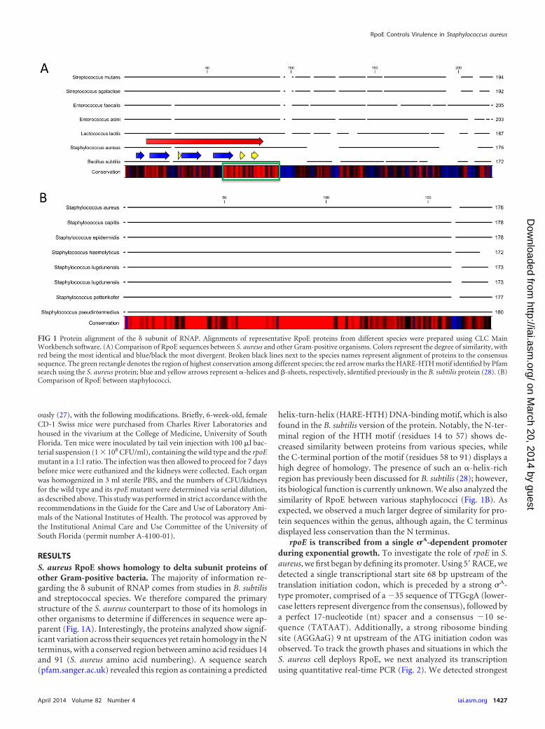
ously (27), with the following modifications. Briefly, 6-week-old, femaleCD-1 Swiss mice were purchased from Charles River Laboratories andhoused in the vivarium at the College of Medicine, University of SouthFlorida. Ten mice were inoculated by tail vein injection with 100 l bac-terial suspension (1 108 CFU/ml), containing the wild type and the rpoEmutant in a 1:1 ratio. The infection was then allowed to proceed for 7 daysbefore mice were euthanized and the kidneys were collected. Each organwas homogenized in 3 ml sterile PBS, and the numbers of CFU/kidneysfor the wild type and its rpoE mutant were determined via serial dilution,as described above. This study was performed in strict accordance with therecommendations in the Guide for the Care and Use of Laboratory Ani-mals of the National Institutes of Health. The protocol was approved bythe Institutional Animal Care and Use Committee of the University ofSouth Florida (permit number A-4100-01).
RESULTSS. aureus RpoE shows homology to delta subunit proteins ofother Gram-positive bacteria. The majority of information re-garding the subunit of RNAP comes from studies in B. subtilisand streptococcal species. We therefore compared the primarystructure of the S. aureus counterpart to those of its homologs inother organisms to determine if differences in sequence were ap-parent (Fig. 1A). Interestingly, the proteins analyzed show signif-icant variation across their sequences yet retain homology in the Nterminus, with a conserved region between amino acid residues 14and 91 (S. aureus amino acid numbering). A sequence search(pfam.sanger.ac.uk) revealed this region as containing a predicted
helix-turn-helix (HARE-HTH) DNA-binding motif, which is alsofound in the B. subtilis version of the protein. Notably, the N-ter-minal region of the HTH motif (residues 14 to 57) shows de-creased similarity between proteins from various species, whilethe C-terminal portion of the motif (residues 58 to 91) displays ahigh degree of homology. The presence of such an -helix-richregion has previously been discussed for B. subtilis (28); however,its biological function is currently unknown. We also analyzed thesimilarity of RpoE between various staphylococci (Fig. 1B). Asexpected, we observed a much larger degree of similarity for pro-tein sequences within the genus, although again, the C terminusdisplayed less conservation than the N terminus.
rpoE is transcribed from a single A-dependent promoterduring exponential growth. To investigate the role of rpoE in S.aureus, we first began by defining its promoter. Using 5=RACE, wedetected a single transcriptional start site 68 bp upstream of thetranslation initiation codon, which is preceded by a strong A-type promoter, comprised of a 35 sequence of TTGcgA (lower-case letters represent divergence from the consensus), followed bya perfect 17-nucleotide (nt) spacer and a consensus 10 se-quence (TATAAT). Additionally, a strong ribosome bindingsite (AGGAaG) 9 nt upstream of the ATG initiation codon wasobserved. To track the growth phases and situations in which theS. aureus cell deploys RpoE, we next analyzed its transcriptionusing quantitative real-time PCR (Fig. 2). We detected strongest
FIG 1 Protein alignment of the subunit of RNAP. Alignments of representative RpoE proteins from different species were prepared using CLC MainWorkbench software. (A) Comparison of RpoE sequences between S. aureus and other Gram-positive organisms. Colors represent the degree of similarity, withred being the most identical and blue/black the most divergent. Broken black lines next to the species names represent alignment of proteins to the consensussequence. The green rectangle denotes the region of highest conservation among different species; the red arrow marks the HARE-HTH motif identified by Pfamsearch using the S. aureus protein; blue and yellow arrows represent -helices and -sheets, respectively, identified previously in the B. subtilis protein (28). (B)Comparison of RpoE between staphylococci.
RpoE Controls Virulence in Staphylococcus aureus
April 2014 Volume 82 Number 4 iai.asm.org 1427
on March 20, 2014 by guest
http://iai.asm.org/
Dow
nloaded from
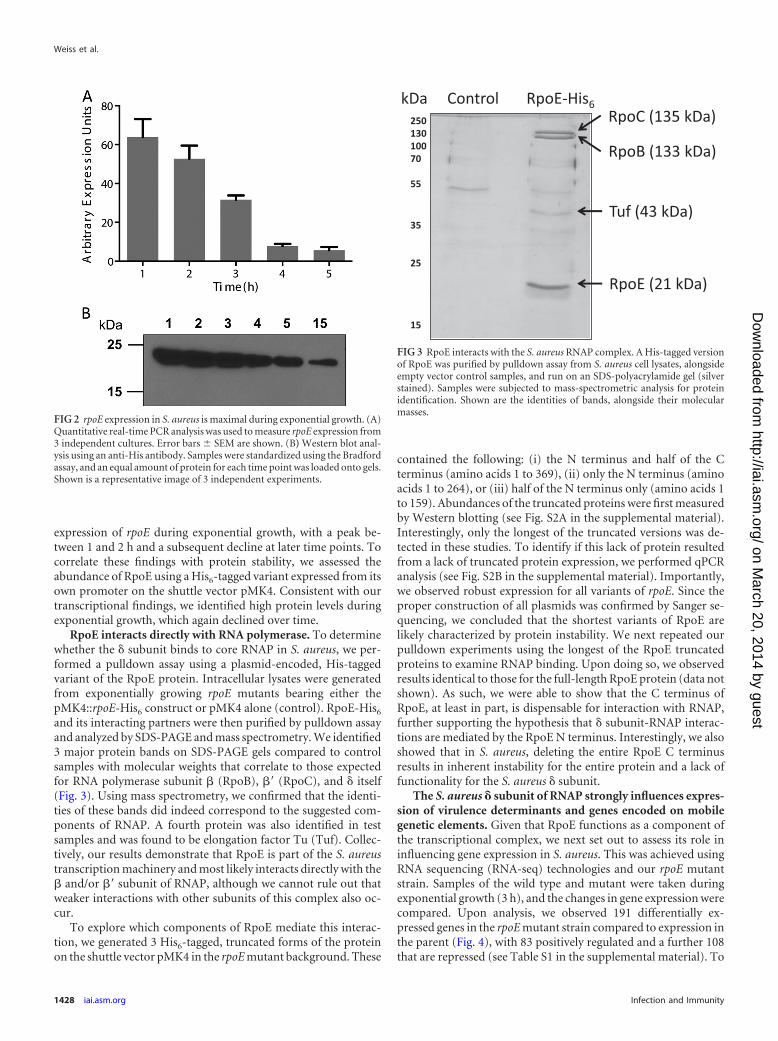
expression of rpoE during exponential growth, with a peak be-tween 1 and 2 h and a subsequent decline at later time points. Tocorrelate these findings with protein stability, we assessed theabundance of RpoE using a His6-tagged variant expressed from itsown promoter on the shuttle vector pMK4. Consistent with ourtranscriptional findings, we identified high protein levels duringexponential growth, which again declined over time.
RpoE interacts directly with RNA polymerase. To determinewhether the subunit binds to core RNAP in S. aureus, we per-formed a pulldown assay using a plasmid-encoded, His-taggedvariant of the RpoE protein. Intracellular lysates were generatedfrom exponentially growing rpoE mutants bearing either thepMK4::rpoE-His6 construct or pMK4 alone (control). RpoE-His6
and its interacting partners were then purified by pulldown assayand analyzed by SDS-PAGE and mass spectrometry. We identified3 major protein bands on SDS-PAGE gels compared to controlsamples with molecular weights that correlate to those expectedfor RNA polymerase subunit (RpoB), = (RpoC), and itself(Fig. 3). Using mass spectrometry, we confirmed that the identi-ties of these bands did indeed correspond to the suggested com-ponents of RNAP. A fourth protein was also identified in testsamples and was found to be elongation factor Tu (Tuf). Collec-tively, our results demonstrate that RpoE is part of the S. aureustranscription machinery and most likely interacts directly with the and/or = subunit of RNAP, although we cannot rule out thatweaker interactions with other subunits of this complex also oc-cur.
To explore which components of RpoE mediate this interac-tion, we generated 3 His6-tagged, truncated forms of the proteinon the shuttle vector pMK4 in the rpoE mutant background. These
contained the following: (i) the N terminus and half of the Cterminus (amino acids 1 to 369), (ii) only the N terminus (aminoacids 1 to 264), or (iii) half of the N terminus only (amino acids 1to 159). Abundances of the truncated proteins were first measuredby Western blotting (see Fig. S2A in the supplemental material).Interestingly, only the longest of the truncated versions was de-tected in these studies. To identify if this lack of protein resultedfrom a lack of truncated protein expression, we performed qPCRanalysis (see Fig. S2B in the supplemental material). Importantly,we observed robust expression for all variants of rpoE. Since theproper construction of all plasmids was confirmed by Sanger se-quencing, we concluded that the shortest variants of RpoE arelikely characterized by protein instability. We next repeated ourpulldown experiments using the longest of the RpoE truncatedproteins to examine RNAP binding. Upon doing so, we observedresults identical to those for the full-length RpoE protein (data notshown). As such, we were able to show that the C terminus ofRpoE, at least in part, is dispensable for interaction with RNAP,further supporting the hypothesis that subunit-RNAP interac-tions are mediated by the RpoE N terminus. Interestingly, we alsoshowed that in S. aureus, deleting the entire RpoE C terminusresults in inherent instability for the entire protein and a lack offunctionality for the S. aureus subunit.
The S. aureus subunit of RNAP strongly influences expres-sion of virulence determinants and genes encoded on mobilegenetic elements. Given that RpoE functions as a component ofthe transcriptional complex, we next set out to assess its role ininfluencing gene expression in S. aureus. This was achieved usingRNA sequencing (RNA-seq) technologies and our rpoE mutantstrain. Samples of the wild type and mutant were taken duringexponential growth (3 h), and the changes in gene expression werecompared. Upon analysis, we observed 191 differentially ex-pressed genes in the rpoE mutant strain compared to expression inthe parent (Fig. 4), with 83 positively regulated and a further 108that are repressed (see Table S1 in the supplemental material). To
FIG 2 rpoE expression in S. aureus is maximal during exponential growth. (A)Quantitative real-time PCR analysis was used to measure rpoE expression from3 independent cultures. Error bars SEM are shown. (B) Western blot anal-ysis using an anti-His antibody. Samples were standardized using the Bradfordassay, and an equal amount of protein for each time point was loaded onto gels.Shown is a representative image of 3 independent experiments.
250 130 100 70 55
35 25
15
RpoE (21 kDa)
kDa Control RpoE-His6 RpoC (135 kDa)
RpoB (133 kDa)
Tuf (43 kDa)
FIG 3 RpoE interacts with the S. aureus RNAP complex. A His-tagged versionof RpoE was purified by pulldown assay from S. aureus cell lysates, alongsideempty vector control samples, and run on an SDS-polyacrylamide gel (silverstained). Samples were subjected to mass-spectrometric analysis for proteinidentification. Shown are the identities of bands, alongside their molecularmasses.
Weiss et al.
1428 iai.asm.org Infection and Immunity
on March 20, 2014 by guest
http://iai.asm.org/
Dow
nloaded from
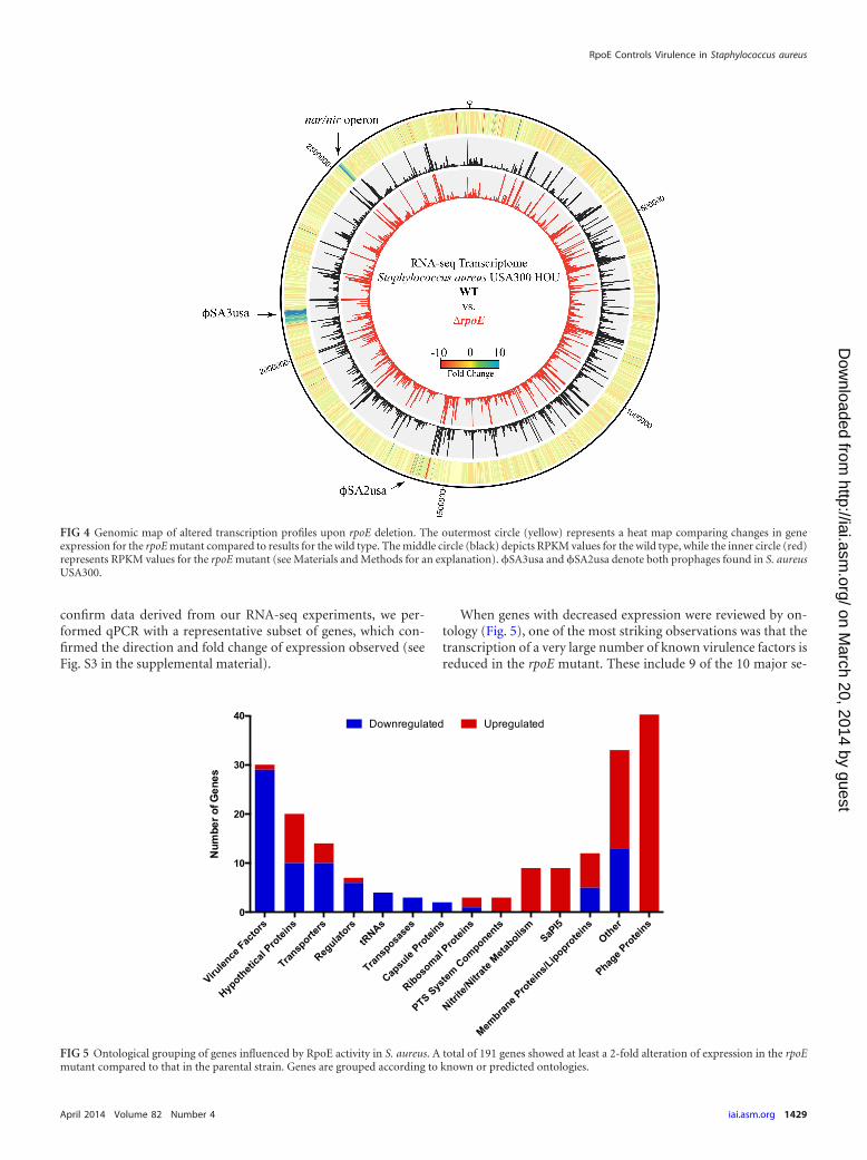
confirm data derived from our RNA-seq experiments, we per-formed qPCR with a representative subset of genes, which con-firmed the direction and fold change of expression observed (seeFig. S3 in the supplemental material).
When genes with decreased expression were reviewed by on-tology (Fig. 5), one of the most striking observations was that thetranscription of a very large number of known virulence factors isreduced in the rpoE mutant. These include 9 of the 10 major se-
FIG 4 Genomic map of altered transcription profiles upon rpoE deletion. The outermost circle (yellow) represents a heat map comparing changes in geneexpression for the rpoE mutant compared to results for the wild type. The middle circle (black) depicts RPKM values for the wild type, while the inner circle (red)represents RPKM values for the rpoE mutant (see Materials and Methods for an explanation). SA3usa and SA2usa denote both prophages found in S. aureusUSA300.
Virulen
ce Fac
tors
Hypotheti
cal P
rotei
ns
Transp
orters
Regulat
ors
tRNAs
Transp
osase
s
Capsu
le Pro
teins
Ribosomal
Protei
ns
PTS System
Componen
ts
Nitrite
/Nitr
ate M
etabolis
mSaP
I5
Membran
e Pro
teins/L
ipoprotei
nsOther
Phage P
rotei
ns0
10
20
30
40
Num
ber o
f Gen
es
Downregulated Upregulated
FIG 5 Ontological grouping of genes influenced by RpoE activity in S. aureus. A total of 191 genes showed at least a 2-fold alteration of expression in the rpoEmutant compared to that in the parental strain. Genes are grouped according to known or predicted ontologies.
RpoE Controls Virulence in Staphylococcus aureus
April 2014 Volume 82 Number 4 iai.asm.org 1429
on March 20, 2014 by guest
http://iai.asm.org/
Dow
nloaded from
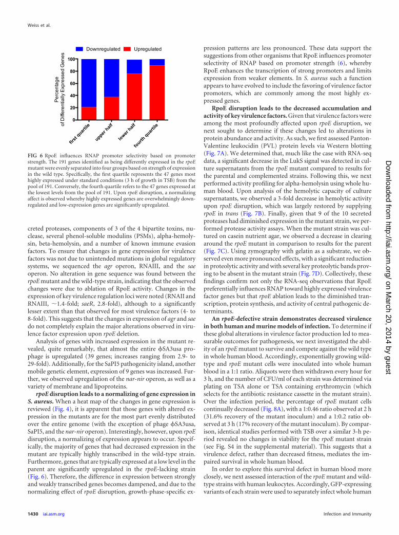
creted proteases, components of 3 of the 4 bipartite toxins, nu-clease, several phenol-soluble modulins (PSMs), alpha-hemoly-sin, beta-hemolysin, and a number of known immune evasionfactors. To ensure that changes in gene expression for virulencefactors was not due to unintended mutations in global regulatorysystems, we sequenced the agr operon, RNAIII, and the saeoperon. No alteration in gene sequence was found between therpoE mutant and the wild-type strain, indicating that the observedchanges were due to ablation of RpoE activity. Changes in theexpression of key virulence regulation loci were noted (RNAII andRNAIII, 1.4-fold; saeR, 2.8-fold), although to a significantlylesser extent than that observed for most virulence factors (4- to8-fold). This suggests that the changes in expression of agr and saedo not completely explain the major alterations observed in viru-lence factor expression upon rpoE deletion.
Analysis of genes with increased expression in the mutant re-vealed, quite remarkably, that almost the entire SA3usa pro-phage is upregulated (39 genes; increases ranging from 2.9- to29-fold). Additionally, for the SaPI5 pathogenicity island, anothermobile genetic element, expression of 9 genes was increased. Fur-ther, we observed upregulation of the nar-nir operon, as well as avariety of membrane and lipoproteins.
rpoE disruption leads to a normalizing of gene expression inS. aureus. When a heat map of the changes in gene expression isreviewed (Fig. 4), it is apparent that those genes with altered ex-pression in the mutants are for the most part evenly distributedover the entire genome (with the exception of phage SA3usa,SaPI5, and the nar-nir operon). Interestingly, however, upon rpoEdisruption, a normalizing of expression appears to occur. Specif-ically, the majority of genes that had decreased expression in themutant are typically highly transcribed in the wild-type strain.Furthermore, genes that are typically expressed at a low level in theparent are significantly upregulated in the rpoE-lacking strain(Fig. 6). Therefore, the difference in expression between stronglyand weakly transcribed genes becomes dampened, and due to thenormalizing effect of rpoE disruption, growth-phase-specific ex-
pression patterns are less pronounced. These data support thesuggestions from other organisms that RpoE influences promoterselectivity of RNAP based on promoter strength (6), wherebyRpoE enhances the transcription of strong promoters and limitsexpression from weaker elements. In S. aureus such a functionappears to have evolved to include the favoring of virulence factorpromoters, which are commonly among the most highly ex-pressed genes.
RpoE disruption leads to the decreased accumulation andactivity of key virulence factors. Given that virulence factors wereamong the most profoundly affected upon rpoE disruption, wenext sought to determine if these changes led to alterations inprotein abundance and activity. As such, we first assessed Panton-Valentine leukocidin (PVL) protein levels via Western blotting(Fig. 7A). We determined that, much like the case with RNA-seqdata, a significant decrease in the LukS signal was detected in cul-ture supernatants from the rpoE mutant compared to results forthe parental and complemented strains. Following this, we nextperformed activity profiling for alpha-hemolysin using whole hu-man blood. Upon analysis of the hemolytic capacity of culturesupernatants, we observed a 3-fold decrease in hemolytic activityupon rpoE disruption, which was largely restored by supplyingrpoE in trans (Fig. 7B). Finally, given that 9 of the 10 secretedproteases had diminished expression in the mutant strain, we per-formed protease activity assays. When the mutant strain was cul-tured on casein nutrient agar, we observed a decrease in clearingaround the rpoE mutant in comparison to results for the parent(Fig. 7C). Using zymography with gelatin as a substrate, we ob-served even more pronounced effects, with a significant reductionin proteolytic activity and with several key proteolytic bands prov-ing to be absent in the mutant strain (Fig. 7D). Collectively, thesefindings confirm not only the RNA-seq observations that RpoEpreferentially influences RNAP toward highly expressed virulencefactor genes but that rpoE ablation leads to the diminished tran-scription, protein synthesis, and activity of central pathogenic de-terminants.
An rpoE-defective strain demonstrates decreased virulencein both human and murine models of infection. To determine ifthese global alterations in virulence factor production led to mea-surable outcomes for pathogenesis, we next investigated the abil-ity of an rpoE mutant to survive and compete against the wild typein whole human blood. Accordingly, exponentially growing wild-type and rpoE mutant cells were inoculated into whole humanblood in a 1:1 ratio. Aliquots were then withdrawn every hour for3 h, and the number of CFU/ml of each strain was determined viaplating on TSA alone or TSA containing erythromycin (whichselects for the antibiotic resistance cassette in the mutant strain).Over the infection period, the percentage of rpoE mutant cellscontinually decreased (Fig. 8A), with a 1:0.46 ratio observed at 2 h(31.6% recovery of the mutant inoculum) and a 1:0.2 ratio ob-served at 3 h (17% recovery of the mutant inoculum). By compar-ison, identical studies performed with TSB over a similar 3-h pe-riod revealed no changes in viability for the rpoE mutant strain(see Fig. S4 in the supplemental material). This suggests that avirulence defect, rather than decreased fitness, mediates the im-paired survival in whole human blood.
In order to explore this survival defect in human blood moreclosely, we next assessed interaction of the rpoE mutant and wild-type strains with human leukocytes. Accordingly, GFP-expressingvariants of each strain were used to separately infect whole human
first
quartile
upper half
lower half
fourth quart
ile0
20
40
60
80
100Pe
rcen
tage
of D
i ffe r
entia
ll y E
xpre
ssed
Gen
e s
UpregulatedDownregulated
FIG 6 RpoE influences RNAP promoter selectivity based on promoterstrength. The 191 genes identified as being differently expressed in the rpoEmutant were evenly separated into four groups based on strength of expressionin the wild type. Specifically, the first quartile represents the 47 genes mosthighly expressed under standard conditions (3 h of growth in TSB) from thepool of 191. Conversely, the fourth quartile refers to the 47 genes expressed atthe lowest levels from the pool of 191. Upon rpoE disruption, a normalizingaffect is observed whereby highly expressed genes are overwhelmingly down-regulated and low-expression genes are significantly upregulated.
Weiss et al.
1430 iai.asm.org Infection and Immunity
on March 20, 2014 by guest
http://iai.asm.org/
Dow
nloaded from
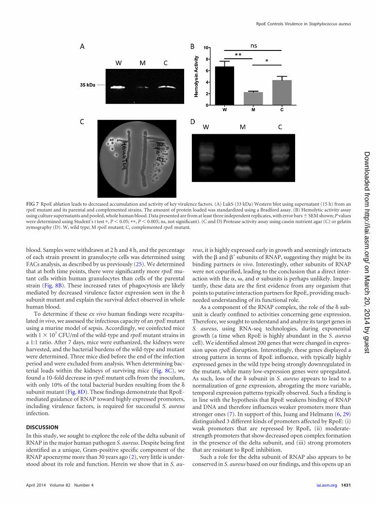
blood. Samples were withdrawn at 2 h and 4 h, and the percentageof each strain present in granulocyte cells was determined usingFACs analysis, as described by us previously (25). We determinedthat at both time points, there were significantly more rpoE mu-tant cells within human granulocytes than cells of the parentalstrain (Fig. 8B). These increased rates of phagocytosis are likelymediated by decreased virulence factor expression seen in the subunit mutant and explain the survival defect observed in wholehuman blood.
To determine if these ex vivo human findings were recapitu-lated in vivo, we assessed the infectious capacity of an rpoE mutantusing a murine model of sepsis. Accordingly, we coinfected micewith 1 107 CFU/ml of the wild-type and rpoE mutant strains ina 1:1 ratio. After 7 days, mice were euthanized, the kidneys wereharvested, and the bacterial burdens of the wild-type and mutantwere determined. Three mice died before the end of the infectionperiod and were excluded from analysis. When determining bac-terial loads within the kidneys of surviving mice (Fig. 8C), wefound a 10-fold decrease in rpoE mutant cells from the inoculum,with only 10% of the total bacterial burden resulting from the subunit mutant (Fig. 8D). These findings demonstrate that RpoE-mediated guidance of RNAP toward highly expressed promoters,including virulence factors, is required for successful S. aureusinfection.
DISCUSSION
In this study, we sought to explore the role of the delta subunit ofRNAP in the major human pathogen S. aureus. Despite being firstidentified as a unique, Gram-positive specific component of theRNAP apoenzyme more than 30 years ago (2), very little is under-stood about its role and function. Herein we show that in S. au-
reus, it is highly expressed early in growth and seemingly interactswith the and = subunits of RNAP, suggesting they might be itsbinding partners in vivo. Interestingly, other subunits of RNAPwere not copurified, leading to the conclusion that a direct inter-action with the , , and subunits is perhaps unlikely. Impor-tantly, these data are the first evidence from any organism thatpoints to putative interaction partners for RpoE, providing much-needed understanding of its functional role.
As a component of the RNAP complex, the role of the sub-unit is clearly confined to activities concerning gene expression.Therefore, we sought to understand and analyze its target genes inS. aureus, using RNA-seq technologies, during exponentialgrowth (a time when RpoE is highly abundant in the S. aureuscell). We identified almost 200 genes that were changed in expres-sion upon rpoE disruption. Interestingly, these genes displayed astrong pattern in terms of RpoE influence, with typically highlyexpressed genes in the wild type being strongly downregulated inthe mutant, while many low-expression genes were upregulated.As such, loss of the subunit in S. aureus appears to lead to anormalization of gene expression, abrogating the more variable,temporal expression patterns typically observed. Such a finding isin line with the hypothesis that RpoE weakens binding of RNAPand DNA and therefore influences weaker promoters more thanstronger ones (7). In support of this, Juang and Helmann (6, 29)distinguished 3 different kinds of promoters affected by RpoE: (i)weak promoters that are repressed by RpoE, (ii) moderate-strength promoters that show decreased open complex formationin the presence of the delta subunit, and (iii) strong promotersthat are resistant to RpoE inhibition.
Such a role for the delta subunit of RNAP also appears to beconserved in S. aureus based on our findings, and this opens up an
FIG 7 RpoE ablation leads to decreased accumulation and activity of key virulence factors. (A) LukS (33 kDa) Western blot using supernatant (15 h) from anrpoE mutant and its parental and complemented strains. The amount of protein loaded was standardized using a Bradford assay. (B) Hemolytic activity assayusing culture supernatants and pooled, whole human blood. Data presented are from at least three independent replicates, with error bars SEM shown; P valueswere determined using Student’s t test , P 0.05; , P 0.005; ns, not significant). (C and D) Protease activity assay using casein nutrient agar (C) or gelatinzymography (D). W, wild type; M rpoE mutant; C, complemented rpoE mutant.
RpoE Controls Virulence in Staphylococcus aureus
April 2014 Volume 82 Number 4 iai.asm.org 1431
on March 20, 2014 by guest
http://iai.asm.org/
Dow
nloaded from
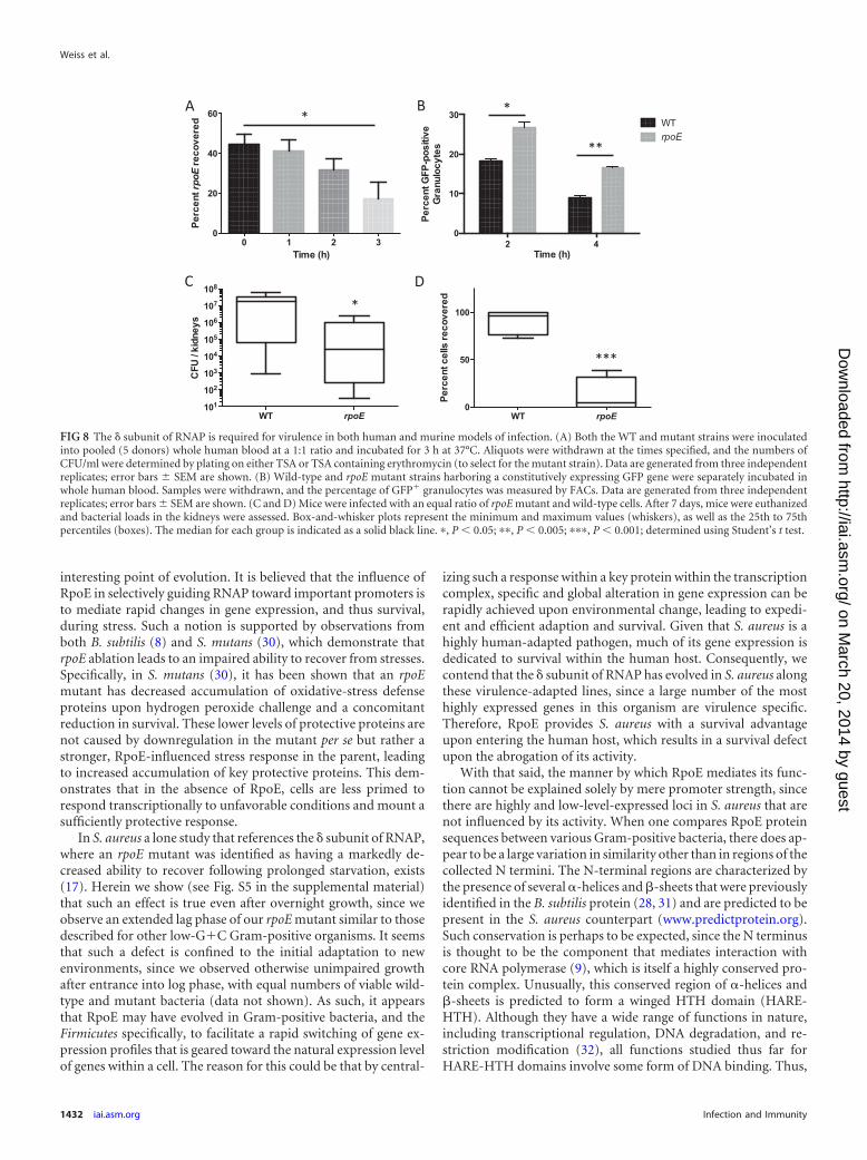
interesting point of evolution. It is believed that the influence ofRpoE in selectively guiding RNAP toward important promoters isto mediate rapid changes in gene expression, and thus survival,during stress. Such a notion is supported by observations fromboth B. subtilis (8) and S. mutans (30), which demonstrate thatrpoE ablation leads to an impaired ability to recover from stresses.Specifically, in S. mutans (30), it has been shown that an rpoEmutant has decreased accumulation of oxidative-stress defenseproteins upon hydrogen peroxide challenge and a concomitantreduction in survival. These lower levels of protective proteins arenot caused by downregulation in the mutant per se but rather astronger, RpoE-influenced stress response in the parent, leadingto increased accumulation of key protective proteins. This dem-onstrates that in the absence of RpoE, cells are less primed torespond transcriptionally to unfavorable conditions and mount asufficiently protective response.
In S. aureus a lone study that references the subunit of RNAP,where an rpoE mutant was identified as having a markedly de-creased ability to recover following prolonged starvation, exists(17). Herein we show (see Fig. S5 in the supplemental material)that such an effect is true even after overnight growth, since weobserve an extended lag phase of our rpoE mutant similar to thosedescribed for other low-GC Gram-positive organisms. It seemsthat such a defect is confined to the initial adaptation to newenvironments, since we observed otherwise unimpaired growthafter entrance into log phase, with equal numbers of viable wild-type and mutant bacteria (data not shown). As such, it appearsthat RpoE may have evolved in Gram-positive bacteria, and theFirmicutes specifically, to facilitate a rapid switching of gene ex-pression profiles that is geared toward the natural expression levelof genes within a cell. The reason for this could be that by central-
izing such a response within a key protein within the transcriptioncomplex, specific and global alteration in gene expression can berapidly achieved upon environmental change, leading to expedi-ent and efficient adaption and survival. Given that S. aureus is ahighly human-adapted pathogen, much of its gene expression isdedicated to survival within the human host. Consequently, wecontend that the subunit of RNAP has evolved in S. aureus alongthese virulence-adapted lines, since a large number of the mosthighly expressed genes in this organism are virulence specific.Therefore, RpoE provides S. aureus with a survival advantageupon entering the human host, which results in a survival defectupon the abrogation of its activity.
With that said, the manner by which RpoE mediates its func-tion cannot be explained solely by mere promoter strength, sincethere are highly and low-level-expressed loci in S. aureus that arenot influenced by its activity. When one compares RpoE proteinsequences between various Gram-positive bacteria, there does ap-pear to be a large variation in similarity other than in regions of thecollected N termini. The N-terminal regions are characterized bythe presence of several -helices and -sheets that were previouslyidentified in the B. subtilis protein (28, 31) and are predicted to bepresent in the S. aureus counterpart (www.predictprotein.org).Such conservation is perhaps to be expected, since the N terminusis thought to be the component that mediates interaction withcore RNA polymerase (9), which is itself a highly conserved pro-tein complex. Unusually, this conserved region of -helices and-sheets is predicted to form a winged HTH domain (HARE-HTH). Although they have a wide range of functions in nature,including transcriptional regulation, DNA degradation, and re-striction modification (32), all functions studied thus far forHARE-HTH domains involve some form of DNA binding. Thus,
2 40
10
20
30
Time (h)
Perc
ent G
FP-p
ositi
ve
Gra
nulo
cyte
s
WTrpoE
WT rpoE0
50
100
Perc
ent c
ells
reco
vere
d
WT rpoE101
102
103
104
105
106
107
108
CFU
/ ki
dney
s
0 1 2 30
20
40
60
Time (h)
Perc
ent rpoE
reco
v ere
d
***
*
*
C D
*
**
FIG 8 The subunit of RNAP is required for virulence in both human and murine models of infection. (A) Both the WT and mutant strains were inoculatedinto pooled (5 donors) whole human blood at a 1:1 ratio and incubated for 3 h at 37°C. Aliquots were withdrawn at the times specified, and the numbers ofCFU/ml were determined by plating on either TSA or TSA containing erythromycin (to select for the mutant strain). Data are generated from three independentreplicates; error bars SEM are shown. (B) Wild-type and rpoE mutant strains harboring a constitutively expressing GFP gene were separately incubated inwhole human blood. Samples were withdrawn, and the percentage of GFP granulocytes was measured by FACs. Data are generated from three independentreplicates; error bars SEM are shown. (C and D) Mice were infected with an equal ratio of rpoE mutant and wild-type cells. After 7 days, mice were euthanizedand bacterial loads in the kidneys were assessed. Box-and-whisker plots represent the minimum and maximum values (whiskers), as well as the 25th to 75thpercentiles (boxes). The median for each group is indicated as a solid black line. , P 0.05; , P 0.005; , P 0.001; determined using Student’s t test.
Weiss et al.
1432 iai.asm.org Infection and Immunity
on March 20, 2014 by guest
http://iai.asm.org/
Dow
nloaded from
even though no direct interaction with nucleic acids has yet beenshown for the subunit of RNAP, a function in recognizing tran-scriptionally relevant DNA features would appear to be possible, ifnot probable, via this HARE-HTH domain. Such a situationwould thus explain the selectivity of RpoE that extends beyondmere promoter strength.
By comparison, the C terminus of the protein shows little con-servation between different species and is in fact the part of theprotein responsible for significant interspecies variation and alter-ations in protein length. Due to its highly acidic nature andstrongly disordered structure, thus far the function of the C ter-minus has remained obscure. Herein we show that deletion of thisC terminus in S. aureus seemingly results in decreased stability ofthe RpoE protein, suggesting its importance for proper foldingand function. In addition, it has been suggested that the C termi-nus may mediate interaction with nucleic acids and have a role inRpoE-dependent displacement of RNA and DNA from the RNAPcomplex (9). As such, it too may influence promoter selection in amanner hitherto not demonstrated.
Another remarkable observation from our RNA-seq analysiswas the simultaneous upregulation of mobile genetic elementswithin the rpoE mutant. Specifically, we observed that most geneson the S. aureus pathogenicity island 5 (SaPI5) and prophageSA3usa displayed strong increases in expression. In the contextof the phage element, it is possible that these effects are mediatedby alterations in transcription of a common regulator encodedwithin the prophage region (33). The shift between lytic and lyso-genic pathways is commonly mediated by an altered abundance ofcompeting regulators, which seek to swing the balance from onelifestyle to the other (reviewed in references 34 and 35). While thespecific molecular details of the lytic/lysogenic decision are notknown for SA3usa, we do observe alterations in regulatory ele-ments on this prophage in the rpoE mutant. Specifically, the phagetranscriptional regulator SAUSA300_1968 is strongly altered inexpression (12-fold). This finding, together with the upregula-tion of most of the adjacent phage genes, leads to the postulationthat SAUSA300_1968 may be a positive and global regulator ofSA3usa gene transcription. In terms of SaPI5, there is limitedunderstanding of regulatory events that modulate expression ofthe genes it encodes. However, it is possible that an event similar tothat suggested for SA3usa occurs, since these elements are be-lieved to have evolved from phages themselves. In line with this,SaPI5 in USA300 contains at least 2 uncharacterized regulatoryelements (33), one of which (SAUSA300_0804) displays elevatedgene expression upon rpoE deletion. As such, it is possible thatupregulation of this element could account for changes in theexpression of the pathogenicity island, as described for SA3usa.
Alternatively, it is possible that RpoE has a wider role in the S.aureus cell in providing cellular immunity against phage infection.Although quite different, immunity systems in bacteria that seekto abrogate phage infections, such as clustered, regularly inter-spaced short palindromic repeat (CRISPR) elements, do exist(36). In such a scenario, RpoE may be involved in silencing foreignelements and therefore protecting the core genome.
In the context of virulence, the various defects observed for therpoE mutant are readily explained. Numerous elements that areknown to play integral roles in S. aureus disease causation, includ-ing Panton-Valentine leukocidin (PVL) (37–40) (reviewed in ref-erence 41), alpha-hemolysin (42–44), and different proteases (21,25), were shown to be decreased in the rpoE mutant. To explore
how these changes are mediated, we first ensured (by Sanger se-quencing and analysis of mRNA transcript fidelity from the RNA-seq reads) that unintended mutations were not present in keyglobal regulators. We next assessed expression of key virulenceregulators and found the agr locus to be unaffected by rpoE de-letion. We did observe downregulation of saeR (2.8-fold) in ourRNA-seq experiment, which may explain some of the effects ob-served (45). Indeed, Nygaard et al. (46) show that deletion ofsaeRS results in a downregulation of various virulence factors,many of which were also affected in the rpoE mutant. However,the limited alteration in saeR expression in the rpoE mutant isunlikely to be the sole reason for the effects observed. Specifically,the changes observed by Nygaard et al. result from complete lossof the saeRS locus (120- and 178-fold downregulation). In ourresults, we see similar effects on virulence factor synthesis, withmuch smaller changes in sae system expression (e.g., for splE,saeRS led to a 1.16-fold change in expression; rpoE led to2.89-fold change). Furthermore, rpoE deletion causes changesin gene expression that are not seen in the saeRS mutant. Forexample, aureolysin and several other virulence factors (e.g., V8protease and PSMs) are either positively affected or unaffected inthe saeRS mutant, while being negatively affected in the rpoE mu-tant. Accordingly, the effects observed in the rpoE mutant appearto be direct and gene specific rather than being caused solely by thedifferential expression of major virulence regulators.
In summary, we herein demonstrate the importance of theRNAP subunit for gene regulation and virulence in S. aureus. Wereveal that rpoE loss results in an uncoupling of well-ordered reg-ulatory circuits, resulting in viable yet less-fit S. aureus cells thatare unable to rapidly adapt to stress, such as that encounteredduring infection. We describe a new layer of transcriptionalregulation in S. aureus that, as in other bacteria, functions in apromoter-specific manner to deploy its global effects and, in thishighly human-adapted pathogenic organism, uniquely targetsthose regions of the genome that are associated with virulence.
ACKNOWLEDGMENTS
We thank Karoly Szekeres for his technical help during FACs analysis.Strain NE646 was obtained through the Network on Antimicrobial
Resistance in Staphylococcus aureus (NARSA) program, supported byNIAID, NIH contract no. HHSN272200700055C. This study was sup-ported in part by grant AI080626 (to L.N.S.) from the National Institute ofAllergies and Infectious Diseases.
REFERENCES1. Sekine S, Tagami S, Yokoyama S. 2012. Structural basis of transcription
by bacterial and eukaryotic RNA polymerases. Curr. Opin. Struct. Biol.22:110 –118. http://dx.doi.org/10.1016/j.sbi.2011.11.006.
2. Pero J, Nelson J, Fox TD. 1975. Highly asymmetric transcription by RNApolymerase containing phage-SP01-induced polypeptides and a new hostprotein. Proc. Natl. Acad. Sci. U. S. A. 72:1589 –1593. http://dx.doi.org/10.1073/pnas.72.4.1589.
3. Achberger EC, Whiteley HR. 1981. The role of the delta peptide of theBacillus subtilis RNA polymerase in promoter selection. J. Biol. Chem.256:7424 –7432.
4. Lampe M, Binnie C, Schmidt R, Losick R. 1988. Cloned gene encodingthe delta subunit of Bacillus subtilis RNA polymerase. Gene 67:13–19.http://dx.doi.org/10.1016/0378-1119(88)90003-0.
5. Achberger EC, Hilton MD, Whiteley HR. 1982. The effect of the deltasubunit on the interaction of Bacillus subtilis RNA polymerase with basesin a SP82 early gene promoter. Nucleic Acids Res. 10:2893–2910. http://dx.doi.org/10.1093/nar/10.9.2893.
6. Juang YL, Helmann JD. 1994. The delta subunit of Bacillus subtilis RNA
RpoE Controls Virulence in Staphylococcus aureus
April 2014 Volume 82 Number 4 iai.asm.org 1433
on March 20, 2014 by guest
http://iai.asm.org/
Dow
nloaded from
polymerase. An allosteric effector of the initiation and core-recyclingphases of transcription. J. Mol. Biol. 239:1–14.
7. Dobinson KF, Spiegelman GB. 1987. Effect of the delta subunit of Bacil-lus subtilis RNA polymerase on initiation of RNA synthesis at two bacte-riophage phi 29 promoters. Biochemistry 26:8206 – 8213. http://dx.doi.org/10.1021/bi00399a028.
8. Rabatinova A, Sanderova H, Jirat Matejckova J, Korelusova J, Sojka L,Barvik I, Papouskova V, Sklenar V, Zidek L, Krasny L. 2013. The deltasubunit of RNA polymerase is required for rapid changes in gene expres-sion and competitive fitness of the cell. J. Bacteriol. 195:2603–2611. http://dx.doi.org/10.1128/JB.00188-13.
9. Lopez de Saro FJ, Woody AY, Helmann JD. 1995. Structural analysis ofthe Bacillus subtilis delta factor: a protein polyanion which displaces RNAfrom RNA polymerase. J. Mol. Biol. 252:189 –202. http://dx.doi.org/10.1006/jmbi.1995.0487.
10. Lopez de Saro FJ, Yoshikawa N, Helmann JD. 1999. Expression, abun-dance, and RNA polymerase binding properties of the delta factor of Ba-cillus subtilis. J. Biol. Chem. 274:15953–15958. http://dx.doi.org/10.1074/jbc.274.22.15953.
11. Gao H, Aronson AI. 2004. The delta subunit of RNA polymerase func-tions in sporulation. Curr. Microbiol. 48:401– 404. http://dx.doi.org/10.1007/s00284-003-4229-8.
12. Xue X, Tomasch J, Sztajer H, Wagner-Dobler I. 2010. The delta subunitof RNA polymerase, RpoE, is a global modulator of Streptococcus mutansenvironmental adaptation. J. Bacteriol. 192:5081–5092. http://dx.doi.org/10.1128/JB.00653-10.
13. Xue X, Sztajer H, Buddruhs N, Petersen J, Rohde M, Talay SR, Wagner-Dobler I. 2011. Lack of the delta subunit of RNA polymerase increasesvirulence related traits of Streptococcus mutans. PLoS One 6:e20075. http://dx.doi.org/10.1371/journal.pone.0020075.
14. Seepersaud R, Needham RH, Kim CS, Jones AL. 2006. Abundance of thedelta subunit of RNA polymerase is linked to the virulence of Streptococ-cus agalactiae. J. Bacteriol. 188:2096 –2105. http://dx.doi.org/10.1128/JB.188.6.2096-2105.2006.
15. Jones AL, Needham RH, Rubens CE. 2003. The Delta subunit of RNApolymerase is required for virulence of Streptococcus agalactiae. In-fect. Immun. 71:4011– 4017. http://dx.doi.org/10.1128/IAI.71.7.4011-4017.2003.
16. Jones AL, Knoll KM, Rubens CE. 2000. Identification of Streptococcusagalactiae virulence genes in the neonatal rat sepsis model using signature-tagged mutagenesis. Mol. Microbiol. 37:1444 –1455. http://dx.doi.org/10.1046/j.1365-2958.2000.02099.x.
17. Watson SP, Antonio M, Foster SJ. 1998. Isolation and characterization ofStaphylococcus aureus starvation-induced, stationary-phase mutants de-fective in survival or recovery. Microbiology 144(Part 11):3159 –3169.http://dx.doi.org/10.1099/00221287-144-11-3159.
18. Carroll RK, Robison TM, Rivera FE, Davenport JE, Jonsson IM, Flo-rczyk D, Tarkowski A, Potempa J, Koziel J, Shaw LN. 2012. Identifica-tion of an intracellular M17 family leucine aminopeptidase that is requiredfor virulence in Staphylococcus aureus. Microbes Infect. 14:989 –999.http://dx.doi.org/10.1016/j.micinf.2012.04.013.
19. Kolar SL, Nagarajan V, Oszmiana A, Rivera FE, Miller HK, DavenportJE, Riordan JT, Potempa J, Barber DS, Koziel J, Elasri MO, Shaw LN.2011. NsaRS is a cell-envelope-stress-sensing two-component system ofStaphylococcus aureus. Microbiology 157:2206 –2219. http://dx.doi.org/10.1099/mic.0.049692-0.
20. Miller HK, Carroll RK, Burda WN, Krute CN, Davenport JE, Shaw LN.2012. The extracytoplasmic function sigma factor sigmaS protects againstboth intracellular and extracytoplasmic stresses in Staphylococcus aureus.J. Bacteriol. 194:4342– 4354. http://dx.doi.org/10.1128/JB.00484-12.
21. Shaw L, Golonka E, Potempa J, Foster SJ. 2004. The role and regulationof the extracellular proteases of Staphylococcus aureus. Microbiology 150:217–228. http://dx.doi.org/10.1099/mic.0.26634-0.
22. Shaw LN, Lindholm C, Prajsnar TK, Miller HK, Brown MC, Golonka E,Stewart GC, Tarkowski A, Potempa J. 2008. Identification and charac-terization of sigma, a novel component of the Staphylococcus aureus stressand virulence responses. PLoS One 3:e3844. http://dx.doi.org/10.1371/journal.pone.0003844.
23. Rivera FE, Miller HK, Kolar SL, Stevens SM, Jr, Shaw LN. 2012. Theimpact of CodY on virulence determinant production in community-associated methicillin-resistant Staphylococcus aureus. Proteomics 12:263–268. http://dx.doi.org/10.1002/pmic.201100298.
24. McClure R, Balasubramanian D, Sun Y, Bobrovskyy M, Sumby P,
Genco CA, Vanderpool CK, Tjaden B. 2013. Computational analysis ofbacterial RNA-Seq data. Nucleic Acids Res. 41:e140. http://dx.doi.org/10.1093/nar/gkt444.
25. Kolar SL, Ibarra JA, Rivera FE, Mootz JM, Davenport JE, Stevens SM,Horswill AR, Shaw LN. 2013. Extracellular proteases are key mediators ofStaphylococcus aureus virulence via the global modulation of virulence-determinant stability. Microbiologyopen 2:18 –34. http://dx.doi.org/10.1002/mbo3.55.
26. Benson MA, Lilo S, Nygaard T, Voyich JM, Torres VJ. 2012. Rot andSaeRS cooperate to activate expression of the staphylococcal superanti-gen-like exoproteins. J. Bacteriol. 194:4355– 4365. http://dx.doi.org/10.1128/JB.00706-12.
27. Nuxoll AS, Halouska SM, Sadykov MR, Hanke ML, Bayles KW, KielianT, Powers R, Fey PD. 2012. CcpA regulates arginine biosynthesis inStaphylococcus aureus through repression of proline catabolism. PLoSPathog. 8:e1003033. http://dx.doi.org/10.1371/journal.ppat.1003033.
28. Motackova V, Sanderova H, Zidek L, Novacek J, Padrta P, Svenkova A,Korelusova J, Jonak J, Krasny L, Sklenar V. 2010. Solution structure ofthe N-terminal domain of Bacillus subtilis delta subunit of RNA polymer-ase and its classification based on structural homologs. Proteins 78:1807–1810. http://dx.doi.org/10.1002/prot.22708.
29. Juang YL, Helmann JD. 1995. Pathway of promoter melting by Bacillussubtilis RNA polymerase at a stable RNA promoter: effects of temperature,delta protein, and sigma factor mutations. Biochemistry 34:8465– 8473.http://dx.doi.org/10.1021/bi00026a030.
30. Xue X, Li J, Wang W, Sztajer H, Wagner-Dobler I. 2012. The globalimpact of the delta subunit RpoE of the RNA polymerase on the proteomeof Streptococcus mutans. Microbiology 158:191–206. http://dx.doi.org/10.1099/mic.0.047936-0.
31. Papouskova V, Kaderavek P, Otrusinova O, Rabatinova A, SanderovaH, Novacek J, Krasny L, Sklenar V, Zidek L. 2013. Structural study of thepartially disordered full-length delta subunit of RNA polymerase fromBacillus subtilis. Chembiochem 14:1772–1779. http://dx.doi.org/10.1002/cbic.201300226.
32. Aravind L, Iyer LM. 2012. The HARE-HTH and associated domains:novel modules in the coordination of epigenetic DNA and protein modi-fications. Cell Cycle 11:119 –131. http://dx.doi.org/10.4161/cc.11.1.18475.
33. Ibarra JA, Perez-Rueda E, Carroll RK, Shaw LN. 2013. Global analysis oftranscriptional regulators in Staphylococcus aureus. BMC Genomics 14:126. http://dx.doi.org/10.1186/1471-2164-14-126.
34. Dodd IB, Shearwin KE, Egan JB. 2005. Revisited gene regulation inbacteriophage lambda. Curr. Opin. Genet. Dev. 15:145–152. http://dx.doi.org/10.1016/j.gde.2005.02.001.
35. Young R. 1992. Bacteriophage lysis: mechanism and regulation. Micro-biol. Rev. 56:430 – 481.
36. Marraffini LA, Sontheimer EJ. 2010. CRISPR interference: RNA-directedadaptive immunity in bacteria and archaea. Nat. Rev. Genet. 11:181–190.http://dx.doi.org/10.1038/nrg2749.
37. Gillet Y, Issartel B, Vanhems P, Fournet JC, Lina G, Bes M, Van-denesch F, Piemont Y, Brousse N, Floret D, Etienne J. 2002. Asso-ciation between Staphylococcus aureus strains carrying gene for Pan-ton-Valentine leukocidin and highly lethal necrotising pneumonia inyoung immunocompetent patients. Lancet 359:753–759. http://dx.doi.org/10.1016/S0140-6736(02)07877-7.
38. Finck-Barbancon V, Duportail G, Meunier O, Colin DA. 1993. Pore for-mation by a two-component leukocidin from Staphylococcus aureus withinthe membrane of human polymorphonuclear leukocytes. Biochim. Biophys.Acta 1182:275–282. http://dx.doi.org/10.1016/0925-4439(93)90069-D.
39. Labandeira-Rey M, Couzon F, Boisset S, Brown EL, Bes M, Benito Y,Barbu EM, Vazquez V, Hook M, Etienne J, Vandenesch F, Bowden MG.2007. Staphylococcus aureus Panton-Valentine leukocidin causes necro-tizing pneumonia. Science 315:1130 –1133. http://dx.doi.org/10.1126/science.1137165.
40. Loffler B, Hussain M, Grundmeier M, Bruck M, Holzinger D, Varga G,Roth J, Kahl BC, Proctor RA, Peters G. 2010. Staphylococcus aureusPanton-Valentine leukocidin is a very potent cytotoxic factor for humanneutrophils. PLoS Pathog. 6:e1000715. http://dx.doi.org/10.1371/journal.ppat.1000715.
41. Yoong P, Torres VJ. 2013. The effects of Staphylococcus aureus leuko-toxins on the host: cell lysis and beyond. Curr. Opin. Microbiol. 16:63– 69.http://dx.doi.org/10.1016/j.mib.2013.01.012.
42. Bramley AJ, Patel AH, O’Reilly M, Foster R, Foster TJ. 1989. Roles of
Weiss et al.
1434 iai.asm.org Infection and Immunity
on March 20, 2014 by guest
http://iai.asm.org/
Dow
nloaded from
alpha-toxin and beta-toxin in virulence of Staphylococcus aureus for themouse mammary gland. Infect. Immun. 57:2489 –2494.
43. McElroy MC, Harty HR, Hosford GE, Boylan GM, Pittet JF, Foster TJ.1999. Alpha-toxin damages the air-blood barrier of the lung in a rat modelof Staphylococcus aureus-induced pneumonia. Infect. Immun. 67:5541–5544.
44. Kebaier C, Chamberland RR, Allen IC, Gao X, Broglie PM, Hall JD,Jania C, Doerschuk CM, Tilley SL, Duncan JA. 2012. Staphylococcusaureus alpha-hemolysin mediates virulence in a murine model of severepneumonia through activation of the NLRP3 inflammasome. J. Infect.Dis. 205:807– 817. http://dx.doi.org/10.1093/infdis/jir846.
45. Voyich JM, Vuong C, DeWald M, Nygaard TK, Kocianova S, Griffith S,Jones J, Iverson C, Sturdevant DE, Braughton KR, Whitney AR, OttoM, DeLeo FR. 2009. The SaeR/S gene regulatory system is essential forinnate immune evasion by Staphylococcus aureus. J. Infect. Dis. 199:1698 –1706. http://dx.doi.org/10.1086/598967.
46. Nygaard TK, Pallister KB, Ruzevich P, Griffith S, Vuong C, Voyich JM.
2010. SaeR binds a consensus sequence within virulence gene promotersto advance USA300 pathogenesis. J. Infect. Dis. 201:241–254. http://dx.doi.org/10.1086/649570.
47. Salisbury V, Hedges RW, Datta N. 1972. Two modes of “curing” trans-missible bacterial plasmids. J. Gen. Microbiol. 70:443– 452. http://dx.doi.org/10.1099/00221287-70-3-443.
48. Sullivan MA, Yasbin RE, Young FE. 1984. New shuttle vectors for Bacillussubtilis and Escherichia coli which allow rapid detection of inserted frag-ments. Gene 29:21–26. http://dx.doi.org/10.1016/0378-1119(84)90161-6.
49. Bae T, Banger AK, Wallace A, Glass EM, Aslund F, Schneewind O,Missiakas DM. 2004. Staphylococcus aureus virulence genes identified bybursa aurealis mutagenesis and nematode killing. Proc. Natl. Acad. Sci.U. S. A. 101:12312–12317. http://dx.doi.org/10.1073/pnas.0404728101.
50. Koprivnjak T, Mlakar V, Swanson L, Fournier B, Peschel A, Weiss JP.2006. Cation-induced transcriptional regulation of the dlt operon ofStaphylococcus aureus. J. Bacteriol. 188:3622–3630. http://dx.doi.org/10.1128/JB.188.10.3622-3630.2006.
RpoE Controls Virulence in Staphylococcus aureus
April 2014 Volume 82 Number 4 iai.asm.org 1435
on March 20, 2014 by guest
http://iai.asm.org/
Dow
nloaded from
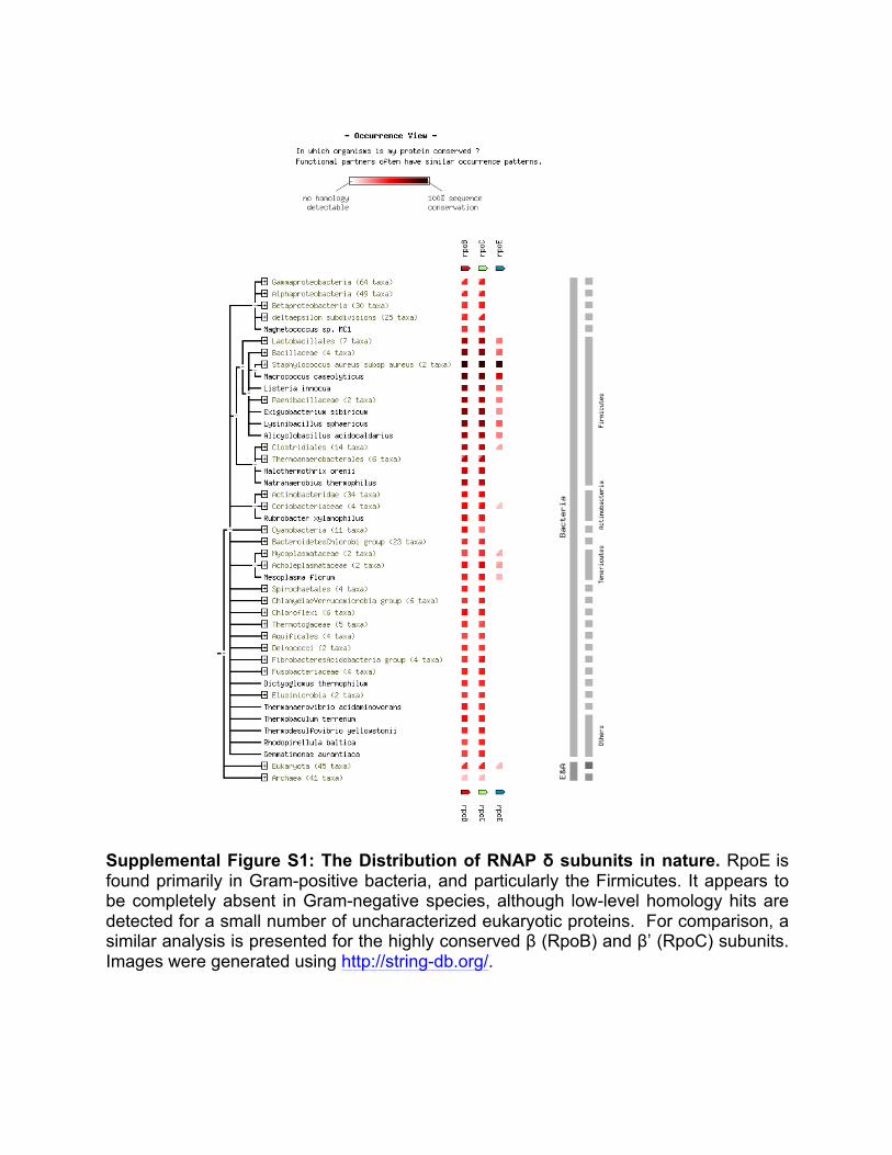
Supplemental Figure S1: The Distribution of RNAP δ subunits in nature. RpoE is found primarily in Gram-positive bacteria, and particularly the Firmicutes. It appears to be completely absent in Gram-negative species, although low-level homology hits are detected for a small number of uncharacterized eukaryotic proteins. For comparison, a similar analysis is presented for the highly conserved β (RpoB) and β’ (RpoC) subunits. Images were generated using http://string-db.org/.
Home • Download • Help • My Data STRING 9.05
Your Input:
rpoB DNA-directed RNA polymerase subunit beta;; DNA-dependent RNA polymerase catalyzes the transcriptionof DNA into RNA using the four ribonucleoside triphosphates as substrates (By similarity) (1183 aa)
rpoC DNA-directed RNA polymerase subunit beta';; DNA-dependent RNA polymerase catalyzes the transcriptionof DNA into RNA using the four ribonucleoside triphosphates as substrates (1198 aa)
rpoE DNA-directed RNA polymerase subunit delta;; Participates in both the initiation and recycling phases oftranscription. In the presence of the delta subunit, RNAP displays an increased specificity of transcription,a decreased affinity for nucleic acids, and an increased efficiency of RNA synthesis because of enhancedrecycling (By similarity) (176 aa)
Supplemental Figure S1: The Distribution of RNAP δ subunits in nature. RpoE is found primarily in Gram-positive bacteria, and particularly the Firmicutes. It appears to be completely absent in Gram-negative species, although low-level homology hits are detected for a small number of uncharacterized eukaryotic proteins. For comparison, a similar analysis is presented for the highly conserved β (RpoB) and β’ (RpoC) subunits. Images were generated using http://string-db.org/.
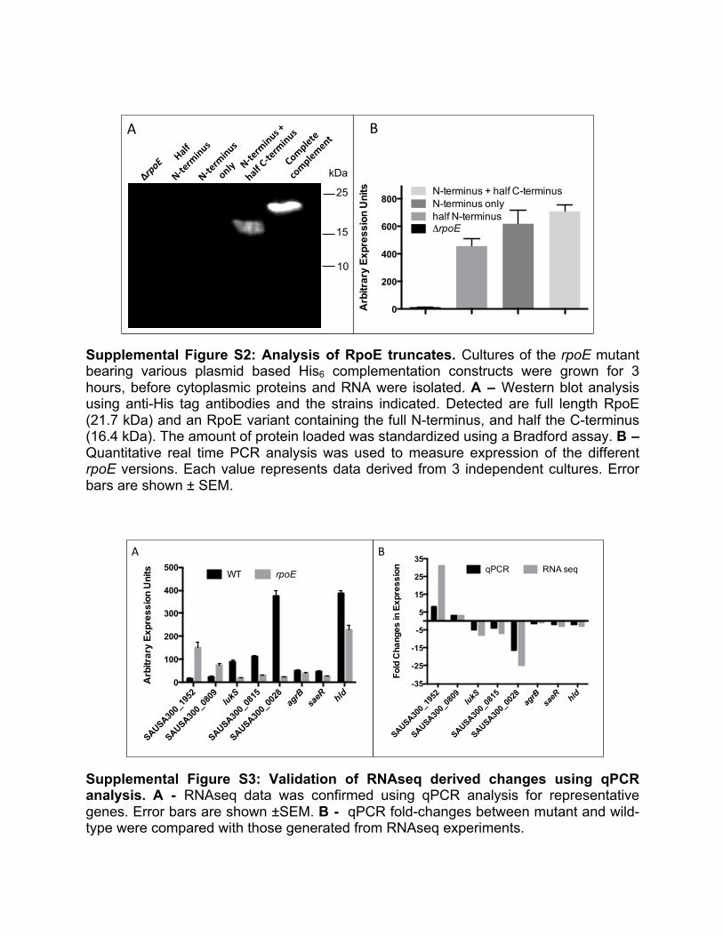
Supplemental Figure S2: Analysis of RpoE truncates. Cultures of the rpoE mutant bearing various plasmid based His6 complementation constructs were grown for 3 hours, before cytoplasmic proteins and RNA were isolated. A – Western blot analysis using anti-His tag antibodies and the strains indicated. Detected are full length RpoE (21.7 kDa) and an RpoE variant containing the full N-terminus, and half the C-terminus (16.4 kDa). The amount of protein loaded was standardized using a Bradford assay. B – Quantitative real time PCR analysis was used to measure expression of the different rpoE versions. Each value represents data derived from 3 independent cultures. Error bars are shown ± SEM.
Supplemental Figure S3: Validation of RNAseq derived changes using qPCR analysis. A - RNAseq data was confirmed using qPCR analysis for representative genes. Error bars are shown ±SEM. B - qPCR fold-changes between mutant and wild-type were compared with those generated from RNAseq experiments.
A B
RpoE
half N
-term
inus
N-term
inus o
nly
N-term
inus +
half
C-te
rmin
us
0
200
400
600
800
Arb
itrar
y E
xpre
ssio
n U
nits
ΔrpoEhalf N-terminusN-terminus onlyN-terminus + half C-terminus
Supplemental Figure S2: Analysis of RpoE truncates. Cultures of the rpoEmutant bearingvarious plasmid based His6 complementa3on constructs were grown for 3 hours, beforecytoplasmic proteins and RNA were isolated. A –Western blot analysis using an3-His tagan3bodies and the strains indicated. Detected are full length RpoE (21.7 kDa) and an RpoEvariant containing the full N-terminus, and half the C-terminus (16.4 kDa). The amount ofprotein loaded was standardized using a Bradford assay. B – Quan3ta3ve real 3me PCRanalysiswasusedtomeasureexpressionofthedifferentrpoEversions.Eachvaluerepresentsdataderivedfrom3independentcultures.Errorbarsareshown±SEM.
kDa
15
25
10
SAUSA300_
1952
SAUSA300_
0809
lukS
SAUSA300_
0815
SAUSA300_
0028
agrB
saeR hld
-35
-25
-15
-5
5
15
25
35
Fold
Cha
nges
in E
xpre
ssio
n qPCR RNA seq
Arb
itrar
y E
xpre
ssio
n U
nits
SAUSA300_
1952
SAUSA300_
0809
lukS
SAUSA300_
0815
SAUSA300_
0028
agrB
saeR hld
0
100
200
300
400
500WT rpoE
Supplemental Figure S3: Valida6on of RNAseq derived changes using qPCR analysis. A -RNAseqdatawasconfirmedusingqPCRanalysisforrepresenta3vegenes.Errorbarsareshown±SEM. B - qPCR fold-changes between mutant and wild-type were compared with thosegeneratedfromRNAseqexperiments.
A B
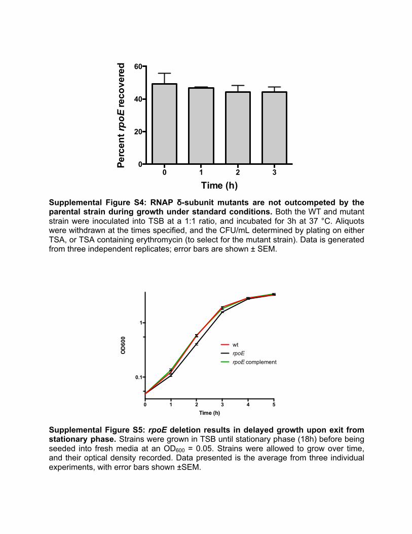
Supplemental Figure S4: RNAP δ-subunit mutants are not outcompeted by the parental strain during growth under standard conditions. Both the WT and mutant strain were inoculated into TSB at a 1:1 ratio, and incubated for 3h at 37 °C. Aliquots were withdrawn at the times specified, and the CFU/mL determined by plating on either TSA, or TSA containing erythromycin (to select for the mutant strain). Data is generated from three independent replicates; error bars are shown ± SEM.
Supplemental Figure S5: rpoE deletion results in delayed growth upon exit from stationary phase. Strains were grown in TSB until stationary phase (18h) before being seeded into fresh media at an OD600 = 0.05. Strains were allowed to grow over time, and their optical density recorded. Data presented is the average from three individual experiments, with error bars shown ±SEM.
Co-culture ΔrpoE and Hou
0 1 2 30
20
40
60
Perc
ent rpoE
reco
vere
d
Time (h)
SupplementalFigureS4:RNAPδ-subunitmutantsarenotoutcompetedbytheparentalstrainduringgrowthunderstandardcondi6ons.BoththeWTandmutantstrainwereinoculatedintoTSBata1:1ra3o,andincubatedfor3hat37°C.Aliquotswerewithdrawnatthe3messpecified,andtheCFU/mLdeterminedbypla3ngoneitherTSA,orTSAcontainingerythromycin(toselectfor the mutant strain). Data is generated from three independent replicates; error bars areshown±SEM.
Supplemental Figure S5: rpoE dele6on results in delayed growth upon exit from sta6onaryphase.Strainsweregrown inTSBun3l sta3onaryphase (18h)beforebeing seeded into freshmedia at anOD600 = 0.05. Strainswere allowed to grow over 3me, and their op3cal densityrecorded. Data presented is the average from three individual experiments, with error barsshown±SEM.
0 1 2 3 4 5
0.1
1
Time (h)
OD
600
wt
rpoE complementrpoE
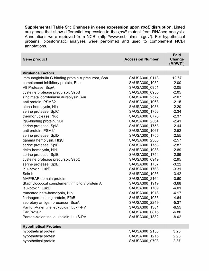
Supplemental Table S1: Changes in gene expression upon rpoE disruption. Listed are genes that show differential expression in the rpoE mutant from RNAseq analysis. Annotations were retrieved from NCBI (http://www.ncbi.nlm.nih.gov/). For hypothetical proteins, bioinformatic analyses were performed and used to complement NCBI annotations.
Gene product Accession Number Fold
Change (Ma/WTb)
Virulence Factors
immunoglobulin G binding protein A precursor, Spa SAUSA300_0113 12.67 complement inhibitory protein, Ehb SAUSA300_1052 -2.00 V8 Protease, SspA SAUSA300_0951 -2.05 cysteine protease precursor, SspB SAUSA300_0950 -2.05 zinc metalloproteinase aureolysin, Aur SAUSA300_2572 -2.07 anti protein, PSMβ2 SAUSA300_1068 -2.15 alpha-hemolysin, Hla SAUSA300_1058 -2.20 serine protease, SplC SAUSA300_1756 -2.34 thermonuclease, Nuc SAUSA300_0776 -2.37 IgG-binding protein, SBI SAUSA300_2364 -2.41 serine protease, SplA SAUSA300_1758 -2.44 anti protein, PSMβ1 SAUSA300_1067 -2.52 serine protease, SplD SAUSA300_1755 -2.55 gamma hemolysin, HlgC SAUSA300_2366 -2.57 serine protease, SplF SAUSA300_1753 -2.87 delta-hemolysin, Hld SAUSA300_1988 -2.89 serine protease, SplE SAUSA300_1754 -2.89 cysteine protease precursor, SspC SAUSA300_0949 -2.95 serine protease, SplB SAUSA300_1757 -3.22 leukotoxin, LukD SAUSA300_1768 -3.31 Scin-b SAUSA300_1056 -3.42 MAP/EAP domain protein SAUSA300_2164 -3.60 Staphylococcal complement inhibitory protein A SAUSA300_1919 -3.68 leukotoxin, LukE SAUSA300_1769 -4.01 truncated beta-hemolysin, Hlb SAUSA300_1918 -4.17 fibrinogen-binding protein, EfbB SAUSA300_1055 -4.64 secretory antigen precursor, SsaA SAUSA300_2249 -5.37 Panton-Valentine leukocidin, LukF-PV SAUSA300_1381 -6.55 Ear Protein SAUSA300_0815 -6.80 Panton-Valentine leukocidin, LukS-PV SAUSA300_1382 -8.02 Hypothetical Proteins
hypothetical protein SAUSA300_2158 3.25 hypothetical protein SAUSA300_1215 2.98 hypothetical protein SAUSA300_0793 2.37
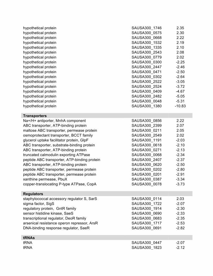
hypothetical protein SAUSA300_1746 2.35 hypothetical protein SAUSA300_0575 2.30 hypothetical protein SAUSA300_0668 2.22 hypothetical protein SAUSA300_1532 2.19 hypothetical protein SAUSA300_1335 2.10 hypothetical protein SAUSA300_2543 2.08 hypothetical protein SAUSA300_0779 2.02 hypothetical protein SAUSA300_0300 -2.25 hypothetical protein SAUSA300_2447 -2.46 hypothetical protein SAUSA300_0471 -2.50 hypothetical protein SAUSA300_0302 -2.64 hypothetical protein SAUSA300_2522 -3.05 hypothetical protein SAUSA300_2524 -3.72 hypothetical protein SAUSA300_0409 -4.67 hypothetical protein SAUSA300_2482 -5.05 hypothetical protein SAUSA300_0048 -5.31 hypothetical protein SAUSA300_1380 -10.83
Transporters Na+/H+ antiporter, MnhA component SAUSA300_0856 2.22
ABC transporter, ATP-binding protein SAUSA300_2399 2.07 maltose ABC transporter, permease protein SAUSA300_0211 2.05 osmoprotectant transporter, BCCT family SAUSA300_2549 2.02 glycerol uptake facilitator protein, GlpF SAUSA300_1191 -2.03 ABC transporter, substrate-binding protein SAUSA300_0618 -2.10 ABC transporter, ATP-binding protein SAUSA300_0271 -2.13 truncated calmodulin exporting ATPase SAUSA300_0068 -2.34 peptide ABC transporter, ATP-binding protein SAUSA300_2407 -2.37 ABC transporter, ATP-binding protein SAUSA300_0620 -2.50 peptide ABC transporter, permease protein SAUSA300_0202 -2.80 peptide ABC transporter, permease protein SAUSA300_0201 -2.91 xanthine permease, PbuX SAUSA300_0387 -3.34 copper-translocating P-type ATPase, CopA SAUSA300_0078 -3.73
Regulators staphylococcal accessory regulator S, SarS SAUSA300_0114 2.03
sigma factor, SigS SAUSA300_1722 -2.07 regulatory protein, GntR family SAUSA300_1914 -2.30 sensor histidine kinase, SaeS SAUSA300_0690 -2.33 transcriptional regulator, DeoR family SAUSA300_0683 -2.35 arsenical resistance operon repressor, ArsR SAUSA300_1717 -2.53 DNA-binding response regulator, SaeR SAUSA300_0691 -2.82
tRNAs tRNA SAUSA300_0447 -2.07
tRNA SAUSA300_1823 -2.12
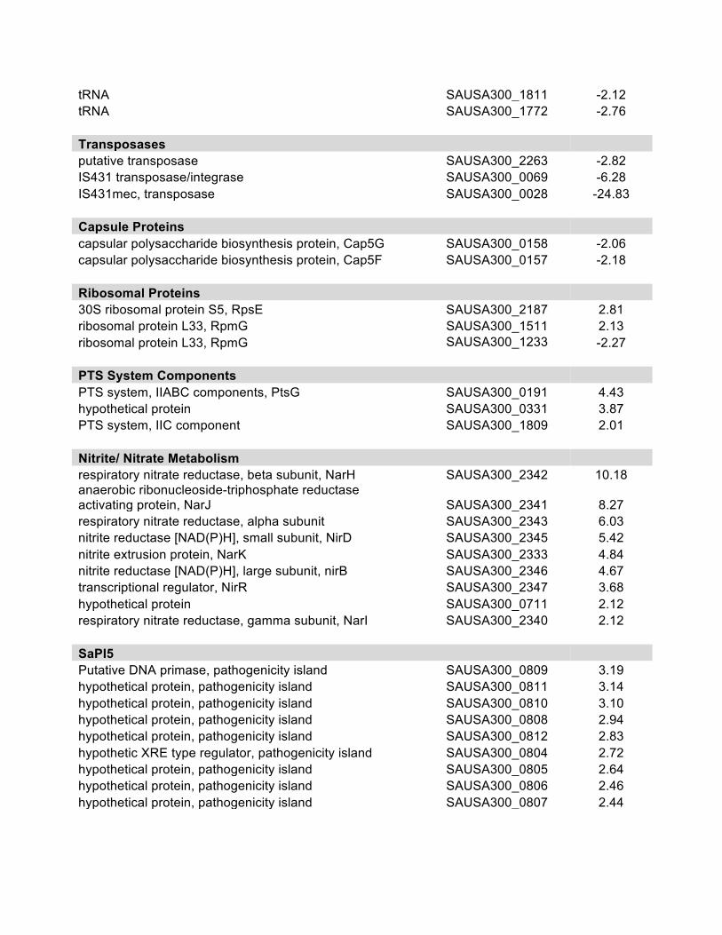
tRNA SAUSA300_1811 -2.12 tRNA SAUSA300_1772 -2.76
Transposases putative transposase SAUSA300_2263 -2.82
IS431 transposase/integrase SAUSA300_0069 -6.28 IS431mec, transposase SAUSA300_0028 -24.83
Capsule Proteins capsular polysaccharide biosynthesis protein, Cap5G SAUSA300_0158 -2.06
capsular polysaccharide biosynthesis protein, Cap5F SAUSA300_0157 -2.18
Ribosomal Proteins 30S ribosomal protein S5, RpsE SAUSA300_2187 2.81
ribosomal protein L33, RpmG SAUSA300_1511 2.13 ribosomal protein L33, RpmG SAUSA300_1233 -2.27
PTS System Components PTS system, IIABC components, PtsG SAUSA300_0191 4.43
hypothetical protein SAUSA300_0331 3.87 PTS system, IIC component SAUSA300_1809 2.01
Nitrite/ Nitrate Metabolism respiratory nitrate reductase, beta subunit, NarH SAUSA300_2342 10.18
anaerobic ribonucleoside-triphosphate reductase activating protein, NarJ SAUSA300_2341 8.27 respiratory nitrate reductase, alpha subunit SAUSA300_2343 6.03 nitrite reductase [NAD(P)H], small subunit, NirD SAUSA300_2345 5.42 nitrite extrusion protein, NarK SAUSA300_2333 4.84 nitrite reductase [NAD(P)H], large subunit, nirB SAUSA300_2346 4.67 transcriptional regulator, NirR SAUSA300_2347 3.68 hypothetical protein SAUSA300_0711 2.12 respiratory nitrate reductase, gamma subunit, NarI SAUSA300_2340 2.12
SaPI5 Putative DNA primase, pathogenicity island SAUSA300_0809 3.19
hypothetical protein, pathogenicity island SAUSA300_0811 3.14 hypothetical protein, pathogenicity island SAUSA300_0810 3.10 hypothetical protein, pathogenicity island SAUSA300_0808 2.94 hypothetical protein, pathogenicity island SAUSA300_0812 2.83 hypothetic XRE type regulator, pathogenicity island SAUSA300_0804 2.72 hypothetical protein, pathogenicity island SAUSA300_0805 2.64 hypothetical protein, pathogenicity island SAUSA300_0806 2.46 hypothetical protein, pathogenicity island SAUSA300_0807 2.44
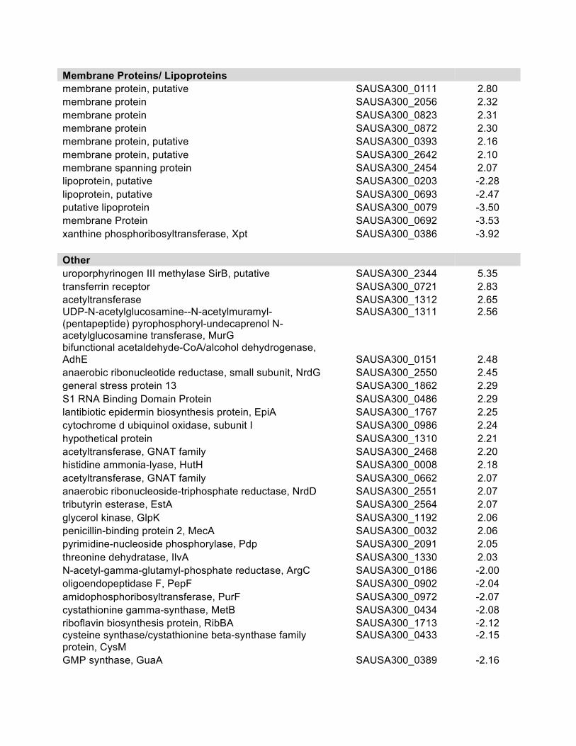
Membrane Proteins/ Lipoproteins membrane protein, putative SAUSA300_0111 2.80
membrane protein SAUSA300_2056 2.32 membrane protein SAUSA300_0823 2.31 membrane protein SAUSA300_0872 2.30 membrane protein, putative SAUSA300_0393 2.16 membrane protein, putative SAUSA300_2642 2.10 membrane spanning protein SAUSA300_2454 2.07 lipoprotein, putative SAUSA300_0203 -2.28 lipoprotein, putative SAUSA300_0693 -2.47 putative lipoprotein SAUSA300_0079 -3.50 membrane Protein SAUSA300_0692 -3.53 xanthine phosphoribosyltransferase, Xpt SAUSA300_0386 -3.92
Other uroporphyrinogen III methylase SirB, putative SAUSA300_2344 5.35
transferrin receptor SAUSA300_0721 2.83 acetyltransferase SAUSA300_1312 2.65 UDP-N-acetylglucosamine--N-acetylmuramyl-(pentapeptide) pyrophosphoryl-undecaprenol N-acetylglucosamine transferase, MurG
SAUSA300_1311
2.56
bifunctional acetaldehyde-CoA/alcohol dehydrogenase, AdhE SAUSA300_0151 2.48 anaerobic ribonucleotide reductase, small subunit, NrdG SAUSA300_2550 2.45 general stress protein 13 SAUSA300_1862 2.29 S1 RNA Binding Domain Protein SAUSA300_0486 2.29 lantibiotic epidermin biosynthesis protein, EpiA SAUSA300_1767 2.25 cytochrome d ubiquinol oxidase, subunit I SAUSA300_0986 2.24 hypothetical protein SAUSA300_1310 2.21 acetyltransferase, GNAT family SAUSA300_2468 2.20 histidine ammonia-lyase, HutH SAUSA300_0008 2.18 acetyltransferase, GNAT family SAUSA300_0662 2.07 anaerobic ribonucleoside-triphosphate reductase, NrdD SAUSA300_2551 2.07 tributyrin esterase, EstA SAUSA300_2564 2.07 glycerol kinase, GlpK SAUSA300_1192 2.06 penicillin-binding protein 2, MecA SAUSA300_0032 2.06 pyrimidine-nucleoside phosphorylase, Pdp SAUSA300_2091 2.05 threonine dehydratase, IlvA SAUSA300_1330 2.03 N-acetyl-gamma-glutamyl-phosphate reductase, ArgC SAUSA300_0186 -2.00 oligoendopeptidase F, PepF SAUSA300_0902 -2.04 amidophosphoribosyltransferase, PurF SAUSA300_0972 -2.07 cystathionine gamma-synthase, MetB SAUSA300_0434 -2.08 riboflavin biosynthesis protein, RibBA SAUSA300_1713 -2.12 cysteine synthase/cystathionine beta-synthase family protein, CysM
SAUSA300_0433
-2.15
GMP synthase, GuaA SAUSA300_0389 -2.16
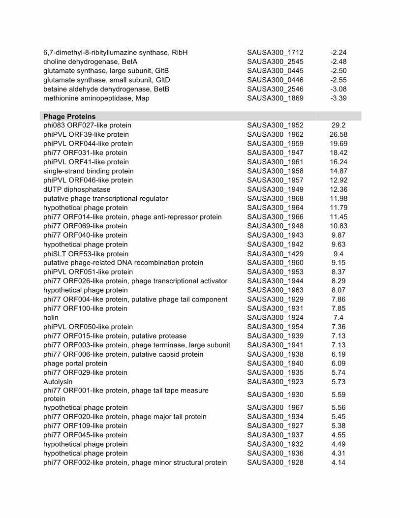
6,7-dimethyl-8-ribityllumazine synthase, RibH SAUSA300_1712 -2.24 choline dehydrogenase, BetA SAUSA300_2545 -2.48 glutamate synthase, large subunit, GltB SAUSA300_0445 -2.50 glutamate synthase, small subunit, GltD SAUSA300_0446 -2.55 betaine aldehyde dehydrogenase, BetB SAUSA300_2546 -3.08 methionine aminopeptidase, Map SAUSA300_1869 -3.39
Phage Proteins phi083 ORF027-like protein SAUSA300_1952 29.2
phiPVL ORF39-like protein SAUSA300_1962 26.58 phiPVL ORF044-like protein SAUSA300_1959 19.69 phi77 ORF031-like protein SAUSA300_1947 18.42 phiPVL ORF41-like protein SAUSA300_1961 16.24 single-strand binding protein SAUSA300_1958 14.87 phiPVL ORF046-like protein SAUSA300_1957 12.92 dUTP diphosphatase SAUSA300_1949 12.36 putative phage transcriptional regulator SAUSA300_1968 11.98 hypothetical phage protein SAUSA300_1964 11.79 phi77 ORF014-like protein, phage anti-repressor protein SAUSA300_1966 11.45 phi77 ORF069-like protein SAUSA300_1948 10.83 phi77 ORF040-like protein SAUSA300_1943 9.87 hypothetical phage protein SAUSA300_1942 9.63 phiSLT ORF53-like protein SAUSA300_1429 9.4 putative phage-related DNA recombination protein SAUSA300_1960 9.15 phiPVL ORF051-like protein SAUSA300_1953 8.37 phi77 ORF026-like protein, phage transcriptional activator SAUSA300_1944 8.29 hypothetical phage protein SAUSA300_1963 8.07 phi77 ORF004-like protein, putative phage tail component SAUSA300_1929 7.86 phi77 ORF100-like protein SAUSA300_1931 7.85 holin SAUSA300_1924 7.4 phiPVL ORF050-like protein SAUSA300_1954 7.36 phi77 ORF015-like protein, putative protease SAUSA300_1939 7.13 phi77 ORF003-like protein, phage terminase, large subunit SAUSA300_1941 7.13 phi77 ORF006-like protein, putative capsid protein SAUSA300_1938 6.19 phage portal protein SAUSA300_1940 6.09 phi77 ORF029-like protein SAUSA300_1935 5.74 Autolysin SAUSA300_1923 5.73 phi77 ORF001-like protein, phage tail tape measure protein SAUSA300_1930 5.59
hypothetical phage protein SAUSA300_1967 5.56 phi77 ORF020-like protein, phage major tail protein SAUSA300_1934 5.45 phi77 ORF109-like protein SAUSA300_1927 5.38 phi77 ORF045-like protein SAUSA300_1937 4.55 hypothetical phage protein SAUSA300_1932 4.49 hypothetical phage protein SAUSA300_1936 4.31 phi77 ORF002-like protein, phage minor structural protein SAUSA300_1928 4.14

phi77 ORF044-like protein SAUSA300_1926 3.26 hypothetical phage protein SAUSA300_1933 3.14 phiPVL ORF17-like protein SAUSA300_1925 2.92 Phage, phiSLT ORF151-like protein, major tail protein SAUSA300_1396 2.9 Phage, phiSLT ORF412-like protein, portal protein SAUSA300_1403 2.8
a USA300 HOU rpoE mutant b USA300 HOU wild-type

129
APPENDIX III
American Society for Microbiology Journals LICENSE
TERMS AND CONDITIONS
Apr 05, 2017
This is a License Agreement between Andy Weiss ("You") and American Society for
Microbiology - Journals ("American Society for Microbiology - Journals") provided by
Copyright Clearance Center ("CCC"). The license consists of your order details, the terms
and conditions provided by American Society for Microbiology - Journals, and the payment
terms and conditions.
All payments must be made in full to CCC. For payment instructions, please see
information listed at the bottom of this form.
License Number 4053651266117
License date Feb 19, 2017
Licensed content publisher American Society for Microbiology Journals
Licensed content title Journal of bacteriology
Licensed content date Jan 1, 1916
Type of Use Thesis/Dissertation
Requestor type Author of requested content
Format Print, Electronic
Portion chapter/article
Title or numeric reference of
the portion(s)
If permission is granted this article would be used as chapter of my
dissertation. The article, as well as the supplementals, would appear
in the appendix of the dissertation
Title of the article or chapter
the portion is from
The ω Subunit Governs RNA Polymerase Stability and
Transcriptional Specificity in Staphylococcus aureus
Editor of portion(s) N/A
Author of portion(s) Andy Weiss, Brittney D. Moore, Miguel H.J. Tremblay, Dale Chaput,
Astrid Kremer and Lindsey N. Shaw
Volume of serial or
monograph.
JB.0045916
Issue, if republishing an
article from a serial
N/A
Page range of the portion
Publication date of portion 31 October 2016 (posted online)
Rights for Main product and any product related to main product
Duration of use Life of current edition
Creation of copies for the
disabled
no
With minor editing privileges yes
For distribution to Worldwide
In the following language(s) Original language of publication
With incidental promotional
use
no
The lifetime unit quantity of
new product
Up to 499
Made available in the
following markets
education
Specified additional
information
The requesting
person/organization is:
Order reference number
Author/Editor
The standard identifier of
New Work
Title of New Work
Publisher of New Work
Expected publication date
Estimated size (pages)
Total (may include CCC user
fee)
Including minor editing privileges.
Andy Weiss
Andy Weiss
University of South Florida
Nonclassical regulators in Staphylococcus aureus
University of South Florida
May 2017
130
0.00 USD
Terms and Conditions
TERMS AND CONDITIONS
The following terms are individual to this publisher:
We ask that you review the copyright information associated with the article that you are
interested in, as some articles in the ASM Journals are covered by creative commons
licenses that would permit free reuse with attribution.
Please note that ASM cannot grant permission to reuse figures or images that are credited to
publications other than ASM journals. For images credited to non-ASM journal publications,
you will need to obtain permission from the journal referenced in the figure or table legend
or credit line before making any use of the image(s) or table(s). ASM is unable to grant
permission to reuse supplemental material as copyright for the supplemental material
remains with the author.
Other Terms and Conditions:
STANDARD TERMS AND CONDITIONS
1. Description of Service; Defined Terms. This Republication License enables the User to
obtain licenses for republication of one or more copyrighted works as described in detail on
the relevant Order Confirmation (the “Work(s)”). Copyright Clearance Center, Inc. (“CCC”)
grants licenses through the Service on behalf of the rightsholder identified on the Order
Confirmation (the “Rightsholder”). “Republication”, as used herein, generally means the
inclusion of a Work, in whole or in part, in a new work or works, also as described on the
Order Confirmation. “User”, as used herein, means the person or entity making such
republication.
2. The terms set forth in the relevant Order Confirmation, and any terms set by the
Rightsholder with respect to a particular Work, govern the terms of use of Works in
connection with the Service. By using the Service, the person transacting for a republication
license on behalf of the User represents and warrants that he/she/it (a) has been duly
authorized by the User to accept, and hereby does accept, all such terms and conditions on
behalf of User, and (b) shall inform User of all such terms and conditions. In the event such
person is a “freelancer” or other third party independent of User and CCC, such party shall
be deemed jointly a “User” for purposes of these terms and conditions. In any event, User
shall be deemed to have accepted and agreed to all such terms and conditions if User
republishes the Work in any fashion.
3. Scope of License; Limitations and Obligations.
3.1 All Works and all rights therein, including copyright rights, remain the sole and
exclusive property of the Rightsholder. The license created by the exchange of an Order
Confirmation (and/or any invoice) and payment by User of the full amount set forth on that
document includes only those rights expressly set forth in the Order Confirmation and in
these terms and conditions, and conveys no other rights in the Work(s) to User. All rights not
expressly granted are hereby reserved.
3.2 General Payment Terms: You may pay by credit card or through an account with us
payable at the end of the month. If you and we agree that you may establish a standing
account with CCC, then the following terms apply: Remit Payment to: Copyright Clearance
Center, 29118 Network Place, Chicago, IL 60673-1291. Payments Due: Invoices are payable
upon their delivery to you (or upon our notice to you that they are available to you for
downloading). After 30 days, outstanding amounts will be subject to a service charge of 1-
1/2% per month or, if less, the maximum rate allowed by applicable law. Unless otherwise
specifically set forth in the Order Confirmation or in a separate written agreement signed by
CCC, invoices are due and payable on “net 30” terms. While User may exercise the rights
licensed immediately upon issuance of the Order Confirmation, the license is automatically
revoked and is null and void, as if it had never been issued, if complete payment for the
license is not received on a timely basis either from User directly or through a payment
agent, such as a credit card company.
3.3 Unless otherwise provided in the Order Confirmation, any grant of rights to User (i) is
“one-time” (including the editions and product family specified in the license), (ii) is non-
exclusive and non-transferable and (iii) is subject to any and all limitations and restrictions
(such as, but not limited to, limitations on duration of use or circulation) included in the
Order Confirmation or invoice and/or in these terms and conditions. Upon completion of the
licensed use, User shall either secure a new permission for further use of the Work(s) or
immediately cease any new use of the Work(s) and shall render inaccessible (such as by
deleting or by removing or severing links or other locators) any further copies of the Work
(except for copies printed on paper in accordance with this license and still in User's stock at
the end of such period).
3.4 In the event that the material for which a republication license is sought includes third
party materials (such as photographs, illustrations, graphs, inserts and similar materials)
which are identified in such material as having been used by permission, User is responsible
for identifying, and seeking separate licenses (under this Service or otherwise) for, any of
such third party materials; without a separate license, such third party materials may not be
used.
3.5 Use of proper copyright notice for a Work is required as a condition of any license
granted under the Service. Unless otherwise provided in the Order Confirmation, a proper
copyright notice will read substantially as follows: “Republished with permission of
[Rightsholder’s name], from [Work's title, author, volume, edition number and year of
copyright]; permission conveyed through Copyright Clearance Center, Inc. ” Such notice
must be provided in a reasonably legible font size and must be placed either immediately
adjacent to the Work as used (for example, as part of a by-line or footnote but not as a
separate electronic link) or in the place where substantially all other credits or notices for the
new work containing the republished Work are located. Failure to include the required notice
results in loss to the Rightsholder and CCC, and the User shall be liable to pay liquidated
damages for each such failure equal to twice the use fee specified in the Order Confirmation,
in addition to the use fee itself and any other fees and charges specified.
3.6 User may only make alterations to the Work if and as expressly set forth in the Order
Confirmation. No Work may be used in any way that is defamatory, violates the rights of
third parties (including such third parties' rights of copyright, privacy, publicity, or other
tangible or intangible property), or is otherwise illegal, sexually explicit or obscene. In
addition, User may not conjoin a Work with any other material that may result in damage to
the reputation of the Rightsholder. User agrees to inform CCC if it becomes aware of any
infringement of any rights in a Work and to cooperate with any reasonable request of CCC
or the Rightsholder in connection therewith.
4. Indemnity. User hereby indemnifies and agrees to defend the Rightsholder and CCC, and
their respective employees and directors, against all claims, liability, damages, costs and
expenses, including legal fees and expenses, arising out of any use of a Work beyond the
scope of the rights granted herein, or any use of a Work which has been altered in any
unauthorized way by User, including claims of defamation or infringement of rights of
copyright, publicity, privacy or other tangible or intangible property.
5. Limitation of Liability. UNDER NO CIRCUMSTANCES WILL CCC OR THE
RIGHTSHOLDER BE LIABLE FOR ANY DIRECT, INDIRECT, CONSEQUENTIAL OR
INCIDENTAL DAMAGES (INCLUDING WITHOUT LIMITATION DAMAGES FOR
LOSS OF BUSINESS PROFITS OR INFORMATION, OR FOR BUSINESS
INTERRUPTION) ARISING OUT OF THE USE OR INABILITY TO USE A WORK,
EVEN IF ONE OF THEM HAS BEEN ADVISED OF THE POSSIBILITY OF SUCH
DAMAGES. In any event, the total liability of the Rightsholder and CCC (including their
respective employees and directors) shall not exceed the total amount actually paid by User
for this license. User assumes full liability for the actions and omissions of its principals,
employees, agents, affiliates, successors and assigns.
6. Limited Warranties. THE WORK(S) AND RIGHT(S) ARE PROVIDED “AS IS”. CCC
HAS THE RIGHT TO GRANT TO USER THE RIGHTS GRANTED IN THE ORDER
CONFIRMATION DOCUMENT. CCC AND THE RIGHTSHOLDER DISCLAIM ALL
OTHER WARRANTIES RELATING TO THE WORK(S) AND RIGHT(S), EITHER
EXPRESS OR IMPLIED, INCLUDING WITHOUT LIMITATION IMPLIED
WARRANTIES OF MERCHANTABILITY OR FITNESS FOR A PARTICULAR
PURPOSE. ADDITIONAL RIGHTS MAY BE REQUIRED TO USE ILLUSTRATIONS,
GRAPHS, PHOTOGRAPHS, ABSTRACTS, INSERTS OR OTHER PORTIONS OF THE
WORK (AS OPPOSED TO THE ENTIRE WORK) IN A MANNER CONTEMPLATED
BY USER; USER UNDERSTANDS AND AGREES THAT NEITHER CCC NOR THE
RIGHTSHOLDER MAY HAVE SUCH ADDITIONAL RIGHTS TO GRANT.
7. Effect of Breach. Any failure by User to pay any amount when due, or any use by User of
a Work beyond the scope of the license set forth in the Order Confirmation and/or these
terms and conditions, shall be a material breach of the license created by the Order
Confirmation and these terms and conditions. Any breach not cured within 30 days of
written notice thereof shall result in immediate termination of such license without further
notice. Any unauthorized (but licensable) use of a Work that is terminated immediately upon
notice thereof may be liquidated by payment of the Rightsholder's ordinary license price
therefor; any unauthorized (and unlicensable) use that is not terminated immediately for any
reason (including, for example, because materials containing the Work cannot reasonably be
recalled) will be subject to all remedies available at law or in equity, but in no event to a
payment of less than three times the Rightsholder's ordinary license price for the most
closely analogous licensable use plus Rightsholder's and/or CCC's costs and expenses
incurred in collecting such payment.
8. Miscellaneous.
8.1 User acknowledges that CCC may, from time to time, make changes or additions to the
Service or to these terms and conditions, and CCC reserves the right to send notice to the
User by electronic mail or otherwise for the purposes of notifying User of such changes or
additions; provided that any such changes or additions shall not apply to permissions already
secured and paid for.
8.2 Use of User-related information collected through the Service is governed by CCC’s
privacy policy, available online here:
http://www.copyright.com/content/cc3/en/tools/footer/privacypolicy.html.
8.3 The licensing transaction described in the Order Confirmation is personal to User.
Therefore, User may not assign or transfer to any other person (whether a natural person or
an organization of any kind) the license created by the Order Confirmation and these terms
and conditions or any rights granted hereunder; provided, however, that User may assign
such license in its entirety on written notice to CCC in the event of a transfer of all or
substantially all of User’s rights in the new material which includes the Work(s) licensed
under this Service.
8.4 No amendment or waiver of any terms is binding unless set forth in writing and signed
by the parties. The Rightsholder and CCC hereby object to any terms contained in any
writing prepared by the User or its principals, employees, agents or affiliates and purporting
to govern or otherwise relate to the licensing transaction described in the Order
Confirmation, which terms are in any way inconsistent with any terms set forth in the Order
Confirmation and/or in these terms and conditions or CCC's standard operating procedures,
whether such writing is prepared prior to, simultaneously with or subsequent to the Order
Confirmation, and whether such writing appears on a copy of the Order Confirmation or in a
separate instrument.
8.5 The licensing transaction described in the Order Confirmation document shall be
governed by and construed under the law of the State of New York, USA, without regard to
the principles thereof of conflicts of law. Any case, controversy, suit, action, or proceeding
arising out of, in connection with, or related to such licensing transaction shall be brought, at

CCC's sole discretion, in any federal or state court located in the County of New York, State
of New York, USA, or in any federal or state court whose geographical jurisdiction covers
the location of the Rightsholder set forth in the Order Confirmation. The parties expressly
submit to the personal jurisdiction and venue of each such federal or state court.If you have
any comments or questions about the Service or Copyright Clearance Center, please contact
us at 978-750-8400 or send an e-mail to [email protected].
v 1.1
Questions? [email protected] or +18552393415 (toll free in the US) or
+19786462777.
The Subunit Governs RNA PolymeraseStability and Transcriptional Specificityin Staphylococcus aureus
Andy Weiss, Brittney D. Moore, Miguel H. J. Tremblay, Dale Chaput,Astrid Kremer, Lindsey N. ShawUniversity of South Florida, Department of Cell Biology, Microbiology and Molecular Biology, Tampa, Florida,USA
ABSTRACT Staphylococcus aureus is a major human pathogen that causes infectionin a wide variety of sites within the human body. Its ability to adapt to the humanhost and to produce a successful infection requires precise orchestration of gene ex-pression. While DNA-dependent RNA polymerase (RNAP) is generally well character-ized, the roles of several small accessory subunits within the complex have yet to befully explored. This is particularly true for the omega ( or RpoZ) subunit, which hasbeen extensively studied in Gram-negative bacteria but largely neglected in Gram-positive counterparts. In Escherichia coli, it has been shown that ppGpp binding, andthus control of the stringent response, is facilitated by . Interestingly, key residuesthat facilitate ppGpp binding by are not conserved in S. aureus, and consequently,survival under starvation conditions is unaffected by rpoZ deletion. Further to this,-lacking strains of S. aureus display structural changes in the RNAP complex, whichresult from increased degradation and misfolding of the = subunit, alterations in
and factor abundance, and a general dissociation of RNAP in the absence of .Through RNA sequencing analysis we detected a variety of transcriptional changesin the rpoZ-deficient strain, presumably as a response to the negative effects of
depletion on the transcription machinery. These transcriptional changes translated toan impaired ability of the rpoZ mutant to resist stress and to fully form a biofilm.Collectively, our data underline, for the first time, the importance of for RNAP sta-bility, function, and cellular physiology in S. aureus.
IMPORTANCE In order for bacteria to adjust to changing environments, such aswithin the host, the transcriptional process must be tightly controlled. Transcriptionis carried out by DNA-dependent RNA polymerase (RNAP). In addition to its majorsubunits (2=) a fifth, smaller subunit, , is present in all forms of life. Althoughthis small subunit is well studied in eukaryotes and Gram-negative bacteria, only lim-ited information is available for Gram-positive and pathogenic species. In this study,we investigated the structural and functional importance of , revealing key roles insubunit folding/stability, complex assembly, and maintenance of transcriptional in-tegrity. Collectively, our data underline, for the first time, the importance of forRNAP function and cellular harmony in S. aureus.
KEYWORDS RNA polymerase subunit omega, RpoZ, Staphylococcus aureus, generegulation
Transcription in all forms of life is a tightly controlled process, necessitated by theessentiality of correct temporal and spatial expression of genes for survival. All
transcriptional activity within a cell is maintained by the DNA-dependent RNA poly-merase (RNAP). This multiprotein complex is structurally and functionally similar indistant forms of life, displaying only minor variations in composition, e.g., the presence/absence of certain subunits (1, 2). In bacteria RNAP consists of four main subunits, i.e.,
Received 10 June 2016 Accepted 26 October2016
Accepted manuscript posted online 31October 2016
Citation Weiss A, Moore BD, Tremblay MHJ,Chaput D, Kremer A, Shaw LN. 2017. The ωsubunit governs RNA polymerase stability andtranscriptional specificity in Staphylococcusaureus. J Bacteriol 199:e00459-16. https://doi.org/10.1128/JB.00459-16.
Editor Victor J. DiRita, Michigan StateUniversity
Copyright © 2016 American Society forMicrobiology. All Rights Reserved.
Address correspondence to Lindsey N. Shaw,[email protected].
RESEARCH ARTICLE
crossm
January 2017 Volume 199 Issue 2 e00459-16 jb.asm.org 1Journal of Bacteriology
on Decem
ber 28, 2016 by guesthttp://jb.asm
.org/D
ownloaded from
two (RpoA) subunits and one subunit each of (RpoB) and = (RpoC), forming the2= complex. Together they facilitate transcriptional elongation, but they require a
factor to initiate the process. Interestingly, a large number of bacteria, and particularlythe Firmicutes, possess several other accessory RNAP subunits (3–6). These are consid-erably smaller than the major subunits, ranging from 8.5 to 21.5 kDa, and include the (RpoE), (RpoY), and (RpoZ) subunits. Deletion of these subunits does not result inlethality for the cell, and thus their diminutive size and nonessential nature haveresulted in their being classified as the “small accessory subunits” (reviewed in refer-ence 6). Nevertheless, for the factor it has been shown that, in various species,deletion is accompanied by a deregulation of the transcriptional process, leading todecreased fitness and impaired virulence in pathogenic organisms (7, 8). While there isan extensive history of research for , has only recently been described as an RNAPsubunit, with only a single study thus far performed in Bacillus subtilis (5), whichsuggested a role in phage immunity. While the presence of these two subunits is largelyconfined to the Firmicutes, homologs of , the smallest of the three subunits, can befound not only in bacteria but also in eukaryotes (RPB6) and archaea (RpoK) (6, 9).Although this conservation might suggest a vital role and perhaps similar functionacross widely different species, there are in fact marked differences in how this subunitinfluences cells across the various kingdoms (6, 10). Most strikingly is the observationthat while it is accessory in bacteria, the subunit is essential in eukaryotic organisms(11).
The majority of studies on have been carried out in Gram-negative bacteria, witha focus on the model organism Escherichia coli (10). In this organism it has been shownthat influences the transcriptional machinery, and thus the transcriptional process, ina variety of ways. Most notably, is known to interact with = to ensure correct foldingof the subunit, as well as to facilitate docking to the 2 complex (9, 12–14). Accord-ingly, deletion of leads to misfolding as well as degradation of = (13, 15), which isalso observed in Mycobacterium smegmatis (16). The crystal structure of in complexwith the other RNAP subunits of both E. coli and Thermus aquaticus has been solvedand confirms the binding of to = (17, 18). Interestingly, these structures also revealspecies-specific differences in the interaction of these two proteins, again highlightingthe heterogeneous nature of function in different organisms. In contrast, no suchcrystal structure is available for , or RNAP at large, in Gram-positive organisms.
Quite strikingly, in terms of functional difference, is the finding that in E. coli, thestringent response, and thus adaption to nutrient-limiting conditions, is dependent on. Specifically, the stringent response-inducing molecule ppGpp is recognized by andbinds to the interface of = and (19–21), which in turn leads to the adjustment oftranscriptional profiles to promote survival under nutrient-limiting conditions. Whilethis role is true for E. coli, the protein in B. subtilis has been suggested to have no suchrole in the stringent response, due to an alternative mechanism of ppGpp recognition.Instead of binding to , the adaption to limiting conditions in B. subtilis is mediated byppGpp-induced alterations of GTP concentrations within the cell (22, 23). Subsequently,these changes lead to alterations in gene expression, driven by the sensitivity of certainpromoters to GTP availability as an initiating nucleotide. In line with this model, whereonset of the stringent response does not require the interaction of ppGpp with theRNAP complex, is the observation that the conserved E. coli residues required forppGpp binding to RNAP are largely absent in the B. subtilis and = subunits (20).
The final major function of described in the literature is a putative role infacilitating factor biding to the RNAP complex. For E. coli and cyanobacteria, it hasbeen reported that depletion of can lead to increased binding of alterative factorsand in turn to increased expression of genes within alternative factor regulons (24,25). Again, structural differences within the -depleted RNAP have been implicated inthis alteration of factor affinity for the complex. As with many of the other
phenotypes, no in-depth studies have been performed in Gram-positive bacteriaregarding this role, further underscoring the need to characterize this diverse protein.
Weiss et al. Journal of Bacteriology
January 2017 Volume 199 Issue 2 e00459-16 jb.asm.org 2
on Decem
ber 28, 2016 by guesthttp://jb.asm
.org/D
ownloaded from
For Gram-positive organisms, only a limited number of studies exist, detailing a fewphenotypic effects resulting from the abrogation of activity. Indeed, none of thesestudies have unraveled the molecular basis for alterations in mutant strains, meaningthat the role of in Gram-positive species is still relatively elusive. Those effects thathave been detailed for rpoZ mutants include alterations in cell wall morphology, cellmotility, protein secretion, and biofilm formation (16, 26–29). Importantly, the role ofthis subunit in the virulence of pathogenic species has yet to be evaluated. Therefore,in this study we explored the role of in Staphylococcus aureus, demonstrating that itis not involved in the stringent response but instead mediates structural integrity of theRNAP complex. Deletion of this factor leads to individual RNAP subunit degradationand an induction of cellular stress responses. The latter effect was characterized byglobal transcriptional analyses, revealing that a significant number of the observedchanges were due, at least in part, to altered factor abundance in the RNAP complex.Finally, we demonstrate that deletion of rpoZ influences the ability of S. aureus to formbiofilms, a process that mediates persistent infections and the capacity to resistantibiotic treatment. We suggest that collectively, our data underline the importance of for RNAP stability, function, and cellular physiology in S. aureus.
RESULTS is cotranscribed with gmk and is highly expressed throughout growth.
Analysis of RNA sequencing data previously generated by our laboratory (31) revealsthat the -encoding gene, rpoZ, is highly expressed during all phases of growth and isseemingly cotranscribed with its upstream gene, gmk, encoding a guanylate kinase (seeFig. S2A in the supplemental material). To validate the latter observation, we performedNorthern blot analysis on RNA extracted from wild-type S. aureus cells grown tomid-exponential phase using rpoZ-specific probes. In so doing, a band of 1100nucleotides (nt) was observed, indicating that rpoZ and gmk are indeed organized in abicistronic operon (Fig. S2B). In order to study the role of in S. aureus, we next createdan unmarked deletion mutant, removing the majority of the rpoZ-encoding gene andleaving gmk intact. Validation of this arrangement and that no deleterious effects wereobserved on gmk expression was obtained by RNA sequencing analysis of the mutantstrain and by Northern blotting (Fig. S2A and B). RNA sequencing data for the-depleted strain showed the expected deletion of the rpoZ gene but confirmed thatthere were no unintended effects on transcript abundance for gmk (see Table S1 in thesupplemental material).
The S. aureus and = proteins lack conserved residues required for ppGppbinding. In E. coli the interaction of ppGpp with RNAP is mediated by conservedresidues within the and = subunits, thus controlling the stringent response. Suchresidues are absent in B. subtilis, suggesting an alternate mechanism for stringentresponse regulation in Gram-positive organisms (20, 32). In corroboration of this,alignment analysis of and = subunits from E. coli, B. subtilis, and S. aureus (Fig. 1A)reveals that, although there is partial conservation of the = and proteins for all threeorganisms, the residues for ppGpp binding are almost entirely absent (a single asparticacid is conserved in = region 2) at the primary sequence level and in the biochemicalcharacteristics of each amino acid. This finding is further validated by the observationthat growth of a ΔrpoZ strain is not impaired under standard, nutrient-limiting, orstringent response-inducing conditions. Specifically, in addition to growth in complexmedium (tryptic soy broth [TSB]) (Fig. 1B), we investigated growth during amino acidlimitation (Fig. 1C) as well as in amino acid-limiting medium completely depleted ofvaline and leucine (Fig. 1D). Importantly, the rpoZ mutant showed growth rates similarto those of the wild type in both TSB and amino acid-limiting medium and only a minorgrowth defect in medium devoid of valine and leucine. This defect, however, suggestsan impaired ability to adapt to changing growth conditions rather than a stringentresponse defect, as stringent response-deficient mutants display a characteristic stallingof growth during the stringent response, rather than delayed growth (33). To furthervalidate this finding, we assessed sensitivity to mupirocin of the rpoZ mutant alongside
The Subunit of RNAP in Staphylococcus aureus Journal of Bacteriology
January 2017 Volume 199 Issue 2 e00459-16 jb.asm.org 3
on Decem
ber 28, 2016 by guesthttp://jb.asm
.org/D
ownloaded from
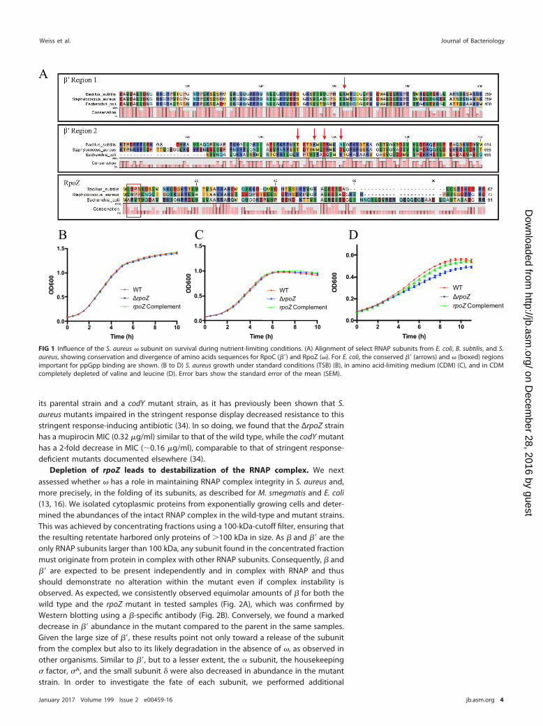
its parental strain and a codY mutant strain, as it has previously been shown that S.aureus mutants impaired in the stringent response display decreased resistance to thisstringent response-inducing antibiotic (34). In so doing, we found that the ΔrpoZ strainhas a mupirocin MIC (0.32 g/ml) similar to that of the wild type, while the codY mutanthas a 2-fold decrease in MIC (0.16 g/ml), comparable to that of stringent response-deficient mutants documented elsewhere (34).
Depletion of rpoZ leads to destabilization of the RNAP complex. We nextassessed whether has a role in maintaining RNAP complex integrity in S. aureus and,more precisely, in the folding of its subunits, as described for M. smegmatis and E. coli(13, 16). We isolated cytoplasmic proteins from exponentially growing cells and deter-mined the abundances of the intact RNAP complex in the wild-type and mutant strains.This was achieved by concentrating fractions using a 100-kDa-cutoff filter, ensuring thatthe resulting retentate harbored only proteins of 100 kDa in size. As and = are theonly RNAP subunits larger than 100 kDa, any subunit found in the concentrated fractionmust originate from protein in complex with other RNAP subunits. Consequently, and= are expected to be present independently and in complex with RNAP and thusshould demonstrate no alteration within the mutant even if complex instability isobserved. As expected, we consistently observed equimolar amounts of for both thewild type and the rpoZ mutant in tested samples (Fig. 2A), which was confirmed byWestern blotting using a -specific antibody (Fig. 2B). Conversely, we found a markeddecrease in = abundance in the mutant compared to the parent in the same samples.Given the large size of =, these results point not only toward a release of the subunitfrom the complex but also to its likely degradation in the absence of , as observed inother organisms. Similar to =, but to a lesser extent, the subunit, the housekeeping factor, A, and the small subunit were also decreased in abundance in the mutantstrain. In order to investigate the fate of each subunit, we performed additional
0 2 4 6 8 100.0
0.2
0.4
0.6
Time (h)O
D60
00 2 4 6 8 10
0.0
0.5
1.0
1.5
OD
600
0 2 4 6 8 100.0
0.5
1.0
1.5
Time (h)
OD
600
WTrpoZ
rpoZ Complement
WTrpoZ
rpoZ Complement
WTrpoZ
rpoZ Complement
C D B
A
Time (h)
FIG 1 Influence of the S. aureus subunit on survival during nutrient-limiting conditions. (A) Alignment of select RNAP subunits from E. coli, B. subtilis, and S.aureus, showing conservation and divergence of amino acids sequences for RpoC (=) and RpoZ (). For E. coli, the conserved = (arrows) and (boxed) regionsimportant for ppGpp binding are shown. (B to D) S. aureus growth under standard conditions (TSB) (B), in amino acid-limiting medium (CDM) (C), and in CDMcompletely depleted of valine and leucine (D). Error bars show the standard error of the mean (SEM).
Weiss et al. Journal of Bacteriology
January 2017 Volume 199 Issue 2 e00459-16 jb.asm.org 4
on Decem
ber 28, 2016 by guesthttp://jb.asm
.org/D
ownloaded from
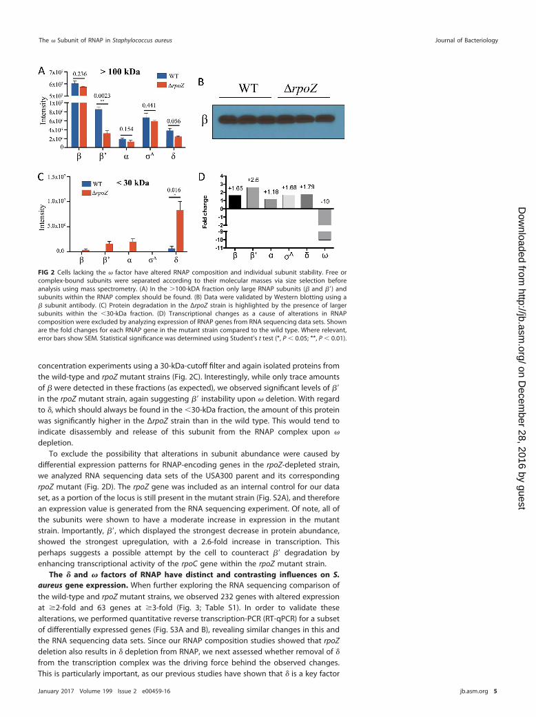
concentration experiments using a 30-kDa-cutoff filter and again isolated proteins fromthe wild-type and rpoZ mutant strains (Fig. 2C). Interestingly, while only trace amountsof were detected in these fractions (as expected), we observed significant levels of =in the rpoZ mutant strain, again suggesting = instability upon deletion. With regardto , which should always be found in the 30-kDa fraction, the amount of this proteinwas significantly higher in the ΔrpoZ strain than in the wild type. This would tend toindicate disassembly and release of this subunit from the RNAP complex upon
depletion.To exclude the possibility that alterations in subunit abundance were caused by
differential expression patterns for RNAP-encoding genes in the rpoZ-depleted strain,we analyzed RNA sequencing data sets of the USA300 parent and its correspondingrpoZ mutant (Fig. 2D). The rpoZ gene was included as an internal control for our dataset, as a portion of the locus is still present in the mutant strain (Fig. S2A), and thereforean expression value is generated from the RNA sequencing experiment. Of note, all ofthe subunits were shown to have a moderate increase in expression in the mutantstrain. Importantly, =, which displayed the strongest decrease in protein abundance,showed the strongest upregulation, with a 2.6-fold increase in transcription. Thisperhaps suggests a possible attempt by the cell to counteract = degradation byenhancing transcriptional activity of the rpoC gene within the rpoZ mutant strain.
The and factors of RNAP have distinct and contrasting influences on S.aureus gene expression. When further exploring the RNA sequencing comparison ofthe wild-type and rpoZ mutant strains, we observed 232 genes with altered expressionat 2-fold and 63 genes at 3-fold (Fig. 3; Table S1). In order to validate thesealterations, we performed quantitative reverse transcription-PCR (RT-qPCR) for a subsetof differentially expressed genes (Fig. S3A and B), revealing similar changes in this andthe RNA sequencing data sets. Since our RNAP composition studies showed that rpoZdeletion also results in depletion from RNAP, we next assessed whether removal of
from the transcription complex was the driving force behind the observed changes.This is particularly important, as our previous studies have shown that is a key factor
FIG 2 Cells lacking the factor have altered RNAP composition and individual subunit stability. Free orcomplex-bound subunits were separated according to their molecular masses via size selection beforeanalysis using mass spectrometry. (A) In the 100-kDA fraction only large RNAP subunits ( and =) andsubunits within the RNAP complex should be found. (B) Data were validated by Western blotting using a subunit antibody. (C) Protein degradation in the ΔrpoZ strain is highlighted by the presence of largersubunits within the 30-kDa fraction. (D) Transcriptional changes as a cause of alterations in RNAPcomposition were excluded by analyzing expression of RNAP genes from RNA sequencing data sets. Shownare the fold changes for each RNAP gene in the mutant strain compared to the wild type. Where relevant,error bars show SEM. Statistical significance was determined using Student’s t test (*, P 0.05; **, P 0.01).
The Subunit of RNAP in Staphylococcus aureus Journal of Bacteriology
January 2017 Volume 199 Issue 2 e00459-16 jb.asm.org 5
on Decem
ber 28, 2016 by guesthttp://jb.asm
.org/D
ownloaded from
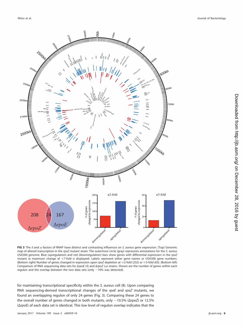
for maintaining transcriptional specificity within the S. aureus cell (8). Upon comparingRNA sequencing-derived transcriptional changes of the rpoE and rpoZ mutants, wefound an overlapping regulon of only 24 genes (Fig. 3). Comparing these 24 genes tothe overall number of genes changed in both mutants, only 10.5% (ΔrpoZ) or 12.5%(ΔrpoE) of each data set is identical. This low level of regulon overlap indicates that the
0kb
100k
b
200k
b
300k
b
400kb
500kb
600kb
700kb
800kb
900kb
1000kb
1100kb
1200kb1300kb
1400kb
1500
kb1600
kb
1700
kb
1800
kb
1900kb
2000kb
2100kb
2200kb
2300kb
2400kb
2500kb
2600kb
2700kb
2800kb
4.5S_RNA
asp23
cap5
E
sdaAB
mal
R
mur
Q
mec
A
putApckA
ptsG
purD
nsaS
purS
purN
hlgA
purH
pknBpt
sG
nusA
plsX
purM
purB
glpF
gntR
treP treC
gntK
treR
xseB
gltA
gntP
typA
rpsO
deoD
rpmB
rumA
rpsB
rplA rpoC
rplM rpsI
rluB
rplT
rpmJ
rpsT
recA
rpsD
fhuA
0045
0431
0170
0208
0777
0471
0575
0538
0209
0229
0332
0816
0465
0941
0210
0621
0392
0703
0328 03
31
0662
0954
0724
0425
0779
0945
0424
0095
0821
0033
0211
0023
0814
0192
0531
0474
0922
0642
0464
0090
0068
2493
2132
2324
246624552408
2037
acpP
2639
2310
2407
2131
acsAopuD
2484
2448
2409
250325302527
2435
2512
lexA
lytN
leuD
sbrB
comK
scrR
sdhC
sarA
clpB
sucB
sdhB
scpB
sucA
infC
1205
1456
1606
1702
1981
1739
1959
1335
1742
1344
1943
1746 1740
1966
1180
1933
1059
1962
1198
1058
1857
1549
17431759
1247
1685
19151042
1701
1582
1968
1986
gap
ald
lip
icd
lukS
-PV
lukF
-PV
nrdDnrdG
panB
pfkA
pfoR
budA
nirR tatC
gpmA
rpoZ
folP
0043
041001
740048
0343
0952
0233
0614
0175
0613
0883
0234
0443
0110
0173
0176
0985
07950805
0177
0824
0119
0135
05342087
2557estA
2234
2475
2355
argR
2316
24532509
2454
2236
splB splE
194519521951
1380
1412
1827
1230
18231748
1948
18631926
1583
1894
pcp
nuc
aur
sak
167
ΔrpoZ
208 24
ΔrpoE
2-fold
# of
gen
es c
hang
ed in
exp
ress
ion
downre
gulat
ed
upreg
ulated
0
50
100
150
200 3-fold
# of
gen
es c
hang
ed in
exp
ress
ion
downre
gulat
ed
upreg
ulated
0
20
40
60
FIG 3 The and factors of RNAP have distinct and contrasting influences on S. aureus gene expression. (Top) Genomicmap of altered transcription in the rpoZ mutant strain. The outermost circle (gray) represents annotations for the S. aureusUSA300 genome. Blue (upregulation) and red (downregulation) bars show genes with differential expression in the rpoZmutant (a maximum change of 7-fold is displayed). Labels represent either gene names or USA300 gene numbers.(Bottom right) Number of genes changed in expression upon rpoZ depletion at 2-fold (232) or 3-fold (63). (Bottom left)Comparison of RNA sequencing data sets for ΔrpoE () and ΔrpoZ () strains. Shown are the number of genes within eachregulon and the overlap between the two data sets (only 10% was detected).
Weiss et al. Journal of Bacteriology
January 2017 Volume 199 Issue 2 e00459-16 jb.asm.org 6
on Decem
ber 28, 2016 by guesthttp://jb.asm
.org/D
ownloaded from
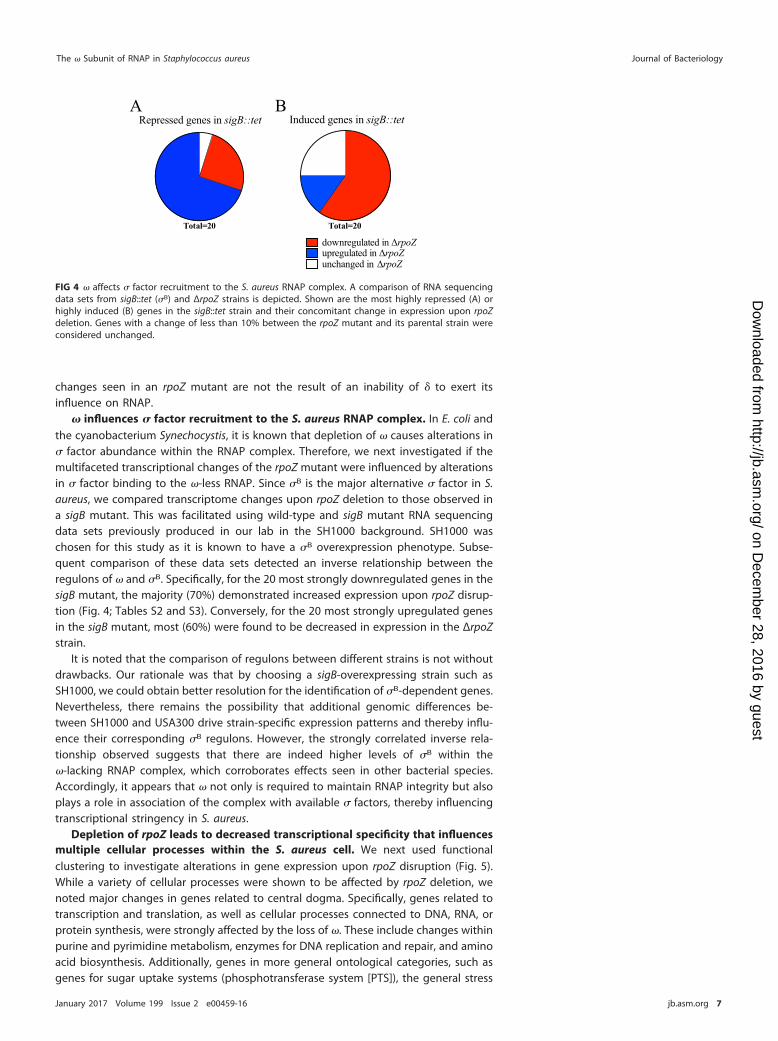
changes seen in an rpoZ mutant are not the result of an inability of to exert itsinfluence on RNAP.
influences factor recruitment to the S. aureus RNAP complex. In E. coli andthe cyanobacterium Synechocystis, it is known that depletion of causes alterations in factor abundance within the RNAP complex. Therefore, we next investigated if themultifaceted transcriptional changes of the rpoZ mutant were influenced by alterationsin factor binding to the -less RNAP. Since B is the major alternative factor in S.aureus, we compared transcriptome changes upon rpoZ deletion to those observed ina sigB mutant. This was facilitated using wild-type and sigB mutant RNA sequencingdata sets previously produced in our lab in the SH1000 background. SH1000 waschosen for this study as it is known to have a B overexpression phenotype. Subse-quent comparison of these data sets detected an inverse relationship between theregulons of and B. Specifically, for the 20 most strongly downregulated genes in thesigB mutant, the majority (70%) demonstrated increased expression upon rpoZ disrup-tion (Fig. 4; Tables S2 and S3). Conversely, for the 20 most strongly upregulated genesin the sigB mutant, most (60%) were found to be decreased in expression in the ΔrpoZstrain.
It is noted that the comparison of regulons between different strains is not withoutdrawbacks. Our rationale was that by choosing a sigB-overexpressing strain such asSH1000, we could obtain better resolution for the identification of B-dependent genes.Nevertheless, there remains the possibility that additional genomic differences be-tween SH1000 and USA300 drive strain-specific expression patterns and thereby influ-ence their corresponding B regulons. However, the strongly correlated inverse rela-tionship observed suggests that there are indeed higher levels of B within the-lacking RNAP complex, which corroborates effects seen in other bacterial species.Accordingly, it appears that not only is required to maintain RNAP integrity but alsoplays a role in association of the complex with available factors, thereby influencingtranscriptional stringency in S. aureus.
Depletion of rpoZ leads to decreased transcriptional specificity that influencesmultiple cellular processes within the S. aureus cell. We next used functionalclustering to investigate alterations in gene expression upon rpoZ disruption (Fig. 5).While a variety of cellular processes were shown to be affected by rpoZ deletion, wenoted major changes in genes related to central dogma. Specifically, genes related totranscription and translation, as well as cellular processes connected to DNA, RNA, orprotein synthesis, were strongly affected by the loss of . These include changes withinpurine and pyrimidine metabolism, enzymes for DNA replication and repair, and aminoacid biosynthesis. Additionally, genes in more general ontological categories, such asgenes for sugar uptake systems (phosphotransferase system [PTS]), the general stress
Repressed genes in sigB::tet
Total=20 Total=20
downregulated in rpoZupregulated in rpoZ
Induced genes in sigB::tet
unchanged in rpoZ
BA
FIG 4 affects factor recruitment to the S. aureus RNAP complex. A comparison of RNA sequencingdata sets from sigB::tet (B) and ΔrpoZ strains is depicted. Shown are the most highly repressed (A) orhighly induced (B) genes in the sigB::tet strain and their concomitant change in expression upon rpoZdeletion. Genes with a change of less than 10% between the rpoZ mutant and its parental strain wereconsidered unchanged.
The Subunit of RNAP in Staphylococcus aureus Journal of Bacteriology
January 2017 Volume 199 Issue 2 e00459-16 jb.asm.org 7
on Decem
ber 28, 2016 by guesthttp://jb.asm
.org/D
ownloaded from
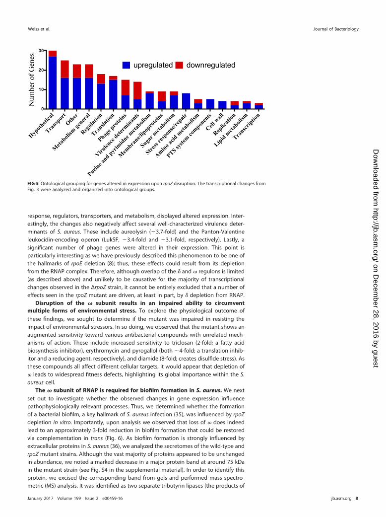
response, regulators, transporters, and metabolism, displayed altered expression. Inter-estingly, the changes also negatively affect several well-characterized virulence deter-minants of S. aureus. These include aureolysin (3.7-fold) and the Panton-Valentineleukocidin-encoding operon (LukSF, 3.4-fold and 3.1-fold, respectively). Lastly, asignificant number of phage genes were altered in their expression. This point isparticularly interesting as we have previously described this phenomenon to be one ofthe hallmarks of rpoE deletion (8); thus, these effects could result from its depletionfrom the RNAP complex. Therefore, although overlap of the and regulons is limited(as described above) and unlikely to be causative for the majority of transcriptionalchanges observed in the ΔrpoZ strain, it cannot be entirely excluded that a number ofeffects seen in the rpoZ mutant are driven, at least in part, by depletion from RNAP.
Disruption of the subunit results in an impaired ability to circumventmultiple forms of environmental stress. To explore the physiological outcome ofthese findings, we sought to determine if the mutant was impaired in resisting theimpact of environmental stressors. In so doing, we observed that the mutant shows anaugmented sensitivity toward various antibacterial compounds with unrelated mech-anisms of action. These include increased sensitivity to triclosan (2-fold; a fatty acidbiosynthesis inhibitor), erythromycin and pyrogallol (both 4-fold; a translation inhib-itor and a reducing agent, respectively), and diamide (8-fold; creates disulfide stress). Asthese compounds all affect different cellular targets, it would appear that depletion of leads to widespread fitness defects, highlighting its global importance within the S.aureus cell.
The subunit of RNAP is required for biofilm formation in S. aureus. We nextset out to investigate whether the observed changes in gene expression influencepathophysiologically relevant processes. Thus, we determined whether the formationof a bacterial biofilm, a key hallmark of S. aureus infection (35), was influenced by rpoZdepletion in vitro. Importantly, upon analysis we observed that loss of does indeedlead to an approximately 3-fold reduction in biofilm formation that could be restoredvia complementation in trans (Fig. 6). As biofilm formation is strongly influenced byextracellular proteins in S. aureus (36), we analyzed the secretomes of the wild-type andrpoZ mutant strains. Although the vast majority of proteins appeared to be unchangedin abundance, we noted a marked decrease in a major protein band at around 75 kDain the mutant strain (see Fig. S4 in the supplemental material). In order to identify thisprotein, we excised the corresponding band from gels and performed mass spectro-metric (MS) analysis. It was identified as two separate tributyrin lipases (the products of
Hypoth
etica
l
Transp
ort
Other
Meta
bolism
genera
l
Regulat
ion
Transla
tion
Phage p
rotei
ns
Virulen
ce dete
rminan
ts
Purine a
nd pyrim
idne meta
bolism
Mem
brane/l
ipopro
teins
Sugar m
etaboli
sm
Stress
respon
se/rep
air
Amino acid
meta
bolism
PTS syste
m compon
ents
Cell w
all
Replic
ation
Lipid meta
bolism
Transcr
iption
0
10
20
30
Num
ber o
f Gen
es
upregulated downregulated
FIG 5 Ontological grouping for genes altered in expression upon rpoZ disruption. The transcriptional changes fromFig. 3 were analyzed and organized into ontological groups.
Weiss et al. Journal of Bacteriology
January 2017 Volume 199 Issue 2 e00459-16 jb.asm.org 8
on Decem
ber 28, 2016 by guesthttp://jb.asm
.org/D
ownloaded from
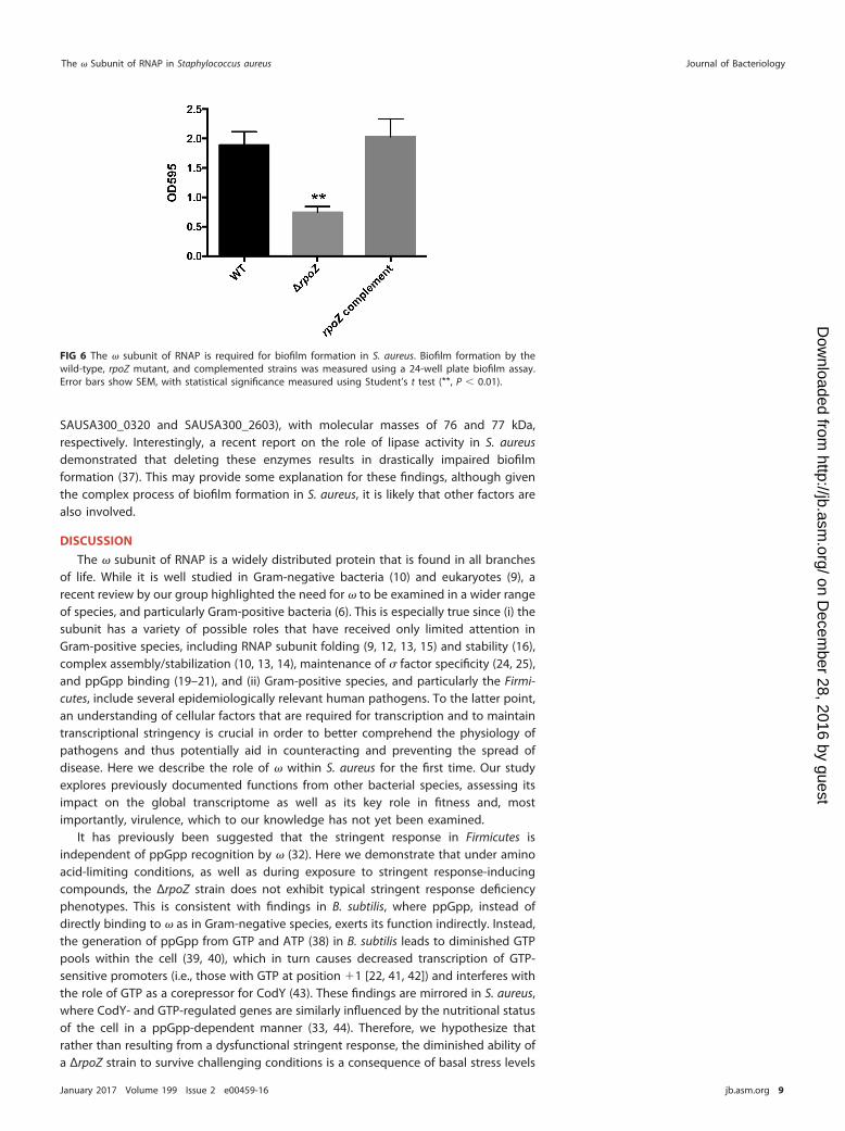
SAUSA300_0320 and SAUSA300_2603), with molecular masses of 76 and 77 kDa,respectively. Interestingly, a recent report on the role of lipase activity in S. aureusdemonstrated that deleting these enzymes results in drastically impaired biofilmformation (37). This may provide some explanation for these findings, although giventhe complex process of biofilm formation in S. aureus, it is likely that other factors arealso involved.
DISCUSSION
The subunit of RNAP is a widely distributed protein that is found in all branchesof life. While it is well studied in Gram-negative bacteria (10) and eukaryotes (9), arecent review by our group highlighted the need for to be examined in a wider rangeof species, and particularly Gram-positive bacteria (6). This is especially true since (i) thesubunit has a variety of possible roles that have received only limited attention inGram-positive species, including RNAP subunit folding (9, 12, 13, 15) and stability (16),complex assembly/stabilization (10, 13, 14), maintenance of factor specificity (24, 25),and ppGpp binding (19–21), and (ii) Gram-positive species, and particularly the Firmi-cutes, include several epidemiologically relevant human pathogens. To the latter point,an understanding of cellular factors that are required for transcription and to maintaintranscriptional stringency is crucial in order to better comprehend the physiology ofpathogens and thus potentially aid in counteracting and preventing the spread ofdisease. Here we describe the role of within S. aureus for the first time. Our studyexplores previously documented functions from other bacterial species, assessing itsimpact on the global transcriptome as well as its key role in fitness and, mostimportantly, virulence, which to our knowledge has not yet been examined.
It has previously been suggested that the stringent response in Firmicutes isindependent of ppGpp recognition by (32). Here we demonstrate that under aminoacid-limiting conditions, as well as during exposure to stringent response-inducingcompounds, the ΔrpoZ strain does not exhibit typical stringent response deficiencyphenotypes. This is consistent with findings in B. subtilis, where ppGpp, instead ofdirectly binding to as in Gram-negative species, exerts its function indirectly. Instead,the generation of ppGpp from GTP and ATP (38) in B. subtilis leads to diminished GTPpools within the cell (39, 40), which in turn causes decreased transcription of GTP-sensitive promoters (i.e., those with GTP at position 1 [22, 41, 42]) and interferes withthe role of GTP as a corepressor for CodY (43). These findings are mirrored in S. aureus,where CodY- and GTP-regulated genes are similarly influenced by the nutritional statusof the cell in a ppGpp-dependent manner (33, 44). Therefore, we hypothesize thatrather than resulting from a dysfunctional stringent response, the diminished ability ofa ΔrpoZ strain to survive challenging conditions is a consequence of basal stress levels
FIG 6 The subunit of RNAP is required for biofilm formation in S. aureus. Biofilm formation by thewild-type, rpoZ mutant, and complemented strains was measured using a 24-well plate biofilm assay.Error bars show SEM, with statistical significance measured using Student’s t test (**, P 0.01).
The Subunit of RNAP in Staphylococcus aureus Journal of Bacteriology
January 2017 Volume 199 Issue 2 e00459-16 jb.asm.org 9
on Decem
ber 28, 2016 by guesthttp://jb.asm
.org/D
ownloaded from
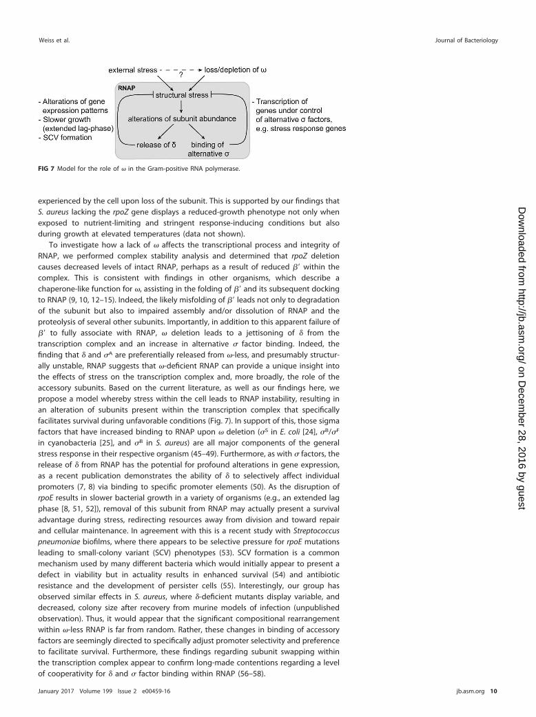
experienced by the cell upon loss of the subunit. This is supported by our findings thatS. aureus lacking the rpoZ gene displays a reduced-growth phenotype not only whenexposed to nutrient-limiting and stringent response-inducing conditions but alsoduring growth at elevated temperatures (data not shown).
To investigate how a lack of affects the transcriptional process and integrity ofRNAP, we performed complex stability analysis and determined that rpoZ deletioncauses decreased levels of intact RNAP, perhaps as a result of reduced = within thecomplex. This is consistent with findings in other organisms, which describe achaperone-like function for , assisting in the folding of = and its subsequent dockingto RNAP (9, 10, 12–15). Indeed, the likely misfolding of = leads not only to degradationof the subunit but also to impaired assembly and/or dissolution of RNAP and theproteolysis of several other subunits. Importantly, in addition to this apparent failure of= to fully associate with RNAP, deletion leads to a jettisoning of from thetranscription complex and an increase in alternative factor binding. Indeed, thefinding that and A are preferentially released from -less, and presumably structur-ally unstable, RNAP suggests that -deficient RNAP can provide a unique insight intothe effects of stress on the transcription complex and, more broadly, the role of theaccessory subunits. Based on the current literature, as well as our findings here, wepropose a model whereby stress within the cell leads to RNAP instability, resulting inan alteration of subunits present within the transcription complex that specificallyfacilitates survival during unfavorable conditions (Fig. 7). In support of this, those sigmafactors that have increased binding to RNAP upon deletion (S in E. coli [24], B/F
in cyanobacteria [25], and B in S. aureus) are all major components of the generalstress response in their respective organism (45–49). Furthermore, as with factors, therelease of from RNAP has the potential for profound alterations in gene expression,as a recent publication demonstrates the ability of to selectively affect individualpromoters (7, 8) via binding to specific promoter elements (50). As the disruption ofrpoE results in slower bacterial growth in a variety of organisms (e.g., an extended lagphase [8, 51, 52]), removal of this subunit from RNAP may actually present a survivaladvantage during stress, redirecting resources away from division and toward repairand cellular maintenance. In agreement with this is a recent study with Streptococcuspneumoniae biofilms, where there appears to be selective pressure for rpoE mutationsleading to small-colony variant (SCV) phenotypes (53). SCV formation is a commonmechanism used by many different bacteria which would initially appear to present adefect in viability but in actuality results in enhanced survival (54) and antibioticresistance and the development of persister cells (55). Interestingly, our group hasobserved similar effects in S. aureus, where -deficient mutants display variable, anddecreased, colony size after recovery from murine models of infection (unpublishedobservation). Thus, it would appear that the significant compositional rearrangementwithin -less RNAP is far from random. Rather, these changes in binding of accessoryfactors are seemingly directed to specifically adjust promoter selectivity and preferenceto facilitate survival. Furthermore, these findings regarding subunit swapping withinthe transcription complex appear to confirm long-made contentions regarding a levelof cooperativity for and factor binding within RNAP (56–58).
FIG 7 Model for the role of in the Gram-positive RNA polymerase.
Weiss et al. Journal of Bacteriology
January 2017 Volume 199 Issue 2 e00459-16 jb.asm.org 10
on Decem
ber 28, 2016 by guesthttp://jb.asm
.org/D
ownloaded from
In spite of these changes in subunit abundance, which we suggest are utilized bythe cell as a survival strategy, rpoZ deletion itself results in wide-reaching cellular stressthat is associated with an impaired transcriptional process. This contention is supportedby our RNA sequencing data, which demonstrate the upregulation of various genescoding for transcriptional and translational factors, putatively in an attempt to over-come diminished or inefficient RNAP function. Furthermore, we identified increasedexpression of genes involved in the general stress response, sugar uptake pathways,and energy metabolism, all of which may be designed to overcome unfavorableconditions (59). The latter group emphasizes that intracellular stress resulting from
deletion, and the subsequent countermeasures it elicits (e.g., production of chaperones,RNAP subunits, and ribosomal subunits), consumes energy and therefore has thepotential to deplete cellular energy pools. This in turn leads to the upregulation ofsugar uptake and energy metabolism, which in our study resulted in a minor increasein ATP pools (data not shown). In accordance with this, studies investigating S. aureusheat stress demonstrate that following exposure to elevated temperatures, cellularenergy levels are in fact not depleted, but rather a steady (60) or transient (61) increasein ATP pools is observed, presumably due to the upregulation of energy-generatingprocesses. It is perhaps not surprising that the transcriptional and physiologicalchanges observed from our in vitro experiments also translate into the impairment ofvirulence-related processes, as shown by the impaired biofilm formation in the rpoZmutant.
We suggest that, collectively, our findings extend the existing knowledge regarding and its role within RNAP, provide a unique insight into structural rearrangementswithin the transcription complex in response to stress, and demonstrate the necessityof this important subunit for the cellular homeostasis.
MATERIALS AND METHODSBacterial strains and growth conditions. All bacterial strains used in this study are listed in Table
1. Unless otherwise indicated, cultures were grown as described by Shaw et al. (62). Briefly, S. aureus wasgrown at 37°C with shaking (250 rpm) in tryptic soy broth (TSB) or chemically defined medium (CDM)(45), while E. coli was cultivated in lysogeny broth (LB) with agitation (250 rpm) at 37°C. Overnightcultures of S. aureus were seeded at a 1:100 dilution into fresh medium. After 3 h of growth, cultures wereused to inoculate fresh TSB to an optical density at 600 nm (OD600) of 0.05 and grown to the desireddensity in a Synergy2 plate reader (BioTek) in 96-well plates.
Mutant construction. In order to investigate the role of the subunit, a strain carrying a markerlessdeletion of the rpoZ gene was created, using a method based on the plasmid pJB38 as outlined by Boseand coworkers (63). Briefly, the majority of the rpoZ gene was removed via allelic replacement, as initiallydescribed elsewhere (64). To do this, both the 5= and 3= ends of the rpoZ gene, together with 1 kb ofup- and downstream DNA, were PCR amplified using primers OL2889 to OL2892 and fused togetherusing MluI sites contained in the amplification primers. This was then cloned into pJB38 using SacI andKpnI sites in primers OL2889 and OL2892. Following several selection and counterselection steps in S.aureus as described previously (63), rpoZ deletion was confirmed using internal (OL2991 and OL2992) andexternal (OL3008 and OL3009) primer pairs relative to the deleted region within the gene.
Complement construction. In order to exclude that secondary mutations were causative forphenotypic changes observed in the ΔrpoZ strain, a plasmid was constructed to complement the rpoZgene in trans. Here, the complete rpoZ operon, including its native promoter, was PCR amplified usingprimers OL3753 and OL3807. After BamHI digestion of the amplicon and the pMK4 shuttle vector, thePCR fragment and plasmid were ligated together and transformed into chemically competent E. coliDH5. The plasmid isolated from ampicillin-resistant clones was confirmed via Sanger sequencing (usingstandard M13 primers). Correct plasmids were transformed into electrocompetent S. aureus RN4220 (65)and finally transduced into the S. aureus USA300 rpoZ mutant using 11 (66).
RNA isolation and Northern blots. The detection of RNA transcripts was performed using aNorthern blotting protocol outlined by Caswell et al. (67). Briefly, RNA from cultures grown for 3 h wasisolated using an RNeasy kit (Qiagen), with DNA removed using a Turbo DNA-free kit (Ambion). RNAconcentrations were determined using an Agilent 2100 Bioanalyzer and an RNA 6000 Nano kit (Agilent).RNA (15 g) was separated by gel electrophoresis (10% polyacrylamide containing 7 M urea and 1Tris-borate-EDTA [TBE]) before being transferred to an Amersham Hybond N membrane (GE Health-care) by electroblotting. In order to cross-link samples, membranes were exposed to UV radiation.Following this, membranes were prehybridized (1 h, 43°C) in ULTRAhyb-Oligo buffer (Ambion) andincubated (16 h, 43°C) with a [-32]ATP-end-labeled oligonucleotide (OL3377) specific to the rpoZ targetsequence. Labeling was carried out using T4 polynucleotide kinase (Thermo Scientific) according to themanufacturer’s protocol. After overnight incubation, each membrane was washed (30 min, 43°C) withdecreasing (2, 1, and 0.5) concentrations of SSC buffer (300 mM sodium chloride, 30 mM sodium
The Subunit of RNAP in Staphylococcus aureus Journal of Bacteriology
January 2017 Volume 199 Issue 2 e00459-16 jb.asm.org 11
on Decem
ber 28, 2016 by guesthttp://jb.asm
.org/D
ownloaded from
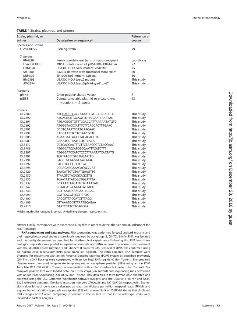
citrate). Finally, membranes were exposed to X-ray film in order to detect the size and abundance of therpoZ transcript.
RNA sequencing and data analyses. RNA sequencing was performed for rpoZ and sigB mutants andtheir respective parental strains as previously outlined by our group (8, 68–70). Briefly, RNA was isolatedand the quality determined as described for Northern blot experiments. Following this, RNA from threebiological replicates was pooled in equimolar amounts and rRNA removed by consecutive treatmentwith the MICROBExpress (Ambion) and RiboZero (Epicentre) kits. Removal of rRNA was confirmed usingan Agilent 2100 Bioanalyzer (RNA 6000 Nano kit; Agilent). The rRNA-depleted RNA samples wereprepared for sequencing with an Ion Personal Genome Machine (PGM) system as described previously(68). First, cDNA libraries were constructed with an Ion Total RNA-seq kit, v2 (Ion Torrent). The preparedlibraries were then used to generate template-positive ion sphere particles (ISPs) using an Ion PGMTemplate OT2 200 kit (Ion Torrent) in combination with an Ion OneTouch 2 system (Ion Torrent). Thetemplate-positive ISPs were loaded onto Ion 318 v2 chips (Ion Torrent) and sequencing runs performedwith an Ion PGM Sequencing 200 kit, v2 (Ion Torrent). Raw data files in fastq format were exported andanalyzed using the CLC Genomics Workbench software (Qiagen) and the USA300_FPR3757 and NCTC8325 reference genomes (GenBank accession numbers CP000255 and NC_007795, respectively). Expres-sion values for each gene were calculated as reads per kilobase per million mapped reads (RPKM), anda quantile normalization approach was applied (71) with a lower limit of 10 RPKM. Genes that displayedfold changes of 2 when comparing expression in the mutant to that in the wild-type strain wereincluded in further analyses.
TABLE 1 Strains, plasmids, and primers
Strain, plasmid, orprimer Description or sequencea
Reference orsource
Species and strainsE. coli DH5 Cloning strain 79
S. aureusRN4220 Restriction-deficient transformation recipient Lab StocksUSA300 HOU MRSA isolate cured of pUSA300-HOU-MRSA 72HKM850 USA300 HOU codY mutant; codY::tet 75SH1000 8325-4 derivate with functional rsbU; rsbU 80MJH502 SH1000 sigB mutant; sigB::tet 80BM2393 USA300 HOU ΔrpoZ mutant This studyAW2394 USA300 HOU ΔrpoZ/pMK4::rpoZ rpoZ This study
PlasmidspMK4 Gram-positive shuttle vector 81pJB38 Counterselectable plasmid to create silent
mutations in S. aureus63
PrimersOL2889 ATGGAGCTCGCCATAATTTATCTTCCACCTTC This studyOL2890 ATGACGCGTGCAGTTGTTGCAATTAAATAC This studyOL2891 ATGACGCGTGTTTCGACCATTAAAAATATGTG This studyOL2892 ATGGGTACCCATTTCTTCAGCACTTTGAAC This studyOL2991 GCGTGAAATTGATGAACAAC This studyOL2992 CAGCAATTTCTTCTAACGCTC This studyOL3008 AGAAGATTAGCTTAGAGAGGTC This studyOL3009 GAAATGCTAATGGTGTCACA This studyOL3377 CGTCAGCAATTTCTTCTAACGCTCTACCAAC This studyOL3753 ATGGGATCCATCGCCAATTTCATCTTT This studyOL3807 ATGGGATCCATCTCCCTTAAATATCACTATG This studyOL2393 TCGTATGTTGTGTGGAATTG This studyOL2394 GTGCTGCAAGGCGATTAAG This studyOL1297 GTGGTGGCGTTTGTGC This studyOL1298 CCGACAGCAAACACACCCAT This studyOL2129 TAACATTCTCTGATGAAGTTG This studyOL2130 TTAAGTCTACAGCAGCTTG This studyOL3136 TCAATATTATCGGTGGATTTA This studyOL3137 GCAAATTATGATGTTGAAATAG This studyOL3747 CGTAGATGCAAATTATTACG This studyOL3748 CGTTAATGAAACAATTGGAC This studyOL4099 GGTTCACGTTCCTTTATC This studyOL4100 CAGGTTTACCATCTTTAGG This studyOL4109 GTTAAATGGTTTAATGCAGAA This studyOL4110 GTATCCATCTTCAGCGA This study
aMRSA, methicillin-resistant S. aureus. Underlining denotes restriction sites.
Weiss et al. Journal of Bacteriology
January 2017 Volume 199 Issue 2 e00459-16 jb.asm.org 12
on Decem
ber 28, 2016 by guesthttp://jb.asm
.org/D
ownloaded from
RT-qPCR. In order to confirm expression values that were generated by RNA sequencing, a subsetof genes were chosen to confirm transcriptional changes via quantitative reverse transcription-PCR(RT-qPCR), as described by us previously (8, 69, 72, 73). PCR amplification was performed using primersfor each gene (Table 1) (glpF, OL1297/OL1298; asp23, OL2129/OL2130; SAUSA300_0174, OL3136/OL3137;aur, OL3747/OL3748; rpoC, OL4099/OL4100; and SAUSA300_0777, OL4109/OL4110), alongside 16S rRNA-specific primers as standard controls (OL1184/OL1185) (74).
Investigation of RNA polymerase composition. In order to determine the effects of loss on thestability and composition of RNAP, we adopted an approach first outlined by Gunnelius et al. (anoverview of this procedure can be found in Fig. S1 in the supplemental material) (25). Wild-type andΔrpoZ strains were grown to exponential phase, and their cytoplasmic protein fractions were isolated asoutlined previously (75). In order to differentiate between RNAP subunits unbound or within the RNAPcomplex, we performed two size selection steps. To do so, we first added 10 ml of phosphate-bufferedsaline (PBS) to the isolated protein fraction and loaded the complete mixture onto an Amicon Ultra-15centrifugal filter unit (EMD Millipore) with a 100-kDa cutoff. After centrifugation (4,000 g, 45 min), theapproximately 300 l of remaining protein fraction was washed with 10 ml of PBS and the centrifugationstep repeated. The fraction on top of the filter was then recovered and stored (80°C) for further analysis.The complete flowthrough of the first filter step was then applied to a second Amicon Ultra-15centrifugal filter unit (EMD Millipore) with a 30-kDa cutoff. The flowthrough was collected and trichlo-roacetic acid (TCA) precipitated (overnight, 4°C). The pelleted proteins were washed three times inice-cold absolute ethanol and subsequently resuspended in PBS. The different fractions were thensubjected to either mass spectrometry or Western blot analysis.
Characterization of protein content using mass spectrometry. Protein extracts isolated via thesize selection protocol outlined above were processed by filter-aided sample preparation (FASP), asdescribed by us previously (73, 75). Proteins were reduced with dithiothreitol (DTT), alkylated withiodoacetamide (IAA), and digested with trypsin–Lys-C (Promega) overnight at 37°C. Peptides werecollected by centrifugation, desalted using Pierce SPE C18 columns with a Supelco vacuum manifold, anddried in a vacuum concentrator (Labconco). Peptides were resuspended in H2O– 0.1% formic acid andseparated using a 75-m by 10-cm C18 reversed-phase high-pressure liquid chromatography (HPLC)column (New Objective) on a NanoLC Ultra (Eksigent) with a 120-min gradient (4 to 40% acetonitrile[ACN] with 0.1% formic acid). Fractions were analyzed on a linear ion trap Orbitrap instrument (OrbitrapXL; Thermo Fisher Scientific), with full MS survey scans acquired at 60,000 resolution. The top 10 mostabundant ions were selected for tandem MS (MS/MS) analysis in the linear ion trap. Raw data files wereprocessed in MaxQuant (www.maxquant.org) and searched against the UniProtKB S. aureus USA300protein sequence database. Search parameters included constant modification of cysteine by carbam-idomethylation and the variable modification methionine oxidation. Proteins were identified usingfiltering criteria of 1% protein and peptide false-discovery rates. Protein levels were normalized to theoverall protein content in each of the investigated fractions.
Western blotting. The assessment of subunit abundance was performed as described by uspreviously (66). Size-selected protein fractions containing proteins and protein complexes of 100 kDawere prepared as for experiments determining RNAP composition. Samples were separated using 12%SDS-polyacrylamide gel electrophoresis (SDS-PAGE), as described by us previously (76). Separatedproteins were then transferred to a polyvinylidene difluoride (PVDF) membrane and detected using amonoclonal mouse RNAP antibody (8RB13; Santa Cruz Biotechnology) and a horseradish peroxidase(HRP)-conjugated secondary antibody. HRP activity was assessed using the SuperSignal West Picosubstrate (Thermo Fisher Scientific) and visualized on X-ray film.
MIC assessment assay. To determine whether transcriptional changes observed within the mutantstrain resulted in detectable physiological sensitivities, we assessed MICs for various antibacterial agents,as described by our group previously (77). Briefly, overnight cultures were diluted 1:1,000 into freshmedium in 96-well plates, and antibiotics at various concentrations were added at amounts no greaterthan 2% of the final volume. Plates were incubated for 12 h (static, 37°C), and turbidity, as a sign ofbacterial growth, was manually assessed. MICs were defined as the minimum concentration of a givenagent to result in no detectable turbidity in individual wells.
Secretome analysis. To evaluate alterations in secreted proteins, bacteria were grown for 24 hbefore being centrifuged (6,000 g, 10 min). Supernatants were removed and subjected to 12%SDS-PAGE as described by us previously (76). Gels were assessed either by silver staining using a Piercesilver stain kit (Thermo Fisher Scientific) according to the manufacturer’s instructions or by Coomassiebrilliant blue staining, in-gel trypsin digestion, and mass spectrometric analysis, as described by uspreviously (76). Briefly, gel pieces were minced and destained before reduction and alkylation withdithiothreitol (DTT) and iodoacetamide (IAA), respectively. Proteins were digested with trypsin–Lys-Covernight at 37°C, and peptides were extracted using 50:50 ACN and H2O– 0.1% formic acid and dried ina vacuum concentrator (Labconco). Peptides were resuspended in H2O– 0.1% formic acid for LC-MS/MSanalysis, which was performed as described above.
Biofilm assay. The ability of strains to form biofilms was assessed as described by us previously (78).The wells of non-tissue-culture treated 12-well polystyrene plates were incubated for 48 h (static, 4°C)with 1 ml of human serum in order to facilitate attachment of cells. Concurrently, bacteria were culturedovernight in biofilm medium (BFM) (TSB supplemented with 0.5% [wt/vol] dextrose and 3% [wt/vol]NaCl) and used to seed fresh BFM to an OD600 of 0.05. After removal of the human serum from the wellsof plates, 1 ml of these fresh cultures was transferred into each well and incubated for 48 h (static, 37°C).After incubation, supernatants were gently removed from each well and the biofilm washed twice withPBS before fixation with absolute ethanol. Biofilms were stained with crystal violet solution (2% [wt/vol])
The Subunit of RNAP in Staphylococcus aureus Journal of Bacteriology
January 2017 Volume 199 Issue 2 e00459-16 jb.asm.org 13
on Decem
ber 28, 2016 by guesthttp://jb.asm
.org/D
ownloaded from
for 10 min, followed by two more rounds of washing with PBS. Residual supernatants were aspirated, andplates were dried overnight. After this time, 300 l of absolute ethanol was added to the wells andincubated for 10 min before being removed, and the absorbance was measured at 570 nm using aSynergy2 plate reader (BioTek).
Accession number(s). All data sets have been deposited to the NCBI Gene Expression Omnibus(GEO) (accession numbers GSE87033 and GSE87036).
SUPPLEMENTAL MATERIAL
Supplemental material for this article may be found at https://doi.org/10.1128/JB.00459-16.
TEXT S1, PDF file, 1.3 MB.TEXT S2, PDF file, 0.1 MB.
ACKNOWLEDGMENTThis study was supported in part by grant AI080626 (to L.N.S.) from the National
Institute of Allergy and Infectious Diseases.
REFERENCES1. Sekine S, Tagami S, Yokoyama S. 2012. Structural basis of transcription
by bacterial and eukaryotic RNA polymerases. Curr Opin Struct Biol22:110 –118. https://doi.org/10.1016/j.sbi.2011.11.006.
2. Ebright RH. 2000. RNA polymerase: structural similarities between bac-terial RNA polymerase and eukaryotic RNA polymerase II. J Mol Biol304:687– 698. https://doi.org/10.1006/jmbi.2000.4309.
3. Burgess RR. 1969. Separation and characterization of the subunits ofribonucleic acid polymerase. J Biol Chem 244:6168 – 6176.
4. Pero J, Nelson J, Fox TD. 1975. Highly asymmetric transcription by RNApolymerase containing phage-SP01-induced polypeptides and a newhost protein. Proc Natl Acad Sci U S A 72:1589 –1593. https://doi.org/10.1073/pnas.72.4.1589.
5. Keller AN, Yang X, Wiedermannová J, Delumeau O, Krásný L, Lewis PJ. 2014., a new subunit of RNA polymerase found in Gram-positive bacteria. JBacteriol 196:3622–3632. https://doi.org/10.1128/JB.02020-14.
6. Weiss A, Shaw LN. 2015. Small things considered: the small accessorysubunits of RNA polymerase in Gram-positive bacteria. FEMS MicrobiolRev 39:541–554. https://doi.org/10.1093/femsre/fuv005.
7. Xue X, Tomasch J, Sztajer H, Wagner-Dobler I. 2010. The delta subunit ofRNA polymerase, RpoE, is a global modulator of Streptococcus mutansenvironmental adaptation. J Bacteriol 192:5081–5092. https://doi.org/10.1128/JB.00653-10.
8. Weiss A, Ibarra JA, Paoletti J, Carroll RK, Shaw LN. 2014. The delta subunitof RNA polymerase guides promoter selectivity and virulence in Staph-ylococcus aureus. Infect Immun 82:1424 –1435. https://doi.org/10.1128/IAI.01508-14.
9. Minakhin L, Bhagat S, Brunning A, Campbell EA, Darst SA, Ebright RH,Severinov K. 2001. Bacterial RNA polymerase subunit omega and eu-karyotic RNA polymerase subunit RPB6 are sequence, structural, andfunctional homologs and promote RNA polymerase assembly. Proc NatlAcad Sci U S A 98:892– 897. https://doi.org/10.1073/pnas.98.3.892.
10. Mathew R, Chatterji D. 2006. The evolving story of the omega subunit ofbacterial RNA polymerase. Trends Microbiol 14:450 – 455. https://doi.org/10.1016/j.tim.2006.08.002.
11. Nouraini S, Archambault J, Friesen JD. 1996. Rpo26p, a subunit commonto yeast RNA polymerases, is essential for the assembly of RNA poly-merases I and II and for the stability of the largest subunits of theseenzymes. Mol Cell Biol 16:5985–5996. https://doi.org/10.1128/MCB.16.11.5985.
12. Gentry DR, Burgess RR. 1993. Cross-linking of Escherichia coli RNApolymerase subunits: identification of beta= as the binding site ofomega. Biochemistry 32:11224 –11227. https://doi.org/10.1021/bi00092a036.
13. Ghosh P, Ishihama A, Chatterji D. 2001. Escherichia coli RNA polymerasesubunit omega and its N-terminal domain bind full-length beta= tofacilitate incorporation into the alpha2beta subassembly. Eur J Biochem268:4621– 4627. https://doi.org/10.1046/j.1432-1327.2001.02381.x.
14. Ganguly A, Chatterji D. 2011. Sequential assembly of an active RNApolymerase molecule at the air-water interface. Langmuir 27:3808 –3814.https://doi.org/10.1021/la200225t.
15. Ghosh P, Ramakrishnan C, Chatterji D. 2003. Inter-subunit recognition
and manifestation of segmental mobility in Escherichia coli RNApolymerase: a case study with omega-beta= interaction. Biophys Chem103:223–237. https://doi.org/10.1016/S0301-4622(02)00271-5.
16. Mathew R, Ramakanth M, Chatterji D. 2005. Deletion of the gene rpoZ,encoding the omega subunit of RNA polymerase, in Mycobacteriumsmegmatis results in fragmentation of the beta= subunit in the enzymeassembly. J Bacteriol 187:6565– 6570. https://doi.org/10.1128/JB.187.18.6565-6570.2005.
17. Murakami KS. 2013. X-ray crystal structure of Escherichia coli RNA poly-merase sigma70 holoenzyme. J Biol Chem 288:9126 –9134. https://doi.org/10.1074/jbc.M112.430900.
18. Zhang G, Campbell EA, Minakhin L, Richter C, Severinov K, Darst SA.1999. Crystal structure of Thermus aquaticus core RNA polymerase at3.3 A resolution. Cell 98:811– 824. https://doi.org/10.1016/S0092-8674(00)81515-9.
19. Mechold U, Potrykus K, Murphy H, Murakami KS, Cashel M. 2013. Differ-ential regulation by ppGpp versus pppGpp in Escherichia coli. NucleicAcids Res 41:6175– 6189. https://doi.org/10.1093/nar/gkt302.
20. Ross W, Vrentas CE, Sanchez-Vazquez P, Gaal T, Gourse RL. 2013. Themagic spot: a ppGpp binding site on E. coli RNA polymerase responsiblefor regulation of transcription initiation. Mol Cell 50:420 – 429. https://doi.org/10.1016/j.molcel.2013.03.021.
21. Zuo Y, Wang Y, Steitz TA. 2013. The mechanism of E. coli RNA polymer-ase regulation by ppGpp is suggested by the structure of their complex.Mol Cell 50:430 – 436. https://doi.org/10.1016/j.molcel.2013.03.020.
22. Krasny L, Gourse RL. 2004. An alternative strategy for bacterial ribosomesynthesis: Bacillus subtilis rRNA transcription regulation. EMBO J 23:4473– 4483. https://doi.org/10.1038/sj.emboj.7600423.
23. Kriel A, Bittner AN, Kim SH, Liu K, Tehranchi AK, Zou WY, Rendon S, ChenR, Tu BP, Wang JD. 2012. Direct regulation of GTP homeostasis by(p)ppGpp: a critical component of viability and stress resistance. Mol cell48:231–241. https://doi.org/10.1016/j.molcel.2012.08.009.
24. Geertz M, Travers A, Mehandziska S, Sobetzko P, Chandra-Janga S,Shimamoto N, Muskhelishvili G. 2011. Structural coupling between RNApolymerase composition and DNA supercoiling in coordinatingtranscription: a global role for the omega subunit? mBio 2:e00034-11.https://doi.org/10.1128/mBio.00034-11.
25. Gunnelius L, Hakkila K, Kurkela J, Wada H, Tyystjarvi E, Tyystjarvi T. 2014.The omega subunit of the RNA polymerase core directs transcriptionefficiency in cyanobacteria. Nucleic Acids Res 42:4606 – 4614. https://doi.org/10.1093/nar/gku084.
26. Santos-Beneit F, Barriuso-Iglesias M, Fernandez-Martinez LT, Martinez-Castro M, Sola-Landa A, Rodriguez-Garcia A, Martin JF. 2011. The RNApolymerase omega factor RpoZ is regulated by PhoP and has an impor-tant role in antibiotic biosynthesis and morphological differentiation inStreptomyces coelicolor. Appl Environ Microbiol 77:7586 –7594. https://doi.org/10.1128/AEM.00465-11.
27. Kojima I, Kasuga K, Kobayashi M, Fukasawa A, Mizuno S, Arisawa A,Akagawa H. 2002. The rpoZ gene, encoding the RNA polymerase omegasubunit, is required for antibiotic production and morphological differ-
Weiss et al. Journal of Bacteriology
January 2017 Volume 199 Issue 2 e00459-16 jb.asm.org 14
on Decem
ber 28, 2016 by guesthttp://jb.asm
.org/D
ownloaded from
entiation in Streptomyces kasugaensis. J Bacteriol 184:6417– 6423.https://doi.org/10.1128/JB.184.23.6417-6423.2002.
28. Kojima I, Kasuga K, Kobayashi M, Tsuchiya KS, Ikeno S. 2006. Thebiosynthesis of kasugamycin, an antibiotic against rice blast disease,with particular reference to the involvement of rpoZ, a gene encodingRNA polymerase omega subunit. Int J Soc Mat Eng Res 14:28 –32. https://doi.org/10.5188/ijsmer.14.28.
29. Mathew R, Mukherjee R, Balachandar R, Chatterji D. 2006. Deletion of therpoZ gene, encoding the omega subunit of RNA polymerase, results inpleiotropic surface-related phenotypes in Mycobacterium smegmatis.Microbiology 152:1741–1750. https://doi.org/10.1099/mic.0.28879-0.
30. Reference deleted.31. Weiss A, Broach WH, Shaw LN. 2016. Characterizing the transcriptional
adaptation of Staphylococcus aureus to stationary phase growth. Pat-hog Dis 74:ftw046. https://doi.org/10.1093/femspd/ftw046.
32. Hauryliuk V, Atkinson GC, Murakami KS, Tenson T, Gerdes K. 2015.Recent functional insights into the role of (p)ppGpp in bacterial physi-ology. Nat Rev Microbiol 13:298 –309. https://doi.org/10.1038/nrmicro3448.
33. Geiger T, Francois P, Liebeke M, Fraunholz M, Goerke C, Krismer B,Schrenzel J, Lalk M, Wolz C. 2012. The stringent response of Staphylo-coccus aureus and its impact on survival after phagocytosis through theinduction of intracellular PSMs expression. PLoS Pathog 8:e1003016.https://doi.org/10.1371/journal.ppat.1003016.
34. Geiger T, Goerke C, Fritz M, Schafer T, Ohlsen K, Liebeke M, Lalk M, WolzC. 2010. Role of the (p)ppGpp synthase RSH, a RelA/SpoT homolog, instringent response and virulence of Staphylococcus aureus. Infect Im-mun 78:1873–1883. https://doi.org/10.1128/IAI.01439-09.
35. Archer NK, Mazaitis MJ, Costerton JW, Leid JG, Powers ME, Shirtliff ME.2011. Staphylococcus aureus biofilms: properties, regulation, and rolesin human disease. Virulence 2:445– 459. https://doi.org/10.4161/viru.2.5.17724.
36. Lister JL, Horswill AR. 2014. Staphylococcus aureus biofilms: recentdevelopments in biofilm dispersal. Front Cell Infect Microbiol 4:178.https://doi.org/10.3389/fcimb.2014.00178.
37. Hu C, Xiong N, Zhang Y, Rayner S, Chen S. 2012. Functional character-ization of lipase in the pathogenesis of Staphylococcus aureus. BiochemBiophys Res Commun 419:617– 620. https://doi.org/10.1016/j.bbrc.2012.02.057.
38. Nanamiya H, Kasai K, Nozawa A, Yun CS, Narisawa T, Murakami K, NatoriY, Kawamura F, Tozawa Y. 2008. Identification and functional analysis ofnovel (p)ppGpp synthetase genes in Bacillus subtilis. Mol Microbiol67:291–304.
39. Lopez JM, Dromerick A, Freese E. 1981. Response of guanosine 5=-triphosphate concentration to nutritional changes and its significancefor Bacillus subtilis sporulation. J Bacteriol 146:605– 613.
40. Ochi K, Kandala J, Freese E. 1982. Evidence that Bacillus subtilis sporu-lation induced by the stringent response is caused by the decrease inGTP or GDP. J Bacteriol 151:1062–1065.
41. Sojka L, Kouba T, Barvik I, Sanderova H, Maderova Z, Jonak J, Krasny L.2011. Rapid changes in gene expression: DNA determinants of promoterregulation by the concentration of the transcription initiating NTP inBacillus subtilis. Nucleic Acids Res 39:4598 – 4611. https://doi.org/10.1093/nar/gkr032.
42. Krasny L, Tiserova H, Jonak J, Rejman D, Sanderova H. 2008. The identityof the transcription 1 position is crucial for changes in gene expressionin response to amino acid starvation in Bacillus subtilis. Mol Microbiol69:42–54. https://doi.org/10.1111/j.1365-2958.2008.06256.x.
43. Ratnayake-Lecamwasam M, Serror P, Wong KW, Sonenshein AL. 2001.Bacillus subtilis CodY represses early-stationary-phase genes by sens-ing GTP levels. Genes Dev 15:1093–1103. https://doi.org/10.1101/gad.874201.
44. Pohl K, Francois P, Stenz L, Schlink F, Geiger T, Herbert S, Goerke C,Schrenzel J, Wolz C. 2009. CodY in Staphylococcus aureus: a regulatorylink between metabolism and virulence gene expression. J Bacteriol191:2953–2963. https://doi.org/10.1128/JB.01492-08.
45. Chan PF, Foster SJ, Ingham E, Clements MO. 1998. The Staphylococcusaureus alternative sigma factor sigmaB controls the environmental stressresponse but not starvation survival or pathogenicity in a mouse abscessmodel. J Bacteriol 180:6082– 6089.
46. Battesti A, Majdalani N, Gottesman S. 2011. The RpoS-mediated generalstress response in Escherichia coli. Annu Rev Microbiol 65:189 –213.https://doi.org/10.1146/annurev-micro-090110-102946.
47. Pollari M, Gunnelius L, Tuominen I, Ruotsalainen V, Tyystjarvi E, Salminen
T, Tyystjarvi T. 2008. Characterization of single and double inactivationstrains reveals new physiological roles for group 2 sigma factors in thecyanobacterium Synechocystis sp. PCC 6803. Plant Physiol 147:1994 –2005. https://doi.org/10.1104/pp.108.122713.
48. Huckauf J, Nomura C, Forchhammer K, Hagemann M. 2000. Stressresponses of Synechocystis sp. strain PCC 6803 mutants impaired ingenes encoding putative alternative sigma factors. Microbiology 146:2877–2889. https://doi.org/10.1099/00221287-146-11-2877.
49. Kullik II, Giachino P. 1997. The alternative sigma factor sigmaB in Staph-ylococcus aureus: regulation of the sigB operon in response to growthphase and heat shock. Arch Microbiol 167:151–159. https://doi.org/10.1007/s002030050428.
50. Prajapati RK, Sengupta S, Rudra P, Mukhopadhyay J. 2016. Bacillussubtilis delta factor functions as a transcriptional regulator by facilitatingthe open complex formation. J Biol Chem 291:1064 –1075. https://doi.org/10.1074/jbc.M115.686170.
51. Lopez de Saro FJ, Yoshikawa N, Helmann JD. 1999. Expression, abun-dance, and RNA polymerase binding properties of the delta factor ofBacillus subtilis. J Biol Chem 274:15953–15958. https://doi.org/10.1074/jbc.274.22.15953.
52. Jones AL, Needham RH, Rubens CE. 2003. The delta subunit of RNApolymerase is required for virulence of Streptococcus agalactiae. InfectImmun 71:4011– 4017. https://doi.org/10.1128/IAI.71.7.4011-4017.2003.
53. Churton NW, Misra RV, Howlin RP, Allan RN, Jefferies J, Faust SN, GharbiaSE, Edwards RJ, Clarke SC, Webb JS. 2016. Parallel evolution in Strepto-coccus pneumoniae biofilms. Genome Biol Evol 8:1316 –1326. https://doi.org/10.1093/gbe/evw072.
54. Mirani ZA, Aziz M, Khan SI. 2015. Small colony variants have a major rolein stability and persistence of Staphylococcus aureus biofilms. J Antibiot68:98 –105. https://doi.org/10.1038/ja.2014.115.
55. Proctor RA, von Eiff C, Kahl BC, Becker K, McNamara P, Herrmann M,Peters G. 2006. Small colony variants: a pathogenic form of bacteria thatfacilitates persistent and recurrent infections. Nat Rev Microbiol4:295–305. https://doi.org/10.1038/nrmicro1384.
56. Achberger EC, Whiteley HR. 1981. The role of the delta peptide of theBacillus subtilis RNA polymerase in promoter selection. J Biol Chem256:7424 –7432.
57. Williamson VM, Doi RH. 1978. Delta factor can displace sigma factor fromBacillus subtilis RNA polymerase holoenzyme and regulate its initiationactivity. Mol Gen Genet 161:135–141.
58. Hyde EI, Hilton MD, Whiteley HR. 1986. Interactions of Bacillus subtilisRNA polymerase with subunits determining the specificity of initiation.Sigma and delta peptides can bind simultaneously to core. J Biol Chem261:16565–16570.
59. Kultz D. 2005. Molecular and evolutionary basis of the cellular stressresponse. Annu Rev Physiol 67:225–257. https://doi.org/10.1146/annurev.physiol.67.040403.103635.
60. Fleury B, Kelley WL, Lew D, Gotz F, Proctor RA, Vaudaux P. 2009.Transcriptomic and metabolic responses of Staphylococcus aureus ex-posed to supra-physiological temperatures. BMC Microbiol 9:76. https://doi.org/10.1186/1471-2180-9-76.
61. Pane-Farre J, Jonas B, Forstner K, Engelmann S, Hecker M. 2006. ThesigmaB regulon in Staphylococcus aureus and its regulation. Int J MedMicrobiol 296:237–258. https://doi.org/10.1016/j.ijmm.2005.11.011.
62. Shaw LN, Lindholm C, Prajsnar TK, Miller HK, Brown MC, Golonka E,Stewart GC, Tarkowski A, Potempa J. 2008. Identification and character-ization of sigmaS, a novel component of the Staphylococcus aureusstress and virulence responses. PLoS One 3:e3844. https://doi.org/10.1371/journal.pone.0003844.
63. Bose JL, Fey PD, Bayles KW. 2013. Genetic tools to enhance the study ofgene function and regulation in Staphylococcus aureus. Appl EnvironMicrobiol 79:2218 –2224. https://doi.org/10.1128/AEM.00136-13.
64. Bae T, Schneewind O. 2006. Allelic replacement in Staphylococcus au-reus with inducible counter-selection. Plasmid 55:58 – 63. https://doi.org/10.1016/j.plasmid.2005.05.005.
65. Monk IR, Shah IM, Xu M, Tan MW, Foster TJ. 2012. Transforming theuntransformable: application of direct transformation to manipulategenetically Staphylococcus aureus and Staphylococcus epidermidis.mBio 3:e00277-11. https://doi.org/10.1128/mBio.00277-11.
66. Carroll RK, Robison TM, Rivera FE, Davenport JE, Jonsson IM, Florczyk D,Tarkowski A, Potempa J, Koziel J, Shaw LN. 2012. Identification of anintracellular M17 family leucine aminopeptidase that is required forvirulence in Staphylococcus aureus. Microbes Infect 14:989 –999. https://doi.org/10.1016/j.micinf.2012.04.013.
The Subunit of RNAP in Staphylococcus aureus Journal of Bacteriology
January 2017 Volume 199 Issue 2 e00459-16 jb.asm.org 15
on Decem
ber 28, 2016 by guesthttp://jb.asm
.org/D
ownloaded from
67. Caswell CC, Gaines JM, Ciborowski P, Smith D, Borchers CH, Roux CM,Sayood K, Dunman PM, Roop Ii RM. 2012. Identification of two smallregulatory RNAs linked to virulence in Brucella abortus 2308. Mol Mi-crobiol 85:345–360. https://doi.org/10.1111/j.1365-2958.2012.08117.x.
68. Carroll RK, Weiss A, Shaw LN. 2016. RNA-sequencing of Staphylococcusaureus messenger RNA. Methods Mol Biol 1373:131–141. https://doi.org/10.1007/7651_2014_192.
69. Krute CN, Carroll RK, Rivera FE, Weiss A, Young RM, Shilling A, Botlani M,Varma S, Baker BJ, Shaw LN. 2015. The disruption of prenylation leads topleiotropic rearrangements in cellular behavior in Staphylococcus au-reus. Mol Microbiol 95:819 – 832. https://doi.org/10.1111/mmi.12900.
70. Weiss A, Broach WH, Lee MC, Shaw LN. 2016. Towards the completesmall RNome of Acinetobacter baumannii. Microb Genomics https://doi.org/10.1099/mgen.0.000045.
71. McClure R, Balasubramanian D, Sun Y, Bobrovskyy M, Sumby P, GencoCA, Vanderpool CK, Tjaden B. 2013. Computational analysis of bacterialRNA-Seq data. Nucleic Acids Res 41:e140. https://doi.org/10.1093/nar/gkt444.
72. Kolar SL, Nagarajan V, Oszmiana A, Rivera FE, Miller HK, Davenport JE,Riordan JT, Potempa J, Barber DS, Koziel J, Elasri MO, Shaw LN. 2011.NsaRS is a cell-envelope-stress-sensing two-component system ofStaphylococcus aureus. Microbiology 157:2206 –2219. https://doi.org/10.1099/mic.0.049692-0.
73. Krute CN, Bell-Temin H, Miller HK, Rivera FE, Weiss A, Stevens SM, ShawLN. 2015. The membrane protein PrsS mimics sigmaS in protectingStaphylococcus aureus against cell wall-targeting antibiotics and DNA-damaging agents. Microbiology 16:1136 –1148.
74. Koprivnjak T, Mlakar V, Swanson L, Fournier B, Peschel A, Weiss JP. 2006.Cation-induced transcriptional regulation of the dlt operon of Staphy-lococcus aureus. J Bacteriol 188:3622–3630. https://doi.org/10.1128/JB.188.10.3622-3630.2006.
75. Rivera FE, Miller HK, Kolar SL, Stevens SM, Jr, Shaw LN. 2012. The impactof CodY on virulence determinant production in community-associatedmethicillin-resistant Staphylococcus aureus. Proteomics 12:263–268.https://doi.org/10.1002/pmic.201100298.
76. Kolar SL, Ibarra JA, Rivera FE, Mootz JM, Davenport JE, Stevens SM,Horswill AR, Shaw LN. 2013. Extracellular proteases are key mediators ofStaphylococcus aureus virulence via the global modulation of virulence-determinant stability. Microbiol Open 2:18 –34. https://doi.org/10.1002/mbo3.55.
77. Fleeman R, LaVoi TM, Santos RG, Morales A, Nefzi A, Welmaker GS,Medina-Franco JL, Giulianotti MA, Houghten RA, Shaw LN. 2015. Com-binatorial libraries as a tool for the discovery of novel, broad-spectrumantibacterial agents targeting the ESKAPE pathogens. J Med Chem58:3340 –3355. https://doi.org/10.1021/jm501628s.
78. Shaw LN, Golonka E, Szmyd G, Foster SJ, Travis J, Potempa J. 2005.Cytoplasmic control of premature activation of a secreted proteasezymogen: deletion of staphostatin B (SspC) in Staphylococcus aureus8325-4 yields a profound pleiotropic phenotype. J Bacteriol 187:1751–1762. https://doi.org/10.1128/JB.187.5.1751-1762.2005.
79. Salisbury V, Hedges RW, Datta N. 1972. Two modes of “curing” trans-missible bacterial plasmids. J Gen Microbiol 70:443– 452. https://doi.org/10.1099/00221287-70-3-443.
80. Horsburgh MJ, Aish JL, White IJ, Shaw L, Lithgow JK, Foster SJ. 2002.sigmaB modulates virulence determinant expression and stressresistance: characterization of a functional rsbU strain derived fromStaphylococcus aureus 8325-4. J Bacteriol 184:5457–5467. https://doi.org/10.1128/JB.184.19.5457-5467.2002.
81. Sullivan MA, Yasbin RE, Young FE. 1984. New shuttle vectors forBacillus subtilis and Escherichia coli which allow rapid detection ofinserted fragments. Gene 29:21–26. https://doi.org/10.1016/0378-1119(84)90161-6.
Weiss et al. Journal of Bacteriology
January 2017 Volume 199 Issue 2 e00459-16 jb.asm.org 16
on Decem
ber 28, 2016 by guesthttp://jb.asm
.org/D
ownloaded from
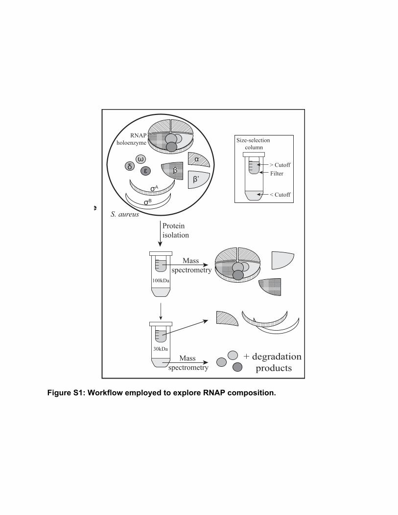
Figure S1: Workflow employed to explore RNAP composition.
Figure S1: Workflow employed to explore RNAP composition.
+ degradation products
100kDa
30kDa
Size-selection column
S. aureus
RNAPholoenzyme
Protein isolation
ωδ ε
σA
σB
ββ’
Filter> Cutoff
< Cutoff
α
Mass spectrometry
Mass spectrometry
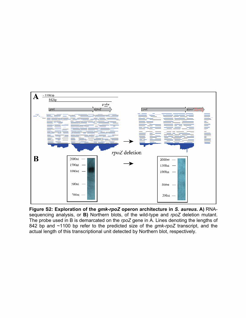
Figure S2: Exploration of the gmk-rpoZ operon architecture in S. aureus. A) RNA-sequencing analysis, or B) Northern blots, of the wild-type and rpoZ deletion mutant. The probe used in B is demarcated on the rpoZ gene in A. Lines denoting the lengths of 842 bp and ~1100 bp refer to the predicted size of the gmk-rpoZ transcript, and the actual length of this transcriptional unit detected by Northern blot, respectively.
Figure S2: Exploration of the gmk-rpoZ operon architecture in S. aureus. A) RNA-sequencing analysis, or B) Northern blots, of the wild-type and rpoZ deletion mutant. The probe used in B is demarcated on the rpoZ gene in A. Lines denoting the lengths of 842 bp and ~1100 bp refer to the predicted size of the gmk-rpoZ ORF, and the actual length of this transcriptional unit detected by Northern blot, respectively.
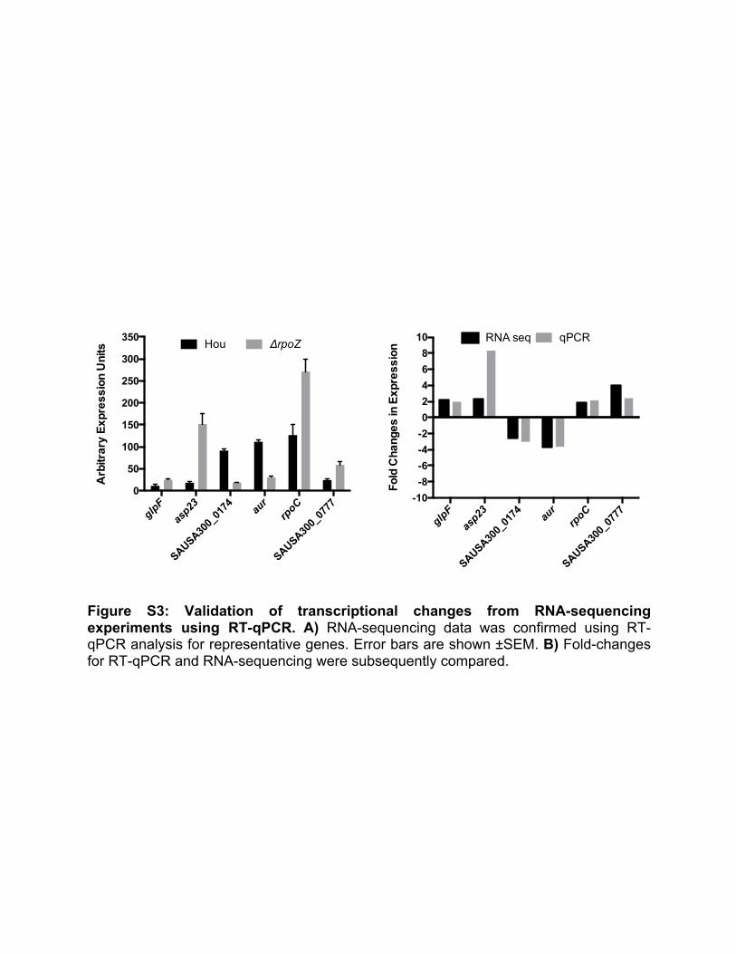
Figure S3: Validation of transcriptional changes from RNA-sequencing experiments using RT-qPCR. A) RNA-sequencing data was confirmed using RT-qPCR analysis for representative genes. Error bars are shown ±SEM. B) Fold-changes for RT-qPCR and RNA-sequencing were subsequently compared.
glpF
asp23
SAUSA300_
0174 au
rrpoC
SAUSA300_
0777
0
50
100
150
200
250
300
350
Arb
itrar
y E
xpre
ssio
n U
nits Hou ΔrpoZ
glpF
asp23
SAUSA300_
0174 au
rrpoC
SAUSA300_
0777
-10-8-6-4-202468
10
Fold
Cha
nges
in E
xpre
ssio
n
RNA seq qPCR
Figure S3: Validation of transcriptional changes from RNA-sequencing experiments using RT-qPCR. A) RNA-sequencing data was confirmed using RT-qPCR analysis for representative genes. Error bars are shown ±SEM. B) Fold-changes for RT-qPCR and RNA-sequencing were subsequently compared.
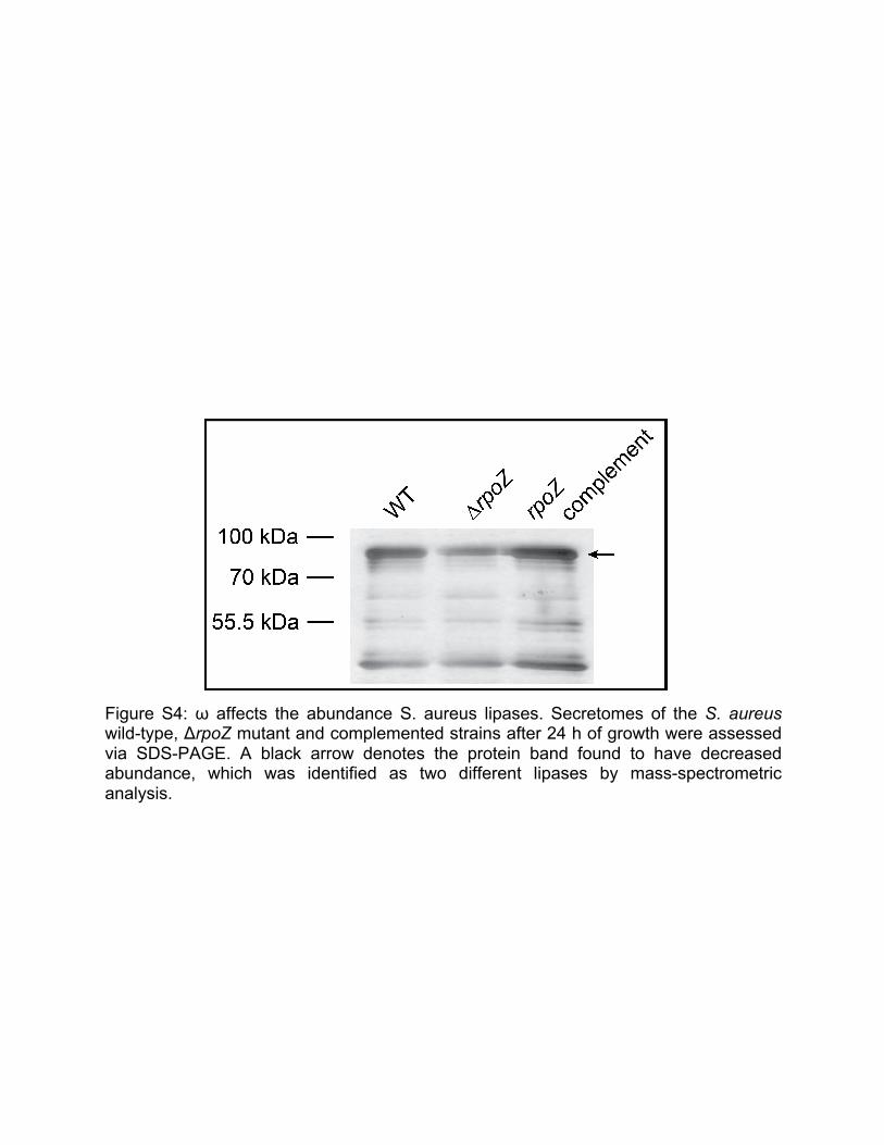
Figure S4: ω affects the abundance S. aureus lipases. Secretomes of the S. aureus wild-type, ΔrpoZ mutant and complemented strains after 24 h of growth were assessed via SDS-PAGE. A black arrow denotes the protein band found to have decreased abundance, which was identified as two different lipases by mass-spectrometric analysis.
Figure S4: ω affects the abundance S. aureus lipases. Secretomes of the S. aureus wild-type, ΔrpoZ mutant and complemented strains after 24 h of growth were assessed via SDS-PAGE. A black arrow denotes the protein band found to have decreased abundance, which was identified as two different lipases by mass-spectrometric analysis.
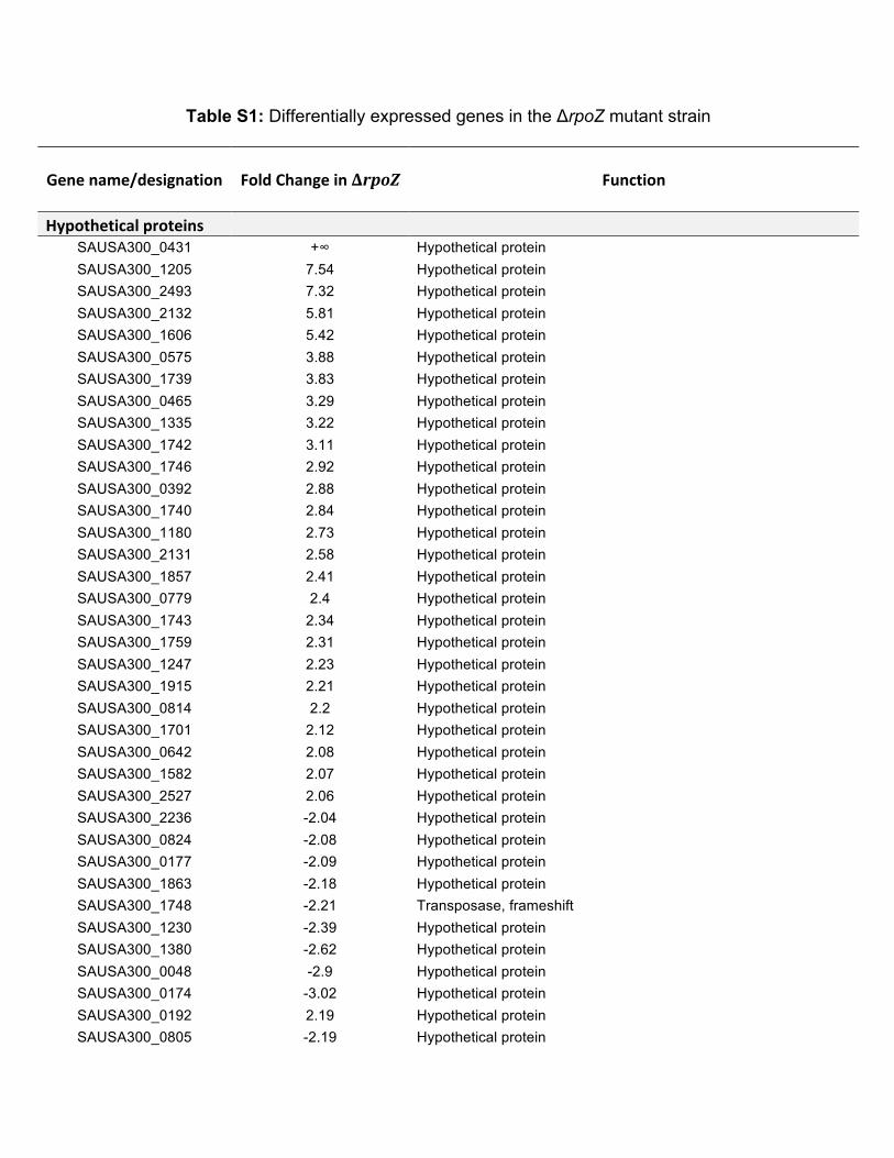
Table S1: Differentially expressed genes in the ΔrpoZ mutant strain
Genename/designation
FoldChangeinΔrpoZ
Function
Hypotheticalproteins
SAUSA300_0431 +∞ Hypothetical protein SAUSA300_1205 7.54 Hypothetical protein SAUSA300_2493 7.32 Hypothetical protein SAUSA300_2132 5.81 Hypothetical protein SAUSA300_1606 5.42 Hypothetical protein SAUSA300_0575 3.88 Hypothetical protein SAUSA300_1739 3.83 Hypothetical protein SAUSA300_0465 3.29 Hypothetical protein SAUSA300_1335 3.22 Hypothetical protein SAUSA300_1742 3.11 Hypothetical protein SAUSA300_1746 2.92 Hypothetical protein SAUSA300_0392 2.88 Hypothetical protein SAUSA300_1740 2.84 Hypothetical protein SAUSA300_1180 2.73 Hypothetical protein SAUSA300_2131 2.58 Hypothetical protein SAUSA300_1857 2.41 Hypothetical protein SAUSA300_0779 2.4 Hypothetical protein SAUSA300_1743 2.34 Hypothetical protein SAUSA300_1759 2.31 Hypothetical protein SAUSA300_1247 2.23 Hypothetical protein SAUSA300_1915 2.21 Hypothetical protein SAUSA300_0814 2.2 Hypothetical protein SAUSA300_1701 2.12 Hypothetical protein SAUSA300_0642 2.08 Hypothetical protein SAUSA300_1582 2.07 Hypothetical protein SAUSA300_2527 2.06 Hypothetical protein SAUSA300_2236 -2.04 Hypothetical protein SAUSA300_0824 -2.08 Hypothetical protein SAUSA300_0177 -2.09 Hypothetical protein SAUSA300_1863 -2.18 Hypothetical protein SAUSA300_1748 -2.21 Transposase, frameshift SAUSA300_1230 -2.39 Hypothetical protein SAUSA300_1380 -2.62 Hypothetical protein SAUSA300_0048 -2.9 Hypothetical protein SAUSA300_0174 -3.02 Hypothetical protein SAUSA300_0192 2.19 Hypothetical protein SAUSA300_0805 -2.19 Hypothetical protein
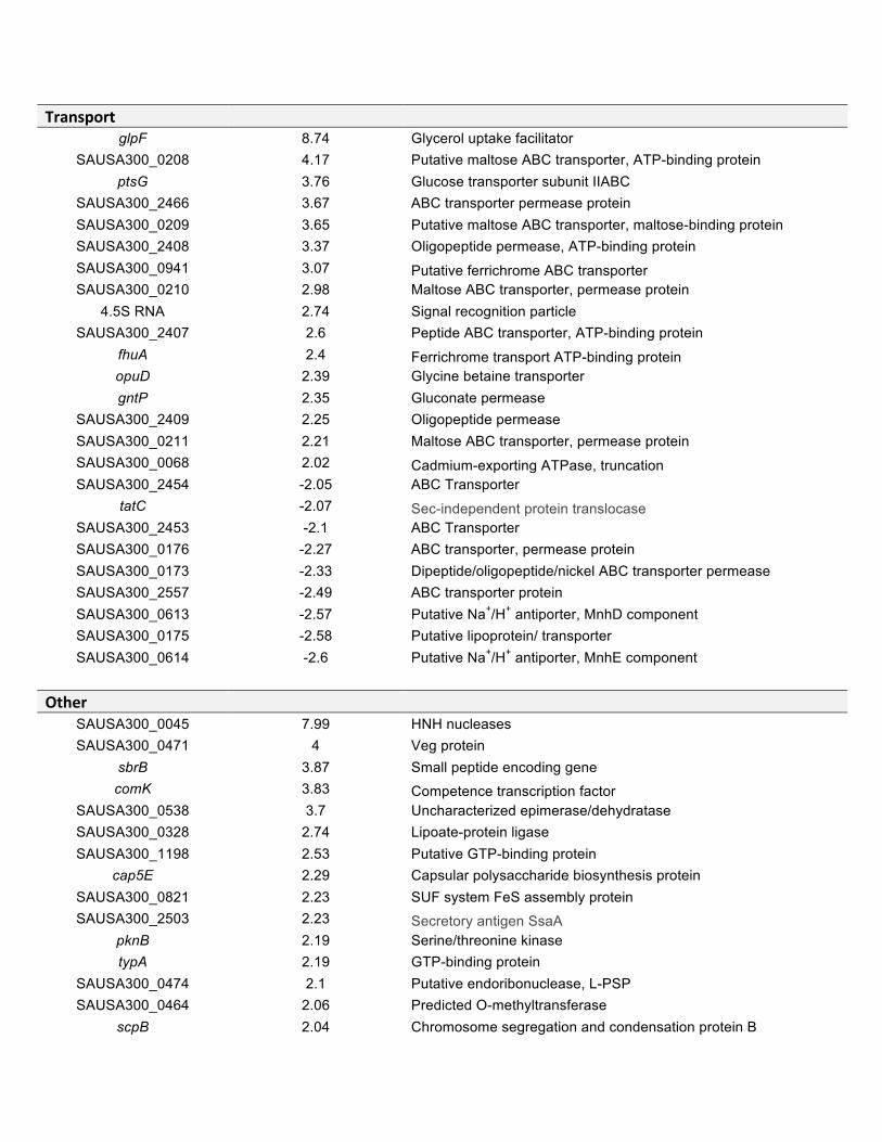
Transport glpF 8.74 Glycerol uptake facilitator
SAUSA300_0208 4.17 Putative maltose ABC transporter, ATP-binding protein ptsG 3.76 Glucose transporter subunit IIABC
SAUSA300_2466 3.67 ABC transporter permease protein SAUSA300_0209 3.65 Putative maltose ABC transporter, maltose-binding protein SAUSA300_2408 3.37 Oligopeptide permease, ATP-binding protein SAUSA300_0941 3.07 Putative ferrichrome ABC transporter SAUSA300_0210 2.98 Maltose ABC transporter, permease protein
4.5S RNA 2.74 Signal recognition particle SAUSA300_2407 2.6 Peptide ABC transporter, ATP-binding protein
fhuA 2.4 Ferrichrome transport ATP-binding protein opuD 2.39 Glycine betaine transporter gntP 2.35 Gluconate permease
SAUSA300_2409 2.25 Oligopeptide permease SAUSA300_0211 2.21 Maltose ABC transporter, permease protein SAUSA300_0068 2.02 Cadmium-exporting ATPase, truncation SAUSA300_2454 -2.05 ABC Transporter
tatC -2.07 Sec-independent protein translocase SAUSA300_2453 -2.1 ABC Transporter SAUSA300_0176 -2.27 ABC transporter, permease protein SAUSA300_0173 -2.33 Dipeptide/oligopeptide/nickel ABC transporter permease SAUSA300_2557 -2.49 ABC transporter protein SAUSA300_0613 -2.57 Putative Na+/H+ antiporter, MnhD component SAUSA300_0175 -2.58 Putative lipoprotein/ transporter SAUSA300_0614 -2.6 Putative Na+/H+ antiporter, MnhE component
Other SAUSA300_0045 7.99 HNH nucleases SAUSA300_0471 4 Veg protein
sbrB 3.87 Small peptide encoding gene comK 3.83 Competence transcription factor
SAUSA300_0538 3.7 Uncharacterized epimerase/dehydratase SAUSA300_0328 2.74 Lipoate-protein ligase SAUSA300_1198 2.53 Putative GTP-binding protein
cap5E 2.29 Capsular polysaccharide biosynthesis protein SAUSA300_0821 2.23 SUF system FeS assembly protein SAUSA300_2503 2.23 Secretory antigen SsaA
pknB 2.19 Serine/threonine kinase typA 2.19 GTP-binding protein
SAUSA300_0474 2.1 Putative endoribonuclease, L-PSP SAUSA300_0464 2.06 Predicted O-methyltransferase
scpB 2.04 Chromosome segregation and condensation protein B
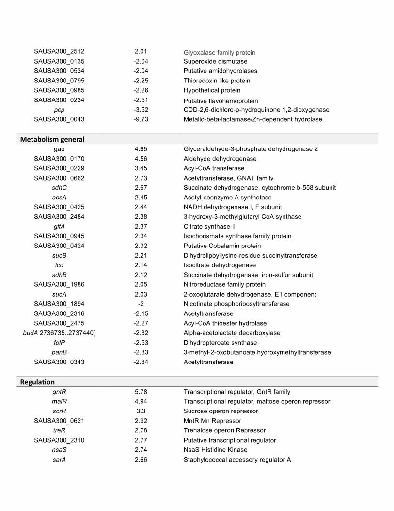
SAUSA300_2512 2.01 Glyoxalase family protein SAUSA300_0135 -2.04 Superoxide dismutase SAUSA300_0534 -2.04 Putative amidohydrolases SAUSA300_0795 -2.25 Thioredoxin like protein SAUSA300_0985 -2.26 Hypothetical protein SAUSA300_0234 -2.51 Putative flavohemoprotein
pcp -3.52 CDD-2,6-dichloro-p-hydroquinone 1,2-dioxygenase SAUSA300_0043 -9.73 Metallo-beta-lactamase/Zn-dependent hydrolase
Metabolismgeneral gap 4.65 Glyceraldehyde-3-phosphate dehydrogenase 2
SAUSA300_0170 4.56 Aldehyde dehydrogenase SAUSA300_0229 3.45 Acyl-CoA transferase SAUSA300_0662 2.73 Acetyltransferase, GNAT family
sdhC 2.67 Succinate dehydrogenase, cytochrome b-558 subunit acsA 2.45 Acetyl-coenzyme A synthetase
SAUSA300_0425 2.44 NADH dehydrogenase I, F subunit SAUSA300_2484 2.38 3-hydroxy-3-methylglutaryl CoA synthase
gltA 2.37 Citrate synthase II SAUSA300_0945 2.34 Isochorismate synthase family protein SAUSA300_0424 2.32 Putative Cobalamin protein
sucB 2.21 Dihydrolipoyllysine-residue succinyltransferase icd 2.14 Isocitrate dehydrogenase
sdhB 2.12 Succinate dehydrogenase, iron-sulfur subunit SAUSA300_1986 2.05 Nitroreductase family protein
sucA 2.03 2-oxoglutarate dehydrogenase, E1 component SAUSA300_1894 -2 Nicotinate phosphoribosyltransferase SAUSA300_2316 -2.15 Acetyltransferase SAUSA300_2475 -2.27 Acyl-CoA thioester hydrolase
budA 2736735..2737440) -2.32 Alpha-acetolactate decarboxylase folP -2.53 Dihydropteroate synthase
panB -2.83 3-methyl-2-oxobutanoate hydroxymethyltransferase SAUSA300_0343 -2.84 Acetyltransferase
Regulation gntR 5.78 Transcriptional regulator, GntR family malR 4.94 Transcriptional regulator, maltose operon repressor scrR 3.3 Sucrose operon repressor
SAUSA300_0621 2.92 MntR Mn Repressor treR 2.78 Trehalose operon Repressor
SAUSA300_2310 2.77 Putative transcriptional regulator nsaS 2.74 NsaS Histidine Kinase sarA 2.66 Staphylococcal accessory regulator A
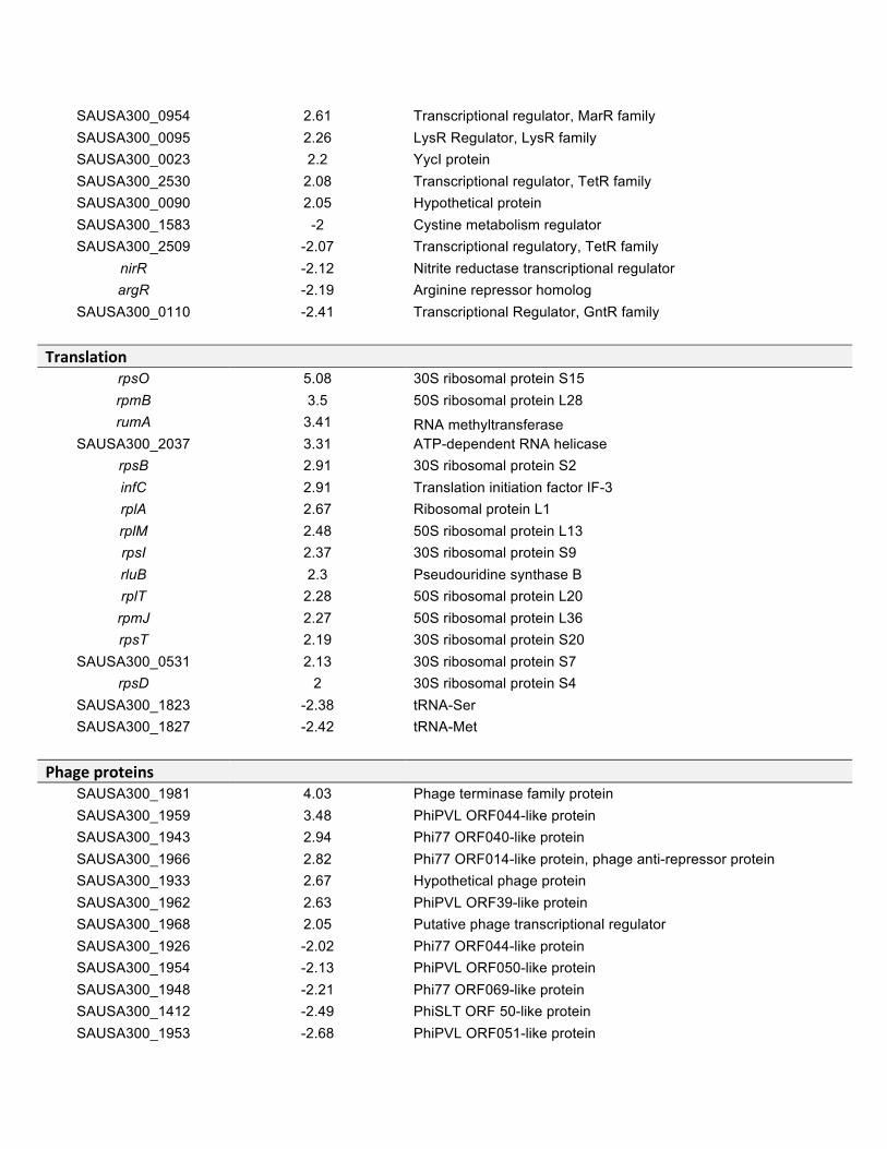
SAUSA300_0954 2.61 Transcriptional regulator, MarR family SAUSA300_0095 2.26 LysR Regulator, LysR family SAUSA300_0023 2.2 YycI protein SAUSA300_2530 2.08 Transcriptional regulator, TetR family SAUSA300_0090 2.05 Hypothetical protein SAUSA300_1583 -2 Cystine metabolism regulator SAUSA300_2509 -2.07 Transcriptional regulatory, TetR family
nirR -2.12 Nitrite reductase transcriptional regulator argR -2.19 Arginine repressor homolog
SAUSA300_0110 -2.41 Transcriptional Regulator, GntR family
Translation rpsO 5.08 30S ribosomal protein S15 rpmB 3.5 50S ribosomal protein L28 rumA 3.41 RNA methyltransferase
SAUSA300_2037 3.31 ATP-dependent RNA helicase rpsB 2.91 30S ribosomal protein S2 infC 2.91 Translation initiation factor IF-3 rplA 2.67 Ribosomal protein L1 rplM 2.48 50S ribosomal protein L13 rpsI 2.37 30S ribosomal protein S9 rluB 2.3 Pseudouridine synthase B rplT 2.28 50S ribosomal protein L20 rpmJ 2.27 50S ribosomal protein L36 rpsT 2.19 30S ribosomal protein S20
SAUSA300_0531 2.13 30S ribosomal protein S7 rpsD 2 30S ribosomal protein S4
SAUSA300_1823 -2.38 tRNA-Ser SAUSA300_1827 -2.42 tRNA-Met
Phageproteins SAUSA300_1981 4.03 Phage terminase family protein SAUSA300_1959 3.48 PhiPVL ORF044-like protein SAUSA300_1943 2.94 Phi77 ORF040-like protein SAUSA300_1966 2.82 Phi77 ORF014-like protein, phage anti-repressor protein SAUSA300_1933 2.67 Hypothetical phage protein SAUSA300_1962 2.63 PhiPVL ORF39-like protein SAUSA300_1968 2.05 Putative phage transcriptional regulator SAUSA300_1926 -2.02 Phi77 ORF044-like protein SAUSA300_1954 -2.13 PhiPVL ORF050-like protein SAUSA300_1948 -2.21 Phi77 ORF069-like protein SAUSA300_1412 -2.49 PhiSLT ORF 50-like protein SAUSA300_1953 -2.68 PhiPVL ORF051-like protein
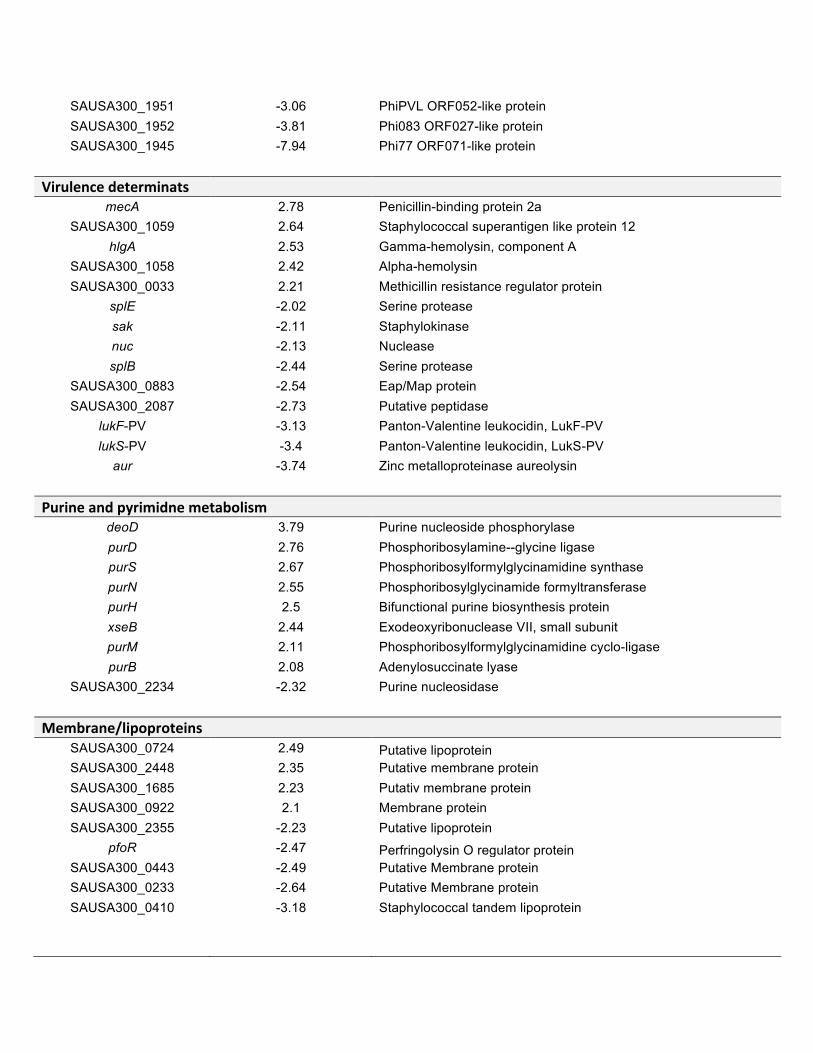
SAUSA300_1951 -3.06 PhiPVL ORF052-like protein SAUSA300_1952 -3.81 Phi083 ORF027-like protein SAUSA300_1945 -7.94 Phi77 ORF071-like protein
Virulencedeterminats mecA 2.78 Penicillin-binding protein 2a
SAUSA300_1059 2.64 Staphylococcal superantigen like protein 12 hlgA 2.53 Gamma-hemolysin, component A
SAUSA300_1058 2.42 Alpha-hemolysin SAUSA300_0033 2.21 Methicillin resistance regulator protein
splE -2.02 Serine protease sak -2.11 Staphylokinase nuc -2.13 Nuclease splB -2.44 Serine protease
SAUSA300_0883 -2.54 Eap/Map protein SAUSA300_2087 -2.73 Putative peptidase
lukF-PV -3.13 Panton-Valentine leukocidin, LukF-PV lukS-PV -3.4 Panton-Valentine leukocidin, LukS-PV
aur -3.74 Zinc metalloproteinase aureolysin
Purineandpyrimidnemetabolism deoD 3.79 Purine nucleoside phosphorylase purD 2.76 Phosphoribosylamine--glycine ligase purS 2.67 Phosphoribosylformylglycinamidine synthase purN 2.55 Phosphoribosylglycinamide formyltransferase purH 2.5 Bifunctional purine biosynthesis protein xseB 2.44 Exodeoxyribonuclease VII, small subunit purM 2.11 Phosphoribosylformylglycinamidine cyclo-ligase purB 2.08 Adenylosuccinate lyase
SAUSA300_2234 -2.32 Purine nucleosidase
Membrane/lipoproteins SAUSA300_0724 2.49 Putative lipoprotein SAUSA300_2448 2.35 Putative membrane protein SAUSA300_1685 2.23 Putativ membrane protein SAUSA300_0922 2.1 Membrane protein SAUSA300_2355 -2.23 Putative lipoprotein
pfoR -2.47 Perfringolysin O regulator protein SAUSA300_0443 -2.49 Putative Membrane protein SAUSA300_0233 -2.64 Putative Membrane protein SAUSA300_0410 -3.18 Staphylococcal tandem lipoprotein
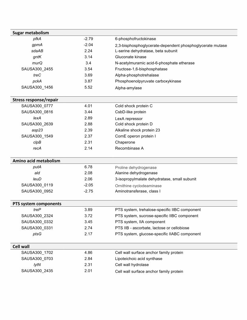
SugarmetabolismpfkA -2.79 6-phosphofructokinase
gpmA -2.04 2,3-bisphosphoglycerate-dependent phosphoglycerate mutase sdaAB 2.24 L-serine dehydratase, beta subunit gntK 3.14 Gluconate kinase murQ 3.4 N-acetylmuramic acid-6-phosphate etherase
SAUSA300_2455 3.54 Fructose-1,6-bisphosphatase treC 3.69 Alpha-phosphotrehalase pckA 3.87 Phosphoenolpyruvate carboxykinase
SAUSA300_1456 5.52 Alpha-amylase
Stressresponse/repair SAUSA300_0777 4.01 Cold shock protein C SAUSA300_0816 3.44 CsbD-like protein
lexA 2.89 LexA repressor SAUSA300_2639 2.88 Cold shock protein D
asp23 2.39 Alkaline shock protein 23 SAUSA300_1549 2.37 ComE operon protein I
clpB 2.31 Chaperone recA 2.14 Recombinase A
Aminoacidmetabolism putA 6.78 Proline dehydrogenase ald 2.08 Alanine dehydrogenase
leuD 2.06 3-isopropylmalate dehydratase, small subunit SAUSA300_0119 -2.05 Ornithine cyclodeaminase SAUSA300_0952 -2.75 Aminotransferase, class I
PTSsystemcomponents treP 3.89 PTS system, trehalose-specific IIBC component
SAUSA300_2324 3.72 PTS system, sucrose-specific IIBC component SAUSA300_0332 3.45 PTS system, IIA component SAUSA300_0331 2.74 PTS IIB - ascorbate, lactose or cellobiose
ptsG 2.17 PTS system, glucose-specific IIABC component
Cellwall SAUSA300_1702 4.86 Cell wall surface anchor family protein SAUSA300_0703 2.84 Lipoteichoic acid synthase
lytN 2.31 Cell wall hydrolase SAUSA300_2435 2.01 Cell wall surface anchor family protein
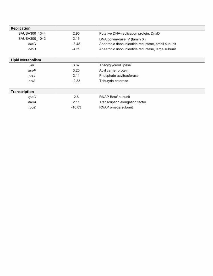
Replication SAUSA300_1344 2.95 Putative DNA-replication protein, DnaD SAUSA300_1042 2.15 DNA polymerase IV (family X)
nrdG -3.48 Anaerobic ribonucleotide reductase, small subunit nrdD -4.59 Anaerobic ribonucleotide reductase, large subunit
LipidMetabolism lip 3.67 Triacyglycerol lipase
acpP 3.25 Acyl carrier protein
plsX 2.11 Phosphate acyltrasferase estA -2.33 Tributyrin esterase
Transcription rpoC 2.6 RNAP Beta' subunit nusA 2.11 Transcription elongation factor rpoZ -10.03 RNAP omega subunit
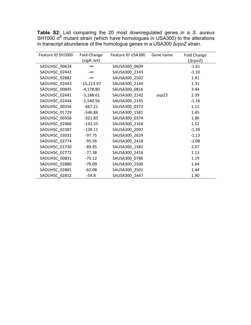
Table S2: List comparing the 20 most downregulated genes in a S. aureus SH1000 σB mutant strain (which have homologues in USA300) to the alterations in transcript abundance of the homologue genes in a USA300 ΔrpoZ strain.
FeatureIDSH1000
Fold-Change(sigB::tet)
FeatureIDUSA300
Genename
FoldChange(ΔrpoZ)
SAOUHSC_00624 -∞ SAUSA300_0609
-1.61SAOUHSC_02442 -∞ SAUSA300_2143
-1.10
SAOUHSC_02882 -∞ SAUSA300_2502
1.41SAOUHSC_02443 -15,213.97 SAUSA300_2144
1.31
SAOUHSC_00845 -4,178.80 SAUSA300_0816
3.44SAOUHSC_02441 -3,188.61 SAUSA300_2142 asp23 2.39SAOUHSC_02444 -1,540.56 SAUSA300_2145
-1.16
SAOUHSC_00356 -667.21 SAUSA300_0372
1.11SAOUHSC_01729 -546.86 SAUSA300_1581
1.45
SAOUHSC_00358 -321.83 SAUSA300_0374
1.86SAOUHSC_02466 -142.55 SAUSA300_2164
1.52
SAOUHSC_02387 -139.11 SAUSA300_2097
-1.39SAOUHSC_03032 -97.75 SAUSA300_2629
-1.13
SAOUHSC_02774 -95.56 SAUSA300_2418
-1.08SAOUHSC_01730 -89.95 SAUSA300_1582
2.07
SAOUHSC_02772 -77.38 SAUSA300_2416
1.13SAOUHSC_00831 -75.12 SAUSA300_0786
1.19
SAOUHSC_02880 -70.09 SAUSA300_2500
1.64SAOUHSC_02881 -62.08 SAUSA300_2501
1.44
SAOUHSC_02812 -54.8 SAUSA300_2447
1.90
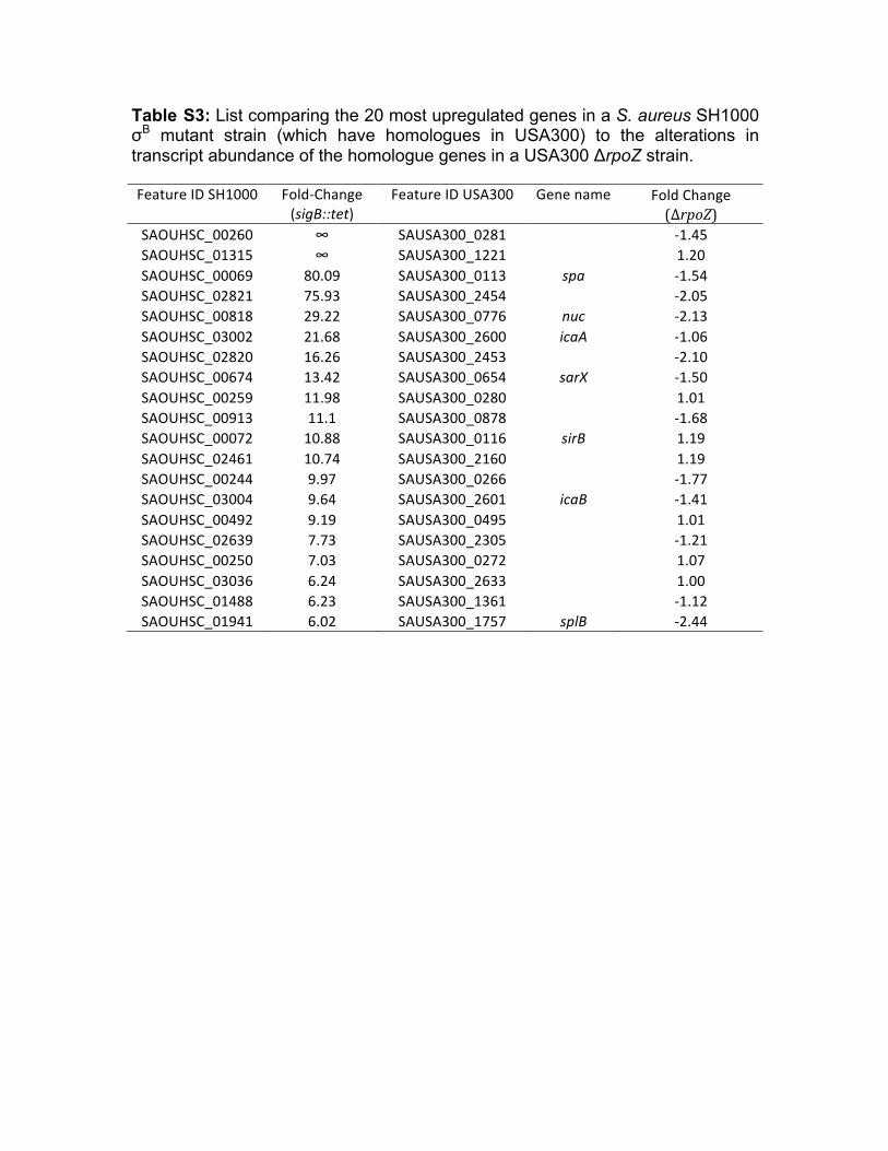
Table S3: List comparing the 20 most upregulated genes in a S. aureus SH1000 σB mutant strain (which have homologues in USA300) to the alterations in transcript abundance of the homologue genes in a USA300 ΔrpoZ strain.
FeatureIDSH1000
Fold-Change(sigB::tet)
FeatureIDUSA300
Genename
FoldChange(ΔrpoZ)
SAOUHSC_00260 ∞ SAUSA300_0281
-1.45SAOUHSC_01315 ∞ SAUSA300_1221
1.20
SAOUHSC_00069 80.09 SAUSA300_0113 spa -1.54SAOUHSC_02821 75.93 SAUSA300_2454
-2.05
SAOUHSC_00818 29.22 SAUSA300_0776 nuc -2.13SAOUHSC_03002 21.68 SAUSA300_2600 icaA -1.06SAOUHSC_02820 16.26 SAUSA300_2453
-2.10
SAOUHSC_00674 13.42 SAUSA300_0654 sarX -1.50SAOUHSC_00259 11.98 SAUSA300_0280
1.01
SAOUHSC_00913 11.1 SAUSA300_0878
-1.68SAOUHSC_00072 10.88 SAUSA300_0116 sirB 1.19SAOUHSC_02461 10.74 SAUSA300_2160
1.19
SAOUHSC_00244 9.97 SAUSA300_0266
-1.77SAOUHSC_03004 9.64 SAUSA300_2601 icaB -1.41SAOUHSC_00492 9.19 SAUSA300_0495
1.01
SAOUHSC_02639 7.73 SAUSA300_2305
-1.21SAOUHSC_00250 7.03 SAUSA300_0272
1.07
SAOUHSC_03036 6.24 SAUSA300_2633
1.00SAOUHSC_01488 6.23 SAUSA300_1361
-1.12
SAOUHSC_01941 6.02 SAUSA300_1757 splB -2.44

164
APPENDIX IV
American Society for Microbiology Journals LICENSE
TERMS AND CONDITIONS
Apr 05, 2017
This is a License Agreement between Andy Weiss ("You") and American Society for
Microbiology - Journals ("American Society for Microbiology - Journals") provided by
Copyright Clearance Center ("CCC"). The license consists of your order details, the terms
and conditions provided by American Society for Microbiology - Journals, and the payment
terms and conditions.
All payments must be made in full to CCC. For payment instructions, please see
information listed at the bottom of this form.
License Number 4053651327049
License date Feb 19, 2017
Licensed content publisher American Society for Microbiology Journals
Licensed content title Infection and immunity
Licensed content date Jan 1, 1970
Type of Use Thesis/Dissertation
Requestor type Author of requested content
Format Print, Electronic
Portion chapter/article
Title or numeric reference of
the portion(s)
This article is intended to be used as a chapter in my dissertation.
The article, as well as the supplemental data, will be placed in the
appendix of the dissertation
Title of the article or chapter
the portion is from
The δ Subunit of RNA Polymerase Guides Promoter Selectivity and
Virulence in Staphylococcus aureus
Editor of portion(s) A. J. Bäumler
Author of portion(s) Andy Weiss, J. Antonio Ibarra, Jessica Paoletti, Ronan K. Carroll,
and Lindsey N. Shaw
Volume of serial or
monograph.
82
Issue, if republishing an
article from a serial
4
Page range of the portion
Publication date of portion 2014 April
Rights for Main product and any product related to main product
Duration of use Life of current edition
Creation of copies for the
disabled
no
With minor editing privileges yes
For distribution to Worldwide
In the following language(s) Original language of publication
With incidental promotional
use
no
The lifetime unit quantity of
new product
Up to 499
Made available in the
following markets
educational
Specified additional
information
The requesting
person/organization is:
Order reference number
Author/Editor
The standard identifier of
New Work
Title of New Work
Publisher of New Work
Expected publication date
Estimated size (pages)
Total (may include CCC user
fee)
Minor changes might required.
Andy Weiss
Andy Weiss, J. Antonio Ibarra, Jessica Paoletti, Ronan K. Carroll,
and Lindsey N. Shaw
University of South Florida
Nonclassical regulators in Staphylococcus aureus
University of South Florida
May 2017
130
0.00 USD
Terms and Conditions
TERMS AND CONDITIONS
The following terms are individual to this publisher:
We ask that you review the copyright information associated with the article that you are
interested in, as some articles in the ASM Journals are covered by creative commons
licenses that would permit free reuse with attribution.
Please note that ASM cannot grant permission to reuse figures or images that are credited to
publications other than ASM journals. For images credited to non-ASM journal publications,
you will need to obtain permission from the journal referenced in the figure or table legend
or credit line before making any use of the image(s) or table(s). ASM is unable to grant
permission to reuse supplemental material as copyright for the supplemental material
remains with the author.
Other Terms and Conditions:
STANDARD TERMS AND CONDITIONS
1. Description of Service; Defined Terms. This Republication License enables the User to
obtain licenses for republication of one or more copyrighted works as described in detail on
the relevant Order Confirmation (the “Work(s)”). Copyright Clearance Center, Inc. (“CCC”)
grants licenses through the Service on behalf of the rightsholder identified on the Order
Confirmation (the “Rightsholder”). “Republication”, as used herein, generally means the
inclusion of a Work, in whole or in part, in a new work or works, also as described on the
Order Confirmation. “User”, as used herein, means the person or entity making such
republication.
2. The terms set forth in the relevant Order Confirmation, and any terms set by the
Rightsholder with respect to a particular Work, govern the terms of use of Works in
connection with the Service. By using the Service, the person transacting for a republication
license on behalf of the User represents and warrants that he/she/it (a) has been duly
authorized by the User to accept, and hereby does accept, all such terms and conditions on
behalf of User, and (b) shall inform User of all such terms and conditions. In the event such
person is a “freelancer” or other third party independent of User and CCC, such party shall
be deemed jointly a “User” for purposes of these terms and conditions. In any event, User
shall be deemed to have accepted and agreed to all such terms and conditions if User
republishes the Work in any fashion.
3. Scope of License; Limitations and Obligations.
3.1 All Works and all rights therein, including copyright rights, remain the sole and
exclusive property of the Rightsholder. The license created by the exchange of an Order
Confirmation (and/or any invoice) and payment by User of the full amount set forth on that
document includes only those rights expressly set forth in the Order Confirmation and in
these terms and conditions, and conveys no other rights in the Work(s) to User. All rights not
expressly granted are hereby reserved.
3.2 General Payment Terms: You may pay by credit card or through an account with us
payable at the end of the month. If you and we agree that you may establish a standing
account with CCC, then the following terms apply: Remit Payment to: Copyright Clearance
Center, 29118 Network Place, Chicago, IL 60673-1291. Payments Due: Invoices are payable
upon their delivery to you (or upon our notice to you that they are available to you for
downloading). After 30 days, outstanding amounts will be subject to a service charge of 1-
1/2% per month or, if less, the maximum rate allowed by applicable law. Unless otherwise
specifically set forth in the Order Confirmation or in a separate written agreement signed by
CCC, invoices are due and payable on “net 30” terms. While User may exercise the rights
licensed immediately upon issuance of the Order Confirmation, the license is automatically
revoked and is null and void, as if it had never been issued, if complete payment for the
license is not received on a timely basis either from User directly or through a payment
agent, such as a credit card company.
3.3 Unless otherwise provided in the Order Confirmation, any grant of rights to User (i) is
“one-time” (including the editions and product family specified in the license), (ii) is non-
exclusive and non-transferable and (iii) is subject to any and all limitations and restrictions
(such as, but not limited to, limitations on duration of use or circulation) included in the
Order Confirmation or invoice and/or in these terms and conditions. Upon completion of the
licensed use, User shall either secure a new permission for further use of the Work(s) or
immediately cease any new use of the Work(s) and shall render inaccessible (such as by
deleting or by removing or severing links or other locators) any further copies of the Work
(except for copies printed on paper in accordance with this license and still in User's stock at
the end of such period).
3.4 In the event that the material for which a republication license is sought includes third
party materials (such as photographs, illustrations, graphs, inserts and similar materials)
which are identified in such material as having been used by permission, User is responsible
for identifying, and seeking separate licenses (under this Service or otherwise) for, any of
such third party materials; without a separate license, such third party materials may not be
used.
3.5 Use of proper copyright notice for a Work is required as a condition of any license
granted under the Service. Unless otherwise provided in the Order Confirmation, a proper
copyright notice will read substantially as follows: “Republished with permission of
[Rightsholder’s name], from [Work's title, author, volume, edition number and year of
copyright]; permission conveyed through Copyright Clearance Center, Inc. ” Such notice
must be provided in a reasonably legible font size and must be placed either immediately
adjacent to the Work as used (for example, as part of a by-line or footnote but not as a
separate electronic link) or in the place where substantially all other credits or notices for the
new work containing the republished Work are located. Failure to include the required notice
results in loss to the Rightsholder and CCC, and the User shall be liable to pay liquidated
damages for each such failure equal to twice the use fee specified in the Order Confirmation,
in addition to the use fee itself and any other fees and charges specified.
3.6 User may only make alterations to the Work if and as expressly set forth in the Order
Confirmation. No Work may be used in any way that is defamatory, violates the rights of
third parties (including such third parties' rights of copyright, privacy, publicity, or other
tangible or intangible property), or is otherwise illegal, sexually explicit or obscene. In
addition, User may not conjoin a Work with any other material that may result in damage to
the reputation of the Rightsholder. User agrees to inform CCC if it becomes aware of any
infringement of any rights in a Work and to cooperate with any reasonable request of CCC
or the Rightsholder in connection therewith.
4. Indemnity. User hereby indemnifies and agrees to defend the Rightsholder and CCC, and
their respective employees and directors, against all claims, liability, damages, costs and
expenses, including legal fees and expenses, arising out of any use of a Work beyond the
scope of the rights granted herein, or any use of a Work which has been altered in any
unauthorized way by User, including claims of defamation or infringement of rights of
copyright, publicity, privacy or other tangible or intangible property.
5. Limitation of Liability. UNDER NO CIRCUMSTANCES WILL CCC OR THE
RIGHTSHOLDER BE LIABLE FOR ANY DIRECT, INDIRECT, CONSEQUENTIAL OR
INCIDENTAL DAMAGES (INCLUDING WITHOUT LIMITATION DAMAGES FOR

LOSS OF BUSINESS PROFITS OR INFORMATION, OR FOR BUSINESS
INTERRUPTION) ARISING OUT OF THE USE OR INABILITY TO USE A WORK,
EVEN IF ONE OF THEM HAS BEEN ADVISED OF THE POSSIBILITY OF SUCH
DAMAGES. In any event, the total liability of the Rightsholder and CCC (including their
respective employees and directors) shall not exceed the total amount actually paid by User
for this license. User assumes full liability for the actions and omissions of its principals,
employees, agents, affiliates, successors and assigns.
6. Limited Warranties. THE WORK(S) AND RIGHT(S) ARE PROVIDED “AS IS”. CCC
HAS THE RIGHT TO GRANT TO USER THE RIGHTS GRANTED IN THE ORDER
CONFIRMATION DOCUMENT. CCC AND THE RIGHTSHOLDER DISCLAIM ALL
OTHER WARRANTIES RELATING TO THE WORK(S) AND RIGHT(S), EITHER
EXPRESS OR IMPLIED, INCLUDING WITHOUT LIMITATION IMPLIED
WARRANTIES OF MERCHANTABILITY OR FITNESS FOR A PARTICULAR
PURPOSE. ADDITIONAL RIGHTS MAY BE REQUIRED TO USE ILLUSTRATIONS,
GRAPHS, PHOTOGRAPHS, ABSTRACTS, INSERTS OR OTHER PORTIONS OF THE
WORK (AS OPPOSED TO THE ENTIRE WORK) IN A MANNER CONTEMPLATED
BY USER; USER UNDERSTANDS AND AGREES THAT NEITHER CCC NOR THE
RIGHTSHOLDER MAY HAVE SUCH ADDITIONAL RIGHTS TO GRANT.
7. Effect of Breach. Any failure by User to pay any amount when due, or any use by User of
a Work beyond the scope of the license set forth in the Order Confirmation and/or these
terms and conditions, shall be a material breach of the license created by the Order
Confirmation and these terms and conditions. Any breach not cured within 30 days of
written notice thereof shall result in immediate termination of such license without further
notice. Any unauthorized (but licensable) use of a Work that is terminated immediately upon
notice thereof may be liquidated by payment of the Rightsholder's ordinary license price
therefor; any unauthorized (and unlicensable) use that is not terminated immediately for any
reason (including, for example, because materials containing the Work cannot reasonably be
recalled) will be subject to all remedies available at law or in equity, but in no event to a
payment of less than three times the Rightsholder's ordinary license price for the most
closely analogous licensable use plus Rightsholder's and/or CCC's costs and expenses
incurred in collecting such payment.
8. Miscellaneous.
8.1 User acknowledges that CCC may, from time to time, make changes or additions to the
Service or to these terms and conditions, and CCC reserves the right to send notice to the
User by electronic mail or otherwise for the purposes of notifying User of such changes or
additions; provided that any such changes or additions shall not apply to permissions already
secured and paid for.
8.2 Use of User-related information collected through the Service is governed by CCC’s
privacy policy, available online here:
http://www.copyright.com/content/cc3/en/tools/footer/privacypolicy.html.
8.3 The licensing transaction described in the Order Confirmation is personal to User.
Therefore, User may not assign or transfer to any other person (whether a natural person or
an organization of any kind) the license created by the Order Confirmation and these terms
and conditions or any rights granted hereunder; provided, however, that User may assign
such license in its entirety on written notice to CCC in the event of a transfer of all or
substantially all of User’s rights in the new material which includes the Work(s) licensed
under this Service.
8.4 No amendment or waiver of any terms is binding unless set forth in writing and signed
by the parties. The Rightsholder and CCC hereby object to any terms contained in any
writing prepared by the User or its principals, employees, agents or affiliates and purporting
to govern or otherwise relate to the licensing transaction described in the Order
Confirmation, which terms are in any way inconsistent with any terms set forth in the Order
Confirmation and/or in these terms and conditions or CCC's standard operating procedures,
whether such writing is prepared prior to, simultaneously with or subsequent to the Order
Confirmation, and whether such writing appears on a copy of the Order Confirmation or in a
separate instrument.
8.5 The licensing transaction described in the Order Confirmation document shall be
governed by and construed under the law of the State of New York, USA, without regard to

the principles thereof of conflicts of law. Any case, controversy, suit, action, or proceeding
arising out of, in connection with, or related to such licensing transaction shall be brought, at
CCC's sole discretion, in any federal or state court located in the County of New York, State
of New York, USA, or in any federal or state court whose geographical jurisdiction covers
the location of the Rightsholder set forth in the Order Confirmation. The parties expressly
submit to the personal jurisdiction and venue of each such federal or state court.If you have
any comments or questions about the Service or Copyright Clearance Center, please contact
us at 978-750-8400 or send an e-mail to [email protected].
v 1.1
Questions? [email protected] or +18552393415 (toll free in the US) or
+19786462777.
Genome-wide Annotation, Identification, and Global TranscriptomicAnalysis of Regulatory or Small RNA Gene Expression inStaphylococcus aureus
Ronan K. Carroll,a Andy Weiss,b William H. Broach,b Richard E. Wiemels,a Austin B. Mogen,c Kelly C. Rice,c Lindsey N. Shawb
Department of Biological Sciences, Ohio University, Athens, Ohio, USAa; Department of Cell Biology, Microbiology and Molecular Biology, University of South Florida,Tampa, Florida, USAb; Department of Microbiology and Cell Science, Institute of Food and Agricultural Sciences, University of Florida, Gainesville, Florida, USAc
R.K.C. and A.W. contributed equally to this work.
ABSTRACT In Staphylococcus aureus, hundreds of small regulatory or small RNAs (sRNAs) have been identified, yet this class ofmolecule remains poorly understood and severely understudied. sRNA genes are typically absent from genome annotation files,and as a consequence, their existence is often overlooked, particularly in global transcriptomic studies. To facilitate improveddetection and analysis of sRNAs in S. aureus, we generated updated GenBank files for three commonly used S. aureus strains(MRSA252, NCTC 8325, and USA300), in which we added annotations for >260 previously identified sRNAs. These files, the firstto include genome-wide annotation of sRNAs in S. aureus, were then used as a foundation to identify novel sRNAs in thecommunity-associated methicillin-resistant strain USA300. This analysis led to the discovery of 39 previously unidentified sR-NAs. Investigating the genomic loci of the newly identified sRNAs revealed a surprising degree of inconsistency in genome anno-tation in S. aureus, which may be hindering the analysis and functional exploration of these elements. Finally, using our newlycreated annotation files as a reference, we perform a global analysis of sRNA gene expression in S. aureus and demonstrate thatthe newly identified tsr25 is the most highly upregulated sRNA in human serum. This study provides an invaluable resource tothe S. aureus research community in the form of our newly generated annotation files, while at the same time presenting the firstexamination of differential sRNA expression in pathophysiologically relevant conditions.
IMPORTANCE Despite a large number of studies identifying regulatory or small RNA (sRNA) genes in Staphylococcus aureus,their annotation is notably lacking in available genome files. In addition to this, there has been a considerable lack of cross-referencing in the wealth of studies identifying these elements, often leading to the same sRNA being identified multiple timesand bearing multiple names. In this work, we have consolidated and curated known sRNA genes from the literature and mappedthem to their position on the S. aureus genome, creating new genome annotation files. These files can now be used by the scien-tific community at large in experiments to search for previously undiscovered sRNA genes and to monitor sRNA gene expressionby transcriptome sequencing (RNA-seq). We demonstrate this application, identifying 39 new sRNAs and studying their expres-sion during S. aureus growth in human serum.
Received 13 November 2015 Accepted 22 December 2015 Published 9 February 2016
Citation Carroll RK, Weiss A, Broach WH, Wiemels RE, Mogen AB, Rice KC, Shaw LN. 2016. Genome-wide annotation, identification, and global transcriptomic analysis ofregulatory or small RNA gene expression in Staphylococcus aureus. mBio 7(1):e01990-15. doi:10.1128/mBio.01990-15.
Invited Editor Paul Dunman, University of Rochester Editor Gerald B. Pier, Harvard Medical School
Copyright © 2016 Carroll et al. This is an open-access article distributed under the terms of the Creative Commons Attribution-Noncommercial-ShareAlike 3.0 Unportedlicense, which permits unrestricted noncommercial use, distribution, and reproduction in any medium, provided the original author and source are credited.
Address correspondence to Ronan K. Carroll, [email protected], or Lindsey Shaw, [email protected].
In recent years, a number of studies have been carried out, em-ploying both experimental and computational methods to iden-
tify regulatory or small RNAs (sRNAs) in Staphylococcus aureus(1–12). Hundreds of sRNAs have been identified, and many, inaddition to the agr effector RNAIII, have been shown to play a rolein gene regulation (while these molecules go by a variety of namessuch as regulatory RNAs or noncoding RNAs, we will use sRNAsto refer to them all, as previously recommended [13]). Despiteadvancements in sRNA identification, the roles of most of thesemolecules remain unknown, because in many cases, limited func-tional information can be gathered from analysis of their sequencealone.
One additional factor that has hampered the study of sRNAs inS. aureus has been the lack of a clear nomenclature and annotation
system. This absence of a systematic identification and annotationprocess has led to the repeated discovery of the same sRNAs onmultiple occasions, the reidentification of already known sRNAs(e.g., RNAIII), and even to important protein-coding genes beingascribed as sRNAs (e.g., the -PSM transcript, which is not anno-tated in most S. aureus genome files). Recent work by Sassi et al.(14) established an online database for staphylococcal sRNAs;however, most sRNAs, including the well-studied RNAIII, are stillnot included in annotated S. aureus GenBank genome files. This isa marked oversight, as annotated genome files serve as the refer-ence for global genomic and transcriptomic studies; thus, the ab-sence of sRNAs from these files severely impedes their study andprevents us from gaining an overarching picture of regulatorycircuits.
RESEARCH ARTICLE
crossmark
January/February 2016 Volume 7 Issue 1 e01990-15 ® mbio.asm.org 1
m
bio.asm.org
on February 9, 2016 - P
ublished by m
bio.asm.org
Dow
nloaded from
In S. aureus, most sRNA identification studies have been per-formed in a single background, the hospital-acquired methicillin-resistant S. aureus (MRSA) strain N315. The existence of many ofthese sRNAs has been demonstrated experimentally in strainN315 (1, 3, 4, 6, 7); however, very few of them have been investi-gated in other S. aureus strains, including the epidemiccommunity-associated MRSA (CA-MRSA) strain USA300 (15).As such, their existence, location, and copy number in most S. au-reus isolates is unknown, preventing us from gaining a sense oftheir role in the physiological and pathogenic differences betweenstrains.
To better understand the sRNA content of multiple S. aureusstrains, we explored the genomes of three well-studied S. aureusstrains (USA300, MRSA252, and NCTC 8325). We identified thelocation(s) of previously discovered sRNAs and created newGenBank genome annotation files for each strain, inserting ~260sRNAs. These newly annotated files serve as a valuable resourceallowing us to do the following: (i) search the genome of eachstrain for as yet unidentified sRNAs without mistakenly reidenti-fying known species, and (ii) calculate expression values for thesegenes using transcriptome sequencing (RNA-seq) data.
To demonstrate the application of these new files, we per-formed RNA-seq on CA-MRSA strain USA300 growing in labo-ratory media and human serum and aligned the data to our newlycreated sRNA annotated genome files. Examining the data, weidentified 39 novel putative sRNAs that had not been previouslyreported. These novel sRNAs were annotated, cross-referenced inthe genomes of strains NCTC 8325 and MRSA252, and added tothe newly created genome files as well. During the cross-referencing process, we observed numerous examples of inconsis-tent genome annotation in different S. aureus strains. We high-light examples clearly demonstrating genome misannotation anddemonstrate how this phenomenon is adversely affecting theidentification and characterization of sRNAs. Finally, we calculateexpression values and examine the global sRNA expression profileof strain USA300, uncovering a wealth of molecules that displaydifferential expression in human serum. The latter point is of sig-nificant importance, as it gives us a unique look into the sRNAtranscriptome, not only during growth of S. aureus but also in apathophysiologically relevant growth environment. The new ge-nome annotation files described in this work have been depositedin the figshare depository and are freely available for download.We suggest that these newly reannotated genomes will be a valu-able resource to the S. aureus research community for sRNA iden-tification and analysis hereafter, as they can be incrementallyadded to as new sRNAs are discovered.
RESULTSAnnotation of sRNAs on the S. aureus genome. Previous studiesin our lab have utilized RNA-seq to determine the global tran-scriptomic profile of S. aureus in the community-associatedMRSA strain USA300 (16, 17). Analysis of these data sets revealeda large number of S. aureus transcripts that map to intergenicregions where no protein-coding genes have been annotated. Wehypothesized that many of these transcripts represent sRNAs be-cause of the following. (i) Most S. aureus genome annotation filesdo not contain annotations for sRNA genes. (ii) Recent studiescarried out in the S. aureus N315 background have demonstratedthat there are several hundred sRNAs encoded in the S. aureusgenome (12, 14). To facilitate improved global transcriptomic
analysis of S. aureus by RNA-seq, we created new GenBank ge-nome annotation files for three commonly used S. aureus strains,NCTC 8325, MRSA252, and USA300. To do this, we elected toexpand the sRNA annotation and nomenclature system alreadypresent for 25 sRNAs in strain MRSA252 (see Table S1 in thesupplemental material) and apply a similar annotation/nomen-clature system to strains USA300 and NCTC 8325 (for details, seesupplemental material). To include annotations for knownsRNAs, we performed a literature search to identify studies inwhich sRNAs in S. aureus were reported. A total of 12 papers thatemployed a variety of methods to identify sRNAs, including com-putational approaches, microarray studies, cDNA cloning, andhigh-throughput sequencing, were investigated (1–12). Using theinformation provided in these publications, a list of 928 potentialsRNAs was assembled (Table S2) (1–12). In order to ensure accu-rate annotation of each sRNA from this list, RNA-seq experimentswere performed for each of the three strains growing under stan-dard laboratory conditions. Reads generated from these workswere aligned to the respective genomes and used as a guide toidentify the specific location of each sRNA for each of the threestrains (see Fig. S1 in the supplemental material).
This work condensed the 928 putative sRNAs to a total of 248annotations for strain MRSA252, 254 annotations for strainNCTC 8325, and 264 annotations for strain USA300 (for a more-comprehensive explanation of how sRNAs were identified andannotated, see Text S1 in the supplemental material). Such a dra-matic reduction in the number of sRNAs points to the scale ofoverlapping identification, reidentification, and duplicate namingthat was extant in the literature for these elements and the poorstate of sRNA curation in S. aureus genomes. Our newly generatedGenBank files for all three strains represent the first comprehen-sive list of sRNAs annotated directly in the S. aureus genome,which will serve as a valuable reference point for future sRNAdiscovery, and more broadly, global transcriptomic analyses ofS. aureus by RNA-seq.
Detection of novel sRNAs in strain USA300. The creation ofGenBank files containing annotations for previously identifiedsRNAs provided us with a unique opportunity to examine RNA-seq data for novel forms of these elements, without mistakenlyreidentifying those that are already known. Accordingly, we setout to perform such an exploration using the community-associated MRSA isolate USA300, an undeniably relevant clinicalstrain for which no sRNA identification studies have yet been per-formed. To maximize the probability of identifying novel sRNAs,we performed RNA-seq with USA300 grown under both labora-tory conditions (tryptic soy broth [TSB]), and in media that wasmore pathophysiologically relevant to its infectious lifestyle (hu-man serum) (18).
RNA-seq reads from bacteria grown under these conditionswere mapped to the newly created USA300 GenBank file (contain-ing 264 sRNA annotations), followed by a thorough examinationfor the presence of novel sRNA transcripts. A total of 39 potentialsRNAs were identified from both conditions, which we named tsr1to tsr39 for Tampa small RNA (Table 1) (for details regarding thecriteria used for our determination of novel sRNAs, see Text S1 inthe supplemental material). Although the majority of the tsr geneswere located in intergenic regions, a number were identified asbeing antisense to annotated genes or as partially overlapping an-notated coding DNA sequence (CDS) genes. The novel sRNAswere also added to our newly created USA300 genome file, using
Carroll et al.
2 ® mbio.asm.org January/February 2016 Volume 7 Issue 1 e01990-15
m
bio.asm.org
on February 9, 2016 - P
ublished by m
bio.asm.org
Dow
nloaded from
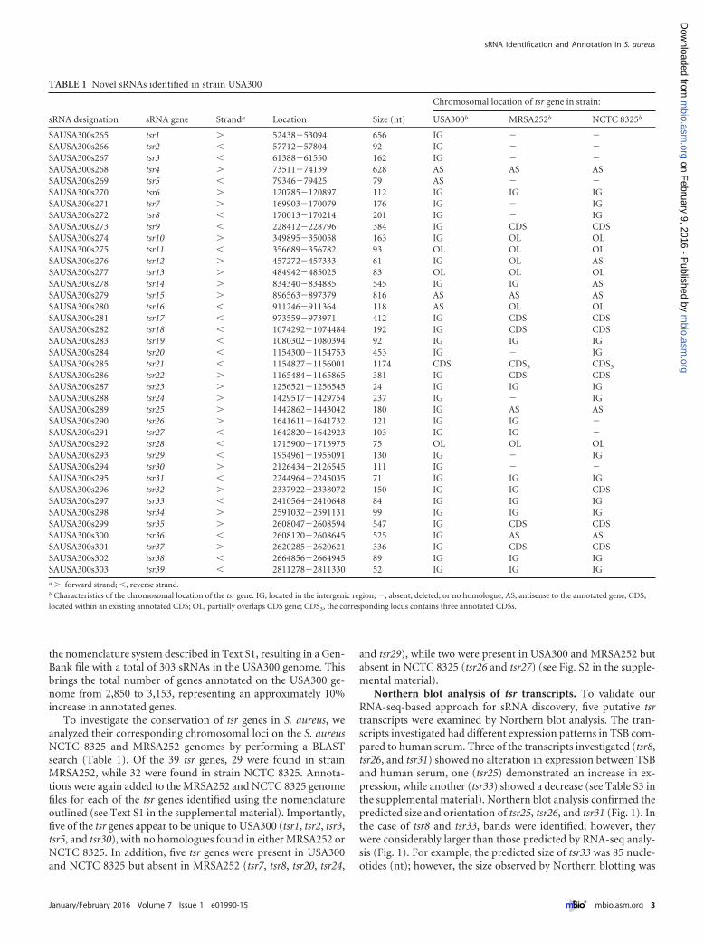
the nomenclature system described in Text S1, resulting in a Gen-Bank file with a total of 303 sRNAs in the USA300 genome. Thisbrings the total number of genes annotated on the USA300 ge-nome from 2,850 to 3,153, representing an approximately 10%increase in annotated genes.
To investigate the conservation of tsr genes in S. aureus, weanalyzed their corresponding chromosomal loci on the S. aureusNCTC 8325 and MRSA252 genomes by performing a BLASTsearch (Table 1). Of the 39 tsr genes, 29 were found in strainMRSA252, while 32 were found in strain NCTC 8325. Annota-tions were again added to the MRSA252 and NCTC 8325 genomefiles for each of the tsr genes identified using the nomenclatureoutlined (see Text S1 in the supplemental material). Importantly,five of the tsr genes appear to be unique to USA300 (tsr1, tsr2, tsr3,tsr5, and tsr30), with no homologues found in either MRSA252 orNCTC 8325. In addition, five tsr genes were present in USA300and NCTC 8325 but absent in MRSA252 (tsr7, tsr8, tsr20, tsr24,
and tsr29), while two were present in USA300 and MRSA252 butabsent in NCTC 8325 (tsr26 and tsr27) (see Fig. S2 in the supple-mental material).
Northern blot analysis of tsr transcripts. To validate ourRNA-seq-based approach for sRNA discovery, five putative tsrtranscripts were examined by Northern blot analysis. The tran-scripts investigated had different expression patterns in TSB com-pared to human serum. Three of the transcripts investigated (tsr8,tsr26, and tsr31) showed no alteration in expression between TSBand human serum, one (tsr25) demonstrated an increase in ex-pression, while another (tsr33) showed a decrease (see Table S3 inthe supplemental material). Northern blot analysis confirmed thepredicted size and orientation of tsr25, tsr26, and tsr31 (Fig. 1). Inthe case of tsr8 and tsr33, bands were identified; however, theywere considerably larger than those predicted by RNA-seq analy-sis (Fig. 1). For example, the predicted size of tsr33 was 85 nucle-otides (nt); however, the size observed by Northern blotting was
TABLE 1 Novel sRNAs identified in strain USA300
sRNA designation sRNA gene Stranda Location Size (nt)
Chromosomal location of tsr gene in strain:
USA300b MRSA252b NCTC 8325b
SAUSA300s265 tsr1 5243853094 656 IG SAUSA300s266 tsr2 5771257804 92 IG SAUSA300s267 tsr3 6138861550 162 IG SAUSA300s268 tsr4 7351174139 628 AS AS ASSAUSA300s269 tsr5 7934679425 79 AS SAUSA300s270 tsr6 120785120897 112 IG IG IGSAUSA300s271 tsr7 169903170079 176 IG IGSAUSA300s272 tsr8 170013170214 201 IG IGSAUSA300s273 tsr9 228412228796 384 IG CDS CDSSAUSA300s274 tsr10 349895350058 163 IG OL OLSAUSA300s275 tsr11 356689356782 93 OL OL OLSAUSA300s276 tsr12 457272457333 61 IG OL ASSAUSA300s277 tsr13 484942485025 83 OL OL OLSAUSA300s278 tsr14 834340834885 545 IG IG ASSAUSA300s279 tsr15 896563897379 816 AS AS ASSAUSA300s280 tsr16 911246911364 118 AS OL OLSAUSA300s281 tsr17 973559973971 412 IG CDS CDSSAUSA300s282 tsr18 10742921074484 192 IG CDS CDSSAUSA300s283 tsr19 10803021080394 92 IG IG IGSAUSA300s284 tsr20 11543001154753 453 IG IGSAUSA300s285 tsr21 11548271156001 1174 CDS CDS3 CDS3
SAUSA300s286 tsr22 11654841165865 381 IG CDS CDSSAUSA300s287 tsr23 12565211256545 24 IG IG IGSAUSA300s288 tsr24 14295171429754 237 IG IGSAUSA300s289 tsr25 14428621443042 180 IG AS ASSAUSA300s290 tsr26 16416111641732 121 IG IG SAUSA300s291 tsr27 16428201642923 103 IG IG SAUSA300s292 tsr28 17159001715975 75 OL OL OLSAUSA300s293 tsr29 19549611955091 130 IG IGSAUSA300s294 tsr30 21264342126545 111 IG SAUSA300s295 tsr31 22449642245035 71 IG IG IGSAUSA300s296 tsr32 23379222338072 150 IG IG CDSSAUSA300s297 tsr33 24105642410648 84 IG IG IGSAUSA300s298 tsr34 25910322591131 99 IG IG IGSAUSA300s299 tsr35 26080472608594 547 IG CDS CDSSAUSA300s300 tsr36 26081202608645 525 IG AS ASSAUSA300s301 tsr37 26202852620621 336 IG CDS CDSSAUSA300s302 tsr38 26648562664945 89 IG IG IGSAUSA300s303 tsr39 28112782811330 52 IG IG IGa , forward strand; , reverse strand.b Characteristics of the chromosomal location of the tsr gene. IG, located in the intergenic region; , absent, deleted, or no homologue; AS, antisense to the annotated gene; CDS,located within an existing annotated CDS; OL, partially overlaps CDS gene; CDS3, the corresponding locus contains three annotated CDSs.
sRNA Identification and Annotation in S. aureus
January/February 2016 Volume 7 Issue 1 e01990-15 ® mbio.asm.org 3
m
bio.asm.org
on February 9, 2016 - P
ublished by m
bio.asm.org
Dow
nloaded from
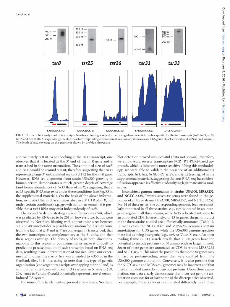
approximately 600 nt. When looking at the tsr33 transcript, oneobserves that it is located at the 3= end of the sarR gene and istranscribed in the same orientation. The combined size of sarRand tsr33 would be around 600 nt, therefore suggesting that tsr33represents a large 3= untranslated region (UTR) for the sarR gene.However, RNA-seq alignment from strain USA300 growing inhuman serum demonstrates a much greater depth of coverage(and hence abundance) of tsr33 than of sarR, suggesting that atsr33-specific RNA may exist under these conditions (see Fig. S3 inthe supplemental material). On the basis of the above informa-tion, we predict that tsr33 is cotranscribed as a 3=UTR of sarR, butunder certain conditions (e.g., growth in human serum), it is pos-sible that a tsr33 RNA may exist independently of sarR.
The second tsr demonstrating a size difference was tsr8, whichwas predicted by RNA-seq to be 201 nt; however, two bands wereobserved by Northern blotting with approximate sizes between300 and 400 nucleotides. A possible explanation for this may comefrom the fact that tsr8 and tsr7 are convergently transcribed, thatthe two transcripts are complementary at the 3= ends, and thatthese regions overlap. The density of reads, in both directions,mapping to this region of complementarity make it difficult topredict the precise location of each transcript based on RNA-seqdata, resulting in an underestimation of tsr8 size. Given our exper-imental findings, the size of tsr8 was amended to ~350 nt in theGenBank files. It is interesting to note that this type of geneticorganization (convergent transcripts overlapping at the 3= end) iscommon among toxin-antitoxin (TA) systems in S. aureus (19,20), hence tsr7 and tsr8 could potentially represent a novel serum-induced TA system.
For some of the tsr elements expressed at low levels, Northern
blot detection proved unsuccessful (data not shown); therefore,we employed a reverse transcriptase PCR (RT-PCR)-based ap-proach, which is inherently more sensitive. Using this methodol-ogy, we were able to validate the presence of an additional sixtranscripts, tsr1, tsr2, tsr18, tsr24, tsr29, and tsr32 (see Fig. S4 in thesupplemental material), suggesting that our RNA-seq-based iden-tification approach is effective at identifying legitimate sRNA mol-ecules.
Inconsistent genome annotation in strains USA300, MRSA252,and NCTC 8325. Twenty-seven tsr genes were found in the ge-nomes of all three strains (USA300, MRSA252, and NCTC 8325).For 14 of these genes, the corresponding genomic loci were simi-larly annotated in all three strains, e.g., tsr6 is located in an inter-genic region in all three strains, while tsr15 is located antisense toan annotated CDS. Interestingly, for 13 tsr genes, the genomic lociin the three strains studied are differentially annotated (Table 1).In many cases, the NCTC 8325 and MRSA252 genomes containannotations for CDS genes, while the USA300 genome specifiesthese loci as being intergenic (e.g., tsr9, tsr17, tsr18, etc.). An openreading frame (ORF) search reveals that 11 tsr genes have thepotential to encode proteins (of 30 amino acids or larger in size).Seven of these genes are annotated as CDS in strains MRSA252and NCTC 8325. This raises the possibility that some tsr genes mayin fact be protein-coding genes that were omitted from theUSA300 genome annotation. Conversely, it is also possible thatthe NCTC 8325 and MRSA252 genomes may be incorrect and thatthese annotated genes do not encode proteins. Upon close exam-ination, our data clearly demonstrate that incorrect genome an-notation accounts for at least some of the discrepancies observed.For example, the tsr12 locus is annotated differently in all three
FIG 1 Northern blot analysis of tsr transcripts. Northern blotting was performed using oligonucleotide probes specific for the tsr transcripts (tsr8, tsr25, tsr26,tsr31, and tsr33). RNA-seq read alignments for each corresponding chromosomal location are shown, as are CDS genes (black arrows), and sRNAs (red arrows).The depth of read coverage on the genome is shown by the blue histograms.
Carroll et al.
4 ® mbio.asm.org January/February 2016 Volume 7 Issue 1 e01990-15
m
bio.asm.org
on February 9, 2016 - P
ublished by m
bio.asm.org
Dow
nloaded from
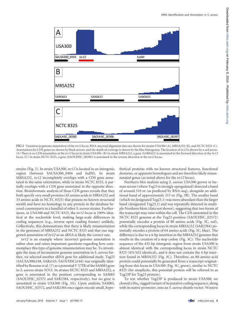
strains (Fig. 2). In strain USA300, tsr12 is located in an intergenicregion (between SAUSA300_0404 and hsdM). In strainMRSA252, tsr12 incompletely overlaps with a CDS gene anno-tated in the same orientation, while in strain NCTC 8325, it par-tially overlaps with a CDS gene annotated in the opposite direc-tion. Bioinformatic analysis of these CDS genes reveals that theyboth specify very small proteins (43 amino acids in MRSA252 and33 amino acids in NCTC 8325) that possess no known structuralmotifs and have no homology to any protein in the database be-yond counterparts in a handful of other S. aureus strains. Further-more, in USA300 and NCTC 8325, the tsr12 locus is 100% iden-tical at the nucleotide level, making large-scale differences incoding sequences (e.g., inverse open reading frames) unlikely.Collectively, this demonstrates that there is likely misannotationin the genomes of MRSA252 and NCTC 8325 and that our sug-gested annotation of tsr12 as an sRNA is likely the correct one.
tsr12 is an example where incorrect genome annotation israther clear and raises important questions regarding how com-monplace this type of genome misannotation may be. To investi-gate the issue of inconsistent genome annotation in S. aureus fur-ther, we selected another sRNA gene for additional study. Teg23(SAUSA300s148, SARs145, SAOUHSCs144) was originally iden-tified by Beaume et al. (7) as a potential 5=UTR of the SAS083 genein S. aureus strain N315. In strains NCTC 8325 and MRSA252, agene is annotated in the position corresponding to SAS083(SAOUHSC_02572 and SAR2384, respectively), but no gene isannotated in strain USA300 (Fig. 3A). Upon analysis, SAS083,SAOUHSC_02572, and SAR2384 once again encode small, hypo-
thetical proteins with no known structural features, functionaldomains, or apparent homologues and are therefore likely misan-notated genes (as noted above for the tsr12 locus).
Northern blot analysis using S. aureus USA300 grown in hu-man serum (where Teg23 is strongly upregulated) detected a bandof around 310 nt (as predicted by RNA-seq), alongside an addi-tional band of approximately 215 nt (Fig. 3B). The smaller band(which we designated Teg23.1) was more abundant than the largerband (designated Teg23.2) and was repeatedly detected in multi-ple Northern blots (data not shown), suggesting that two forms ofthe transcript may exist within the cell. The CDS annotated in theNCTC 8325 genome at the Teg23 position (SAOUHSC_02572)potentially encodes a protein of 80 amino acids (Fig. 3C, red),while the corresponding locus in strain MRSA252 (SAR2384) po-tentially encodes a protein of 64 amino acids (Fig. 3C, blue). Thedifference is due to a 4-bp insertion in the MRSA252 genome thatresults in the creation of a stop codon (Fig. 3C). The nucleotidesequence of the 432-bp intergenic region from strain USA300 isalmost identical with the corresponding locus in strain NCTC8325 (431/432 identical), and it does not contain the 4-bp inser-tion found in MRSA252 (Fig. 3C). Therefore, an 80-amino-acidprotein could potentially be generated from a transcript originat-ing from this locus in USA300 (Fig. 3C, green), similar to NCTC8325 (for simplicity, this potential protein will be referred to asTeg23P for Teg23 protein).
To test whether Teg23P is produced in strain USA300, wecloned a His6-tagged variant of its putative coding sequence, alongwith its native promoter, into an S. aureus shuttle vector. Western
FIG 2 Variation in genome annotation of the tsr12 locus. RNA-seq read alignment data are shown for strains USA300 (A), MRSA252 (B), and NCTC 8325 (C).Annotations for CDS genes are shown by black arrows, and the depth of coverage is shown by the blue histograms. The location of tsr12 is shown by a red arrow.(A) There is no CDS annotation at the tsr12 locus in strain USA300. (B) In strain MRSA252, a gene (SAR0432) is annotated in the forward direction at the tsr12locus. (C) In strain NCTC 8325, a gene (SAOUHSC_00396) is annotated in the reverse direction at the tsr12 locus.
sRNA Identification and Annotation in S. aureus
January/February 2016 Volume 7 Issue 1 e01990-15 ® mbio.asm.org 5
m
bio.asm.org
on February 9, 2016 - P
ublished by m
bio.asm.org
Dow
nloaded from
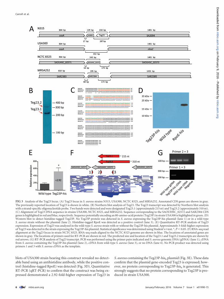
blots of USA300 strain bearing this construct revealed no detect-able band using an antihistidine antibody, while the positive con-trol (histidine-tagged RpoE) was detected (Fig. 3D). QuantitativeRT-PCR (qRT-PCR) to confirm that the construct was being ex-pressed demonstrated a 2.92-fold-higher expression of Teg23 in
S. aureus containing the Teg23P-his6 plasmid (Fig. 3E). These dataconfirm that the plasmid gene-encoded Teg23 is expressed; how-ever, no protein corresponding to Teg23P-his6 is generated. Thisstrongly suggests that no protein corresponding to Teg23P is pro-duced in strain USA300.
FIG 3 Analysis of the Teg23 locus. (A) Teg23 locus in S. aureus strains N315, USA300, NCTC 8325, and MRSA252. Annotated CDS genes are shown in gray.The previously reported location of Teg23 is shown in white. (B) Northern blot analysis of Teg23. The Teg23 transcript was detected by Northern blot analysiswith a strand-specific oligonucleotide probe. Two bands were detected and were designated Teg23.1 (approximately 215 nt) and Teg23.2 (approximately 310 nt).(C) Alignment of Teg23 DNA sequence in strains USA300, NCTC 8325, and MRSA252. Sequence corresponding to the SAOUHSC_02572 and SAR2384 CDSgenes is highlighted in red and blue, respectively. Sequence potentially encoding an 80-amino-acid protein (Teg23P) in strain USA300 is highlighted in green. (D)Western blot to detect histidine-tagged Teg23P. No Teg23P protein was detected in S. aureus expressing the Teg23P-his plasmid (lane 1) or in a wild-typeS. aureus strain without the plasmid (lane 2). Histidine-tagged RpoE was detected as a positive control (lane 3). (E) Quantitative RT-PCR analysis of Teg23expression. Expression of Teg23 was analyzed in the wild-type S. aureus strain with or without the Teg23P-his plasmid. Approximately 3-fold-higher expressionof Teg23 was detected in the strain expressing the Teg23P-his plasmid. Statistical significance was determined using Student’s t-test. *, P 0.05. (F) RNA-seq readalignment at the Teg23 locus in strain NCTC 8325. RNA-seq reads aligned to the NCTC 8325 genome are shown in blue. The locations of annotated genes areshown in gray. The locations of primers used for RT-PCR are shown in red. The predicted size and location of the Teg23.1 and Teg23.2 transcripts are shown byred arrows. (G) RT-PCR analysis of Teg23 transcript. PCR was performed using the primer pairs indicated and S. aureus genomic DNA (gDNA) (lane 1), cDNAfrom S. aureus containing the Teg23P-his plasmid (lane 2), cDNA from wild-type S. aureus (lane 3), or no DNA (lane 4). No PCR product was detected usingprimers 1 and 3 with S. aureus cDNA as the template.
Carroll et al.
6 ® mbio.asm.org January/February 2016 Volume 7 Issue 1 e01990-15
m
bio.asm.org
on February 9, 2016 - P
ublished by m
bio.asm.org
Dow
nloaded from
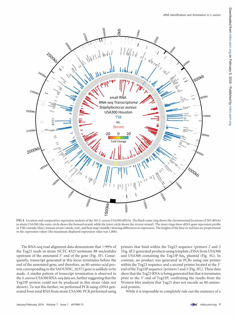
The RNA-seq read alignment data demonstrate that 99% ofthe Teg23 reads in strain NCTC 8325 terminate 88 nucleotidesupstream of the annotated 3= end of the gene (Fig. 3F). Conse-quently, transcript generated at this locus terminates before theend of the annotated gene, and therefore, an 80-amino-acid pro-tein corresponding to the SAOUHSC_02572 gene is unlikely to bemade. A similar pattern of transcript termination is observed inthe S. aureus USA300 RNA-seq data set, further suggesting that theTeg23P protein could not be produced in this strain (data notshown). To test this further, we performed PCR using cDNA gen-erated from total RNA from strain USA300. PCR performed using
primers that bind within the Teg23 sequence (primers 2 and 3[Fig. 4F]) generated products using template cDNA from USA300and USA300 containing the Teg23P-his6 plasmid (Fig. 3G). Incontrast, no product was generated in PCRs using one primerwithin the Teg23 sequence and a second primer located at the 3=end of the Teg23P sequence (primers 1 and 3 [Fig. 3F]). These datashow that the Teg23 RNA is being generated but that it terminatesprior to the 3= end of Teg23P, confirming the results from theWestern blot analysis that Teg23 does not encode an 80-amino-acid protein.
While it is impossible to completely rule out the existence of a
0kb
100k
b
200k
b
300k
b
400kb
500kb
600kb
700kb
800kb
900kb
1000kb
1100kb
1200kb1300kb
1400kb15
00kb16
00kb
1700
kb
1800
kb
1900kb
2000kb
2100kb
2200kb
2300kb
2400kb
2500kb
2600kb
2700kb
2800kb
s001
s002
s003
s004
s005
s006
s007
s008
s009
s010
s011s012
s013
s014
s015
s016
s017
s018
s019
s020s021
s022
s023
s024
s025s0
26
s027
s028
s029s030
s031s032s033s037
s034
s035s036
s038
s039
s040
s041
s042
s043
s044
s045
s046
s047
s048
s049
s050
s051s052
s053
s054
s055
s056
s057
s058
s059
s060
s061s062
s063
s064
s065
s066
s067
s068
s069
s070
s071
s072
s073
s074
s075
s076
s077
s078
s079
s080
s081
s082
s083
s084
s085
s086
s087
s088
s089
s090s091
s092s093s094
s095
s096
s097
s098
s099
s100
s101
s102s103
s104
s105
s106
s107
s108
s109s110s111
s112
s113
s114
s115
s116
s117
s118
s119
s120
s121
s122s123
s124
s125
s126
s127
s128
s129
s130
s131
s132
s133
s134
s135
s136
s137
s138
s139
s140
s141
s142
s143
s144
s145
s146
s147
s148
s149
s150
s151
s152
s153
s154
s155
s156
s157
s158
s159
s160s161
s162
s163
s164
s165
s166
s167
s168
s169
s170
s171
s172
s173
s174
s175
s176
s177
s178 s179s180
s181
s182s183
s184
s185
s186
s187
s188
s189
s190
s191
s192
s193
s194
s195
s196
s197
s200
s198
s199
s201
s202
s203
s204
s205
s206
s207
s208
s209
s210
s211 s212
s213 s214
s215
s216
s217
s218
s219s220
s221s222s223s224
s225s226
s227
s228s229
s231s232
s233
s234
s236
s237
s238
s239
s240
s241
s242
s243
s244
s245
s246s247
s248s249
s250
s251
s252
s253
s254
s255
s256s257 s258
s259
s260s261
s262
s263
s264
s265
s266
s267
s268
s269
s270
s271
s272
s273
s274
s275
s276
s277
s278
s279s280
s281
s282
s283
s284s285
s286
s287
s288s289
s290
s291
s292
s293
s294
s295
s296
s297
s298s299
s300s301
s302
s303
s230
small RNA RNA-seq TranscriptomeStaphylococcus aureus
USA300 HoustonTSBvs.
Serum
-20 200
Fold Change
FIG 4 Location and comparative expression analysis of the 303 S. aureus USA300 sRNAs. The black outer ring shows the chromosomal locations of 303 sRNAsin strain USA300 (the outer circle shows the forward strand, while the inner circle shows the reverse strand). The inner rings show sRNA gene expression profilein TSB (outside, blue), human serum (inside, red), and heat map (middle) showing differences in expression. The heights of the blue or red bars are proportionalto the expression values (the maximum displayed expression value was 1,000).
sRNA Identification and Annotation in S. aureus
January/February 2016 Volume 7 Issue 1 e01990-15 ® mbio.asm.org 7
m
bio.asm.org
on February 9, 2016 - P
ublished by m
bio.asm.org
Dow
nloaded from
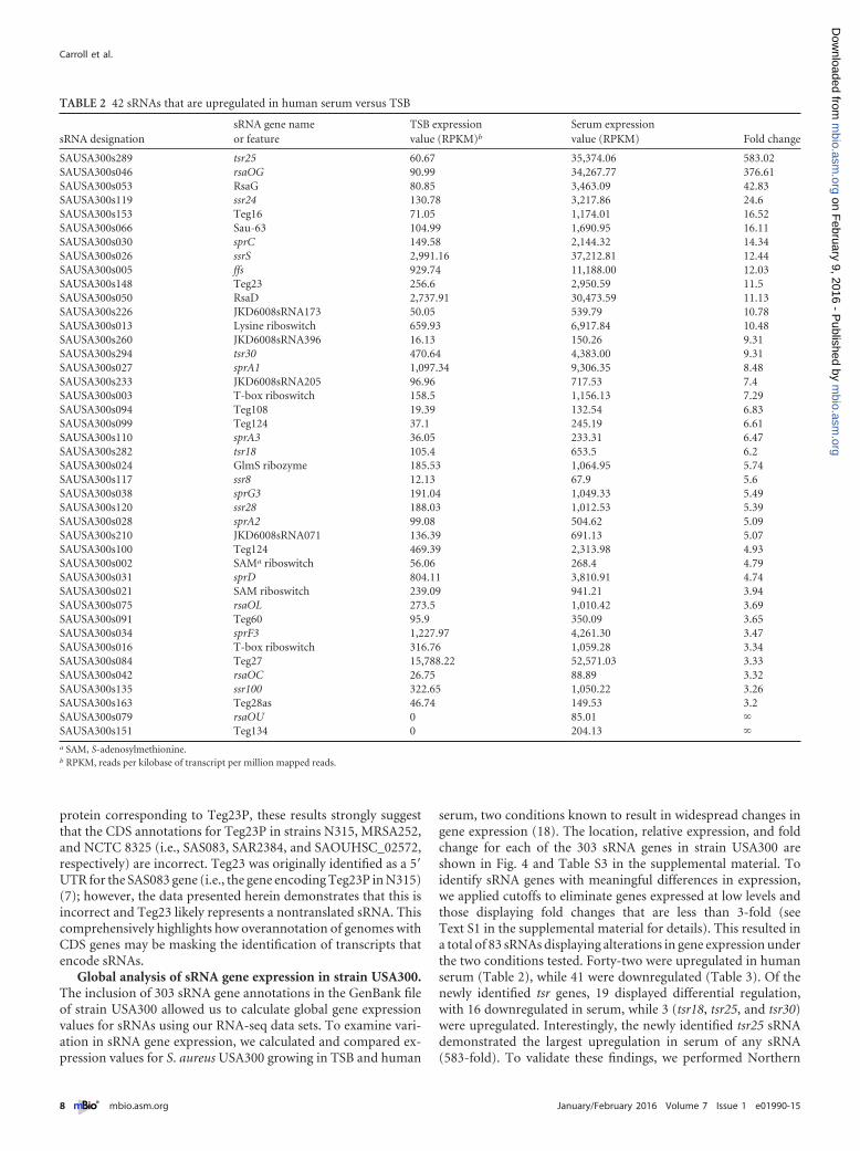
protein corresponding to Teg23P, these results strongly suggestthat the CDS annotations for Teg23P in strains N315, MRSA252,and NCTC 8325 (i.e., SAS083, SAR2384, and SAOUHSC_02572,respectively) are incorrect. Teg23 was originally identified as a 5=UTR for the SAS083 gene (i.e., the gene encoding Teg23P in N315)(7); however, the data presented herein demonstrates that this isincorrect and Teg23 likely represents a nontranslated sRNA. Thiscomprehensively highlights how overannotation of genomes withCDS genes may be masking the identification of transcripts thatencode sRNAs.
Global analysis of sRNA gene expression in strain USA300.The inclusion of 303 sRNA gene annotations in the GenBank fileof strain USA300 allowed us to calculate global gene expressionvalues for sRNAs using our RNA-seq data sets. To examine vari-ation in sRNA gene expression, we calculated and compared ex-pression values for S. aureus USA300 growing in TSB and human
serum, two conditions known to result in widespread changes ingene expression (18). The location, relative expression, and foldchange for each of the 303 sRNA genes in strain USA300 areshown in Fig. 4 and Table S3 in the supplemental material. Toidentify sRNA genes with meaningful differences in expression,we applied cutoffs to eliminate genes expressed at low levels andthose displaying fold changes that are less than 3-fold (seeText S1 in the supplemental material for details). This resulted ina total of 83 sRNAs displaying alterations in gene expression underthe two conditions tested. Forty-two were upregulated in humanserum (Table 2), while 41 were downregulated (Table 3). Of thenewly identified tsr genes, 19 displayed differential regulation,with 16 downregulated in serum, while 3 (tsr18, tsr25, and tsr30)were upregulated. Interestingly, the newly identified tsr25 sRNAdemonstrated the largest upregulation in serum of any sRNA(583-fold). To validate these findings, we performed Northern
TABLE 2 42 sRNAs that are upregulated in human serum versus TSB
sRNA designationsRNA gene nameor feature
TSB expressionvalue (RPKM)b
Serum expressionvalue (RPKM) Fold change
SAUSA300s289 tsr25 60.67 35,374.06 583.02SAUSA300s046 rsaOG 90.99 34,267.77 376.61SAUSA300s053 RsaG 80.85 3,463.09 42.83SAUSA300s119 ssr24 130.78 3,217.86 24.6SAUSA300s153 Teg16 71.05 1,174.01 16.52SAUSA300s066 Sau-63 104.99 1,690.95 16.11SAUSA300s030 sprC 149.58 2,144.32 14.34SAUSA300s026 ssrS 2,991.16 37,212.81 12.44SAUSA300s005 ffs 929.74 11,188.00 12.03SAUSA300s148 Teg23 256.6 2,950.59 11.5SAUSA300s050 RsaD 2,737.91 30,473.59 11.13SAUSA300s226 JKD6008sRNA173 50.05 539.79 10.78SAUSA300s013 Lysine riboswitch 659.93 6,917.84 10.48SAUSA300s260 JKD6008sRNA396 16.13 150.26 9.31SAUSA300s294 tsr30 470.64 4,383.00 9.31SAUSA300s027 sprA1 1,097.34 9,306.35 8.48SAUSA300s233 JKD6008sRNA205 96.96 717.53 7.4SAUSA300s003 T-box riboswitch 158.5 1,156.13 7.29SAUSA300s094 Teg108 19.39 132.54 6.83SAUSA300s099 Teg124 37.1 245.19 6.61SAUSA300s110 sprA3 36.05 233.31 6.47SAUSA300s282 tsr18 105.4 653.5 6.2SAUSA300s024 GlmS ribozyme 185.53 1,064.95 5.74SAUSA300s117 ssr8 12.13 67.9 5.6SAUSA300s038 sprG3 191.04 1,049.33 5.49SAUSA300s120 ssr28 188.03 1,012.53 5.39SAUSA300s028 sprA2 99.08 504.62 5.09SAUSA300s210 JKD6008sRNA071 136.39 691.13 5.07SAUSA300s100 Teg124 469.39 2,313.98 4.93SAUSA300s002 SAMa riboswitch 56.06 268.4 4.79SAUSA300s031 sprD 804.11 3,810.91 4.74SAUSA300s021 SAM riboswitch 239.09 941.21 3.94SAUSA300s075 rsaOL 273.5 1,010.42 3.69SAUSA300s091 Teg60 95.9 350.09 3.65SAUSA300s034 sprF3 1,227.97 4,261.30 3.47SAUSA300s016 T-box riboswitch 316.76 1,059.28 3.34SAUSA300s084 Teg27 15,788.22 52,571.03 3.33SAUSA300s042 rsaOC 26.75 88.89 3.32SAUSA300s135 ssr100 322.65 1,050.22 3.26SAUSA300s163 Teg28as 46.74 149.53 3.2SAUSA300s079 rsaOU 0 85.01 SAUSA300s151 Teg134 0 204.13 a SAM, S-adenosylmethionine.b RPKM, reads per kilobase of transcript per million mapped reads.
Carroll et al.
8 ® mbio.asm.org January/February 2016 Volume 7 Issue 1 e01990-15
m
bio.asm.org
on February 9, 2016 - P
ublished by m
bio.asm.org
Dow
nloaded from
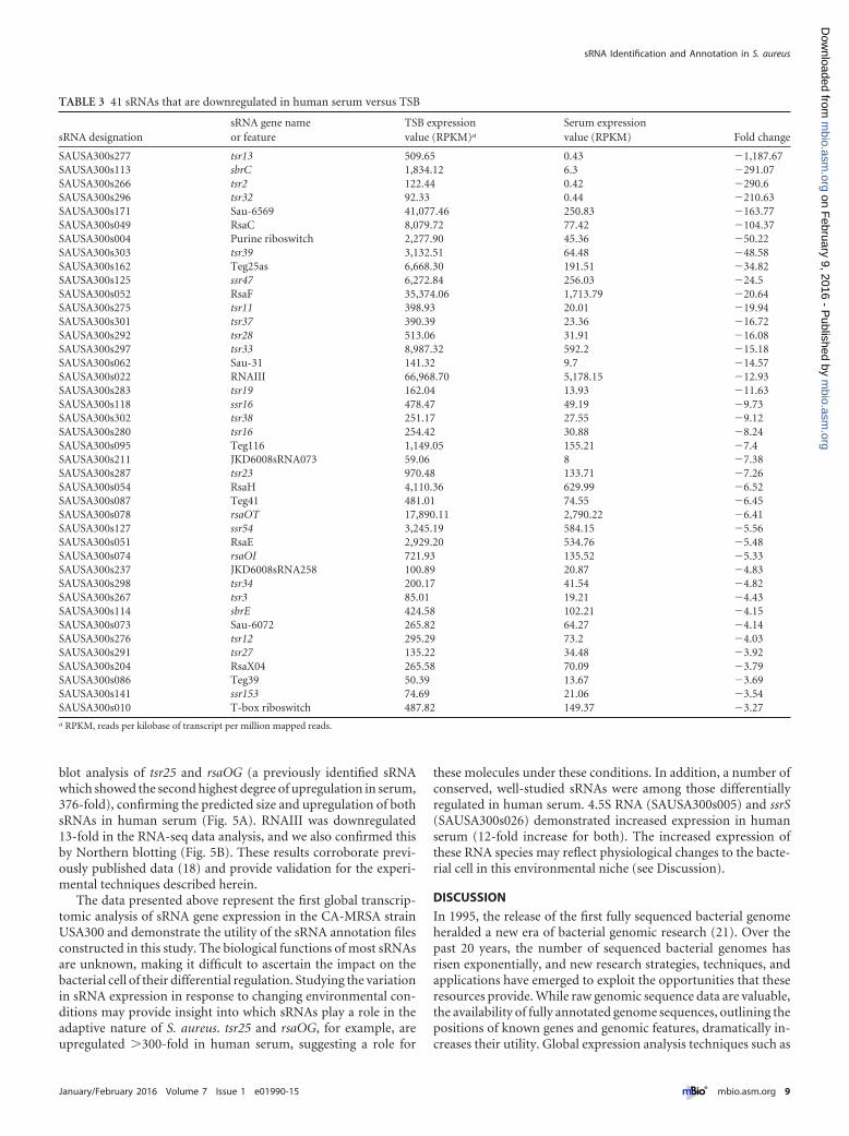
blot analysis of tsr25 and rsaOG (a previously identified sRNAwhich showed the second highest degree of upregulation in serum,376-fold), confirming the predicted size and upregulation of bothsRNAs in human serum (Fig. 5A). RNAIII was downregulated13-fold in the RNA-seq data analysis, and we also confirmed thisby Northern blotting (Fig. 5B). These results corroborate previ-ously published data (18) and provide validation for the experi-mental techniques described herein.
The data presented above represent the first global transcrip-tomic analysis of sRNA gene expression in the CA-MRSA strainUSA300 and demonstrate the utility of the sRNA annotation filesconstructed in this study. The biological functions of most sRNAsare unknown, making it difficult to ascertain the impact on thebacterial cell of their differential regulation. Studying the variationin sRNA expression in response to changing environmental con-ditions may provide insight into which sRNAs play a role in theadaptive nature of S. aureus. tsr25 and rsaOG, for example, areupregulated 300-fold in human serum, suggesting a role for
these molecules under these conditions. In addition, a number ofconserved, well-studied sRNAs were among those differentiallyregulated in human serum. 4.5S RNA (SAUSA300s005) and ssrS(SAUSA300s026) demonstrated increased expression in humanserum (12-fold increase for both). The increased expression ofthese RNA species may reflect physiological changes to the bacte-rial cell in this environmental niche (see Discussion).
DISCUSSION
In 1995, the release of the first fully sequenced bacterial genomeheralded a new era of bacterial genomic research (21). Over thepast 20 years, the number of sequenced bacterial genomes hasrisen exponentially, and new research strategies, techniques, andapplications have emerged to exploit the opportunities that theseresources provide. While raw genomic sequence data are valuable,the availability of fully annotated genome sequences, outlining thepositions of known genes and genomic features, dramatically in-creases their utility. Global expression analysis techniques such as
TABLE 3 41 sRNAs that are downregulated in human serum versus TSB
sRNA designationsRNA gene nameor feature
TSB expressionvalue (RPKM)a
Serum expressionvalue (RPKM) Fold change
SAUSA300s277 tsr13 509.65 0.43 1,187.67SAUSA300s113 sbrC 1,834.12 6.3 291.07SAUSA300s266 tsr2 122.44 0.42 290.6SAUSA300s296 tsr32 92.33 0.44 210.63SAUSA300s171 Sau-6569 41,077.46 250.83 163.77SAUSA300s049 RsaC 8,079.72 77.42 104.37SAUSA300s004 Purine riboswitch 2,277.90 45.36 50.22SAUSA300s303 tsr39 3,132.51 64.48 48.58SAUSA300s162 Teg25as 6,668.30 191.51 34.82SAUSA300s125 ssr47 6,272.84 256.03 24.5SAUSA300s052 RsaF 35,374.06 1,713.79 20.64SAUSA300s275 tsr11 398.93 20.01 19.94SAUSA300s301 tsr37 390.39 23.36 16.72SAUSA300s292 tsr28 513.06 31.91 16.08SAUSA300s297 tsr33 8,987.32 592.2 15.18SAUSA300s062 Sau-31 141.32 9.7 14.57SAUSA300s022 RNAIII 66,968.70 5,178.15 12.93SAUSA300s283 tsr19 162.04 13.93 11.63SAUSA300s118 ssr16 478.47 49.19 9.73SAUSA300s302 tsr38 251.17 27.55 9.12SAUSA300s280 tsr16 254.42 30.88 8.24SAUSA300s095 Teg116 1,149.05 155.21 7.4SAUSA300s211 JKD6008sRNA073 59.06 8 7.38SAUSA300s287 tsr23 970.48 133.71 7.26SAUSA300s054 RsaH 4,110.36 629.99 6.52SAUSA300s087 Teg41 481.01 74.55 6.45SAUSA300s078 rsaOT 17,890.11 2,790.22 6.41SAUSA300s127 ssr54 3,245.19 584.15 5.56SAUSA300s051 RsaE 2,929.20 534.76 5.48SAUSA300s074 rsaOI 721.93 135.52 5.33SAUSA300s237 JKD6008sRNA258 100.89 20.87 4.83SAUSA300s298 tsr34 200.17 41.54 4.82SAUSA300s267 tsr3 85.01 19.21 4.43SAUSA300s114 sbrE 424.58 102.21 4.15SAUSA300s073 Sau-6072 265.82 64.27 4.14SAUSA300s276 tsr12 295.29 73.2 4.03SAUSA300s291 tsr27 135.22 34.48 3.92SAUSA300s204 RsaX04 265.58 70.09 3.79SAUSA300s086 Teg39 50.39 13.67 3.69SAUSA300s141 ssr153 74.69 21.06 3.54SAUSA300s010 T-box riboswitch 487.82 149.37 3.27a RPKM, reads per kilobase of transcript per million mapped reads.
sRNA Identification and Annotation in S. aureus
January/February 2016 Volume 7 Issue 1 e01990-15 ® mbio.asm.org 9
m
bio.asm.org
on February 9, 2016 - P
ublished by m
bio.asm.org
Dow
nloaded from
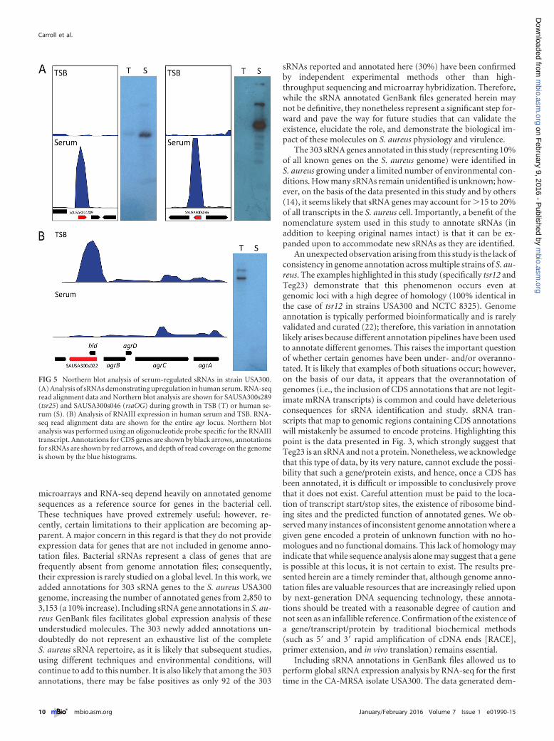
microarrays and RNA-seq depend heavily on annotated genomesequences as a reference source for genes in the bacterial cell.These techniques have proved extremely useful; however, re-cently, certain limitations to their application are becoming ap-parent. A major concern in this regard is that they do not provideexpression data for genes that are not included in genome anno-tation files. Bacterial sRNAs represent a class of genes that arefrequently absent from genome annotation files; consequently,their expression is rarely studied on a global level. In this work, weadded annotations for 303 sRNA genes to the S. aureus USA300genome, increasing the number of annotated genes from 2,850 to3,153 (a 10% increase). Including sRNA gene annotations in S. au-reus GenBank files facilitates global expression analysis of theseunderstudied molecules. The 303 newly added annotations un-doubtedly do not represent an exhaustive list of the completeS. aureus sRNA repertoire, as it is likely that subsequent studies,using different techniques and environmental conditions, willcontinue to add to this number. It is also likely that among the 303annotations, there may be false positives as only 92 of the 303
sRNAs reported and annotated here (30%) have been confirmedby independent experimental methods other than high-throughput sequencing and microarray hybridization. Therefore,while the sRNA annotated GenBank files generated herein maynot be definitive, they nonetheless represent a significant step for-ward and pave the way for future studies that can validate theexistence, elucidate the role, and demonstrate the biological im-pact of these molecules on S. aureus physiology and virulence.
The 303 sRNA genes annotated in this study (representing 10%of all known genes on the S. aureus genome) were identified inS. aureus growing under a limited number of environmental con-ditions. How many sRNAs remain unidentified is unknown; how-ever, on the basis of the data presented in this study and by others(14), it seems likely that sRNA genes may account for 15 to 20%of all transcripts in the S. aureus cell. Importantly, a benefit of thenomenclature system used in this study to annotate sRNAs (inaddition to keeping original names intact) is that it can be ex-panded upon to accommodate new sRNAs as they are identified.
An unexpected observation arising from this study is the lack ofconsistency in genome annotation across multiple strains of S. au-reus. The examples highlighted in this study (specifically tsr12 andTeg23) demonstrate that this phenomenon occurs even atgenomic loci with a high degree of homology (100% identical inthe case of tsr12 in strains USA300 and NCTC 8325). Genomeannotation is typically performed bioinformatically and is rarelyvalidated and curated (22); therefore, this variation in annotationlikely arises because different annotation pipelines have been usedto annotate different genomes. This raises the important questionof whether certain genomes have been under- and/or overanno-tated. It is likely that examples of both situations occur; however,on the basis of our data, it appears that the overannotation ofgenomes (i.e., the inclusion of CDS annotations that are not legit-imate mRNA transcripts) is common and could have deleteriousconsequences for sRNA identification and study. sRNA tran-scripts that map to genomic regions containing CDS annotationswill mistakenly be assumed to encode proteins. Highlighting thispoint is the data presented in Fig. 3, which strongly suggest thatTeg23 is an sRNA and not a protein. Nonetheless, we acknowledgethat this type of data, by its very nature, cannot exclude the possi-bility that such a gene/protein exists, and hence, once a CDS hasbeen annotated, it is difficult or impossible to conclusively provethat it does not exist. Careful attention must be paid to the loca-tion of transcript start/stop sites, the existence of ribosome bind-ing sites and the predicted function of annotated genes. We ob-served many instances of inconsistent genome annotation where agiven gene encoded a protein of unknown function with no ho-mologues and no functional domains. This lack of homology mayindicate that while sequence analysis alone may suggest that a geneis possible at this locus, it is not certain to exist. The results pre-sented herein are a timely reminder that, although genome anno-tation files are valuable resources that are increasingly relied uponby next-generation DNA sequencing technology, these annota-tions should be treated with a reasonable degree of caution andnot seen as an infallible reference. Confirmation of the existence ofa gene/transcript/protein by traditional biochemical methods(such as 5= and 3= rapid amplification of cDNA ends [RACE],primer extension, and in vivo translation) remains essential.
Including sRNA annotations in GenBank files allowed us toperform global sRNA expression analysis by RNA-seq for the firsttime in the CA-MRSA isolate USA300. The data generated dem-
FIG 5 Northern blot analysis of serum-regulated sRNAs in strain USA300.(A) Analysis of sRNAs demonstrating upregulation in human serum. RNA-seqread alignment data and Northern blot analysis are shown for SAUSA300s289(tsr25) and SAUSA300s046 (rsaOG) during growth in TSB (T) or human se-rum (S). (B) Analysis of RNAIII expression in human serum and TSB. RNA-seq read alignment data are shown for the entire agr locus. Northern blotanalysis was performed using an oligonucleotide probe specific for the RNAIIItranscript. Annotations for CDS genes are shown by black arrows, annotationsfor sRNAs are shown by red arrows, and depth of read coverage on the genomeis shown by the blue histograms.
Carroll et al.
10 ® mbio.asm.org January/February 2016 Volume 7 Issue 1 e01990-15
m
bio.asm.org
on February 9, 2016 - P
ublished by m
bio.asm.org
Dow
nloaded from
onstrated that 83 sRNAs are differentially expressed in TSB versushuman serum (Tables 2 and 3). This represents 27% of the knownsRNAs in strain USA300. It is not easy to interpret how theirdifferential regulation affects the bacterial cell (because the bio-logical functions of most of them are unknown); however, certaininferences can be made. The newly identified tsr25 sRNA demon-strated a 582-fold increase in expression in human serum. It istempting to speculate that increased expression of tsr25 in serumsuggests that it plays an important role during S. aureus blood-stream infections. A small number of conserved, well-studiedsRNAs were also among the differentially regulated sRNAs in se-rum. 4.5S RNA, a component of the signal recognition particle,was upregulated 12-fold in human serum, perhaps reflecting al-tered protein secretion and/or protein composition in the cellmembrane in this environment. Another important cellular RNAthat has been well explored is 6S RNA (ssrS), which we demon-strate also has a 12-fold increase in expression during growth inserum. In Escherichia coli, 6S RNA binds to the housekeepingsigma factor 70 and inhibits transcription from 70-dependentpromoters. It is thought that this diverts RNA polymerase to al-ternative sigma factors (such as the stress response sigma factor),resulting in increased expression of adaptive and stress-circumventing genes (23). The upregulation of ssrS in human se-rum suggests that a similar situation occurs in S. aureus, wherebyA-dependent genes are downregulated and B-dependent genesare upregulated by the action of 6S RNA.
Examining the global transcriptome can provide valuable in-sights into bacterial physiology and adaptation to environmentalconditions. In the past, global transcriptomic analysis has focusedon protein-coding genes, but here, we conduct global transcrip-tomic analysis and include newly annotated sRNA genes. Likeprotein-coding genes, sRNA genes display differential regulationthat allow bacteria to adapt to environmental changes. The anno-tation files presented herein, which facilitate this kind of globalanalysis will prove to be a valuable resource for the future study ofsRNAs in S. aureus and will more generally broaden our under-standing of regulatory circuits.
MATERIALS AND METHODSStrains, plasmids, and primers. Bacterial strains, plasmids, and primersused in this study are listed in Table 4. S. aureus and E. coli were grownroutinely at 37°C with shaking in tryptic soy broth (TSB) and lysogenybroth (LB), respectively. Pooled human serum from anonymous donors,purchased from MP Biomedicals, was used for growth of S. aureus strainUSA300. When necessary, antibiotics were added at the following concen-trations: ampicillin, 100 g ml1; chloramphenicol, 5 g ml1.
RNA-seq. Samples for transcriptome sequencing (RNA-seq) wereprepared as follows for S. aureus USA300 and SH1000. Overnight cultureswere diluted 1:100 in 100 ml of fresh TSB and grown at 37°C with shakingfor 3 h. Exponentially growing cultures were then diluted and synchro-nized by inoculating fresh 100-ml flasks of TSB at an optical density at 600nm (OD600) of 0.05, or in the case of strain USA300 growing in serum,10 ml of human serum in a 50-ml tube. Synchronized cultures were grownfor 3 h at 37°C with shaking, at which time bacteria were pelleted bycentrifugation and stored at 80°C prior to RNA isolation. S. aureusUAMS-1 (a USA200 strain and close relative of MRSA252) overnightcultures were diluted to an optical density of 0.05 in 40 ml of TSB con-taining no dextrose (1:12.5 volume-to-flask volume ratio). Cultures weregrown at 37°C with shaking for 4 h, before cells were harvested and storedas described above. For each RNA-seq sample, three replicate cultureswere grown, and three biological replicate RNA isolations were performed(using the procedure outlined in reference 24). For each sample, equimo-
lar amounts of the three biological replicate RNA preparations weremixed prior to rRNA reduction. The subsequent RNA-seq analysis there-fore represents the average of three biological replicates. RNA-seq anddata analysis were carried out using the protocol previously published byour research group (24).
Bioinformatics, sRNA identification, and genome annotation. TheCLC Genomics Workbench software platform (Qiagen) was used for allRNA-seq data analysis and for construction of new GenBank files. Anno-tated genome files for S. aureus N315, USA300, MRSA252, and NCTC8325 (GenBank accession numbers NC_002745, CP000255, NC_002952,and NC_007795, respectively) were downloaded from NCBI. The loca-tion, sequence, and orientation of previously described sRNAs on theN315 genome were calculated based on the information provided in pub-lished manuscripts and supplemental information files of the relevantstudies (1–12). Using the CLC Genomics Workbench built-in BLASTfeature, the corresponding positions for sRNAs were identified in thegenomes of strains USA300, MRSA252, and NCTC 8325. The locationand orientation of each sRNA was then annotated.
Northern blots. Northern blots to identify the size and abundance ofsRNAs were performed by the method of Caswell et al. (25). Briefly, 10 gof total RNA isolated from a 3 h USA300 culture in TSB were loaded on a10% polyacrylamide gel (7 M urea, 1 Tris-borate-EDTA [TBE]) andseparated by gel electrophoresis. The samples were then transferred viaelectroblotting to an Amersham Hybond N membrane (GE Health-care). The membrane was exposed to UV light to cross-link samples to themembrane. Subsequently, membranes were prehybridized (1 h, 45°C) inULTRAhyb-Oligo buffer (Ambion) and then incubated (16 h, 45°C) withsRNA-specific oligonucleotide probes end labeled with [-32P]ATP andT4 polynucleotide kinase (Thermo Scientific). After incubation, mem-branes were washed with 2, 1, and 0.5 SSC buffer (1 SSC is 300mM sodium chloride and 30 mM sodium citrate) at 45°C for 30 min eachto remove unspecific bound probes. Finally, X-ray film was exposed tomembranes for sRNA detection.
Cloning of histidine-tagged Teg23P. The genomic region containingTeg23P (including its native promoter) was amplified using USA300genomic DNA and primers OL3222 and OL3223. The reverse primerOL3223, in addition to the gene-specific region, carries a sequence thatencodes a hexahistidine (His6) tag, which allows the detection of a possi-ble encoded protein via Western blotting. The amplified 761-bp productwas cloned into shuttle vector pMK4, and the plasmid was transformedinto chemically competent E. coli DH5. The resulting colonies werescreened for correct constructs employing a colony PCR approach usingidentical primers to those used for the amplification of the initial frag-ment. After identification of positive clones, the plasmid was verified viaSanger sequencing with the plasmid-specific standard primers M13Fw(Fw stands for forward) and M13Rv (Rv stands for reverse) (EurofinsMWG Operon). This construct was then transformed into S. aureusRN4220 and confirmed via PCR. Finally, the plasmid was transduced intoS. aureus USA300 using a 11 phage lysate, and after final confirmation ofthe construct using PCR, the strain was utilized for subsequent analysis.
qRT-PCR. Quantitative reverse transcriptase PCR (qRT-PCR) wasconducted as described by our research group previously (26), employingthe teg23-specific primers OL3281, OL3282, OL3232, and OL3282 andRNA isolated from S. aureus USA300 cultures grown in TSB as describedabove for RNA-seq experiments. As a reference, 16S rRNA was amplifiedusing primers OL1184 and OL1185 (27). All experiments were performedin triplicate.
Western blots. The evaluation of Teg23 protein abundance was per-formed by Western immunoblotting as described previously (28).USA300 cells harboring the His6-tagged version of Teg23P were grown for3 h in TSB, before they were pelleted and their cytoplasmic proteins wereisolated. Following SDS-polyacrylamide gel electrophoresis and transferof the separated proteins to a polyvinylidene difluoride (PVDF) mem-brane, detection was performed using anti-His monoclonal mouse anti-body (Covance) and horseradish peroxidase (HRP)-conjugated anti-
sRNA Identification and Annotation in S. aureus
January/February 2016 Volume 7 Issue 1 e01990-15 ® mbio.asm.org 11
m
bio.asm.org
on February 9, 2016 - P
ublished by m
bio.asm.org
Dow
nloaded from
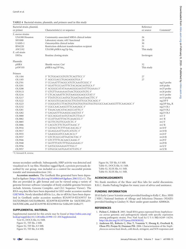
mouse secondary antibody. Subsequently, HRP activity was detected andvisualized on X-ray film. Histidine-tagged RpoE, a protein previously de-scribed by our group, was included as a control for successful proteintransfer and immunodetection (16).
Accession numbers. The GenBank files generated have been depos-ited in figshare (https://dx.doi.org/10.6084/m9.figshare.2061132.v1). Thefiles are provided in .gkb format and can be viewed using a variety ofgenome browser software (examples of freely available genome browsersinclude Artemis, Genome Compiler, and CLC Sequence Viewer). TheRNA-seq data files have been deposited in GEO under accession numberGSE74936. Newly identified sRNAs (i.e., the tsr sRNAs) have been depos-ited in GenBank under accession numbers KU639719-KU639757 forSAUSA300s265-SAUSA300s303, KU639758-KU639789 for SAOUHSCs255-SAOUHSCs286, and KU639790-KU639818 for SARs249-SARs277.
SUPPLEMENTAL MATERIALSupplemental material for this article may be found at http://mbio.asm.org/lookup/suppl/doi:10.1128/mBio.01990-15/-/DCSupplemental.
Text S1, DOCX file, 0.2 MB.Figure S1, TIF file, 2 MB.Figure S2, TIF file, 0.5 MB.Figure S3, TIF file, 0.4 MB.
Figure S4, TIF file, 0.5 MB.Table S1, DOCX file, 0.1 MB.Table S2, XLSX file, 0.1 MB.Table S3, XLSX file, 0.1 MB.
ACKNOWLEDGMENTS
We thank members of the Shaw and Rice labs for useful discussions.R.K.C. thanks Padraig Deighan for many years of advice and assistance.
FUNDING INFORMATIONIFAS Early Career Scientist award provided funding to Kelly C. Rice. HHS| NIH | National Institute of Allergy and Infectious Diseases (NIAID)provided funding to Lindsey N. Shaw under grant number AI080626.
REFERENCES1. Pichon C, Felden B. 2005. Small RNA genes expressed from Staphylococ-
cus aureus genomic and pathogenicity islands with specific expressionamong pathogenic strains. Proc Natl Acad Sci U S A 102:14249 –14254.http://dx.doi.org/10.1073/pnas.0503838102.
2. Anderson KL, Roberts C, Disz T, Vonstein V, Hwang K, Overbeek R,Olson PD, Projan SJ, Dunman PM. 2006. Characterization of the Staph-ylococcus aureus heat shock, cold shock, stringent, and SOS responses and
TABLE 4 Bacterial strains, plasmids, and primers used in this study
Bacterial strain, plasmid,or primer Characteristic(s) or sequence
Referenceor source Commenta
S. aureus strainsUSA300 Houston Community-associated MRSA clinical isolate 26SH1000 Laboratory strain; rsbU functional 29UAMS-1 Osteomyelitis clinical isolate 30RN4220 Restriction-deficient transformation recipient 31AW2192 USA300 pMK4::teg23p-his6 This study
E. coli strainDH5 Routine cloning strain Invitrogen
PlasmidspMK4 Shuttle vector; Cmr 32pAW105 pMK4::teg23P-his6 This study
PrimersOL1184 5= TCTGGACCGTGTCTCAGTTCC 3= 27OL1185 5= AGCCGACCTGAGAGGGTGA 3= 27OL2701 5= CCAAATTTAGGCATGTCAAATCGGC 3= teg23 probeOL3201 5= GGATTCCCAATTTCTACAGACAATGCA 3= tsr8 probeOL3208 5= ACGGGCATATAAAAGGGGAATATTTGAAAA 3= tsr25 probeOU0121 5= GTGTTAAAAAAATAACTGGGATGTG 3= tsr26 probeOL3216 5= CTCACAAATTCTGTAAGGGGAGCGTAT 3= tsr31 probeOL3217 5= TTATGTCCCAATGCTGAATAAATAACTTC 3= tsr33 probeOL3222 5= ACGCGTCGACGCGCTTGTATTCGCTGCAGG 3= teg23P FOL3223 5= CGGGATCCTTAGTGGTGGTGGTGGTGGTGCGCCAACAAGGTTTCAAGAGC 3= teg23P-his6 ROL3232 5= CGCCAACAAGGTTTCAAGAGC 3= teg23 OL1OL3281 5= TAAACAACATACAGCCATTG 3= teg23 OL2OL3282 5= GAGAATTTGAAGGCAAGTAT 3= teg23 OL3OL3880 5= GCCAGGATAATGTAGTCTTAA 3= tsr1 FOL3882 5= CCATTAATTTACTCAAACCG 3= tsr1 ROL3885 5= GCTTCTGTTCGATCTC 3= tsr2 FOL3886 5= CACGTCTTCTGATTAAAC 3= tsr2 ROL3916 5= CATACCTCTTTAACAACAG 3= tsr18 FOL3917 5= GGAGGAATTAATCATGTC 3= tsr18 ROL3935 5= GAAGGGATCCAACACA 3= tsr24 FOL3937 5= GTCTCGCCATTAATACTAC 3= tsr24 ROL3946 5= GTCTTTTCACAACCAAAG 3= tsr29 FOL3948 5= GGTTTTATCTTTGGAAAAAG 3= tsr29 ROL3956 5= GATGCGGAAAATTTGG 3= tsr32 FOL3957 5= GTGCGCAATGAATATTATG 3= tsr32 R
a F, forward; R, reverse.
Carroll et al.
12 ® mbio.asm.org January/February 2016 Volume 7 Issue 1 e01990-15
m
bio.asm.org
on February 9, 2016 - P
ublished by m
bio.asm.org
Dow
nloaded from
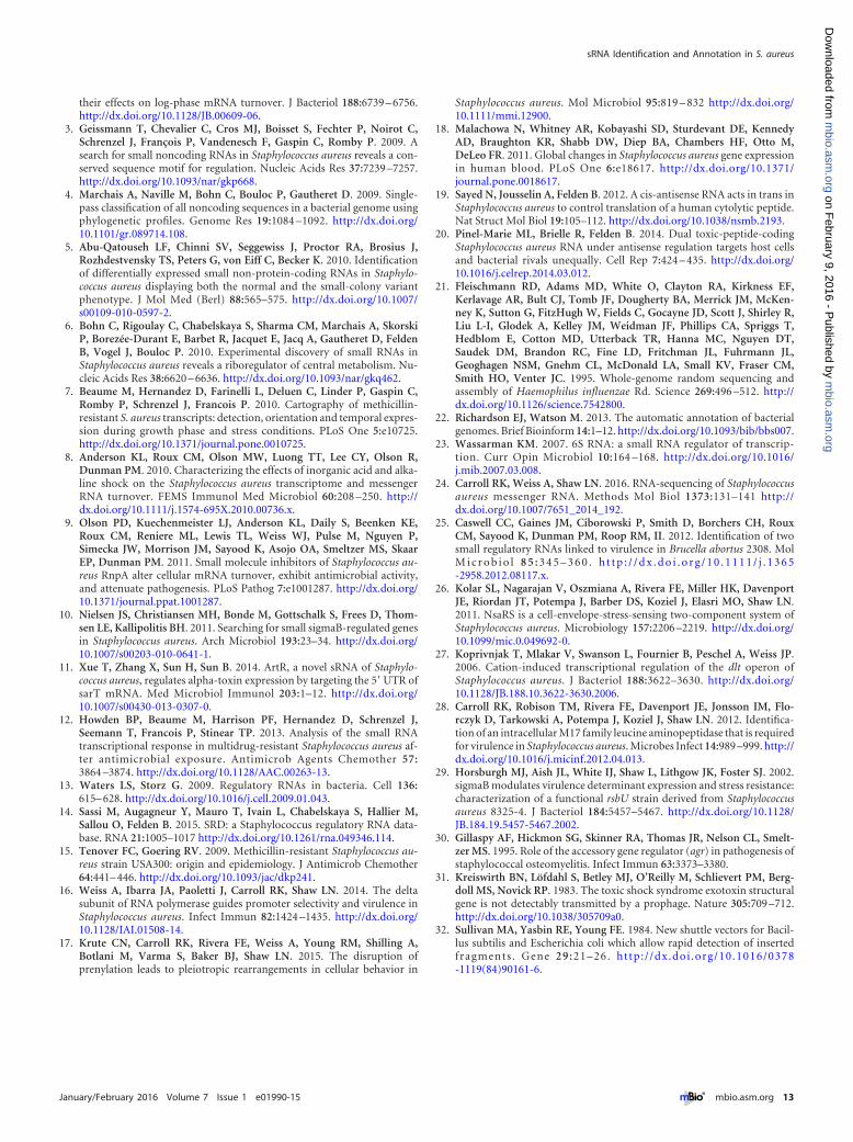
their effects on log-phase mRNA turnover. J Bacteriol 188:6739 – 6756.http://dx.doi.org/10.1128/JB.00609-06.
3. Geissmann T, Chevalier C, Cros MJ, Boisset S, Fechter P, Noirot C,Schrenzel J, François P, Vandenesch F, Gaspin C, Romby P. 2009. Asearch for small noncoding RNAs in Staphylococcus aureus reveals a con-served sequence motif for regulation. Nucleic Acids Res 37:7239 –7257.http://dx.doi.org/10.1093/nar/gkp668.
4. Marchais A, Naville M, Bohn C, Bouloc P, Gautheret D. 2009. Single-pass classification of all noncoding sequences in a bacterial genome usingphylogenetic profiles. Genome Res 19:1084 –1092. http://dx.doi.org/10.1101/gr.089714.108.
5. Abu-Qatouseh LF, Chinni SV, Seggewiss J, Proctor RA, Brosius J,Rozhdestvensky TS, Peters G, von Eiff C, Becker K. 2010. Identificationof differentially expressed small non-protein-coding RNAs in Staphylo-coccus aureus displaying both the normal and the small-colony variantphenotype. J Mol Med (Berl) 88:565–575. http://dx.doi.org/10.1007/s00109-010-0597-2.
6. Bohn C, Rigoulay C, Chabelskaya S, Sharma CM, Marchais A, SkorskiP, Borezée-Durant E, Barbet R, Jacquet E, Jacq A, Gautheret D, FeldenB, Vogel J, Bouloc P. 2010. Experimental discovery of small RNAs inStaphylococcus aureus reveals a riboregulator of central metabolism. Nu-cleic Acids Res 38:6620 – 6636. http://dx.doi.org/10.1093/nar/gkq462.
7. Beaume M, Hernandez D, Farinelli L, Deluen C, Linder P, Gaspin C,Romby P, Schrenzel J, Francois P. 2010. Cartography of methicillin-resistant S. aureus transcripts: detection, orientation and temporal expres-sion during growth phase and stress conditions. PLoS One 5:e10725.http://dx.doi.org/10.1371/journal.pone.0010725.
8. Anderson KL, Roux CM, Olson MW, Luong TT, Lee CY, Olson R,Dunman PM. 2010. Characterizing the effects of inorganic acid and alka-line shock on the Staphylococcus aureus transcriptome and messengerRNA turnover. FEMS Immunol Med Microbiol 60:208 –250. http://dx.doi.org/10.1111/j.1574-695X.2010.00736.x.
9. Olson PD, Kuechenmeister LJ, Anderson KL, Daily S, Beenken KE,Roux CM, Reniere ML, Lewis TL, Weiss WJ, Pulse M, Nguyen P,Simecka JW, Morrison JM, Sayood K, Asojo OA, Smeltzer MS, SkaarEP, Dunman PM. 2011. Small molecule inhibitors of Staphylococcus au-reus RnpA alter cellular mRNA turnover, exhibit antimicrobial activity,and attenuate pathogenesis. PLoS Pathog 7:e1001287. http://dx.doi.org/10.1371/journal.ppat.1001287.
10. Nielsen JS, Christiansen MH, Bonde M, Gottschalk S, Frees D, Thom-sen LE, Kallipolitis BH. 2011. Searching for small sigmaB-regulated genesin Staphylococcus aureus. Arch Microbiol 193:23–34. http://dx.doi.org/10.1007/s00203-010-0641-1.
11. Xue T, Zhang X, Sun H, Sun B. 2014. ArtR, a novel sRNA of Staphylo-coccus aureus, regulates alpha-toxin expression by targeting the 5=UTR ofsarT mRNA. Med Microbiol Immunol 203:1–12. http://dx.doi.org/10.1007/s00430-013-0307-0.
12. Howden BP, Beaume M, Harrison PF, Hernandez D, Schrenzel J,Seemann T, Francois P, Stinear TP. 2013. Analysis of the small RNAtranscriptional response in multidrug-resistant Staphylococcus aureus af-ter antimicrobial exposure. Antimicrob Agents Chemother 57:3864 –3874. http://dx.doi.org/10.1128/AAC.00263-13.
13. Waters LS, Storz G. 2009. Regulatory RNAs in bacteria. Cell 136:615– 628. http://dx.doi.org/10.1016/j.cell.2009.01.043.
14. Sassi M, Augagneur Y, Mauro T, Ivain L, Chabelskaya S, Hallier M,Sallou O, Felden B. 2015. SRD: a Staphylococcus regulatory RNA data-base. RNA 21:1005–1017 http://dx.doi.org/10.1261/rna.049346.114.
15. Tenover FC, Goering RV. 2009. Methicillin-resistant Staphylococcus au-reus strain USA300: origin and epidemiology. J Antimicrob Chemother64:441– 446. http://dx.doi.org/10.1093/jac/dkp241.
16. Weiss A, Ibarra JA, Paoletti J, Carroll RK, Shaw LN. 2014. The deltasubunit of RNA polymerase guides promoter selectivity and virulence inStaphylococcus aureus. Infect Immun 82:1424 –1435. http://dx.doi.org/10.1128/IAI.01508-14.
17. Krute CN, Carroll RK, Rivera FE, Weiss A, Young RM, Shilling A,Botlani M, Varma S, Baker BJ, Shaw LN. 2015. The disruption ofprenylation leads to pleiotropic rearrangements in cellular behavior in
Staphylococcus aureus. Mol Microbiol 95:819 – 832 http://dx.doi.org/10.1111/mmi.12900.
18. Malachowa N, Whitney AR, Kobayashi SD, Sturdevant DE, KennedyAD, Braughton KR, Shabb DW, Diep BA, Chambers HF, Otto M,DeLeo FR. 2011. Global changes in Staphylococcus aureus gene expressionin human blood. PLoS One 6:e18617. http://dx.doi.org/10.1371/journal.pone.0018617.
19. Sayed N, Jousselin A, Felden B. 2012. A cis-antisense RNA acts in trans inStaphylococcus aureus to control translation of a human cytolytic peptide.Nat Struct Mol Biol 19:105–112. http://dx.doi.org/10.1038/nsmb.2193.
20. Pinel-Marie ML, Brielle R, Felden B. 2014. Dual toxic-peptide-codingStaphylococcus aureus RNA under antisense regulation targets host cellsand bacterial rivals unequally. Cell Rep 7:424 – 435. http://dx.doi.org/10.1016/j.celrep.2014.03.012.
21. Fleischmann RD, Adams MD, White O, Clayton RA, Kirkness EF,Kerlavage AR, Bult CJ, Tomb JF, Dougherty BA, Merrick JM, McKen-ney K, Sutton G, FitzHugh W, Fields C, Gocayne JD, Scott J, Shirley R,Liu L-I, Glodek A, Kelley JM, Weidman JF, Phillips CA, Spriggs T,Hedblom E, Cotton MD, Utterback TR, Hanna MC, Nguyen DT,Saudek DM, Brandon RC, Fine LD, Fritchman JL, Fuhrmann JL,Geoghagen NSM, Gnehm CL, McDonald LA, Small KV, Fraser CM,Smith HO, Venter JC. 1995. Whole-genome random sequencing andassembly of Haemophilus influenzae Rd. Science 269:496 –512. http://dx.doi.org/10.1126/science.7542800.
22. Richardson EJ, Watson M. 2013. The automatic annotation of bacterialgenomes. Brief Bioinform 14:1–12. http://dx.doi.org/10.1093/bib/bbs007.
23. Wassarman KM. 2007. 6S RNA: a small RNA regulator of transcrip-tion. Curr Opin Microbiol 10:164 –168. http://dx.doi.org/10.1016/j.mib.2007.03.008.
24. Carroll RK, Weiss A, Shaw LN. 2016. RNA-sequencing of Staphylococcusaureus messenger RNA. Methods Mol Biol 1373:131–141 http://dx.doi.org/10.1007/7651_2014_192.
25. Caswell CC, Gaines JM, Ciborowski P, Smith D, Borchers CH, RouxCM, Sayood K, Dunman PM, Roop RM, II. 2012. Identification of twosmall regulatory RNAs linked to virulence in Brucella abortus 2308. MolM i c r o b i o l 8 5 : 3 4 5 – 3 6 0 . h t t p : / / d x . d o i . o r g / 1 0 . 1 1 1 1 / j . 1 3 6 5-2958.2012.08117.x.
26. Kolar SL, Nagarajan V, Oszmiana A, Rivera FE, Miller HK, DavenportJE, Riordan JT, Potempa J, Barber DS, Koziel J, Elasri MO, Shaw LN.2011. NsaRS is a cell-envelope-stress-sensing two-component system ofStaphylococcus aureus. Microbiology 157:2206 –2219. http://dx.doi.org/10.1099/mic.0.049692-0.
27. Koprivnjak T, Mlakar V, Swanson L, Fournier B, Peschel A, Weiss JP.2006. Cation-induced transcriptional regulation of the dlt operon ofStaphylococcus aureus. J Bacteriol 188:3622–3630. http://dx.doi.org/10.1128/JB.188.10.3622-3630.2006.
28. Carroll RK, Robison TM, Rivera FE, Davenport JE, Jonsson IM, Flo-rczyk D, Tarkowski A, Potempa J, Koziel J, Shaw LN. 2012. Identifica-tion of an intracellular M17 family leucine aminopeptidase that is requiredfor virulence in Staphylococcus aureus. Microbes Infect 14:989 –999. http://dx.doi.org/10.1016/j.micinf.2012.04.013.
29. Horsburgh MJ, Aish JL, White IJ, Shaw L, Lithgow JK, Foster SJ. 2002.sigmaB modulates virulence determinant expression and stress resistance:characterization of a functional rsbU strain derived from Staphylococcusaureus 8325-4. J Bacteriol 184:5457–5467. http://dx.doi.org/10.1128/JB.184.19.5457-5467.2002.
30. Gillaspy AF, Hickmon SG, Skinner RA, Thomas JR, Nelson CL, Smelt-zer MS. 1995. Role of the accessory gene regulator (agr) in pathogenesis ofstaphylococcal osteomyelitis. Infect Immun 63:3373–3380.
31. Kreiswirth BN, Löfdahl S, Betley MJ, O’Reilly M, Schlievert PM, Berg-doll MS, Novick RP. 1983. The toxic shock syndrome exotoxin structuralgene is not detectably transmitted by a prophage. Nature 305:709 –712.http://dx.doi.org/10.1038/305709a0.
32. Sullivan MA, Yasbin RE, Young FE. 1984. New shuttle vectors for Bacil-lus subtilis and Escherichia coli which allow rapid detection of insertedfragments . Gene 29:21–26. http:/ /dx.doi .org/10.1016/0378-1119(84)90161-6.
sRNA Identification and Annotation in S. aureus
January/February 2016 Volume 7 Issue 1 e01990-15 ® mbio.asm.org 13
m
bio.asm.org
on February 9, 2016 - P
ublished by m
bio.asm.org
Dow
nloaded from
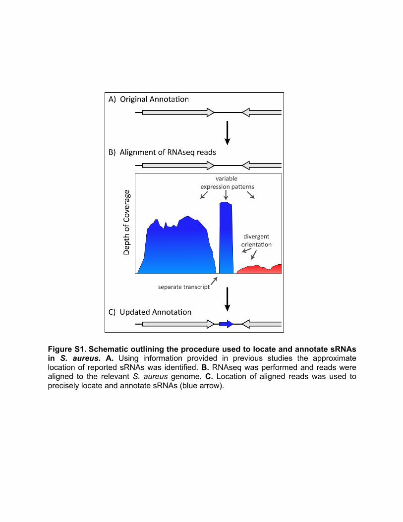
Figure S1. Schematic outlining the procedure used to locate and annotate sRNAs in S. aureus. A. Using information provided in previous studies the approximate location of reported sRNAs was identified. B. RNAseq was performed and reads were aligned to the relevant S. aureus genome. C. Location of aligned reads was used to precisely locate and annotate sRNAs (blue arrow).
FigureS1.
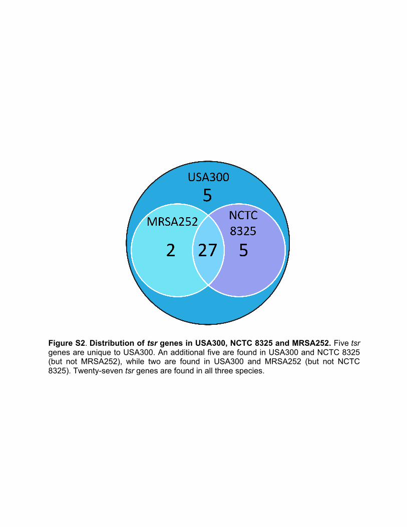
Figure S2. Distribution of tsr genes in USA300, NCTC 8325 and MRSA252. Five tsr genes are unique to USA300. An additional five are found in USA300 and NCTC 8325 (but not MRSA252), while two are found in USA300 and MRSA252 (but not NCTC 8325). Twenty-seven tsr genes are found in all three species.
FigureS2.
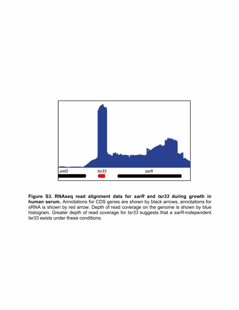
Figure S3. RNAseq read alignment data for sarR and tsr33 during growth in human serum. Annotations for CDS genes are shown by black arrows, annotations for sRNA is shown by red arrow. Depth of read coverage on the genome is shown by blue histogram. Greater depth of read coverage for tsr33 suggests that a sarR-independent tsr33 exists under these conditions.
FigureS3.
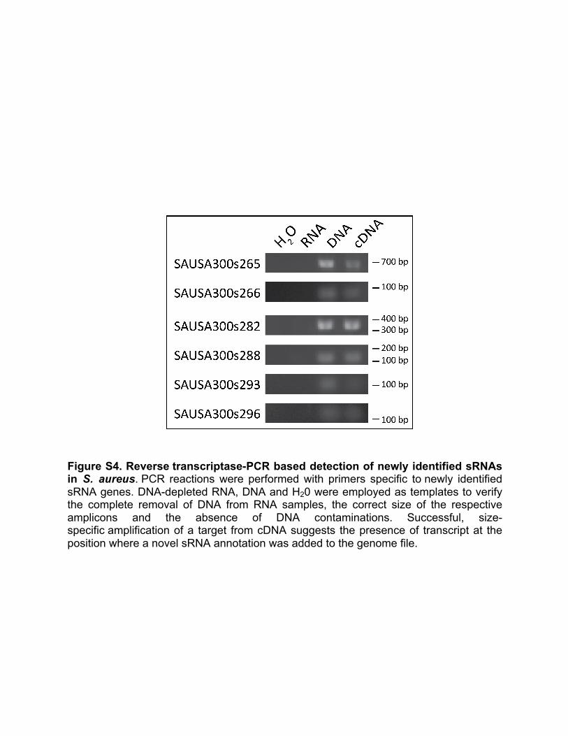
Figure S4. Reverse transcriptase-PCR based detection of newly identified sRNAs in S. aureus. PCR reactions were performed with primers specific to newly identified sRNA genes. DNA-depleted RNA, DNA and H20 were employed as templates to verify the complete removal of DNA from RNA samples, the correct size of the respective amplicons and the absence of DNA contaminations. Successful, size-specific amplification of a target from cDNA suggests the presence of transcript at the position where a novel sRNA annotation was added to the genome file.
FigureS4.
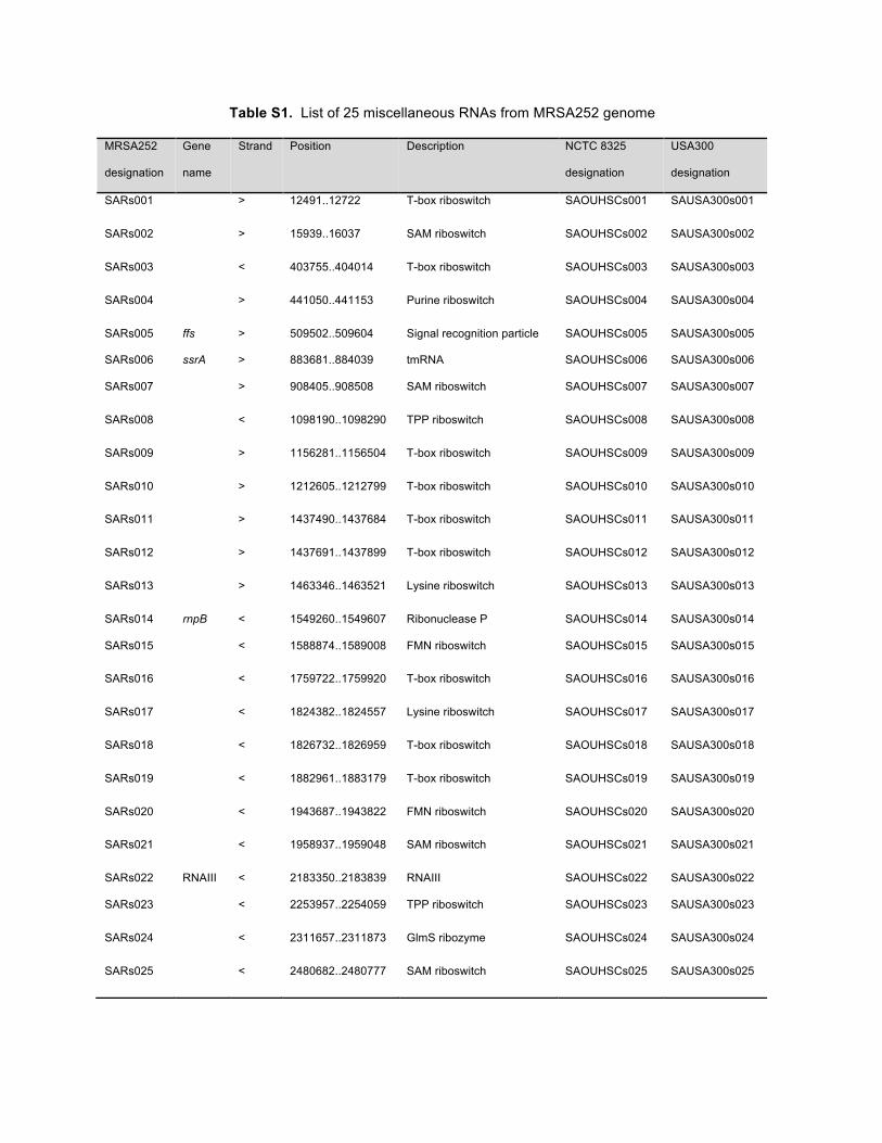
Table S1. List of 25 miscellaneous RNAs from MRSA252 genome
MRSA252
designation
Gene
name
Strand Position Description NCTC 8325
designation
USA300
designation
SARs001 > 12491..12722 T-box riboswitch SAOUHSCs001 SAUSA300s001
SARs002 > 15939..16037 SAM riboswitch SAOUHSCs002 SAUSA300s002
SARs003 < 403755..404014 T-box riboswitch SAOUHSCs003 SAUSA300s003
SARs004 > 441050..441153 Purine riboswitch SAOUHSCs004 SAUSA300s004
SARs005 ffs > 509502..509604 Signal recognition particle SAOUHSCs005 SAUSA300s005
SARs006 ssrA > 883681..884039 tmRNA SAOUHSCs006 SAUSA300s006
SARs007 > 908405..908508 SAM riboswitch SAOUHSCs007 SAUSA300s007
SARs008 < 1098190..1098290 TPP riboswitch SAOUHSCs008 SAUSA300s008
SARs009 > 1156281..1156504 T-box riboswitch SAOUHSCs009 SAUSA300s009
SARs010 > 1212605..1212799 T-box riboswitch SAOUHSCs010 SAUSA300s010
SARs011 > 1437490..1437684 T-box riboswitch SAOUHSCs011 SAUSA300s011
SARs012 > 1437691..1437899 T-box riboswitch SAOUHSCs012 SAUSA300s012
SARs013 > 1463346..1463521 Lysine riboswitch SAOUHSCs013 SAUSA300s013
SARs014 rnpB < 1549260..1549607 Ribonuclease P SAOUHSCs014 SAUSA300s014
SARs015 < 1588874..1589008 FMN riboswitch SAOUHSCs015 SAUSA300s015
SARs016 < 1759722..1759920 T-box riboswitch SAOUHSCs016 SAUSA300s016
SARs017 < 1824382..1824557 Lysine riboswitch SAOUHSCs017 SAUSA300s017
SARs018 < 1826732..1826959 T-box riboswitch SAOUHSCs018 SAUSA300s018
SARs019 < 1882961..1883179 T-box riboswitch SAOUHSCs019 SAUSA300s019
SARs020 < 1943687..1943822 FMN riboswitch SAOUHSCs020 SAUSA300s020
SARs021 < 1958937..1959048 SAM riboswitch SAOUHSCs021 SAUSA300s021
SARs022 RNAIII < 2183350..2183839 RNAIII SAOUHSCs022 SAUSA300s022
SARs023 < 2253957..2254059 TPP riboswitch SAOUHSCs023 SAUSA300s023
SARs024 < 2311657..2311873 GlmS ribozyme SAOUHSCs024 SAUSA300s024
SARs025 < 2480682..2480777 SAM riboswitch SAOUHSCs025 SAUSA300s025
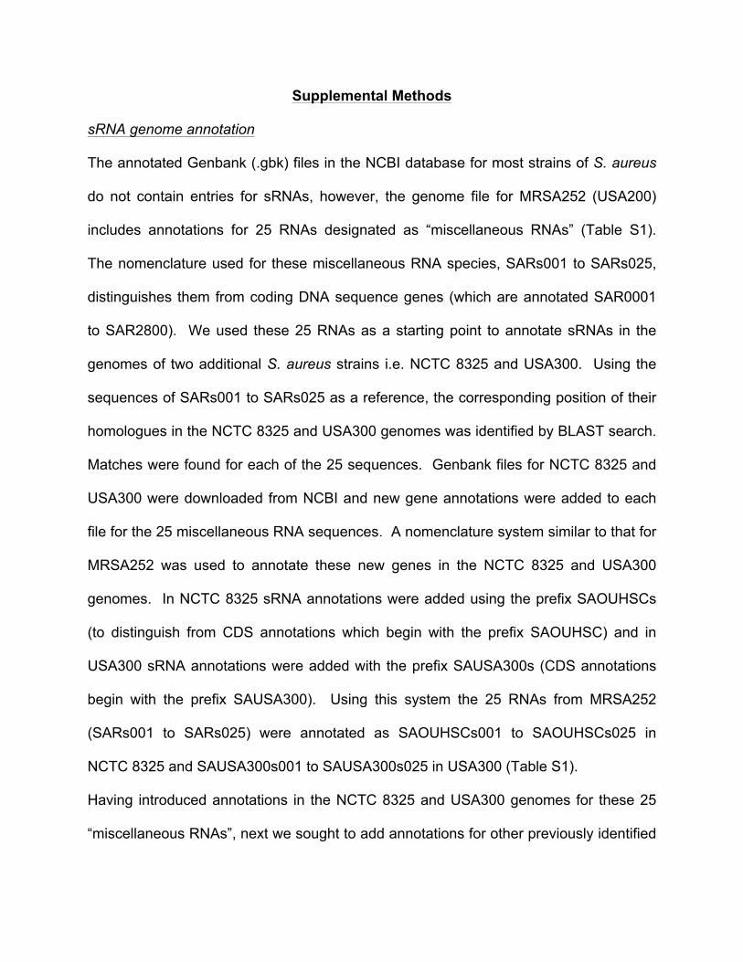
Supplemental Methods
sRNA genome annotation
The annotated Genbank (.gbk) files in the NCBI database for most strains of S. aureus
do not contain entries for sRNAs, however, the genome file for MRSA252 (USA200)
includes annotations for 25 RNAs designated as “miscellaneous RNAs” (Table S1).
The nomenclature used for these miscellaneous RNA species, SARs001 to SARs025,
distinguishes them from coding DNA sequence genes (which are annotated SAR0001
to SAR2800). We used these 25 RNAs as a starting point to annotate sRNAs in the
genomes of two additional S. aureus strains i.e. NCTC 8325 and USA300. Using the
sequences of SARs001 to SARs025 as a reference, the corresponding position of their
homologues in the NCTC 8325 and USA300 genomes was identified by BLAST search.
Matches were found for each of the 25 sequences. Genbank files for NCTC 8325 and
USA300 were downloaded from NCBI and new gene annotations were added to each
file for the 25 miscellaneous RNA sequences. A nomenclature system similar to that for
MRSA252 was used to annotate these new genes in the NCTC 8325 and USA300
genomes. In NCTC 8325 sRNA annotations were added using the prefix SAOUHSCs
(to distinguish from CDS annotations which begin with the prefix SAOUHSC) and in
USA300 sRNA annotations were added with the prefix SAUSA300s (CDS annotations
begin with the prefix SAUSA300). Using this system the 25 RNAs from MRSA252
(SARs001 to SARs025) were annotated as SAOUHSCs001 to SAOUHSCs025 in
NCTC 8325 and SAUSA300s001 to SAUSA300s025 in USA300 (Table S1).
Having introduced annotations in the NCTC 8325 and USA300 genomes for these 25
“miscellaneous RNAs”, next we sought to add annotations for other previously identified
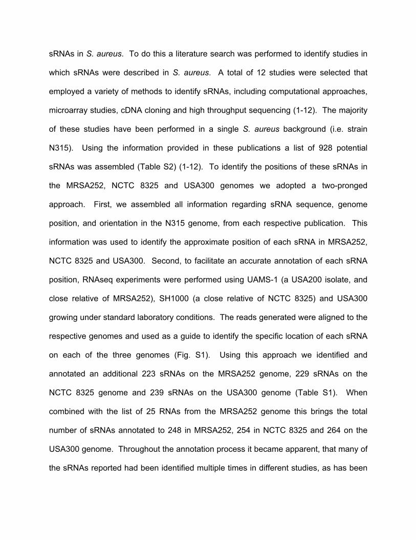
sRNAs in S. aureus. To do this a literature search was performed to identify studies in
which sRNAs were described in S. aureus. A total of 12 studies were selected that
employed a variety of methods to identify sRNAs, including computational approaches,
microarray studies, cDNA cloning and high throughput sequencing (1-12). The majority
of these studies have been performed in a single S. aureus background (i.e. strain
N315). Using the information provided in these publications a list of 928 potential
sRNAs was assembled (Table S2) (1-12). To identify the positions of these sRNAs in
the MRSA252, NCTC 8325 and USA300 genomes we adopted a two-pronged
approach. First, we assembled all information regarding sRNA sequence, genome
position, and orientation in the N315 genome, from each respective publication. This
information was used to identify the approximate position of each sRNA in MRSA252,
NCTC 8325 and USA300. Second, to facilitate an accurate annotation of each sRNA
position, RNAseq experiments were performed using UAMS-1 (a USA200 isolate, and
close relative of MRSA252), SH1000 (a close relative of NCTC 8325) and USA300
growing under standard laboratory conditions. The reads generated were aligned to the
respective genomes and used as a guide to identify the specific location of each sRNA
on each of the three genomes (Fig. S1). Using this approach we identified and
annotated an additional 223 sRNAs on the MRSA252 genome, 229 sRNAs on the
NCTC 8325 genome and 239 sRNAs on the USA300 genome (Table S1). When
combined with the list of 25 RNAs from the MRSA252 genome this brings the total
number of sRNAs annotated to 248 in MRSA252, 254 in NCTC 8325 and 264 on the
USA300 genome. Throughout the annotation process it became apparent, that many of
the sRNAs reported had been identified multiple times in different studies, as has been
noted recently (14). Where multiple copies of sRNAs exist, each copy was individually
annotated to distinguish them from each other. In addition, we observed that the α-
phenol soluble modulins (α-PSM) transcript was mistakenly identified as a sRNA (6).
The most likely reason for this is that the α-PSMs are not annotated in the S. aureus
genome and therefore any transcript mapping to that location was thought to represent
a sRNA. To avoid future confusion we have included annotations for the PSMs in the
updated MRSA252, NCTC 8325 and USA300 Genbank files.
sRNA gene expression analysis by RNAseq
Expression values for each annotated gene were calculated as RPKM (reads per
kilobase material per million reads) values, using the CLC Genomics Workbench
software platform (Qiagen). TSB and serum data sets were normalized by quantile
normalization (16). To identify sRNA genes differentially expressed in serum Vs TSB,
eliminate lowly expressed genes, and reduce the impact of non-unique reads, three cut
off criteria were applied to the data.
1. Percent unique reads. RPKM values are calculated based upon the number of
reads mapping to a gene. These reads can be unique or non-unique. Non-unique
reads map to multiple locations on the genome and consequently their mapping location
cannot be precisely determined. To avoid artificially inflating RPKM values due to the
incorporation of ambiguous non-unique reads we imposed a cut off whereby >80% of
the reads mapping to a gene (in both data sets) must be unique.
2. Expression values. Small differences in expression values can translate into large
fold-differences for lowly expressed genes. To eliminate lowly expressed genes we
imposed a cut-off whereby the RPKM expression value of a gene must be greater than
or equal to 50 in at least one data set.
3. Fold-change. We imposed a cut-off of 3-fold to identify genes showing differentially
expressed in serum Vs TSB.
sRNA identification in USA300
RNAseq reads generated for USA300 growing in TSB and human serum were aligned
to the newly created Genbank file containing annotations for known sRNAs. These
read alignment files were subsequently examined for transcripts that did not map to
annotated genes. Two types of potential sRNA were identified (i) those mapping to
intergenic regions and (ii) those that were antisense to, and located within annotated
genes. The following criteria were applied during the selection process to avoid
excessive false positive identifications. The minimum number of unique reads mapping
to a potential sRNA, and minimum RPKM expression value generated must exceed 25
and 40 respectively. Using these selection criteria 39 potential sRNAs were identified
which we named tsr1-39.
Reverse transcriptase polymerase chain reaction
Overnight cultures of wild-type S. aureus USA300 were diluted 1:100 into 5 mL of fresh
TSB and incubated for 3 hours at 37°C in a rotating incubator at 250 RPM. The cultures
were diluted to an OD600 of 0.05 in 100 mL of fresh TSB and incubated for 3 hours at
37°C in a rotating incubator at 250 RPM. Duplicate samples were immediately removed
into 3 volumes of ice cold PBS and centrifuged for 10 minutes at 4150 RPM. RNA was
extracted as previously outlined (16) and contaminating DNA removed using TURBO™
DNase (ThermoFisher Scientific) per their protocol. Finally, 1 µg DNA-free RNA was
used to create cDNA using the iScript™ Reverse Transcription Supermix Kit (BioRad)
per their instructions.
To isolate genomic DNA the pelleted cells were re-suspended in 600 µL of TE buffer in
screw cap tubes with glass beads. The cells were then beaten for a total of 3 minutes in
30-second bursts. The cells were then centrifuged at full speed for 2 minutes. Next, 500
µL of supernatant was transferred to a sterile microfuge tube with 100 µL of 1.6% n-
lauroylsarcosine and 25 µg Proteinase K (US Biologicals) then incubated for 1 hour at
60°C. After incubation 500 µL of phenol:chloroform (1:1), briefly vortexed, and then
centrifuged at full speed for 2 minutes. The aqueous layer was removed and the DNA
was precipitated with 3 M sodium acetate and isopropanol overnight at -80°C. The DNA
was centrifuged at 4°C at full speed for 20 minutes. DNA pellets were rinsed with ice
cold 70% ethanol, dried at 45°C, and re-suspended in 500 µL of nuclease free water.
Green GoTaq (Promega) was used in an amplification reaction containing 1 mM primers
and 100 ng nucleic acid template (DNase-treated RNA, genomic DNA, or cDNA) or
water. All PCR reactions were performed in a DNA Engine Peltier Thermal Cycler
(BioRad) with an annealing temperature of 45°C and an extension time of 1 minute at
72°C for 35 cycles. PCR products were evaluated for size and specificity by separation
on a 2% (w/v) agarose gel with a 100 bp DNA Molecular Marker (Promega).