Embed Size (px)
Citation preview
J. Microbiol. Biotechnol. (2012), 22(1), 84–91http://dx.doi.org/10.4014/jmb.1107.07060First published online October 15, 2011
Characterization of Lipases from Staphylococcus aureus and Staphylococcusepidermidis Isolated from Human Facial Sebaceous Skin
Xie, Winny1,2
, Vivia Khosasih1,2
, Antonius Suwanto2, and Hyung Kwoun Kim
1*
1Division of Biotechnology, The Catholic University of Korea, Bucheon 420-743, Korea2Faculty of Biotechnology, Atma Jaya Catholic University of Indonesia, Jenderal Sudirman 51, Jakarta 12930, Indonesia
Received: July 27, 2011 / Revised: September 7, 2011 / Accepted: September 15, 2011
Two staphylococcal lipases were obtained from Staphylococcus
epidermidis S2 and Staphylococcus aureus S11 isolated
from sebaceous areas on the skin of the human face. The
molecular mass of both enzymes was estimated to be
45 kDa by SDS-PAGE. S2 lipase displayed its highest
activity in the hydrolysis of olive oil at 32o
C and pH 8,
whereas S11 lipase showed optimal activity at 31o
C and pH
8.5. The S2 lipase showed the property of cold-adaptation,
with activation energy of 6.52 kcal/mol. In contrast, S11
lipase’s activation energy, at 21 kcal/mol, was more
characteristic of mesophilic lipases. S2 lipase was stable
up to 45o
C and within the pH range from 5 to 9, whereas
S11 lipase was stable up to 50o
C and from pH 6 to 10. Both
enzymes had high activity against tributyrin, waste
soybean oil, and fish oil. Sequence analysis of the S2 lipase
gene showed an open reading frame of 2,067 bp encoding
a signal peptide (35 aa), a pro-peptide (267 aa), and a
mature enzyme (386 aa); the S11 lipase gene, at 2,076 bp,
also encoded a signal peptide (37 aa), pro-peptide (255 aa),
and mature enzyme (399 aa). The two enzymes maintained
amino acid sequence identity of 98-99% with other
similar staphylococcal lipases. Their microbial origins and
biochemical properties may make these staphylococcal
lipases isolated from facial sebaceous skin suitable for use
as catalysts in the cosmetic, medicinal, food, or detergent
industries.
Keywords: Staphylococcus, lipase, oil hydrolysis
A group of enzymes used widely in industrial and
household conversion processes are the lipases (E.C.
3.1.1.3). Enzymes belonging to this group are biocatalysts
in the hydrolysis of triacylglycerols into free fatty acids
and glycerols. These enzymes exhibit high substrate specificity
according to their diverse chemo-, regio-, and enantio-
selective properties [9]. Lipases are also selective in their
recognition of fatty acid species and can be used for
interesterification reactions to perform cocoa butter
substitutions and in the production of specialty fats or
biodiesel [9, 15, 20].
Among the microbial lipases, staphylococcal lipases are
classified as belonging to family I, subfamily 5 [10]. They
are produced as pre-pro-enzymes in which the pre-region
acts as a signal peptide, and are secreted as precursors to
form a mature protein of approximately 400 amino acid
residues after cleavage of the peptide bond between the
pro-region and mature enzyme by a specific protease.
Staphylococcal lipases have been applied industrially to
produce flavor esters. Lipases from Staphylococcus xylosus
play a role in aroma production in fermented food [23]. S.
xylosus is commonly used in lipolytic starter cultures for
fermented meat products such as sausages and ham [11].
Staphylococcus capitis lipase has been used in a hair
treatment formulation to suppress dandruff and itching
[24]. These properties have led to the adoption of
staphylococcal lipases for catalytic processes in the food
and medicinal industries.
The endogenous role of lipases in Staphylococcus bacteria
can be pathogenic in nature as well as to metabolize lipids,
and the opportunistic pathogen Staphylococcus aureus can
produce a lipase interfering with phagocytosis of human
granulocytes [17]. An immune response towards S. aureus
lipase is also reported [3].
On the other hand, their detailed three-dimensional
structures, as well as their specific pathogenic mechanisms,
have not yet been addressed. Further intensive biochemical
and structural studies on these lipases are necessary for
their cost-effective industrial application.
Over 200 different genera have been identified from human
skin [7]. Corynebacteria, staphylococci, and propionibacteria
*Corresponding authorPhone: +82 2 2164 4890; Fax: +82 2 2164 4865;E-mail: [email protected]
# Supplementary data for this paper are available on-line only athttp://jmb.or.kr.
85 Xie et al.
comprise the major portion of the microbiota of normal
human skin. Staphylococci in particular are commonly
found on sebaceous skin, which includes areas such as the
alar crease, back of the scalp, upper chest, and back.
Staphylococci isolated from such sebaceous areas are
likely to produce lypolytic enzymes and to metabolize
sebum. Human sebaceous skin is therefore a suitable site
to isolate lipase-producing staphylococcal strains.
The objectives of this research were to measure lipase
activity from Staphylococcus strains isolated from human
facial skin, to characterize the lipases responsible, and to
analyze their genetic sequence.
MATERIALS AND METHODS
Screening for Lipase Activity
Microbes isolated from human facial sebaceous skin were grown on
tributyrin [1% (v/v)] and tricaprylin [1% (v/v)] agar plates containing
1× gum arabic solution, 1% tryptone, 0.5% yeast extract, 0.5%
NaCl, and 1.5% agar. Gum arabic stock solution (10×) contained
10% (w/v) gum arabic, 200 mM NaCl, and 50 mM CaCl2.
Staphylococcus epidermidis S2 and Staphylococcus aureus S11,
which formed distinct clear zones around their colonies after 24 h
incubation at 37oC, were selected for further study.
Production and Concentration of S2 and S11 Lipases
S. epidermidis S2 and S. aureus S11 strains were cultivated in
800 ml of LB broth (1% tryptone, 0.5% yeast extract, and 1%
NaCl) at 37oC for 20 h with shaking at 200 rpm. Extracellular
enzymes were separated from bacterial cells by centrifugation
(7,000 ×g, 10 min) at 4oC. Supernatants containing extracellular
lipases were collected and added with ammonium sulfate to 30%
saturation. Centrifugation (10,000 ×g, 10 min) was performed to
remove non-protein polymers and protein aggregates from supernatants.
Addition of ammonium sulfate was continued until 70% saturation.
Protein precipitates were collected by centrifugation (10,000 ×g,
10 min), dissolved in distilled water, and dialyzed with Spectra/Por
4 membrane (Spectrum Labs, USA) to remove ammonium sulfate.
The dialysates were concentrated by an ultrafiltration kit using an
Amicon PLGC 47 mm membrane with the cut-off size of 10,000
MW (Millipore, USA).
Lipase Activity Assay and Estimation of Protein Concentration
An olive oil emulsion containing 1% (v/v) olive oil (Sigma, USA)
and 1% gum arabic was prepared by blending in a Waring blender
(model 51BL31) at maximum speed for 2 min. Lipase activity was
measured at 37oC using the pH-STAT method. Substrate emulsions
were adjusted to pH 8.0 before the addition of enzyme. Reactions
were initiated after addition of an appropriate amount of enzyme
(0.5 - 5 U). Titration of free fatty acids with 10 mM of NaOH
solution was performed during the reaction to maintain the pH of
the reaction at 8.0 for 5 min. The hydrolysis rate for lipase
conversion of olive oil into free fatty acids was measured with a
718 Titrino pH titrator (Metrohm, Switzerland). The amount of
enzyme catalyzing the release of 1 µmol fatty acid per minute was
defined as one lipase unit.
Protein concentration was measured using a Bradford assay kit
(Bio-Rad Lab., USA), and was calculated relative to a standard
curve of bovine serum albumin.
Molecular Mass Determinations
SDS-PAGE and zymograms were performed to determine the
molecular masses of S2 and S11 lipases. SDS-PAGE was performed
using polyacrylamide gels (10%) as described by Laemmli [13].
Proteins were stained with Coomassie Brilliant Blue R-250.
Gels used for zymograms were washed with 50 mM Tris-HCl
(pH 8.0) containing 1% Triton X-100 for 10 min with shaking. A
second 10 min wash step was performed with 50 mM Tris-HCl (pH
8.0) containing 0.1% Triton X-100; and a final 10 min wash was
performed with distilled water. Renatured proteins were checked for
activity by attaching gels to a tricaprylin agar plate and incubating at
37oC for 2 h.
Effects of Temperature on Lipase Activity and Stability
The optimal reaction temperatures of S2 and S11 lipases were
determined by assaying their hydrolytic activities toward olive oil at
various temperatures (10 - 60oC) using the pH-STAT method.
Lipase temperature stability was examined by their pre-incubation at
various temperatures for 30 min before assay, with a pH-STAT
instrument, for optimal temperature.
Effects of pH on Lipase Activity and Stability
The optimal pHs for S2 and S11 lipase activity were determined by
assaying their hydrolytic activities toward olive oil or p-nitrophenyl
caprylate (pNPC) at various pHs (pH 6-10) using pH-STAT and
spectrophotometry, respectively. The activity of the S11 lipase was
determined spectrophotometrically using pNPC, as accurate titration
of fatty acids released was difficult to determine above pH 9 by pH-
STAT. The result of pNPC assay for S11 lipase were normalized
with the result of the pH-STAT assay. The stability of the lipases at
various pHs was examined by pre-incubating 25 µl (corresponding
to about 2 U) of the S2 and S11 lipases in 225 µl of 0.1 M sodium
acetate (pH 4-6), 0.1 M potassium phosphate (pH 6-7.5), 0.1 M
Tris-HCl (pH 7.5-9), 0.1 M KCl-glycine-KOH (pH 9-10), or 0.1 M
potassium phosphate (pH 10-12) for 30 min and assaying with a
pH-STAT machine at their optimal temperature.
Analysis of Substrate Specificity
Tributyrin, tricaprylin, olive oil, soybean oil, sunflower oil, fish oil
(Sigma, USA), home waste cooking oil, and waste soybean oil
(National Fisheries Research and Development Institute, Busan,
South Korea) were selected for use in substrate emulsions. S2 and
S11 lipase activity toward various substrates was measured using the
pH-STAT method at their optimal temperature and pH.
PCR Cloning of S2 and S11 Lipase Genes
To obtain the S2 and S11 lipase genetic sequence, four primers were
designed based on the 5'- and 3'-terminal sequences of the S.
epidermidis 9 (GenBank: M95577) and S. aureus B56 lipase genes
(GenBank: AY028918) [5, 12]. The primer sequences for S11 (AF
and AR) and S2 (EF and ER) were as follows: AF, 5'-GAA CAT
ATG TTA AGA GGA CAA GAA-3'; AR, 5'-CTT GGA TCC ATA
CTT GCT TTC AAT TGT GT-3'; EF, 5'-GAA CCA TGG TGA
AGA CAA GAC AAA A -3'; ER, 5'-TCC GGA TCC ATT TTA
TTT GTT GAT GTT AAT TG-3'.
STAPHYLOCOCCUS AUREUS AND STAPHYLOCCUS EPIDERMIDIS LIPASES 86
PCR conditions were as follows: pre-denaturation at 95oC for
5 min, followed by 30 cycles of denaturation at 95oC for 1 min,
annealing at 41oC for 0.5 min, and extension at 72
oC for 1.5 min,
and a post-extension step of 5 min at 72oC.
PCR products were inserted into pGEM-T vectors (Promega,
USA) and the recombinant vectors were transformed by electroporation
into E. coli XL1-Blue cells. Purified plasmids were sequenced with
primers directed against the T7 and SP6 promoters.
Lipase Gene-Sequence Analysis
The genetic sequences of the S2 and S11 lipases were analyzed for
homology to other organisms by NCBI BLAST. The DNA sequences
were translated into amino acid sequences by using the EditSeq
application from the DNASTAR program. Amino acid sequences
were aligned against other known staphylococcal lipases using the
ClustalW method in the DNASTAR MegAlign application. Protein
divergence (in millions of years since species divergence) was
calculated based on amino acid substitutions using cytochrome S as
a “molecular clock” [14]. Protein divergence (π) at each node in the
phylogenetic tree (Fig. 5) was calculated according to the formula
π = Σ πij/2n, where πij is the pairwise divergence between the ith
protein in one branch and the jth protein in the other.
RESULTS
Isolation of Lipase-Producing Staphylococcal Strains
We isolated microbial strains from human facial sebaceous
skin as follows. The condition of sample source was oily
face skin with some acne on the face skin. The oily part of
the face (forehead and cheek) was swabbed using a cotton
bath. Then, the cotton bath was streaked on an LB agar
plate and incubated overnight at 37oC. Approximately 14
colonies showing different morphologies were selected
and cultivated on Rhodamine B agar plate at 37oC for 48 h.
Then 9 colonies forming orange fluorescent halos under
UV irradiation were finally selected. We assessed their
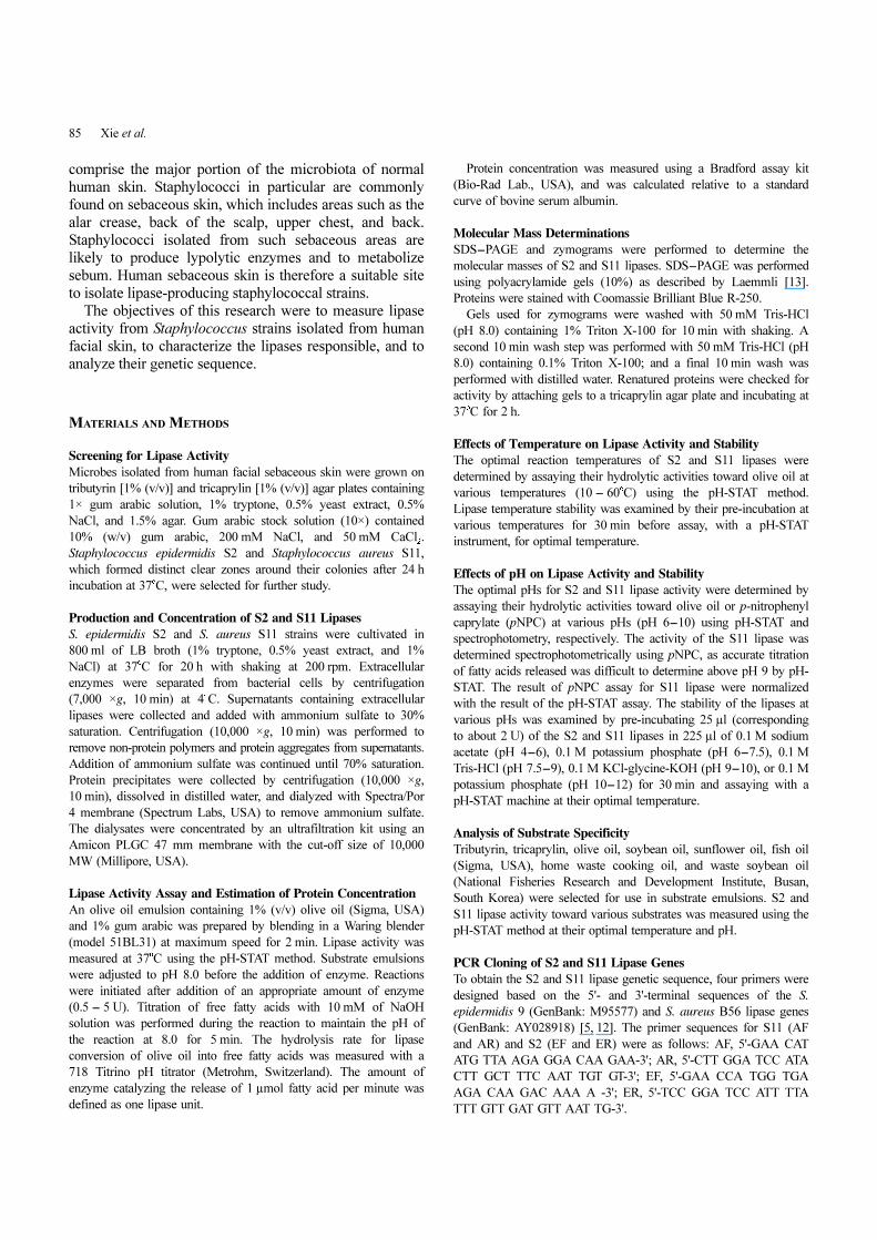
ability to form clear zones around colonies grown on
tributyrin (TBN) (Fig. 1A and 1B) and tricaprylin (TCN)
LB plates. Of these strains, the two colonies showing the
largest halos were selected and designated as S2 and S11.
16S rRNA analysis identified these strains as Staphylococcus
epidermidis and Staphylococcus aureus, respectively. The
GenBank accession numbers for the 16S rRNAs of these
bacteria are JN245969 and JN245970, respectively.
Determination of Lipase Molecular Mass
S. epidermidis S2 and S. aureus S11 were cultured and the
culture supernatants were partially purified by ammonium
sulfate precipitation, dialysis, and ultrafiltration. The
concentrated enzymes were loaded into polyacrylamide gels
for separation and zymogram analysis. TCN zymograms
demonstrated formation of a single distinct band in each
lane (Fig. 1D). The molecular mass of each enzyme was
estimated at approximately 45 kDa by comparison of marker
size and zymogram results (Fig. 1C and 1D).
Effects of Temperature and pH on Lipase Activity and
Stability
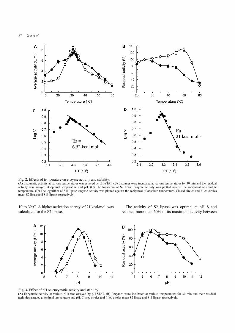
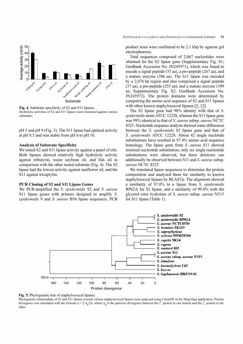
The S. epidermidis S2 lipase activity against olive oil
reached its optimum at 32oC, but it could stably retain
activity relatively up to 45oC after 30 min incubation at
various temperatures; activity decreased beyond pre-
incubation at 50oC. S11 lipase showed optimal activity at
31oC and was stable up to 50oC pre-incubation (Fig. 2A
and 2B).
In contrast to S2 lipase, which exhibited diminished
stability above 45oC and activity above 50oC, the activity
of S11 lipase increased upon high-temperature pre-
incubation. Although the activity continued to increase
with pre-incubation of up to 50oC, it declined rapidly
beyond 55oC.
We calculated the lipase activation energy with the
Arrhenius equation according to their activity at various
temperatures (Fig. 2C and 2D). The activation energy of
S2 lipase was 6.52 kcal/mol in the temperature range from
Fig. 1. Characterization of lipase activities and molecular mass. Clear zones were formed around S. epidermidis S2 (A) and S. aureus S11
(B) colonies. (C) SDS-PAGE and (D) TCN zymograms were performed
with partially purified S2 and S11 enzymes. The arrow indicates distinct 45
kDa bands.
87 Xie et al.
10 to 32oC. A higher activation energy, of 21 kcal/mol, was
calculated for the S2 lipase.
The activity of S2 lipase was optimal at pH 8 and
retained more than 60% of its maximum activity between
Fig. 2. Effects of temperature on enzyme activity and stability. (A) Enzymatic activity at various temperatures was assayed by pH-STAT. (B) Enzymes were incubated at various temperatures for 30 min and the residual
activity was assayed at optimal temperature and pH. (C) The logarithm of S2 lipase enzyme activity was plotted against the reciprocal of absolute
temperature. (D) The logarithm of S11 lipase enzyme activity was plotted against the reciprocal of absolute temperature. Closed circles and filled circles
mean S2 lipase and S11 lipase, respectively.
Fig. 3. Effect of pH on enzymatic activity and stability. (A) Enzymatic activity at various pHs was assayed by pH-STAT. (B) Enzymes were incubated at various temperatures for 30 min and their residual
activities assayed at optimal temperature and pH. Closed circles and filled circles mean S2 lipase and S11 lipase, respectively.
STAPHYLOCOCCUS AUREUS AND STAPHYLOCCUS EPIDERMIDIS LIPASES 88
pH 5 and pH 9 (Fig. 3). The S11 lipase had optimal activity
at pH 8.5 and was stable from pH 6 to pH 10.
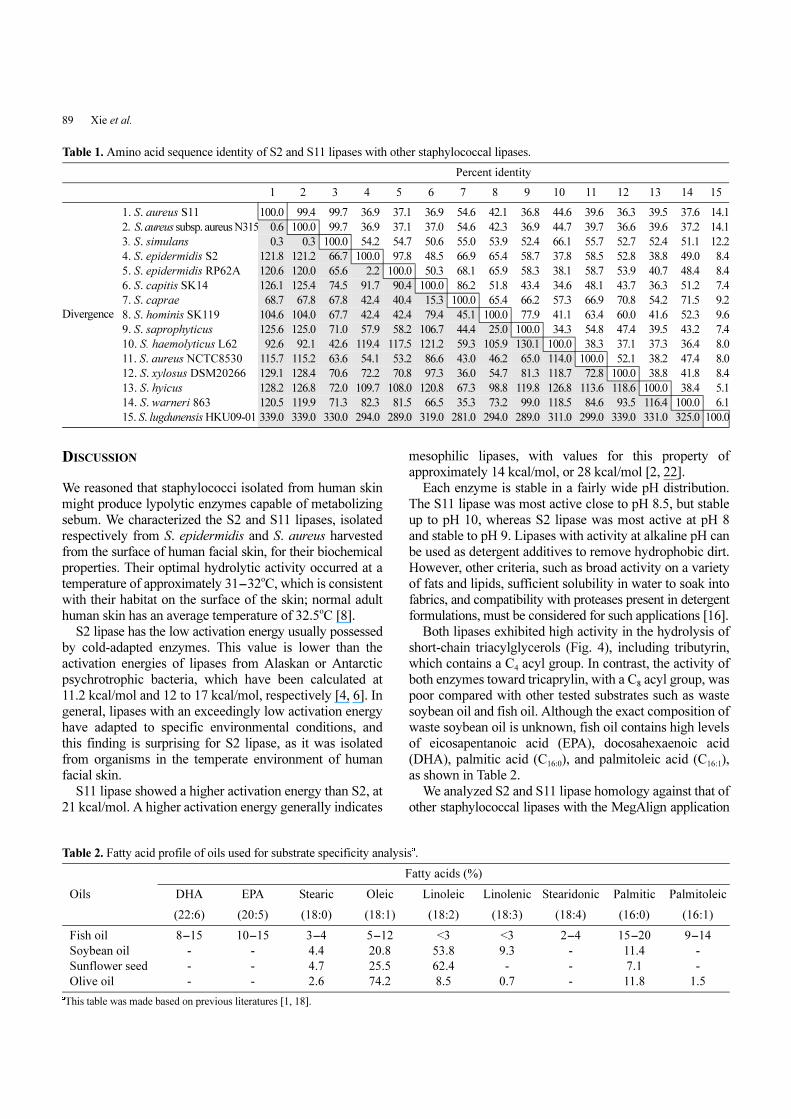
Analysis of Substrate Specificity
We tested S2 and S11 lipase activity against a panel of oils.
Both lipases showed relatively high hydrolytic activity
against tributyrin, waste soybean oil, and fish oil in
comparison with the other tested substrate (Fig. 4). The S2
lipase had the lowest activity against sunflower oil, and the
S11 against tricaprylin.
PCR Cloning of S2 and S11 Lipase Genes
We PCR-amplified the S. epidermidis S2 and S. aureus
S11 lipase genes with primers designed to amplify S.
epidermidis 9 and S. aureus B56 lipase sequences. PCR
product sizes were confirmed to be 2.1 kbp by agarose gel
electrophoresis.
Total sequences composed of 2,067 nucleotides were
obtained for the S2 lipase gene (Supplementary Fig. S1;
GenBank Accession No. JN245971), which was found to
encode a signal peptide (35 aa), a pro-peptide (267 aa), and
a mature enzyme (386 aa). The S11 lipase was encoded
by a 2,076 bp region and also comprised a signal peptide
(37 aa), a pro-peptide (255 aa), and a mature enzyme (399
aa; Supplementary Fig. S2; GenBank Accession No.
JN245972). The protein domains were determined by
compairing the amino acid sequence of S2 and S11 lipases
with other known staphylococcal lipases [5, 12].
The S2 lipase gene had 98% identity with that of S.
epidermidis strain ATCC 12228, whereas the S11 lipase gene
was 99% identical to that of S. aureus subsp. aureus NCTC
8325. Nucleotide sequence analysis showed some differences
between the S. epidermidis S2 lipase gene and that of
S. epidermidis ATCC 12228. About 42 single nuclotide
substitutions have resulted in 97.4% amino acid sequence
homology. The lipase gene from S. aureus S11 showed
minimal nucleotide substitution; only six single nucleotide
substitutions were observed, but three deletions can
additionally be observed between S11 and S. aureus subsp.
aureus NCTC 8325.
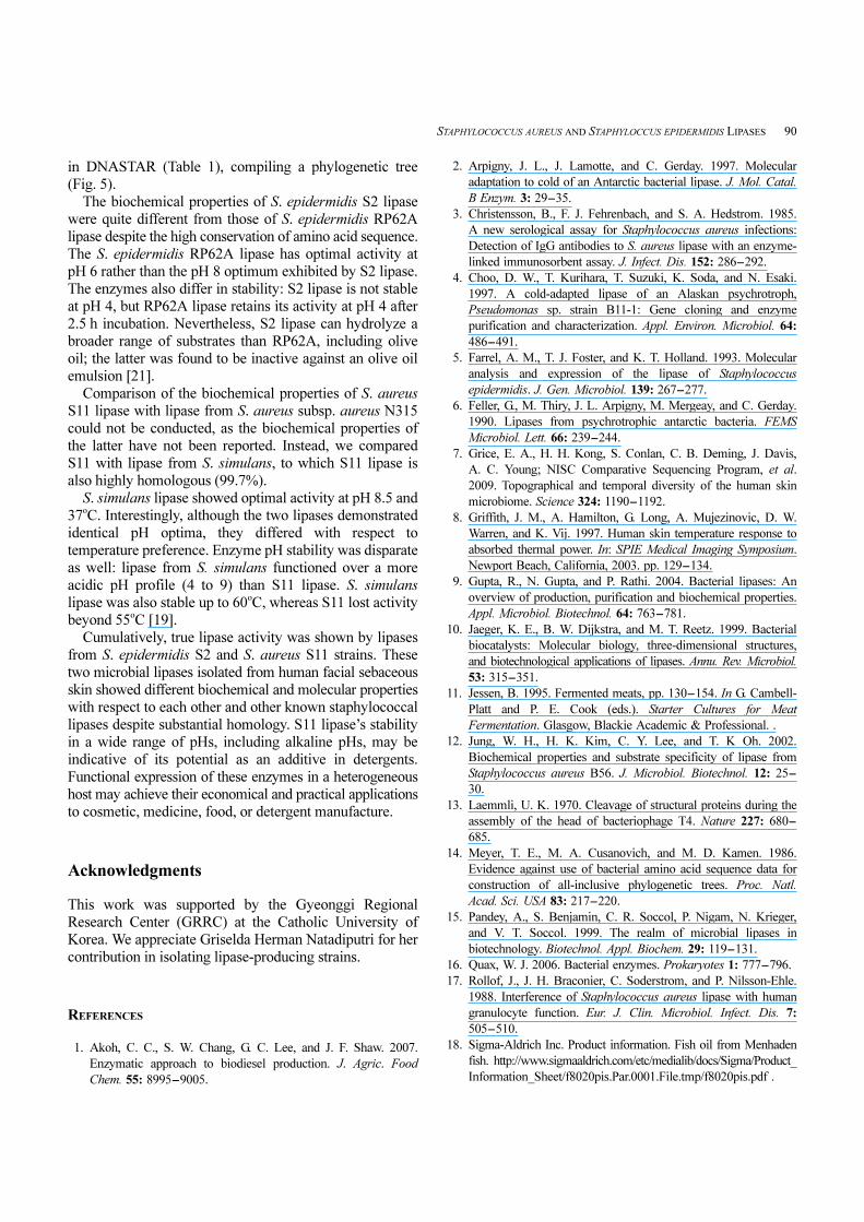
We translated lipase sequences to determine the protein
composition and analyzed them for similarity to known
staphylococcal lipases by BLASTp. The alignment showed
a similarity of 97.8% to a lipase from S. epidermidis
RP62A for S2 lipase, and a similarity of 99.4% with the
glycerol ester hydrolase of S. aureus subsp. aureus N315
for S11 lipase (Table 1).
Fig. 4. Substrate specificity of S2 and S11 lipases. Hydrolytic activities of S2 and S11 lipases were measured against various
substrates.
Fig. 5. Phylogenetic tree of staphylococcal lipases. Phylogenetic relationships of S2 and S11 lipases toward various staphylococcal lipases were analyzed using ClustalW in the MegAlign application. Protein
divergence was calculated with the formula π = Σ πij/2n, where πij is the pairwise divergence between the ith protein in one branch and the j
th protein in the
other.
89 Xie et al.
DISCUSSION
We reasoned that staphylococci isolated from human skin
might produce lypolytic enzymes capable of metabolizing
sebum. We characterized the S2 and S11 lipases, isolated
respectively from S. epidermidis and S. aureus harvested
from the surface of human facial skin, for their biochemical
properties. Their optimal hydrolytic activity occurred at a
temperature of approximately 31-32oC, which is consistent
with their habitat on the surface of the skin; normal adult
human skin has an average temperature of 32.5oC [8].
S2 lipase has the low activation energy usually possessed
by cold-adapted enzymes. This value is lower than the
activation energies of lipases from Alaskan or Antarctic
psychrotrophic bacteria, which have been calculated at
11.2 kcal/mol and 12 to 17 kcal/mol, respectively [4, 6]. In
general, lipases with an exceedingly low activation energy
have adapted to specific environmental conditions, and
this finding is surprising for S2 lipase, as it was isolated
from organisms in the temperate environment of human
facial skin.
S11 lipase showed a higher activation energy than S2, at
21 kcal/mol. A higher activation energy generally indicates
mesophilic lipases, with values for this property of
approximately 14 kcal/mol, or 28 kcal/mol [2, 22].
Each enzyme is stable in a fairly wide pH distribution.
The S11 lipase was most active close to pH 8.5, but stable
up to pH 10, whereas S2 lipase was most active at pH 8
and stable to pH 9. Lipases with activity at alkaline pH can
be used as detergent additives to remove hydrophobic dirt.
However, other criteria, such as broad activity on a variety
of fats and lipids, sufficient solubility in water to soak into
fabrics, and compatibility with proteases present in detergent
formulations, must be considered for such applications [16].
Both lipases exhibited high activity in the hydrolysis of
short-chain triacylglycerols (Fig. 4), including tributyrin,
which contains a C4 acyl group. In contrast, the activity of
both enzymes toward tricaprylin, with a C8 acyl group, was
poor compared with other tested substrates such as waste
soybean oil and fish oil. Although the exact composition of
waste soybean oil is unknown, fish oil contains high levels
of eicosapentanoic acid (EPA), docosahexaenoic acid
(DHA), palmitic acid (C16:0), and palmitoleic acid (C16:1),
as shown in Table 2.
We analyzed S2 and S11 lipase homology against that of
other staphylococcal lipases with the MegAlign application
Table 1. Amino acid sequence identity of S2 and S11 lipases with other staphylococcal lipases.
Percent identity
1 2 3 4 5 6 7 8 9 10 11 12 13 14 15
Divergence
1. S. aureus S11 100.0 99.4 99.7 36.9 37.1 36.9 54.6 42.1 36.8 44.6 39.6 36.3 39.5 37.6 14.1
2. S. aureus subsp. aureus N315 0.6 100.0 99.7 36.9 37.1 37.0 54.6 42.3 36.9 44.7 39.7 36.6 39.6 37.2 14.1
3. S. simulans 0.3 0.3 100.0 54.2 54.7 50.6 55.0 53.9 52.4 66.1 55.7 52.7 52.4 51.1 12.2
4. S. epidermidis S2 121.8 121.2 66.7 100.0 97.8 48.5 66.9 65.4 58.7 37.8 58.5 52.8 38.8 49.0 8.4
5. S. epidermidis RP62A 120.6 120.0 65.6 2.2 100.0 50.3 68.1 65.9 58.3 38.1 58.7 53.9 40.7 48.4 8.4
6. S. capitis SK14 126.1 125.4 74.5 91.7 90.4 100.0 86.2 51.8 43.4 34.6 48.1 43.7 36.3 51.2 7.4
7. S. caprae 68.7 67.8 67.8 42.4 40.4 15.3 100.0 65.4 66.2 57.3 66.9 70.8 54.2 71.5 9.2
8. S. hominis SK119 104.6 104.0 67.7 42.4 42.4 79.4 45.1 100.0 77.9 41.1 63.4 60.0 41.6 52.3 9.6
9. S. saprophyticus 125.6 125.0 71.0 57.9 58.2 106.7 44.4 25.0 100.0 34.3 54.8 47.4 39.5 43.2 7.4
10. S. haemolyticus L62 92.6 92.1 42.6 119.4 117.5 121.2 59.3 105.9 130.1 100.0 38.3 37.1 37.3 36.4 8.0
11. S. aureus NCTC8530 115.7 115.2 63.6 54.1 53.2 86.6 43.0 46.2 65.0 114.0 100.0 52.1 38.2 47.4 8.0
12. S. xylosus DSM20266 129.1 128.4 70.6 72.2 70.8 97.3 36.0 54.7 81.3 118.7 72.8 100.0 38.8 41.8 8.4
13. S. hyicus 128.2 126.8 72.0 109.7 108.0 120.8 67.3 98.8 119.8 126.8 113.6 118.6 100.0 38.4 5.1
14. S. warneri 863 120.5 119.9 71.3 82.3 81.5 66.5 35.3 73.2 99.0 118.5 84.6 93.5 116.4 100.0 6.1
15. S. lugdunensis HKU09-01 339.0 339.0 330.0 294.0 289.0 319.0 281.0 294.0 289.0 311.0 299.0 339.0 331.0 325.0 100.0
Table 2. Fatty acid profile of oils used for substrate specificity analysisa.
Fatty acids (%)
Oils DHA EPA Stearic Oleic Linoleic Linolenic Stearidonic Palmitic Palmitoleic
(22:6) (20:5) (18:0) (18:1) (18:2) (18:3) (18:4) (16:0) (16:1)
Fish oil 8-15 10-15 3-4 5-12 <3 <3 2-4 15-20 9-14
Soybean oil - - 4.4 20.8 53.8 9.3 - 11.4 -
Sunflower seed - - 4.7 25.5 62.4 - - 7.1 -
Olive oil - - 2.6 74.2 8.5 0.7 - 11.8 1.5
aThis table was made based on previous literatures [1, 18].
STAPHYLOCOCCUS AUREUS AND STAPHYLOCCUS EPIDERMIDIS LIPASES 90
in DNASTAR (Table 1), compiling a phylogenetic tree
(Fig. 5).
The biochemical properties of S. epidermidis S2 lipase
were quite different from those of S. epidermidis RP62A
lipase despite the high conservation of amino acid sequence.
The S. epidermidis RP62A lipase has optimal activity at
pH 6 rather than the pH 8 optimum exhibited by S2 lipase.
The enzymes also differ in stability: S2 lipase is not stable
at pH 4, but RP62A lipase retains its activity at pH 4 after
2.5 h incubation. Nevertheless, S2 lipase can hydrolyze a
broader range of substrates than RP62A, including olive
oil; the latter was found to be inactive against an olive oil
emulsion [21].
Comparison of the biochemical properties of S. aureus
S11 lipase with lipase from S. aureus subsp. aureus N315
could not be conducted, as the biochemical properties of
the latter have not been reported. Instead, we compared
S11 with lipase from S. simulans, to which S11 lipase is
also highly homologous (99.7%).
S. simulans lipase showed optimal activity at pH 8.5 and
37oC. Interestingly, although the two lipases demonstrated
identical pH optima, they differed with respect to
temperature preference. Enzyme pH stability was disparate
as well: lipase from S. simulans functioned over a more
acidic pH profile (4 to 9) than S11 lipase. S. simulans
lipase was also stable up to 60oC, whereas S11 lost activity
beyond 55oC [19].
Cumulatively, true lipase activity was shown by lipases
from S. epidermidis S2 and S. aureus S11 strains. These
two microbial lipases isolated from human facial sebaceous
skin showed different biochemical and molecular properties
with respect to each other and other known staphylococcal
lipases despite substantial homology. S11 lipase’s stability
in a wide range of pHs, including alkaline pHs, may be
indicative of its potential as an additive in detergents.
Functional expression of these enzymes in a heterogeneous
host may achieve their economical and practical applications
to cosmetic, medicine, food, or detergent manufacture.
Acknowledgments
This work was supported by the Gyeonggi Regional
Research Center (GRRC) at the Catholic University of
Korea. We appreciate Griselda Herman Natadiputri for her
contribution in isolating lipase-producing strains.
REFERENCES
1. Akoh, C. C., S. W. Chang, G. C. Lee, and J. F. Shaw. 2007.
Enzymatic approach to biodiesel production. J. Agric. Food
Chem. 55: 8995-9005.
2. Arpigny, J. L., J. Lamotte, and C. Gerday. 1997. Molecular
adaptation to cold of an Antarctic bacterial lipase. J. Mol. Catal.
B Enzym. 3: 29-35.
3. Christensson, B., F. J. Fehrenbach, and S. A. Hedstrom. 1985.
A new serological assay for Staphylococcus aureus infections:
Detection of IgG antibodies to S. aureus lipase with an enzyme-
linked immunosorbent assay. J. Infect. Dis. 152: 286-292.
4. Choo, D. W., T. Kurihara, T. Suzuki, K. Soda, and N. Esaki.
1997. A cold-adapted lipase of an Alaskan psychrotroph,
Pseudomonas sp. strain B11-1: Gene cloning and enzyme
purification and characterization. Appl. Environ. Microbiol. 64:
486-491.
5. Farrel, A. M., T. J. Foster, and K. T. Holland. 1993. Molecular
analysis and expression of the lipase of Staphylococcus
epidermidis. J. Gen. Microbiol. 139: 267-277.
6. Feller, G., M. Thiry, J. L. Arpigny, M. Mergeay, and C. Gerday.
1990. Lipases from psychrotrophic antarctic bacteria. FEMS
Microbiol. Lett. 66: 239-244.
7. Grice, E. A., H. H. Kong, S. Conlan, C. B. Deming, J. Davis,
A. C. Young; NISC Comparative Sequencing Program, et al.
2009. Topographical and temporal diversity of the human skin
microbiome. Science 324: 1190-1192.
8. Griffith, J. M., A. Hamilton, G. Long, A. Mujezinovic, D. W.
Warren, and K. Vij. 1997. Human skin temperature response to
absorbed thermal power. In: SPIE Medical Imaging Symposium.
Newport Beach, California, 2003. pp. 129-134.
9. Gupta, R., N. Gupta, and P. Rathi. 2004. Bacterial lipases: An
overview of production, purification and biochemical properties.
Appl. Microbiol. Biotechnol. 64: 763-781.
10. Jaeger, K. E., B. W. Dijkstra, and M. T. Reetz. 1999. Bacterial
biocatalysts: Molecular biology, three-dimensional structures,
and biotechnological applications of lipases. Annu. Rev. Microbiol.
53: 315-351.
11. Jessen, B. 1995. Fermented meats, pp. 130-154. In G. Cambell-
Platt and P. E. Cook (eds.). Starter Cultures for Meat
Fermentation. Glasgow, Blackie Academic & Professional. .
12. Jung, W. H., H. K. Kim, C. Y. Lee, and T. K Oh. 2002.
Biochemical properties and substrate specificity of lipase from
Staphylococcus aureus B56. J. Microbiol. Biotechnol. 12: 25-
30.
13. Laemmli, U. K. 1970. Cleavage of structural proteins during the
assembly of the head of bacteriophage T4. Nature 227: 680-
685.
14. Meyer, T. E., M. A. Cusanovich, and M. D. Kamen. 1986.
Evidence against use of bacterial amino acid sequence data for
construction of all-inclusive phylogenetic trees. Proc. Natl.
Acad. Sci. USA 83: 217-220.
15. Pandey, A., S. Benjamin, C. R. Soccol, P. Nigam, N. Krieger,
and V. T. Soccol. 1999. The realm of microbial lipases in
biotechnology. Biotechnol. Appl. Biochem. 29: 119-131.
16. Quax, W. J. 2006. Bacterial enzymes. Prokaryotes 1: 777-796.
17. Rollof, J., J. H. Braconier, C. Soderstrom, and P. Nilsson-Ehle.
1988. Interference of Staphylococcus aureus lipase with human
granulocyte function. Eur. J. Clin. Microbiol. Infect. Dis. 7:
505-510.
18. Sigma-Aldrich Inc. Product information. Fish oil from Menhaden
fish. http://www.sigmaaldrich.com/etc/medialib/docs/Sigma/Product_
Information_Sheet/f8020pis.Par.0001.File.tmp/f8020pis.pdf .
91 Xie et al.
19. Sayari, A., N. Agrebi, S. Jaoua, and Y. Gargouri. 2001.
Biochemical and molecular characterization of Staphylococcus
simulans lipase. Biochimie 83: 863-871.
20. Sharma, R., Y. Chisti, and U. C. Banerjee. 2001. Production,
purification, characterization, and applications of lipases. Biotechnol.
Adv. 19: 627-662.
21. Simons, J. W., M. D. Kampen, S. Riel, F. Gotz, M. R. Egmond,
and H. M. Verheij. 1998. Cloning, purification and chracterisation
of the lipase from Staphylococcus epidermidis: Implications
for structure-function relationships of staphylococci. Eur. J.
Biochem. 253: 675-683.
22. Suzuki, T., T. Nakayama, T. Kurihara, T. Nishino, and N. Esaki.
2002. Primary structure and catalytic properties of a cold-active
esterase from a psychrotroph, Acinetobacter sp. strain no. 6.
isolated from Siberian soil. Biosci. Biotechnol. Biochem. 8:
1682-1690.
23. Talon, R. and M. C. Montel. 1997. Hydrolysis of esters by
staphylococci. Int. J. Food Microbiol. 36: 207-214.
24. Yoshizumi, H., T. Amachi, T. Kusumi, T. Tanaka, and H.
Ishigooka. 1988. Hair treatment composition. US patent 4737362.