Embed Size (px)
Citation preview
Molecular Regulation of UVB-Induced Cutaneous Angiogenesis
Diane R. Bielenberg, Corazon D. Bucana, Ricardo Sanchez, Cherrie K. Donawho,* Margaret L. Kripke,* andIsaiah J. FidlerDepartments of Cell Biology and * Immunology, The University of Texas MD Anderson Cancer Center, Houston, Texas, U.S.A.
We determined whether cutaneous angiogenesis inducedby exposure of mice to ultraviolet-B (UVB) radiationis associated with an imbalance between positive andnegative angiogenesis-regulating molecules. UnshavedC3H/HeN mice were exposed to a single dose (15 kJ perm2) of UVB. At various times, the mice were killed, andtheir external ears were processed for routine histologyand immunohistochemistry. Antibodies against proli-ferating cell nuclear antigen and bromodeoxyuridineidentified dividing cells. Antibodies against CD31/PECAM-1 identified endothelial cells, and antibodiesagainst basic fibroblast growth factor (bFGF), vascularendothelial growth factor/vascular permeability factor,and interferon-β (IFN-β) identified angiogenesis-regulat-ing molecules. Epidermal hyperplasia was documentedby 48 h and reached a maximum on day 7 after exposure
The formation of blood vessels, i.e., angiogenesis, isessential for normal reproduction, development, woundhealing and repair, and tumor growth (Folkman, 1995).To form a new blood vessel, endothelial cells mustdegrade the basement membrane of existing vessels,
invade through the walls of the venules, migrate toward an angiogenicstimulus, proliferate as they align in a linear fashion to form sprouts,and differentiate to form tubes through which blood will ultimatelyflow (Folkman, 1995). Factors that stimulate angiogenesis includemembers of the fibroblast growth factor family, vascular endothelialgrowth factor/vascular permeability factor (VEGF/VPF), plateletderived endothelial cell growth factor, interleukin-8 (IL-8), platelet-derived growth factor, and matrix metalloproteinases (Folkman andKlagsbrun, 1987; Auerbach and Auerbach, 1994; Fidler and Ellis, 1994;Singh et al, 1995b). Negative regulators of angiogenesis includeinterferons, thrombospondin, platelet factor 4, prolactin fragment,angiostatin, endostatin, and tissue inhibitors of metalloproteinases(Auerbach and Auerbach, 1994; Fidler and Ellis, 1994; O’Reilly et al,1994, 1997; Singh et al, 1995a). The extent of angiogenesis is determinedby the balance between positive and negative endogenous molecules.Pathologic conditions such as psoriasis, retinopathy, and vascular
Manuscript received March 31, 1998; revised July 3, 1998; accepted forpublication July 3, 1998.
Reprint requests to: Dr. Isaiah J. Fidler, Department of Cell Biology-173,The University of Texas M.D. Anderson Cancer Center, 1515 HolcombeBoulevard, Houston, TX 77030.
Abbreviations: MVD, microvessel density; PCNA, proliferating cell nuclearantigen; VEGF/VPF, vascular endothelial growth factor/vascular permeabilityfactor.
0022-202X/98/$10.50 · Copyright © 1998 by The Society for Investigative Dermatology, Inc.
864
to UVB. The expression of bFGF increased by 24 h,whereas the expression of IFN-β decreased by 72 h afterexposure to UVB. The expression of vascular endothelialgrowth factor/vascular permeability factor increasedslightly after irradiation. The altered balance betweenbFGF and IFN-β was associated with increased endothel-ial cell proliferation (bromodeoxyuridineF CD31F cells)within existing blood vessels, leading to telangiectasiaand new blood vessels. UV-induced epidermal hyperplasiaand cutaneous angiogenesis were highest in IFN-α/βreceptor knockout mice. These results demonstrate thatin response to UVB radiation, dividing keratinocytesproduce a positive angiogenic molecule (bFGF) but nota negative angiogenic molecule (IFN-β), and that thisaltered balance is associated with enhanced cutaneousangiogenesis. Key words: bFGF/IFN-β/UVB radiation. JInvest Dermatol 111:864–872, 1998
neoplasms, e.g., hemangioma and angiofibroma, are associated with animbalance between angiogenesis-regulating molecules (Folkman, 1995).
Cutaneous infantile hemangiomas represent a relatively pure formof pathologic angiogenesis. These benign tumors are characterized byrapid proliferation of capillaries in the first year of life (proliferativephase), slow growth during the next 5 y (involuting phase), andeventual involution or complete regression by 10–15 y of age (involutedphase) (Ezekowitz et al, 1992). In a previous study, we examined thehypothesis that the progression and involution of hemangiomas areassociated with the relative levels of expression of positive and negativeangiogenic molecules. We found that proliferating and involutingtumors expressed high levels of basic fibroblast growth factor (bFGF)and VEGF/VPF but not interferon-β (IFN-β) (mRNA or protein).Moreover, the epidermis directly overlying proliferating and earlyinvoluting hemangiomas was hyperplastic and expressed high levels ofbFGF and VEGF/VPF but not IFN-β. In contrast, the epidermis fromage-matched normal individuals, the epidermis directly overlyinginvoluted lesions, and the epidermis at sites distant from proliferatinghemangiomas, were not hyperplastic and expressed IFN-β as wellas bFGF and VEGF/VPF. These findings suggest that epidermalhyperplasia surrounding proliferating hemangioma produces bFGF andVEGF/VPF but not IFN-β, and thus contributes to the angiogenesisof the neoplasms.1
To further examine the relationship between epidermal hyperplasiaand cutaneous angiogenesis, we studied the well-documented effectsof ultraviolet (UV) radiation on the skin. Exposure to UV radiation,
1Bielenberg, DR, Bucana CD, Mulliken J, Folkman J, Fidler IJ: Loss of anendogenous negative angiogenic factor in the epidermis overlying hemangiomalesions. Proc Am Assoc Cancer Res 38:524, 1997 (abstr.)

VOL. 111, NO. 5 NOVEMBER 1998 UVB-INDUCED CUTANEOUS ANGIOGENESIS 865
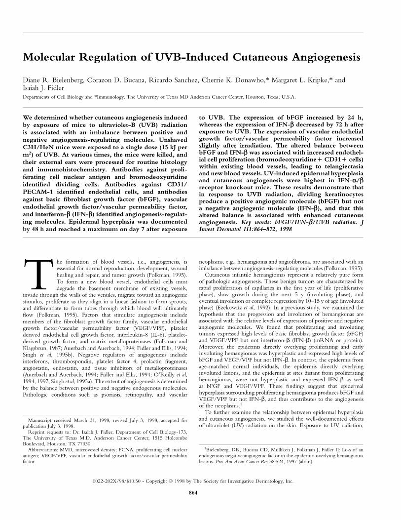
Figure 1. Induction of epidermal hyperplasia and cutaneous angiogenesis by UVB radiation. C3H/HeN mice were exposed to a single 15 kJ per m2
dose of UVB and killed 1, 2, 3, 5, or 7 d after irradiation. Their ears were resected and either formalin-fixed and paraffin-embedded for histologic (hematoxylinand eosin stain) and for immunohistochemical staining with antibodies to PCNA, or frozen in OCT compound for immunohistochemical staining with antibodiesto CD31/PECAM-1. Age-matched, unirradiated mice served as controls. Note increased thickening of the epidermis and increased numbers of PCNA1 cells(brown) after UV exposure. CD31/PECAM-1 staining (pink) reveals an increase in the size and number of blood vessels after UV irradiation. Scale bar: 50 µm.
especially UVB radiation (280–320 nm), causes sunburn, immuno-suppression, and ultimately skin cancer (Kripke, 1994). A singleexposure to UVB radiation can induce erythema (Cox et al, 1992;Kripke, 1994; Naylor, 1997), epidermal hyperplasia, and increasedcutaneous vascularization (Soffen and Blum, 1961; Pearse et al, 1987;Donawho et al, 1994; Berton et al, 1997). Subsequent to exposure toUVB radiation, keratinocytes produce a plethora of proinflammatorycytokines such as IL-1 (Schwarz and Luger, 1989; Kripke, 1994), IL-3(Schwarz and Luger, 1989), IL-6 (Schwarz and Luger, 1989; Chunget al, 1996), tumor necrosis factor-α (Schwarz and Luger, 1989; Rivasand Ullrich, 1994; Strickland et al, 1997), granulocyte-macrophagecolony-stimulating factor (Schwarz and Luger, 1989), and IL-12 (Enket al, 1996); immunosuppressive cytokines such as IL-4 and IL-10(Rivas and Ullrich, 1994); and angiogenic molecules such as bFGF(Kramer et al, 1993), VEGF/VPF (Brauchle et al, 1996), IL-8 (Kondo
et al, 1993; Singh et al, 1995b; Strickland et al, 1997), and matrixmetalloproteinases (Singh et al, 1995b).
We examined whether cutaneous angiogenesis induced by exposureof mice to UVB radiation is associated with an imbalance betweenpositive and negative angiogenic molecules. We show that a singleexposure to UVB radiation (15 kJ per m2) produces first an increasein expression of bFGF and then a decrease in expression of IFN-βin association with epidermal hyperplasia and angiogenesis in theunderlying tissues.
MATERIALS AND METHODS
Animals Specific pathogen-free, female C3H/HeN (MTV-) mice werepurchased from the Animal Production Area of the National Cancer Institute-Frederick Cancer Research Facility (Frederick, MD) and used when they were8–12 wk of age. Breeding pairs of A129 mice lacking the receptor for type I
866 BIELENBERG ET AL THE JOURNAL OF INVESTIGATIVE DERMATOLOGY
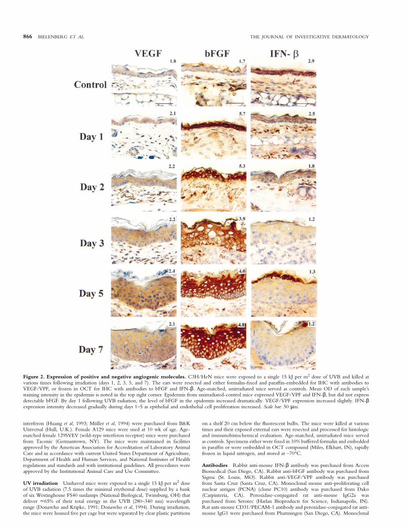
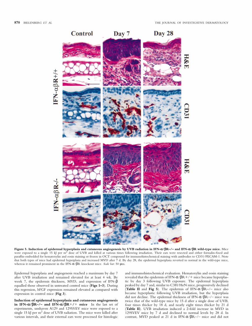
Figure 2. Expression of positive and negative angiogenic molecules. C3H/HeN mice were exposed to a single 15 kJ per m2 dose of UVB and killed atvarious times following irradiation (days 1, 2, 3, 5, and 7). The ears were resected and either formalin-fixed and paraffin-embedded for IHC with antibodies toVEGF/VPF, or frozen in OCT for IHC with antibodies to bFGF and IFN-β. Age-matched, unirradiated mice served as controls. Mean OD of each sample’sstaining intensity in the epidermis is noted in the top right corner. Epidermis from unirradiated-control mice expressed VEGF/VPF and IFN-β, but did not expressdetectable bFGF. By day 1 following UVB radiation, the level of bFGF in the epidermis increased dramatically. VEGF/VPF expression increased slightly. IFN-βexpression intensity decreased gradually during days 1–5 as epithelial and endothelial cell proliferation increased. Scale bar: 50 µm.
interferon (Huang et al, 1993; Muller et al, 1994) were purchased from B&KUniversal (Hull, U.K.). Female A129 mice were used at 10 wk of age. Age-matched female 129SVEV (wild-type interferon receptor) mice were purchasedfrom Taconic (Germantown, NY). The mice were maintained in facilitiesapproved by the American Association for Accreditation of Laboratory AnimalCare and in accordance with current United States Department of Agriculture,Department of Health and Human Services, and National Institutes of Healthregulations and standards and with institutional guidelines. All procedures wereapproved by the Institutional Animal Care and Use Committee.
UV irradiation Unshaved mice were exposed to a single 15 kJ per m2 doseof UVB radiation (7.5 times the minimal erythemal dose) supplied by a bankof six Westinghouse FS40 sunlamps (National Biological, Twinsburg, OH) thatdeliver µ65% of their total energy in the UVB (280–340 nm) wavelengthrange (Donawho and Kripke, 1991; Donawho et al, 1994). During irradiation,the mice were housed five per cage but were separated by clear plastic partitions
on a shelf 20 cm below the fluorescent bulbs. The mice were killed at varioustimes and their exposed external ears were resected and processed for histologicand immunohistochemical evaluation. Age-matched, unirradiated mice servedas controls. Specimens either were fixed in 10% buffered formalin and embeddedin paraffin or were embedded in OCT compound (Miles, Elkhart, IN), rapidlyfrozen in liquid nitrogen, and stored at –70°C.
Antibodies Rabbit anti-mouse IFN-β antibody was purchased from AccessBiomedical (San Diego, CA). Rabbit anti-bFGF antibody was purchased fromSigma (St. Louis, MO). Rabbit anti-VEGF/VPF antibody was purchasedfrom Santa Cruz (Santa Cruz, CA). Monoclonal mouse anti-proliferating cellnuclear antigen (PCNA) (clone PC10) antibody was purchased from Dako(Carpinteria, CA). Peroxidase-conjugated rat anti-mouse IgG2a waspurchased from Serotec (Harlan Bioproducts for Science, Indianapolis, IN).Rat anti-mouse CD31/PECAM-1 antibody and peroxidase-conjugated rat anti-mouse IgG1 were purchased from Pharmingen (San Diego, CA). Monoclonal
VOL. 111, NO. 5 NOVEMBER 1998 UVB-INDUCED CUTANEOUS ANGIOGENESIS 867
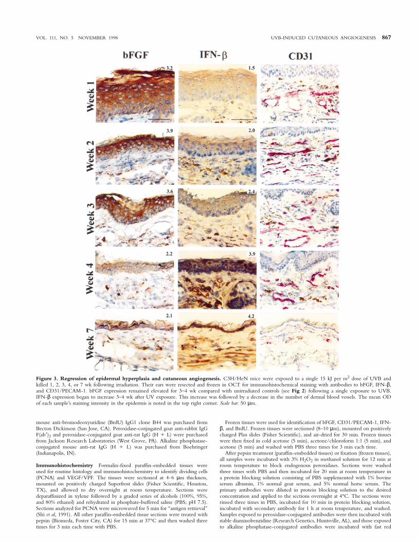
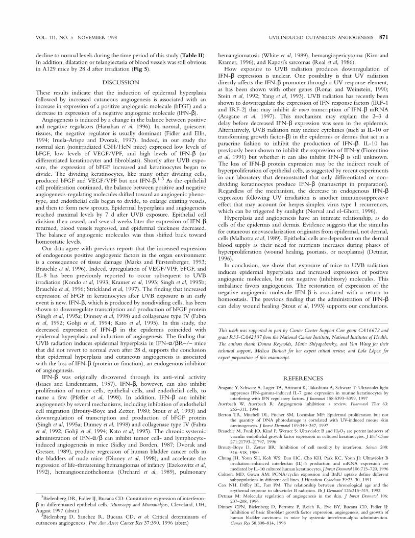
Figure 3. Regression of epidermal hyperplasia and cutaneous angiogenesis. C3H/HeN mice were exposed to a single 15 kJ per m2 dose of UVB andkilled 1, 2, 3, 4, or 7 wk following irradiation. Their ears were resected and frozen in OCT for immunohistochemical staining with antibodies to bFGF, IFN-β,and CD31/PECAM-1. bFGF expression remained elevated for 3–4 wk compared with unirradiated controls (see Fig 2) following a single exposure to UVB.IFN-β expression began to increase 3–4 wk after UV exposure. This increase was followed by a decrease in the number of dermal blood vessels. The mean ODof each sample’s staining intensity in the epidermis is noted in the top right corner. Scale bar: 50 µm.
mouse anti-bromodeoxyuridine (BrdU) IgG1 clone B44 was purchased fromBecton Dickinson (San Jose, CA). Peroxidase-conjugated goat anti-rabbit IgGF(ab9)2 and peroxidase-conjugated goat anti-rat IgG (H 1 L) were purchasedfrom Jackson Research Laboratories (West Grove, PA). Alkaline phosphatase-conjugated mouse anti-rat IgG (H 1 L) was purchased from Boehringer(Indianapolis, IN).
Immunohistochemistry Formalin-fixed paraffin-embedded tissues wereused for routine histology and immunohistochemistry to identify dividing cells(PCNA) and VEGF/VPF. The tissues were sectioned at 4–6 µm thickness,mounted on positively charged Superfrost slides (Fisher Scientific, Houston,TX), and allowed to dry overnight at room temperature. Sections weredeparaffinized in xylene followed by a graded series of alcohols (100%, 95%,and 80% ethanol) and rehydrated in phosphate-buffered saline (PBS; pH 7.5).Sections analyzed for PCNA were microwaved for 5 min for ‘‘antigen retrieval’’(Shi et al, 1991). All other paraffin-embedded tissue sections were treated withpepsin (Biomeda, Foster City, CA) for 15 min at 37°C and then washed threetimes for 3 min each time with PBS.
Frozen tissues were used for identification of bFGF, CD31/PECAM-1, IFN-β, and BrdU. Frozen tissues were sectioned (8–10 µm), mounted on positivelycharged Plus slides (Fisher Scientific), and air-dried for 30 min. Frozen tissueswere then fixed in cold acetone (5 min), acetone/chloroform 1:1 (5 min), andacetone (5 min) and washed with PBS three times for 3 min each time.
After pepsin treatment (paraffin-embedded tissues) or fixation (frozen tissues),all samples were incubated with 3% H2O2 in methanol solution for 12 min atroom temperature to block endogenous peroxidases. Sections were washedthree times with PBS and then incubated for 20 min at room temperature ina protein blocking solution consisting of PBS supplemented with 1% bovineserum albumin, 1% normal goat serum, and 5% normal horse serum. Theprimary antibodies were diluted in protein blocking solution to the desiredconcentration and applied to the sections overnight at 4°C. The sections wererinsed three times in PBS, incubated for 10 min in protein blocking solution,incubated with secondary antibody for 1 h at room temperature, and washed.Samples exposed to peroxidase-conjugated antibodies were then incubated withstable diaminobenzidine (Research Genetics, Huntsville, AL), and those exposedto alkaline phosphatase-conjugated antibodies were incubated with fast red
868 BIELENBERG ET AL THE JOURNAL OF INVESTIGATIVE DERMATOLOGY
Table I. Quantitation of epidermal thickness, proliferativeindex, and MVD during the progression of hyperplasia andangiogenesis following UVB irradiation in C3H/HeN micea
Epidermal ProliferativeDays thickness (µm) index (%) MVD
NR 11.7 6 2.6 12.7 6 4.0 5.2 6 1.51 15.9 6 3.8 20.3 6 5.2 6.5 6 1.92 27.0 6 6.9b 27.0 6 6.4 6.8 6 1.93 48.2 6 12b 29.3 6 4.4c 5.7 6 1.35 53.7 6 8.8b 55.0 6 6.4c 7.8 6 1.1c
7 68.9 6 10.9b 60.5 6 11.5c 9.9 6 2.6c
aThe values are mean 6 SD. NR, unirradiated (control) tissue. The proliferative indexwas calculated as the number of PCNA1 epidermal cells/the total number of epidermalcells within 20 2003 fields on the dorsal side of the ear. MVD was calculated as the meannumber of blood vessels (CD31/PECAM-11) in a 20,000 µm2 area of the dermis on thedorsal side of the ear.
bp , 0.001 by the Kruskal–Wallis nonparametric analysis of variance test in comparisonwith control tissue.
cp , 0.001 by the Student’s t test in comparison with control tissue.
chromogenic substrate (Biomeda, Foster City, CA). Staining was monitoredunder a bright field microscope, and the reaction was stopped by washingwith distilled water. Sections were counterstained with Gill’s 3 hematoxylin(Sigma) and mounted with Universal Mount (Research Genetics). The con-centrations of primary and secondary antibodies were predetermined usingseparate specimens that were included in the assay as controls. Control specimensexposed to secondary antibody alone showed no specific staining. The intensityof the cytoplasm staining was quantitated using Optimas Image Analysis software(Bioscan). Optical density (OD) was defined as the integrated log of the inversegray value. OD was measured in the epidermis of control and UV irradiatedears after bFGF, VEGF/VPF, and IFN-β immunohistochemical staining.
Identification of dividing endothelial cells To identify dividing cellsin vivo, mice were injected twice intravenously with 0.2 ml saline containing250 µg BrdU (Sigma) 4 h and 1 h before they were euthanized (Fujimaki et al,1993). The mice’s ears were resected and embedded as described above.Immunohistochemical staining was performed on the frozen tissues as describedabove with rat anti-mouse CD31/PECAM-1 antibody, peroxidase-conjugatedgoat anti-rat IgG, and diaminobenzidine chromogen (brown precipitate). Thesections were then washed with PBS three times for 3 min each time, treatedwith 1% Triton X-100 in PBS for 8 min, and rinsed again with PBS threetimes for 3 min each time. Samples were incubated with 2N HCl in PBS at37°C for 30 min. Sections were next rinsed with PBS three times for 3 mineach time, treated with protein blocking solution and primary monoclonalmouse anti-BrdU antibody overnight at 4°C, and then treated with proteinblocking solution and secondary rat anti-mouse IgG1 for 1 h. The sectionswere then incubated in a second chromogen, 3-amino-9-ethylcarbazole (redprecipitate) (BioGenex, San Ramon, CA) for 5–20 min and mounted asdescribed above. Dividing endothelial cells exhibited red nuclei and browncytoplasm.
Epidermal thickness and proliferative index Sections of formalin-fixed,paraffin-embedded ears were stained with hematoxylin and eosin stain or withantibodies to PCNA. Images of five fields at 2003 magnification were capturedfor each ear using a Sony 3-chip camera (Sony, Montvale, NJ), mounted on aZeiss universal microscope (Carl Zeiss, Thornwood, NY), and Optimas ImageAnalysis software (Bioscan, Bothell, WA) installed in a PC computer with aPentium chip, a frame grabber, an optical disk storage system, and a Sony colorprinter. To minimize changes in background and color intensities, all imagesto be used for analysis were digitized and stored for further analysis. Epidermalthickness was determined from an average of 100 separate measurements (10thickness measurements along each of five 200 µm lengths of tissue from eachof two different mouse ears). The significance of differences in epidermalthickness was calculated using the Kruskal–Wallis nonparametric analysis ofvariance test with Dunn’s multiple comparisons test (InSTAT 2.0 software).The proliferative index, expressed as a percentage, was calculated as the numberof proliferating cells (PCNA1)/the total number of epidermal cells in 20 2003fields. The thickness and proliferative index were measured in the dorsalepidermis of the ear. The significance of differences in PCNA1 cells/field andproliferative index was calculated using the Student’s t test (two-tailed). Allvalues are reported as means 6 SD.
Quantitation of microvessel density (MVD) Control and UVB-exposedears were embedded in OCT compound and frozen, sectioned, fixed, andstained with antibodies to CD31/PECAM-1 (Veechi et al, 1994). Images of
10 1003 fields per ear were digitized and stored for further analysis. Thenumber of blood vessels was counted in each field in the dermis on the dorsal(UVB-exposed) side of the ears. The total area of dermis in each field wasdetermined with the Optimas software program. MVD was calculated as themean number of blood vessels (CD31/PECAM-11 cells) in a 20,000 µm2 areaof dermis. The significance of differences was calculated using the Student’s ttest (two-tailed).
RESULTS
Induction of epidermal hyperplasia by UVB radiation In thefirst set of experiments, unshaved C3H/HeN mice were exposed to asingle 15 kJ per m2 dose of UVB radiation. The mice were killed aftervarious intervals, and their external ears were processed for histologicand immunohistochemical evaluation. Hematoxylin and eosin stainingrevealed that epidermal hyperplasia began by 48 h after UVB exposureand peaked on day 7 (Figs 1, 2) and began to decline by 2 wk afterexposure (Fig 3). The mean thickness of the epidermis progressivelyincreased from 11.7 6 2.6 µm to 68.9 6 10.9 µm (Table I) and wasaccompanied by an apparent thickening of the dermis (Figs 1–3). TheUVB-induced epidermal hyperplasia was local because ventral skinthickness was unchanged (data not shown).
UVB radiation induces proliferation of keratinocytes Immuno-histochemical analysis using antibodies against PCNA, a proteinexpressed during the S phase of the cell cycle (Coltrera and Gown,1991; Wolf and Dittrich, 1992), revealed that the normal (unirradiated)epidermis contained few PCNA1 keratinocytes in the basal cell layer(Fig 1). By 24 and 48 h after UVB irradiation, an increased numberof PCNA1 keratinocytes were found in the basal layer. The numberof PCNA1 cells continued to increase, and by day 3 the cells weredispersed throughout the epidermis. The mean number of PCNA1cells per field in the epidermis increased gradually from 7.2 6 1.3(12.7%), and by day 7 after UVB exposure it reached 124.4 6 31(60.5%) (Fig 1, Table I).
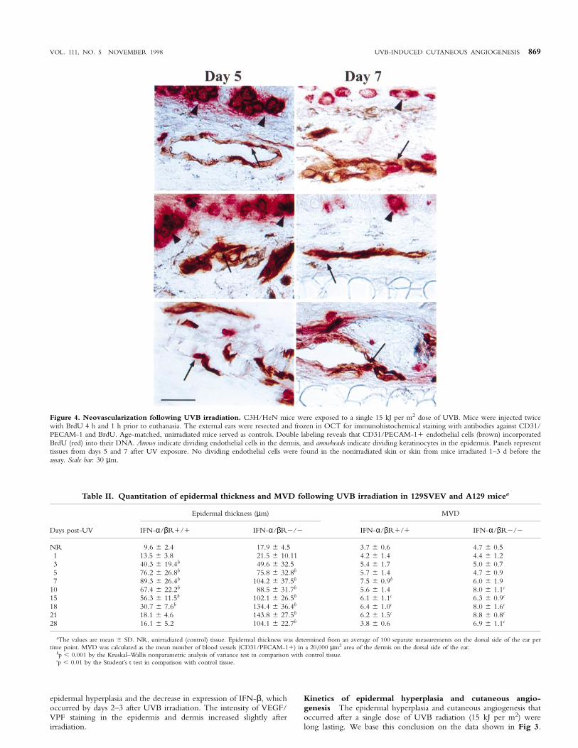
UVB radiation induces endothelial cell proliferation Immuno-staining with antibodies against CD31/PECAM-1 identifies endothelialcells in murine tissues (Veechi et al, 1994). In our study, UVB irradiationfirst induced dilatation of blood vessels (telangiectasia) and then induceda 2-fold increase in the number of blood vessels per area, i.e., their density(Figs 1, 4, Table I). These data indicate that the increase in the size ofthe skin (overlying the dorsal surface of the ear) was accompanied by anincrease in vasculature, i.e., angiogenesis.
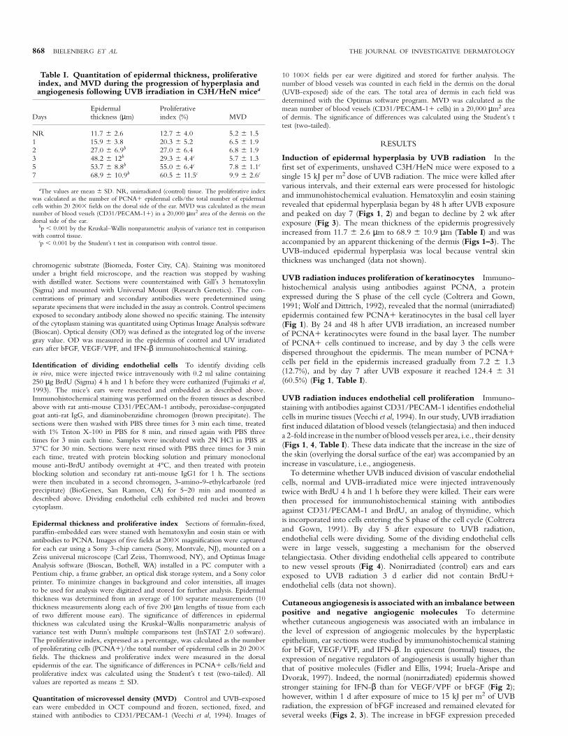
To determine whether UVB induced division of vascular endothelialcells, normal and UVB-irradiated mice were injected intravenouslytwice with BrdU 4 h and 1 h before they were killed. Their ears werethen processed for immunohistochemical staining with antibodiesagainst CD31/PECAM-1 and BrdU, an analog of thymidine, whichis incorporated into cells entering the S phase of the cell cycle (Coltreraand Gown, 1991). By day 5 after exposure to UVB radiation,endothelial cells were dividing. Some of the dividing endothelial cellswere in large vessels, suggesting a mechanism for the observedtelangiectasia. Other dividing endothelial cells appeared to contributeto new vessel sprouts (Fig 4). Nonirradiated (control) ears and earsexposed to UVB radiation 3 d earlier did not contain BrdU1endothelial cells (data not shown).
Cutaneous angiogenesis is associated with an imbalance betweenpositive and negative angiogenic molecules To determinewhether cutaneous angiogenesis was associated with an imbalance inthe level of expression of angiogenic molecules by the hyperplasticepithelium, ear sections were studied by immunohistochemical stainingfor bFGF, VEGF/VPF, and IFN-β. In quiescent (normal) tissues, theexpression of negative regulators of angiogenesis is usually higher thanthat of positive molecules (Fidler and Ellis, 1994; Iruela-Arispe andDvorak, 1997). Indeed, the normal (nonirradiated) epidermis showedstronger staining for IFN-β than for VEGF/VPF or bFGF (Fig 2);however, within 1 d after exposure of mice to 15 kJ per m2 of UVBradiation, the expression of bFGF increased and remained elevated forseveral weeks (Figs 2, 3). The increase in bFGF expression preceded
VOL. 111, NO. 5 NOVEMBER 1998 UVB-INDUCED CUTANEOUS ANGIOGENESIS 869
Figure 4. Neovascularization following UVB irradiation. C3H/HeN mice were exposed to a single 15 kJ per m2 dose of UVB. Mice were injected twicewith BrdU 4 h and 1 h prior to euthanasia. The external ears were resected and frozen in OCT for immunohistochemical staining with antibodies against CD31/PECAM-1 and BrdU. Age-matched, unirradiated mice served as controls. Double labeling reveals that CD31/PECAM-11 endothelial cells (brown) incorporatedBrdU (red) into their DNA. Arrows indicate dividing endothelial cells in the dermis, and arrowheads indicate dividing keratinocytes in the epidermis. Panels representtissues from days 5 and 7 after UV exposure. No dividing endothelial cells were found in the nonirradiated skin or skin from mice irradiated 1–3 d before theassay. Scale bar: 30 µm.
Table II. Quantitation of epidermal thickness and MVD following UVB irradiation in 129SVEV and A129 micea
Epidermal thickness (µm) MVD
Days post-UV IFN-α/βR1/1 IFN-α/βR2/2 IFN-α/βR1/1 IFN-α/βR2/2
NR 9.6 6 2.4 17.9 6 4.5 3.7 6 0.6 4.7 6 0.51 13.5 6 3.8 21.5 6 10.11 4.2 6 1.4 4.4 6 1.23 40.3 6 19.4b 49.6 6 32.5 5.4 6 1.7 5.0 6 0.75 76.2 6 26.8b 75.8 6 32.8b 5.7 6 1.4 4.7 6 0.97 89.3 6 26.4b 104.2 6 37.5b 7.5 6 0.9b 6.0 6 1.9
10 67.4 6 22.2b 88.5 6 31.7b 5.6 6 1.4 8.0 6 1.1c
15 56.3 6 11.5b 102.1 6 26.5b 6.1 6 1.1c 6.3 6 0.9c
18 30.7 6 7.6b 134.4 6 36.4b 6.4 6 1.0c 8.0 6 1.6c
21 18.1 6 4.6 143.8 6 27.5b 6.2 6 1.5c 8.8 6 0.8c
28 16.1 6 5.2 104.1 6 22.7b 3.8 6 0.6 6.9 6 1.1c
aThe values are mean 6 SD. NR, unirradiated (control) tissue. Epidermal thickness was determined from an average of 100 separate measurements on the dorsal side of the ear pertime point. MVD was calculated as the mean number of blood vessels (CD31/PECAM-11) in a 20,000 µm2 area of the dermis on the dorsal side of the ear.
bp , 0.001 by the Kruskal–Wallis nonparametric analysis of variance test in comparison with control tissue.cp , 0.01 by the Student’s t test in comparison with control tissue.
epidermal hyperplasia and the decrease in expression of IFN-β, whichoccurred by days 2–3 after UVB irradiation. The intensity of VEGF/VPF staining in the epidermis and dermis increased slightly afterirradiation.
Kinetics of epidermal hyperplasia and cutaneous angio-genesis The epidermal hyperplasia and cutaneous angiogenesis thatoccurred after a single dose of UVB radiation (15 kJ per m2) werelong lasting. We base this conclusion on the data shown in Fig 3.
870 BIELENBERG ET AL THE JOURNAL OF INVESTIGATIVE DERMATOLOGY
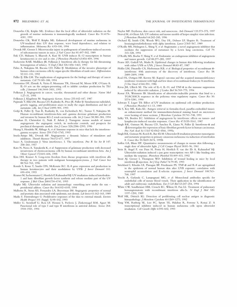
Figure 5. Induction of epidermal hyperplasia and cutaneous angiogenesis by UVB radiation in IFN-α/βR–/– and IFN-α/βR wild-type mice. Micewere exposed to a single 15 kJ per m2 dose of UVB and killed at various times following irradiation. Their ears were resected and either formalin-fixed andparaffin-embedded for hematoxylin and eosin staining or frozen in OCT compound for immunohistochemical staining with antibodies to CD31/PECAM-1. Notethat both types of mice had epidermal hyperplasia and increased MVD after 7 d. By day 28, the epidermal hyperplasia reverted to normal in the wild-type mice,whereas it remained prominent in the IFN-α/βR knockout mice. Scale bar: 50 µm.
Epidermal hyperplasia and angiogenesis reached a maximum by day 7after UVB irradiation and remained elevated for at least 4 wk. Byweek 7, the epidermis thickness, MVD, and expression of IFN-βequalled those observed in untreated control mice (Figs 1–3). Duringthis regression, bFGF expression remained elevated as compared withexpression in control mice (Fig 3).
Induction of epidermal hyperplasia and cutaneous angiogenesisin IFN-α/βR–/– and IFN-α/βR1/1 mice In the last set ofexperiments, unshaven A129 and 129SVEV mice were exposed to asingle 15 kJ per m2 dose of UVB radiation. The mice were killed aftervarious intervals, and their external ears were processed for histologic
and immunohistochemical evaluation. Hematoxylin and eosin stainingrevealed that the epidermis of IFN-α/βR1/1 mice became hyperplas-tic by day 3 following UVB exposure. The epidermal hyperplasiapeaked by day 7 and, similar to C3H/HeN mice, progressively declined(Table II and Fig 5). The epidermis of IFN-α/βR–/– mice alsobecame hyperplastic following UVB irradiation, but the hyperplasiadid not decline. The epidermal thickness of IFN-α/βR–/– mice wastwice that of the wild-type mice by 15 d after a single dose of UVB,four times thicker by 18 d, and nearly eight times thicker by 21 d(Table II). UVB irradiation induced a 2-fold increase in MVD in129SVEV mice by 7 d and declined to normal levels by 28 d. Incontrast, MVD peaked at 21 d in IFN-α/βR–/– mice and did not
VOL. 111, NO. 5 NOVEMBER 1998 UVB-INDUCED CUTANEOUS ANGIOGENESIS 871
decline to normal levels during the time period of this study (Table II).In addition, dilatation or telangiectasia of blood vessels was still obviousin A129 mice by 28 d after irradiation (Fig 5).
DISCUSSION
These results indicate that the induction of epidermal hyperplasiafollowed by increased cutaneous angiogenesis is associated with anincrease in expression of a positive angiogenic molecule (bFGF) and adecrease in expression of a negative angiogenic molecule (IFN-β).
Angiogenesis is induced by a change in the balance between positiveand negative regulators (Hanahan et al, 1996). In normal, quiescenttissues, the negative regulator is usually dominant (Fidler and Ellis,1994; Iruela-Arispe and Dvorak, 1997). Indeed, in our study thenormal skin (nonirradiated C3H/HeN mice) expressed low levels ofbFGF, low levels of VEGF/VPF, and high levels of IFN-β (indifferentiated keratinocytes and fibroblasts). Shortly after UVB expo-sure, the expression of bFGF increased and keratinocytes began todivide. The dividing keratinocytes, like many other dividing cells,produced bFGF and VEGF/VPF but not IFN-β.1–3 As the epithelialcell proliferation continued, the balance between positive and negativeangiogenesis-regulating molecules shifted toward an angiogenic pheno-type, and endothelial cells began to divide, to enlarge existing vessels,and then to form new sprouts. Epidermal hyperplasia and angiogenesisreached maximal levels by 7 d after UVB exposure. Epithelial celldivision then ceased, and several weeks later the expression of IFN-βreturned, blood vessels regressed, and epidermal thickness decreased.The balance of angiogenic molecules was thus shifted back towardhomeostatic levels.
Our data agree with previous reports that the increased expressionof endogenous positive angiogenic factors in the organ environmentis a consequence of tissue damage (Marks and Furstenberger, 1993;Brauchle et al, 1996). Indeed, upregulation of VEGF/VPF, bFGF, andIL-8 has been previously reported to occur subsequent to UVBirradiation (Kondo et al, 1993; Kramer et al, 1993; Singh et al, 1995b;Brauchle et al, 1996; Strickland et al, 1997). The finding that increasedexpression of bFGF in keratinocytes after UVB exposure is an earlyevent is new. IFN-β, which is produced by nondividing cells, has beenshown to downregulate transcription and production of bFGF protein(Singh et al, 1995a; Dinney et al, 1998) and collagenase type IV (Fabraet al, 1992; Gohji et al, 1994; Kato et al, 1995). In this study, thedecreased expression of IFN-β in the epidermis coincided withepidermal hyperplasia and induction of angiogenesis. The finding thatUVB radiation induces epidermal hyperplasia in IFN-α/βR–/– micethat did not revert to normal even after 28 d, supports the conclusionthat epidermal hyperplasia and cutaneous angiogenesis is associatedwith the loss of IFN-β (protein or function), an endogenous inhibitorof angiogenesis.
IFN-β was originally discovered through its anti-viral activity(Isaacs and Lindenmann, 1957). IFN-β, however, can also inhibitproliferation of tumor cells, epithelial cells, and endothelial cells, toname a few (Pfeffer et al, 1998). In addition, IFN-β can inhibitangiogenesis by several mechanisms, including inhibition of endothelialcell migration (Brouty-Boye and Zetter, 1980; Stout et al, 1993) anddownregulation of transcription and production of bFGF protein(Singh et al, 1995a; Dinney et al, 1998) and collagenase type IV (Fabraet al, 1992; Gohji et al, 1994; Kato et al, 1995). The chronic systemicadministration of IFN-α/β can inhibit tumor cell- and lymphocyte-induced angiogenesis in mice (Sidky and Borden, 1987; Dvorak andGresser, 1989), produce regression of human bladder cancer cells inthe bladders of nude mice (Dinney et al, 1998), and accelerate theregression of life-threatening hemangiomas of infancy (Ezekowitz et al,1992), hemangioendotheliomas (Orchard et al, 1989), pulmonary
2Bielenberg DR, Fidler IJ, Bucana CD: Constitutive expression of interferon-β in differentiated epithelial cells. Microscopy and Microanalysis, Cleveland, OH,August 1997 (abstr.)
3Bielenberg D, Sanchez R, Bucana CD, et al: Critical determinants ofcutaneous angiogenesis. Proc Am Assoc Cancer Res 37:390, 1996 (abstr.)
hemangiomatosis (White et al, 1989), hemangiopericytoma (Kirn andKramer, 1996), and Kaposi’s sarcomas (Real et al, 1986).
How exposure to UVB radiation produces downregulation ofIFN-β expression is unclear. One possibility is that UV radiationdirectly affects the IFN-β promoter through a UV response element,as has been shown with other genes (Ronai and Weinstein, 1990;Stein et al, 1992; Yang et al, 1993). UVB radiation has recently beenshown to downregulate the expression of IFN response factors (IRF-1and IRF-2) that may inhibit de novo transcription of IFN-β mRNA(Aragane et al, 1997). This mechanism may explain the 2–3 ddelay before decreased IFN-β expression was seen in the epidermis.Alternatively, UVB radiation may induce cytokines (such as IL-10 ortransforming growth factor-β) in the epidermis or dermis that act in aparacrine fashion to inhibit the production of IFN-β. IL-10 haspreviously been shown to inhibit the expression of IFN-γ (Fiorentinoet al, 1991) but whether it can also inhibit IFN-β is still unknown.The loss of IFN-β protein expression may be the indirect result ofhyperproliferation of epithelial cells, as suggested by recent experimentsin our laboratory that demonstrated that only differentiated or non-dividing keratinocytes produce IFN-β (manuscript in preparation).Regardless of the mechanism, the decrease in endogenous IFN-βexpression following UV irradiation is another immunosuppressiveeffect that may account for herpes simplex virus type 1 recurrences,which can be triggered by sunlight (Norval and el-Ghorr, 1996).
Hyperplasia and angiogenesis have an intimate relationship, as docells of the epidermis and dermis. Evidence suggests that the stimulusfor cutaneous neovascularization originates from epidermal, not dermal,cells (Malhotra et al, 1989). Epithelial cells are dependent on the dermalblood supply as their need for nutrients increases during phases ofhyperproliferation (wound healing, psoriasis, or neoplasms) (Detmar,1996).
In conclusion, we show that exposure of mice to UVB radiationinduces epidermal hyperplasia and increased expression of positiveangiogenic molecules, but not negative (inhibitory) molecules. Thisimbalance favors angiogenesis. The restoration of expression of thenegative angiogenic molecule IFN-β is associated with a return tohomeostasis. The previous finding that the administration of IFN-βcan delay wound healing (Stout et al, 1993) supports our conclusions.
This work was supported in part by Cancer Center Support Core grant CA16672 andgrant R35-CA42107 from the National Cancer Institute, National Institutes of Health.The authors thank Donna Reynolds, Maria Shlyapobersky, and Yun Wang for theirtechnical support, Melissa Burkett for her expert critical review, and Lola Lopez forexpert preparation of this manuscript.
REFERENCES
Aragane Y, Schwarz A, Luger TA, Ariizumi K, Takashima A, Schwarz T: Ultraviolet lightsuppresses IFN-gamma-induced IL-7 gene expression in murine keratinocytes byinterfering with IFN regulatory factors. J Immunol 158:5393–5399, 1997
Auerbach W, Auerbach R: Angiogenesis inhibition: a review. Pharmacol Ther 63:265–311, 1994
Berton TR, Mitchell DL, Fischer SM, Locniskar MF: Epidermal proliferation but notthe quantity of DNA photodamage is correlated with UV-induced mouse skincarcinogenesis. J Invest Dermatol 109:340–347, 1997
Brauchle M, Funk JO, Kind P, Werner S: Ultraviolet B and H2O2 are potent inducers ofvascular endothelial growth factor expression in cultured keratinocytes. J Biol Chem271:21793–21797, 1996
Brouty-Boye D, Zetter BR: Inhibition of cell motility by interferon. Science 208:516–518, 1980
Chung JH, Youn SH, Koh WS, Eun HC, Cho KH, Park KC, Youn JI: Ultraviolet Birradiation-enhanced interleukin (IL)-6 production and mRNA expression aremediated by IL-1α cultured human keratinocytes. J Invest Dermatol 106:715–720, 1996
Coltrera MD, Gown AM: PCNA/cyclin expression and BrdU uptake define differentsubpopulations in different cell lines. J Histochem Cytochem 39:23–30, 1991
Cox NH, Diffey BL, Farr PM: The relationship between chronological age and theerythemal response to ultraviolet B radiation. Br J Dermatol 126:315–319, 1992
Detmar M: Molecular regulation of angiogenesis in the skin. J Invest Dermatol 106:207–208, 1996
Dinney CPN, Bielenberg D, Perrotte P, Reich R, Eve BY, Bucana CD, Fidler IJ:Inhibition of basic fibroblast growth factor expression, angiogenesis, and growth ofhuman bladder carcinoma in mice by systemic interferon-alpha administration.Cancer Res 58:808–814, 1998
872 BIELENBERG ET AL THE JOURNAL OF INVESTIGATIVE DERMATOLOGY
Donawho CK, Kripke ML: Evidence that the local effect of ultraviolet radiation on thegrowth of murine melanoma is immunologically mediated. Cancer Res 51:4176–4181, 1991
Donawho CK, Wolf P, Kripke ML: Enhanced development of murine melanoma inUV-irradiated skin: UV dose response, wave band dependence, and relation toinflammation. Melanoma Res 4:93–100, 1994
Dvorak HF, Gresser I: Microvascular injury in pathogenesis of interferon-induced necrosisof subcutaneous tumors in mice. J Natl Cancer Inst 81:497–502, 1989
Enk CD, Mahanty S, Blauvelt A, Katz SI: UVB induces IL-12 transcription in humankeratinocytes in vivo and in vitro. J Photochem Photobiol 63:854–859, 1996
Ezekowitz RAB, Mulliken JB, Folkman J: Interferon alfa-2a therapy for life-threateninghemangiomas of infancy. N Engl J Med 326:1456–1463, 1992
Fabra A, Nakajima M, Bucana CD, Fidler IJ: Modulation of the invasive phenotype ofhuman colon carcinoma cells by organ specific fibroblasts of nude mice. Differentiation52:101–110, 1992
Fidler IJ, Ellis LM: The implications of angiogenesis for the biology and therapy of cancermetastasis. Cell 79:185–188, 1994
Fiorentino DF, Zlotnik A, Vieira P, Mosmann TR, Howard M, Moore KW, O’Garra A:IL-10 acts on the antigen-presenting cell to inhibit cytokine production by Th1cells. J Immunol 146:3444–3451, 1991
Folkman J: Angiogenesis in cancer, vascular, rheumatoid and other disease. Nature Med1:27–31, 1995
Folkman J, Klagsbrun M: Angiogenic factors. Science 235:442–447, 1987Fujimaki T, Ellis LM, Bucana CD, Radinsky R, Price JE, Fidler IJ: Simultaneous radiolabel,
genetic tagging, and proliferation assays to study the organ distribution and fate ofmetastatic cells. Int J Oncol 2:895–901, 1993
Gohji K, Fidler IJ, Tsan R, Radinsky R, von Eschenbach AC, Tsuruo T, Nakajima M:Human recombinant interferons-beta and -gamma decrease gelatinase productionand invasion by human KG-2 renal-carcinoma cells. Int J Cancer 58:380–384, 1994
Hanahan D, Christofori G, Naik P, Arbeit J: Transgenic mouse models of tumorangiogenesis: the angiogenic switch, its molecular controls, and prospects forpreclinical therapeutic models. Eur J Cancer 32A:2386–2393, 1996
Huang S, Hendriks W, Althage A, et al: Immune response in mice that lack the interferon-gamma receptor. Science 259:1742–1745, 1993
Iruela-Arispe ML, Dvorak HF: Angiogenesis: a dynamic balance of stimulators andinhibitors. Thromb Haemost 78:672–677, 1997
Isaacs A, Lindenmann J: Virus interference. I. The interferon. Proc R Soc Ser B 147:258–260, 1957
Kato N, Nawa A, Tamakoshi K, et al: Suppression of gelatinase production with decreasedinvasiveness of choriocarcinoma cells by human recombinant interferon beta. Am JObstet Gynecol 172:601–606, 1995
Kirn DH, Kramer A: Long-term freedom from disease progression with interferon alfatherapy in two patients with malignant hemangiopericytoma. J Natl Cancer Inst88:764–765, 1996
Kondo S, Kono T, Sauder DN, McKenzie RC: IL-8 gene expression and production inhuman keratinocytes and their modulation by UVB. J Invest Dermatol 101:690–694, 1993
Kramer M, Sachsenmaier C, Herrlich P, Rahmsdorf HJ: UV irradiation-induced interleukin-1 and basic fibroblast growth factor synthesis and release mediate part of the UVresponse. J Biol Chem 268:6734–6741, 1993
Kripke ML: Ultraviolet radiation and immunology: something new under the sun –presidential address. Cancer Res 54:6102–6105, 1994
Malhotra R, Stenn KS, Fernandez LA, Braverman IM: Angiogenic properties of normaland psoriatic skin associated with epidermis, not dermis. Lab Invest 61:162–165, 1989
Marks F, Furstenberger G: Proliferative responses of the skin to external stimuli. EnvironHealth Perspect 101 (Suppl. 5):95–102, 1993
Muller U, Steinhoff U, Reis LF, Hemmi S, Pavlovic J, Zinkermagel RM, Aguet M:Functional role of type I and type II interferons in antiviral defense. Science 264:1918–1921, 1994
Naylor MF: Erythema, skin cancer risk, and sunscreens. Arch Dermatol 133:373–375, 1997Norval M, el-Ghorr AA: UV radiation and mouse models of herpes simplex virus infection.
J Photochem Photobiol 64:242–245, 1996Orchard PJ, Smith CM, Woods WG, Day DL, Dehner LP, Shapiro R: Treatment of
heamangioendotheliomas with alpha interferon. Lancet 2:565–567, 1989O’Reilly MS, Holmgren L, Shing Y, et al: Angiostatin: a novel angiogenesis inhibitor that
mediates the suppression of metastases by a Lewis lung carcinoma. Cell 79:315–328, 1994
O’Reilly MS, Boehn T, Shing Y, et al: Endostatin: an endogenous inhibitor of angiogenesisand tumor growth. Cell 88:277–285, 1997
Pearse AD, Gaskell SA, Marks R: Epidermal changes in human skin following irradiationwith either UVB or UVA. J Invest Dermatol 88:83–87, 1987
Pfeffer LM, Dinarello CA, Herberman RB, et al: Biological properties of recombinant α-interferons: 40th anniversary of the discovery of interferons. Cancer Res 58:2489–2499, 1998
Real FX, Oettgen HF, Krown SE: Kaposi’s sarcoma and the acquired immunodeficiencysyndrome: treatment with high and low doses of recombinant leukocyte A interferon.J Clin Oncol 4:544–551, 1986
Rivas JM, Ullrich SE: The role of IL-4, IL-10, and TNF-α in the immune suppressioninduced by ultraviolet radiation. J Leukoc Biol 56:769–775, 1994
Ronai ZA, Weinstein IB: Identification of ultraviolet-inducible proteins that bind to aTGACAACA sequence in the polyoma virus regulatory region. Cancer Res 50:5374–5381, 1990
Schwarz T, Luger TA: Effect of UV irradiation on epidermal cell cytokine production.J Photochem Photobiol 4:1–13, 1989
Shi S, Key ME, Kalra KL: Antigen retrieval in formalin-fixed, paraffin-embedded tissues:an enhancement method for immunohistochemical staining based on microwaveoven heating of tissue sections. J Histochem Cytochem 39:741–748, 1991
Sidky YA, Borden EC: Inhibition of angiogenesis by interferons: effects on tumor- andlymphocyte-induced vascular responses. Cancer Res 47:5155–5161, 1987
Singh RK, Gutman M, Bucana CD, Sanchez R, Llansa N, Fidler IJ: Interferons-α and-β downregulate the expression of basic fibroblast growth factor in human carcinomas.Proc Natl Acad Sci USA 92:4562–4566, 1995a
Singh RK, Gutman M, Reich R, Bar-Eli M: Ultraviolet B irradiation promotes tumorigenicand metastatic properties in primary cutaneous melanoma via induction of interleukin8. Cancer Res 55:3669–3674, 1995b
Soffen GA, Blum HF: Quantitative measurements of changes in mouse skin following asingle dose of ultraviolet light. J Cell Compar Physiol 58:81–96, 1961
Stein B, Angel P, van Dam H, Ponta H, Herrlich P, van der Eb A, Rahmsdorf HJ:Ultraviolet-radiation induced c-jun gene transcription: two AP-1 like binding sitesmediate the response. Photochem Photobiol 55:409–415, 1992
Stout AJ, Gresser I, Thompson WD: Inhibition of wound healing in mice by localinterferon-β injection. Int J Exp Pathol 74:79–85, 1993
Strickland I, Rhodes LE, Flanagan BF, Friedmann PS: TNF-α and IL-8 are upregulatedin the epidermis of normal human skin after UVB exposure: correlation withneutrophil accumulation and E-selectin expression. J Invest Dermatol 108:763–768, 1997
Veechi A, Garlanda C, Lampugnani MG, et al: Monoclonal antibodies specific forendothelial cells of mouse blood vessels. Their application in the identification ofadult and embryonic endothelium. Eur J Cell Biol 63:247–254, 1994
White CW, Sondheimer HM, Crouch EC, Wilson H, Fan LL: Treatment of pulmonaryhemangiomatosis with recombinant interferon alfa-2a. N Engl J Med 320:1197–1200, 1989
Wolf HK, Dittrich KL: Detection of proliferating cell nuclear antigen in diagnostichistopathology. J Histochem Cytochem 40:1269–1273, 1992
Yang YM, Rutberg SE, Luo FC, Spratt TE, Halaban R, Ferrone S, Ronai Z: Atranscriptional inhibitor induced in human melanoma cells upon ultravioletirradiation. Cell Growth Differ 4:595–602, 1993


