Embed Size (px)
Citation preview

1994 84: 3524-3531
MA Grijzenhout, MI Aarts-Riemens, FR de Gruijl, H van Weelden and HC van Prooijen alloimmunization in recipientsUVB irradiation of human platelet concentrates does not prevent HLA
http://bloodjournal.hematologylibrary.org/site/misc/rights.xhtml#repub_requestsInformation about reproducing this article in parts or in its entirety may be found online at:
http://bloodjournal.hematologylibrary.org/site/misc/rights.xhtml#reprintsInformation about ordering reprints may be found online at:
http://bloodjournal.hematologylibrary.org/site/subscriptions/index.xhtmlInformation about subscriptions and ASH membership may be found online at:
reserved.Copyright 2011 by The American Society of Hematology; all rights900, Washington DC 20036.weekly by the American Society of Hematology, 2021 L St, NW, Suite Blood (print ISSN 0006-4971, online ISSN 1528-0020), is published
For personal use only. by guest on July 13, 2011. bloodjournal.hematologylibrary.orgFrom
UVB Irradiation of Human Platelet Concentrates Does Not Prevent HLA Alloimmunization in Recipients
By M.A. Grijzenhout, M.I. Aarts-Riemens, F.R. de Gruijl, H. van Weelden, and H.C. van Prooijen
Exposure of platelet concentrates (PCs) to ultraviolet B radia- tion (UVB) has been advocated as an alternative method for prevention of the onset of HLA sensitization in recipients. In this study, pooled PCs were irradiated in a Haemonetics UV irradiator (Haemonetics Corp. Braintree, MA) at a dose that did not induce platelet activation. The effect of UVB irradiation on prevention of primary HLA sensitization was evaluated in a prospective controlled clinical study per- formed in cardiac patients undergoing cardiopulmonary by- pass. Patients were treated with filtered red blood cells and a single transfusion of either standard (control group) or UVB-irradiated (UVB group) pooled platelets prepared from 12 donors. Five of 39 patients in the control group and 6 of 62 patients in the UVB group developed allo-antibodies against HLA antigens, which is not significantly different ( P = .62). This unexpected finding prompted us t o check the efficacy of UVB irradiation. We determined UVB-specific
ONTAMINATING LEUKOCYTES in"p1atelet concen- trates (PCs) are responsible for the induction of anti-
bodies against HLA class I antigens in recipients.'.' These antibodies may induce platelet refractoriness and, for this reason, techniques have been developed to prevent the onset of HLA alloimmunization (HLA-AI). At present, removal of leukocytes from PCs by filtration is the best approach to reduce the incidence of primary HLA-AI.1-7 Exposure of leukocytes to UV radiation has been proposed in recent stud- ies as an alternative procedure for prevention of platelet refractoriness. UV radiation abolishes the ability of antigen presenting cells (APCs) to stimulate responder T-cells in the mixed lymphocyte reaction (MLR) and it is thought that this inactivation prevents the induction of anti-HLA antibodies in vivo.8"2 This hypothesis was confirmed by Slichter et all3 in studies performed in dogs; the incidence of HLA alloimmunization decreased from 86% to 8% when the ani- mals were transfused with UV-irradiated platelets.
The clinical application of UV radiation for treatment of PCs on a large scale was delayed by the observation that platelets were activated by UVB radiati~n. ' '~ '~"~ Because
C
From the Departments of Immunohaematology, Haematology, and Dermatology, University Hospital Utrecht, The Netherlands; and the Laboratory for Medicines and Medical Devices, National Institute oj Public Health Care and Environmental Hygiene, Bilthoven, The Netherlands.
Submitted November 30, 1993; accepted July 19, 1994. Supported in part by The Red Cross Bloodbank Utrecht, The
Address reprint requests to H.C. van Prooijen, MD, PhD, Depart- ment of Immunohematology, University Hospital Utrecht, PO BOX 85500, 3500 GA Utrecht, The Netherlands.
The publication costs of this article were defrayed in part by page charge payment. This article must therefore be hereby marked "advertisement" in accordance with 18 U.S.C. section 1734 solely to indicate this fact.
Netherlands.
0 1994 by The American Society of Hematology. 0006-4971/94/8410-0030$3.00/0
3524
DNA damage in cells by measuring the fluorescence from a labeled specific monoclonal antibody against thymine di- mers. With this novel flow cytometer technique, we esti- mated in UVB-irradiated leukocytes in saline that a mean fluorescence intensity (MFI) of 47 c 2 arbitrary units (n = 6) correlated with abolition of alloreactivity in mixed lympho- cyte cultures and delayed cell death (within 72 hours). MFI in leukocytes suspended in plasma and exposed to the clinical dose of UVB was sixfold higher (310 +_ 41 arbitrary units) and resulted in early cell death (within 24 hours). We hypothesize that this high level of UVB radiation induces fragmentation of the leukocytes. As a consequence, the poor results of UVB irradiation may be explained by the onset of HLA-alloimmu- nization induced by soluble donor HLA class I antigens pro- cessed and presented by host antigen-presenting cells. 0 1994 by The American Society of Hematology.
platelet activation was induced at higher doses than required for inhibition of leukocyte function,".'2 the use of an inter- mediate dose of UVB was found to be most appropriate."
In this report, pooled PCs were exposed to UVB in a commercially available UV irradiator. Initially, we per- formed a series of studies to estimate the intermediate dose of UVB needed to avoid platelet activation. Subsequently, we did a prospective controlled clinical study in cardiac patients undergoing cardiopulmonary bypass, to evaluate the effect of UVB radiation on prevention of HLA alloimmuni- zation. For this purpose, patients were treated with filtered red blood cells (RBCs) and a single transfusion of either standard or UVB-irradiated pooled platelets prepared from 12 donors. The data did not show a beneficial effect of UVB irradiation on the incidence of HLA alloimmunization. For better understanding of this unexpected finding, we per- formed additional studies to correlate UVB-induced aboli- tion of leukocyte function with the level of UVB-specific DNA damage and cell viability. UVB-specific DNA damage was measured in a novel flow cytometer technique, using a unique monoclonal antibody (MoAb) against thymine di- mers.
MATERIALS AND METHODS
Determination of the Intermediate Dose of UVB Pooled PCs were exposed to UVB in a commercially available
UV irradiator (Platelet Treatment System; Haemonetics, Braintree, MA). For determining the intermediate dose of UVB, the effect of UVB on platelets and lymphocytes was evaluated in platelet activa- tion studies and mixed lymphocyte cultures, respectively.
UVB source and dose calculations. The Haemonetics UV irradi- ator was equipped with a bank of nine UVB fluorescent tubes (Philips FSTS) emitting a continuous spectrum between 270 and 340 nm with a maximum around 310 nm. Before irradiation, platelet and leukocyte suspensions (300 mL) were transferred to a Stericell large cell culture bag (Stericell; DuPont, Wilmington, DE; UVB transmis- sion, 80%; cross-sectional layer depth, 4.3 mm) using the Sterile Docking Device from Haemonetics. The bags were placed on a quartz plate and irradiated from below under continuous agitation
Blood, Vol84, No 10 (November 15). 1994: pp 3524-3531
For personal use only. by guest on July 13, 2011. bloodjournal.hematologylibrary.orgFrom

PREVENTION OF ALLOIMMUNIZATION BY UVB RADIATION 3525
(70 cycledmin). Irradiance above the quartz plate was 5.2 mW/ cm’, measured with a Waldmann UVB detector (Waldmann AG, Schwenningen, Germany). The dose (surface exposure) of UVB radiation (J/cmz) delivered on the cell suspensions was calculated as the product of the irradiance (mW/cm2) and the exposure times. UVB doses were corrected for the transmission through plastic mate- rial.
Mixed lymphocyte cultures and platelet-activation studies. Pe- ripheral blood mononuclear cells (PBMCs) were prepared from fresh buffy coats by density gradient centrifugation (Ficoll Isopaque; Phar- macia, Uppsala, Sweden). The cells were suspended in plasma (300 mL) at a concentration of 1 X lo6 cells/mL and transferred to Steri- cell bags for UVB irradiation. For dose-response studies, samples (3 mL) were taken from the bag during irradiation at different expo- sure times. In some experiments, leukocytes were irradiated in phos- phate-buffered saline (PBS) at a concentration of 1 X lo6 cells/mL. The effect of UVB irradiation on the ability of lymphocytes to stimulate responder cells was evaluated in the one-way MLR as reported before.” In this assay, stimulator cells were also y irradiated to block their allogeneic proliferative response. The ability of lym- phocytes to stimulate in the MLR is not affected by y irradiation. Lymphoproliferation induced by UVB-exposed stimulator cells was measured by incorporation of 3H-thymidine (specific activity, 2.1 GBq/mmol; Radiochemical Centre, Amersham, UK) and expressed as a percentage of control.
For platelet activation studies, leukodepleted platelets were col- lected from buffy coats as reported before.I6 In this study, platelets were collected from 18 buffy coats and pooled to obtain a PC with a volume of 900 mL. This PC was equally divided over three Steri- cell bags; one PC served as control, and two PCs were exposed to either 1.5 or 3.0 J/cm2 in the Haemonetics UV irradiator. After UV exposure PCs were stored in 1000-mL Fenwal PL-732 bags (Baxter Healthcare, Fenwal Division, Deerfield, IL) for 96 hours at 22°C on a Helmer shaker (Helmer Labs, Noblesville, IN). UV-induced platelet activation was evaluated by measuring platelet counts, lactate levels, pH, and the expression of platelet-membrane activation markers dur- ing postirradiation storage.I6 Flow cytometry was done in a FACScan flow cytometer (Becton Dickinson, Mountain View, CA) with plate- lets fixed with paraformaldehyde and stained with fluorescein iso- thiocyanate (FITC)-labeled MoAbs (RUU-PL 6.66 reacting with gly- coprotein Ib and MoAb RUU-SP 2.15 specific for P-selectin).
Clinical Study Patients and transfusion policy. Between October 1990 and
September 1992, a total of 557 consecutive male patients undergoing elective surgery for coronary bypass grafting or valve replacement requiring cardiopulmonary bypass were selected for the study. Only patients with a history of blood transfusions were excluded to avoid secondary HLA-AI. The patients were transfused on clinical indica- tion with buffy coat-depleted RBC concentrates, leukocyte depleted by filtration, and with pooled platelets prepared from 12 buffy coats. All blood components were delivered by the Red Cross Bloodbank Utrecht. Leukocyte depletion of RBCs was performed by filtration with BPF4 filters (Pall Biomedical, Glencove, NY) in a closed sys- tem. Leukocyte counts, measured in quality-control procedures, were always below 1 X IO6 cellslU.” Pooled PCs were prepared within 12 hours after collection. The average leukocyte count in PCs pre- pared from 6 buffy coats was 2 X 10’ cells.17 Fresh PCs were transferred to Stericell bags for UVB irradiation, stored in Fenwall PL 732 hags, and transfused within 24 hours after preparation.
Anti-HLA antibody screening. Patients transfused with RBCs and/or PCs were selected for follow-up. From these patients, serum samples were taken before operation 1 week and 3 to 6 weeks after
operation and kept at -70°C until tested for the presence of anti- HLA antibodies in the complement-dependent lymphocytotoxicity assay against a panel of 30 selected donors covering most of the defined HLA-A and B specificities.“ For the Ig class determination, dithiotreitol ( D m ) was used. A serum with a positive reaction in the panel that turns into a negative reaction after IgM reduction with D l T is considered to contain IgM antibodies only. A positive panel reactivity despite D l T treatment indicates the presence of IgG anti- bodies. Anti-HLA antibodies were considered positive when the percentage of lysed cells per well was above 25% and when panel reactivity was at least 10%.
Effects of UVB Radiation on Leukocytes Detection of thymine dimers. UVB radiation of cells induces
alterations in DNA; the formation of cyclobutyl thymine dimers (T<>T) is very paramount. The amount of T<>T in DNA can be measured by flow cytometry using a MoAb’’ (H3-Moab, kind gift of Dr L. Roza; “NO; Rijswijk; The Netherlands) labeled for green fluorescence (FITC), against T<>T, as described by Berg et al.’’ For accurate measurements we only detected T<>T in nuclei with 2 n DNA. For this purpose, DNA was stained with the red fluorescent dye 7-amino-actinomycin D (7-AAD) that allowed gating for nuclei with 2 n DNA.
Analysis of T<>T was done in PBMCs. The cells were sus- pended in plasma or in some experiments in PBS, irradiated in the Haemonetics UV irradiator and subsequently fixed in ice-cold 96% ethanol (1 X lo6 cells/mL). For analysis of T<>T in DNA, fixed cells were sedimented and incubated with 0.02% pepsin in 2 N HC1 for 40 minutes to make the DNA accessible to the H3-Moab. Incubation was terminated with an excess of 0.1 m o m Na2B207. Nuclei were then incubated with 100 pL PBS containing 5% fetal calf serum (FCS) and 0.5% Tween 20 with 2.5% culture supernatant of the H3-MoAb. Bound H3-Moabs were stained with 100 pL 1% voUvol FITC-conjugated rabbit-antimouse Igs (Dakopatts, Copenha- gen, Denmark). Isotype- and fluorochrome-matched control Moabs were used in each experiment to determine nonspecific background Moab binding. The nuclei were also incubated for 30 minutes at 37°C with 25 pg/mL 7-AAD in PBS to stain DNA. Fluorescence (l0,OOO events) was analyzed in the flow cytometer; FITC fluores- cence was detected in the FLl channel (530 2 15 nm), 7-AAD fluorescence was recorded in the FL3 channel (>650 nm). Gates were set in the 7-AAD fluorescence to select events containing 2 n DNA (>80% of all events).
Measurement of PBMC viability. Viability was determined in PBMCs, T-cell-depleted with AET-treated sheep RBCs. The cells were suspended in 300 mL PBS or plasma at a concentration of 1 X 10⁶/mL, and UV-irradiated with the Haemonetics UV irradiator. Control and UVB samples (20 mL) were washed twice and sus- pended in RPM1 1640 (GIBCO, Life Technologies, Ltd, Paisley, UK) supplemented with 2% FCS and 1% human serum albumin (cell concentration 2 X 10⁶/mL). Each sample (12 mL) was then divided over three handmade Teflon bags (4 d a g ; Fluorplast, Raamsdonksveer, The Netherlands) to avoid adherence of mono- cytes” and cultured (5% COz; 37°C; humidified air) for up to 3 days. For each dose of UVB, one of the bags was opened daily and the number of viable cells was counted by trypan blue dye exclusion. Viability and cell count of control suspensions did not change during incubation. The number of viable cells in UV-exposed suspensions was expressed as a percentage of age-matched control.
Statistical analysis. Differences in the expression of surface markers between UV-irradiated platelets and age-matched control platelets were analyzed with the paired t-test. Differences in the
For personal use only. by guest on July 13, 2011. bloodjournal.hematologylibrary.orgFrom

3526 GRIJZENHOUT ET AL
Table 1. The Effect of UV Radiation on Ability of Lymphocytes to Stimulate in MLR
Dose (Jlcrn') 'HTdR-Incorporation I % of controll
0.12 0.25 0.37 0.50
39 f 13 15 -+ 5 5 2 2 <l
PBMCs were suspended in plasma and exposed to increasing doses of UVB. Data are means i- SEM In = 6).
incidence of HLA sensitization between the control group and in the UVB group were analyzed with the x* test.
RESULTS
Determination of the Optimal Dose of UVB Radiation
For successful irradiation of PCs, it is essential to select a dose that abolishes the ability of passenger leukocytes to stimulate in the MLR and spares the hemostatic function of the platelets. In this study, PBMCs, suspended in 300 mL plasma, were exposed to increasing doses of UVB delivered by the Haemonetics UV irradiator. Table 1 shows that a dose of 0.5 J/cm2 was sufficient to abolish alloreactive responses in the MLR (responder cell proliferation below 1% of con- trol). Addition of platelets (3 X l O l l / m L ) to the leukocyte suspensions did not change the dose of UVB required for abolition of alloreactive responses and indicates that platelets do not interfere with the effect of UVB on leukocytes sus- pended in plasma.
Recent studies have shown that further increase of the dose results in progressive activation of platelet^'^"^''^ during storage, leading to poor posttransfusion recoveries.I6 There- fore, PCs were exposed to increasing doses of UVB and aliquots were taken from each PC before UV exposure and during postirradiation storage at 3, 48, and 96 hours. The samples were analyzed for platelet count, pH, lactate, and surface expression of P-selectin. Table 2 shows that 3.0 J/cm2 of UVB induces a progressive increase in activated platelets during storage. The dose of 1.5 J/cm2 did not affect biochem- ical parameters and expression of P-selectin on the platelets during storage for up to 96 hours, and was selected for clinical use.
Clinical Study
The effect of UVB irradiation on the incidence of HLA alloimminization was evaluated in a prospective controlled clinical study, performed in cardiac patients undergoing car- diopulmonary bypass. A total of 557 consecutive male pa- tients was selected for the study; 372 patients were not trans- fused, 22 patients were transfused only with plasma, 62 patients were transfused with plasma and filtered RBCs, and 101 patients were transfused with plasma, filtered RBCs, and platelets. Patients were only transfused on clinical indi- cation. If platelets were required, the patients were transfused with a single transfusion of pooled platelets prepared from a total of 12 donors. Platelets were either standard or UVB irradiated, but never filtered or y irradiated. Anti-HLA anti-
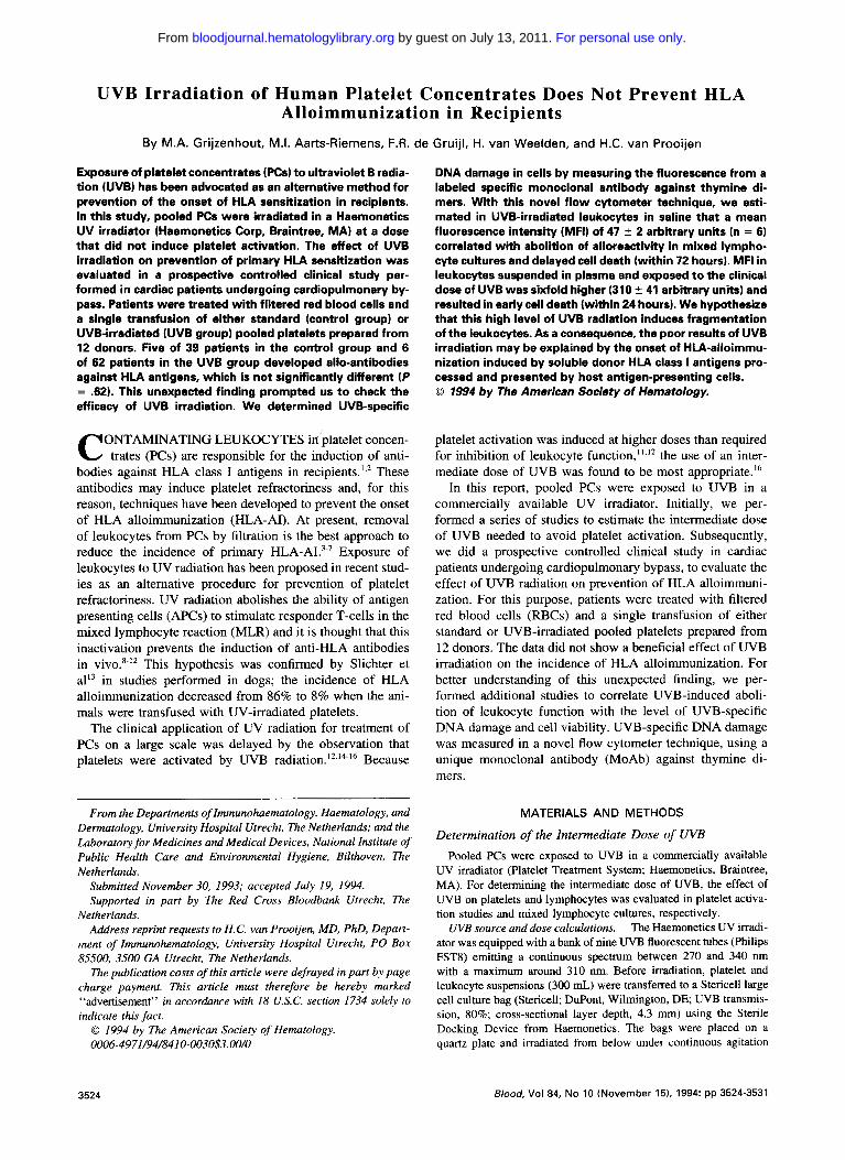
body screens were performed before operation, 1 week after operation and 3 to 6 weeks after operation in patients trans- fused with cellular blood components. Patients with anti- HLA antibodies before operation were excluded for further evaluation. In the group of patients transfused with plasma and RBCs, 53 patients received an average of 2.1 U (range, 1 to 4 U) of RBCs and 9 patients received an average of 8.5 U (range, 6 to 11 U) of RBCs. In all these patients, anti- HLA antibody screens were negative. In the group of patients transfused with platelets, 39 were transfused with standard PCs (control group) and 62 patients were transfused with UV irradiated PCs (UVB group). In the control group, 17 patients were transfused with an average of 4.3 -+ 2.5 U (SD) of RBC (range, 1 to 8 U) and 5 patients received two transfusions of PCs prepared from 12 donors each. In the UVB group, 29 patients were transfused with an average of 3.7 2.5 U (SD) of RBCs (range, 1 to 11 U) and 2 patients received two transfusions of PCs prepared from 12 donors each. Table 3 shows transfusion characteristics of the pa- tients who developed anti-HLA antibodies. Figure l shows the panel reactivity of the antibodies in each patient; the presence of IgG anti-HLA antibodies alone was evaluated in sera after IgM reduction with DDT. Patient 2 in the control group developed auto-antibodies; panel reactivity was 100% 1 week and 3 to 6 weeks after operation, but disappeared after IgM reduction with DDT. Thus, the incidence of alloan- tibodies against HLA antigens is 12.8% ( 5 of 39 patients) in the control group and 9.7% (6 of 62 patients) in the UVB group ( P = .62). Induction of IgM anti-HLA antibodies was found in 4 patients in the control group and 5 patients in the UVB group. Specificity of anti-HLA antibodies is listed in Table 3.
Effects of UVB Radiation on Leukocytes
Association between loss of stimulatory activity in the MLR and amount of thymine dimers in DNA. The outcome
Table 2. Effect of UVB-Radiation on Platelet Activation With Storage
~ ~~
Storage Time (hrs ) Control 1.5 Jlcm' 3.0 Jlcm'
Platelet count 3 9 7 5 4 96 i- 4 96 + 4 (% of initial) 48 94 ir 5 94 t 3 96 t- 4
96 96 +- 4 95 t 5 100 t 2 pH 3 7.30 t- 0.03 7.30 i- 0.02 7.29 t 0.04
48 7.11 t- 0.04 7.04 t- 0.03 6.89 -+ 0.04* 96 6.93 t 0.09 6.86 ir 0.07 6.36 t- 0.04*
Lactate (mmol/L) 3 5.1 t- 0.4 5.1 t- 0.2 5.5 ir 0.5 48 9.0 t- 0.5 10.9 ? 0.8 12.9 t- 0.4* 96 14.6 2 1.1 16.1 i- 0.5 25.6 t- 0.5*
P-selectin (% 3 2 2 + 1 21 i- 2 22 t- 2
positive cells) 48 16 t 2 16 2 3 23 t- 3 96 19 i 1 18 t- 3 47 t 2"
Platelets exposed to UVB doses of 1.5 and 3.0 J/cmz and control platelets were stored for up to 96 hours. Data are means i- SEM, n =
6. * P i .01 (paired t-test for age-matched data).
For personal use only. by guest on July 13, 2011. bloodjournal.hematologylibrary.orgFrom

PREVENTION OF ALLOIMMUNIZATION BY UVB RADIATION
Table 3. No. of Transfusions and Specificities of Panel-Reactive Antibodies
Anti-HLA RBCs* PCst Plasma Antibodies
Patient No. (donors) (12 pool) (donors) (specificities)
Control group 1 5 1 8 B18 2 3 1 1 - A2, A28
4 1 - B12 ,
5 1 - Broad reactive
6 5 2 6 A l l , B13
- 1 2 Autoreactive
- -
UVB group 1 6 2 4 B17 2 3 4 1 2 87. B13 4 1 1 - A2, A28
5 2 1 - Broad reactive
6 1 1 1 Broad reactive
Depicted are all patients with positive anti-HLA antibody screens
* No. of red blood cell concentrates prepared from one donor. t No. of platelet concentrates prepared from 12 donors.
- 1 2 AI , AS
after operation.
of the clinical study indicates that transfusion of UVB irradi- ated PCs does not reduce the incidence of HLA-AI. For better understanding of this finding we measured the amount of UV-induced T<>T in DNA of individual cells. This was done with a novel flow-cytometric technique that became available to us at the end of the clinical study. The histograms shown in Fig 2 are measurements of PBMCs prepared from one donor and are representative for a series of experiments. Figure 2A shows T<>T histograms of UVB-irradiated leu- kocytes that were suspended in plasma during the irradiation. The distribution of dimers in these cells is wide and indicates the large variation in UVB-induced damage per cell, which can be ascribed to a combination of poor mixing of cells and poor transmission of UVB radiation” through plasma. In subsequent experiments (Fig 2B), we suspended leuko-
3521
cytes in PBS (good UVB transmission) and exposed these suspensions to UVB to establish more precisely the level of T< >T that corresponds to the loss of stimulatory responses in the MLR. As expected, histograms are narrow indicating that the variation in the amount of dimers in the cells is small. Mean fluorescence intensity (MFI) increased linearly with the dose of UVB. The capacity of these cells to stimu- late in the MLR is shown in Table 4. The amount of dimers in the cells exposed to 36 mJ/cm2 corresponds to complete loss of alloreactivity in the MLR. Figure 2C is a composite plot of two T<>T histograms obtained from leukocytes suspended in plasma and exposed to 0.5 and 1.5 J/cm2 and one T< >T histogram obtained from leukocytes suspended in PBS and exposed to 36 mJ/cm2. Leukocytes exposed to 0.5 J/cm2 (in plasma) and 36 mJ/cm2 (in PBS) had com- pletely lost their ability to induce alloreactive responses in the MLR. About 30% of the cells suspended in plasma and exposed to 0.5 J/cm2 showed T<>T levels, which were below the level of dimers found in the cells exposed to 36 mJ/cm2. All leukocytes exposed to the clinical dose (1.5 J/ cm’) showed T<>T levels which were equal or above the level of dimers found in cells exposed to 36 mJ/cm2 and indicates that this clinical dose should have been adequate to render all antigen-presenting cells dysfunctional.
Association between UVB dose, amount of thymine di- mers, and viability. Several studies have shown that UVB radiation induces loss of viability in a dose-dependent man- ner.23324 Measurement of T<>T enables us to associate the level of DNA damage with cell death. T-cell-depleted PBMCs were suspended in saline and exposed to 6, 12, 36, and 72 mJ/cm2 of UVB. After UV exposure, the cells were cultured in Teflon bags for up to 3 days. Every 24 hours, aliquots were taken from control and UV-exposed suspen- sions to determine viability. Table 4 shows the correlation between the amount of dimers (arbitrary units) in the cells after UVB exposure and the loss of viability during culture for 3 days. Each dose of UVB induced a progressive decrease in viability over time. The dose of 36 mJ/cm2 resulted in abolition of alloreactivity in the MLR and in less than 5%
120 , Fig 1. HLA antibody screens
were performed before opera- tion, 1 week, and 3 to 6 wwks after operation. Patients with anti-HLA antibodies before oper- ation were excluded from the study. HLA antibody screens were positive in six patients in the control group and six pa- tients in the UVB group. The presence of IgG anti-HLA anti- bodies alone was analyzed in sera collected 3 to 6 weeks after operation after IgM reduction with D l l (lane 3 of each patient). Autoreactive antibodies were found in the sera collected from patient 2 in the control group.
100
80 8 t 8 *a 60 .LI
20
n 1 2 3 4 5 6 1 2 3 4 5 6
patients
1 Oweek 1, IgM+IgG l w e e k 3-6, IgM+IgG @week 3-6, IgG I
For personal use only. by guest on July 13, 2011. bloodjournal.hematologylibrary.orgFrom

GRIJZENHOUT ET AL
Y
....... A control
0.5 J/crn2 ,: i: - plasma .I
...... . . . . / f
) ;
/ / 1.5 J/cm2
i j 3.0 J/crn2 ; j ....
B . . . . . control
:: ...... 12 mJlCrn2
Y
C ... 36 mJlcm2 (W)
- 0.5 J/cm2
: 1.5 JIcm2 ( p l a m l
(p laml . . . . . . .
. . . .
. . / /
. . . . . .
Log Fluorescence Intensity Fig 2. Distribution curves of T<>T obtained from PBMCs exposed to different doses of UVB (J/cm2). Histograms were obtained with the
leukocytes from one donor and are representative for a series of experiments. (A) UVB irradiation of PBMCs suspended in plasma. (B) UVB irradiation of PBMCs suspended in PBS. MFI increased linearly with the dose of UVB. IC1 Composite plot of histograms obtained from PBMCs irradiated either in PBS or in plasma. About 25% of cells irradiated in plasma at 0.5 Jlcm' showed T<>T levels below the level found in cells
PBS. irradiated in PBS at 36 d l c m ' . All cells irradiated in plasma at 1.5 Jlcm' showed T<>T levels above the levels found in cells irradiated in
of viable cells after 72 hours of culture. We also determined viability of cells suspended in plasma and exposed to 0.5 and 3.0 J/cm2. The dose of 0.5 k m 2 resulted in loss of alloreactivity in the MLR, but not in complete loss of viabil- ity; 25% of cells were still viable after 3 days of culture. It is plausible that part of the cells with T<>T levels below the levels found in cells exposed to 36 mJ/cm2 recover from the deleterious effects of UVB and remain viable. The clini- cal dose of 3.0 J/cm* resulted in less than 5% of viable cells after 24 hours of culture. The MFI in these cells was 3 10 2 41 arbitrary units because of the wide distribution of dimers in these cells. Almost all cells showed T<>T levels that were equal or above the level of dimers found in cells sus- pended in saline and exposed to 36 mJ/cm2 of UVB (Fig 2 ) .
DISCUSSION
In this report, we have evaluated the administration of UVB-irradiated PCs for the prevention of HLA allosensitiza-
tion. All previous studies from our laboratory that were de- signed to determine the optimal dose of UVB were per- formed with an experimental irradiation ~abine t . '* . '~ - '~ In this study, PCs were exposed to UVB radiation delivered by the UV irradiator provided by Haemonetics Corp. We found that a dose of 0.5 J/cm2 was needed to abolish alloreactivity of passenger leukocytes in MLR (Table 1). This observation is in concert with data from other studies. Pamphilon et al" and Andreu et all' exposed PCs to similar sources of UVB and reported that 0.3 to 0.5 k m 2 of UVB was sufficient to abolish MLR alloreactivity.
The selection of the dose of W B for irradiation of PCs in the clinical study was made on the basis of data obtained from studies on APCs. The most striking effect of UVB is the inhibition of APCs to stimulate responder cells in the allogeneic MLR. In this assay, class I1 major histocompat- ibility complex (MHC) determinants on the APCs bind to the T-cell receptor in an antigen-specific manner. As a conse-
Table 4. Effect of UVB Radiation on Alloreactivity, DNA Damage, and Viability
UVB Dose 3HTdR-lncorporation TOT ImJ/cmz) (% of control) (arbitrary units) 24 h 48 h 72 h
Viability 1%)
6 48 f 13 82 c 7 63 c 3 55 f 2 12 26 2 3 22.1 2 1.5 38 ? 5 27 ? 2 21 i 1 18 15 f 5 24 9 2 4 34.3 f 1.5 29 2 6 16 f 4 10 r 2 30 4 - c 3 36 < l 47.0 2 2.3 16 f 5 7 2 3 3 2 1 72 11 r 3 3 2 1 2 2 1
PEMCs suspended in PES were exposed to increasing doses of UVB. The ability of UVB-exposed cells t o stimulate responder cells was evaluated in the MLR. Alloreactivity was expressed as a percentage of control. DNA damage and viability were evaluated in T-cell-depleted PBMCs. For evaluation of DNA damage, the level of thymine dimers (TOT) was measured by flow cytometry and expressed in arbitrary units. Viability of UVB-exposed cells was determined after culture for up to 72 hours and expressed as a percentage of age-matched controls. Data are means t SEM (n = 6).
For personal use only. by guest on July 13, 2011. bloodjournal.hematologylibrary.orgFrom

PREVENTION OF ALLOIMMUNIZATION BY UVB RADIATION 3529
quence, costimulatory signals are transmitted from the APCs to the T-cells. UV irradiation interferes with a number of processes involved in the MLR. Studies in plastic adherent blood mononuclear cells, which are enriched for monocytes, showed that low doses of W B (5 mJ/cm2) are deleterious to their accessory function in antigen and mitogen-induced T-cell response^.^^ In later studies from the same laboratory, Krutmann et alZ4 evaluated monocyte accessory cell function in supporting anti-CD3 mitogenesis. They described a dose- dependent reduction in intercellular adhesion molecule I(ICAM- 1) surface expression on UVB-irradiated mono- cytes associated with a corresponding decrease in cluster formation and T-cell proliferation. A dose-dependent recov- ery of ICAM-l expression was seen 1 to 3 days after UV irradiation, indicating that viability of the cells was not im- paired.25 However, higher doses of UVB reduced the viabil- ity of adherent mononuclear cells in proportion to the dose administered. Based on these considerations, we decided to select the highest possible dose in order to induce irreversible inactivation of the leukocytes. According to Table 2, a dose of 1.5 J/cm2 did not affect the platelets and was found most appropriate.
Recently, Young et a126 evaluated the sensitivity of human blood dendritic cells (DCs) for UVB radiation. At given doses of UVB, DCs were found to be more resistant than monocytes or B-cells in reducing alloreactive T-cell re- sponses in the MLR. UVB irradiation of DCs prevented the upregulation of costimulatory ligands B7/BB1 and ICAM- 1/CD54 in a dose-dependent manner after alloreactive T-cell binding, but did not affect cluster formation between DCs and T-cells during the first l to 2 days of the allogeneic MLR. UVB-exposed cells remained viable and class I1 MHC molecule expression was not decreased. Although UVB pre- vented upregulation of ICAM-1 and B7BB 1 expression on DCs in the clusters, interleukin-2 (IL-2) receptor surface expression on individual T-cells was not affected. However, there was a profound diminution in T-cell autocrine IL-2 secretion, after their aggregation with UVB-irradiated DCs, in a dose-dependent manner. It is presumed that the abolition of T-cell proliferation in the MLR by UVB is caused by the low expression of B7/BB1 on DCs. The interaction between B7/BB1 and CD28 on T-cells is the only one known to stimulate T-cell proliferation by a direct effect on IL-2 gene transcription and produ~tion.~’~** Thus, for selection of the optimal dose, we should consider that DCs, which are the most potent stimulators, are more resistant to the deleterious effects of UVB than monocytes. We should also consider that UV-irradiated DCs with intact viability may recover from the deleterious effects of UVB and regain their ability to upregulate the expression of costimulatory ligands. These considerations warrant the use of the highest dose of UVB that does not induce platelet activation.
The clinical study showed that a single transfusion of pooled platelets (4 X lo8 leukocytes) induced HLA sensitiza- tion in 5 of 39 patients (12.8%). This incidence is rather low when compared with the incidence of alloimmunization in patients transfused with 1 U of leukocyte-poor blood (5 X 10* leukocytes). In these studies, Lagaay et a129 found anti-
HLA antibodies in 18 of 30 patients (60%) 2 weeks after transfusion. Data from this study suggests that the transfu- sion of UVB-irradiated platelets results in a trend towards lower sensitization rates (9.7%), taking into account that the numbers studied are small statistically. Preliminary results in patients with acute leukaemia, who were transfused with UV-exposed PCs and filtered RBCs, also suggested that UV irradiation may lead to lower sensitization rate^.^',^'
In view of the generally accepted concept, that viable donor APCs are responsible for the onset of HLA alloimmu- nization via the direct pathway of allorecognition, we did not expect that UVB-inactivated APCs were able to induce anti-HLA antibodies. For better understanding of this obser- vation, we analyzed the effect of UVB on the cells by mea- suring the level of T< >T in DNA with a specific MoAb; a technique that came available to us at the end of the clinical study. The level of T< >T in the cells can easily be measured by flow cytometry and provides a tool for determining the association between the level of DNA damage and loss of viability. In control experiments, we first exposed cells sus- pended in PBS to different doses of UVB just to show that increasing levels of T<>T were associated with decreases in viability (Table 4). This measurement was used to show that the level of T<>T in cells suspended in plasma and exposed to the clinical dose of UVB (1.5 J/cm2) was above the level required for complete loss of viability in all cells. Despite these findings, we found production of anti-HLA antibodies in patients transfused with UVB-exposed plate- lets. For an explanation of this finding, we should consider triggering of the indirect pathway of allorecognition. Heavily UV-irradiated cells may disintegrate in the circulation and release large amounts of soluble class I HLA antigens, which are then processed and presented by host APCs. The antigen- specific pathway of allorecognition was also reported by Pellegrino et al.32 The authors found that transfusion of plasma containing leukocyte fragments from selected donors resulted in the onset of anti-HLA antibodies. Presentation of donor MHC peptides by host APCs resulting in CD4’ T- helper cell responses has also been found in murine immuni- zation model^.'^,'^ The amount of soluble class I HLA anti- gens released from the leukocytes is probably important. The clinical observation that platelets do not induce anti-HLA antibodies is most likely explained by the poor expression of class I HLA antigens on the platelet membrane. When platelets disintegrate, the release of these antigens probably remains below the threshold required for HLA alloimmuni- zation.
We conclude from the data presented in this study that UVB irradiation of PCs results in a trend toward lower sensi- tization rates. The relatively high dose of UVB (1.5 J/cmZ), used for inactivation of leukocytes in PCs, may induce frag- mentation of cells and alloimmunization according to the indirect pathway of allorecognition. We propose that frag- mentation of the cells is associated with the observation that the cells are exposed to a wide range of UVB energy, in concert with the wide distribution of UVB-induced DNA damage. As a result, large numbers of cells are heavily over- exposed to UVB. To reduce the number of overexposed
For personal use only. by guest on July 13, 2011. bloodjournal.hematologylibrary.orgFrom

3530 GRIJZENHOUT ET AL
cells, we would suggest UVB irradiation of cells suspended in a crystalloid medium. Recently, electrolyte solutions have been used for storage of platelet concentrates.3s These solu- tions allow the application of relatively low doses of UVB for inactivation of all leukocytes and reduction in the number of heavily overexposed cells.
ACKNOWLEDGMENT
We thank Dr Hans Wesenhagen from the Department of Cardio- anaesthesiology, University Hospital Utrecht, Utrecht, The Nether- lands, for his collaboration; and Mieke Moes and Marijke Bamstijn for collection of all data from the clinical study.
REFERENCES
1. Claas FHJ, Smeenk RJT, Schmidt R, Van Steenbrugge GJ, Eernisse JG: Alloimmunization against the MHC antigens after platelet transfusions is due to contaminating leukocytes in the platelet suspension. Exp Hematol 9:84, 1981
2. Batchelor DR, Welsh KI, Burgos H: Transplantation antigens per se are poor immunogens within a species. Nature 27354, 1978
3. Murphy MF, Metcalfe P, Thomas H, Eve J, Ord J, Lister TA, Waters AH: Use of leukocyte-poor blood components and HLA- matched-platelet donors to prevent HLA alloimmunization. Br J Hematol 62529, 1986
4. Sniecinski I, O’Donnell MR, Nowicki B, Hill LR: Prevention of refractoriness and HLA-alloimmunization using filtered blood products. Blood 71 : 1402. 1988
5 . Andreu G, Dewailly J, Leberre C, Quarre MC, Bidet ML, Tardive1 R, Devers L, Lam Y, Soreau E, Boccaccio C, Piard N, Bidet JM, Genetet B, Fauchet R: Prevention of HLA immunization with leukocyte-poor packed red cells and platelet concentrates oh- tained by filtration. Blood 72:964, 1988
6. Saarinen UM, Kekomaki R, Siimes MA, Myllyld G: Effective prophylaxis against platelet refractoriness in multitransfused patients by use of leukocyte-free blood components. Blood 75:512, 1990
7. Van Marwijk-Kooy M, Van Prooijen HC, Moes M, Bosma- Stants I , Akkerman JW: Use of leukocyte-depleted platelet concen- trates for the prevention of refractoriness and primary HLA-sensiti- zation: A prospective, randomized trial. Blood 77:201, 1991
8. Lindahl-Kiessling K, Safwenberg J: Inability of UV-irradiated lymphocytes to stimulate allogeneic cells in mixed lymphocyte cul- ture. Int Arch Allergy 41:670, 1971
9. Kahn RA, Duffy BF, Rodey GG: Ultraviolet irradiation of platelet concentrates abrogates lymphocyte function without affect- ing platelet function in vitro. Transfusion 25547, 1985
I O . Pamphilon DH, Corbin SA, Saunders J, Tandy MP: Applica- tions of ultraviolet light in the preparation of platelet concentrates. Transfusion 29:379, 1989
1 I . Andreu G, Boccaccio C, Lecrubier C, Fretault J, Coursaget J, LeGuen JP, Oleggini M, Fournel J, Samama M: Ultraviolet irradia- tion of platelet concentrates: Feasibility in transfusion practice. Transfusion 30:40 I , 1990
12. Van Prooyen HC, Van Marwijk-Kooy M, Van Weelden H, Aarts-Riemens MI, Borghuis L, Akkerman JWN: Evaluation of a new UVB source for irradiation of platelet concentrates. Br J Hema- to1 7.5573, 1990
13. Slichter SJ, Deeg HJ, Kennedy MS: Prevention of platelet alloimmunization in dogs with systemic cyclosporine and by UV- irradiation or cyclosporine-loading of donor platelets. Blood 69:414, 1987
14. Van Manvijk-Kooy M, Borghuis L, Van Prooijen HC, Aarts- Riemens MI, Akkerman JWN: Irradiation of platelets with UV-B
light exposes fibrinogen binding sites via an intracellular mechanism. Br J Hematol 76531, 1990
IS. Van Marwijk-Kooy M, Akkerman JWN, Van A\beck S, Borghuis L, Van Prooijen HC: UVB radiation exposes fibrinogen binding sites on platelets by activating protein kinase C via reactive oxygen species. Br J Hematol 83:253, 1993
16. Grijzenhout MA, Aarts-Riemens MI, Akkerman JW, Van Weelden H, Nieuwenhuis HK, Van Prooijen HC: UV-B irradiation of platelet concentrates induces a dose-dependent increase in the expression of activation markers with storage. Br J Hematol 83:627. 1993
17. Van Prooijen HC, Visser JJ, Van Oostendorp WR, De Cast GC, Verdonck LF: Prevention of primary transfusion-associated cy- tomegalovirus infection in bone marrow transplant recipients by the removal of white blood cells from blood components with high- efficiency filters. Br J Hematol 87:144, 1994
18. Mittal KD. Mickey MR, Singal DP. Terasaki PI: Serotyping for homotransplantation XVIII: Refinement of microdroplet lympho- cyte cytotoxicity test. Transplantation 6:913, 1986
19. Roza L, Van der Wulp KJM, MacFarlane SJ, Lohman PHM, Baan RA: Detection of cyclobutane thymine dimers in DNA of human cells with monoclonal antibodies raised against a thymine dimer-containing tetranucleotide. Photochem Photobiol 48:627. I988
20. Berg RJW, De Gruijl FR, Roza L, Van der Leun JC: Flow cytometric immunofluorescence assay for quantification of cyclobu- tyldithymine dimers in separate phases of the cell cycle. Carcinogen- esis 14:103, 1993
21. Te Velde AA, Klomp JPG, Yard BA, De Vries JE, Figdor CG: Modulation of phenotypic and functional properties of human peripheral blood monocytes by IL-4. J Immunol 140: 1548, 1988
22. Enninga IC, Groenendijk RTL, Filon AR: The wavelength dependence of UV-induced pyrimidine dimer formation, cell killing and mutation induction in human diploid skin fibroblasts. Carcino- genesis 7: 1829, 1986
23. Rich EA, Elmets CA, Fujiwara H, Wallis RS, Ellner JJ: Dele- terious effect of ultraviolet-B radiation on accessory function of human blood adherent mononuclear cells. Clin Exp Immunol 70: I 16. 1987
24. Krutmann J, Khan IU, Wallis RS, Zhang F, Rich EA, Elner JJ, Elmets CA: Cell membrane is a major locus for ultraviolet B- induced alterations in accessory cells. J Clin Invest 85:1529, 1990
25. Krutmann J, Czech W, Parlow F, Trefzer U. Kapp A, Schopf E, Luger TA: Ultraviolet radiation effects on human keratinocyte ICAM-I expression: UV-induced inhibition of cytokine-induced ICAM-I mRNA expression is transient, differentially restored for IFN-gamma versus TNF alfa and followed by ICAM-I induction via a TNF alfa-like pathway. J Invest Dermatol 98:923, 1992
26. Young WJ, Baggers J, Soergel SA: High-dose UV-B radiation alters human dendritic cell costimulatory activity but does not allow dendriticcells to tolerize T lymphocytes to alloantigen in vitro. Blood 8 l :2987, I993
27. Thompson CB, Lindsten T, Ledbetter JA, Kunkel SL, Young HA, Emerson SG, Leiden JM, June CH: CD28 activation pathway regulates the production of multiple T cell-derived lymphokines/ cytokines. Proc Natl Acad Sci USA 86:1333, 1989
28. Fraser JD, Irving BA, Crabtree GR, Weiss A: Regulation of interleukin-2 gene enhancer activity by the T-cell accessory molecule CD28. Science 251:313, 1991
29. Lagaay EL, Henneman PH, Ruigrok M, De Haan MW, Persijn GD, Termijtelen A. Hendriks GFJ, Weimar W, Claas FHJ, Van Rood JJ: Effect of one HLA-DR-antigen-matched and completely HLA-DR-mismatched blood transfusions on survival of heart and kidney allografts. N Engl J Med 321:701, 1989
For personal use only. by guest on July 13, 2011. bloodjournal.hematologylibrary.orgFrom

PREVENTION OF ALLOIMMUNIZATION BY UVB RADIATION 3531
30. Menitove JE, Kagen LR, Aster RH, Greenwalt TJ, Allen C, Snyder EL, Napychank P, Lin A, Hedberg S, Buchholz DH: Alloim- munization is decreased in patients receiving UV-B irradiated plate- let concentrates and leukocyte-depleted red cells. Blood 76: 1607, 1990 (abstr, suppl 1)
31. Blundell EL, Pamphilon DH, Fraser ID, Kagen L, Menitove JE, Aster RH, Greemwalt TJ, Snyder EL, Repucci A, Hedberg S, Anderson J, Buchholz DH: A prospective randomized study of plate- let concentrates irradiated with ultraviolet (UV)-B light in patients with high grade haematological malignancy Blood 80:215a, 1992 (abstr, suppl 1)
32. Pellegrino MA, Indiveri F, Fagilolo U, Antonello A, Ferrone
S : Immunogenicity of serum HLA antigens in allogeneic combina- tions. Transplantation 33:530, 1982
33. Benichou G , Takizawa PA, Olson CA, McMillan M, Sercarz EE: Donor major histocompatibility complex (MHC) peptides are presented by recipient MHC molecules during graft rejection. J Exp Med 175:305, 1992
34. Sherwood RA, Brent L, Rayfield LS: Presentation of alloanti- gens by host cells. Eur J Immunol 16:569, 1986
35. Bertolini F, Rebulla P, Porretti L, Murphy S: Platelet quality after 15-day storage of platelet concentrates prepared from buffy- coats and stored in a glucose-free crystalloid medium. Transfusion 32:9, 1992
For personal use only. by guest on July 13, 2011. bloodjournal.hematologylibrary.orgFrom


