Embed Size (px)
Citation preview

JOURNAL OF VIROLOGY,0022-538X/01/$04.0010 DOI: 10.1128/JVI.75.1.458–468.2001
Jan. 2001, p. 458–468 Vol. 75, No. 1
Copyright © 2001, American Society for Microbiology. All Rights Reserved.
Modulation of Cellular and Viral Gene Expression by theLatency-Associated Nuclear Antigen of Kaposi’s
Sarcoma-Associated HerpesvirusROLF RENNE,1* CHRIS BARRY,2 DIRK DITTMER,3 NICOLE COMPITELLO,1
PATRICK O. BROWN,2 AND DON GANEM4
Division of Hematology/Oncology, Case Western Reserve University, Cleveland, Ohio 441061; Department ofBiochemistry, Stanford University School of Medicine, Stanford, California 943052; Department of
Microbiology and Immunology, University of Oklahoma Health Sciences Center,Oklahoma City, Oklahoma 731903; and Howard Hughes Medical Institute
and Departments of Microbiology and Medicine, University ofCalifornia, San Francisco, California 94143-04144
Received 14 June 2000/Accepted 29 September 2000
Kaposi’s sarcoma-associated herpesvirus (KSHV), also called human herpesvirus 8 (HHV-8), is the likelyetiological agent of Kaposi’s sarcoma and primary effusion lymphoma. Common to these malignancies is thattumor cells are latently infected with KSHV. Viral gene expression is limited to a few genes, one of which is thelatency-associated nuclear antigen (LANA), the product of ORF73. Examination of the primary sequence ofLANA reveals some structural features reminiscent of transcription factors, leading us to hypothesize thatLANA may regulate viral and cellular transcription during latency. In reporter gene-based transient trans-fection assays, we found that LANA can have either positive or negative effects on gene expression. Whileexpression of a reporter gene from several synthetic promoters was increased in the presence of LANA,expression from the human immunodeficiency virus (HIV) long terminal repeat (LTR)—and from NF-kB-dependent reporter genes—was reduced by LANA expression. In addition, the promoter of KSHV ORF73 itselfis activated up to 5.5-fold by LANA. This autoregulation may be important in tumorigenesis, because two othergenes (v-cyclin and v-FLIP) with likely roles in cell growth and survival are also controlled by this element. Toidentify cellular genes influenced by LANA, we employed cDNA array-based expression profiling. Six knowngenes (and nine expressed sequence tags) were found to be upregulated in LANA-expressing cell lines. One ofthese, Staf-50, is known to inhibit expression from the HIV LTR; most of the other known genes are interferoninducible, although the interferon genes themselves were not induced by LANA. These data demonstrate thatLANA expression has effects on cellular and viral gene expression. We suggest that, whether direct or indirectin origin, these effects may play important roles in the pathobiology of KSHV infection.
Kaposi’s sarcoma (KS)-associated herpesvirus (KSHV), alsocalled human herpesvirus 8 (HHV-8), is associated with KSand with two lymphoproliferative diseases: primary effusionlymphomas (PEL) and multicentric Castleman’s disease (18).Common to these neoplasms is the fact that the majority oftumor cells are latently infected (5, 46). Viral gene expressionin this stage is restricted to a small number of genes, one ofwhich is the latency-associated nuclear antigen (LANA). Thisantigen was first identified by reactivity with sera from KSpatients in immunofluorescence assays (IFA) on latently in-fected PEL cell lines (19, 25). Using Northern blot analysis andexpression cloning, it was subsequently shown that LANA isencoded by ORF73 of KSHV (24, 26, 36). ORF73 encodes aprotein of about 1,162 amino acids (aa) and is expressed froma singly spliced mRNA of 5.7 kb which also bears ORF72 andORF71 coding sequences. In situ hybridization revealed thatnearly all cells in the KS lesion express ORF73 mRNA (12),and the expression of LANA protein has been demonstrated
by immunohistochemistry in all malignancies associated withKSHV (13).
The identification of ORF73 as LANA opened the door forstudies addressing the function of this protein during latency.Its nuclear localization and restriction to latency suggested thatLANA was a formal analog of the Epstein-Barr virus (EBV)nuclear antigens (EBNAs). EBNAs play an important role inthe pathogenesis of EBV, contributing directly to plasmidmaintenance, as well as to host cell transformation (for reviewssee references 28 and 35). However, comparison of the pre-dicted primary structure of ORF73 did not reveal any aminoacid homology to EBV, genes. Examination of the predictedamino acid sequence of LANA reveals several interesting fea-tures. ORF73 encodes a polypeptide with a predicted molec-ular mass of about 132 kDa. Sequence inspection suggests thatthe protein can be divided into three distinct domains (Fig. 1).The N-terminal 340 aa are extremely proline rich and containseveral PXXP motifs, which are potential binding sites for SH3domain-containing proteins (1). The central region containsthree different highly repetitive blocks of acidic residues; sim-ilar domains often function in transcriptional activation in viraland cellular transcription factors (33, 42). The C-terminal do-main contains a putative nuclear localization site, and partiallyoverlapping with the central domain is a leucine zipper repeat
* Corresponding author. Mailing address: Division of Hematology/Oncology, Case Western Reserve University, 10900 Euclid Ave., Cleve-land, OH 44106. Phone: (216) 368-1190. Fax: (216) 368-1166. E-mail:[email protected].
458

motif, raising the possibility of homo- and hetero-oligomeriza-tion. Both the N- and C-terminal domains contain several po-tential phosphorylation sites.
Many of these features are also found in proteins involved inregulation of gene expression and recall similar features ofEBNA-1 and EBNA-2. EBNA-1 is the oriP binding proteinrequired for plasmid maintenance during the latency of EBV.EBNA-1 also transactivates viral promoters while bound tooriP during latency (17, 34, 43). EBNA-2 is a transcriptionaltransactivator which regulates viral transcription during la-tency by up-regulating the oncogenic latency-associated mem-brane proteins (LMP1, LMP2A, and LMP2B). In addition,EBNA-2 regulates a variety of cellular target genes withinlatently infected cells. Most prominent is the strong activationof CD23; other EBNA-2 target genes have important roles inadhesion (ICAM-1) and the control of apoptosis (Bcl-2), tomention two examples. While EBNA-1 binds specific targetsites on DNA, EBNA-2 activates promoters primarily throughinteraction with other transcription factors. In addition toEBNA-1 and EBNA-2, the EBNA-3 gene encodes three dif-ferent proteins (two of which are important for transforma-tion) generated by alternative splicing. EBV thus encodes anentire family of latency-associated nuclear proteins (for a re-view see reference 28 and references therein).
So far, LANA is the only latency-associated nuclear antigenidentified for KSHV. We speculated that in view of its largesize and modular architecture, LANA might have subsumed inone polypeptide many of the functions which in EBV aredistributed among the several EBNAs. Indeed, it was recentlyshown that, like EBNA-1, LANA plays a role in plasmid main-tenance (4, 8). Specifically, we hypothesized that (by analogy toEBNA-1 and EBNA-2) LANA may function as a transcrip-tional regulator and thereby modify viral and cellular geneexpression during latency. Here we show, by using transienttransfection assays in combination with cDNA array-based ex-pression profiling, that LANA can both positively and nega-tively regulate viral and cellular gene expression, and we iden-tify several targets of this regulation.
MATERIALS AND METHODS
Cell lines. COS-7 and 293 cells were obtained from the American TypeCulture Collection. Cell monolayers were maintained in Dulbecco modifiedEagle medium supplemented with 10% fetal bovine serum, penicillin, and strep-tomycin at 37°C under a 5% CO2 atmosphere. BJAB cells (kindly provided byElliot Kieff, Harvard University) were cultured in RPMI 1640 supplemented with10% fetal bovine serum, 0.05 mM 2-mercaptoethanol, 1 mM sodium bicarbon-ate, 2 mM L-glutamine, penicillin, and streptomycin at 37°C under a 5% CO2
atmosphere.Plasmids. KSHV DNA was derived from a KSHV l library derived from a KS
lesion (47) unless otherwise indicated. The effector plasmid pcDNA3/73, which
we had generated during our initial studies on the identification of LANA (25),contains viral sequences from nucleotide (nt) 127394 to nt 123663 (all nucleotidenumbering refers to GenBank sequence no. U75698 [41]). The ORF73 fragmentwas cloned from a l clone as a KpnI/NheI fragment into the KpnI/XbaI sites ofthe pcDNA3 polylinker (Invitrogen). PCEP4/73 was generated similarly by clon-ing this fragment into the KpnI/NheI sites of the pCEP4 (Invitrogen) polylinker.In IFA, COS-7 and 293 cells transfected with pcDNA3/73 produce the charac-teristic nuclear punctate pattern. By Western blot analysis we detected a doubletrunning at about 200 and 220 kDa, which is identical to the pattern observed inBCBL-1 cells (25). The unusually slow mobility of this protein (computed mo-lecular mass, 132 kDa) was also observed by other investigators and is believedto be, in part, due to its central acidic region and possible posttranslationalmodifications (24, 36; A. Polson and D. Ganem, unpublished data). The syntheticpromoter vectors were a gift from J. Alwine, University of Pennsylvania (29). Thehuman immunodeficiency virus (HIV) long terminal repeat (LTR) reporterconstruct (pC15CAT) was obtained from the National Institutes of Health(NIH) AIDS Research and Reference Program. The HIV LTR luciferase re-porter was generated by ligating a HindIII fragment from pC15CAT into pGL3/Basic (Promega). PDD83 contains the LANA promoter harboring KSHV se-quences from nt 128159 to nt 127607 cloned into pGL3/Basic (Promega) aspreviously described (12). The NF-kB luciferase reporters contained dimerizedNF-kB consensus sites from the major histocompatibility complex class I(MHC-I) promoter and the HIV promoter upstream of a simian virus 40 (SV40)promoter without enhancer.
Transient transfection assays. COS-7 and 293 cells were plated at 4 3 105 cellsper well in 6-well plates 8 to 12 h prior to transfection. DNA for reporter andeffector plasmids was transfected using Fugene (Gibco-BRL) according to themanufacturer’s instructions. To monitor transfection efficiency, we transfectedpcDNA3/lacZ into parallel wells and stained fixed cells for b-galactosidase ac-tivity. Transfection efficiencies were generally between 25 and 35%.
We did not use internal standards in each transfection for normalizationbecause the presence of LANA influenced a wide range of reporters in theseexperiments. An SV40 promoter as well as a cytomegalovirus (CMV) promoterdriving b-galactosidase was activated by LANA at low concentrations but wasinhibited at higher concentrations. Additionally, we tested pGKb-gal (a gift fromJ. Jung, Harvard University), driving b-galactosidase expression from a phos-phoglucokinase promoter, a housekeeping gene, as an internal standard. Thisreporter was consistently down-regulated between two- and fivefold by the pres-ence of different concentrations of LANA expression plasmid—even in experi-ments where the LANA promoter (pDD83) was up-regulated by the sameconstruct (see Fig. 3). We therefore performed Bradford assays on all lysates andnormalized all relative light unit (RLU) values to the protein concentrations aspreviously reported for other proteins which have the ability to affect a widerange of different reporters in transient transfection assays (40a).
For chloramphenicol acetyltransferase (CAT) assays, cells were harvested 48 hafter transfection and CAT activity was determined as previously described (39).Briefly, cells were washed with phosphate-buffered saline (PBS) and scraped intoPBS. After centrifugation cells were resuspended in 100 ml of 0.25 M Tris-HCland lysed by three freeze-thaw cycles. Debris was spun down, and cell extracts(30 ml) were assayed for CAT activity in a reaction mixture containing 14C-labeled chloramphenicol and n-butyryl coenzyme A (CoA) in 0.25 M Tris-HCl.Acetylated chloramphenicol was collected by organic extraction using hexene-xylene (2:1) and transferred into scintillation fluid; then counts per minute weremeasured in a Beckman scintillation counter. Luciferase assays were performedas recommended by the manufacturer’s manual. Briefly, 48 h after transfection,cells were washed with PBS and lysed using 200 ml of lysis buffer (Promega).After centrifugation of cell debris, 10 ml of cell extracts was used to determinerelative light units in a Monolight 2010 luminometer. Protein content in lysateswas measured by Bradford assays as recommended by the supplier (Bio-rad), andall relative light unit values were normalized to protein concentrations.
Expression profiling. One microgram of mRNA was used for synthesis of thefluorescently labeled cDNA probes for hybridization to the microarrays using theprotocol described previously (11). cDNA probes from BJAB/ORF73 and BJAB/pCEP4 cells were synthesized in the presence of Cy3 and Cy5 fluorescentlylabeled deoxynucleoside triphosphates (dNTPs). Ten micrograms of yeast tRNA,10 mg of polydeoxyadenylic acid, and 20 mg of human CoT1 DNA (Gibco-BRL)were added to the mixture of labeled probes in a solution containing 33 SSC (13SSC is 0.15 M NaCl plus 0.015 M sodium citrate) and 0.3% sodium dodecylsulfate (SDS) and allowed to prehybridize at room temperature for 30 minbefore the probe was added to the surface of the microarray. Hybridizations,washes, and fluorescent scans were performed as described previously (11, 14).All measurements were stored in a computer database for analysis and interpre-tation.
FIG. 1. Domain structure of LANA. The central domain has threefamilies of simple repeats: EEDD, DEQQQ or DEEQQ, and LEEQEQEL. The length of the second repeat is variable between isolates(20, 24).
VOL. 75, 2001 LANA AND CELLULAR GENE EXPRESSION 459

Northern blot analysis and reverse transcription-PCR (RT-PCR). Northernblotting and hybridization have been described previously (16). Total cellularRNA was isolated with RNAzol (Tel-Test Inc., Friendswood, Tex.) and poly(A)enriched using Oligotex beads (Oiagen) as recommended by the supplier. RNAwas quantified, electrophoretically separated on denaturing agarose gels, blottedto Hybond membranes (Amersham), hybridized to specific probes overnight at65°C in Church buffer (5) (1% [wt/vol] bovine serum albumin [BSA], 1 mMEDTA, 0.5 M NaHPO4 [pH 7.2], 7% [wt/vol] SDS), washed in a solution of 40mM NaHPO4 (pH 7.2), 0.1% SDS, and 1 mM EDTA, and exposed to film for48 h. Probes were randomly labeled with [32P]dCTP by using the Redivuerandom priming kit (Amersham).
RT-PCR assays. One microgram of total RNA was reverse transcribed byusing 200 U of Moloney murine leukemia virus reverse transcriptase (GibcoBRL) in a total volume of 20 ml containing 125 mM dATP, dGTP, and dTTP, 20U of RNasin (Promega), and 120 pmol of random hexanucleotide primers(Boehringer Mannheim). After incubation at 42°C for 60 min, the reaction wasstopped by heating to 95°C for 5 min. Five microliters of this cDNA pool wasamplified in 50 ml of a PCR mix containing 103 PCR buffer and 100 pmol ofeach primer, and 5 U of Taq polymerase (Perkin-Elmer) was added. Eachreaction mixture was overlaid with 50 ml of mineral oil prior to amplification for30 cycles (30 s at 94°C, 1 min at 58°C, and 1 min 30 s at 72°C). To perform semi-quantitative RT-PCR, we serially diluted (in threefold steps) cDNA pools gen-erated from BJAB/pCEP4 and BJAB/LANA cells and performed PCR amplifi-cation for glyceraldehyde-3-phosphate dehydrogenase (GAPDH) and STAT1 asdescribed above. Amplification products were electrophoresed in 1.5% agarosegels. Primers for STAT1 amplification were STAT1/F (CGGTTGAACCCTACACGAAG) and STAT1/R (CAAGTTCCATTGGCTCTGGT), and primers forthe amplification of GAPDH were GAPDH/F (CGACCACTTTGTCAAGCTCA) and GAPDH/R (AGGGGAGATTCAGTGTGGTG).
IFA. Cells were washed twice in PBS and resuspended at a density of 5 3 106
cells/ml in PBS. Ten microliters of the cells was added to Teflon-coated 32-wellIFA slides, and cells were allowed to settle for 30 min. After careful aspiration,slides were transferred into precooled methanol-acetone (1:1) for 10 min. Slideswere dried and rehydrated with PBS containing 3% BSA. For blocking, cellswere incubated with PBS containing 3% BSA and 1% glycine for 30 min. Todetect LANA, we used a polyclonal rabbit antibody raised against a syntheticpeptide from the acidic domain of LANA which is highly specific (Polson andGanem, unpublished). The primary antibody was diluted 1:400 in blocking buffer,and cells were incubated for 1 h, followed by two washes with PBS–4% Tween 20for 30 min. The secondary antibody was a fluorescein isothiocyanate (FITC)-conjugated goat anti-rabbit whole antibody used at a dilution of 1:300. Incuba-tion was carried out for 1 h, followed by washing as described above. Slides weremounted and microscopy was performed using a Zeiss IFA microscope.
RESULTS
LANA can function as a transcriptional modulator. To testour hypothesis we first performed transient transfection assaysusing a LANA expression vector and asked whether LANAaugments transcription from a set of reporter plasmids.
The effector plasmid pcDNA3/73 contains viral sequencesfrom nt 127394 to nt 123663 (all nucleotide numbering refersto sequence U75698 [40]) and was previously shown to expressLANA (24). As reporter plasmids we used a series of syntheticpromoter constructs each containing a single transcription fac-tor binding site linked in cis to the minimal TATA box fromthe SV40 early promoter; these promoters drive expression ofthe CAT reporter gene. COS-7 cells were transiently trans-fected by lipofection with 1 mg of reporter plasmid and 2 mg ofeither pcDNA3/73 or empty vector (pcDNA3). After incuba-tion for 48 h, cell extracts were prepared and analyzed for theamount of CAT activity as previously described (39). As shownin Fig. 2A, the presence of LANA transactivates most of theconstructs between two- and sixfold. A construct containingonly the basic TATA box was significantly activated by thepresence of LANA (Fig. 2A), suggesting that, directly or indi-rectly, LANA can affect the basal transcription machinery.Constructs bearing additional upstream activating sequences
(UAS) were similarly up-regulated, although we cannot deter-mine if this effect is due to the action of LANA at the TATAelement or the UAS. Using a second set of reporter constructscontaining identical transcription factor binding sites tetheredto a different TATA box (HSP70 versus SV40 early), we alsofound a similar pattern of transactivation in these assays, in-cluding an activation of the HSP70 TATA element alone up tosevenfold (data not shown). These results demonstrate thatLANA can function, directly or indirectly, to regulate tran-scription.
In addition to these artificial promoter constructs, we testedtwo more complex viral promoters: the adenovirus E2 pro-moter and the HIV LTR. The adenovirus E2 promoter con-struct was not up-regulated by ORF73 (Fig. 2A), indicatingthat not all promoters are subject to LANA-mediated regula-tion. Surprisingly, however, basal transcription from the HIVLTR was dramatically repressed (30-fold) by the presence ofLANA (Fig. 2A). To confirm this observation, we tested theHIV LTR in a reporter driving the luciferase gene and co-transfected a constant amount of the HIV reporter constructtogether with increasing amounts of the LANA expressionconstruct pcDNA3/73. As little as 0.5 mg of LANA expressionvector led to a fivefold inhibition compared to transfectionwith the control vector. Increasing the amount of the LANAexpression construct pcDNA3/73 progressively increased theinhibition of this promoter up to 21-fold (Fig. 2B). Takentogether, these data demonstrate that LANA can augmenttranscription from some promoters while it can function toantagonize gene expression from others.
The HIV LTR promoter is controlled by a complex en-hancer containing several transcription factor binding sites(41). In activated T cells, the basal level of HIV transcriptionis mainly regulated by a tandem element which contains twoNF-kB binding sites. The inhibitory effect of alpha interferon(IFN-a) on HIV transcription is also thought to be mediatedthrough these NF-kB sites (32). We therefore asked whetherthe inhibitory effect of LANA on the basal activity of the HIVLTR might also be NF-kB mediated, at least in part. Accord-ingly, we measured gene expression from an NF-kB reporter inthe presence and absence of LANA. In addition to the NF-kBconsensus sequence found in the HIV LTR, we also tested aconstruct containing an NF-kB site from the MHC-I promoter.Transcription from both luciferase reporter constructs was in-hibited 7.9- and 6-fold by the presence of LANA; in contrast,a reporter containing a basic SV40 promoter was inhibited only2.4-fold under these conditions (Fig. 2C). The magnitude ofthis reduction is less than that observed for the intact HIVLTR, suggesting that other factors may also play a role in thedown-regulation of the latter; evidence consistent with thisinference will be presented below.
We have previously identified and mapped the KSHV ORF73promoter, which drives the expression of v-cyclin (ORF72) andv-FLIP (ORF71) in addition to LANA (12). As we have re-ported earlier, the LANA promoter is active in 293 cells, whichare semipermissive for KSHV infection (15, 37). To investigatewhether LANA can regulate its own synthesis, we cotrans-fected a luciferase reporter construct driven by the LANApromoter with increasing amounts of pcDNA3/73 into 293cells. Expression of LANA transactivates this promoter con-sistently up to 5.5-fold in a dose-dependent manner (high lev-
460 RENNE ET AL. J. VIROL.
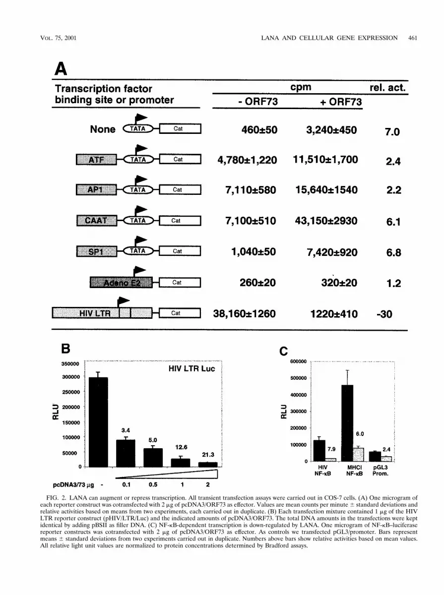
FIG. 2. LANA can augment or repress transcription. All transient transfection assays were carried out in COS-7 cells. (A) One microgram ofeach reporter construct was cotransfected with 2 mg of pcDNA3/ORF73 as effector. Values are mean counts per minute 6 standard deviations andrelative activities based on means from two experiments, each carried out in duplicate. (B) Each transfection mixture contained 1 mg of the HIVLTR reporter construct (pHIV/LTR/Luc) and the indicated amounts of pcDNA3/ORF73. The total DNA amounts in the transfections were keptidentical by adding pBSII as filler DNA. (C) NF-kB-dependent transcription is down-regulated by LANA. One microgram of NF-kB–luciferasereporter constructs was cotransfected with 2 mg of pcDNA3/ORF73 as effector. As controls we transfected pGL3/promoter. Bars representmeans 6 standard deviations from two experiments carried out in duplicate. Numbers above bars show relative activities based on mean values.All relative light unit values are normalized to protein concentrations determined by Bradford assays.
VOL. 75, 2001 LANA AND CELLULAR GENE EXPRESSION 461
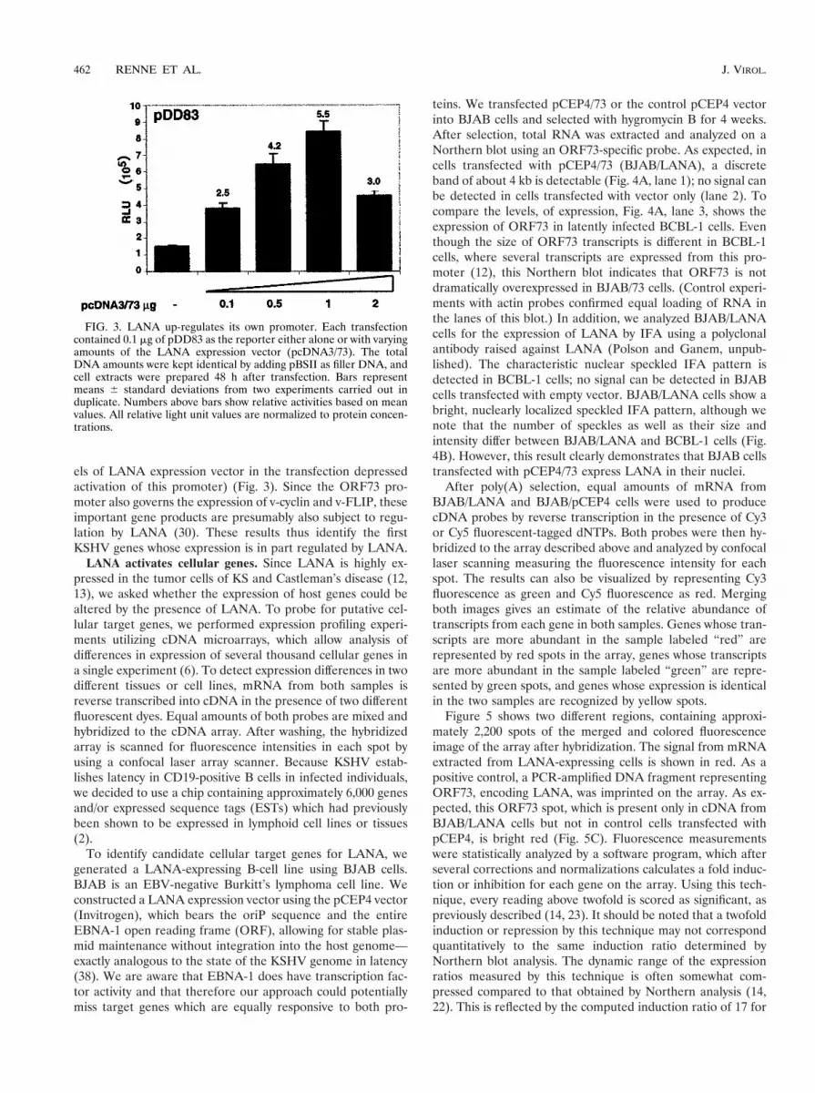
els of LANA expression vector in the transfection depressedactivation of this promoter) (Fig. 3). Since the ORF73 pro-moter also governs the expression of v-cyclin and v-FLIP, theseimportant gene products are presumably also subject to regu-lation by LANA (30). These results thus identify the firstKSHV genes whose expression is in part regulated by LANA.
LANA activates cellular genes. Since LANA is highly ex-pressed in the tumor cells of KS and Castleman’s disease (12,13), we asked whether the expression of host genes could bealtered by the presence of LANA. To probe for putative cel-lular target genes, we performed expression profiling experi-ments utilizing cDNA microarrays, which allow analysis ofdifferences in expression of several thousand cellular genes ina single experiment (6). To detect expression differences in twodifferent tissues or cell lines, mRNA from both samples isreverse transcribed into cDNA in the presence of two differentfluorescent dyes. Equal amounts of both probes are mixed andhybridized to the cDNA array. After washing, the hybridizedarray is scanned for fluorescence intensities in each spot byusing a confocal laser array scanner. Because KSHV estab-lishes latency in CD19-positive B cells in infected individuals,we decided to use a chip containing approximately 6,000 genesand/or expressed sequence tags (ESTs) which had previouslybeen shown to be expressed in lymphoid cell lines or tissues(2).
To identify candidate cellular target genes for LANA, wegenerated a LANA-expressing B-cell line using BJAB cells.BJAB is an EBV-negative Burkitt’s lymphoma cell line. Weconstructed a LANA expression vector using the pCEP4 vector(Invitrogen), which bears the oriP sequence and the entireEBNA-1 open reading frame (ORF), allowing for stable plas-mid maintenance without integration into the host genome—exactly analogous to the state of the KSHV genome in latency(38). We are aware that EBNA-1 does have transcription fac-tor activity and that therefore our approach could potentiallymiss target genes which are equally responsive to both pro-
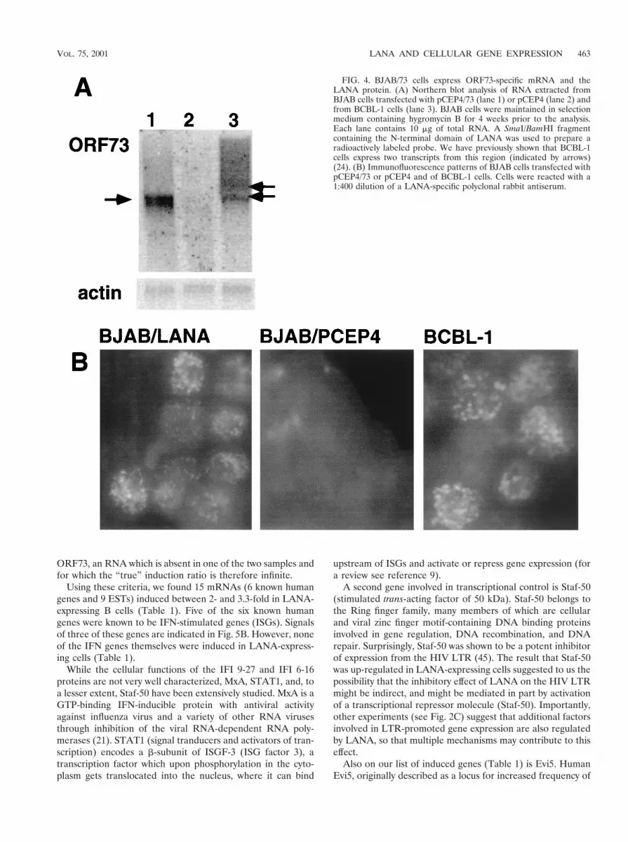
teins. We transfected pCEP4/73 or the control pCEP4 vectorinto BJAB cells and selected with hygromycin B for 4 weeks.After selection, total RNA was extracted and analyzed on aNorthern blot using an ORF73-specific probe. As expected, incells transfected with pCEP4/73 (BJAB/LANA), a discreteband of about 4 kb is detectable (Fig. 4A, lane 1); no signal canbe detected in cells transfected with vector only (lane 2). Tocompare the levels, of expression, Fig. 4A, lane 3, shows theexpression of ORF73 in latently infected BCBL-1 cells. Eventhough the size of ORF73 transcripts is different in BCBL-1cells, where several transcripts are expressed from this pro-moter (12), this Northern blot indicates that ORF73 is notdramatically overexpressed in BJAB/73 cells. (Control experi-ments with actin probes confirmed equal loading of RNA inthe lanes of this blot.) In addition, we analyzed BJAB/LANAcells for the expression of LANA by IFA using a polyclonalantibody raised against LANA (Polson and Ganem, unpub-lished). The characteristic nuclear speckled IFA pattern isdetected in BCBL-1 cells; no signal can be detected in BJABcells transfected with empty vector. BJAB/LANA cells show abright, nuclearly localized speckled IFA pattern, although wenote that the number of speckles as well as their size andintensity differ between BJAB/LANA and BCBL-1 cells (Fig.4B). However, this result clearly demonstrates that BJAB cellstransfected with pCEP4/73 express LANA in their nuclei.
After poly(A) selection, equal amounts of mRNA fromBJAB/LANA and BJAB/pCEP4 cells were used to producecDNA probes by reverse transcription in the presence of Cy3or Cy5 fluorescent-tagged dNTPs. Both probes were then hy-bridized to the array described above and analyzed by confocallaser scanning measuring the fluorescence intensity for eachspot. The results can also be visualized by representing Cy3fluorescence as green and Cy5 fluorescence as red. Mergingboth images gives an estimate of the relative abundance oftranscripts from each gene in both samples. Genes whose tran-scripts are more abundant in the sample labeled “red” arerepresented by red spots in the array, genes whose transcriptsare more abundant in the sample labeled “green” are repre-sented by green spots, and genes whose expression is identicalin the two samples are recognized by yellow spots.
Figure 5 shows two different regions, containing approxi-mately 2,200 spots of the merged and colored fluorescenceimage of the array after hybridization. The signal from mRNAextracted from LANA-expressing cells is shown in red. As apositive control, a PCR-amplified DNA fragment representingORF73, encoding LANA, was imprinted on the array. As ex-pected, this ORF73 spot, which is present only in cDNA fromBJAB/LANA cells but not in control cells transfected withpCEP4, is bright red (Fig. 5C). Fluorescence measurementswere statistically analyzed by a software program, which afterseveral corrections and normalizations calculates a fold induc-tion or inhibition for each gene on the array. Using this tech-nique, every reading above twofold is scored as significant, aspreviously described (14, 23). It should be noted that a twofoldinduction or repression by this technique may not correspondquantitatively to the same induction ratio determined byNorthern blot analysis. The dynamic range of the expressionratios measured by this technique is often somewhat com-pressed compared to that obtained by Northern analysis (14,22). This is reflected by the computed induction ratio of 17 for
FIG. 3. LANA up-regulates its own promoter. Each transfectioncontained 0.1 mg of pDD83 as the reporter either alone or with varyingamounts of the LANA expression vector (pcDNA3/73). The totalDNA amounts were kept identical by adding pBSII as filler DNA, andcell extracts were prepared 48 h after transfection. Bars representmeans 6 standard deviations from two experiments carried out induplicate. Numbers above bars show relative activities based on meanvalues. All relative light unit values are normalized to protein concen-trations.
462 RENNE ET AL. J. VIROL.
ORF73, an RNA which is absent in one of the two samples andfor which the “true” induction ratio is therefore infinite.
Using these criteria, we found 15 mRNAs (6 known humangenes and 9 ESTs) induced between 2- and 3.3-fold in LANA-expressing B cells (Table 1). Five of the six known humangenes were known to be IFN-stimulated genes (ISGs). Signalsof three of these genes are indicated in Fig. 5B. However, noneof the IFN genes themselves were induced in LANA-express-ing cells (Table 1).
While the cellular functions of the IFI 9-27 and IFI 6-16proteins are not very well characterized, MxA, STAT1, and, toa lesser extent, Staf-50 have been extensively studied. MxA is aGTP-binding IFN-inducible protein with antiviral activityagainst influenza virus and a variety of other RNA virusesthrough inhibition of the viral RNA-dependent RNA poly-merases (21). STAT1 (signal tranducers and activators of tran-scription) encodes a b-subunit of ISGF-3 (ISG factor 3), atranscription factor which upon phosphorylation in the cyto-plasm gets translocated into the nucleus, where it can bind
upstream of ISGs and activate or repress gene expression (fora review see reference 9).
A second gene involved in transcriptional control is Staf-50(stimulated trans-acting factor of 50 kDa). Staf-50 belongs tothe Ring finger family, many members of which are cellularand viral zinc finger motif-containing DNA binding proteinsinvolved in gene regulation, DNA recombination, and DNArepair. Surprisingly, Staf-50 was shown to be a potent inhibitorof expression from the HIV LTR (45). The result that Staf-50was up-regulated in LANA-expressing cells suggested to us thepossibility that the inhibitory effect of LANA on the HIV LTRmight be indirect, and might be mediated in part by activationof a transcriptional repressor molecule (Staf-50). Importantly,other experiments (see Fig. 2C) suggest that additional factorsinvolved in LTR-promoted gene expression are also regulatedby LANA, so that multiple mechanisms may contribute to thiseffect.
Also on our list of induced genes (Table 1) is Evi5. HumanEvi5, originally described as a locus for increased frequency of
FIG. 4. BJAB/73 cells express ORF73-specific mRNA and theLANA protein. (A) Northern blot analysis of RNA extracted fromBJAB cells transfected with pCEP4/73 (lane 1) or pCEP4 (lane 2) andfrom BCBL-1 cells (lane 3). BJAB cells were maintained in selectionmedium containing hygromycin B for 4 weeks prior to the analysis.Each lane contains 10 mg of total RNA. A SmaI/BamHI fragmentcontaining the N-terminal domain of LANA was used to prepare aradioactively labeled probe. We have previously shown that BCBL-1cells express two transcripts from this region (indicated by arrows)(24). (B) Immunofluorescence patterns of BJAB cells transfected withpCEP4/73 or pCEP4 and of BCBL-1 cells. Cells were reacted with a1:400 dilution of a LANA-specific polyclonal rabbit antiserum.
VOL. 75, 2001 LANA AND CELLULAR GENE EXPRESSION 463

murine retroviral integration events, is associated with humancancers, often in combination with chromosomal transloca-tions. Evi5 is a homolog of the murine Tre-2 oncogene, whichshows sequence homology to cell cycle regulators (27). At this
point we have not further characterized any of the nine in-duced ESTs.
Confirmation by Northern blot analysis. To confirm theobserved up-regulated genes, we performed Northern blot
FIG. 5. LANA can activate cellular genes. Shown are two sections of the microarray after hybridization to cDNA prepared from LANA-expressing and non-LANA-expressing cells. LANA-expressing cells are labeled in red, and non-LANA-expressing cells are labeled in green. (A)Larger area of the chip showing a representative result of this hybridization experiment. (B) Enlargement of an area of panel A where genesapparently induced by LANA expression (red spots) are indicated by arrows. (C) An area of the chip containing the positive control ORF73, whosetranscripts are present only in LANA-expressing cells; therefore, this spot is bright red.
464 RENNE ET AL. J. VIROL.
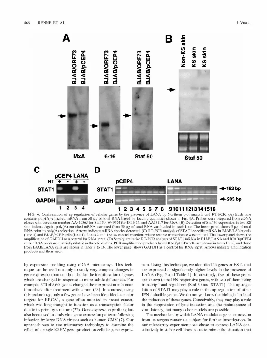
analysis on RNA extracted from BJAB/LANA cells and fromcontrol cells (BJAB/pCEP4). MxA mRNA was detectable atextremely low levels in control cells but was clearly inducedin BJAB/73 cells. IFI 6-16 and Staf-50 were detectable onlyin cells expressing LANA. However, the expression level ofStaf-50 was close to the detection level of our Northern blotassays (Fig. 6A). This low level of expression is in agreementwith recently published data on the induction of Staf-50 by IFNin HT1080 cells (human fibrosarcoma) (10). We were not ableto detect mRNA for STAT1, IFI 9-27, or the Evi5 gene byNorthern blot analysis. However, we detected STAT1 mRNAexpression in BJAB/LANA cells by RT-PCR assays. TotalRNAs from LANA-expressing and control cells were reversetranscribed, and the resulting cDNAs were amplified withSTAT1-specific primers. As shown in Fig. 6C, STAT1 mRNAexpression is slightly higher in LANA-expressing cells (lanes 1and 3). To show that we indeed amplified RNA, we carried outcontrol experiments with no reverse transcriptase, which didnot lead to any amplification (Fig. 6C, lanes 2 and 4). We alsoamplified GAPDH to control for equal RNA amounts in thereaction mixture (Fig. 6C). To determine differences in STAT1expression in a more quantitative analysis, we performed dilu-tion PCR assays. cDNA pools from LANA-expressing andcontrol cells were diluted in threefold steps and analyzed forthe presence of STAT1 and GAPDH by PCR. A GAPDH-specific band is detectable throughout the entire dilution seriesin both BJAB/pCEP4 cells (Fig. 6 D, lanes 1 to 8) and BJAB/LANA cells (lanes 9 to 16), demonstrating equal amounts ofinput RNA. In contrast, in BJAB/pCEP4 cells a STAT1-spe-cific band can be detected only in the first three lanes, while inBJAB/LANA cells this signal can be detected up to lane 4,confirming the induction of this gene (Fig. 6D).
Detection of Staf-50 mRNA in KS tumors. To validate thatthe up-regulation of Staf-50 in LANA-expressing cells in cul-ture reflects events in vivo, we asked whether Staf-50 is ex-pressed in primary dermal KS lesions. We analyzed mRNAextracted from KS lesions of two patients and, as a control,from a healthy skin necropsy specimen. A specific band forStaf-50 was detectable in both skin lesions but not in healthyskin (Fig. 6B). These data suggest that Staf-50 is indeed up-regulated in KS tissues.
DISCUSSION
By performing transient transcription assays, we have shownthat LANA expression can up-regulate its own promoter aswell as synthetic promoter constructs as much as sixfold (Fig.2). In contrast, the basal transcriptional activity of the HIVLTR was drastically repressed in the presence of LANA, in adose-dependent fashion. At least two mechanisms can be in-voked to explain the suppression of LTR-based transcription.First, LANA expression had a negative effect on NF-kB-de-pendent transcription, which is known to be important for LTRfunction; second, LANA induces Staf-50, a known inhibitor ofLTR-driven expression. We do not know which of these mech-anisms predominates, nor can we exclude additional contribu-tions from as yet unexplained mechanisms. However, theseinitial experiments clearly demonstrate that LANA can mod-ulate transcription both positively and negatively.
It was recently shown that LANA can interact with bothRING3 (a homolog of the Drosophila female sterile homeotic(fsh) gene product) and the tumor suppressor p53. Fsh belongsto a class of proteins implicated in chromatin structure andtranscriptional regulation (31). p53 is a known transcriptionalactivator, and binding of LANA to p53 impairs its transactiva-tion activity (16). Both of these interactions are consistentwith a role for LANA in the regulation of gene expression.Recently, it was demonstrated that LANA is required formaintenance of the episomal viral DNA in dividing cells, pre-sumably by tethering episomal KSHV DNA to mitotic chro-mosomes (4). In agreement with this finding, it was demon-strated that LANA binds in an in vitro assay to a putative oriPat the left side of the genome overlapping with the terminalrepeats (8). Taken together with our data, these findings indi-cate that LANA, like EBNA-1, has at least two activities:transcriptional regulation and episome maintenance.
In cells latently infected by EBV, EBNA-1 is also subject toautoregulation at the transcriptional level (3, 43). Our obser-vation that the ORF73 promoter can be up-regulated by LANAcoexpression suggests the existence of LANA autoregulationin KSHV, although we do not know if this autoregulation isbased on direct binding of LANA to DNA targets in its ownpromoter. It should be noted that the basal activity of theLANA promoter in 293 cells is very high; accordingly, auto-regulation by LANA might be more dramatic in cells in whichthis promoter is less active. For example, it has been shownthat the LANA promoter is much weaker in cells of endothe-lial origin—a major target for KSHV infection in KS patho-genesis (44). Experiments to study the autoregulation of LANAin endothelial and lymphoid cell lines are currently in progress.However, our data provide the first identification of a LANA-responsive KSHV promoter. Moreover, this regulation by LANAis also expected to augment the expression of ORF72 (encod-ing a viral cyclin D homolog) and ORF71 (which encodes aputative FLICE-inhibitory protein, v-FLIP). Since both ofthese genes have potential roles in the deregulation of cellulargrowth and survival, this regulation may play an important rolein driving proliferation in KSHV-linked disorders (12, 30).
To identify putative cellular target genes whose expression isaltered by the presence of LANA, we first generated cellsconstitutively expressing LANA and then analyzed these cells
TABLE 1. Genes or ESTs up-regulated in the presenceof LANA in BJAB cells
Gene Inductionratio
KSHV ORF73 (positive control).................................................. 17.30EST (example)a............................................................................... 3.30Staf-50 (IFN induced) .................................................................... 3.20Evi5................................................................................................... 2.99STAT1 (ISGF-3 b-subunit) ........................................................... 2.71IFI 6-16 (IFN induced).................................................................. 2.46MxA (IFN induced) ....................................................................... 2.43IFI 9-27 (IFN induced).................................................................. 2.08
IFN-a................................................................................................ 1.18IFN-b1.............................................................................................. 0.78IFN-g ................................................................................................ 0.78
a In addition, eight ESTs showed induction levels ranging between 2.0 and 2.5.
VOL. 75, 2001 LANA AND CELLULAR GENE EXPRESSION 465
by expression profiling using cDNA microarrays. This tech-nique can be used not only to study very complex changes ingene expression patterns but also for the identification of geneswhich are changed in response to more subtle differences. Forexample, 570 of 8,600 genes changed their expression in humanfibroblasts after treatment with serum (23). In contrast, usingthis technology, only a few genes have been identified as majortargets for BRCA1, a gene often mutated in breast cancer,which was long thought to function as a transcription factordue to its primary structure (22). Gene expression profiling hasalso been used to study viral gene expression patterns followinginfection by large DNA viruses such as human CMV (7). Ourapproach was to use microarray technology to examine theeffect of a single KSHV gene product on cellular gene expres-
sion. Using this technique, we identified 15 genes or ESTs thatare expressed at significantly higher levels in the presence ofLANA (Fig. 5 and Table 1). Interestingly, five of these genesare known to be IFN-responsive genes, with two of them beingtranscriptional regulators (Staf-50 and STAT1). The up-regu-lation of STAT1 may play a role in the up-regulation of otherIFN-inducible genes. We do not yet know the biological role ofthe induction of these genes. Conceivably, they may play a rolein the suppression of lytic induction and the maintenance ofviral latency, but many other models are possible.
The mechanism by which LANA modulates gene expressionfrom its targets remains a subject for further investigation. Inour microarray experiments we chose to express LANA con-stitutively in stable cell lines, so as to mimic the situation that
FIG. 6. Confirmation of up-regulation of cellular genes by the presence of LANA by Northern blot analysis and RT-PCR. (A) Each lanecontains poly(A)-enriched mRNA from 50 mg of total RNA based on loading quantities shown in Fig. 4A. Probes were prepared from cDNAclones with accession number AA419365 for Staf-50, W49674 for IFI 6-16, and AA53117 for MxA. (B) Detection of Staf-50 expression in two KSskin lesions. Again, poly(A)-enriched mRNA extracted from 50 mg of total RNA was loaded in each lane. The lower panel shows 5 mg of totalRNA prior to poly(A) selection. Arrows indicate mRNA species detected. (C) RT-PCR analysis of STAT1-specific mRNA in BJAB/LANA cells(lane 3) and BJAB/pCEP cells (lane 1). Lanes 2 and 4 show control reactions where reverse transcriptase was omitted. The lower panel shows theamplification of GAPDH as a control for RNA input. (D) Semiquantitative RT-PCR analysis of STAT1 mRNA in BJAB/LANA and BJAB/pCEP4cells. cDNA pools were serially diluted in threefold steps. PCR amplification products from BJAB/pCEP4 cells are shown in lanes 1 to 8, and thosefrom BJAB/LANA cells are shown in lanes 9 to 16. The lower panel shows GAPDH as a control for RNA input. Arrows indicate amplificationproducts and their sizes.
466 RENNE ET AL. J. VIROL.
obtains in latent infection in vivo. While the up-regulation weobserved in this system could be due to the direct action ofLANA on its targets, it is equally possible that these genes areinduced by other regulatory molecules that are themselvescontrolled by LANA. The binding of p53 by LANA (16) wouldbe one example of such a mechanism; our experiments simi-larly show that LANA expression can affect NF-kB-dependenttranscription (Fig 2C) and can up-regulate known transcriptionfactors STAT1 and Staf-50. We are currently developing celllines in which LANA expression is inducible in order to exam-ine gene regulation by LANA more directly. But whether ac-tivation of these targets is direct or indirect, the target genesidentified herein are likely to be in latently infected cells—aninference we have directly confirmed for Staf-50 in KS tumors.A better understanding of the mechanisms by which LANAmodifies cellular and viral gene expression is likely to be im-portant for deciphering the role(s) of this protein in the patho-biology of KSHV infection.
ACKNOWLEDGMENTS
We thank Andy Polson for providing the LANA antibody and KarenE. Tucker for help with microscopy and digital imaging.
R.R. is a fellow of the Leukemia Society of America and a MountSinai Healthcare foundation scholar. This work was supported by theHoward Hughes Medical Institute (HHMI) and grants from the NIHto D.G. (CA73506-04) and R.R. (CA CA88763-01). D.G. is an inves-tigator and P.O.B. is an associate investigator of the HHMI.
REFERENCES
1. Alexandropoulos, K., G. Cheng, and D. Baltimore. 1995. Proline-rich se-quences that bind to Src homology 3 domains with individual specificities.Proc. Natl. Acad. Sci. USA 92:3110–3114.
2. Alizadeh, A., M. Eisen, D. Botstein, P. O. Brown, and L. M. Staudt. 1998.Probing lymphocyte biology by genomic-scale gene expression analysis.J. Clin. Immunol. 18:373–379.
3. Ambinder, R. F., M. A. Mullen, Y. N. Chang, G. S. Hayward, and S. D.Hayward. 1991. Functional domains of Epstein-Barr virus nuclear antigenEBNA-1. J. Virol. 65:1466–1478.
4. Ballestas, M. E., P. A. Chatis, and K. M. Kaye. 1999. Efficient persistence ofextrachromosomal KSHV DNA mediated by latency-associated nuclear an-tigen. Science 284:641–644.
5. Boshoff, C., T. F. Schulz, M. M. Kennedy, A. K. Graham, C. Fisher, A.Thomas, J. O. McGee, R. A. Weiss, and J. J. O’Leary. 1995. Kaposi’s sarco-ma-associated herpesvirus infects endothelial and spindle cells. Nat. Med. 1:1274–1278.
6. Brown, P. O., and D. Botstein. 1999. Exploring the new world of the genomewith DNA microarrays. Nat. Genet. 21(Suppl. 1):33–37.
7. Chambers, J., A. Angulo, D. Amaratunga, H. Guo, Y. Jiang, J. S. Wan, A.Bittner, K. Frueh, M. R. Jackson, P. A. Peterson, M. G. Erlander, and P.Ghazal. 1999. DNA microarrays of the complex human cytomegalovirusgenome: profiling kinetic class with drug sensitivity of viral gene expression.J. Virol. 73:5757–5766.
8. Cotter, M. A., II, and E. S. Robertson. 1999. The latency-associated nuclearantigen tethers the Kaposi’s sarcoma-associated herpesvirus genome to hostchromosomes in body cavity-based lymphoma cells. Virology 264:254–264.
9. Darnell, J. E., Jr., I. M. Kerr, and G. R. Stark. 1994. Jak-STAT pathways andtranscriptional activation in response to IFNs and other extracellular signal-ing proteins. Science 264:1415–1421.
10. Der, S. D., A. Zhou, B. R. Williams, and R. H. Silverman. 1998. Identificationof genes differentially regulated by interferon alpha, beta, or gamma usingoligonucleotide arrays. Proc. Natl. Acad. Sci. USA 95:15623–15628.
11. DeRisi, J. L., V. R. Iyer, and P. O. Brown. 1997. Exploring the metabolic andgenetic control of gene expression on a genomic scale. Science 278:680–686.
12. Dittmer, D., M. Lagunoff, R. Renne, K. Staskus, A. Haase, and D. Ganem.1998. A cluster of latently expressed genes in Kaposi’s sarcoma-associatedherpesvirus. J. Virol. 72:8309–8315.
13. Dupin, N., C. Fisher, P. Kellam, S. Ariad, M. Tulliez, N. Franck, E. vanMarck, D. Salmon, I. Gorin, J. P. Escande, R. A. Weiss, K. Alitalo, and C.Boshoff. 1999. Distribution of human herpesvirus-8 latently infected cells inKaposi’s sarcoma, multicentric Castleman’s disease, and primary effusionlymphoma. Proc. Natl. Acad. Sci. USA 96:4546–4551.
14. Eisen, M. B., and P. O. Brown. 1999. DNA arrays for analysis of geneexpression. Methods Enzymol. 303:179–205.
15. Foreman, K. E., J. Friborg, Jr., W. P. Kong, C. Woffendin, P. J. Polverini,B. J. Nickoloff, and G. J. Nabel. 1997. Propagation of a human herpesvirusfrom AIDS-associated Kaposi’s sarcoma. N. Engl. J. Med. 336:163–171.
16. Friborg, J., W. Kong, M. O. Hottiger, and G. J. Nabel. 2000. p53 inhibition bythe LANA protein of KSHV protects against cell death. Nature 402:889–894.
17. Gahn, T. A., and B. Sugden. 1995. An EBNA-1-dependent enhancer actsfrom a distance of 10 kilobase pairs to increase expression of the Epstein-Barr virus LMP gene. J. Virol. 69:2633–2636.
18. Ganem, D. 1997. KSHV and Kaposi’s sarcoma: the end of the beginning?Cell 91:157–160.
19. Gao, S. J., L. Kingsley, M. Li, W. Zheng, C. Parravicini, J. Ziegler, R.Newton, C. R. Rinaldo, A. Saah, J. Phair, R. Detels, Y. Chang, and P. S.Moore. 1996. KSHV antibodies among Americans, Italians and Ugandanswith and without Kaposi’s sarcoma. Nat. Med. 2:925–928.
20. Gao, S. J., Y. J. Zhang, J. H. Deng, C. S. Rabkin, O. Flore, and H. B. Jenson.1999. Molecular polymorphism of Kaposi’s sarcoma-associated herpesvirus(human herpesvirus 8) latent nuclear antigen: evidence for a large repertoireof viral genotypes and dual infection with different viral genotypes. J. Infect.Dis. 180:1466–1476.
21. Haller, O., M. Frese, and G. Kochs. 1998. Mx proteins: mediators of innateresistance to RNA viruses. Rev. Sci. Tech. 17:220–230.
22. Harkin, D. P., J. M. Bean, D. Miklos, Y. H. Song, V. B. Truong, C. Englert,F. C. Christians, L. W. Ellisen, S. Maheswaran, J. D. Oliner, and D. A.Haber. 1999. Induction of GADD45 and JNK/SAPK-dependent apoptosisfollowing inducible expression of BRCA1. Cell 97:575–586.
23. Iyer, V. R., M. B. Eisen, D. T. Ross, G. Schuler, T. Moore, J. C. F. Lee, J. M.Trent, L. M. Staudt, J. Hudson, Jr., M. S. Boguski, D. Lashkari, D. Shalon,D. Botstein, and P. O. Brown. 1999. The transcriptional program in theresponse of human fibroblasts to serum. Science 283:83–87.
24. Kedes, D. H., M. Lagunoff, R. Renne, and D. Ganem. 1997. Identification ofthe gene encoding the major latency-associated nuclear antigen of Kaposi’ssarcoma-associated herpesvirus. J. Clin. Investig. 100:2606–2610.
25. Kedes, D. H., E. Operskalski, M. Busch, R. Kohn, J. Flood, and D. Ganem.1996. The seroepidemiology of human herpesvirus 8 (Kaposi’s sarcoma-associated herpesvirus): distribution of infection in KS risk groups and evi-dence for sexual transmission. Nat. Med. 2:918–924. (Erratum, 2:1041.)
26. Kellam, P., C. Boshoff, D. Whitby, S. Matthews, R. A. Weiss, and S. J. Talbot.1997. Identification of a major latent nuclear antigen, LNA-1, in the humanherpesvirus 8 genome. J. Hum. Virol. 1:19–29.
27. Liao, X., Y. Du, H. C. Morse III, N. A. Jenkins, and N. G. Copeland. 1997.Proviral integrations at the Evi5 locus disrupt a novel 90-kDa protein withhomology to the Tre2 oncogene and cell-cycle regulatory proteins. Oncogene14:1023–1029.
28. Liebowitz, D., and E. Kieff. 1993. Epstein-Barr virus, p. 107–172. In B.Roizman, R. J. Whitley, and C. Lopez (ed.), The human herpesviruses.Raven Press, New York, N.Y.
29. Lukac, D. M., J. R. Manuppello, and J. C. Alwine. 1994. Transcriptionalactivation by the human cytomegalovirus immediate-early proteins: require-ments for simple promoter structures and interactions with multiple com-ponents of the transcription complex. J. Virol. 68:5184–5193.
30. Moore, P. S., C. Boshoff, R. A. Weiss, and Y. Chang. 1996. Molecularmimicry of human cytokine and cytokine response pathway genes by KSHV.Science 274:1739–1744.
31. Platt, G. M., G. R. Simpson, S. Mittnacht, and T. F. Schulz. 1999. Latentnuclear antigen of Kaposi’s sarcoma-associated herpesvirus interacts withRING3, a homolog of the Drosophila female sterile homeotic (fsh) gene.J. Virol. 73:9789–9795.
32. Popik, W., and P. M. Pitha. 1992. Transcriptional activation of the tat-defective human immunodeficiency virus type-1 provirus: effect of inter-feron. Virology 189:435–447.
33. Ptashne, M. 1992. A genetic switch, 2nd ed. Cell Press & Blackwell ScientificPublications, Cambridge, Mass.
34. Puglielli, M. T., N. Desai, and S. H. Speck. 1997. Regulation of EBNA genetranscription in lymphoblastoid cell lines: characterization of sequencesdownstream of BCR2 (Cp). J. Virol. 71:120–128.
35. Raab-Traub, N. 1996. Pathogenesis of Epstein-Barr virus and its associatedmalignancies. Virology 7:315–323.
36. Rainbow, L., G. M. Platt, G. R. Simpson, R. Sarid, S. J. Gao, H. Stoiber, C. S.Herrington, P. S. Moore, and T. F. Schulz. 1997. The 222- to 234-kilodaltonlatent nuclear protein (LNA) of Kaposi’s sarcoma-associated herpesvirus(human herpesvirus 8) is encoded by orf73 and is a component of thelatency-associated nuclear antigen. J. Virol. 71:5915–5921.
37. Renne, R., D. Blackbourn, D. Whitby, J. Levy, and D. Ganem. 1998. Limitedtransmission of Kaposi’s sarcoma-associated herpesvirus in cultured cells.J. Virol. 72:5182–5188.
38. Renne, R., M. Lagunoff, W. Zhong, and D. Ganem. 1996. The size andconformation of Kaposi’s sarcoma-associated herpesvirus (human herpesvi-rus 8) DNA in infected cells and virions. J. Virol. 70:8151–8154.
39. Renne, R., A. Mergia, L. W. Renshaw-Gegg, D. Neumann-Haefelin, and P. A.Luciw. 1993. Regulatory elements in the long terminal repeat (LTR) ofsimian foamy virus type 3 (SFV-3). Virology 192:365–369.
40. Russo, J. J., R. A. Bohenzky, M. C. Chien, J. Chen, M. Yan, D. Maddalena,
VOL. 75, 2001 LANA AND CELLULAR GENE EXPRESSION 467

J. P. Parry, D. Peruzzi, I. S. Edelman, Y. Chang, and P. S. Moore. 1996.Nucleotide sequence of the Kaposi sarcoma-associated herpesvirus (HHV8).Proc. Natl. Acad. Sci. USA 93:14862–14867.
40a.Shen, Y., and T. Shenk. 1994. Relief of p53-mediated transcriptional repres-sion by the adenovirus E1B 19-kDa protein or the cellular Bcl-2 protein.Proc. Natl. Acad. Sci. USA 91:8940–8944.
41. Sheridan, P. L., C. T. Sheline, L. H. Milocco, and K. A. Jones. 1993. Tat andthe HIV-1 promoter: a model for RNA-mediated regulation of transcription.Semin. Virol. 4:69–80.
42. Struhl, K. 1995. Yeast transcriptional regulatory mechanisms. Annu. Rev.Genet. 29:651–674.
43. Sugden, B., and N. Warren. 1989. A promoter of Epstein-Barr virus that can
function during latent infection can be transactivated by EBNA-1, a viralprotein required for viral DNA replication during latent infection. J. Virol.63:2644–2649.
44. Talbot, S. J., R. A. Weiss, P. Kellam, and C. Boshoff. 1999. Transcriptionalanalysis of human herpesvirus-8 open reading frames 71, 72, 73, K14, and 74in a primary effusion lymphoma cell line. Virology 257:84–94.
45. Tissot, C., and N. Mechti. 1995. Molecular cloning of a new interferon-induced factor that represses human immunodeficiency virus type 1 longterminal repeat expression. J. Biol. Chem. 270:14891–14898.
46. Zhong, W., H. Wang, B. Herndier, and D. Ganem. 1996. Restricted expres-sion of Kaposi sarcoma-associated herpesvirus (human herpesvirus 8) genesin Kaposi sarcoma. Proc. Natl. Acad. Sci. USA 93:6641–6646.
468 RENNE ET AL. J. VIROL.