Embed Size (px)
Citation preview

Published Ahead of Print 15 May 2013. 2013, 87(14):8004. DOI: 10.1128/JVI.00506-13. J. Virol.
Kellam and Thomas F. SchulzWeidner-Glunde, Thomas Rothämel, Adam Grundhoff, Paul Semra Kati, Edward H. Tsao, Thomas Günther, Magdalena Sarcoma-Associated Herpesvirus in B CellsTriggers Reactivation of Latent Kaposi's Activation of the B Cell Antigen Receptor
http://jvi.asm.org/content/87/14/8004Updated information and services can be found at:
These include:
REFERENCEShttp://jvi.asm.org/content/87/14/8004#ref-list-1at:
This article cites 75 articles, 35 of which can be accessed free
CONTENT ALERTS more»articles cite this article),
Receive: RSS Feeds, eTOCs, free email alerts (when new
http://journals.asm.org/site/misc/reprints.xhtmlInformation about commercial reprint orders: http://journals.asm.org/site/subscriptions/To subscribe to to another ASM Journal go to:
on June 12, 2014 by guesthttp://jvi.asm
.org/D
ownloaded from
on June 12, 2014 by guest
http://jvi.asm.org/
Dow
nloaded from
Activation of the B Cell Antigen Receptor Triggers Reactivation ofLatent Kaposi’s Sarcoma-Associated Herpesvirus in B Cells
Semra Kati,a Edward H. Tsao,b Thomas Günther,c Magdalena Weidner-Glunde,a Thomas Rothämel,d Adam Grundhoff,c Paul Kellam,b,e
Thomas F. Schulza
Institute of Virology, Hanover Medical School, Hanover, Germanya; MRC Centre for Medical Molecular Virology, University College London, London, United Kingdomb;Heinrich Pette Institute, Leibniz Institute for Experimental Virology, Hamburg, Germanyc; Institute of Forensic Medicine, Hanover Medical School, Hanover, Germanyd;Wellcome Trust Sanger Institute, Wellcome Trust Genome Campus, Hinxton, Cambridge, United Kingdome
Kaposi’s sarcoma-associated herpesvirus (KSHV) is an oncogenic herpesvirus and the cause of Kaposi’s sarcoma, primary effu-sion lymphoma (PEL) and multicentric Castleman’s disease. Latently infected B cells are the main reservoir of this virus in vivo,but the nature of the stimuli that lead to its reactivation in B cells is only partially understood. We established stable BJAB celllines harboring latent KSHV by cell-free infection with recombinant virus carrying a puromycin resistance marker. Our latentlyinfected B cell lines, termed BrK.219, can be reactivated by triggering the B cell receptor (BCR) with antibodies to surface IgM, astimulus imitating antigen recognition. Using this B cell model system we studied the mechanisms that mediate the reactivationof KSHV in B cells following the stimulation of the BCR and could identify phosphatidylinositol 3-kinase (PI3K) and X-boxbinding protein 1 (XBP-1) as proteins that play an important role in the BCR-mediated reactivation of latent KSHV.
Kaposi’s sarcoma-associated herpesvirus (KSHV), also referredto as human herpesvirus 8 (HHV8), and Epstein-Barr virus
(EBV; HHV4) belong to the gammaherpesvirus subfamily and areassociated with B cell lymphoproliferative diseases (1, 2). Al-though EBV is the causative agent of endemic Burkitt’s lym-phoma, Hodgkin’s lymphoma, and posttransplant lymphoprolif-erative disorder (3), KSHV causes primary effusion lymphoma(PEL) (4) and multicentric Castleman’s disease (5) in addition toKaposi’s sarcoma.
B cells are the main latent reservoir for both viruses in vivo (6,7). EBV can transform and immortalize primary B lymphocytes invitro, which results in the formation of lymphoblastoid cell lines(8). EBV-infected cell lines, derived directly from patients or gen-erated by in vitro infection of either EBV-negative B cell lines ornaive B cells, contributed significantly to the understanding ofEBV biology in B cells.
In contrast, B cell lines and primary B cells are poorly permis-sive to infection by KSHV in vitro (9–11). Introduction of the viralDNA via electroporation into a B cell line resulted in latent per-sistence of KSHV (12). Only recently, KSHV- and EBV-negativeBJAB cells were successfully infected with KSHV via cell-mediatedtransmission (13). Latently infected cells could be generated un-der selection (13, 14) but were poorly permissive to lytic/produc-tive replication after stimulation with valproate, sodium butyrate,or 12-O-tetradecanoyl-phorbol-13-acetate (TPA) (13). Most ofour knowledge on the lytic/productive replication of KSHV in Bcells stems from work on PEL cell lines. It is possible to reactivateKSHV in these cells using chemical stimuli such as phorbol esters(e.g., TPA) or histone deacetylase inhibitors (e.g., sodium bu-tyrate) (15–17). As for possible physiological trigger(s), reactiva-tion of both KSHV in PEL cell lines and EBV in the EBV-positivenasopharyngeal carcinoma cell line HONE-1 and lymphoblastoidcell lines have been shown to be induced by the active form of thetranscription factor X-box binding protein 1 (XBP-1) (18–21).XBP-1 is a key regulator of unfolded protein response (UPR) andis also essential for the terminal differentiation of B cells intoplasma cells (49). Any perturbation that leads to the accumulation
of unfolded or misfolded proteins in the endoplasmic reticulum(ER) initiates the removal of a 26-nucleotide intron from theXBP-1 mRNA by the ER transmembrane protein endoribonu-clease inositol-requiring enzyme 1� (IRE1�) (22). This results in atranscriptional frameshift that generates the active XBP-1, whichupregulates UPR genes to enhance protein folding capacity ofcells. UPR activation during antibody production has been pro-posed to provide a link between plasma cell differentiation (23,24) and gammaherpesviral reactivation (18, 21). Overexpressionof spliced XBP-1, or its artificial induction with dithiothreitol(DTT), leads to reactivation of KSHV in PEL cells in vitro (18–21).
In the case of EBV-infected B cells, reactivation of the lytic cyclecan be triggered by activating the B cell antigen receptor (BCR) bycross-linking surface immunoglobulins on the B cell surface withanti-Ig antibodies (25, 26). This, together with the involvement ofplasma cell differentiation-associated cellular factors such asXBP-1, has led to the notion that triggering of the BCR on thesurface of latently infected memory B cells and the ensuing plasmacell differentiation could provide the physiological stimulus forthe reactivation of EBV in latently infected memory B cells (27–30). Evidence for the reactivation of murine herpesvirus 68(MHV68) in B cells following triggering of the BCR also exists(31). Reactivation of EBV in B cells as a result of triggering theBCR involves the phosphatidylinositol 3-kinase (PI3K) pathway(28), which is also known to interact with the spliced form ofXBP-1 (32, 33). Whether contact with antigen also plays a role inthe reactivation of KSHV in latently infected B cells has so far notbeen addressed, since PEL cells lack the B cell immunoglobulinreceptor on their surface (34–38).
Received 20 February 2013 Accepted 5 May 2013
Published ahead of print 15 May 2013
Address correspondence to Thomas F. Schulz, [email protected].
Copyright © 2013, American Society for Microbiology. All Rights Reserved.
doi:10.1128/JVI.00506-13
8004 jvi.asm.org Journal of Virology p. 8004–8016 July 2013 Volume 87 Number 14
on June 12, 2014 by guesthttp://jvi.asm
.org/D
ownloaded from
In this study, we therefore wanted to develop an experimentalsystem in which to study a possible role of the BCR in KSHVreactivation from latency. We established stable latent KSHV in-fection in an immortalized B cell line (BJAB) using a recombinantKSHV and either cell-free or cell-associated infection. Character-ization of these stably infected B cell lines, named BrK.219, re-vealed an expression pattern of viral proteins similar to that of PELcell lines. These cells express surface IgM and treating them withantibodies against human IgM led to a reactivation of the lyticcycle, resulting in the release of significant titers of infectiousprogeny. Inhibition of PI3K and XBP-1 splicing with chemicalinhibitors decreased the expression of viral lytic proteins and in-fectious progeny production after anti-IgM treatment.
Our findings indicate that, as for EBV, the contact of latentlyKSHV-infected B cells with their cognate antigen might provide atrigger for viral reactivation.
MATERIALS AND METHODSCell culture and reagents. HEK 293 cells and TE671 were cultured inDulbecco’s modified Eagle medium (Gibco) supplemented with 10%heat-inactivated fetal calf serum (FCS; HyClone). Vero cells were grownin minimum essential medium (Cytogen) containing 10% FCS. The re-combinant rKSHV.219 carries a constitutively expressed green fluores-cent protein (GFP), a red fluorescent protein (RFP) under the control ofthe lytic PAN promoter, and a puromycin resistance gene (39). Vero cellsstably infected with rKSHV.219 (referred to as Vero rKSHV.219) (39)were grown in the presence of 5 �g of puromycin (Sigma)/ml. A KSHV-and EBV-negative BJAB cell line (40), KSHV-positive and EBV-negativePEL cell lines (BC-3 and BCBL-1) (16, 41), and the KSHV- and EBV-double positive PEL cell line BC-1 (42) were maintained in RPMI 1640medium (Gibco) containing 10% FCS without antibiotics. BJAB cell linesstably infected with recombinant KSHV (39) (referred to as BrK.219)were additionally treated with 4.2 �g of puromycin/ml. All cell lines werekept in a humidified incubator at 37°C and 5% CO2 and were routinelymonitored for contamination with mycoplasma using a VenorGEM-My-coplasma detection kit (Minerva-Biolabs) according to the manufactur-er’s guidelines.
Preparation of concentrated rKSHV.219 virus stocks in Vero cells.Preparation of recombinant virus was performed as described previously(39). Briefly, rKSHV.219 production was induced in Vero rKSHV.219 byrecombinant baculovirus expressing KSHV RTA (ORF50; replication andtranscription activator) (39) and sodium butyrate (1.25 mM). After 3days, infectious supernatants were harvested and centrifuged for 10 min at4°C at 1,237 � g, filtered (0.45-�m pore size; Nalgene), and ultracentri-fuged (L8-70 ultracentrifuge; Beckman) at 15,000 � g at 4°C for 4 to 6 husing a Beckman type 19 rotor. The viral pellets were resuspended inmedium. The concentrated virus stocks were analyzed by infectivity assay.
Generation of KSHV-infected B cells by cell-mediated and cell-freetransmission of rKSHV.219 in vitro. For cell-mediated transmission,BJAB cells (40) were cocultured with Vero rKSHV.219 cells, and the lyticcycle of KSHV was induced by treatment with ectopically expressed bacu-loviral RTA (ORF50) (39) and sodium butyrate (1.25 mM). After 2 to 3days, infected B cells were separated from the adherent cell lines by gentlyshaking the tissue culture plates and harvesting suspension cells. The lym-phoblastoid cells were then resuspended in selection medium. For cell-free infection, BJAB cells were incubated with concentrated cell-free virusat a multiplicity of infection (MOI) from 1 to 10 in the presence of Poly-brene (8 �g/ml). The cells were then spinoculated at 450 � g for 20 min at32°C and incubated at 37°C for 2 to 3 days before the addition of puro-mycin. In both cases, the medium was changed twice a week. After 3weeks, polyclonal BJAB cells latently infected with rKSHV.219 could becultured and were designated as BrK.219 cells.
Infectivity assays. To determine the titers of infectious progeny in thesupernatants of reactivated KSHV-positive cells, 2.3 � 103 HEK 293 cells
were seeded in each well of a 96-well plate. The next day, the cells wereexposed to serial dilutions of infectious supernatants in a total volume of100 �l. Plates were incubated at room temperature for 20 min while rock-ing, centrifuged at 450 � g for 20 min at 32°C, and incubated at 37°C for3 days. GFP-expressing cells were counted, and the number of GFP-pos-itive cells per ml of infectious supernatant was calculated and expressed inIU/ml. All samples were measured at least in duplicates. Error bars repre-sent the standard deviations.
For titration of infectious progeny in the supernatants of reactivatedKSHV-positive cells derived from experiments with the IRE1�-inhibitorIRE1i, TE671 cells (105 cells) were seeded into each well of a 12-well plate.The next day, the cells were mixed with culture supernatants, and theplates were centrifuged at 500 � g for 1 h at room temperature, followedby 37°C incubation for 2 days. The number of GFP-expressing cells wasdetermined by flow cytometry.
STR analysis. Cell line identification and control for contaminationwas accomplished by DNA analysis using forensic standards. DNA wasextracted from cell sediments by an overnight proteinase K digestion in aChelex in water (DNA-grade) suspension (10%) at 56°C. A total of nineshort-tandem-repeat (STR) polymorphisms were analyzed using theAmpFLSTR Profiler (Applied Biosystems) multiplex PCR kit, includingAmelogenin for gender determination. PCR was run on the MJ ResearchThermocycler PTC-200 with the standard AmpFLSTR Profiler cyclingconditions as proposed by the manufacturer. STR fragments were sepa-rated by capillary electrophoresis on an ABI Prism 310 genetic analyzer(Applied Biosystems) in POP-4 polymer; the data were processed with theABI Prism 310 Genetic Analyzer Data Collection (v2.1) and GeneScan(v3.1) software. Allele calling was performed with the ABI Prism Geno-typer (v2.5.2) software.
MTT cell viability assay. A total of 4 � 104 cells were incubated withindicated inhibitors or control substances in each well of a 96-well plate.After 3 days of incubation the medium was removed, and 100 �l of Opti-MEM I reduced serum medium (Invitrogen) with 0.8 mM MTT [3-(4,5-dimethylthiazol-2-yl)-2,5-diphenyltetrazolium bromide; Sigma-Aldrich]was added to each well. After 1 h of incubation at 37°C in the dark, theMTT medium was removed, and the cells were resuspended in isopropa-nol (100 �l/well). Finally, the absorbance of the dyed solutions was mea-sured at an optical density at 550 nm (OD550) and normalized to OD620.Each sample was tested at least in triplicates. Graphs display the averagemetabolism relative to the vehicle treated sample. Error bars represent thestandard deviations.
CellTitreGlo cell viability assay. BrK.219 cells were treated with eitherdimethyl sulfoxide (DMSO), the IRE1�-inhibitor IRE1i, anti-IgM plusDMSO, or anti-IgM plus IRE1i. All samples were measured for cell viabil-ity at 72 h posttreatment using the CellTiter-Glo assay (Promega) accord-ing to the manufacturer’s instructions.
Kinase inhibitor assay. The toxicity of all inhibitors was tested byMTT or CellTitreGlo cell viability assay before use in BrK.219, and DMSOconcentrations did not exceed 0.1%. For the PI3K experiment, 1 h prior toinduction of the lytic cycle, 5 � 105 BrK.219 cells in each well of a six-wellplate were treated with the PI3K inhibitors LY294002 (Biomol) and PI-103 (Calbiochem) at the indicated concentrations, an equivalent amountof DMSO (Applichem) or not treated. For the protein kinase/endoribo-nuclease IRE1� experiment, 1.5 � 106 BrK.219 cells in each well of asix-well plate were treated with the IRE1�-specific inhibitor (IRE1i;Chembridge) at 25 �M, an equivalent amount of DMSO or mock treated.The KSHV lytic cycle was induced by cross-linking of the BCR with anti-human IgM (�-chain specific; 2.5 �g/ml; Southern Biotech) for 3 days.Finally, supernatants and cells were collected and processed for furtheranalysis by infectivity assay, immunoblotting, or reverse transcription-PCR (RT-PCR).
Use of qPCR to analyze KSHV copy numbers per cell. To determinethe KSHV copy numbers per cell, TaqMan real-time quantitative PCRs(qPCR) directed against the viral open reading frame (ORF) K6 of KSHVwere performed as described previously (43). A TaqMan qPCR for the
BCR Activation Triggers KSHV Reactivation in B Cells
July 2013 Volume 87 Number 14 jvi.asm.org 8005
on June 12, 2014 by guesthttp://jvi.asm
.org/D
ownloaded from
cellular C-reactive protein (CRP) was used for normalization (44). Briefly,genomic DNA was extracted using the DNA blood kit (Qiagen) accordingto the manufacturer’s instructions and eluted in 50 �l of elution buffer EB.A qPCR of KSHV was carried out in a total volume of 50 �l containing aready-to-use master mix (QuantiTect multiplex PCR kit; Qiagen), 0.5 �Mconcentrations of each primer, 10 �l of DNA from the sample of interest,and 0.4 �M FAM-labeled KSHV K6 probe. Amplification was performedin the Applied Biosystems 7500 thermal cycler and visualized with ABI7500 software. qPCR of CRP was carried out in a total volume of 20 �lcontaining a ready-to-use master mix (LightCycler FastStart DNA MasterHybProbe; Roche), 0.3 mM MgCl2, 0.5 �M concentrations of eachprimer, 0.2 �M FAM-labeled CRP probe, and 5 �l of DNA from thesample of interest. Amplification was performed in the LightCycler 2.0Instrument and analyzed with the LightCycler software. The primers(Sigma) and probes (Eurogentec) used for the quantification of KSHVand CRP had the following sequences: KSHV K6 forward (CGC CTA ATAGCT GCT GCT ACG G), HHV8 K6 reverse (TGC ATC AGC TGC CTAACC CAG), CRP forward (CTT GAC CAG CCT CTC TCA TGC), CRPreverse (TGC AGT CTT AGA CCC CAC CC), K6 probe [5=-(6 FAM)-CAG CCA CCG CCC GTC CAA ATT C-TAMARA], and CRP probe[5=-(6 FAM)-TTT GGC CAG ACA GGT AAG GGC CAC C-TAMARA].
Immunoblotting. For immunoblotting, cells were lysed in 1� sodiumdodecyl sulfate (SDS) sample buffer (62.5 mM Tris-HCl [pH 6.8], 2%[wt/vol] SDS, 10% glycerol, 50 mM DTT, 0.01% [wt/vol] bromophenolblue) with freshly added protease inhibitor (1 mM phenylmethylsulfonylfluoride [PMSF], 50 �M leupeptin, 100 U of aprotinin/ml, 200 �M ben-zamidine, 1 �M pepstatin A [in DMSO]) and protein kinase inhibitors(0.5 mM sodium orthovanadate, 0.5 mM NaF, 0.5 mM pyrophosphate,0.5 mM sodium molybdate, and 0.5 mM �-glycerophosphate) when anal-ysis of the phosphorylation stage of AKT kinase was intended. Sampleswere briefly sonicated on ice and then boiled at 100°C for 3 min.
For the detection of vIRF-3, the samples were lysed in 1% Triton, 500mM NaCl, 2 mM EDTA, and 2 mM EGTA in phosphate-buffered saline(PBS), and protease inhibitors (1 mM PMSF), 50 �M leupeptin, 100 U ofaprotinin/ml, 200 �M benzamidine, 1 �M pepstatin A [in DMSO]). Thecell lysates were briefly sonicated and subsequently cleared by centrifuga-tion at 20,000 � g at 4°C for 10 min. Proteins were separated by SDS-PAGE and electrophoretically transferred onto nitrocellulose mem-branes. Proteins were detected by the following primary antibodies:mouse anti-�-actin (Chemicon), rabbit anti-phospho AKT (Ser473)(193H12) (Cell Signaling Technology), rabbit anti-AKT1 (Cell SignalingTechnology), anti-LANA-1 (KSHV-ORF73 [LNA-1]; Advanced Biotech-nologies, Inc. [ABI]), rabbit anti-ORF50/RTA (45), mouse anti-KbZIP(Santa Cruz), mouse anti-K8.1 A/B (ABI), and rat anti-vIRF-3/K10.5(46).
Chromatin immunoprecipitation and ChIP-on-chip analysis.ChIP-on-chip analysis was essentially carried out as described previously(47). Briefly, chromatin was prepared from KSHV-infected BrK.219 cellsand subjected to immunoprecipitation with antibodies specific for his-tone H3 trimethylated at lysine 4, lysine 9, or lysine 27 or acetylated atlysine 9 and/or lysine 14 (H3K4-me3, H3K9-me3, H3K27-me3, andH3K9/14-ac [Upstate/Millipore]) or with a matched isotype control an-tibody (IgG). The immunoprecipitated material was hybridized togetherwith differentially labeled input DNA from the same sample to a tiledhigh-resolution microarray covering the complete KSHV genome(NC_009333) and normalization controls (spike-in DNA). For each sam-ple, enrichment was calculated as the fold normalized probe fluorescencein the precipitated versus the input channels. Probes were matched to asliding 250-bp window, shifted along the KSHV genome in 100-bp incre-ments, and averaged values of matching probes were plotted against thewindow’s center position. We confirmed the array data by performingquantitative real-time PCR from the immunoprecipitated material usingprimers specific for several loci across the genome (data not shown).
Lentiviral vector preparation and transduction. The “control” lenti-viral vector (IRES-GFP alone) and the “active XBP-1” lentiviral vector
(encoding XBP-1 IRES-GFP) were generated as described previously (20).Both lentiviral vectors were titrated on HEK 293T cells by flow cytometryto determine the number of GFP-expressing cells. BJAB cells were alsotransduced with both lentiviral vectors and analyzed by fluorescence-ac-tivated cell sorting (FACS) to ensure comparable infection levels. BrK.219cells (2 � 105 cells) were then transduced with either the “control” or the“active XBP-1” vectors at an input equivalent to an MOI of 1 on HEK 293Tcells.
RNA extraction and RT-PCR. Total RNA was purified and reversetranscribed as previously described (69) with modification. Briefly, RNAwas extracted from 2 � 106 BrK.219 cells with RNAzol (Sigma), followedby lithium precipitation. The splicing of XBP-1 mRNA was analyzed byPstI restriction as described previously (20). For the detection of theKSHV late gene ORF29, serial dilution (3 � 10-fold) of oligo(dT) (Qia-gen)-primed cDNA was used for PCR amplification (Promega) using for-ward (5=-GCA CAC ACG TAA CTG ACC AC-3=) and reverse (5=-CATTGG GTA CGT AGC CCA CC-3=) primers. All samples were normalizedby PCR amplification of �2-microglobulin (�2M) using serially dilutedcDNA with forward (5=-TGA CTT TGT CAC AGC CCA AG-3=) andreverse (5=-TCT CTG CTC CCC ACC TCT AA-3=) primers. Both sets ofprimers amplify across splice junctions and the sizes of the expected prod-ucts were as follows: ORF29 (cDNA, 399 bp; DNA, 3,650 bp) and �2M(cDNA, 301 bp; DNA, 2,178 bp).
5=-RACE. Total RNA was extracted using an RNeasy minikit (Qia-gen), and potential residual DNA was digested by using RNase-free DNaseI (Ambion) according to the manufacturer’s instructions. The RNA con-centration was quantified by using a NanoDrop ND-1000 UV/Vis-Spec-trophotometer (Peqlab). For the reverse transcription of the total RNAand the subsequent PCR of 5= ends, the SMARTer RACE kit (Clontech)was used according to the manufacturer’s guidelines. MasterAmp TaqDNA polymerase (Epicentre) was used for the RACE (rapid amplificationof cDNA ends) PCR with the gene-specific primer 5-RACE 286 (5=-TCGTAC TCT TCA CCT GGC AA-3=) under the following conditions: 10cycles at 94°C for 30 s, 68°C for 30 s and 72°C for 180 s, 40 cycles at 94°Cfor 30 s, 64°C for 30 s, and 72°C for 180 s, and subsequently, 1 cycle at 72°Cfor 600 s. Each sample was subjected to agarose gel electrophoresis inlow-melting-point agarose and ethidium bromide staining. Fragmentswere excised and extracted using the QIAquick gel extraction kit (Qiagen)according to the manufacturer’s instructions. Recovered DNA fragmentswere cloned into a pGEM-T Easy Vector system (Promega), and plasmidDNA was prepared. Sequences were obtained for several clones of eachfragment with M13 for primer (5=-CGC CAG GGT TTT CCC AGT CACGAC-3=) using an ABI Prism 310 genetic analyzer.
Immunofluorescence assay. Suspension cells were washed twice withPBS and resuspended in a low volume of PBS. Then, 2 �l of the cell suspen-sion was plated onto SuperFrost Plus (Fisher Scientific) slides. Samples weredried, encircled with a PAP pen (G. Kisker), fixed with 3% paraformaldehydefor 10 min at room temperature, quenched immediately with 50 mM ammo-nium chloride solution (NH4Cl4) for 10 min at room temperature, andwashed three times with PBS for 10 min each time. After blocking with 10%FCS and 1% bovine serum albumin in PBS, the samples were stained with amouse monoclonal antibody specific for LANA-1 (NCL-HHV8-LNA [No-vocastra], 1:100) and a Cy5-conjugated anti-mouse antibody (Jackson Im-munoResearch) as the secondary antibody (1:200).
RESULTSEpigenetic patterns of latent KSHV in infected BJAB cells. Weinfected the KSHV- and EBV-negative lymphoblastoid cell lineBJAB with a recombinant KSHV, rKSHV.219 (39), by either cell-to-cell transmission (data not shown) or with cell-free virus (Fig.1), with the intention of establishing a surface IgM expressing Bcell line that would be stably infected with KSHV. The recombi-nant rKSHV.219 contains a puromycin resistance gene, a consti-tutively expressed GFP, and an RFP under the control of the lyticPAN promoter (39). Infection of BJAB cells with rKSHV.219 virus
Kati et al.
8006 jvi.asm.org Journal of Virology
on June 12, 2014 by guesthttp://jvi.asm
.org/D
ownloaded from

at an MOI of 1 to 15 in the presence of Polybrene and subsequent3-week puromycin selection resulted in the establishment ofKSHV- and GFP-positive cultures, here referred to as BrK.219 celllines. Immunofluorescence staining for LANA (LANA-1 [latency-associated nuclear antigen 1]) in these cells showed the typicalspeckled nuclear pattern seen in KSHV-infected cells (Fig. 1A).Expression of LANA could also be detected by Western blotting(Fig. 1B, Fig. 3A and C, Fig. 4A and B, and Fig. 6B). We used STRanalysis to confirm that the KSHV-infected cell lines generated inthis manner were indeed of BJAB origin (Fig. 1C).
Unlike PEL cell lines, which remain KSHV-positive duringcontinued passaging, BrK.219 cells showed a rapid decline ofKSHV episome number in the absence of puromycin selection(Fig. 1D). The episomal loss was also reflected in a loss of GFPexpression and was not due to lytic replication, since no increaseof RFP expression was observed (data not shown). Therefore, wewere able to infect BJAB cells stably with recombinant KSHV, butthis required continuous antibiotic selection.
It has been suggested that the stable persistence of EBV (48), aswell as of KSHV (49), in newly infected cells may be linked to
FIG 1 Establishment of BrK.219 cells, LANA expression, and persistence of viral genomes. (A) Immunofluorescence staining of the latent viral nuclear antigenLANA was carried out in BrK.219 cells, the uninfected parental BJAB cell line, and the PEL cell line BCBL-1. Cells infected with rKSHV.219 express GFPconstitutively. Samples were fixed, blocked, and incubated either with primary antibody against LANA, or without primary antibody, followed by a Cy5-conjugated secondary antibody. Nuclei were stained with DAPI (4=,6=-diamidino-2-phenylindole). Magnified images of the boxed regions B1 and B2 are shownon the right. (B) Expression of LANA shown by immunoblotting. (C) BrK.219 cells are BJAB-derived. BrK.219 cells established by coculture with rKSHV.219-infected Vero cells as described in Materials and Methods were analyzed using STR analysis for two gene loci, CSF1PO and FGA. The result indicates that BrK.219cells and BJAB cells share the same genetic profile but differ from Vero cells. (D) Persistence of KSHV genomes in the presence or absence of puromycin. Sampleswere taken twice a week, and the number of KSHV genomes was measured by qPCR and normalized to CRP (a cellular gene) copy numbers.
BCR Activation Triggers KSHV Reactivation in B Cells
July 2013 Volume 87 Number 14 jvi.asm.org 8007
on June 12, 2014 by guesthttp://jvi.asm
.org/D
ownloaded from

(so-far-unidentified) epigenetic modifications of the viral ge-nome. Minor differences in the histone modification patterns ofPEL cell lines and de novo-infected endothelial cells have beenreported before (47). We therefore compared the histone modifi-cation pattern of BrK.219 cells with patterns previously reportedfor a PEL cell line, BCBL-1 (Fig. 2). ChIP-on-chip analysis of
histone modification patterns revealed global H3K4-me3, H3K9/K14-ac, and H3K27-me3 profiles that are highly similar (Pearsoncorrelation coefficients of 0.67, 0.49, and 0.65, respectively) tothose seen in PEL cell lines (47). In contrast, although a fewgenomic regions exhibit H3K9 trimethylation (H3K9met) in PELcells, this constitutive heterochromatin mark is largely absent
FIG 2 Comparison of global histone modification patterns of KSHV genomes in BrK.219 and PEL cells. Global patterns of histone H3 trimethylated at lysine 4(H3K4-me3), lysine 9 (H3K9-me3), or lysine 27 (H3K27-me3) or acetylated at lysine 9 and/or lysine 14 (H3K27-me3 and H3K9/14-ac). Modification patternsin BrK.219 cells were analyzed by ChIP-on-chip analysis using high-resolution KSHV tiling microarrays. Values shown on the y axis represent relative enrichmentof normalized signals from the immunoprecipitated over input material. Histone modification patterns in the PEL line BCBL-1 (47) are shown for comparison.Regions highlighted in gray indicate the position of the ORF50/Rta and the ORF73/LANA promoters, both of which carry activating H3K4-me3 and H3K9/14-acmarks. In addition, an H3K19/14-ac peak, located upstream of the vIRF-3 coding region and marked by arrows, is more prominent in BCBL-1 compared toBrK.219 cells, which may reflect the lower basal expression levels of vIRF-3 during latency in BrK.219 cells. The ORF50 promoter additionally acquires repressiveH3K27-me3 marks and thus exhibits the hallmarks of bivalent chromatin, which is transcriptionally silent, but “poised” for reactivation (47).
Kati et al.
8008 jvi.asm.org Journal of Virology
on June 12, 2014 by guesthttp://jvi.asm
.org/D
ownloaded from
from BrK.219 cells, as already previously reported for de novo-infected endothelial cells (Fig. 2) (47).
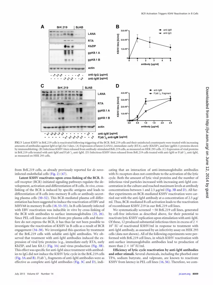
Latent KSHV reactivates upon cross-linking of the BCR. B-cell receptor (BCR)-initiated signaling pathways regulate the de-velopment, activation and differentiation of B cells. In vivo, cross-linking of the BCR is induced by specific antigens and leads todifferentiation of B cells into memory B cells or antibody-secret-ing plasma cells (50–52). This BCR-mediated plasma cell differ-entiation has been suggested to induce the reactivation of EBV andMHV68 in memory B cells (18, 53–55). In B cells latently infectedwith EBV reactivation was inducible in vitro by cross-linking ofthe BCR with antibodies to surface immunoglobulins (25, 26).Since PEL cell lines are derived from pre-plasma cells and there-fore do not express the BCR, it has thus far not been possible toinvestigate the reactivation of KSHV in B cells as a result of BCRengagement (34–38). We investigated this question by treatmentof the BrK.219 cells with soluble anti-IgM antibodies. We ob-served that treatment with anti-IgM antibodies induced the ex-pression of viral lytic proteins (e.g., immediate-early RTA, earlyKbZIP, and late K8.1) (Fig. 3A) and virus production (Fig. 3B).This effect was specific for anti-IgM since treatment with antibod-ies to IgG did not induce the KSHV lytic cycle in the BrK.219 cells(Fig. 3A and B). F(ab=)2 fragments of anti-IgM antibodies were aseffective as complete anti-IgM antibodies (Fig. 3C and D), indi-
cating that an interaction of anti-immunoglobulin antibodieswith Fc receptors does not contribute to the activation of the lyticcycle. Both the amount of lytic viral proteins and the number ofinfectious viral particles increased with increasing anti-IgM con-centration in the culture and reached maximum levels at antibodyconcentrations between 1 and 2.5 �g/ml (Fig. 3B and D). All fur-ther experiments on BCR-mediated KSHV reactivation were car-ried out with the anti-IgM antibody at a concentration of 2.5 �g/ml. Thus, BCR-mediated B cell activation leads to the reactivationof recombinant KSHV.219 in our BrK.219 cell lines.
We systematically screened �50 BrK.219 cell lines, generatedby cell-free infection as described above, for their potential toreactivate lytic KSHV replication upon stimulation with anti-IgM.Of these, 12 produced substantial infectious titers in excess of 2 �105 IU of reactivated KSHV/ml in response to treatment withanti-IgM antibody, as assessed by an infectivity assay on HEK 293cells (data not shown). All of the following experiments were per-formed with BrK.219 cell lines, in which KSHV reactivation withanti-surface immunoglobulin antibodies lead to production ofmore than 2 � 105 IU/ml.
Efficiency of lytic cycle reactivation by anti-IgM antibodiesand other stimuli. Several chemicals, including the phorbol esterTPA, sodium butyrate, and valproate, are known to reactivateKSHV from latency in PEL cell lines (16, 56). Therefore, we com-
FIG 3 Latent KSHV in BrK.219 cells is reactivated following triggering of the BCR. BrK.219 cells and their uninfected counterparts were treated with increasingamounts of antibodies against IgM or IgG for 3 days. (A) Expression of latent (LANA), immediate-early (RTA), early (KbZIP), and late (gpK8.1) proteins shownby immunoblotting. (B) Infectious KSHV titers released from antibody-stimulated BrK.219 cells, as measured on HEK 293 cells. (C) Expression of viral proteinsin BrK.219 cells treated with anti-IgM and F(ab=)2 anti-IgM. (D) Infectious KSHV titers released from BrK.219 cells treated with anti-IgM or F(ab=)2 anti-IgM,as measured on HEK 293 cells.
BCR Activation Triggers KSHV Reactivation in B Cells
July 2013 Volume 87 Number 14 jvi.asm.org 8009
on June 12, 2014 by guesthttp://jvi.asm
.org/D
ownloaded from
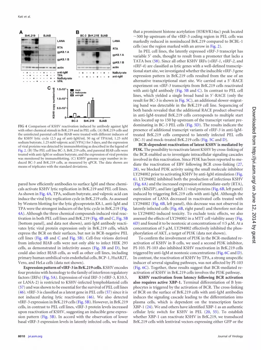
pared how efficiently antibodies to surface IgM and these chemi-cals activate KSHV lytic replication in BrK.219 and PEL cell lines.As shown in Fig. 4A, TPA, sodium butyrate, and valproic acid caninduce the viral lytic replication cycle in BrK.219 cells. As assessedby Western blotting for the lytic glycoprotein K8.1, anti-IgM andTPA were the strongest inducers of the lytic cycle in BrK.219 (Fig.4A). Although the three chemical compounds induced viral reac-tivation in both PEL cell lines and BrK.219 (Fig. 4B and C, Fig. 5B[bottom panel], and data not shown), anti-IgM antibody reacti-vates lytic viral protein expression only in BrK.219 cells, whichexpress the BCR on their surfaces, but not in BCR-negative PELcell lines (Fig. 4B and C and Fig. 5B). Cell-free virions derivedfrom infected BJAB cells were not only able to infect HEK 293cells, as demonstrated in infectivity assays (Fig. 3B and D), butcould also infect BJAB cells, as well as other cell lines, includingprimary human umbilical vein endothelial cells, BCP-1, HuAR2T,Vero, and HeLa cells (data not shown).
Expression pattern of vIRF-3 in BrK.219 cells. KSHV encodesfour proteins with homology to the family of interferon regulatoryfactors (IRFs) (Fig. 5A). Expression of viral IRF-3 (vIRF-3, K10.5,or LANA-2) is restricted to KSHV-infected lymphoblastoid cells(57) and was shown to be essential for the survival of PEL cell lines(46). vIRF-3 is classified as a latent gene in PEL cells (57) since it isnot induced during lytic reactivation (46). We also detectedvIRF-3 expression in BrK.219 cells (Fig. 5B). However, in BrK.219cells, in contrast to PEL cell lines, vIRF-3 protein levels increasedupon reactivation of KSHV, suggesting an inducible gene expres-sion pattern (Fig. 5B). In accord with the observation of lowerbasal vIRF-3 expression levels in latently infected cells, we found
that a prominent histone acetylation (H3K9/K14ac) peak located�500 bp upstream of the vIRF-3 coding region in PEL cells wasmarkedly reduced in noninduced BrK.219 compared to BCBL-1cells (see the region marked with an arrow in Fig. 2).
In PEL cell lines, the latently expressed vIRF-3 transcript hasvariable 5= ends, thought to result from a promoter that lacks aTATA box (58). Since all other KSHV IRFs (vIRF-1, vIRF-2, andvIRF-4) are classified as lytic genes with a well-defined transcrip-tional start site, we investigated whether the inducible vIRF-3 geneexpression pattern in BrK.219 cells resulted from the use of analternative transcriptional start site. We carried out a 5=-RACEexperiment on vIRF-3 transcripts from BrK.219 cells reactivatedwith anti-IgM antibody (Fig. 5B and C). In contrast to PEL celllines, which yielded a single broad band in 5=-RACE (only theresult for BC-3 is shown in Fig. 5C), an additional slower-migrat-ing band was detectable in the BrK.219 cell line. Sequencing ofboth bands revealed that the additional RACE product observedin anti-IgM-treated BrK.219 cells corresponds to multiple startsites located up to 150 bp upstream of the transcript variant pre-dominating in BC-3 PEL cells (Fig. 5D). The results indicate thepresence of additional transcript variants of vIRF-3 in anti-IgM-treated BrK.219 cells compared to latently infected PEL cells(BC-3) and mock-treated BrK.219 cells (Fig. 5C and D).
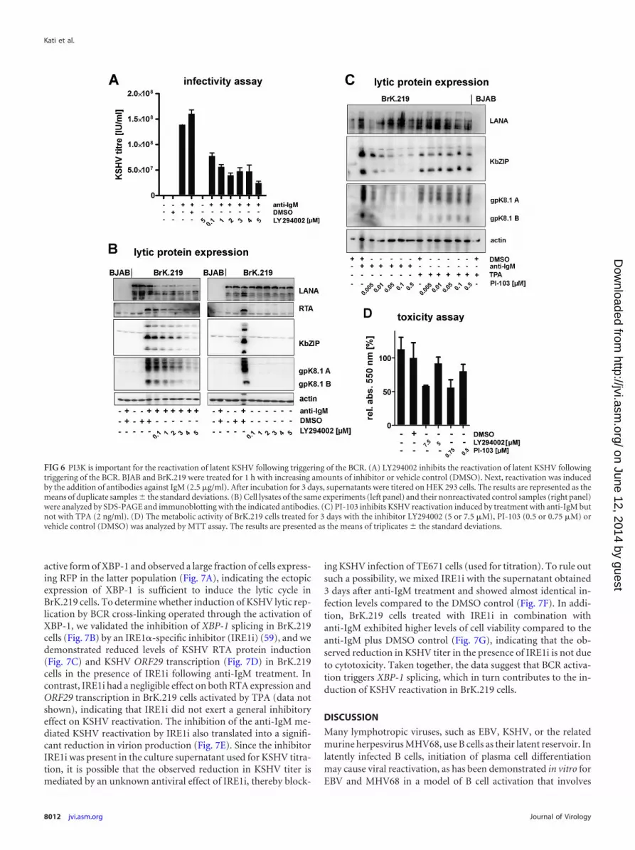
BCR-dependent reactivation of latent KSHV is mediated byPI3K. The possibility to reactivate latent KSHV by cross-linking ofthe BCR enabled us to investigate intracellular signaling cascadesinvolved in this reactivation. Since PI3K has been reported to me-diate the reactivation of EBV following BCR cross-linking (27,28), we blocked PI3K activity using the small molecule inhibitorLY294002 prior to activating KSHV by anti-IgM stimulation (Fig.6). LY294002 inhibited both the production of infectious KSHV(Fig. 6A) and the increased expression of immediate-early (RTA),early (KbZIP), and late (gpK8.1) viral proteins (Fig. 6B, left panel)induced by triggering BrK.219 cells with anti-IgM. Although theexpression of LANA decreased in reactivated cells treated withLY294002 (Fig. 6B, left panel), this decrease was not observed inlatently infected cells (Fig. 6B, right panel) and therefore not dueto LY294002-induced toxicity. To exclude toxic effects, we alsoassessed the effects of LY294002 in a MTT cell viability assay (Fig.6D) and found it to be nontoxic at concentrations of �5 �M. At aconcentration of 5 �M, LY294002 effectively inhibited the phos-phorylation of AKT, a target of PI3K (data not shown).
To confirm the involvement of PI3K in the BCR-mediated re-activation of KSHV in B cells, we used a second PI3K inhibitor,PI-103. PI-103 also inhibited KSHV reactivation in BrK.219 cellstriggered by anti-IgM at nontoxic concentrations (Fig. 6C and D).In contrast, the reactivation of KSHV by TPA, a strong unspecificinducer of several signaling pathways, was not affected by PI-103(Fig. 6C). Together, these results suggest that BCR-mediated re-activation of KSHV in BrK.219 cells involves the PI3K pathway.
KSHV reactivation from latency following BCR activationalso requires active XBP-1. Terminal differentiation of B lym-phocytes is triggered by the activation of BCR. The cross-linkingof BCR on the surface of BrK.219 cells with anti-IgM antibodiesinduces the signaling cascade leading to the differentiation intoplasma cells, which is dependent on the transcription factorXBP-1 (24). We and others have identified XBP-1 as an authenticcellular lytic switch for KSHV in PEL (20, 55). To establishwhether XBP-1 can reactivate KSHV in BrK.219, we transducedBrK.219 cells with lentiviral vectors expressing either GFP or the
FIG 4 Comparison of KSHV reactivation induced by antibody against IgMwith other chemical stimuli in BrK.219 and in PEL cells. (A) BrK.219 cells andthe uninfected parental cell line BJAB were treated with different inducers ofthe KSHV lytic cycle (2.5 �g of anti-IgM/ml, 50 ng of TPA/ml, 1.25 mMsodium butyrate, 1.25 mM valproic acid [VPA]) for 3 days, and the expressionof viral proteins was detected by immunoblotting as described in the legend ofFig. 2. (B) The PEL cell line BC-3, BrK.219 cells, and parental BJAB cells weretreated with anti-IgM or sodium butyrate, and the expression of viral proteinswas monitored by immunoblotting. (C) KSHV genome copy number in in-duced BC-3 and BrK.219 cells, as measured by qPCR. The data shown aremeans of triplicates with the standard deviations.
Kati et al.
8010 jvi.asm.org Journal of Virology
on June 12, 2014 by guesthttp://jvi.asm
.org/D
ownloaded from
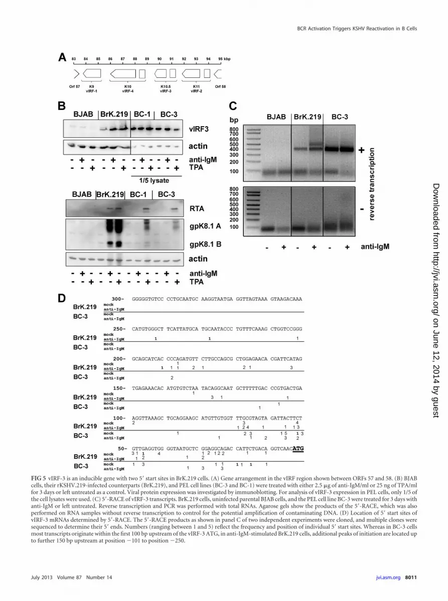
FIG 5 vIRF-3 is an inducible gene with two 5= start sites in BrK.219 cells. (A) Gene arrangement in the vIRF region shown between ORFs 57 and 58. (B) BJABcells, their rKSHV.219-infected counterparts (BrK.219), and PEL cell lines (BC-3 and BC-1) were treated with either 2.5 �g of anti-IgM/ml or 25 ng of TPA/mlfor 3 days or left untreated as a control. Viral protein expression was investigated by immunoblotting. For analysis of vIRF-3 expression in PEL cells, only 1/5 ofthe cell lysates were used. (C) 5=-RACE of vIRF-3 transcripts. BrK.219 cells, uninfected parental BJAB cells, and the PEL cell line BC-3 were treated for 3 days withanti-IgM or left untreated. Reverse transcription and PCR was performed with total RNAs. Agarose gels show the products of the 5=-RACE, which was alsoperformed on RNA samples without reverse transcription to control for the potential amplification of contaminating DNA. (D) Location of 5= start sites ofvIRF-3 mRNAs determined by 5=-RACE. The 5=-RACE products as shown in panel C of two independent experiments were cloned, and multiple clones weresequenced to determine their 5= ends. Numbers (ranging between 1 and 5) reflect the frequency and position of individual 5= start sites. Whereas in BC-3 cellsmost transcripts originate within the first 100 bp upstream of the vIRF-3 ATG, in anti-IgM-stimulated BrK.219 cells, additional peaks of initiation are located upto further 150 bp upstream at position �101 to position �250.
BCR Activation Triggers KSHV Reactivation in B Cells
July 2013 Volume 87 Number 14 jvi.asm.org 8011
on June 12, 2014 by guesthttp://jvi.asm
.org/D
ownloaded from
active form of XBP-1 and observed a large fraction of cells express-ing RFP in the latter population (Fig. 7A), indicating the ectopicexpression of XBP-1 is sufficient to induce the lytic cycle inBrK.219 cells. To determine whether induction of KSHV lytic rep-lication by BCR cross-linking operated through the activation ofXBP-1, we validated the inhibition of XBP-1 splicing in BrK.219cells (Fig. 7B) by an IRE1�-specific inhibitor (IRE1i) (59), and wedemonstrated reduced levels of KSHV RTA protein induction(Fig. 7C) and KSHV ORF29 transcription (Fig. 7D) in BrK.219cells in the presence of IRE1i following anti-IgM treatment. Incontrast, IRE1i had a negligible effect on both RTA expression andORF29 transcription in BrK.219 cells activated by TPA (data notshown), indicating that IRE1i did not exert a general inhibitoryeffect on KSHV reactivation. The inhibition of the anti-IgM me-diated KSHV reactivation by IRE1i also translated into a signifi-cant reduction in virion production (Fig. 7E). Since the inhibitorIRE1i was present in the culture supernatant used for KSHV titra-tion, it is possible that the observed reduction in KSHV titer ismediated by an unknown antiviral effect of IRE1i, thereby block-
ing KSHV infection of TE671 cells (used for titration). To rule outsuch a possibility, we mixed IRE1i with the supernatant obtained3 days after anti-IgM treatment and showed almost identical in-fection levels compared to the DMSO control (Fig. 7F). In addi-tion, BrK.219 cells treated with IRE1i in combination withanti-IgM exhibited higher levels of cell viability compared to theanti-IgM plus DMSO control (Fig. 7G), indicating that the ob-served reduction in KSHV titer in the presence of IRE1i is not dueto cytotoxicity. Taken together, the data suggest that BCR activa-tion triggers XBP-1 splicing, which in turn contributes to the in-duction of KSHV reactivation in BrK.219 cells.
DISCUSSION
Many lymphotropic viruses, such as EBV, KSHV, or the relatedmurine herpesvirus MHV68, use B cells as their latent reservoir. Inlatently infected B cells, initiation of plasma cell differentiationmay cause viral reactivation, as has been demonstrated in vitro forEBV and MHV68 in a model of B cell activation that involves
FIG 6 PI3K is important for the reactivation of latent KSHV following triggering of the BCR. (A) LY294002 inhibits the reactivation of latent KSHV followingtriggering of the BCR. BJAB and BrK.219 were treated for 1 h with increasing amounts of inhibitor or vehicle control (DMSO). Next, reactivation was inducedby the addition of antibodies against IgM (2.5 �g/ml). After incubation for 3 days, supernatants were titered on HEK 293 cells. The results are represented as themeans of duplicate samples � the standard deviations. (B) Cell lysates of the same experiments (left panel) and their nonreactivated control samples (right panel)were analyzed by SDS-PAGE and immunoblotting with the indicated antibodies. (C) PI-103 inhibits KSHV reactivation induced by treatment with anti-IgM butnot with TPA (2 ng/ml). (D) The metabolic activity of BrK.219 cells treated for 3 days with the inhibitor LY294002 (5 or 7.5 �M), PI-103 (0.5 or 0.75 �M) orvehicle control (DMSO) was analyzed by MTT assay. The results are presented as the means of triplicates � the standard deviations.
Kati et al.
8012 jvi.asm.org Journal of Virology
on June 12, 2014 by guesthttp://jvi.asm
.org/D
ownloaded from

cross-linking of the B cell receptor (BCR) with anti-immunoglob-ulins (25, 26, 31).
De novo KSHV infection occurs in CD19-positive B cells (60),especially in IgM(�) B cells (61). The commonly used model sys-tem for KSHV latency, the PEL cells, are (post)germinal center(GC)-derived B cells, displaying only a few markers of B cell dif-ferentiation (62, 63). They lack the BCR on their surface but typ-ically express the postgerminal center B cell marker CD138 (syn-decan-1), suggesting that they are in a pre-plasma cell-like stage(34–38). In contrast, the human germinal center-like BJAB lym-phoma cells (64), used to establish our system, are mature B cells
with the expression of IgM (65) and CD19 on their surface. Con-sequently, KSHV-infected BJAB cells might serve as a model toinvestigate KSHV effects in mature B cells that respond to trigger-ing of their surface IgM receptor by cognate antigen. Here, weshow for the first time that BCR activation also triggers KSHVreactivation and that this involves a PI3K-dependent signalingpathway and the active XBP-1.
Most of the research conducted on KSHV reactivation in Bcells has thus far been carried out in cell lines established from PELsamples. These lines are stably infected with KSHV, and the virallytic replication cycle can be induced with chemical compounds
FIG 7 Lytic KSHV induction following BCR activation requires the splicing of XBP-1 mRNA. (A) Confocal images of BrK.219 cells transduced with lentiviralvectors expressing either GFP (“control”) only or the spliced form of XBP-1 (“active XBP-1”). Expression of RFP marks RTA-expressing BrK.219 cells. (B)RT-PCR analysis of the DTT-induced splicing of XBP-1 transcript in BrK.219 cells with or without treatment with IRE1� inhibitor (IRE1i; 25 �M). cDNAproducts derived from the spliced XBP-1 mRNA are resistant to PstI digestion (upper band, 223 bp), whereas the products for the unspliced XBP-1 mRNA arecleaved (the two lower bands, 104 and 145 bp). WB denotes water blank control. The band sizes (in bp) of the DNA ladder are as indicated. (C) Western blotanalysis of KSHV RTA induction by anti-IgM antibodies (1 �g/ml) with or without IRE1i (25 �M) treatment. (D) Semiquantitative RT-PCR analysis of a KSHVlate gene (ORF29) expression at 48 h posttreatment (upper panel), using serially 10-fold-diluted cDNA obtained from BrK.219 cells treated with either DMSO,anti-IgM antibodies (1 �g/ml), or anti-IgM antibodies plus IRE1i (25 �M). Input cDNA was normalized to a cellular gene (�2M lower panel). Primers for bothvirus and cellular genes span across intron(s), and no amplification of genomic DNA was observed under the PCR conditions used. The data from onerepresentative experiment out of two independent experiments are shown. (E) Relative KSHV titer in supernatants from BrK.219 cells 3 days after BCRcross-linking, with or without the presence of IRE1i. Cell supernatants from BrK.219 cells treated with either DMSO, anti-IgM, or anti-IgM plus IRE1i weretitrated on TE671 cells. The data are shown as the relative infectious titer where all samples were normalized to the positive control (anti-IgM) as 100% andplotted as means � the standard deviations from three independent experiments. (F) Effect of IRE1i on the relative KSHV titer of anti-IgM induced supernatantfrom BrK.219 cells. The cell supernatant from BrK.219 cells induced with anti-IgM was split into two, and each half mixed with either DMSO or IRE1i; theresulting supernatants were titrated on TE671 cells. The data are shown as relative infectious titer where all samples were normalized to the positive control(anti-IgM plus DMSO) as 100% and plotted as means � the standard deviations from three independent experiments. (G) The relative cell viability of BrK.219cells treated with either DMSO, IRE1i, anti-IgM plus DMSO, or anti-IgM plus IRE1i is plotted as the percent concentration of ATP (proportional to the numberof live cells) normalized to the DMSO control as 100%. The data (means � the standard deviations) from one representative experiment out of two independentexperiments are shown.
BCR Activation Triggers KSHV Reactivation in B Cells
July 2013 Volume 87 Number 14 jvi.asm.org 8013
on June 12, 2014 by guesthttp://jvi.asm
.org/D
ownloaded from

such as TPA, sodium butyrate, and valproate and/or the overex-pression of the viral lytic cycle activator RTA (16, 56, 66, 67).However, owing to their pre-plasma cell differentiation stage (34–38) and consequent absence of surface IgM, the role of the BCRcannot be studied in PEL cells.
We therefore generated BJAB cell lines stably infected withrecombinant KSHV.219 using either a cell-free infection protocol(see Materials and Methods) or cell-cell transmission (data notshown). The KSHV genome copy number in the derived BrK.219cell lines is comparable to PEL cell lines (Fig. 4C), but the stablepersistence of viral episomes requires continuous selection withpuromycin (Fig. 1D). This resembles what has been observed withother cell lines that were experimentally infected with KSHV. Ithas been suggested that epigenetic modifications of the latent viralgenome may be responsible for successful persistence in the ab-sence of a selection marker (49), but the nature of such modifica-tions is still unknown. Similar to de novo-infected endothelial cells(47), KSHV episomes in latently infected BrK.219 cells adopt epi-genetic profiles that are akin to those observed in PEL lines. No-tably, activation-associated histone modifications are not simplyexcluded from lytic gene regions during latency. Rather, latentepisomes acquire abundant H3K27-me3, a facultative hetero-chromatin mark that mediates transcriptional repression by poly-comb group (PcG) proteins. A number of loci, including theORF50/Rta promoter, become bivalently modified (i.e., they carryboth activating H3K4-me3 and repressive H3K27-me3 marks)and hence are repressed but remain “poised” for rapid reactiva-tion. Interestingly, although a few regions exhibit H3K9 trimethy-lation (H3K9-me3) in PEL cells, this constitutive heterochroma-tin mark is largely absent from de novo-infected BrK.219 andendothelial cells (Fig. 2) (47). These data suggest that H3K9-me3is not acquired during primary latency establishment or within thefirst few weeks of a de novo infection but rather may progressivelyreplace H3K27-me3 during long-term latent infection at a selectnumber of loci, potentially leading to further stabilization of virallatency.
Treatment of BrK.219 cells with anti-IgM, but not anti-IgGresults in a strong reactivation of KSHV, as demonstrated by theexpression of immediate-early (RTA), early (KbZIP), or late(gpK8.1) viral proteins and the production of significant titers ofinfectious KSHV (Fig. 3 and 4). The amount of infectious progenyproduced after stimulation with anti-IgM ranges from 2 � 105 to106 IU/ml in 12 of �50 independently established KSHV-infectedB cell lines (data not shown). Reactivation of BrK.219 cells is twiceor even three times more efficient than reactivation of the PEL celllines BC-1 and BC-3, measured by the KSHV copy numbers percell (data not shown) and by the expression of viral lytic proteins(Fig. 5B, lower panel). However, reactivation of our most potentKSHV production cell line, Vero rKSHV.219, yielded KSHV copynumbers per cell that were up to 15 to 20 times those found inreactivated BrK.219 (data not shown). When examining the ex-pression of viral proteins in BrK.219 cell lines, we noticed thatvIRF-3/K10.5/LANA-2, a constitutively expressed latent proteinin PEL cells (57, 68), is more strongly expressed after activation ofthe lytic replication cycle in BrK.219 cells. This is accompanied bythe expression of a vIRF-3/K10.5 transcript with an extended 5=-untranslated region (Fig. 5). Similar to the latent vIRF-3 transcriptin PEL cells, where expression is controlled by a promoter lackinga TATA box and does not initiate at a single, well-defined tran-scriptional start site (58), the “lytic” vIRF-3/K10.5 transcript has
multiple start sites located �100 bp upstream of the start sitesmost commonly used by the latent transcript in PEL cells (Fig.5D). The inducible gene expression pattern in BrK.219 cells isaccompanied by a reduced H3K9/K14 acetylation upstream of thevIRF-3 gene (Fig. 2). Since all other KSHV vIRFs show increasedexpression after activation of the lytic replication cycle (68–72),and since vIRF-3 expression has been shown to be crucial for thesurvival of PEL cells (46), we speculate that the “default” expres-sion pattern of vIRF-3 in B cells may be similar to BrK.219 cells(i.e., may be inducible) but that its expression has been fixed in aconstitutive manner in PEL cells by increased H3K9/K14 acetyla-tion to allow their survival and expansion as a monoclonal B cellpopulation. In this, the constitutive expression of vIRF-3 in PELcells would resemble the EBV latent Wp promoter-driven expres-sion of the lytic viral bcl-2 homologue, BHRF1, which has beenshown to result from alternative splicing and contribute to thesurvival of Burkitt’s lymphoma cells (73).
It is well established that BCR stimulation/B cell activationleads to plasma cell differentiation. Several studies on both EBVand KSHV focused on cellular proteins involved in plasma celldifferentiation to understand their role in both inhibition andinitiation of viral reactivation (74). A recent report showed thatEBV latent membrane protein 1 (LMP-1) downregulates the B-lymphocyte-induced maturation protein 1 (BLIMP-1) expressionin germinal center (GC) B cells and thereby inhibits their differ-entiation into plasma cells and subsequently the reactivation ofEBV (75). In the case of MHV68, it was shown that the viral pro-tein M2 can directly induce the expression of cellular proteins forplasma cell differentiation and thus viral reactivation (54). In vivoBCR-mediated initiation of plasma cell differentiation dependson IRE-1 and XBP-1, both of which are important mediators ofBCR signaling (23, 24). Ectopic expression of the active XBP-1 haspreviously been demonstrated to promote KSHV reactivation inPEL cell lines (20, 55).
Consistent with these reports, we show that XBP-1 is also re-quired for the reactivation of KSHV in BCR-positive B cells fol-lowing BCR stimulation. It is possible that PI3K and XBP-1 act ina common pathway, since recent publications demonstrate an in-teraction of the regulatory PI3K subunits with the active XBP-1(32, 33). This promotes the unfolded protein response (32, 33),leading to the initiation of plasma cell differentiation in primary Bcells and, in the case of BrK.219 cells, lytic replication of KSHV.
Based on these findings, we established an infection model sys-tem that allows investigation of the entire process of KSHV reac-tivation starting from BCR cross-linking, continuing through sig-naling cascades involving partners such as PI3K and XBP-1, andgoing all the way to infectious virion production.
ACKNOWLEDGMENTS
We thank Christian Michaelis and Jessica Rückert for their support withDNA preparations, Sabine Hübner for qPCR to analyze CRP and KSHVcopy numbers per cell, Sandra Flucht and Jenny Witthuhn for sequencing,and Martin Mynarek for the critical revision of the manuscript. We alsothank Jeffrey Vieira and Patricia O’Hearn for the rKSHV.219 construct.The vIRF-3 antibody was a generous gift from Elisabeth Kremmer andFrank Neipel.
This study was supported by the Collaborative Research Center 900 ofthe Deutsche Forschungsgemeinschaft and the European Union Inte-grated Project INCA (LSHC-CT-2005-018704).
Kati et al.
8014 jvi.asm.org Journal of Virology
on June 12, 2014 by guesthttp://jvi.asm
.org/D
ownloaded from
REFERENCES1. Cesarman E. 2011. Gammaherpesvirus and lymphoproliferative disor-
ders in immunocompromised patients. Cancer Lett. 305:163–174.2. Taylor GS, Blackbourn DJ. 2011. Infectious agents in human cancers:
lessons in immunity and immunomodulation from gammaherpesvirusesEBV and KSHV. Cancer Lett. 305:263–278.
3. Saha A, Robertson ES. 2011. Epstein-Barr virus-associated B-cell lym-phomas: pathogenesis and clinical outcomes. Clin. Cancer Res. 17:3056 –3063.
4. Cesarman E, Chang Y, Moore PS, Said JW, Knowles DM. 1995. Kaposi’ssarcoma-associated herpesvirus-like DNA sequences in AIDS-relatedbody-cavity-based lymphomas. N. Engl. J. Med. 332:1186 –1191.
5. Soulier J, Grollet L, Oksenhendler E, Cacoub P, Cazals-Hatem D,Babinet P, d’Agay MF, Clauvel JP, Raphael M, Degos L. 1995. Kaposi’ssarcoma-associated herpesvirus-like DNA sequences in multicentricCastleman’s disease. Blood 86:1276 –1280.
6. Nealy MS, Coleman CB, Li H, Tibbetts SA. 2010. Use of a virus-encodedenzymatic marker reveals that a stable fraction of memory B cells expresseslatency-associated nuclear antigen throughout chronic gammaherpesvi-rus infection. J. Virol. 84:7523–7534.
7. Thorley-Lawson DA. 2001. Epstein-Barr virus: exploiting the immunesystem. Nat. Rev. 1:75– 82.
8. Pope JH, Scott W, Moss DJ. 1973. Human lymphoid cell transformationby Epstein-Barr virus. Nat. New Biol. 246:140 –141.
9. Bechtel JT, Liang Y, Hvidding J, Ganem D. 2003. Host range of Kaposi’ssarcoma-associated herpesvirus in cultured cells. J. Virol. 77:6474 – 6481.
10. Blackbourn DJ, Lennette E, Klencke B, Moses A, Chandran B, Wein-stein M, Glogau RG, Witte MH, Way DL, Kutzkey T, Herndier B, LevyJA. 2000. The restricted cellular host range of human herpesvirus 8. AIDS14:1123–1133.
11. Renne R, Blackbourn D, Whitby D, Levy J, Ganem D. 1998. Limitedtransmission of Kaposi’s sarcoma-associated herpesvirus in cultured cells.J. Virol. 72:5182–5188.
12. Chen L, Lagunoff M. 2005. Establishment and maintenance of Kaposi’ssarcoma-associated herpesvirus latency in B cells. J. Virol. 79:14383–14391.
13. Myoung J, Ganem D. 2011. Infection of lymphoblastoid cell lines byKaposi’s sarcoma-associated herpesvirus: critical role of cell-associatedvirus. J. Virol. 85:9767–9777.
14. Cho H, Kang H. 2012. KSHV infection of B-cell lymphoma using amodified KSHV BAC36 and coculturing system. J. Microbiol. (Seoul) 50:285–292.
15. Luka J, Kallin B, Klein G. 1979. Induction of the Epstein-Barr virus(EBV) cycle in latently infected cells by n-butyrate. Virology 94:228 –231.
16. Renne R, Zhong W, Herndier B, McGrath M, Abbey N, Kedes D,Ganem D. 1996. Lytic growth of Kaposi’s sarcoma-associated herpesvirus(human herpesvirus 8) in culture. Nat. Med. 2:342–346.
17. zur Hausen H, O’Neill FJ, Freese UK, Hecker E. 1978. Persisting onco-genic herpesvirus induced by the tumor promoter TPA. Nature 272:373–375.
18. Bhende PM, Dickerson SJ, Sun X, Feng WH, Kenney SC. 2007. X-box-binding protein 1 activates lytic Epstein-Barr virus gene expression incombination with protein kinase D. J. Virol. 81:7363–7370.
19. Dalton-Griffin L, Wilson SJ, Kellam P. 2009. X-box binding protein 1contributes to induction of the Kaposi’s sarcoma-associated herpesviruslytic cycle under hypoxic conditions. J. Virol. 83:7202–7209.
20. Wilson SJ, Tsao EH, Webb BL, Ye H, Dalton-Griffin L, Tsantoulas C,Gale CV, Du MQ, Whitehouse A, Kellam P. 2007. X box binding proteinXBP-1s transactivates the Kaposi’s sarcoma-associated herpesvirus(KSHV) ORF50 promoter, linking plasma cell differentiation to KSHVreactivation from latency. J. Virol. 81:13578 –13586.
21. Yu F, Feng J, Harada JN, Chanda SK, Kenney SC, Sun R. 2007. B cellterminal differentiation factor XBP-1 induces reactivation of Kaposi’s sar-coma-associated herpesvirus. FEBS Lett. 581:3485–3488.
22. Calfon M, Zeng H, Urano F, Till JH, Hubbard SR, Harding HP, ClarkSG, Ron D. 2002. IRE1 couples endoplasmic reticulum load to secretorycapacity by processing the XBP-1 mRNA. Nature 415:92–96.
23. Iwakoshi NN, Lee AH, Glimcher LH. 2003. The X-box binding protein-1transcription factor is required for plasma cell differentiation and the un-folded protein response. Immunol. Rev. 194:29 –38.
24. Reimold AM, Iwakoshi NN, Manis J, Vallabhajosyula P, Szomolanyi-Tsuda E, Gravallese EM, Friend D, Grusby MJ, Alt F, Glimcher LH.
2001. Plasma cell differentiation requires the transcription factor XBP-1.Nature 412:300 –307.
25. Takada K. 1984. Cross-linking of cell surface immunoglobulins inducesEpstein-Barr virus in Burkitt lymphoma lines. Int. J. Cancer 33:27–32.
26. Tovey MG, Lenoir G, Begon-Lours J. 1978. Activation of latent Epstein-Barr virus by antibody to human IgM. Nature 276:270 –272.
27. Goswami R, Gershburg S, Satorius A, Gershburg E. 2012. Protein kinaseinhibitors that inhibit induction of lytic program and replication of Ep-stein-Barr virus. Antivir. Res. 96:296 –304.
28. Iwakiri D, Takada K. 2004. Phosphatidylinositol 3-kinase is a determi-nant of responsiveness to B cell antigen receptor-mediated Epstein-Barrvirus activation. J. Immunol. 172:1561–1566.
29. Limon JJ, Fruman DA. 2012. Akt and mTOR in B cell activation anddifferentiation. Front. Immunol. 3:228.
30. Pauls SD, Lafarge ST, Landego I, Zhang T, Marshall AJ. 2012. Thephosphoinositide 3-kinase signaling pathway in normal and malignant Bcells: activation mechanisms, regulation, and impact on cellular functions.Front. Immunol. 3:224.
31. Moser JM, Upton JW, Gray KS, Speck SH. 2005. Ex vivo stimulation ofB cells latently infected with gammaherpesvirus 68 triggers reactivationfrom latency. J. Virol. 79:5227–5231.
32. Park SW, Zhou Y, Lee J, Lu A, Sun C, Chung J, Ueki K, Ozcan U. 2010.The regulatory subunits of PI3K, p85alpha and p85beta, interact withXBP-1 and increase its nuclear translocation. Nat. Med. 16:429 – 437.
33. Winnay JN, Boucher J, Mori MA, Ueki K, Kahn CR. 2010. A regulatorysubunit of phosphoinositide 3-kinase increases the nuclear accumulationof X-box-binding protein-1 to modulate the unfolded protein response.Nat. Med. 16:438 – 445.
34. Carbone A, Gaidano G. 1997. HHV-8-positive body-cavity-based lym-phoma: a novel lymphoma entity. Br. J. Haematol. 97:515–522.
35. Gaidano G, Carbone A. 2001. Primary effusion lymphoma: a liquid phaselymphoma of fluid-filled body cavities. Adv. Cancer Res. 80:115–146.
36. Gaidano G, Gloghini A, Gattei V, Rossi MF, Cilia AM, Godeas C, DeganM, Perin T, Canzonieri V, Aldinucci D, Saglio G, Carbone A, Pinto A.1997. Association of Kaposi’s sarcoma-associated herpesvirus-positiveprimary effusion lymphoma with expression of the CD138/syndecan-1antigen. Blood 90:4894 – 4900.
37. Jenner RG, Maillard K, Cattini N, Weiss RA, Boshoff C, Wooster R,Kellam P. 2003. Kaposi’s sarcoma-associated herpesvirus-infected pri-mary effusion lymphoma has a plasma cell gene expression profile. Proc.Natl. Acad. Sci. U. S. A. 100:10399 –10404.
38. O’Connell FP, Pinkus JL, Pinkus GS. 2004. CD138 (syndecan-1), aplasma cell marker immunohistochemical profile in hematopoietic andnonhematopoietic neoplasms. Am. J. Clin. Pathol. 121:254 –263.
39. Vieira J, O’Hearn PM. 2004. Use of the red fluorescent protein as amarker of Kaposi’s sarcoma-associated herpesvirus lytic gene expression.Virology 325:225–240.
40. Menezes J, Leibold W, Klein G, Clements G. 1975. Establishment andcharacterization of an Epstein-Barr virus (EBC)-negative lymphoblastoidB cell line (BJA-B) from an exceptional, EBV-genome-negative AfricanBurkitt’s lymphoma. Biomedicine 22:276 –284.
41. Arvanitakis L, Mesri EA, Nador RG, Said JW, Asch AS, Knowles DM,Cesarman E. 1996. Establishment and characterization of a primary effu-sion (body cavity-based) lymphoma cell line (BC-3) harboring Kaposi’ssarcoma-associated herpesvirus (KSHV/HHV-8) in the absence of Ep-stein-Barr virus. Blood 88:2648 –2654.
42. Cesarman E, Moore PS, Rao PH, Inghirami G, Knowles DM, Chang Y.1995. In vitro establishment and characterization of two acquired immu-nodeficiency syndrome-related lymphoma cell lines (BC-1 and BC-2)containing Kaposi’s sarcoma-associated herpesvirus-like (KSHV) DNAsequences. Blood 86:2708 –2714.
43. Dedicoat M, Newton R, Alkharsah KR, Sheldon J, Szabados I, NdlovuB, Page T, Casabonne D, Gilks CF, Cassol SA, Whitby D, Schulz TF.2004. Mother-to-child transmission of human herpesvirus-8 in South Af-rica. J. Infect. Dis. 190:1068 –1075.
44. Wandinger K, Jabs W, Siekhaus A, Bubel S, Trillenberg P, Wagner H,Wessel K, Kirchner H, Hennig H. 2000. Association between clinicaldisease activity and Epstein-Barr virus reactivation in MS. Neurology 55:178 –184.
45. Lukac DM, Kirshner JR, Ganem D. 1999. Transcriptional activation bythe product of open reading frame 50 of Kaposi’s sarcoma-associated her-pesvirus is required for lytic viral reactivation in B cells. J. Virol. 73:9348 –9361.
BCR Activation Triggers KSHV Reactivation in B Cells
July 2013 Volume 87 Number 14 jvi.asm.org 8015
on June 12, 2014 by guesthttp://jvi.asm
.org/D
ownloaded from
46. Wies E, Mori Y, Hahn A, Kremmer E, Sturzl M, Fleckenstein B, NeipelF. 2008. The viral interferon-regulatory factor-3 is required for the sur-vival of KSHV-infected primary effusion lymphoma cells. Blood 111:320 –327.
47. Gunther T, Grundhoff A. 2010. The epigenetic landscape of latent Kaposisarcoma-associated herpesvirus genomes. PLoS Pathog. 6:e1000935. doi:10.1371/journal.ppat.1000935.
48. Leight ER, Sugden B. 2001. Establishment of an oriP replicon is depen-dent upon an infrequent, epigenetic event. Mol. Cell. Biol. 21:4149 – 4161.
49. Grundhoff A, Ganem D. 2004. Inefficient establishment of KSHV latencysuggests an additional role for continued lytic replication in Kaposi sar-coma pathogenesis. J. Clin. Invest. 113:124 –136.
50. Hamel KM, Liarski VM, Clark MR. 2012. Germinal center B cells. Au-toimmunity 45:333–347.
51. Shlomchik MJ, Weisel F. 2012. Germinal center selection and the devel-opment of memory B and plasma cells. Immunol. Rev. 247:52– 63.
52. Yoshida T, Mei H, Dorner T, Hiepe F, Radbruch A, Fillatreau S, HoyerBF. 2010. Memory B and memory plasma cells. Immunol. Rev. 237:117–139.
53. Laichalk LL, Thorley-Lawson DA. 2005. Terminal differentiation intoplasma cells initiates the replicative cycle of Epstein-Barr virus in vivo. J.Virol. 79:1296 –1307.
54. Liang X, Collins CM, Mendel JB, Iwakoshi NN, Speck SH. 2009.Gammaherpesvirus-driven plasma cell differentiation regulates virus re-activation from latently infected B lymphocytes. PLoS Pathog.5:e1000677. doi:10.1371/journal.ppat.1000677.
55. Sun CC, Thorley-Lawson DA. 2007. Plasma cell-specific transcriptionfactor XBP-1s binds to and transactivates the Epstein-Barr virus BZLF1promoter. J. Virol. 81:13566 –13577.
56. Shaw RN, Arbiser JL, Offermann MK. 2000. Valproic acid induceshuman herpesvirus 8 lytic gene expression in BCBL-1 cells. AIDS (Lon-don) 14:899 –902.
57. Rivas C, Thlick AE, Parravicini C, Moore PS, Chang Y. 2001. Kaposi’ssarcoma-associated herpesvirus LANA2 is a B-cell-specific latent viralprotein that inhibits p53. J. Virol. 75:429 – 438.
58. Cunningham C, Barnard S, Blackbourn DJ, Davison AJ. 2003. Tran-scription mapping of human herpesvirus 8 genes encoding viral interferonregulatory factors. J. Gen. Virol. 84:1471–1483.
59. Cross BC, Bond PJ, Sadowski PG, Jha BK, Zak J, Goodman JM,Silverman RH, Neubert TA, Baxendale IR, Ron D, Harding HP. 2012.The molecular basis for selective inhibition of unconventional mRNAsplicing by an IRE1-binding small molecule. Proc. Natl. Acad. Sci. U. S. A.109:E869 –E878.
60. Rappocciolo G, Hensler HR, Jais M, Reinhart TA, Pegu A, Jenkins FJ,Rinaldo CR. 2008. Human herpesvirus 8 infects and replicates in primarycultures of activated B lymphocytes through DC-SIGN. J. Virol. 82:4793–4806.
61. Hassman LM, Ellison TJ, Kedes DH. 2010. KSHV infects a subset ofhuman tonsillar B cells, driving proliferation and plasmablast differentia-tion. J. Clin. Invest. 121:752–768.
62. Hamoudi R, Diss TC, Oksenhendler E, Pan L, Carbone A, Ascoli V,
Boshoff C, Isaacson P, Du MQ. 2004. Distinct cellular origins of primaryeffusion lymphoma with and without EBV infection. Leukemia Res. 28:333–338.
63. Matolcsy A, Nador RG, Cesarman E, Knowles DM. 1998. Immunoglob-ulin VH gene mutational analysis suggests that primary effusion lympho-mas derive from different stages of B cell maturation. Am. J. Pathol. 153:1609 –1614.
64. Wei F, Zaprazna K, Wang J, Atchison ML. 2009. PU. 1 can recruit BCL6to DNA to repress gene expression in germinal center B cells. Mol. Cell.Biol. 29:4612– 4622.
65. Singer PA, Williamson AR. 1980. Cell surface immunoglobulin mu andgamma chains of human lymphoid cells are of higher apparent molecularweight than their secreted counterparts. Eur. J. Immunol. 10:180 –186.
66. Lukac DM, Renne R, Kirshner JR, Ganem D. 1998. Reactivation ofKaposi’s sarcoma-associated herpesvirus infection from latency by ex-pression of the ORF50 transactivator, a homolog of the EBV R protein.Virology 252:304 –312.
67. Sun R, Lin SF, Gradoville L, Yuan Y, Zhu F, Miller G. 1998. A viral genethat activates lytic cycle expression of Kaposi’s sarcoma-associated herpes-virus. Proc. Natl. Acad. Sci. U. S. A. 95:10866 –10871.
68. Fakhari FD, Dittmer DP. 2002. Charting latency transcripts in Kaposi’ssarcoma-associated herpesvirus by whole-genome real-time quantitativePCR. J. Virol. 76:6213– 6223.
69. Jenner RG, Alba MM, Boshoff C, Kellam P. 2001. Kaposi’s sarcoma-associated herpesvirus latent and lytic gene expression as revealed by DNAarrays. J. Virol. 75:891–902.
70. Moore PS, Boshoff C, Weiss RA, Chang Y. 1996. Molecular mimicry ofhuman cytokine and cytokine response pathway genes by KSHV. Science274:1739 –1744.
71. Paulose-Murphy M, Ha NK, Xiang C, Chen Y, Gillim L, Yarchoan R,Meltzer P, Bittner M, Trent J, Zeichner S. 2001. Transcription programof human herpesvirus 8 (Kaposi’s sarcoma-associated herpesvirus). J. Vi-rol. 75:4843– 4853.
72. Sarid R, Flore O, Bohenzky RA, Chang Y, Moore PS. 1998. Transcrip-tion mapping of the Kaposi’s sarcoma-associated herpesvirus (humanherpesvirus 8) genome in a body cavity-based lymphoma cell line (BC-1).J. Virol. 72:1005–1012.
73. Kelly GL, Long HM, Stylianou J, Thomas WA, Leese A, Bell AI,Bornkamm GW, Mautner J, Rickinson AB, Rowe M. 2009. An Epstein-Barr virus antiapoptotic protein constitutively expressed in transformedcells and implicated in Burkitt lymphomagenesis: the Wp/BHRF1 link.PLoS Pathog. 5:e1000341. doi:10.1371/journal.ppat.1000341.
74. Kurosaki T. 2011. Regulation of BCR signaling. Mol. Immunol. 48:1287–1291.
75. Vrzalikova K, Vockerodt M, Leonard S, Bell A, Wei W, Schrader A,Wright KL, Kube D, Rowe M, Woodman CB, Murray PG. 2011.Downregulation of BLIMP1� by the EBV oncogene, LMP-1, disrupts theplasma cell differentiation program and prevents viral replication in Bcells: implications for the pathogenesis of EBV-associated B-cell lympho-mas. Blood 117:5907–5917.
Kati et al.
8016 jvi.asm.org Journal of Virology
on June 12, 2014 by guesthttp://jvi.asm
.org/D
ownloaded from