Embed Size (px)
Citation preview

Reactivation of stalled polyribosomes insynaptic plasticityTyson E. Grabera, Sarah Hébert-Seropianb, Arkady Khoutorskyc,d, Alexandre Davide, Jonathan W. Yewdelle,Jean-Claude Lacailleb, and Wayne S. Sossina,1
aDepartment of Neurology and Neurosurgery, Montreal Neurological Institute, McGill University, Montreal, QC, Canada, H3A 2B4; bDepartment ofNeuroscience, Groupe de recherche sur le système nerveux central, Université de Montréal, Montreal, QC, Canada, H3T 1J4; cDepartment of Biochemistry anddGoodman Cancer Research Centre, McGill University, Montreal, QC, Canada, H3A 1A3; and eLaboratory of Viral Diseases, National Institute of Allergy andInfectious Diseases, National Institutes of Health, Bethesda, MD 20892
Edited* by Richard Scheller, Genentech, Inc., South San Francisco, CA, and approved August 22, 2013 (received for review April 24, 2013)
Some forms of synaptic plasticity require rapid, local activation ofprotein synthesis. Although this is thought to reflect recruitmentof mRNAs to free ribosomes, this would limit the speed andmagnitude of translational activation. Here we provide compellingin situ evidence supporting an alternative model in which synapticmRNAs are transported as stably paused polyribosomes. Remark-ably, we show that metabotropic glutamate receptor activationallows the synthesis of proteins that lead to a functional long-termdepression phenotype even when translation initiation has beengreatly reduced. Thus, neurons evolved a unique mechanism toswiftly translate synaptic mRNAs into functional protein uponsynaptic signaling using stalled polyribosomes to bypass the rate-limiting step of translation initiation. Because dysregulated plas-ticity is implicated in neurodevelopmental and psychiatric disor-ders such as fragile X syndrome, this work uncovers a uniquetranslational target for therapies.
RNA granule | mGluR-LTD | translation elongation |microtubule-associated protein 1b
Most studies of translational control focus on initiation, theprocess where mRNAs recruit ribosomes and catalyze the
first step of translation (1). This highly regulated and normallyrate-limiting step of translation is followed by elongation andtermination, resulting in completed proteins. Although multipleribosomes on a given mRNA (a polyribosome) imply active pep-tide synthesis, we and others identified neuronal RNA granules—motile aggregates of nontranslating ribosomes (2, 3). These elec-tron-dense bodies contain single copies of synaptic mRNAs thatare translationally silenced during their transport from soma tosynapse (1, 4).Many models assume that neuronally transported mRNAs are
translationally paused before completion of the initiation step oftranslation during transport. An appropriate synaptic signalwould then activate translation (initiation/elongation/termina-tion) of the granule mRNA. However, it is not clear how manyfree ribosomal subunits are present at synapses to supporttranslation initiation. Further, at a typical translation elongationrate of six amino acids per s (5, 6), synthesis of larger synapticproteins (e.g., microtubule-associated protein 1b; MAP1b) wouldtake over 5 min even if initiation were immediate. These twofactors constrain the speed and magnitude of synaptic translationand, thus, plasticity. As some forms of synaptic plasticity requirerapid (<10 min) and localized activation of protein synthesis, analternative model is wanting (7–9).We have previously proposed the concept of a neuronal RNA
granule as a stalled polyribosome (10, 11). Ribosomal stallinghas been shown to occur in lysates from a mouse neuroblastomacell line and in an in vitro rabbit reticulocyte lysate translationassay programmed with brain homogenate (12). Whether neuro-nal ribosome stalling occurs in vivo is uncertain. We hypothesizedthat neuronal RNA granules contain paused ribosomes with in-complete proteins initiated in the soma before their packagingand transport to dendrites, where translation can be rapidlyand locally completed on demand. Here we show that reactivation
of translation on stalled polyribosomes is a unique feature of theneuronal landscape that functions in metabotropic glutamatereceptor (mGluR) long-term depression (LTD), providing theneuron with the ability to rapidly and specifically respond tostimuli independently of translation initiation.
ResultsAn appropriate assay is required to determine whether stalledpolyribosomes are present in neurons and, if so, whether they aredynamically regulated by neuronal activity. We reasoned that ifpolyribosomes are stalled at the level of elongation and/or ter-mination, they should be unaffected by inhibitors that act atinitiation or during the first round of elongation at the initiationcodon, whereas those actively synthesizing protein would disso-ciate due to ribosome runoff and could not be replaced. Toperform this type of runoff assay, we required a robust method tovisualize polyribosome complexes within intact neurons. Here weuse the recently described ribopuromycylation (RPM) method(13–15) to visualize ribosomes associated with nascent peptidechains within formaldehyde-fixed primary hippocampal neurons.In this technique, live cells are coincubated with the irreversibletranslation elongation inhibitor emetine, together with puromycin,a tyrosyl-tRNA analog that covalently attaches to the carboxylterminus of nascent chains. Although puromycylation is a peptidechain-terminating event, emetine added in this context (throughan imperfectly understood mechanism) prevents puromycylatednascent chain release from ribosomes (14). This enables standard
Significance
In neurons, many mRNAs are transported to synapses in atranslationally repressed state, allowing for the spatial andtemporal regulation of protein synthesis required for synapticplasticity. It has been assumed that these mRNAs are repressedat the initiation step of translation. Here we provide evidencefor a second mechanism whereby these mRNAs are insteadrepressed at elongation/termination awaiting translational reac-tivation upon appropriate synaptic signals. Our results establishthat a form of translation-dependent synaptic plasticity, whichis dysregulated in neurodevelopmental and psychiatric patho-logies, occurs independently of translation initiation. Elucidatingthe upstream pathways that lead to repression and reac-tivation of elongation/termination on these mRNAs mayprovide new avenues for the design of therapies targetingneurodevelopmental disorders.
Author contributions: T.E.G., A.K., J.-C.L., and W.S.S. designed research; T.E.G., S.H.-S., andA.K. performed research; A.D. and J.W.Y. contributed new reagents/analytic tools; T.E.G.,S.H.-S., A.K., J.-C.L., and W.S.S. analyzed data; and T.E.G., A.K., A.D., J.W.Y., J.-C.L., andW.S.S. wrote the paper.
The authors declare no conflict of interest.
*This Direct Submission article had a prearranged editor.1To whom correspondence should be addressed. E-mail: [email protected].
This article contains supporting information online at www.pnas.org/lookup/suppl/doi:10.1073/pnas.1307747110/-/DCSupplemental.
www.pnas.org/cgi/doi/10.1073/pnas.1307747110 PNAS Early Edition | 1 of 6
NEU
ROSC
IENCE

immunofluorescence visualization of ribosome-bound nascentchains in fixed and permeabilized cells using a puromycin-specificmonoclonal antibody.
Puromycylated Puncta in Rat Hippocampal Neurons RepresentNeuronal RNA Granules. As shown in Fig. 1A, RPM staining indissociated rat hippocampal neuron cultures revealed puromy-cin-specific staining indicative of polyribosomes throughout thesoma and neurites (“−puro” and “+puro” conditions demon-strate the strict puromycin dependence on staining). Closer in-spection revealed intensely stained puncta throughout theneurites (Fig. 1B, arrowheads), consistent with electron micro-graphs of polyribosomes in neurites (2). Further characterizationof neuronal RPM staining revealed that puncta colocalize withthe RNA-specific dye Syto14 (Fig. 1C; quantified in Fig. 1 H andI), consistent with previous detection of RNA granules in liveneurons using this dye (16). Syto14 also stains mitochondria;however, the Syto14-mitochondria puncta (imaged with an an-tibody targeting translocase of outer mitochondrial membrane20 (Tom20), a mitochondrial outer-membrane protein) do notcolocalize with RPM staining (Fig. S1, arrowheads). Colocali-zation of RPM signal with ribosomal protein S6 has been pre-viously shown in a fibroblast cell line (15), and we also observedsignificant colocalization of S6 with RPM puncta in primaryneurons (Fig. 1D; quantified in Fig. 1 H and I). Further, wefound that RPM puncta colocalize with punctate fragileX mental retardation protein (FMRP), a component of RNAgranules, where it is thought to negatively regulate translation bystalling ribosome processivity (12, 17–19) (Fig. 1E; quantified in
Fig. 1 H and I). We found similar colocalization of transientlyexpressed GFP fused to the RNA-binding protein Staufen 2(GFP-Stau2), a component of an RNA granule that regulatestransport and translation of MAP1b mRNA (3, 11) (Fig. 1F;quantified in Fig. 1 H and I). RPM staining is significantly morecolocalized with all of the RNA granule markers than it is withthe mitochondrial marker Tom20 (Fig. 1G; quantified in Fig. 1 Hand I). However, not all puncta stained with the RNA granulemarkers colocalized with RPM (Fig. S2), consistent with thepresence of multiple types of RNA transport particles that do notcontain polyribosomes (10, 19). We also ruled out the possibilitythat RPM puncta reflect puromycylation on mitochondrial orcontaminating bacterial ribosomes by inhibiting prokaryotic/mi-tochondrial translation or by assessing colocalization of RPMpuncta with any bacterial DNA that might be present (Figs. S3 andS4). Together, these data demonstrate that the puromycin-stainedpuncta show characteristics of neuronal RNA granules.We next performed experiments using the human embryonic
kidney (HEK) 293T cell line to verify that we could inducerunoff of polyribosomes in intact cells (measured by RPM) aspreviously described (13–15). Because RNA granules (includingputative stalled polyribosomes) have so far been observed only inoligodendrocytes (20) and neurons (16), we did not expectstalled polyribosomes to be abundant in 293T cells. To inducerunoff, we used pateamine A (PatA), which binds the RNAhelicase eukaryotic initiation factor (eIF)4A, blocking its essen-tial function in eIF4F-mediated translation initiation (21), andhomoharringtonine (HHT), which uniquely inhibits the firstround of translation elongation by impeding translocation of the
Fig. 1. Puromycylated puncta in rat hippocampal neurons represent neuronal RNA granules. (A) Confocal images of polyribosome labeling by RPM in rathippocampal neurons. Arrows denote high-intensity puncta in neurites. (B) Magnified and straightened neurites from A with arrowheads denoting high-intensity puromycin puncta in distal locales. (C–F) Representative confocal images of neurites illustrating colocalization of RPM puncta (circles) with markersof either ribosomal RNA (Syto14) or RNA granules including ribosomal protein S6, FMRP, or GFP-Stau2. (G) Representative confocal images of neurites il-lustrating weak colocalization of RPM puncta with mitochondria (Tom20). (H) The fraction of RPM signal that colocalizes with RNA granule and mitochondrialmarkers as quantified by calculating the Manders colocalization split coefficient; n = 20 neurites from three independent experiments. (I) The correlation ofRPM puncta signal with RNA granule and mitochondrial marker signals as quantified using the Pearson correlation function; n = 50 puncta from three in-dependent experiments. All bars represent mean ± SEM, and P values were calculated using a one-way ANOVA with Dunnett’s post hoc tests.
2 of 6 | www.pnas.org/cgi/doi/10.1073/pnas.1307747110 Graber et al.

ribosome from the initiation codon while leaving downstreamribosomes unaffected, thus depleting elongating ribosomes andpreventing any further initiation (Fig. S5) (22, 23). To assess theability of PatA and HHT to inhibit peptide synthesis, we meta-bolically labeled neurons with the methionine analog L-azidoho-moalanine (AHA) for 60 min, followed by fixation and couplingof AHA-labeled proteins to a fluorophore via click chemistry (24).As expected, preincubation with either PatA or HHT severelyrepressed active protein synthesis in 293T cells by ∼85% (Fig.2A; quantified in Fig. 2B). Next, we measured protein syn-thesis at the level of the polyribosome using RPM in 293T cells.As expected, preincubation with PatA or HHT blocked a signif-icant proportion (∼70%) of cytoplasmic RPM staining, consis-tent with the majority of polyribosomes in nonneuronal cellsbeing actively engaged in protein synthesis (Fig. 2C; quantifiedin Fig. 2D). Critically, we were able to create an artificiallystalled polyribosome phenotype in 293T cells by irreversiblyblocking elongation with emetine before treatment with HHT(Fig. 2 C and D). Under these conditions, HHT was unable toreduce RPM staining, confirming that this technique reliablydetects stalled polyribosomes.
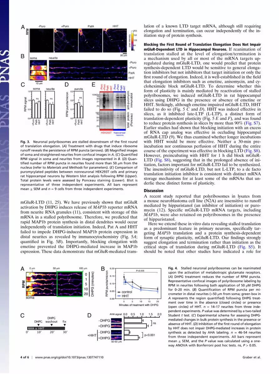
Neuronal Polyribosomes Are Stalled Downstream of the First Roundof Translation Elongation. With a reliable assay to detect stalledpolyribosomes (preincubation with PatA or HHT followed by
RPM), we next asked whether they are present in primary neu-rons. First, we confirmed that both PatA and HHT could reduceactive peptide synthesis in both the soma and neurites as mea-sured by AHA labeling (Fig. 2 E and F; quantified in Fig. 2G).To detect stalled polyribosomes, we examined whether RPMstaining was resistant to runoff by preincubating with the inhib-itors for 10 min. Despite the ability of either PatA or HHT toblock most (∼90%) ongoing translation throughout the neuron(Fig. 2G) and the ability of PatA and HHT to induce poly-ribosome runoff in 293T cells (Fig. 2D), virtually all RPMstaining in neuronal soma and neurites remained (Fig. 3 A and B;quantified in Fig. 3C). We next objectively measured the high-intensity RPM puncta by setting an intensity threshold and foundthat their number in neurites was significantly affected by neitherPatA nor HHT (Fig. 3D). Although we observed a small (∼10–20%) decrease in both diffuse and punctate RPM staining (Fig. 3C and D), the difference was not statistically significant whencorrected for multiple comparisons. Thus, a majority of neuronalpolyribosomes are stalled and resistant to runoff. Importantly,we were able to recapitulate the properties of RPM immunos-taining by immunoblotting RPM-treated cell extracts with anti-puromycin antibody (Fig. 3E). The resulting broad pattern oflabeled peptides confirmed that puromycylation indeed takesplace in both 293T cells and neurons. However, although pre-incubation with PatA or HHT reduces puromycylation in 293Tcells to near-background levels, these inhibitors failed to signif-icantly reduce puromycylation in neurons (Fig. 3E). Together,these observations point to a distinct ribosomal landscape inneurons, exemplified by the presence of polyribosomes stalled atthe level of elongation/termination.
mGluR Activation Reduces the Number of Stalled Polyribosomes andInduces Initiation-Independent Peptide Synthesis in Neurites.mGluRactivation by 3,5-dihydrophenylglycine (DHPG) induces a formof plasticity called mGluR-LTD that depends on local (i.e.,synaptic) translation. Because two mRNA-binding proteins im-plicated in mGluR-LTD, FMRP and Staufen 2, are localized toRPM puncta (Fig. 1 E and F), we hypothesized that DHPGcould reactivate stalled polyribosomes, thereby synthesizing pro-teins necessary for mGluR-LTD. Contrary to a model where DHPGwould increase the number of RPM puncta (i.e., polyribosomes)through translation initiation and elongation, our stalled poly-ribosome hypothesis would predict that DHPG would insteadcause a decrease in the number of RPM puncta as elongationand termination proceed. To test this, we applied DHPG to hip-pocampal neuronal cultures for 0, 1, 5, 10, and 20 min followed byRPM. We found a progressive decrease in the number of RPM-positive puncta per micrometer in neurites >50 μm from the cellsoma (Fig. 4A; quantified in Fig. 4B). As expected, this decreasewas also seen in the presence of HHT, suggesting that it is in-dependent of initiation events (Fig. 4B, open circles). A similardecrease in the number of puncta was also seen in proximaldendrites (Fig. S6).If DHPG induces release of stalled polyribosomes and,
moreover, if the latter represent a sizeable proportion of theribosomes present in neurites, one might expect to see an initi-ation-independent increase in protein synthesis in the presenceof DHPG. We used AHA labeling in hippocampal neurites todetermine whether DHPG could increase peptide synthesis inthe presence of HHT (see Fig. 4C for experimental schema).Despite the pronounced reduction of AHA incorporation upontreatment of neurons with HHT alone, we observed that DHPGsignificantly increased AHA labeling in the presence of HHT (Fig.4D). The loss of RPM puncta and the initiation-independent in-crease in bulk protein synthesis following DHPG treatment, isconsistent with a stimulus-dependent reactivation of stalled poly-ribosomes mediated through mGluRs.
A Concurrent Block in Translation Initiation Does Not Impair mGluR-Dependent MAP1b Protein Synthesis. MAP1b mRNA is one ofseveral transcripts whose local translation is required for
Fig. 2. Inhibitors of initiation and the first round of elongation inducepolyribosome runoff that can be detected by ribopuromycylation in a non-neuronal cell line. (A) Confocal images demonstrating that nascent poly-peptide chain labeling with the methionine analog AHA in HEK293T cells isprevented by preincubation with 100 nM PatA or 5 mM HHT. Cells wereincubated in the presence of AHA for 60 min before fixation. Omittingthe copper catalyst (−Cu) prevented fluorophore coupling to AHA peptides,thus serving as a nonspecific staining control. (B) Quantitation of cytoplasmicAHA signal from images in A. (C) RPM labeling of polyribosomes in HEK293Tcells in the presence of PatA, HHT, and emetine. Cells were pretreated withinhibitors before RPM as detailed in Materials and Methods. For “Emetine+HHT” treatment, cells were incubated with emetine for 5 min before sup-plementing with HHT for an additional 10 min. (D) Quantitation of cyto-plasmic RPM signal from images represented in C. (E) Confocal imagesdemonstrating that PatA and HHT reduce active translation (measured bymetabolic AHA labeling) in rat hippocampal neurons to the same extent asseen in HEK293T cells. (F) Magnified soma and straightened neurites fromimages in E. (G) Quantification of AHA signal in the soma and neurites fromimages represented in F. All bars represent mean ± SEM and n = 9 cells fromthree independent experiments. ***P < 0.001. ns, P > 0.05 calculated usinga one-way ANOVA with Dunnett’s post hoc tests.
Graber et al. PNAS Early Edition | 3 of 6
NEU
ROSC
IENCE
mGluR-LTD (11, 25). We have previously shown that mGluRactivation by DHPG induces release of MAP1b reporter mRNAfrom neurite RNA granules (11), consistent with storage of thismRNA in a stalled polyribosome. Therefore, we predicted thatrapid MAP1b protein synthesis in distal dendrites would occurindependently of translation initiation. Indeed, Pat A and HHTfailed to impede DHPG-induced MAP1b protein expression indistal neurites as revealed by immunocytochemistry (Fig. 5A;quantified in Fig. 5B). Importantly, blocking elongation withemetine prevented the DHPG-mediated increase in MAP1bexpression. These data demonstrate that mGluR-mediated trans-
lation of a known LTD target mRNA, although still requiringelongation and termination, can occur independently of the in-itiation step of protein synthesis.
Blocking the First Round of Translation Elongation Does Not ImpairmGluR-Dependent LTD in Hippocampal Neurons. If reanimation oftranslation stalled at the level of elongation/termination isa mechanism used by all or most of the mRNA targets up-regulated during mGluR-LTD, one would predict that proteinsynthesis-dependent LTD would be sensitive to general elonga-tion inhibitors but not inhibitors that target initiation or only thefirst round of elongation. Indeed, it is well-established in the fieldthat elongation inhibitors such as emetine, anisomycin, and cy-cloheximide block mGluR-LTD. To determine whether thisform of plasticity is mainly mediated by reactivation of stalledpolyribosomes, we induced mGluR-LTD in rat hippocampalslices using DHPG in the presence or absence of emetine orHHT. Strikingly, although emetine impaired mGluR-LTD, HHTfailed to do so (Fig. 5 C and D). HHT was indeed effective inslices, as it inhibited late-LTP (L-LTP), a distinct form oftranslation-dependent plasticity (Fig. 5 E and F), and was foundto reduce protein synthesis in slices by more than 90% (Fig. S7).Earlier studies had shown that blocking initiation with an excessof RNA cap analog was effective in occluding hippocampalmGluR-LTD (9). We thus examined whether longer incubationswith HHT would be more effective. Neither a 30-min pre-incubation nor continuous perfusion of HHT during the entirecourse of the experiment was effective in blocking LTD (Fig. S8).However, preincubating with HHT for 1 h did block mGluR-LTD (Fig. S8), suggesting that in the prolonged absence of ini-tiation, factors important for mGluR-LTD fail to be synthesized.The insensitivity of mGluR-LTD, but not L-LTP, to a de factotranslation initiation inhibitor is consistent with distinct mRNAstorage mechanisms for at least some of the mRNAs that un-derlie these distinct forms of plasticity.
DiscussionA recent study reported that polyribosomes in lysates froma mouse neuroblastoma cell line (N2A) are insensitive to runoffmediated by hippuristanol (an inhibitor of initiation) or puro-mycin (12). Specific mGluR-LTD mRNA targets, includingMAP1b, were also retained on polyribosomes in the presenceof hippuristanol.Here we extend these in vitro data revealing stalled translation
as a predominant feature in primary neurons, specifically tar-geting MAP1b translation and a protein synthesis-dependentform of synaptic plasticity, mGluR-LTD. Our findings stronglysuggest elongation and termination rather than initiation as thecritical steps of translation during mGluR-LTD (Fig. S5). Itshould be noted that other studies have indicated a role for
Fig. 3. Neuronal polyribosomes are stalled downstream of the first roundof translation elongation. (A) Treatment with drugs that induce ribosomerunoff reveals the persistence of RPM puncta (arrows). (B) Magnified imagesof soma and straightened neurites from confocal images in A. (C) QuantifiedRPM signal in soma and neurites from images represented in B. (D) Quan-tified number of RPM puncta in neurites found more than 50 μm from thenucleus (refer to Materials and Methods for parameters). (E) Comparison ofpuromycylated peptides between nonneuronal HEK293T cells and primaryrat hippocampal neurons by Western blot analysis following RPM (Upper).Total protein levels were assessed by Ponceau staining (Lower). Blot isrepresentative of three independent experiments. All bars representmean ± SEM and n = 9 cells from three independent experiments.
Fig. 4. Stalled neuronal polyribosomes can be reanimatedupon the activation of metabotropic glutamate receptors.(A) DHPG treatment reduces the number of RPM puncta.Representative confocal images of polyribosome labeling byRPM in neurites following bath application of 50 μM DHPGfor 0–20 min. (B) Quantification of RPM puncta per mi-crometer in distal neurites (>50 μm from soma; green box inA represents the region quantified) following DHPG treat-ment over time in the absence (closed circles) or presence(open circles) of HHT. n = 14–17 neurites from three inde-pendent experiments. P value was determined by a two-tailedStudent t test. (C) Experimental schema for assessing DHPG-mediated changes in bulk protein synthesis in the presence orabsence of HHT. (D) Inhibition of the first round of elongationby HHT does not impair DHPG-mediated increases in proteinsynthesis as detected by AHA labeling. n = 46–54 neuritesfrom three independent experiments. All bars representmean ± SEM, and the P value was calculated using a one-way ANOVA with Bonferroni post hoc tests. ns, P > 0.05.
4 of 6 | www.pnas.org/cgi/doi/10.1073/pnas.1307747110 Graber et al.

initiation in mGluR-LTD (9). While we observed that pre-incubation with HHT for 1 h blocks mGluR-LTD (Fig. S8),blocking translation initiation at least 30 min before and duringstimulation of mGluRs has no effect, and suggests that DHPG-mediated increases in initiation are not required for mGluR-LTD. Alternatively, differences in experimental conditions mayalter the rate-limiting step that underlies this form of plasticity(11, 26, 27). Nevertheless, under the conditions used in thisstudy, mGluR-LTD is resistant to an inhibitor that blocks thetranslation of newly initiated mRNAs, demonstrating thatrelease of stalled polyribosomes is an important and sufficientstep in producing the proteins necessary for this form of plas-ticity. Moreover, it highlights an important mechanistic differ-ence between mGluR-LTD and L-LTP; although both types ofplasticity are translation-dependent, the latter shows a stricterrequirement for translation of newly initiated mRNAs. Ourresults are consistent with recent studies using the initiation in-hibitor 4EGI-1, which report a block in L-LTP (28) but notstriatal mGluR-dependent LTD (28, 29).How does the puromycin reaction take place on stalled poly-
ribosomes (i.e., engaged but not translocating ribosomes)? Onemight predict that stalled polyribosomes would be resistant topuromycin as it competes with aminoacyl-tRNAs that cannotundergo A- to P-site translocation. We speculate that the highconcentration of puromycin used in this study allows it to dis-
place the charged tRNA present at the A site. Another possi-bility is that the stalling involves occupation of the A site byother proteins that could be competed out with the addition ofpuromycin. Alternatively, a single 3′ proximal ribosome (per-haps at the stop codon) is stalled, causing the remaining up-stream ribosomes to slow but perhaps not stall completely, thusallowing puromycylation.If most neuronal ribosomes are occupied in stalled poly-
ribosomes, what constitutes the large amount of basal translationin dendrites that is blocked by HHT and PatA (Fig. 2 E–G)? Wesuggest that most of this translation is mediated by monosomesthat fall below the detection limit of the RPM technique. In thiscontext, a slow rate of initiation relative to elongation wouldfavor a small number of ribosomes processing an entire mRNAbefore another is initiated. It should also be noted that our AHAlabeling measures translation products accumulated over a pe-riod of 60 min (Fig. 2G), whereas RPM offers a snapshot ofpolyribosomes. Therefore, even a small percentage of ribo-somes occupied in active translation could mediate the initiation-dependent basal translation observed in this study.Although the colocalization of RPM puncta in neurites with
FMRP and Staufen 2 establishes the puncta as RNA granules,we also observed that many of the Staufen 2 and FMRP punctain neurites do not label with RPM (Fig. S2). These data dem-onstrate the heterogeneity of these neuronal structures (30).
Fig. 5. Transient block in translation initiation or the first round of elongation does not impair mGluR-dependent MAP1b protein synthesis or LTD in hippocampalneurons. (A) Confocal images of MAP1b protein expression in hippocampal neurons assessed by immunocytochemistry. Neurons were incubated with PatA, HHT, oremetine for 10 min before coincubation with DHPG for an additional 10 min. (B) Quantification of MAP1b protein expression from experiments in A. Intensity ofMAP1b signal was assessed at least 50 μm from the soma (delineated by the yellow circles in A; boxed areas indicate the neurite regions quantified). (C) Time plots ofnormalized field excitatory postsynaptic potential (fEPSP) slope evoked by Schaffer collateral stimulation in hippocampal slices incubated with vehicle only (No DHPG;n = 8) or DHPG (100 μΜ) alone for 5 min to inducemGluR-mediated long-term depression (DHPG; n = 9), or in the presence of emetine (DHPG+Emetine; n = 9) or HHT(n = 9). (D) Summary bar graph showing mGluR-LTD (at 40–70 min postinduction) was prevented by emetine but not HHT. (E) Time plots of normalized fEPSP slopeshowing that L-LTP induced by theta-burst stimulation (TBS) is impaired by HHT. (F) Summary bar graph showing differences in L-LTP between the control and HHTgroup (at 150–180min postinduction). (C and E) (Insets) Average fEPSPs (n = 5 traces) taken during the baseline period (1; dotted trace) and after DHPG or TBS (2; solidtrace). (Scale bars, 0.2 mV, 5 ms.) Results in B are means ± SEM; n = 72 neurites from three independent experiments. *P < 0.05, ***P < 0.001 were calculated usinga one-way ANOVA with Dunnett’s post hoc tests. Results in D and F are means of fEPSP slopes normalized to baseline ± SEM. Statistical significances were calculatedusing a one-way ANOVA with Bonferroni post hoc tests (D) and Student t test (F).
Graber et al. PNAS Early Edition | 5 of 6
NEU
ROSC
IENCE
Indeed, these two RNA-binding proteins play important roles inother ribonucleoprotein structures, including RNA interferencecomplexes (31) and RNA transport complexes blocked at initi-ation (32). What determines whether a particular RNA, such asMAP1b, is stored in a stalled polyribosome is probably not de-fined by binding to a single RNA-binding protein, and may re-quire some combinatorial code (33).There is now mounting evidence that dysregulated translation
affecting mGluR-LTD is responsible for the overt autistic-likebehaviors found in mice and humans lacking FMRP, althoughthe exact mechanism(s) by which this protein negatively regulatestranslation is unknown (34, 35). We therefore anticipate that elu-cidating the upstream pathways regulating elongation of mGluR-LTD target proteins will offer a tractable strategy for the design ofnew therapies targeting neurodevelopmental disorders such asfragile X syndrome and autism spectrum disorders in general.Reactivation of translation stalled at elongation/termination is
an elegant solution to the general problem of inducing proteinsynthesis with maximal alacrity when needed in special circum-stances. It seems likely that this mechanism is widely applicableto vertebrate cell biology and beyond, and our findings demon-strate that the combined use of RPM with initiation inhibitorsprovides a robust strategy for examining this phenomenon.
Materials and MethodsDetailed descriptions can be found in SI Materials and Methods.
Animals and Cell Culture. Sprague–Dawley rats were obtained from CharlesRiver Canada. All experiments were approved by the Animal EthicsCommittee of the Montreal Neurological Institute, and abided by the
guidelines of the Canadian Council on Animal Care. Rat primary hippo-campal neurons were dissected from embryonic day 18 Sprague–Dawleyembryos and cultured as previously described for 8–10 d before experi-mentation (11). HEK293T cells were cultured in DMEM (Life Technologies)supplemented with 10% (vol/vol) FBS, sodium pyruvate, penicillin,and streptomycin.
AHA Labeling. To assess active protein synthesis in primary rat hippocampalneurons at 8–10 d in vitro (DIV) or HEK293T cells, the methionine analogAHA (Life Technologies) was incubated with methionine-starved cells for 60min before conjugation of AHA peptides to a fluorophore using the Click-ITCell Labeling Kit (Life Technologies). Cells were treated with inhibitors oftranslation 10 min before and during the entire AHA incubation period. SeeSI Materials and Methods for details.
Ribopuromycylation. RPM was performed in HEK293T cells as previously de-scribed (14) and in 8–10 DIV primary rat hippocampal neurons using 0.0003%digitonin for the extraction step. The cells were preincubated with inhibitorsof translation for 10 min before and during the 5-min RPM procedure. SeeSI Materials and Methods for details.
ACKNOWLEDGMENTS. Pateamine A was a generous gift of Dr. JerryPelletier. The GFP-FMRP expression plasmid was a kind gift of Dr. KeithMurai. T.E.G. is supported by a Jeanne Timmins-Costello Fellowshipfrom the Montreal Neurological Institute and a Postdoctoral Fellowshipfrom the Fonds de recherche du Québec-Santé. S.H.-S. is supported bya Graduate Studentship from Université de Montréal. This work wasfunded by the Canadian Institutes of Health Research (J.-C.L. andW.S.S.) and Fonds de recherche du Québec-Santé (J.-C.L. and W.S.S.).J.-C.L. is the recipient of the Canada Research Chair in Cellular andMolecular Neurophysiology.
1. Costa-Mattioli M, Sossin WS, Klann E, Sonenberg N (2009) Translational control oflong-lasting synaptic plasticity and memory. Neuron 61(1):10–26.
2. Krichevsky AM, Kosik KS (2001) Neuronal RNA granules: A link between RNA locali-zation and stimulation-dependent translation. Neuron 32(4):683–696.
3. Elvira G, et al. (2006) Characterization of an RNA granule from developing brain. MolCell Proteomics 5(4):635–651.
4. Batish M, van den Bogaard P, Kramer FR, Tyagi S (2012) Neuronal mRNAs travel singlyinto dendrites. Proc Natl Acad Sci USA 109(12):4645–4650.
5. Ingolia NT, Lareau LF, Weissman JS (2011) Ribosome profiling of mouse embryonic stemcells reveals the complexity and dynamics of mammalian proteomes. Cell 147(4):789–802.
6. Boström K, et al. (1986) Pulse-chase studies of the synthesis and intracellular transportof apolipoprotein B-100 in Hep G2 cells. J Biol Chem 261(29):13800–13806.
7. Villareal G, Li Q, Cai D, Glanzman DL (2007) The role of rapid, local, postsynapticprotein synthesis in learning-related synaptic facilitation in aplysia. Curr Biol 17(23):2073–2080.
8. Mameli M, Balland B, Luján R, Lüscher C (2007) Rapid synthesis and synaptic in-sertion of GluR2 for mGluR-LTD in the ventral tegmental area. Science 317(5837):530–533.
9. Huber KM, Kayser MS, Bear MF (2000) Role for rapid dendritic protein synthesis inhippocampal mGluR-dependent long-term depression. Science 288(5469):1254–1257.
10. Sossin WS, DesGroseillers L (2006) Intracellular trafficking of RNA in neurons. Traffic7(12):1581–1589.
11. Lebeau G, et al. (2011) Staufen 2 regulates mGluR long-term depression and Map1bmRNA distribution in hippocampal neurons. Learn Mem 18(5):314–326.
12. Darnell JC, et al. (2011) FMRP stalls ribosomal translocation on mRNAs linked tosynaptic function and autism. Cell 146(2):247–261.
13. David A, et al. (2011) RNA binding targets aminoacyl-tRNA synthetases to translatingribosomes. J Biol Chem 286(23):20688–20700.
14. David A, et al. (2012) Nuclear translation visualized by ribosome-bound nascent chainpuromycylation. J Cell Biol 197(1):45–57.
15. Willett M, Brocard M, Davide A, Morley SJ (2011) Translation initiation factors andactive sites of protein synthesis co-localize at the leading edge of migrating fibro-blasts. Biochem J 438(1):217–227.
16. Knowles RB, et al. (1996) Translocation of RNA granules in living neurons. J Neurosci16(24):7812–7820.
17. Primerano B, et al. (2002) Reduced FMR1 mRNA translation efficiency in fragile Xpatients with premutations. RNA 8(12):1482–1488.
18. Antar LN, Dictenberg JB, Plociniak M, Afroz R, Bassell GJ (2005) Localizationof FMRP-associated mRNA granules and requirement of microtubules for
activity-dependent trafficking in hippocampal neurons. Genes Brain Behav 4(6):350–359.
19. Napoli I, et al. (2008) The fragile X syndrome protein represses activity-dependenttranslation through CYFIP1, a new 4E-BP. Cell 134(6):1042–1054.
20. Barbarese E, et al. (1995) Protein translation components are colocalized in granulesin oligodendrocytes. J Cell Sci 108(Pt 8):2781–2790.
21. Bordeleau M-E, et al. (2006) RNA-mediated sequestration of the RNA helicase eIF4Aby Pateamine A inhibits translation initiation. Chem Biol 13(12):1287–1295.
22. HuangMT (1975) Harringtonine, an inhibitor of initiation of protein biosynthesis.MolPharmacol 11(5):511–519.
23. Fresno M, Jiménez A, Vázquez D (1977) Inhibition of translation in eukaryotic systemsby harringtonine. Eur J Biochem 72(2):323–330.
24. Dieterich DC, et al. (2010) In situ visualization and dynamics of newly synthesizedproteins in rat hippocampal neurons. Nat Neurosci 13(7):897–905.
25. Davidkova G, Carroll RC (2007) Characterization of the role of microtubule-associatedprotein 1B in metabotropic glutamate receptor-mediated endocytosis of AMPA re-ceptors in hippocampus. J Neurosci 27(48):13273–13278.
26. Auerbach BD, Osterweil EK, Bear MF (2011) Mutations causing syndromic autismdefine an axis of synaptic pathophysiology. Nature 480(7375):63–68.
27. Hou L, Klann E (2004) Activation of the phosphoinositide 3-kinase-Akt-mam-malian target of rapamycin signaling pathway is required for metabotropicglutamate receptor-dependent long-term depression. J Neurosci 24(28):6352–6361.
28. Hoeffer CA, et al. (2013) Multiple components of eIF4F are required for proteinsynthesis-dependent hippocampal long-term potentiation. J Neurophysiol 109(1):68–76.
29. Santini E, et al. (2013) Exaggerated translation causes synaptic and behaviouralaberrations associated with autism. Nature 493(7432):411–415.
30. Miller LC, et al. (2009) Combinations of DEAD box proteins distinguish distinct typesof RNA:protein complexes in neurons. Mol Cell Neurosci 40(4):485–495.
31. Jin P, et al. (2004) Biochemical and genetic interaction between the fragile X mentalretardation protein and the microRNA pathway. Nat Neurosci 7(2):113–117.
32. Wang H, et al. (2008) Dynamic association of the fragile X mental retardation proteinas a messenger ribonucleoprotein between microtubules and polyribosomes.Mol BiolCell 19(1):105–114.
33. Keene JD (2007) RNA regulons: Coordination of post-transcriptional events. Nat RevGenet 8(7):533–543.
34. Bhakar AL, Dölen G, Bear MF (2012) The pathophysiology of fragile X (and what itteaches us about synapses). Annu Rev Neurosci 35:417–443.
35. Kelleher RJ III, Bear MF (2008) The autistic neuron: Troubled translation? Cell 135(3):401–406.
6 of 6 | www.pnas.org/cgi/doi/10.1073/pnas.1307747110 Graber et al.


