Embed Size (px)
Citation preview
�����������������
Citation: Gulcin, I.; Alwasel, S.H.
Metal Ions, Metal Chelators and
Metal Chelating Assay as
Antioxidant Method. Processes 2022,
10, 132.
https://doi.org/10.3390/pr10010132
Academic Editor: Grzegorz Bartosz
Received: 23 November 2021
Accepted: 3 January 2022
Published: 10 January 2022
Publisher’s Note: MDPI stays neutral
with regard to jurisdictional claims in
published maps and institutional affil-
iations.
Copyright: © 2022 by the authors.
Licensee MDPI, Basel, Switzerland.
This article is an open access article
distributed under the terms and
conditions of the Creative Commons
Attribution (CC BY) license (https://
creativecommons.org/licenses/by/
4.0/).
processes
Review
Metal Ions, Metal Chelators and Metal Chelating Assay asAntioxidant MethodIlhami Gulcin 1,* and Saleh H. Alwasel 2
1 Department of Chemistry, Faculty of Sciences, Atatürk University, Erzurum 25240, Turkey2 Department of Zoology, College of Science, King Saud University, Riyadh 11451, Saudi Arabia;
[email protected]* Correspondence: [email protected]; Tel.: +90-4422314375
Abstract: Heavy metals are essential for a wide range of biological processes, including the growthand reproduction of cells, synthesis of biomolecules, many enzymatic reactions, and the body’simmunity, but their excessive intake is harmful. Specifically, they cause oxidative stress (OS) andgenerate free radicals and reactive oxygen species (ROS) in metabolism. In addition, the accumulationof heavy metals in humans can cause serious damage to different organs, especially respiratory,nervous and reproductive and digestive systems. Biologically, metal chelation therapy is oftenused to treat metal toxicity. This process occurs through the interaction between the ligand and acentral metal atom, forming a complex ring-like structure. After metals are chelated with appropriatechelating agents, their damage in metabolism can be prevented and efficiently removed from the body.On the other hand, heavy metals, including Zn, Fe and Cu, are necessary for the suitable functioningof different proteins including enzymes in metabolism. However, when the same metals accumulateat levels higher than the optimum level, they can easily become toxic and have harmful effects towardbiomolecules. In this case, it induces the formation of ROS and nitrogen species (RNS) resulting inperoxidation of biological molecules such as lipids in the plasma membrane. Antioxidants have anincreasing interest in many fields due to their protective effects, especially in food and pharmaceuticalproducts. Screening of antioxidant properties of compounds needs appropriate methods includingmetal chelating assay. In this study, a general approach to the bonding and chelating properties ofmetals is described. For this purpose, the basic principles and chemical principles of metal chelationmethods, both in vivo and in vitro, are outlined and discussed. Hence, in the main sections of thisreview, the descriptions related to metal ions, metal chelating, antioxidants, importance of metalchelating in biological system and definitions of metal chelating assays as widely used methods todetermine antioxidant ability of compounds are provided. In addition, some chemical properties,technical and critical details of the used chelation methods are given.
Keywords: metals; antioxidants; metal chelating; antioxidant activity; antioxidant methods
1. Introduction1.1. Heavy Metals
Metal ions are necessary for the continuation of the vital functions of living organisms.For thousands of years, people have widely used metals for daily needs without consid-ering their drawbacks and consequences. As a result, in addition to destroying the entireecosystem, metal ions pollute the water resources and have seriously affected plant andanimal life. Today, metal pollution is mostly caused by mining, industrial sewage, urbanwastes, acid rain, fossil fuel residues, fertilizers and pesticides [1].
Heavy metals (HMs) are elements with a density above 5 g/mL [2,3]. They havebeen used in many different areas for thousands of years. The most common ones are Cr,Pb, Cd, Hg, Cu and Zn. In addition, as can be included in this group due to its similarphysical and chemical properties to heavy metals [4–6]. Fe, Co and Mn are less common
Processes 2022, 10, 132. https://doi.org/10.3390/pr10010132 https://www.mdpi.com/journal/processes
Processes 2022, 10, 132 2 of 16
heavy metals. HMs can be examined under two groups as essential and non-essentialHMs according to their toxicity. Essential HMs, including Zn, Cu, Fe and Co, are lesseffective or relatively harmless at low quantity. However, non-essential HMs, includingCd, Hg, As, and Cr are highly toxic even at low quantities [7,8]. On the other hand,HMs, including Zn, Cu and Fe, are obligatory for the biological activities of differentproteins and enzymes as cofactors in distinct biological and physiological processes. Forinstance, Cu, Fe, Zn and Co demonstrate a vital role in the use of oxygen in the electron (e−)transport chain, cell growth and differentiation, many enzymatic reactions, synthesis ofbiomolecules and the continuity of the immune system. The excess of HMs in the cytoplasmcan disrupt the intracellular redox balance and also cause changes in the cytoplasm’s pH,alter protein conformation and inhibit enzyme’s function. This situation can easily lead tocell dysfunction, necrosis or apoptosis. In addition, HMs can interact with proteins thiol,carboxyl and imidazole groups [9,10].
Iron (Fe) is the best example of the metals that are taken or exposed to excessively inour daily life. An average person includes 4–5 g of elemental iron. Two-third of this amountexists in the hemoglobin as oxygen transporter protein and another one-third is stored inthe iron-keeping proteins including hemosiderin or ferritin [11]. Fe exists in cytochromes,hemoglobin, myoglobin, and is essential for many enzymes, including peroxidases, catalase,succinate dehydrogenase, aconitase, aldehyde oxidase and oxygenase [3,12]. However,although the human body can tolerate relatively high iron levels, excess iron is quite toxic.Metal poisoning has become quite common in young children as a result of excessiveiron intake due to iron-enriched food supplements. In addition, acute poisoning is lesswidespread in adults, but chronic Fe overload is usually encountered in β-thalassemiapatients due to the mandatory and regular intake of whole blood transfusions [11,13]. Thefatally accumulated iron level primarily affects the heart and liver. The regular evacua-tion of metal ions can be increased by the application of a convenient sequestering agent.Desferrioxamine B is one of the current drugs of choice for Fe3+ removal. However, Des-ferrioxamine B is able to reduce the available iron level to about ten times of the normallevel [11,13,14]. In humans, iron overloads can be decreased by the management of agentsthat can compete with the transferrin protein, which binds and transfers metal ions. Asdescribed in this study, drugs that have a higher Fe3+ affinity than transferrin for effectivechelation may damage natural iron stores under physiological conditions. Enterobactin asnatural siderophore had high affinity toward Fe3+ ions [15]. Recent studies have proventhat siderophores, which are small molecules produced by some microorganisms, makeiron soluble and thus usable by plants. Siderophores from microbial origin are good ironchelating agents primarily due to powerful iron chelating components, such as hydrox-amate, catecholate and α-hydroxocarboxylates. On the other hand, phytosiderophoressuch as mugineic acid and its derivatives are polydentate ligands with carboxylate andamine groups as metal chelators. Gramibactin has been reported to effectively form Fe3+
as an impressive example for a new group of diazeniumdiolate siderophores establishedon its ability to isolate iron. In the aforementioned study, it was reported that gramibactinforms quite stable complexes with Fe3+ ions in a broad pH range (Table 1) [16]. Althoughthere are many reasons for Fe pollution, it occurs especially with the corrosion of waterpipes. The groundwater and soil are contaminated through industrial and agriculturalhuman wastes including Fe. In addition, today, intense air pollution from the steel industryincludes particulate iron and iron oxide together. Vomiting, nausea, diarrhea, abdominalpain, lethargy, and dehydration are the most common symptoms of iron toxicity [17,18].
Aluminum (Al) is the second plentiful metal in the earth’s crust and constitutes about8% of the total mineral quantity. It has an important place today because it is widelyused in different industries. The acceptable daily intake limit of Al in humans is about3 to 10 mg. Therefore, excessive and irregular intake of Al causes dangerous effects forliving creatures [19]. Al3+ toxicity is a major factor in living organisms. Due to its chemicalproperties, Al3+ leads to an imbalance of free radical metabolism, resulting in the oxidativeinjury of polysaccharides, proteins, nucleic acids and membrane lipids, and disrupts the
Processes 2022, 10, 132 3 of 16
normal cell activities. Al toxicity is still not completely unraveled at the molecular level,but some potential mechanisms have been detailed. For example, it is known that Alexhibits an important pro-oxidant effect in living systems [20]. Al toxicity induces anexcessive increase in ROS levels. Especially, it promotes different neurodegenerativediseases including dementia and encephalopathy in humans. This toxicity also causesserious damage to biomolecules. The presence of Al in living systems creates differenttoxic effects. Another effect is the change in the natural structure and roles of proteins andenzymes in the glycolysis and TCA pathways, cells, tissues, central nervous system (CNS),and other organs [21]. Al as a strong Lewis acid prefers oxygen donor ligands, includingphosphates, nucleotides, carboxylates and nucleic acids. It promotes hyperphosphorylationof normal proteins. In a recently proposed paradigm, it has been suggested that Al caninteract directly with the backbone of proteins. In this study, it was suggested that Alcoordinates directly to the carbonyl oxygen and protonated peptide nitrogen, occurring instable structures with a 5-membered ring that forms strong covalent bonds, and can interactdirectly with the backbone of proteins [22]. It was reported that the patients affected by Alintoxication were treated successfully with the ethylenediaminetetraacetic acid (EDTA) aschelating agent over a short period (Table 1) [23].
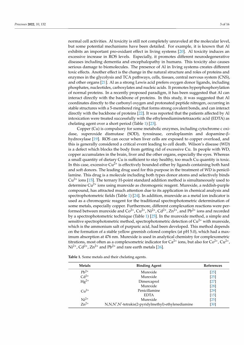
Copper (Cu) is compulsory for some metabolic enzymes, including cytochrome c oxi-dase, superoxide dismutase (SOD), tyrosinase, ceruloplasmin and dopamine-β-hydroxylase [19]. ROS can occur when liver cells are exposed to copper overload, andthis is generally considered a critical event leading to cell death. Wilson’s disease (WD)is a defect which blocks the body from getting rid of excessive Cu. In people with WD,copper accumulates in the brain, liver and the other organs, especially the eyes. Whereasa small quantity of dietary Cu is sufficient to stay healthy, too much Cu quantity is toxic.In this case, excessive Cu2+ is effectively bounded either by ligands containing both hardand soft donors. The leading drug used for this purpose in the treatment of WD is penicil-lamine. This drug is a molecule including both types donor atoms and selectively bindsCu2+ ions [15]. The ternary H-point standard addition method is simultaneously used todetermine Cu2+ ions using murexide as chromogenic reagent. Murexide, a reddish-purplecompound, has attracted much attention due to its application in chemical analysis andspectrophotometric fields (Table 1) [24]. In addition, murexide as a metal ion indicator isused as a chromogenic reagent for the traditional spectrophotometric determination ofsome metals, especially copper. Furthermore, different complexation reactions were per-formed between murexide and Co2+, Cu2+, Ni2+, Cd2+, Zn2+, and Pb2+ ions and recordedby a spectrophotometric technique (Table 1) [25]. In the murexide method, a simple andsensitive spectrophotometric method, spectrophotometric detection of Cu2+ with murexide,which is the ammonium salt of purpuric acid, had been developed. This method dependson the formation of a stable yellow greenish colored complex (at pH 5.0), which had a max-imum absorption at 476 nm. Murexide is used in analytical chemistry for complexometrictitrations, most often as a complexometric indicator for Ca2+ ions, but also for Co2+, Cu2+,Ni2+, Cd2+, Zn2+ and Pb2+ and rare earth metals [26].
Table 1. Some metals and their chelating agents.
Metals Binding Agent References
Pb2+ Murexide [25]Cd2+ Murexide [25]Hg2+ Dimercaprol [27]
Cu2+Murexide [28]
Penicillamine [29]EDTA [15]
Ni2+ Murexide [25]Zn2+ N,N,N′,N′-tetrakis(2-pyridylmethyl)-ethylenediamine [30]
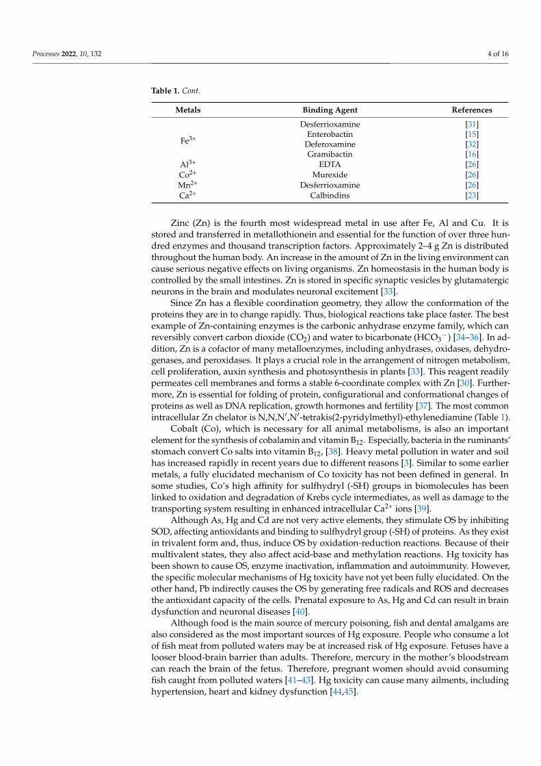
Processes 2022, 10, 132 4 of 16
Table 1. Cont.
Metals Binding Agent References
Fe3+
Desferrioxamine [31]Enterobactin [15]
Deferoxamine [32]Gramibactin [16]
Al3+ EDTA [26]Co2+ Murexide [26]Mn2+ Desferrioxamine [26]Ca2+ Calbindins [23]
Zinc (Zn) is the fourth most widespread metal in use after Fe, Al and Cu. It isstored and transferred in metallothionein and essential for the function of over three hun-dred enzymes and thousand transcription factors. Approximately 2–4 g Zn is distributedthroughout the human body. An increase in the amount of Zn in the living environment cancause serious negative effects on living organisms. Zn homeostasis in the human body iscontrolled by the small intestines. Zn is stored in specific synaptic vesicles by glutamatergicneurons in the brain and modulates neuronal excitement [33].
Since Zn has a flexible coordination geometry, they allow the conformation of theproteins they are in to change rapidly. Thus, biological reactions take place faster. The bestexample of Zn-containing enzymes is the carbonic anhydrase enzyme family, which canreversibly convert carbon dioxide (CO2) and water to bicarbonate (HCO3
−) [34–36]. In ad-dition, Zn is a cofactor of many metalloenzymes, including anhydrases, oxidases, dehydro-genases, and peroxidases. It plays a crucial role in the arrangement of nitrogen metabolism,cell proliferation, auxin synthesis and photosynthesis in plants [33]. This reagent readilypermeates cell membranes and forms a stable 6-coordinate complex with Zn [30]. Further-more, Zn is essential for folding of protein, configurational and conformational changes ofproteins as well as DNA replication, growth hormones and fertility [37]. The most commonintracellular Zn chelator is N,N,N′,N′-tetrakis(2-pyridylmethyl)-ethylenediamine (Table 1).
Cobalt (Co), which is necessary for all animal metabolisms, is also an importantelement for the synthesis of cobalamin and vitamin B12. Especially, bacteria in the ruminants’stomach convert Co salts into vitamin B12, [38]. Heavy metal pollution in water and soilhas increased rapidly in recent years due to different reasons [3]. Similar to some earliermetals, a fully elucidated mechanism of Co toxicity has not been defined in general. Insome studies, Co’s high affinity for sulfhydryl (-SH) groups in biomolecules has beenlinked to oxidation and degradation of Krebs cycle intermediates, as well as damage to thetransporting system resulting in enhanced intracellular Ca2+ ions [39].
Although As, Hg and Cd are not very active elements, they stimulate OS by inhibitingSOD, affecting antioxidants and binding to sulfhydryl group (-SH) of proteins. As they existin trivalent form and, thus, induce OS by oxidation-reduction reactions. Because of theirmultivalent states, they also affect acid-base and methylation reactions. Hg toxicity hasbeen shown to cause OS, enzyme inactivation, inflammation and autoimmunity. However,the specific molecular mechanisms of Hg toxicity have not yet been fully elucidated. On theother hand, Pb indirectly causes the OS by generating free radicals and ROS and decreasesthe antioxidant capacity of the cells. Prenatal exposure to As, Hg and Cd can result in braindysfunction and neuronal diseases [40].
Although food is the main source of mercury poisoning, fish and dental amalgams arealso considered as the most important sources of Hg exposure. People who consume a lotof fish meat from polluted waters may be at increased risk of Hg exposure. Fetuses have alooser blood-brain barrier than adults. Therefore, mercury in the mother’s bloodstreamcan reach the brain of the fetus. Therefore, pregnant women should avoid consumingfish caught from polluted waters [41–43]. Hg toxicity can cause many ailments, includinghypertension, heart and kidney dysfunction [44,45].
Processes 2022, 10, 132 5 of 16
Although Calcium (Ca) is not a heavy metal, it is tightly bound by calbindins, which area putative class of Ca2+-binding proteins. Calbindins belong to the Ca2+ messenger system,which reply to the transitory in intracellular Ca2+ concentration (Table 1). A structuralproperty of calbindins is their functional domain, which consists of two interacting bindingsites. So, they have cooperative binding [46]. A similar situation to calbindins-Ca2+
co-binding is also observed between laurate and human serum albumin (HSA) that iscommonly used as standard protein in biochemical assays [47–50]. It is known that HSAbinds to a wide range of ligands, especially fatty acids. In addition, in another study,multiple binding equilibria were searched for HSA and laurate binding using by a dialysis-exchange method [46].
1.2. The Importance of Metal Chelating in Biological Systems
Metal chelating therapy is the most important and primary clinical treatment in caseof heavy metal poisoning. Chelation is the process of linking existing ions or moleculesof a ligand to a central metal atom or ion through an acyclic or ring-like coordinationbond. A ligand is a molecule or ion with two or more atoms, which can easily donate twoelectrons to form a covalent bond. Ligands can be classified in three different ways basedon the properties of the bond between the ligand and the covalent atom. The complexes’stability varies with the metal ions and ligand interactions. Although Hg and Pb ionshave higher affinity for sulfur and nitrogen than for oxygen ligands, the opposite is thecase for Ca atoms. In addition, these differences that occur in affinity procedures, are thebasic principle in the selection of chelating agents [3,51]. For this purpose, some drugs,such as dimercaprol, EDTA, deferoxamine, penicillamine, dimercaptosuccinic acid andtheir analogues, are used as chelating agents, which are widely used in the treatment ofmetal toxicities [3].
1.3. Reactive Oxygen Species (ROS) and Oxidative Stress (OS)
Oxygen is a highly reactive atom and a powerful oxidizing agent that can easilyform oxides with many other elements and compounds. In the atmosphere, it exists inthe ground state and undergoes a gradual reduction process [52–55]. Molecular oxygencontains a pair of electrons with parallel spins located in two separate anti-bonding orbitals.Therefore, it can easily accept two electrons from any ordinary electron donor [56–58].In addition, redox reactions are a very important metabolic process in living organisms,where electrons can be easily transferred from one species to another. This process is thebasic reaction in most biological systems. In this case, the series of chemical reactions inliving organisms uses molecular oxygen in the air for oxidation and, as a result, providesan immediate usable form of energy such as ATP [59–61]. Oxygen is commonly used inreduction-oxidation reactions and the enzymatic biocatalysis process in cells and tissues.Furthermore, it has interatomic electron transfer ability. It is an important structural elementfor aerobic creatures and living metabolism. In addition, it is the final electron acceptorin the electron transport system [62–65]. So far, everything is very normal, but the mainproblem arises when the electron flow becomes disconnected. This situation results in theformation of free radicals having an odd electron. Free radicals are highly unstable andactive reagents against molecules and intermediates [66–68]. These unstable and short-livedspecies derive from the three basic elements of oxygen, sulphur and nitrogen. For example,ROS include hydroxyl (HO·), superoxide anion (O2·−), alkoxyl (RO·), nitric oxide (NO·),peroxyl (ROO·) and lipid hydroperoxides (LOO·) radicals. Of these O2·−, NO· and LOO·had less reactivity [69–71]. In addition, in living systems, hydrogen peroxide (H2O2), singletoxygen (1O2) and hypochlorous acid (HOCl) are nonradical ROS forms [70,71]. In addition,elemental ions such as Fe2+ can initiate ROS production in living systems [72,73]. OS occursas a result of an imbalance between ROS production and antioxidant system. OS disrupts anumber of cellular functions and leads to different pathological events in organisms [74–76].This situation leads to oxidative modification of proteins, DNA, RNA, and lipids [77,78].OS has long been known to pose an increased risk for many diseases, including cancer,
Processes 2022, 10, 132 6 of 16
arthrosclerosis, diabetes, aging, arthritis and some neurodegenerative diseases [79,80].Nevertheless, antioxidants have a very important role in health by inhibiting oxidativeprocesses and reducing the harmful effects of ROS [81].
When HMs accumulate at toxic levels in the human body, they cause serious hazardouseffects in different organs, including the nervous, respiratory, reproductive and digestivesystems [38]. As a result of this situation, in the plasma membrane, lipid peroxidationoccurs and stimulates the formation of RNS and ROS. Transition metals such as Fe andCu also trigger Fenton and Haber–Weiss reactions and the formation of ROS such asOH· [82–84]. In the presence of metal ions and O2, H2O2 can form OH· by the renownedFenton reaction [85]. On the other hand, the Haber–Weiss reaction produces OH· fromO2•− and H2O2, which is catalyzed by ferrous ions. The reaction was first suggested by
Fritz Haber and his student [86]. In later studies, it was determined that both reactions arethe main sources of radicals and responsible for the cellular damage.
Fe2+ + H2O2 → Fe3+ + OH− + OH• (Fenton reaction)
O•−2 + H2O2 → O2 + H2O + OH• (Haber–Weiss reaction)
The metal ions chelation can be important in order to avoid ROS formation and radicalproduction which can induce damage to biomolecules. In addition, natural metal chelatingcompounds including phenolics and flavonoids are desired over synthetic chelating agents,which are associated with the problem of toxicity [87].
1.4. Antioxidants
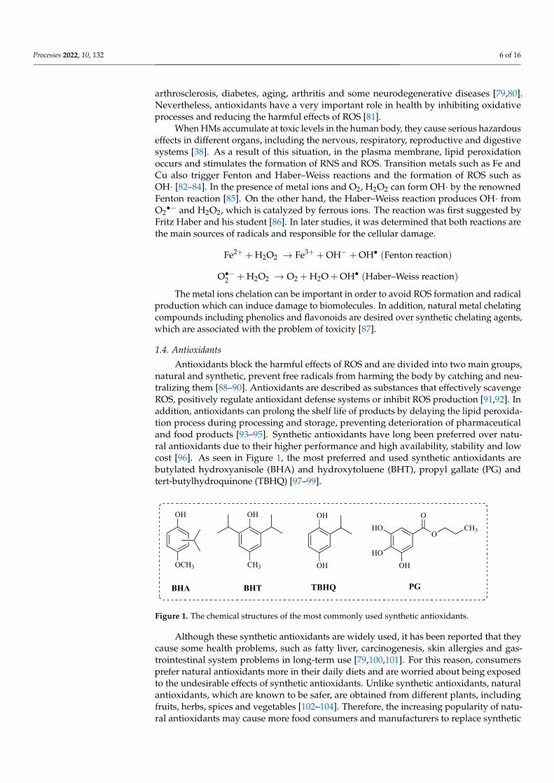
Antioxidants block the harmful effects of ROS and are divided into two main groups,natural and synthetic, prevent free radicals from harming the body by catching and neu-tralizing them [88–90]. Antioxidants are described as substances that effectively scavengeROS, positively regulate antioxidant defense systems or inhibit ROS production [91,92]. Inaddition, antioxidants can prolong the shelf life of products by delaying the lipid peroxida-tion process during processing and storage, preventing deterioration of pharmaceuticaland food products [93–95]. Synthetic antioxidants have long been preferred over natu-ral antioxidants due to their higher performance and high availability, stability and lowcost [96]. As seen in Figure 1, the most preferred and used synthetic antioxidants arebutylated hydroxyanisole (BHA) and hydroxytoluene (BHT), propyl gallate (PG) andtert-butylhydroquinone (TBHQ) [97–99].
Processes 2022, 10, x FOR PEER REVIEW 6 of 16
[70,71]. In addition, elemental ions such as Fe2+ can initiate ROS production in living sys-tems [72,73]. OS occurs as a result of an imbalance between ROS production and antioxi-dant system. OS disrupts a number of cellular functions and leads to different pathological events in organisms [74–76]. This situation leads to oxidative modification of proteins, DNA, RNA, and lipids [77,78]. OS has long been known to pose an increased risk for many diseases, including cancer, arthrosclerosis, diabetes, aging, arthritis and some neuro-degenerative diseases [79,80]. Nevertheless, antioxidants have a very important role in health by inhibiting oxidative processes and reducing the harmful effects of ROS [81].
When HMs accumulate at toxic levels in the human body, they cause serious hazard-ous effects in different organs, including the nervous, respiratory, reproductive and di-gestive systems [38]. As a result of this situation, in the plasma membrane, lipid peroxi-dation occurs and stimulates the formation of RNS and ROS. Transition metals such as Fe and Cu also trigger Fenton and Haber–Weiss reactions and the formation of ROS such as OH· [82–84]. In the presence of metal ions and O2, H2O2 can form OH· by the renowned Fenton reaction [85]. On the other hand, the Haber–Weiss reaction produces OH· from O2•− and H2O2, which is catalyzed by ferrous ions. The reaction was first suggested by Fritz Haber and his student [86]. In later studies, it was determined that both reactions are the main sources of radicals and responsible for the cellular damage. Fe + H O → Fe + OH + OH• (Fenton reaction) O• + H O → O + H O + OH• (Haber–Weiss reaction)
The metal ions chelation can be important in order to avoid ROS formation and rad-ical production which can induce damage to biomolecules. In addition, natural metal che-lating compounds including phenolics and flavonoids are desired over synthetic chelating agents, which are associated with the problem of toxicity [87].
1.4. Antioxidants Antioxidants block the harmful effects of ROS and are divided into two main groups,
natural and synthetic, prevent free radicals from harming the body by catching and neu-tralizing them [88–90]. Antioxidants are described as substances that effectively scavenge ROS, positively regulate antioxidant defense systems or inhibit ROS production [91,92]. In addition, antioxidants can prolong the shelf life of products by delaying the lipid pe-roxidation process during processing and storage, preventing deterioration of pharma-ceutical and food products [93–95]. Synthetic antioxidants have long been preferred over natural antioxidants due to their higher performance and high availability, stability and low cost [96]. As seen in Figure 1, the most preferred and used synthetic antioxidants are butylated hydroxyanisole (BHA) and hydroxytoluene (BHT), propyl gallate (PG) and tert-butylhydroquinone (TBHQ) [97–99].
Figure 1. The chemical structures of the most commonly used synthetic antioxidants.
Although these synthetic antioxidants are widely used, it has been reported that they cause some health problems, such as fatty liver, carcinogenesis, skin allergies and gastro-
OH
OCH3
BHA
OH
CH3
BHT
OH
OH
TBHQ PG
OH
O
OCH3HO
HO
Figure 1. The chemical structures of the most commonly used synthetic antioxidants.
Although these synthetic antioxidants are widely used, it has been reported that theycause some health problems, such as fatty liver, carcinogenesis, skin allergies and gas-trointestinal system problems in long-term use [79,100,101]. For this reason, consumersprefer natural antioxidants more in their daily diets and are worried about being exposedto the undesirable effects of synthetic antioxidants. Unlike synthetic antioxidants, naturalantioxidants, which are known to be safer, are obtained from different plants, includingfruits, herbs, spices and vegetables [102–104]. Therefore, the increasing popularity of natu-ral antioxidants may cause more food consumers and manufacturers to replace synthetic
Processes 2022, 10, 132 7 of 16
ones [105,106]. For example, aqueous tea, anise and fennel extracts were used as naturalantioxidant sources due to their rich content of various components, including tannins,catechins, theines and flavonoids [107–109]. However, the quality and antioxidant capacityof natural antioxidant and extracts depend not only on the quality of the natural source,but also on the applied processes and technologies for the extraction. In addition, thesafety of proven natural antioxidants has been determined taking into account informa-tion on chemical compounds and potential cumulative effects assessed by the results oftoxicity studies [79,110,111].
1.5. Metal Chelating Ability
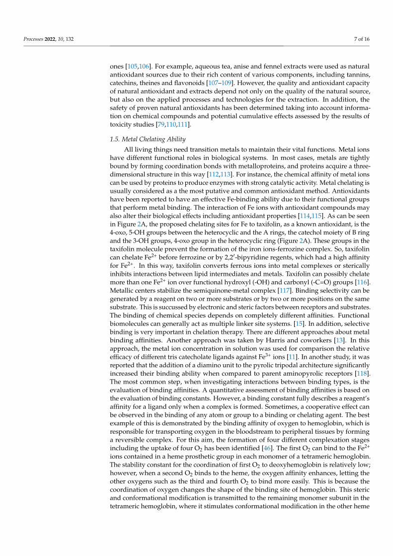
All living things need transition metals to maintain their vital functions. Metal ionshave different functional roles in biological systems. In most cases, metals are tightlybound by forming coordination bonds with metalloproteins, and proteins acquire a three-dimensional structure in this way [112,113]. For instance, the chemical affinity of metal ionscan be used by proteins to produce enzymes with strong catalytic activity. Metal chelating isusually considered as a the most putative and common antioxidant method. Antioxidantshave been reported to have an effective Fe-binding ability due to their functional groupsthat perform metal binding. The interaction of Fe ions with antioxidant compounds mayalso alter their biological effects including antioxidant properties [114,115]. As can be seenin Figure 2A, the proposed chelating sites for Fe to taxifolin, as a known antioxidant, is the4-oxo, 5-OH groups between the heterocyclic and the A rings, the catechol moiety of B ringand the 3-OH groups, 4-oxo group in the heterocyclic ring (Figure 2A). These groups in thetaxifolin molecule prevent the formation of the iron ions-ferrozine complex. So, taxifolincan chelate Fe2+ before ferrozine or by 2,2′-bipyridine regents, which had a high affinityfor Fe2+. In this way, taxifolin converts ferrous ions into metal complexes or stericallyinhibits interactions between lipid intermediates and metals. Taxifolin can possibly chelatemore than one Fe2+ ion over functional hydroxyl (-OH) and carbonyl (-C=O) groups [116].Metallic centers stabilize the semiquinone-metal complex [117]. Binding selectivity can begenerated by a reagent on two or more substrates or by two or more positions on the samesubstrate. This is succussed by electronic and steric factors between receptors and substrates.The binding of chemical species depends on completely different affinities. Functionalbiomolecules can generally act as multiple linker site systems. [15]. In addition, selectivebinding is very important in chelation therapy. There are different approaches about metalbinding affinities. Another approach was taken by Harris and coworkers [13]. In thisapproach, the metal ion concentration in solution was used for comparison the relativeefficacy of different tris catecholate ligands against Fe3+ ions [11]. In another study, it wasreported that the addition of a diamino unit to the pyrolic tripodal architecture significantlyincreased their binding ability when compared to parent aminopyrolic receptors [118].The most common step, when investigating interactions between binding types, is theevaluation of binding affinities. A quantitative assessment of binding affinities is based onthe evaluation of binding constants. However, a binding constant fully describes a reagent’saffinity for a ligand only when a complex is formed. Sometimes, a cooperative effect canbe observed in the binding of any atom or group to a binding or chelating agent. The bestexample of this is demonstrated by the binding affinity of oxygen to hemoglobin, which isresponsible for transporting oxygen in the bloodstream to peripheral tissues by forminga reversible complex. For this aim, the formation of four different complexation stagesincluding the uptake of four O2 has been identified [46]. The first O2 can bind to the Fe2+
ions contained in a heme prosthetic group in each monomer of a tetrameric hemoglobin.The stability constant for the coordination of first O2 to deoxyhemoglobin is relatively low;however, when a second O2 binds to the heme, the oxygen affinity enhances, letting theother oxygens such as the third and fourth O2 to bind more easily. This is because thecoordination of oxygen changes the shape of the binding site of hemoglobin. This stericand conformational modification is transmitted to the remaining monomer subunit in thetetrameric hemoglobin, where it stimulates conformational modification in the other heme
Processes 2022, 10, 132 8 of 16
regions, thus making it easier for oxygen to bind to these regions. In addition, this effect iscalled the cooperative effect as well as the positive homotropic allosteric effect [15].
Processes 2022, 10, x FOR PEER REVIEW 8 of 16
in each monomer of a tetrameric hemoglobin. The stability constant for the coordination of first O2 to deoxyhemoglobin is relatively low; however, when a second O2 binds to the heme, the oxygen affinity enhances, letting the other oxygens such as the third and fourth O2 to bind more easily. This is because the coordination of oxygen changes the shape of the binding site of hemoglobin. This steric and conformational modification is transmitted to the remaining monomer subunit in the tetrameric hemoglobin, where it stimulates con-formational modification in the other heme regions, thus making it easier for oxygen to bind to these regions. In addition, this effect is called the cooperative effect as well as the positive homotropic allosteric effect [15].
Figure 2. The suggested ferrous ions (Fe2+) binding mechanism of taxifolin (A), usnic acid (B), resveratrol (C) and curcumin (D).
In addition, curcumin, which is used as a food ingredient and is abundant in ginger and turmeric, chelates Fe2+ ions and prevents the formation of the Fe2+-ferrozine complex. In this way, curcumin can capture iron ions with a high binding affinity such as ferrozine. It has been suggested that a curcumin molecule binds three Fe2+ ions, as seen in Figure 2D. It has been reported that curcumin chelates iron ions with biological active -OH and -OCH3 groups [119]. In addition, the compounds containing functional groups such as C=O and C–OH can easily bind metal ions. In another study, Kazazica et al. showed that kaempferol binds to Fe2+ and Cu2+ ions. They also stated that this binding was mediated by functional -OH and -OCH3 groups [120]. Compounds containing two or more -OH, -COOH, -SH, -OCH3, -C=O, -PO3H2, -NR2, -O- and -S- functional groups in a suitable func-tion-structure configuration can easily chelate Fe2+ ions [121–124]. In another study, Fio-rucci and coworkers showed that quercetin, as an abundant phenolic compound in plants, had similar metal chelating ability [125]. Recently, the possible Fe2+ binding mechanism of usnic acid was proven by our research group [126]. It was reported that usnic acid pre-vented the formation of the complex of Fe2+-ferrozine (Figure 2B). As shown, usnic acid
Figure 2. The suggested ferrous ions (Fe2+) binding mechanism of taxifolin (A), usnic acid (B),resveratrol (C) and curcumin (D).
In addition, curcumin, which is used as a food ingredient and is abundant in gingerand turmeric, chelates Fe2+ ions and prevents the formation of the Fe2+-ferrozine complex.In this way, curcumin can capture iron ions with a high binding affinity such as ferrozine.It has been suggested that a curcumin molecule binds three Fe2+ ions, as seen in Figure 2D.It has been reported that curcumin chelates iron ions with biological active -OH and -OCH3 groups [119]. In addition, the compounds containing functional groups such asC=O and C–OH can easily bind metal ions. In another study, Kazazica et al. showedthat kaempferol binds to Fe2+ and Cu2+ ions. They also stated that this binding wasmediated by functional -OH and -OCH3 groups [120]. Compounds containing two ormore -OH, -COOH, -SH, -OCH3, -C=O, -PO3H2, -NR2, -O- and -S- functional groups in asuitable function-structure configuration can easily chelate Fe2+ ions [121–124]. In anotherstudy, Fiorucci and coworkers showed that quercetin, as an abundant phenolic compoundin plants, had similar metal chelating ability [125]. Recently, the possible Fe2+ bindingmechanism of usnic acid was proven by our research group [126]. It was reported thatusnic acid prevented the formation of the complex of Fe2+-ferrozine (Figure 2B). As shown,usnic acid can chelate Fe2+ ions with -OH and -COOH groups attached to the phenolic ring.In another effective study, it was observed that resveratrol binds Fe2+ ions on their -OHgroups at meta positions [127]. In this way, it has been reported that the main antioxidantability of resveratrol, a strong and natural antioxidant, may be related to its iron bindingcapacity. In this study, it was clearly demonstrated that resveratrol binds Fe2+ and interferesto form the Fe2+-ferrozine complex.
One of the strategies for estimating the chelation capacity is to measure free ironions (Fe2+) using a chelating agent such as ferrozine or 2,2′-bipyridine (Figure 3), forming
Processes 2022, 10, 132 9 of 16
an easily detectable complex by spectroscopic analysis. Metal chelators form complexesand reduce the reactivity of metals such as iron, making them inactive [128]. The maincontribution to metal binding is because of the catechol moiety, as sampled by the morepronounced bathochromic shift produced by Cu binding to quercetin when compared tothe chelating ability of kaempferol [129]. Flavonoids show bioavailability by chelatingexcess metal ions in the human body. Such a metal chelating effect of flavonoids plays animportant role in the detoxification of other HMs, such as Cr, Sn, Cd and Pb as well asbinding excess Al. The chelating agents effectively chelate the toxic metal ions formingthe complexes [114,130].
Processes 2022, 10, x FOR PEER REVIEW 9 of 16
can chelate Fe2+ ions with -OH and -COOH groups attached to the phenolic ring. In an-other effective study, it was observed that resveratrol binds Fe2+ ions on their -OH groups at meta positions [127]. In this way, it has been reported that the main antioxidant ability of resveratrol, a strong and natural antioxidant, may be related to its iron binding capacity. In this study, it was clearly demonstrated that resveratrol binds Fe2+ and interferes to form the Fe2+-ferrozine complex.
One of the strategies for estimating the chelation capacity is to measure free iron ions (Fe2+) using a chelating agent such as ferrozine or 2,2′-bipyridine (Figure 3), forming an easily detectable complex by spectroscopic analysis. Metal chelators form complexes and reduce the reactivity of metals such as iron, making them inactive [128]. The main contri-bution to metal binding is because of the catechol moiety, as sampled by the more pro-nounced bathochromic shift produced by Cu binding to quercetin when compared to the chelating ability of kaempferol [129]. Flavonoids show bioavailability by chelating excess metal ions in the human body. Such a metal chelating effect of flavonoids plays an im-portant role in the detoxification of other HMs, such as Cr, Sn, Cd and Pb as well as bind-ing excess Al. The chelating agents effectively chelate the toxic metal ions forming the complexes [114,130].
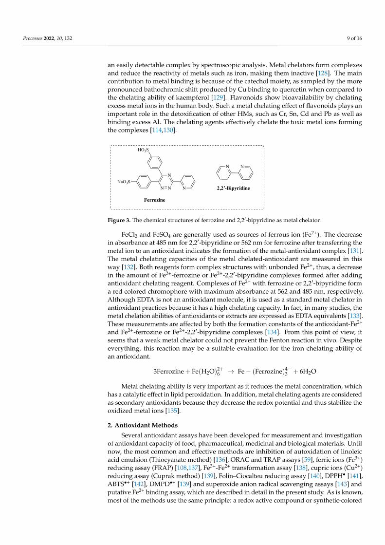
Figure 3. The chemical structures of ferrozine and 2,2′-bipyridine as metal chelator.
FeCl2 and FeSO4 are generally used as sources of ferrous ion (Fe2+). The decrease in absorbance at 485 nm for 2,2′-bipyridine or 562 nm for ferrozine after transferring the metal ion to an antioxidant indicates the formation of the metal-antioxidant complex [131]. The metal chelating capacities of the metal chelated-antioxidant are measured in this way [132]. Both reagents form complex structures with unbonded Fe2+, thus, a decrease in the amount of Fe2+-ferrozine or Fe2+-2,2′-bipyridine complexes formed after adding antioxi-dant chelating reagent. Complexes of Fe2+ with ferrozine or 2,2′-bipyridine form a red col-ored chromophore with maximum absorbance at 562 and 485 nm, respectively. Although EDTA is not an antioxidant molecule, it is used as a standard metal chelator in antioxidant practices because it has a high chelating capacity. In fact, in many studies, the metal che-lation abilities of antioxidants or extracts are expressed as EDTA equivalents [133]. These measurements are affected by both the formation constants of the antioxidant-Fe2+ and Fe2+-ferrozine or Fe2+-2,2′-bipyridine complexes [134]. From this point of view, it seems that a weak metal chelator could not prevent the Fenton reaction in vivo. Despite every-thing, this reaction may be a suitable evaluation for the iron chelating ability of an antiox-idant. 3Ferrozine + Fe(H O) → Fe − (Ferrozine) + 6H O
Metal chelating ability is very important as it reduces the metal concentration, which has a catalytic effect in lipid peroxidation. In addition, metal chelating agents are consid-ered as secondary antioxidants because they decrease the redox potential and thus stabi-lize the oxidized metal ions [135].
Figure 3. The chemical structures of ferrozine and 2,2′-bipyridine as metal chelator.
FeCl2 and FeSO4 are generally used as sources of ferrous ion (Fe2+). The decreasein absorbance at 485 nm for 2,2′-bipyridine or 562 nm for ferrozine after transferring themetal ion to an antioxidant indicates the formation of the metal-antioxidant complex [131].The metal chelating capacities of the metal chelated-antioxidant are measured in thisway [132]. Both reagents form complex structures with unbonded Fe2+, thus, a decreasein the amount of Fe2+-ferrozine or Fe2+-2,2′-bipyridine complexes formed after addingantioxidant chelating reagent. Complexes of Fe2+ with ferrozine or 2,2′-bipyridine forma red colored chromophore with maximum absorbance at 562 and 485 nm, respectively.Although EDTA is not an antioxidant molecule, it is used as a standard metal chelator inantioxidant practices because it has a high chelating capacity. In fact, in many studies, themetal chelation abilities of antioxidants or extracts are expressed as EDTA equivalents [133].These measurements are affected by both the formation constants of the antioxidant-Fe2+
and Fe2+-ferrozine or Fe2+-2,2′-bipyridine complexes [134]. From this point of view, itseems that a weak metal chelator could not prevent the Fenton reaction in vivo. Despiteeverything, this reaction may be a suitable evaluation for the iron chelating ability ofan antioxidant.
3Ferrozine + Fe(H2O)2+6 → Fe− (Ferrozine)4−
3 + 6H2O
Metal chelating ability is very important as it reduces the metal concentration, whichhas a catalytic effect in lipid peroxidation. In addition, metal chelating agents are consideredas secondary antioxidants because they decrease the redox potential and thus stabilize theoxidized metal ions [135].
2. Antioxidant Methods
Several antioxidant assays have been developed for measurement and investigationof antioxidant capacity of food, pharmaceutical, medicinal and biological materials. Untilnow, the most common and effective methods are inhibition of autoxidation of linoleicacid emulsion (Thiocyanate method) [136], ORAC and TRAP assays [59], ferric ions (Fe3+)reducing assay (FRAP) [108,137], Fe3+-Fe2+ transformation assay [138], cupric ions (Cu2+)reducing assay (Cuprak method) [139], Folin–Ciocalteu reducing assay [140], DPPH• [141],ABTS•+ [142], DMPD•+ [139] and superoxide anion radical scavenging assays [143] andputative Fe2+ binding assay, which are described in detail in the present study. As is known,most of the methods use the same principle: a redox active compound or synthetic-colored
Processes 2022, 10, 132 10 of 16
radical is produced; then, the ability of a biological sample to scavenge or reduce theredox-active compounds is measured by a spectrophotometer, applying a suitable standardto measure the antioxidant ability [144].
3. Metal Chelating Assays3.1. Metal Chelating Assay by Ferrozine Reagent
The described spectroscopic methods were developed taking into account the bindingaffinity between a reagent and a metal ion such as Fe2+. The Fe2+ chelation by Ferrozinewas evaluated by the Dinis method [145]. Briefly, a different quantity of sample andstandard compounds was transferred to a 0.05 mL of FeCl2 solution (2 mM). In this way,the interaction between the sample and Fe2+ is supplied, that is, Fe2+ ions are chelated bythe sample. The reaction was started by adding 0.2 mL of ferrozine reagent (5 mm) andthe mixture was stirred and left standing at 25 ◦C for 10 min. Then, absorbance values ofsolution were recorded at 562 nm [146].
3.2. Metal Chelating Assay by 2,2′-Bipyridine Reagent
The Fe2+ chelating by 2,2′-bipyridine are generally performed according to the methodof Re et al. [147]. Briefly, different quantities of the sample and standard compounds weretransferred to a solution of 0.25 mL FeSO4 (2 mM). Thus, the interaction of the sampleand Fe2+ ions is ensured. So, Fe2+ ions are chelated by the sample. Then, 1.5 mL of 0.2%bipyridyl solution dissolved in 1 mL of Tris-HCl solution (pH 7.4) and HCl (0.2 M) weretransferred to the mixture, sequentially. After the solution was incubated for 30 min, 2.5 mLof ethyl alcohol and 0.63 mL of deionized water were transferred. Their absorbances wererecorded at 522 nm against the blank consisting of the Tris-HCl buffer [148].
3.3. The Percentage Metal Chelating
The percentage chelating ability of samples and standards is determined using thefollowing equation:
Percentage chelating effect (%) = [(A0 − A1)/A0)] × 100
where A0 and A1 are the absorbances of the control and sample, respectively. The controldid not include FeCl2, ferrozine, and 2,2′-bipyridine [149].
3.4. The Importance of IC50 Value in Binding Affinity
The IC50 value is commonly used in biochemistry to compare metal chelating [11].IC50 values describe binding affinity quantitatively. This parameter is widely used inbiochemical applications and is an alternative approach based on a suitable principle. Theidea of evaluating half of the binding property does not require extra knowledge of bindingconstants. The lower IC50 value indicates the higher binding affinity and the easier it isto use. For all these reasons, the IC50 value is the most practical way to evaluate bindingaffinities. In most cases, it reports on biological effects [14,46]. In functional antagonists’studies, the IC50 value of a drug is the concentration required to inhibit half of the maximumbiological activity of the agonist by formation a dose-response curve. It can be determinedby examining the effect of different concentrations of antagonists. The IC50 value is not adirect indicator of binding affinity, but at least for competitive agonists and antagonists, IC50and affinity are correlated with the Cheng–Prusoff equation [150], which is given below.
Ki =IC50
1 + [S]Ki
(Cheng–Prusoff equation)
In this formula, the Ki value can be easily calculated from the IC50 value for a com-petitive inhibitor in single-substrate enzymatic reactions. In this formula, IC50 is the halfmaximal concentration of the competitive inhibitor and shows a 50% inhibition. S is the sub-strate concentration. Km is the Michaelis–Menten constant of the substrate for enzymatic
Processes 2022, 10, 132 11 of 16
reaction. The IC50 value of a compound may vary depending on experimental conditionsand parameters such as temperature and pressure, but Ki is a relatively stable value [151].
4. Conclusions
Heavy metals have different functional roles in biological systems and are necessaryfor many different biological events, such as cell growth, development and proliferation,synthesis of biomolecules, catalysis of many enzymatic reactions and immunity of thebody; however, excessive uptake of metals by different pathways is extremely harmfuland can create applications that cannot be repaired. In addition, heavy metals such asFe2+ can simplify the production of ROS in living systems. The metal chelating abilityof the agents used for this purpose can be extremely valuable for antioxidant properties.Antioxidants play a crucial to reduce oxidative damage and hazardous effects of ROS. Metalchelating activity is one of the most applied methods in food, biological and pharmaceuticalapplications. In this review article, metals, heavy metals, the effects of excessive metalexposure, the importance of metal chelating in biological systems, reactive oxygen species,OS, Antioxidants, metal chelating ability, antioxidant methods, and two distinct in vitrometal chelating assays were explained in details.
Author Contributions: Investigation, writing—original draft preparation and writing—review andediting, I.G. and S.H.A. All authors have read and agreed to the published version of the manuscript.
Funding: This research received no external funding.
Institutional Review Board Statement: Not applicable.
Informed Consent Statement: Not applicable.
Data Availability Statement: Data available in a publicly accessible repository.
Acknowledgments: I. Gulcin would like to extend his sincere appreciation to the Turkish Academyof Sciences (TÜBA). S.H. Alwasel would like to extend his sincere appreciation to the ResearchersSupporting Project (RSP-2022/59), King Saud University, Saudi Arabia.
Conflicts of Interest: The authors declare no conflict of interest.
References1. Nurchi, V.M.; Cappai, R.; Crisponi, G.; Sanna, G.; Alberti, G.; Biesuz, R.; Gama, S. Chelating agents in soil remediation: A new
method for a pragmatic choice of the right chelator. Front. Chem. 2020, 8, 597400. [CrossRef]2. Jarup, L. Hazards of heavy metal contamination. Br. Med. Bull. 2003, 68, 167–182. [CrossRef] [PubMed]3. Kim, J.J.; Kim, Y.S.; Kumar, V. Heavy metal toxicity: An update of chelating therapeutic strategies. J. Trace Elem. Med. Biol. 2019,
54, 226–231. [CrossRef] [PubMed]4. Küçük, M.; Gulcin, I. Purification and characterization of carbonic anhydrase enzyme from black sea trout (Salmo trutta Labrax
Coruhensis) kidney and inhibition effects of some metal ions on the enzyme activity. Environ. Toxicol. Pharmacol. 2016, 44, 134–139.[CrossRef] [PubMed]
5. Kocyigit, U.M.; Taslimi, P.; Gulcin, I. Characterization and inhibition effects of some metal ions on carbonic anhydrase enzymefrom Kangal Akkaraman sheep. J. Biochem. Mol. Toxicol. 2018, 32, e22172. [CrossRef] [PubMed]
6. Caglayan, C.; Taslimi, P.; Türk, C.; Kandemir, F.M.; Demir, Y.; Gulcin, I. Purification and characterization of the carbonic anhydraseenzyme from horse mackerel (Trachurus trachurus) muscle and the impact of some metal ions and pesticides on enzyme activity.Comp. Biochem. Physiol. 2018, 226, 108605. [CrossRef]
7. Festa, R.A.; Thiele, D.J. Copper: An essential metal in biology. Curr. Biol. 2011, 21, R877–R883. [CrossRef] [PubMed]8. Haase, H.; Rink, L. The immune system and the impact of zinc during aging. Immun. Ageing 2011, 6, 9. [CrossRef]9. Jomova, K.; Valko, M. Advances in metal-induced oxidative stress and human disease. Toxicology 2011, 283, 65–87. [CrossRef]10. Caglayan, C.; Taslimi, P.; Turk, C.; Gulcin, I.; Kandemir, F.M.; Demir, Y.; Beydemir, S. Inhibition effects of some pesticides and
heavy metals on carbonic anhydrase enzyme activity purified from horse mackerel (Trachurus trachurus) gill tissues. Environ. Sci.Pollut. Res. 2020, 27, 10607–10616. [CrossRef]
11. Vacca, A.; Nativi, C.; Cacciarini, M.; Pergoli, R.; Roelens, S. A new tripodal receptor for molecular recognition of monosaccharides.A paradigm for assessing glycoside binding affinities and selectivities by 1H NMR spectroscopy. J. Am. Chem. Soc. 2004, 126,16456–16465. [CrossRef]
12. Beard, J.L. Iron biology in immune function, muscle metabolism and neuronal functioning. J. Nutr. 2001, 131, 568S–579S.[CrossRef]
Processes 2022, 10, 132 12 of 16
13. Harris, W.R.; Raymond, K.N.; Weitl, F.L. Ferric ion sequestering agents. 6. The spectrophotometric and potentiometric evaluationof sulfonated tricatecholate ligands. J. Am. Chem. Soc. 1981, 103, 2667–2675. [CrossRef]
14. Roelens, S.; Vacca, A.; Venturi, C. Binding of ionic species: A general approach to measuring binding constants and assessingaffinities. Chem. Eur. J. 2009, 15, 2635–2644. [CrossRef] [PubMed]
15. Bazzicalupi, C.; Bianchi, A.; Giorgia, C.; Clares, M.P.; Garcia-Espana, E. Addressing selectivity criteria in binding equilibria. Coord.Chem. Rev. 2012, 256, 13–27. [CrossRef]
16. Gama, S.; Hermenau, R.; Frontauria, M.; Milea, D.; Sammartano, S.; Hertweck, C.; Plass, W. Iron coordination properties ofgramibactin as model for the new class of diazeniumdiolate based siderophores. Chem. Eur. J. 2021, 27, 2724–2733. [CrossRef][PubMed]
17. Baranwal, A.K.; Singhi, S.C. Acute iron poisoning: Management guidelines. Ind. Pediatr. 2003, 40, 534–540.18. Hershko, C. Mechanism of iron toxicity. Food Nutr. Bull. 2007, 28, S500–S509. [CrossRef]19. Desai, V.; Kaler, S.G. Role of copper in human neurological disorders. Am. J. Clin. Nutr. 2008, 88, 855S–858S. [CrossRef] [PubMed]20. Formoso, E.; Grande-Aztatzi, R.; Lopez, X. Does phosphorylation increase the binding affinity of aluminum? A computational
study on the aluminum interaction with serine and O-phosphoserine. J. Inorg. Biochem. 2019, 92, 33–44. [CrossRef]21. David, C.I.; Jayaraj, H.; Prabakara, G.; Velmurugan, K.; Devi, D.P.; Kayalvizhi, R.; Abiram, A.; Kannan, V.R.; Nandhakumar, N. A
photoswitchable “turn-on” fluorescent chemosensor: Quinoline-naphthalene duo for nanomolar detection of aluminum andbisulfite ions and its multifarious applications. Food Chem. 2022, 371, 131130. [CrossRef]
22. Mujika, J.I.; Torre, G.D.; Formoso, E.; Grande-Aztatzi, R.; Grabowski, S.J.; Exley, C.; Lopez, X. Aluminum’s preferential bindingsite in proteins: Sidechain of amino acids versus backbone interactions. J. Inorg. Biochem. 2018, 181, 111–116. [CrossRef]
23. Fulgenzi, A.; De Giuseppe, R.; Bamonti, F.; Vietti, D.; Ferrero, M.E. Efficacy of chelation therapy to remove aluminium intoxication.J. Inorg. Biochem. 2015, 152, 214–218. [CrossRef]
24. Masoud, M.S.; Kassem, T.S.; Shaker, M.A.; Ali, A.E. Studies on transition metal murexide complexes. J. Therm. Anal. Calorim.2006, 84, 549–555. [CrossRef]
25. Grudpan, K.; Jakmunee, J.; Vaneesorn, Y.; Watanesk, S.; Maung, U.A.; Sooksamiti, P. Flow-injection spectrophotometric determi-nation of calcium using murexide as a color agent. Talanta 1998, 46, 1245–1257. [CrossRef]
26. Martin, R.L.; White, A.H.; Willis, A.C. Structural studies in metal–purpurate complexes. Part 1. Crystal structures of potassiumpurpurate trihydrate and ammonium purpurate monohydrate (murexide). J. Chem. Soc. Dalton Trans. 1977, 14, 1336–1342.[CrossRef]
27. Sigel, A.; Sigel, H. (Eds.) Metal Ions in Biological Systems; Marcel Dekker: New York, NY, USA, 2004.28. Ghasemi, I.; Shamsipur, M. Spectrophotometric study of the thermodynamics of interaction of some metal ions with murexide in
binary acetonitrile-dimethylsulfoxide mixtures. J. Coord. Chem. 1995, 36, 183–194. [CrossRef]29. Crisponi, G.; Nurchi, V.M.; Fanni, D.; Gerosa, C.; Nemolato, S.; Faa, G. Copper-related diseases: From chemistry to molecular
pathology. Coord. Chem. Rev. 2010, 254, 876–889. [CrossRef]30. Radford, R.J.; Lippard, S.J. Chelators for investigating zinc metalloneurochemistry. Curr. Opin. Chem. Biol. 2013, 17, 129–136.
[CrossRef]31. Domingo, J.L. The use of chelating agents in the treatment of aluminum overload. J. Toxicol. Clin. Toxicol. 1989, 27, 355–367.
[CrossRef]32. Soybir, G.; Köksoy, F.; Ekiz, F.; Yalçin, O.; Ozseker, A.; Cokneseli, B. Effect of mangan-desferrioxamin in the prevention of
peritoneal adhesions. J. R. Coll. Surg. Edinb. 1998, 43, 26–28.33. Wirosoedarmo, R.; Anugroho, F.; Hanggara, S.D.; Gustinasari, K. Effect of adding chelating agents on the absorption of zinc from
polluted soil sludge textile industrial waste by sunflower plant (Helianthus annuus L.). Appl. Environ. Soil Sci. 2018, 2018, 8259520.[CrossRef]
34. Turkan, F.; Cetin, A.; Taslimi, P.; Karaman, M.; Gulcin, I. Synthesis, biological evaluation and molecular docking of novel pyrazolederivatives as potent carbonic anhydrase and acetylcholinesterase inhibitors. Bioorg. Chem. 2019, 86, 420–427. [CrossRef]
35. Ozgeris, B.; Goksu, S.; Kose Polat, L.; Gulcin, I.; Salmas, R.E.; Durdagi, S.; Tumer, F.; Supuran, C.T. Acetylcholinesterase andcarbonic anhydrase inhibitory properties of novel urea and sulfamide derivatives incorporating dopaminergic 2-aminotetralinscaffolds. Bioorg. Med. Chem. 2016, 24, 2318–2329. [CrossRef]
36. Gulcin, I.; Abbasova, M.; Taslimi, P.; Huyut, Z.; Safarova, L.; Sujayev, A.; Farzaliyev, V.; Beydemir, S.; Alwasel, S.H.; Supuran, C.T.Synthesis and biological evaluation of aminomethyl and alkoxymethyl derivatives as carbonic anhydrase, acetylcholinesteraseand butyrylcholinesterase inhibitors. J. Enzyme Inhib. Med. Chem. 2017, 32, 1174–1182. [CrossRef] [PubMed]
37. Foresta, C.; Garolla, A.; Cosci, I.; Menegazzo, M.; Ferigo, M.; Gandin, V.; DeToni, L. Role of zinc trafficking in male fertility: Fromgerm to sperm. Hum. Reprod. 2014, 29, 1134–1145. [CrossRef]
38. Huat, T.J.; Camats-Perna, J.; Newcombe, E.A.; Valmas, N.; Kitazawa, M.; Medeiros, R. Metal toxicity links to Alzheimer’s diseaseand neuroinflammation. J. Mol. Biol. 2019, 431, 1843–1868. [CrossRef]
39. Devlin, J.J.; Pomerleau, A.C.; Brent, J.; Morgan, B.W.; Deitchman, S.; Schwartz, M. Clinical features, testing, and management ofpatients with suspected prosthetic hip-Associated cobalt toxicity: A systematic review of cases. J. Med. Toxicol. 2013, 9, 405–415.[CrossRef]
40. Flora, S.J.; Pachauri, V. Chelation in metal intoxication. Int. J. Environ. Res. Public Health 2010, 7, 2745–2788. [CrossRef] [PubMed]
Processes 2022, 10, 132 13 of 16
41. Gilman, C.L.; Soon, R.; Sauvage, L.; Ralston, N.V.; Berry, M.J. Umbilical cord blood and placental mercury, selenium andselenoprotein expression in relation to maternal fish consumption. J. Trace Elem. Med. Biol. 2015, 30, 17–24. [CrossRef] [PubMed]
42. Kozikowska, I.; Binkowski, L.J.; Szczepanska, K.; Slawska, H.; Miszczuk, K.; Sliwinska, M.; Laciak, T.; Stawarz, R. Mercuryconcentrations in human placenta, umbilical cord, cord blood and amniotic fluid and their relations with body parameters ofnewborns. Environ. Pollut. 2013, 182, 256–262. [CrossRef] [PubMed]
43. Chen, Z.; Myers, R.; Wei, T.; Bind, E.; Kassim, P.; Wang, G.; Ji, Y.; Hong, X.; Caruso, D.; Bartell, T.; et al. Placental transfer andconcentrations of cadmium, mercury, lead, and selenium in mothers, newborns, and young children. J. Expo. Sci. Environ.Epidemiol. 2014, 24, 537–544. [CrossRef]
44. Bernhoft, R.A. Mercury toxicity and treatment: A review of the literature. J. Environ. Public Health 2012, 2012, 460508. [CrossRef]45. Kosnett, M.J. The role of chelation in the treatment of arsenic and mercury poisoning. J. Med. Toxicol. 2013, 9, 347–354. [CrossRef]46. Vacca, A.; Francesconi, O.; Roelens, S. BC50: A generalized, unifying affinity descriptor. Chem. Rec. 2012, 12, 544–566. [CrossRef]47. Nar, M.; Cetinkaya, Y.; Gulcin, I.; Menzek, A. (3,4-Dihydroxyphenyl)(2,3,4-trihydroxyphenyl)methanone and its derivatives as
carbonic anhydrase isoenzymes inhibitors. J. Enzyme Inhib. Med. Chem. 2013, 28, 402–406. [CrossRef]48. Koksal, E.; Gulcin, I. Purification and characterization of peroxidase from cauliflower (Brassica oleracea L.) buds. Protein Peptide
Lett. 2008, 15, 320–326. [CrossRef]49. Erdemir, F.; Barut Celepci, D.; Aktas, A.; Taslimi, P.; Gök, Y.; Karabıyık, H.; Gulcin, I. 2-Hydroxyethyl substituted NHC
precursors: Synthesis, characterization, crystal structure and carbonic anhydrase, α-glycosidase, butyrylcholinesterase, andacetylcholinesterase inhibitory properties. J. Mol. Struct. 2008, 1155, 797–806. [CrossRef]
50. Boztas, M.; Cetinkaya, Y.; Topal, M.; Gulcin, I.; Menzek, A.; Sahin, E.; Tanc, M.; Supuran, C.T. Synthesis and carbonic anhydraseisoenzymes I, II, IX, and XII inhibitory effects of dimethoxy-bromophenol derivatives incorporating cyclopropane moieties. J.Med. Chem. 2015, 58, 640–650. [CrossRef] [PubMed]
51. Flora, S.J.; Flora, G.; Saxena, G.; Mishra, M. Arsenic and lead induced free radical generation and their reversibility followingchelation. Cell. Mol. Biol. 2007, 53, 26–47. [PubMed]
52. Topal, M.; Gocer, H.; Topal, F.; Kalin, P.; Polat Kose, P.; Gulcin, I.; Cakmak, K.C.; Kucuk, M.; Durmaz, L.; Goren, A.C.; et al.Antioxidant, antiradical and anticholinergic properties of cynarin purified from the illyrian thistle (Onopordum illyricum L.). J.Enzyme Inhib. Med. Chem. 2016, 31, 266–275. [CrossRef]
53. Kiziltas, H.; Bingol, Z.; Goren, A.C.; Alwasel, S.H.; Gulcin, I. Anticholinergic, antidiabetic and antioxidant activities of Ferulaorientalis L.-Analysis of its polyphenol contents by LC-HRMS. Rec. Nat. Prod. 2021, 15, 513–528. [CrossRef]
54. Gulcin, I.; Bursal, E.; Sehitoglu, H.M.; Bilsel, M.; Goren, A.C. Polyphenol contents and antioxidant activity of lyophilized aqueousextract of propolis from Erzurum, Turkey. Food Chem. Toxicol. 2010, 48, 2227–2238. [CrossRef]
55. Bursal, E.; Taslimi, P.; Gören, A.; Gulcin, I. Assessments of anticholinergic, antidiabetic, antioxidant activities and phenolic contentof Stachys annua. Biocat. Agric. Biotechnol. 2020, 28, 101711. [CrossRef]
56. Aksu, K.; Topal, F.; Gulcin, I.; Tumer, F.; Goksu, S. Acetylcholinesterase inhibitory and antioxidant activities of novel symmetricsulfamides derived from phenethylamines. Arch. Pharm. 2015, 348, 446–455. [CrossRef]
57. Koksal, E.; Gulcin, I. Antioxidant activity of cauliflower (Brassica oleracea L.). Turk. J. Agric. For. 2008, 32, 65–78.58. Tohma, H.; Altay, A.; Koksal, E.; Gören, A.C.; Gulcin, I. Measurement of anticancer, antidiabetic and anticholinergic properties of
sumac (Rhus coriaria)—Analysis of its phenolic compounds by LC-MS/MS. J. Food Meas. Charac. 2019, 13, 1607–1619. [CrossRef]59. Gulcin, I. Antioxidants and antioxidant methods-An updated overview. Arch. Toxicol. 2020, 94, 651–715. [CrossRef]60. Taslimi, P.; Koksal, E.; Goren, A.C.; Bursal, E.; Aras, A.; Kılıc, O.; Alwasel, S.; Gulcin, I. Anti-Alzheimer, antidiabetic and
antioxidant potential of Satureja cuneifolia and analysis of its phenolic contents by LC-MS/MS. Arab. J. Chem. 2020, 13, 4528–4537.[CrossRef]
61. Artunc, T.; Menzek, A.; Taslimi, P.; Gulcin, I.; Kazaz, C.; Sahin, E. Synthesis and antioxidant activities of phenol derivatives from1,6-bis(dimethoxyphenyl)hexane-1,6-dione. Bioorg. Chem. 2020, 100, 103884. [CrossRef] [PubMed]
62. Turkan, F.; Atalar, M.N.; Aras, A.; Gulçin, I.; Bursal, E. ICP-MS and HPLC analyses, enzyme inhibition and antioxidant potentialof Achillea schischkinii Sosn. Bioorg. Chem. 2020, 94, 103333. [CrossRef]
63. Gulcin, I.; Goren, A.C.; Taslimi, P.; Akyuz, B.; Tuzun, B. Anticholinergic, antidiabetic and antioxidant activities of Anatolianpennyroyal (Mentha pulegium) -Analysis of its polyphenol contents by LC-MS/MS. Biocat. Agric. Biotechnol. 2020, 23, 101441.[CrossRef]
64. Altay, A.; Tohma, H.; Durmaz, L.; Taslimi, P.; Korkmaz, M.; Gulcin, I.; Koksal, E. Preliminary phytochemical analysis andevaluation of in vitro antioxidant, antiproliferative, antidiabetic and anticholinergics effects of endemic Gypsophila taxa fromTurkey. J. Food Biochem. 2019, 43, e12908. [CrossRef] [PubMed]
65. Gulcin, I.; Kirecci, E.; Akkemik, E.; Topal, F.; Hisar, O. Antioxidant and antimicrobial activities of an aquatic plant: Duckweed(Lemna minor L.). Turk. J. Biol. 2010, 34, 175–188.
66. Polat Kose, L.; Gulçin, I.; Gören, A.C.; Namiesnik, J.; Martinez-Ayala, A.L.; Gorinstein, S. LC-MS/MS analysis, antioxidant andanticholinergic properties of galanga (Alpinia officinarum Hance) rhizomes. Ind. Crops Prod. 2015, 74, 712–721. [CrossRef]
67. Gulcin, I. Antioxidant and antiradical activities of L-carnitine. Life Sci. 2006, 78, 803–811. [CrossRef]68. Ames, B.N.; Shigenaga, M.K.; Hagen, T.M. Oxidants, antioxidants, and the degenerative diseases of aging. Proc. Natl. Acad. Sci.
USA 1993, 90, 7915–7922. [CrossRef]
Processes 2022, 10, 132 14 of 16
69. Gulcin, I.; Kaya, R.; Goren, A.C.; Akıncıoglu, H.; Topal, M.; Bingol, Z.; Cetin Cakmak, K.; Ozturk Sarikaya, S.B.; Durmaz, L.;Alwasel, S. Anticholinergic, antidiabetic and antioxidant activities of cinnamon (Cinnamomum verum) bark extracts: Polyphenolcontents analysis by LC-MS/MS. Int. J. Food Prop. 2019, 22, 1511–1526. [CrossRef]
70. Serbetci Tohma, H.; Gulcin, I. Antioxidant and radical scavenging activity of aerial parts and roots of Turkish liquorice (Glycyrrhizaglabra L.). Int. J. Food Prop. 2010, 13, 657–671. [CrossRef]
71. Gulcin, I. Comparison of in vitro antioxidant and antiradical activities of L-tyrosine and L-Dopa. Amino Acids 2007, 32, 431–843.[CrossRef]
72. Pietta, P.G. Flavonoids as antioxidants. J. Nat. Prod. 2000, 63, 1035–1042. [CrossRef] [PubMed]73. Balaydın, H.T.; Gulcin, I.; Menzek, A.; Goksu, S.; Sahin, E. Synthesis and antioxidant properties of diphenylmethane derivative
bromophenols including a natural product. J. Enzyme Inhib. Med. Chem. 2010, 25, 685–695. [CrossRef] [PubMed]74. Gulcin, I.; Beydemir, S.; Sat, I.G.; Kufrevioglu, O.I. Evaluation of antioxidant activity of cornelian cherry (Cornus mas L.). Acta
Aliment. Hung. 2005, 34, 193–202. [CrossRef]75. Tohma, H.; Gulcin, I.; Bursal, E.; Goren, A.C.; Alwasel, S.H.; Koksal, E. Antioxidant activity and phenolic compounds of ginger
(Zingiber officinale Rosc.) determined by HPLC-MS/MS. J. Food Meas. Charac. 2017, 11, 556–566. [CrossRef]76. Cetinkaya, Y.; Gocer, H.; Menzek, A.; Gulcin, I. Synthesis and antioxidant properties of (3,4-dihydroxyphenyl) (2,3,4-
trihydroxyphenyl)methanone and its derivatives. Arch. Pharm. 2012, 345, 323–334. [CrossRef]77. Karaman, S.; Tutem, E.; Baskan, K.S.; Apak, R. Comparison of total antioxidant capacity and phenolic composition of some apple
juices with combined HPLC-CUPRAC assay. Food Chem. 2009, 120, 1201–1209. [CrossRef]78. Oztaskin, N.; Kaya, R.; Maras, A.; Sahin, E.; Gulcin, I.; Goksu, S. Synthesis and characterization of novel bromophenols:
Determination of their anticholinergic, antidiabetic and antioxidant activities. Bioorg. Chem. 2019, 87, 91–102. [CrossRef]79. Lourenco, S.C.; Moldao-Martins, M.; Alves, V.D. Antioxidants of natural plant origins: From sources to food industry applications.
Molecules 2019, 24, 4132. [CrossRef]80. Gulcin, I. Antioxidant activity of eugenol-a structure and activity relationship study. J. Med. Food 2011, 14, 975–985. [CrossRef]81. Taslimi, P.; Gulcin, I. Antioxidant and anticholinergic properties of olivetol. J. Food Biochem. 2018, 42, e12516. [CrossRef]82. Bursal, E.; Gulcin, I. Polyphenol contents and in vitro antioxidant activities of lyophilized aqueous extract of kiwifruit (Actinidia
deliciosa). Food Res. Int. 2011, 44, 1482–1489. [CrossRef]83. Gulcin, I.; Topal, F.; Cakmakçı, R.; Goren, A.C.; Bilsel, M.; Erdogan, U. Pomological features, nutritional quality, polyphenol
content analysis and antioxidant properties of domesticated and three wild ecotype forms of raspberries (Rubus idaeus L.). J. FoodSci. 2011, 76, C585–C593. [CrossRef]
84. Gulcin, I.; Alici, H.A.; Cesur, M. Determination of in vitro antioxidant and radical scavenging activities of propofol. Chem. Pharm.Bull. 2005, 53, 281–285. [CrossRef]
85. Fenton, H.J.H. Oxidation of tartaric acid in the presence of iron. J. Chem. Soc. Trans. 1984, 65, 899–910. [CrossRef]86. Haber, F.; Weiss, J. The catalytic decomposition of hydrogen peroxide by iron salts. Proc. Roy. Soc. Lond. Ser. A 1934, 147, 332–351.87. Malacari, L.; la Torre, C.; Furia, E.; Fazio, A.; Caroleo, M.C.; Cione, E.; Gallelli, L.; Marino, T.; Plastina, P. Aluminum(III), iron(III)
and copper(II) complexes of luteolin: Stability, antioxidant, and anti-inflammatory properties. J. Mol. Liq. 2022, 345, 117895.[CrossRef]
88. Gulcin, I.; Tel, A.Z.; Goren, A.C.; Taslimi, P.; Alwasel, S. Sage (Salvia pilifera): Determination its polyphenol contents, anticholiner-gic, antidiabetic and antioxidant activities. J. Food Meas. Charac. 2019, 13, 2062–2074. [CrossRef]
89. Elmastas, M.; Turkekul, I.; Ozturk, L.; Gulcin, I.; Isıldak, O.; Aboul-Enein, H.Y. The antioxidant activity of two wild ediblemushrooms (Morchella vulgaris and Morchella esculanta). Comb. Chem. High Throughput Screen. 2006, 9, 443–448. [CrossRef][PubMed]
90. Gulcin, I.; Elias, R.; Gepdiremen, A.; Taoubi, K.; Koksal, E. Antioxidant secoiridoids from fringe tree (Chionanthus virginicus L.).Wood Sci. Technol. 2009, 43, 195–212. [CrossRef]
91. Maharramova, G.; Taslimi, P.; Sujayev, A.; Farzaliyev, F.; Durmaz, L.; Gulcin, I. Synthesis, characterization, antioxidant, antidia-betic, anticholinergic, and antiepileptic properties of novel N-substituted tetrahydropyrimidines based on phenylthiourea. J.Biochem. Mol. Toxicol. 2018, 32, e22221. [CrossRef] [PubMed]
92. Rezai, M.; Bayrak, Ç.; Taslimi, P.; Gulcin, I.; Menzek, A. The first synthesis, antioxidant and anticholinergic activities of 1-(4,5-dihydroxybenzyl)pyrrolidin-2-one derivative bromophenols including natural products. Turk. J. Chem. 2018, 42, 808–825.
93. Halliwell, B. Antioxidants in human health and disease. Ann. Rev. Nut. 1997, 16, 33–50. [CrossRef]94. Elmastas, M.; Celik, S.M.; Genc, N.; Aksit, H.; Erenler, R.; Gulcin, I. Antioxidant activity of an Anatolian herbal tea-Origanum
minutiflorum: Isolation and characterization of its secondary metabolites. Int. J. Food Prop. 2018, 21, 374–384. [CrossRef]95. Oztaskin, N.; Cetinkaya, Y.; Taslimi, P.; Goksu, S.; Gulcin, I. Antioxidant and acetylcholinesterase inhibition properties of novel
bromophenol derivatives. Bioorg. Chem. 2015, 60, 49–57. [CrossRef] [PubMed]96. Tohma, H.; Koksal, E.; Kılıc, O.; Alan, Y.; Yılmaz, M.A.; Gulcin, I.; Bursal, E.; Alwasel, S.H. RP-HPLC/MS/MS analysis of the
phenolic compounds, antioxidant and antimicrobial activities of Salvia L. species. Antioxidants 2016, 5, 38. [CrossRef]97. Hamad, H.O.; Alma, M.H.; Gulcin, I.; Yılmaz, M.A.; Karaogul, E. Evaluation of phenolic contents and bioactivity of root and
nutgall extracts from Iraqian Quercus infectoria Olivier. Rec. Nat. Prod. 2017, 11, 205–210.98. Koksal, E.; Bursal, E.; Gulcin, I.; Korkmaz, M.; Caglayan, C.; Goren, A.C.; Alwasel, S.H. Antioxidant activity and polyphenol
content of Turkish thyme (Thymus vulgaris) monitored by LC-MS/MS. Int. J. Food Prop. 2017, 20, 514–525. [CrossRef]
Processes 2022, 10, 132 15 of 16
99. Gulcin, I. Antioxidant activity of food constituents: An overview. Arch. Toxicol. 2012, 86, 345–391. [CrossRef]100. Koksal, E.; Gulcin, I.; Ozturk Sarikaya, S.B.; Bursal, E. On the in vitro antioxidant activity of silymarin. J. Enzyme Inhib. Med. Chem.
2009, 24, 395–405. [CrossRef]101. Gulcin, I. Antioxidant activity of L-Adrenaline: An activity-structure insight. Chem. Biol. Interact. 2009, 179, 71–80. [CrossRef]102. Bulut, N.; Koçyigit, U.M.; Gecibesler, I.H.; Dastan, T.; Karci, H.; Taslimi, P.; Durna Dastan, S.; Gulcin, I.; Cetin, A. Synthesis of
some novel pyridine compounds containing bis-1,2,4-triazole moiety and investigation of their antioxidant properties, carbonicanhydrase and acetylcholinesterase enzymes inhibition profiles. J. Biochem. Mol. Toxicol. 2018, 32, e22006. [CrossRef]
103. Gulcin, I.; Beydemir, S.; Topal, F.; Gagua, N.; Bakuridze, A.; Bayram, R.; Gepdiremen, A. Apoptotic, antioxidant and antiradicaleffects of majdine and isomajdine from Vinca herbacea Waldst. and kit. J. Enzyme Inhib. Med. Chem. 2012, 27, 587–594. [CrossRef]
104. Gulcin, I.; Elias, R.; Gepdiremen, A.; Boyer, L.; Koksal, E. A comparative study on the antioxidant activity of fringe tree(Chionanthus virginicus L.) extracts. Afr. J. Biotechnol. 2007, 6, 410–418.
105. Gulcin, I. The antioxidant and radical scavenging activities of black pepper (Piper nigrum) seeds. Int. J. Food Sci. Nutr. 2005, 56,491–499. [CrossRef]
106. Polat Kose, L.; Gulcin, I. Evaluation of the antioxidant and antiradical properties of some phyto and mammalian lignans. Molecules2021, 26, 7099. [CrossRef]
107. Yin, J.; Becker, E.M.; Andersen, M.L.; Skibsted, L.H. Green tea extract as food antioxidant. Synergism and antagonism withα-tocopherol in vegetable oils and their colloidal systems. Food Chem. 2012, 135, 2195–2202. [CrossRef]
108. Gulcin, I.; Oktay, M.; Kirecci, E.; Kufrevioglu, O.I. Screening of antioxidant and antimicrobial activities of anise (Pimpinella anisumL.) seed extracts. Food Chem. 2003, 83, 371–382. [CrossRef]
109. Oktay, M.; Gulcin, I.; Kufrevioglu, O.I. Determination of in vitro antioxidant activity of fennel (Foeniculum vulgare) seed extracts.Lebens. Wissen. Technol. 2003, 36, 263–271. [CrossRef]
110. Bingol, Z.; Kızıltas, H.; Goren, A.C.; Polat Köse, L.; Topal, M.; Durmaz, L.; Alwasel, S.H.; Gulcin, I. Antidiabetic, anticholinergicand antioxidant activities of aerial parts of shaggy bindweed (Convulvulus betonicifolia Miller subsp.)-profiling of phenoliccompounds by LC-HRMS. Heliyon 2021, 7, e06986. [CrossRef] [PubMed]
111. Bursal, E.; Aras, A.; Kılıc, O.; Taslimi, P.; Goren, A.C.; Gulcin, I. Phytochemical content, antioxidant activity and enzyme inhibitioneffect of Salvia eriophora Boiss. & Kotschy against acetylcholinesterase, α-amylase, butyrylcholinesterase and α-glycosidaseenzymes. J. Food Biochem. 2019, 43, e12776.
112. Talaz, O.; Gulcin, I.; Goksu, S.; Saracoglu, N. Antioxidant activity of 5,10-dihydroindeno[1,2-b]indoles containing substituents ondihydroindeno part. Bioorg. Med. Chem. 2009, 17, 6583–6589. [CrossRef]
113. Gulcin, I.; Dastan, A. Synthesis of dimeric phenol derivatives and determination of in vitro antioxidant and radical scavengingactivities. J. Enzyme Inhib. Med. Chem. 2007, 22, 685–695. [CrossRef]
114. Ghosh, N.; Chakraborty, T.; Mallick, S.; Mana, S.; Singha, D.; Ghosh, B.; Roy, S. Synthesis, characterization and study of antioxidantactivity of quercetin-magnesium complex. Spectrochim. Acta Part A Mol. Biomol. Spectroscop. 2015, 151, 807–813. [CrossRef]
115. Han, H.; Yılmaz, H.; Gulcin, I. Antioxidant activity of flaxseed (Linum usitatissimum L.) and analysis of its polyphenol contents byLC-MS/MS. Rec. Nat. Prod. 2018, 12, 397–402. [CrossRef]
116. Topal, F.; Topal, M.; Gocer, H.; Kalın, P.; Kocyigit, U.M.; Gulcin, I.; Alwasel, S.H. Antioxidant activity of taxifolin: An activity-structure relationship. J. Enzyme Inhib. Med. Chem. 2016, 31, 674–683. [CrossRef]
117. Aksu, K.; Ozgeris, B.; Taslimi, P.; Naderi, A.; Gulcin, I.; Goksu, S. Antioxidant activity, acetylcholinesterase and carbonic anhydraseinhibitory properties of novel ureas derived from phenethylamines. Arch. Pharm. 2016, 349, 944–954. [CrossRef]
118. Nativi, C.; Francesconi, O.; Gabrielli, G.; Vacca, A.; Roelens, S. Chiral diaminopyrrolic receptors for selective recognition ofmannosides, Part 1: Design, synthesis, and affinities of second-generation tripodal receptors. Chem. Eur. J. 2011, 17, 4814–4820.[CrossRef] [PubMed]
119. Ak, T.; Gulcin, I. Antioxidant and radical scavenging properties of curcumin. Chem. Biol. Interact. 2008, 174, 27–37. [CrossRef][PubMed]
120. Kazazica, S.P.; Butkovica, V.; Srazica, D.; Klasinc, L. Gas-phase ligation of Fe+ and Cu+ ions with some flavonoids. J. Agric. FoodChem. 2006, 54, 8391–8396. [CrossRef] [PubMed]
121. Lindsay, D.; Kerr, W. Cobalt close-up. Nat. Chem. 2014, 3, 494. [CrossRef]122. Gocer, H.; Gulcin, I. Caffeic acid phenethyl ester (CAPE): Correlation of structure and antioxidant properties. Int. J. Food Sci. Nutr.
2011, 62, 821–825. [CrossRef]123. Gulcin, I. Antioxidant activity of caffeic acid (3,4-dihydroxycinnamic acid). Toxicology 2006, 217, 213–220. [CrossRef]124. Eruygur, N.; Atas, M.; Tekin, M.; Taslimi, P.; Kocyigit, U.M.; Gulcin, I. In vitro antioxidant, antimicrobial, anticholinesterase and
antidiabetic activities of Turkish endemic Achillea cucullata (Asteraceae) from ethanol extract. S. Afr. J. Bot. 2019, 120, 141–145.[CrossRef]
125. Fiorucci, S.B.; Golebiowski, J.; Cabrol-Bass, D.; Antonczak, S. DFT study of quercetin activated forms involved in antiradical,antioxidant, and prooxidant biological processes. J. Agric. Food Chem. 2007, 55, 903–911. [CrossRef]
126. Cetin Cakmak, K.; Gulcin, I. Anticholinergic and antioxidant activities of usnic acid-An activity-structure insight. Toxicol. Rep.2019, 6, 1273–1280. [CrossRef] [PubMed]
127. Gulcin, I. Antioxidant properties of resveratrol: A structure-activity insight. Innov. Food Sci. Emerg. 2010, 11, 210–218. [CrossRef]
Processes 2022, 10, 132 16 of 16
128. Almhjell, P.J.; Mills, J.H. Metal-chelating non-canonical amino acids in metalloprotein engineering and design. Curr. Opin. Struct.Biol. 2018, 51, 170–176. [CrossRef] [PubMed]
129. Van Acker, S.A.B.E.; van den Berg, D.Z.; Tromp, M.N.J.L.; Griffoen, D.H.; van Bennekom, W.P.; van der Vijgh, W.J.F.; Bast, A.Structural aspects of antioxidant activity of flavonoids. Free Radical Biol. Med. 1996, 20, 331–342. [CrossRef]
130. Dehghan, G.; Khoshkam, Z. Tin(II)-quercetin complex: Synthesis, spectral characterisation and antioxidant activity. Food Chem.2012, 131, 422–426. [CrossRef]
131. Sujayev, A.; Garibov, E.; Taslimi, P.; Gulcin, I.; Gojayeva, S.; Farzaliyev, V.; Alwasel, S.H.; Supuran, C.T. Synthesis of sometetrahydropyrimidine-5-carboxylates, determination of their metal chelating effects and inhibition profiles against acetyl-cholinesterase, butyrylcholinesterase and carbonic anhydrase. J. Enzyme Inhib. Med. Chem. 2016, 31, 1531–1539. [CrossRef]
132. Ozbey, F.; Taslimi, P.; Gulcin, I.; Maras, A.; Goksu, S.; Supuran, C.T. Synthesis, acetylcholinesterase, butyrilcholinesterase, carbonicanhydrase inhibitory and metal chelating properties of some novel diaryl ether. J. Enzyme Inhib. Med. Chem. 2016, 31, 79–85.[CrossRef]
133. Sarı, Y.; Aktas, A.; Taslimi, P.; Gok, Y.; Caglayan, C.; Gulcin, I. Novel N-propylphthalimide and 4-vinylbenzyl substitutedbenzimidazole salts: Synthesis, characterization and determination of their metal chelating effects and inhibition profiles againstacetylcholinesterase, and carbonic anhydrase enzymes. J. Biochem. Mol. Toxicol. 2018, 32, e22009. [CrossRef]
134. Gulcin, I.; Buyukokuroglu, M.E.; Kufrevioglu, O.I. Metal chelating and hydrogen peroxide scavenging effects of melatonin. J.Pineal Res. 2003, 34, 278–281. [CrossRef]
135. Kalin, P.; Gulcin, I.; Goren, A.C. Antioxidant activity and polyphenol content of cranberries (Vaccinium macrocarpon). Rec. Nat.Prod. 2015, 9, 496–502.
136. Gulcin, I.; Buyukokuroglu, M.E.; Oktay, M.; Kufrevioglu, O.I. On the in vitro antioxidant properties of melatonin. J. Pineal Res.2002, 33, 167–171. [CrossRef] [PubMed]
137. Polat Kose, L.; Bingol, Z.; Kaya, R.; Goren, A.C.; Akincioglu, H.; Durmaz, L.; Koksal, E.; Alwasel, S.; Gulcin, I. Anticholinergicand antioxidant activities of avocado (Folium perseae) leaves—Phytochemical content by LC-MS/MS analysis. Int. J. Food Prop.2020, 23, 878–893. [CrossRef]
138. Gulcin, I.; Berashvili, D.; Gepdiremen, A. Antiradical and antioxidant activity of total anthocyanins from Perilla pankinensis decne.J. Ethnopharmacol. 2005, 101, 287–293. [CrossRef]
139. Gulcin, I. Measurement of antioxidant ability of melatonin and serotonin by the DMPD and CUPRAC methods as troloxequivalent. J. Enzyme Inhib. Med. Chem. 2008, 23, 871–876. [CrossRef]
140. Gulcin, I.; Sat, I.G.; Beydemir, S.; Kufrevioglu, O.I. Evaluation of the in vitro antioxidant properties of extracts of broccoli (Brassicaoleracea L.). Ital. J. Food Sci. 2004, 16, 17–30.
141. Gulcin, I.; Küfrevioglu, O.I.; Oktay, M.; Buyukokuroglu, M.E. Antioxidant, antimicrobial, antiulcer and analgesic activities ofnettle (Urtica dioica L.). J. Ethnopharmacol. 2004, 90, 205–215. [CrossRef] [PubMed]
142. Gulcin, I.; Huyut, Z.; Elmastas, M.; Aboul-Enein, H.Y. Radical scavenging and antioxidant activity of tannic acid. Arab. J. Chem.2010, 3, 43–53. [CrossRef]
143. Gulcin, I.; Mshvildadze, V.; Gepdiremen, A.; Elias, R. Antioxidant activity of saponins isolated from ivy: (-Hederin, hederasaponin-C, hederacolchiside-E and hederacolchiside F. Planta Med. 2004, 70, 561–563. [CrossRef] [PubMed]
144. Floegel, A.; Kim, D.O.; Chung, S.J.; Koo, S.I.; Chun, O.K. Comparison of ABTS/DPPH assays to measure antioxidant capacity inpopular antioxidant-rich US foods. J. Food Comp. Anal. 2011, 24, 1043–1048. [CrossRef]
145. Dinis, T.C.P.; Maderia, V.M.C.; Almeida, L.M. Action of phenolic derivates (acetaminophen, salicylate and 5-aminosalicylate)as inhibitors of membrane lipid peroxidation and as peroxyl radical scavengers. Arch. Biochem. Biophys. 1994, 315, 161–169.[CrossRef]
146. Turan, B.; Sendil, K.; Sengul, E.; Gultekin, M.S.; Taslimi, P.; Gulcin, I.; Supuran, C.T. The synthesis of some β-lactams andinvestigation of their metal chelating activity, carbonic anhydrase and achetylcholinesterase inhibition profiles. J. Enzyme Inhib.Med. Chem. 2016, 31, 79–88. [CrossRef]
147. Re, R.; Pellegrini, N.; Proteggente, A.; Pannala, A.; Yang, M.; Rice-Evans, C. Antioxidant activity applying an improved ABTSradical cation decolorization assay. Free Radic. Biol. Med. 1999, 26, 1231–1237. [CrossRef]
148. Taslimi, P.; Sujayev, A.; Garibov, E.; Nazarov, N.; Huyut, Z.; Alwasel, S.H.; Gulcin, I. The synthesis of new cyclic thioureas andevaluation of their metal-chelating activity, acetylcholinesterase, butyrylcholinesterase and carbonic anhydrase inhibition profiles.J. Biochem. Mol. Toxicol. 2017, 31, e21897. [CrossRef] [PubMed]
149. Gulcin, I.; Elmastas, M.; Aboul-Enein, H.Y. Determination of antioxidant and radical scavenging activity of basil (Ocimumbasilicum) assayed by different methodologies. Phytother. Res. 2007, 21, 354–361. [CrossRef]
150. Cheng, Y.C.; Prusoff, W.H. Relationship between the inhibition constant (Ki) and the concentration of inhibitor which causes 50percent inhibition (I50) of an enzymatic reaction. Biochem. Pharmacol. 1973, 22, 3099–3108.
151. Cheng, H.C. The power issue: Determination of KB or Ki from IC50 A closer look at the Cheng–Prusoff equation, the Schild plotand related power equations. J. Pharmacol. Toxicol. Methods 2002, 46, 61–71. [CrossRef]


![[Metal on metal bearing for total hip arthroplasty]](https://img.dokumen.tips/doc/110x75/63525e1b3284c4f0db098bd8/metal-on-metal-bearing-for-total-hip-arthroplasty.jpg)


