Embed Size (px)
Citation preview
1995 86: 4295-4306
DR Richardson, EH Tran and P Ponka hydrazone class as effective antiproliferative agentsThe potential of iron chelators of the pyridoxal isonicotinoyl
http://bloodjournal.hematologylibrary.org/site/misc/rights.xhtml#repub_requestsInformation about reproducing this article in parts or in its entirety may be found online at:
http://bloodjournal.hematologylibrary.org/site/misc/rights.xhtml#reprintsInformation about ordering reprints may be found online at:
http://bloodjournal.hematologylibrary.org/site/subscriptions/index.xhtmlInformation about subscriptions and ASH membership may be found online at:
reserved.Copyright 2011 by The American Society of Hematology; all rights900, Washington DC 20036.weekly by the American Society of Hematology, 2021 L St, NW, Suite Blood (print ISSN 0006-4971, online ISSN 1528-0020), is published
For personal use only. by guest on July 15, 2011. bloodjournal.hematologylibrary.orgFrom

The Potential of Iron Chelators of the Pyridoxal Isonicotinoyl Hydrazone Class as Effective Antiproliferative Agents
By D.R. Richardson, E.H. Tran, and P. Ponka
Numerous studies have suggested that iron (Fe) chelators such as desferrioxamine (DFO) may be useful antitumor agents (Blatt and Stitely, Cancer Res 47:1749,1987; Becton and Bryles, Cancer Res 48.7189, 19881. Recent work with several analogues of the lipophilic Fe chelator, pyridoxal isonicotinoyl hydrazone (PIH), indicate that some of these ligands are considerably more efficient than DFO both in terms of their Fe chelation efficacy and at preventing 3H- thymidine incorporation by neuroblastoma (NB) cells (Rich- ardson and Ponka, ./ Lab Clin Med 124660,1994). Consider- ing this fact, the present study was designed to test the antiproliferative effect of a wide range of PIH analogues to identify the most active compounds. A total of 36 ligands have been examined that were synthesized by condensation of three types of aromatic aldehydes (pyridoxal, salicylalde- hyde, and 2-hydroxy-l-naphthylaldehyde) with a range of acid hydrazides. The effects of these chelators were as- sessed using the human NB cell line, SK-N-MC. Although PIH was far more effective than DFO at preventing Fe uptake from transferrin, it was less effective than DFO at preventing cellular proliferation (DFO IDSO = 22 pmol/L; PIH ID50 = 75
RON (Fe) IS ESSENTIAL for cellular growth and divi- sion, as Fe-containing proteins catalyze key reactions
involving energy metabolism, respiration, and DNA synthe- sis. In fact, without Fe, cells are unable to proceed from the G, to the S phase of the cell cycle.’ Therefore, all cells require Fe and neoplastic cells have a high Fe requirement related to their rapid rate of repli~ation.~.~ Several studies have suggested that limiting Fe uptake by tumor cells may be one strategy to prevent cellular A number of reports with the Fe(II1) chelator, desferrioxamine (DFO), have shown that this ligand has a potent cytotoxic effect in vitro and in vivo on the common childhood cancer, neuro- blastoma (NB).7”’ In addition to its effects on NB cells, DFO has also been shown to have a favorable antitumor effect on human melanoma cells in culture,I2 on human hepa- toma xenografts in nude mice,I3 and in hematopoietic tu- m o r ~ . ’ ~ Moreover, when DFO was used in combination with cytotoxic agents in a patient suffering from refractory leuke- mia, a partial response was obtained.”
Apart from DFO, a number of other Fe chelators have also been examined for their antitumor effects, including parabactim,6 a-ketohydroxypyridones,“j and pyridoxal isoni- cotinoyl hydrazone (PIH)I7 and its analogue^.^,^ At present, PIH and its analogues show particular promise as alternative Fe chelators to the drug in current clinical use, DF0.18 The high activity of PIH both in vitro and in has prompted a clinical trial, where PIH showed no evidence of toxicity and produced significant Fe excretiomZ6 Further- more, some analogues of PIH have higher affinities for Fe than PIH,27 show high Fe chelation activity both in
and in vivo,23.30-32 and have cytotoxic effects on neoplastic cells!,’7 Hence, these chelators may have some therapeutic potential for the treatment of tumors.
In addition to the antineoplastic effect of apochelators, ligand-metal complexes with both nonphysiologic metals (eg, platin~rn)’~ and physiologic metals (eg, ~ o p p e r ) ~ can also exhibit potent cytotoxicity. One of the best examples of
I
vitr020,24,28-30
Blood, Vol 86, No 11 (December l), 1995: pp 4295-4306
pmol/L). In contrast, 14 PIH analogues were far more effi- cient than DFO at preventing proliferation (IDm = 1 to 7 pmol/L) and may have potential as antitumor agents. The most effective compounds were those hydrazones derived from 2-hydroxy-l-naphthylaldehyde. Most of the PIH ana- logues were considerably more effective than DFO at both preventing 59Fe uptake from 59Fe-transferrin and in mobiliz- ing 59Fe from prelabeled NB cells. In addition, a linear rela- tionship between Fe chelation efficacy and antiproliferative activity was found only for hydrazones derived from salicyl- aldehyde. Apart from gallium (Gal nitrate having an antipro- liferative effect by itself, this metal potentiated the antiprolif- erative effect of PIH but not that of DFO. Spectrophotometric studies showed that PIH could chelate Ga, and it can be suggested that, like the PIH-Fe complex that donates Fe to reticulocytes (Ponka et al. Biochim Biophys Acta 718151, 1982). the PIH-Ga complex may efficiently bestow Ga to NB cells. The results suggest that analogues of PIH deserve fur- ther vigorous investigation because they may be useful ther- apeutic agents for the treatment of cancer. 0 1995 by The American society of Hematology.
these compounds is cisplatin (cis-dichlorodiamineplatinum 11),34 which is used clinically for the treatment of genitouri- nary cancers.
Considering the antineoplastic effects of chelators and their metal complexes, the marked Fe chelation activity of ligands of the PIH class, and the high sensitivity of NB to Fe chelators, the present study was designed to examine the antiproliferative effects of a broad range of PIH analogues. Moreover, the effect of the chelators at inhibiting 59Fe uptake from s9Fe-transferrin (Tf) and mobilizing ”Fe from prela- beled cells has also been examined to investigate the correla- tion between Fe chelation efficacy and antiproliferative ac- tivity. Because gallium (Ga) has been shown by itself to inhibit cellular p r~ l i f e ra t ion ,~~ .~~ the effect of adding PIH together with Ga has also been investigated.
MATERIALS AND METHODS
Materials. Iron-59 was purchased from Dupont-NEN Products (Boston, MA). Human Tf and Pronase were purchased from Boeh-
From the Lady Davis Institute for Medical Research of the Sir Mortimer B. Davis-Jewish General Hospital, Montreal, Quebec; and the Departments of Medicine and Physiology, McGill University, Montrial, Quibec, Canada.
Submitted November 28, 1994; accepted July 21, 1995. Supported by an operating grant from the Medical Research
Council of Canada. D.R.R. is the recipient of a Medical Research Council of Canada Scholarship. E.H.Tis a recipient of a “Challenge 1994” Summer Studentship.
Address reprint requests to D.R. Richardson, BSc, MSc, PhD, Lady Davis Institute for Medical Research, 3755 Chemin de la Cote- Ste-Catherine, Montrial, Quibec, H3T IE2 Canada.
The publication costs of this article were defrayed in part by page charge payment. This article must therefore be hereby marked “advertisement” in accordance with 18 U.S.C. section 1734 solely to indicate this fact. 0 1995 by The American Society of Hematology. OOO6-4971/95/861 I -0029$3.OO/O
4295
For personal use only. by guest on July 15, 2011. bloodjournal.hematologylibrary.orgFrom
4296 RICHARDSON, TRAN, AND PONKA
ringer Mannheim (Mannheim, Germany). Eagle's Modified Mini- mum Essential Medium (MEM), Hanks' balanced salt solution, and penicillin-streptomycin were obtained from GIBCO Laboratories Ltd (Grand Island, NY). 3-(4,5-dimethylthiazol-2-yl)-2,5-diphenyl tetra- zolium (MTT) was obtained from Sigma Chemical CO (St Louis, MO). Gallium(1II) nitrate hydrate (99.999% pure) was purchased from Aldrich Chemical CO (Milwaukee, WI). DFO was obtained from Ciba-Geigy Pharmaceutical CO (Summit, NJ). All other chemi- cals were of analytical reagent quality.
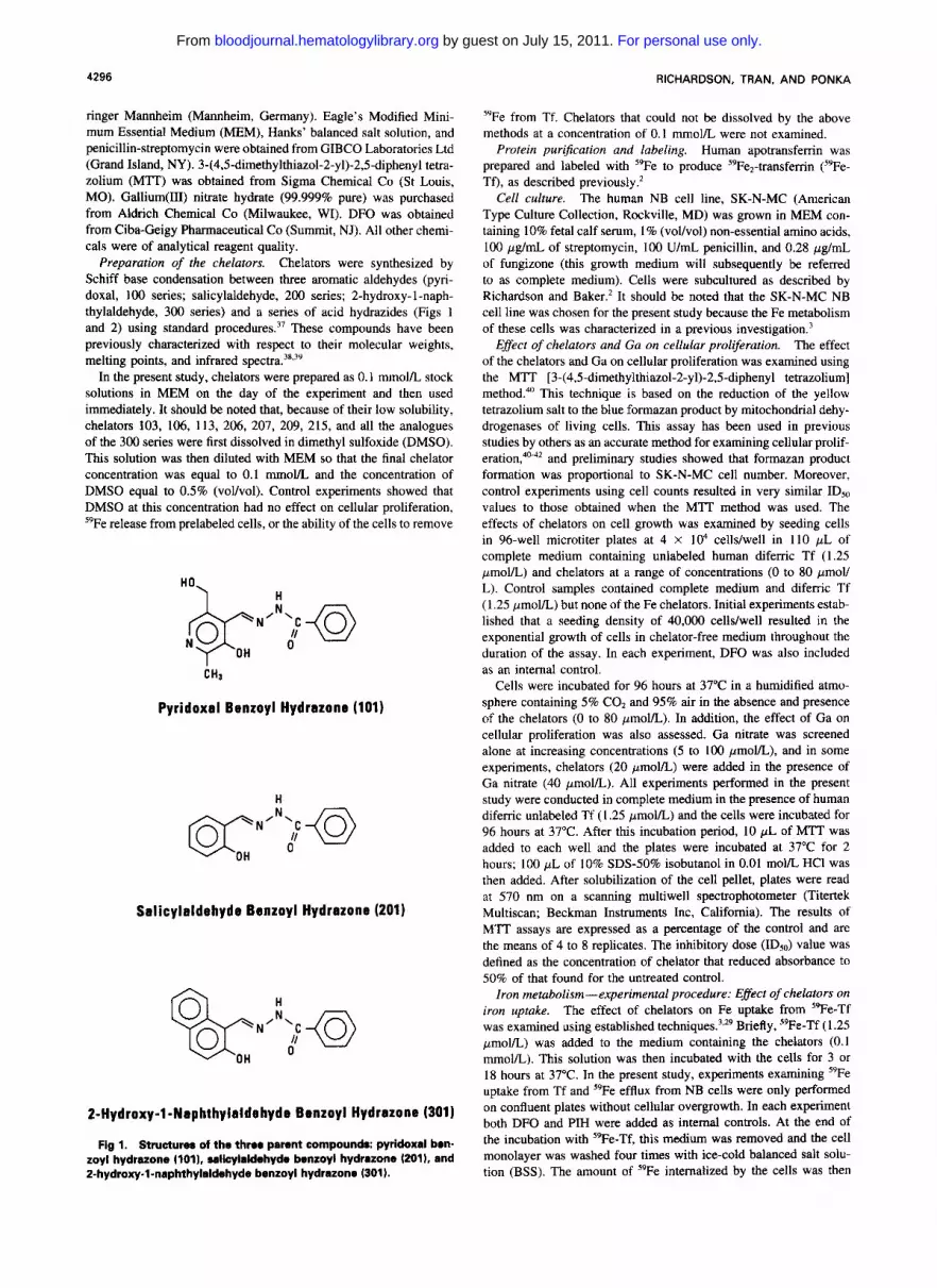
Preparation of the chelators. Chelators were synthesized by Schiff base condensation between three aromatic aldehydes (pyri- doxal, 100 series; salicylaldehyde, 200 series; 2-hydroxy-l-naph- thylaldehyde, 300 series) and a series of acid hydrazides (Figs 1 and 2) using standard procedure^.^' These compounds have been previously characterized with respect to their molecular weights, melting points, and infrared spectra.'*^"
In the present study, chelators were prepared as 0.1 mmol/L stock solutions in MEM on the day of the experiment and then used immediately. It should be noted that, because of their low solubility, chelators 103, 106, 113, 206, 207, 209, 215, and all the analogues of the 300 series were first dissolved in dimethyl sulfoxide (DMSO). This solution was then diluted with MEM so that the final chelator concentration was equal to 0.1 mmovL and the concentration of DMSO equal to 0.5% (vol/vol). Control experiments showed that DMSO at this concentration had no effect on cellular proliferation, "Fe release from prelabeled cells, or the ability of the cells to remove
H0 H
Pyridoxal Benzoyl Hydrazone (101)
H
Salicylaldehyde Benzoyl Hydrazone (201)
2-Hydroxy-l-Naphthylaldehyda Benzoyl Hydrazone (301)
Fig 1. Structurea of the three parent compounda: pyridoxal ben- zoyl hydrazone (1011. salicylaldehyde benzoyl hydrazone (2011, and 2-hydroxy-l-naphthylaldehyde benzoyl hydrazone (3011.
5yFe from Tf. Chelators that could not be dissolved by the above methods at a concentration of 0.1 mmol/L were not examined.
Protein pur$cation and labeling. Human apotransferrin was prepared and labeled with 5yFe to produce S9Fe2-transferrin (59Fe- Tf), as described previously.2
Cell culture. The human NB cell line, SK-N-MC (American Type Culture Collection, Rockville, MD) was grown in MEM con- taining 10% fetal calf serum, I % (voVvol) non-essential amino acids, 100 pg/mL of streptomycin, 100 U/mL penicillin, and 0.28 pg/mL of fungizone (this growth medium will subsequently be referred to as complete medium). Cells were subcultured as described by Richardson and Baker.2 It should be noted that the SK-N-MC NB cell line was chosen for the present study because the Fe metabolism of these cells was characterized in a previous investigation.'
Effect of chelators and Ga on cellular proliferation. The effect of the chelators and Ga on cellular proliferation was examined using the MTT [3-(4,5-dimethylthiazol-2-yl)-2,5-diphenyl tetrazolium] method.'"' This technique is based on the reduction of the yellow tetrazolium salt to the blue formazan product by mitochondrial dehy- drogenases of living cells. This assay has been used in previous studies by others as an accurate method for examining cellular prolif- erati~n,"~' and preliminary studies showed that formazan product formation was proportional to SK-N-MC cell number. Moreover, control experiments using cell counts resulted in very similar IDso values to those obtained when the M'M method was used. The effects of chelators on cell growth was examined by seeding cells in 96-well microtiter plates at 4 X IO4 cells/well in I I O pL of complete medium containing unlabeled human diferric Tf (1.25 pmol/L) and chelators at a range of concentrations (0 to 80 pmoV L). Control samples contained complete medium and difemc Tf (1.25 pmol/L) but none of the Fe chelators. Initial experiments estab- lished that a seeding density of 40,000 cells/well resulted in the exponential growth of cells in chelator-free medium throughout the duration of the assay. In each experiment, DFO was also included as an internal control.
Cells were incubated for 96 hours at 37°C in a humidified atmo- sphere containing 5% CO, and 95% air in the absence and presence of the chelators (0 to 80 pmoIL). In addition, the effect of Ga on cellular proliferation was also assessed. Ga nitrate was screened alone at increasing concentrations (5 to 100 pmoVL), and in some experiments, chelators (20 pmoVL) were added in the presence of Ga nitrate (40 pmoVL). All experiments performed in the present study were conducted in complete medium in the presence of human diferric unlabeled Tf (1.25 pmol/L) and the cells were incubated for 96 hours at 37°C. After this incubation period, 10 pL of M'M was added to each well and the plates were incubated at 37°C for 2 hours; 100 pL of 10% SDS-50% isobutanol in 0.01 m o m HCl was then added. After solubilization of the cell pellet, plates were read at 570 nm on a scanning multiwell spectrophotometer (Titertek Multiscan; Beckman Instruments Inc, California). The results of MTT assays are expressed as a percentage of the control and are the means of 4 to 8 replicates. The inhibitory dose ( I D d value was defined as the concentration of chelator that reduced absorbance to 50% of that found for the untreated control.
Iron metabolism-experimental procedure: Effect of chelators on iron uptake. The effect of chelators on Fe uptake from "Fe-Tf was examined using established technique^.','^ Briefly, We-Tf (1.25 pnol/L) was added to the medium containing the chelators (0.1 mmol/L). This solution was then incubated with the cells for 3 or 18 hours at 37°C. In the present study, experiments examining 5yFe uptake from Tf and "Fe efflux from NB cells were only performed on confluent plates without cellular overgrowth. In each experiment both DFO and PIH were added as internal controls. At the end of the incubation with 59Fe-Tf, this medium was removed and the cell monolayer was washed four times with ice-cold balanced salt solu- tion (BSS). The amount of "Fe internalized by the cells was then
For personal use only. by guest on July 15, 2011. bloodjournal.hematologylibrary.orgFrom

CHELATORS AS ANTIPROLIFERATIVE AGENTS 4291
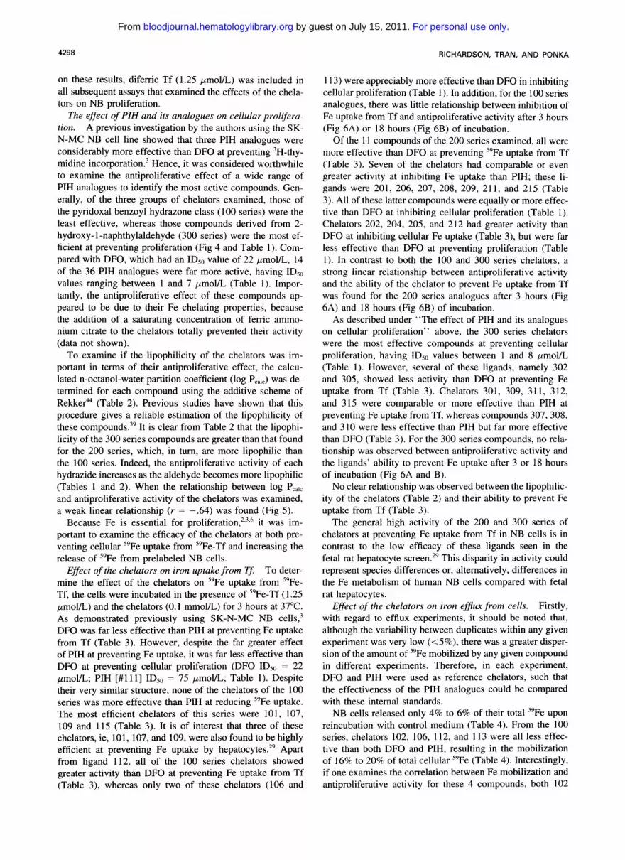
102 - 110
Fig 2. Structures of ana- logues derived from Schiff base condensation of pyridoxal (100 series) with the series of acid hy- drazides. Numbers 102-107: R2 = HO H; 102. R, = -OH; 103, R, -CHI; 104. R, = - N 4 ; 105, R, = -NHS 106, R1 = -C(CHS),; 107, R, = -0CHS; 108-110: R, H; 108, R2 N @ " 0 \N/N,
111, isonicotinoyl; 112. acetyl; CH3
= Cl; 109, Rz = F; 110, RI = Er;
113,2-pyridyl; 114, P-furoyl; 115, 2-thiophenecarboxyl. 113
measured by incubation with the general protease, Pronase (1 mgl mL), for 30 minutes at 4°C to remove membrane-bound s9Fe.2.43 The cells were removed from the plates in the Pronase solution using a teflon spatula, transferred to ice-cold microcentrifuge tubes, and centrifuged for I minute at 14,000 rpm in an Eppendorf microcentri- fuge (Brinkman Instruments Inc, Westbury, NY) to separate intemal- ized from membrane-bound 59Fe in the supernatant. The cell pellet was resuspended in 1 mL of BSS and sonicated for 15 seconds. An aliquot of 1 mL was used to determine protein concentration by the bicinchoninic acid (BCA) method (Pierce Chemical CO, Rockford, IL) as a measure of cell number.' Radioactivity was measured in both the cell pellet and membrane compartment.
The effect of the chelators on iron release from prelabeled cells. The effect of chelators on Fe release from cells was studied using standard procedures.3~24~'5~2' Briefly, cells were prelabeled for 3 hours with MEM containing "Fe-Tf (1.25 pmol/L). After this incubation, the cells were washed four times with ice-cold BSS and reincubated for 3 or 18 hours at 37°C with medium alone or medium containing the chelators (0.1 mmol/L). At the end of the reincubation period, the overlying medium was removed and placed in counting tubes to estimate the release of 59Fe. The cells were then scraped from the plates in 1 mL of BSS using a teflon spatula and placed in counting tubes to estimate cellular "Fe.
UV-Vis spectrophotometry. Spectrophotometric studies were conducted to examine whether PIH bound Ca. Increasing concentra- tions of Ga nitrate (5 to 50 pmoUL) were added to PIH (50 pmoll L) in phosphate-buffered saline (PBS; pH 7.4). After thorough mix- ing, the solutions were then examined spectrophotometrically be- tween 200 and 525 nm on a Hewlett Packard 845lA Diode Array Spectrophotometer (Hewlett Packard, Idaho) using a l-cm quartz cuvette. Spectra were recorded against PBS (pH 7.4) when examin- ing the spectrum of PIH only, or against PBS (pH 7.4) and Ga nitrate (5 to 50 pmol/L) when recording the spectrum of the PIH- Ga complex.
Calculation of the n-octunobwater partition coeficients (log P<J by fhe udditive scheme of Rekker.44 The n-octanol-water partition coefficients of the 100, 200, and 300 series have been calculated by the additive scheme of Rekker,@ that was described in detail in Ponka et al."
RESULTS
The effect of DFO on cellular proliferation in the presence and absence of human diferric T$ Many previous studies
112
have examined the effect of DFO on cell growth in the absence of human diferric Tf.7.8,45 As a result, at least part of the antiproliferative effect observed may be due to the inability of the cells to obtain Fe; hence, the results may have little physiologic relevance. Considering this fact, ex- periments were conducted with DFO in the presence of hu- man diferric Tf at a concentration of 1.25 pmol/L ([Fe] = 2.5 pmol/L). This Tf concentration was chosen because it is well in excess of that required for saturation of the Tf receptor in these cells.'
At DFO concentrations less than 40 pmol/L, the presence of diferric Tf appreciably prevented the effect of the chelator (Fig 3). In contrast, at DFO concentrations greater than 40 pmol/L, diferric Tf had little effect on its antiproliferative action, suggesting that the rate of Fe uptake from Tf was not sufficient to equal the rate of Fe chelation by DFO. Based
I I I I I 100
+ Tf
40
20
0 ' I I 1 I
0 20 40 60 80
Deafemoxamino Concentration (PM)
Fig 3. Effect of DFO on the proliferation of SK-N-MC neuro- blastoma cells in the presence and absence of human diferric Tf (1.25 pmol/L). Cells were incubated with DFO (0 to 80 pmol/L) in the presence and absence of diferric Tf for 96 hours at 37°C. After this incubation period, cell density was then measured via the MlT assay (see Materials and Methods for details). Each data point is the mean of 8 replicates in a typical experiment.
For personal use only. by guest on July 15, 2011. bloodjournal.hematologylibrary.orgFrom
4298 RICHARDSON, TRAN, AND PONKA
on these results, diferric Tf (1.25 pmol/L) was included in all subsequent assays that examined the effects of the chela- tors on NB proliferation.
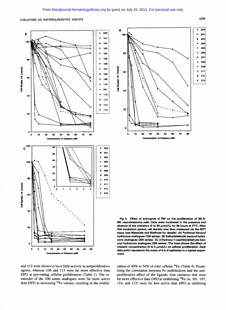
The effect of PIH and its analogues on cellular prolifercr- tion. A previous investigation by the authors using the SK- N-MC NB cell line showed that three PIH analogues were considerably more effective than DFO at preventing 3H-thy- midine incorporation.' Hence, it was considered worthwhile to examine the antiproliferative effect of a wide range of PIH analogues to identify the most active compounds. Gen- erally, of the three groups of chelators examined, those of the pyridoxal benzoyl hydrazone class (100 series) were the least effective, whereas those compounds derived from 2- hydroxy-l-naphthylaldehyde (300 series) were the most ef- ficient at preventing proliferation (Fig 4 and Table 1). Com- pared with DFO, which had an ID5" value of 22 pmol/L, 14 of the 36 PIH analogues were far more active, having ID5,] values ranging between 1 and 7 pmol/L (Table 1). Impor- tantly, the antiproliferative effect of these compounds ap- peared to be due to their Fe chelating properties, because the addition of a saturating concentration of ferric ammo- nium citrate to the chelators totally prevented their activity (data not shown).
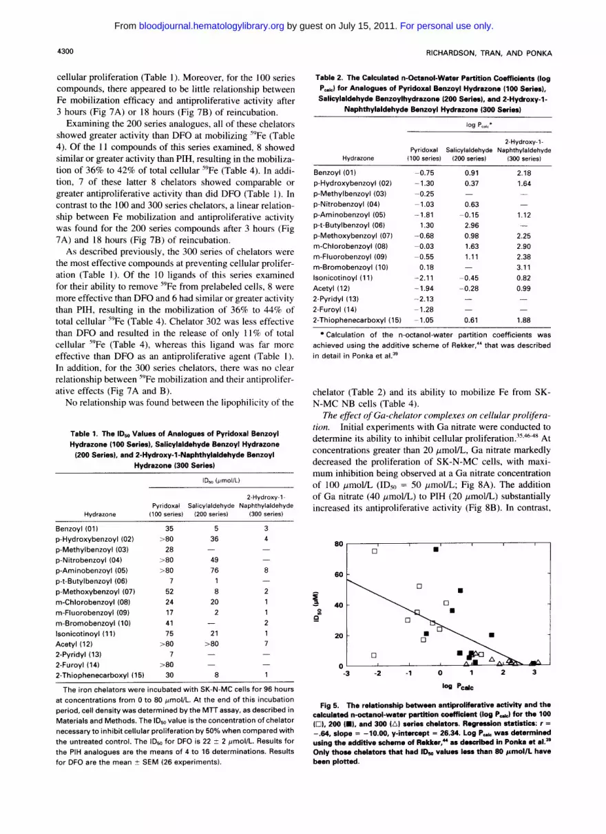
To examine if the lipophilicity of the chelators was im- portant in terms of their antiproliferative effect, the calcu- lated n-octanol-water partition coefficient (log Pcalc) was de- termined for each compound using the additive scheme of Rekker" (Table 2). Previous studies have shown that this procedure gives a reliable estimation of the lipophilicity of these compounds.'' It is clear from Table 2 that the lipophi- licity of the 300 series compounds are greater than that found for the 200 series, which, in turn, are more lipophilic than the 100 series. Indeed, the antiproliferative activity of each hydrazide increases as the aldehyde becomes more lipophilic (Tables 1 and 2). When the relationship between log P,,,, and antiproliferative activity of the chelators was examined, a weak linear relationship ( r = -.64) was found (Fig 5) .
Because Fe is essential for proliferation,*^'^' it was im- portant to examine the efficacy of the chelators at both pre- venting cellular 59Fe uptake from 59Fe-Tf and increasing the release of 59Fe from prelabeled NB cells.
Effect of the chelators on iron uptake from T$ To deter- mine the effect of the chelators on 5yFe uptake from 59Fe- Tf, the cells were incubated in the presence of 59Fe-Tf (1.25 pmol/L) and the chelators (0.1 mmol/L) for 3 hours at 37°C. As demonstrated previously using SK-N-MC NB cells,3 DFO was far less effective than PIH at preventing Fe uptake from Tf (Table 3). However, despite the far greater effect of PIH at preventing Fe uptake, it was far less effective than DFO at preventing cellular proliferation (DFO ID5" = 22 pmol/L; PIH [#l 1 l ] IDSo = 75 pmol/L; Table 1). Despite their very similar structure, none of the chelators of the 100 series was more effective than PIH at reducing "Fe uptake. The most efficient chelators of this series were 101, 107, 109 and 115 (Table 3). It is of interest that three of these chelators, ie, 101, 107, and 109, were also found to be highly efficient at preventing Fe uptake by hepatocyte^.'^ Apart from ligand 112, all of the 100 series chelators showed greater activity than DFO at preventing Fe uptake from Tf (Table 3), whereas only two of these chelators (106 and
1 13) were appreciably more effective than DFO in inhibiting cellular proliferation (Table l). In addition, for the 100 series analogues, there was little relationship between inhibition of Fe uptake from Tf and antiproliferative activity after 3 hours (Fig 6A) or 18 hours (Fig 6B) of incubation.
Of the 11 compounds of the 200 series examined, all were more effective than DFO at preventing "Fe uptake from Tf (Table 3). Seven of the chelators had comparable or even greater activity at inhibiting Fe uptake than PIH; these li- gands were 201, 206, 207, 208, 209, 211, and 215 (Table 3). All of these latter compounds were equally or more effec- tive than DFO at inhibiting cellular proliferation (Table 1). Chelators 202, 204, 205, and 212 had greater activity than DFO at inhibiting cellular Fe uptake (Table 3), but were far less effective than DFO at preventing proliferation (Table I ) . In contrast to both the 100 and 300 series chelators, a strong linear relationship between antiproliferative activity and the ability of the chelator to prevent Fe uptake from Tf was found for the 200 series analogues after 3 hours (Fig 6A) and 18 hours (Fig 6B) of incubation.
As described under "The effect of PIH and its analogues on cellular proliferation" above, the 300 series chelators were the most effective compounds at preventing cellular proliferation, having IDs0 values between 1 and 8 pmoVL (Table 1). However, several of these ligands, namely 302 and 305, showed less activity than DFO at preventing Fe uptake from Tf (Table 3). Chelators 301, 309, 3 1 1, 312, and 315 were comparable or more effective than PIH at preventing Fe uptake from Tf, whereas compounds 307,308, and 3 10 were less effective than PIH but far more effective than DFO (Table 3). For the 300 series compounds, no rela- tionship was observed between antiproliferative activity and the ligands' ability to prevent Fe uptake after 3 or 18 hours of incubation (Fig 6A and B).
No clear relationship was observed between the lipophilic- ity of the chelators (Table 2) and their ability to prevent Fe uptake from Tf (Table 3).
The general high activity of the 200 and 300 series of chelators at preventing Fe uptake from Tf in NB cells is in contrast to the low efficacy of these ligands seen in the fetal rat hepatocyte screen.*' This disparity in activity could represent species differences or, alternatively, differences in the Fe metabolism of human NB cells compared with fetal rat hepatocytes.
Effect c$ the chelators on iron efflux from cells. Firstly, with regard to efflux experiments, it should be noted that, although the variability between duplicates within any given experiment was very low (<5%), there was a greater disper- sion of the amount of 59Fe mobilized by any given compound in different experiments. Therefore, in each experiment, DFO and PIH were used as reference chelators, such that the effectiveness of the PIH analogues could be compared with these internal standards.
NB cells released only 4% to 6% of their total "Fe upon reincubation with control medium (Table 4). From the 100 series, chelators 102, 106, 112, and 1 13 were all less effec- tive than both DFO and PIH, resulting in the mobilization of 16% to 20% of total cellular "Fe (Table 4). Interestingly, if one examines the correlation between Fe mobilization and antiproliferative activity for these 4 compounds, both 102
For personal use only. by guest on July 15, 2011. bloodjournal.hematologylibrary.orgFrom
CHELATORS AS ANTIPROLIFERATIVE AGENTS
A
100
Bo
3 f” 40
20
0 0 l 0 70 80
C 120
I
H E
i 3
1 W
60
40-
20 -
0 ,
\ \ \ \ \ *
l . . . . , . . . . , . . . , . . . . , . . . . , . . . . , . . . . , . l
C o n c m n t n t l o n of clwmon W) 0 5 10 l 6 20 26 30 36 40 46
* DFO
0 101
0 102
8 103
v 104
v 105
v 106
0 107
108
0 109
A 110
A PIH
A 112
0 l13
114
@ 115
* WO
301
v 302
v 306
0 307
308
308
A 310
A 311
0 312
0 316
and 112 were shown to have little activity as antiproliferative agents, whereas 106 and 113 were far more effective than DFO at preventing cellular proliferation (Table 1). The re- mainder of the 100 series analogues were far more active than DFO at increasing J9Fe release, resulting in the mobili-
100
80
80
40
20
0
4299
A 208
A 209
4 211
212
@ 215
Fig 4. Effect of analogues of PIH on the proliferation of SK-N- MC neuroblastoma cells. Cells were incubated in the presence and absence of the chelators (0 to 80 pmollL) for 96 hours at 37°C. After this incubation period, cell density wes then measured via the MTT assay (see Materials and Methods for details). (A) Pyridoxal benzoyl hydrazone analogues (100 series). (B) Salicylaldehyde benzoyl hydra- zone analoguas (200 series). (C) 2-Hydroxy-l-naphthyleldehyde ben- zoyl hydrazone analogues (300 series). The inset shows the effect of chelator concentration (0 to 5 pmollL) on cellular proliferation. Each data point represents the mean of 4 8 a to replicates in typical experi- ment.
zation of 40% to 54% of total cellular 59Fe (Table 4). Exam- ining the correlation between Fe mobilization and the anti- proliferative effect of the ligands, four chelators that were far more effective than DFO at mobilizing 59Fe (ie, 101, 107, 114, and 115) were far less active than DFO at inhibiting
For personal use only. by guest on July 15, 2011. bloodjournal.hematologylibrary.orgFrom
4300
cellular proliferation (Table 1) . Moreover, for the 100 series compounds, there appeared to be little relationship between Fe mobilization efficacy and antiproliferative activity after 3 hours (Fig 7A) or 18 hours (Fig 7B) of reincubation.
Examining the 200 series analogues, all of these chelators showed greater activity than DFO at mobilizing "Fe (Table 4). Of the 1 1 compounds of this series examined, 8 showed similar or greater activity than PIH, resulting in the mobiliza- tion of 36% to 42% of total cellular 59Fe (Table 4). In addi- tion, 7 of these latter 8 chelators showed comparable or greater antiproliferative activity than did DFO (Table l). In contrast to the 100 and 300 series chelators, a linear relation- ship between Fe mobilization and antiproliferative activity was found for the 200 series compounds after 3 hours (Fig 7A) and 18 hours (Fig 7B) of reincubation.
As described previously, the 300 series of chelators were the most effective compounds at preventing cellular prolifer- ation (Table 1). Of the 10 ligands of this series examined for their ability to remove 5yFe from prelabeled cells, 8 were more effective than DFO and 6 had similar or greater activity than PIH, resulting in the mobilization of 36% to 44% of total cellular "Fe (Table 4). Chelator 302 was less effective than DFO and resulted in the release of only 11 % of total cellular "Fe (Table 4), whereas this ligand was far more effective than DFO as an antiproliferative agent (Table 1). In addition, for the 300 series chelators, there was no clear relationship between 5yFe mobilization and their antiprolifer- ative effects (Fig 7A and B).
No relationship was found between the lipophilicity of the
Table 1. The ID,, Values of Analogues of Pyridoxal Benzoyl Hydrazone (100 Series), Salicylaldehyde Benzoyl Hydrazone
(200 Series), and 2-Hydroxy-l-Naphthylaldehyde Benzoyl Hydrazone (300 Series)
IDso IprnollL)
Pyridoxal Salicylaldehyde Naphthylaldehyde 2-Hydroxy-l-
Hydrazone 1100 series) (200 series) 1300 series)
Benzoyl (01) 35 5 3
p-Hydroxybenzoyl (02) >80 36 4
p-Methylbenzoyl (03) 28 - -
p-Nitrobenzoyl (04) >80 49 -
p-Aminobenzoyl (05) >80 76 8
p-t-Butylbenzoyl (06) 7 1 - p-Methoxybenzoyl (07) 52 8 2 m-Chlorobenzoyl (08) 24 20 1 m-Fluorobenzoyl (09) l ? 2 1 m-Bromobenzoyl (IO) 41 - 2 lsonicotinoyl (11) 75 21 1 Acetyl (12) >80 280 7 2-Pyridyl (13) 7 - -
2-Furoyl (14) >80 - -
2-Thiophenecarboxyl (15) 30 8 1
The iron chelators were incubated with SK-N-MC cells for 96 hours at concentrations from 0 to 80 pnol/L. At the end of this incubation period, cell density was determined by the MTT assay, as described in Materials and Methods. The IDso value is the concentration of chelator necessary to inhibit cellular proliferation by 50% when compared with the untreated control. The IDso for DFO is 22 2 2 pmol/L. Results for the PIH analogues are the means of 4 to 16 determinations. Results for DFO are the mean 2 SEM (26 experiments).
RICHARDSON, TRAN, AND PONKA
Table 2. The Calculated n-Octanol-Water Partition Coefficients (log P d for Analogues of Pyridoxal Benzoyl Hydrazone (100 Series), Salicylaldehyde Benzoylhydrazone (200 Seriss), and 2-Hydroxy-l-
Naphthylaldehyde Benzoyl Hydrazone (300 Series)
1% P,,I,*
2-Hydroxy-l-
Hydrazone (100 series) 1200 series) 1300 series) Pyridoxal Salicylaldehyde Naphthylaldehyde
Benzoyl (01) -0.75 0.91 2.18 p-Hydroxybenzoyl (02) -1.30 0.37 1.64 p-Methylbenzoyl (03) -0.25 p-Nitrobenzoyl (04) -1.03 0.63 p-Aminobenzoyl (05) p-t-Butylbenzoyl (06) 1.30 2.96 p-Methoxybenzoyl (07) -0.68 0.98 2.25 m-Chlorobenzoyl (08) -0.03 1.63 2.90 m-Fluorobenzoyl (09) -0.55 1.11 2.38 m-Bromobenzoyl (10) 0.18 - 3.1 1 lsonicotinoyl (11) "2.11 -0.45 0.82 Acetyl ( 12) -1.94 -0.28 0.99 2-Pyridyl (13) -2.13 - - 2-Furoyl (14) - 1.28 2-Thiophenecarboxyl (15) -1.05 0.61 1.88
- - -
- 1.81 -0.15 1.12 -
- -
Calculation of the n-octanol-water partition coefficients was achieved using the additive scheme of Rekker,44 that was described in detail in Ponka et al.39
chelator (Table 2) and its ability to mobilize Fe from SK- N-MC NB cells (Table 4).
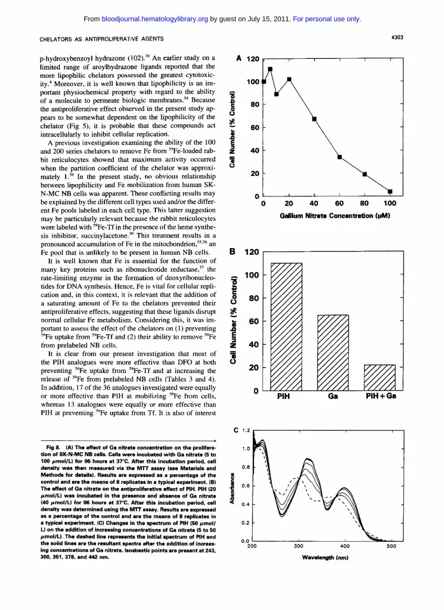
The effect of Gu-chelator complexes on cellular prolifera- tion. Initial experiments with Ga nitrate were conducted to determine its ability to inhibit cellular p r o l i f e r a t i ~ n . ' ~ ~ ~ ~ - ~ ~ At concentrations greater than 20 pmol/L, Ga nitrate markedly decreased the proliferation of SK-N-MC cells, with maxi- mum inhibition being observed at a Ga nitrate concentration of 100 pmol/L (IDSo = 50 pmoVL; Fig 8A). The addition of Ga nitrate (40 pmol/L) to PIH (20 pmoVL) substantially increased its antiproliferative activity (Fig 8B). In contrast,
80 I I I I
0 B
60 -
I n &m\ " -3 -2 -1 0 1 2 3
'W pcalc
Fig 5. The relationship between antiproliferative activity and the calculated n-octanol-water partition coefficient (log P&) for the 100 10). 200 W, and 300 (A) series chelators. Regression statistics: r = -.M, slope = -10.00. y-intercept = 26.34. Log Pab was determined using the additive scheme of Rekker,- as described in Ponka et al." Only those chelators that had IDw values less than 80 pmollL have been plotted.
For personal use only. by guest on July 15, 2011. bloodjournal.hematologylibrary.orgFrom

CHELATORS AS ANTIPROLIFERATIVE AGENTS
the addition of Ga nitrate to DFO did not enhance its antipro- liferative effect (data not shown).
TO further investigate the mechanism by which Ga was able to potentiate the antiproliferative effect of PIH, spectro- photometric studies were performed to determine whether this chelator could bind Ga. Upon the addition of Ga nitrate to PIH, a marked spectral change was evident (Fig 8C) that was consistent with complexation. The addition of Ga re- sulted in a shift of the maximum absorbance peaks to the right, with isosbestic points being present at 243, 300, 351, 378, and 442 nm (Fig 8C).
DISCUSSION
Effect of the chelators on cellular proliferation, iron up- take from TJ and iron release from NB cells. Furst4' has suggested that many drugs that inhibit the growth of cancer cells are, or have the potential to be, Fe chelators. In fact, the high activity of DFO at preventing the proliferation of tumor cells has suggested that this chelator may be of bene- ficial use in the treatment of several neoplastic disease^."'^ However, it is well known that chelation therapy with DFO suffers from several serious problem^,^' including high cost, the need for subcutaneous infusion of the drug (12 to 24 N d, 5 to 6 nightdwk), and poor intestinal absorption preclud- ing oral administration. Hence, these serious disadvantages of DFO have stimulated the search for alternative chelators that are economical, orally effective, and highly efficient. One group of Fe chelators that satisfies all these three criteria are those of the PIH cl as^.'^^^"^^^ These ligands show high
Table 3. The Effect of Analogues of PIH on Internalized Iron Uptake From Tf by SK-N-MC NB Cells
Internalized Iron Uptake (% control)
Pyridoxal Salicylaldehyde Naphthylaldehyde 2-Hydroxy-l-
Hydrazone (100 series) (200 series) (300 series)
Benzoyl (01) 24 5 8 p-Hydroxybenzoyl (02) 69 32 90 p-Methylbenzoyl (03) 32 p-Nitrobenzoyl (04) 46 44 p-Aminobenzoyl (05) 49 61 78 p-t-Butylbenzoyl (06) 75 4 - p-Methoxybenzoyl (07) 25 5 21 m-Chlorobenzoyl (08) 46 13 30 m-Fluorobenzoyl (09) 25 3 14 m-Bromobenzoyl (10) 49 - 41 lsonicotinoyl (1 1) - 8 7 Acetyl (12) 92 62 9 2-Pyridyl (13) 51 2-Furoyl (14) 31 2-Thiophenecarboxyl (15) 19 5
Control 100 1 oa 100 DFO reference 80 71 71 PIH reference 19 14 14
- - -
- -
- - 8
Cells were incubated for 3 hours at 37°C with %Fe-Tf (1.25 pmol/L) in the presence or absence of the chelators (0.1 mmol/L). After this incubation, the cells were washed and incubated with pronase (1 mg/ mL) for 30 minutes at 4°C to separate internalized from membrane- bound 59Fe. Results are the means of duplicate determinations in a typical experiment.
80
60
40
20
4301
0 0 30 60 90 120 Internalised Iron Uptake (nmoles FelgPR)
80
60
40
20
0 0 200 400 Internalised Iron Uptake (nmoles FelgPR)
Fig 6. The relationship between the abi l i i of the chelator to pre- vent internalized iron uptake from =Fe-Tf and its antiproliierative activity in SK-N-MC NB cells. Plot of the IDm values of chelators of the 100 10). 200 (m), and 300 (A) series versus their ability to inhibit internalized "Fe uptake from '*Fe-Tf after (A) 3 hours of incubation or (B) after 18 hours of incubation. It should be notad that low intar- nalized Fe uptake results from high chelation activity. Regression statistics for the 200 series chelators after 3 hours of incubation: r = .97, slope = 0.81, y-intercept = 0.45; or after 18 hours of incubation: r = .98, slope = 0.32. y-intercept = -0.79. Only those chelators that had IDso values less than 80 pmollL have been plotted.
biologic activity probably due to the following: ( 1 ) an opti- mal hydrophilicAipophilic balan~e'~; (2) high affinity and selectivity for Fe(III)27351; and (3) the apochelator is predomi- nately in the neutral form at pH 7.4,52 allowing easy access through biologic membranes to intracellular Fe pools.
Previous studies have shown that PIH and several of its analogues show high activity at preventing Fe uptake from Tf and increasing the release of Fe from both n ~ r m a l ~ ~ . ' ~ and neoplastic cell^,'^^^ being far more effective than DFO.
For personal use only. by guest on July 15, 2011. bloodjournal.hematologylibrary.orgFrom

4302
Table 4. Mobilization of "Fe From SK-N-MC NB Cells by Analogues of PIH
% "Fe Released From Cells
2-Hydroxy-l-
Hydrazone (100 series) (200 series) (300 series) Pyridoxal Salicylaldehyde Naphthylaldehyde
Benzoyl (01) 53 41 39 p-Hydroxybenzoyl (02) 16 41 11 p-Methylbenzoyl (03) 49 p-Nitrobenzoyl (04) 40 21 p-Aminobenzoyl (05) 45 24 14 p-t-Butylbenzoyl (06) 18 38 p-Methoxybenzoyl (07) 53 41 36 m-Chlorobenzoyl (08) 47 36 32 m-Fluorobenzoyl (09) 52 42 38 m-Bromobenzoyl (IO) 46 - 27 lsonicotinoyl (11) - 39 44 Acetyl ( 12) 16 25 42 2-Pyridyl (13) 20 - -
2-Furoyl (14) 2-Thiophenecarboxyl (15) 54 42 41
Control 6 4 4 DFO reference 29 15 15 PIH reference 52 38 38
- -
-
-
52 - -
Cells were incubated for 3 hours at 37°C with -Fe-Tf (1.25 pmol/L) and then washed and reincubated in the presence of the chelators (0.1 mmol/L) for 3 hours at 37°C. After this incubation period, the reincubation medium was removed and placed into counting tubes. The cell monolayer was then scraped from the plates in 1 mL of BSS using a teflon spatula and transferred into counting tubes. Results are means of duplicate determinations in a typical experiment.
Moreover, several analogues of PIH were also far more effi- cient than DFO at preventing the 3H-thymidine incorporation by NB cells3 Considering the possible therapeutic potential of these chelators, the present study was designed to investi- gate the antiproliferative activity of a broad range of PIH analogues to identify the most effective compounds that could then be investigated in detail. In addition, the effects of the chelators on preventing 59Fe uptake from 5qe-Tf and mobilizing 59Fe from prelabeled cells was also examined.
As shown in Figs 1 and 2, all of the chelators apart from the three 2-pyridyl hydrazones possess the same Fe-binding site as that found in PIH, viz, the carbonyl oxygen, aldimine nitrogen, and aromatic ring hydroxyl. Hence, all of these chelators should be highly effective Fe-chelating agents. However, these chelators show marked differences in both antiproliferative activity and Fe chelation efficacy even within the same series of compounds (Tables 1, 3, and 4 and Fig 4). In terms of the activity of a chelator as an antipro- liferative agent, the lipophilicity of the molecule appears to be an important factor in determining efficacy. Examining the 100, 200, and 300 series compounds (Figs 1 and 2), it is evident that the 100 series chelators derived from the pyridoxal ring are the least lipophilic due to the presence of the hydroxymethyl oxygen atom and 1 nitrogen atom, whereas the 300 series chelators derived from 2-hydroxy- 1- naphthylaldehyde are the most lipophilic due to the presence of the hydrophobic aromatic rings of the naphthylaldehyde group (Table 2). Certainly, the antiproliferative activity of
RICHARDSON, TRAN, AND PONKA
every hydrazide increases as the aldehyde becomes more lipophilic (Table 1) . For example, pyridoxal p-hydroxyben- zoyl hydrazone (102) is less effective than salicylaldehyde p-hydroxybenzoyl hydrazone (202), which, in turn, is less effective than 2-hydroxy- 1 -naphthylaldehyde p-hydroxyben- zoyl hydrazone (302). In addition, even within the same series of compounds, eg, the 100 series, it is obvious that the most lipophilic derivative, pyridoxal p-t-butylbenzoyl hydrazone (106),39 is far more effective as an antiprolifera- tive agent than more hydrophilic analogues such as pyridoxal
80
60
40
20
0
\
H \ 0
80
60
40
20
0
0 10 20 30 40 50 60
% Total Iron Released from Cells
Fia 7. The
L
30 40 50 60 70 % Total Iron Released from Cells
lationship between the a b i l i of a chelator to release Fe from pralabeled SK-NiMC NB cells and its antiproliferative activity. Plot of the IDso values of the 100 (U), 200 (W, and 300 (A) series versus their ability to release 69Fe from NB cells prelabeled with "Fe- Tf (1.25 pmol/L) for 3 hours followed by reincubation in the presence of the chelators for (A) 3 hours or (B) 18 hours. Regression statistics for the 200 series chelators after 3 hours of reincubation: r = -.83, slope = -2.64, y-intercept = 118.96; or after 18 hours of reincubation: r = -.80, slope = -6.08, y-intercept = 376.07. Only those chelators that had IDso values less than 80 pmol/L have been plotted.
re
For personal use only. by guest on July 15, 2011. bloodjournal.hematologylibrary.orgFrom
CHELATORS AS ANTIPROLIFERATIVE AGENTS
p-hydroxybenzoyl hydrazone (102).39 An earlier study on a limited range of aroylhydrazone ligands reported that the more lipophilic chelators possessed the greatest cytotoxic- ity." Moreover, it is well known that lipophilicity is an im- portant physiochemical property with regard to the ability of a molecule to permeate biologic membrane^.'^ Because the antiproliferative effect observed in the present study ap- pears to be somewhat dependent on the lipophilicity of the chelator (Fig 5), it is probable that these compounds act intracellularly to inhibit cellular replication.
A previous investigation examining the ability of the 100 and 200 series chelators to remove Fe from 59Fe-loaded rab- bit reticulocytes showed that maximum activity occurred when the partition coefficient of the chelator was approxi- mately In the present study, no obvious relationship between lipophilicity and Fe mobilization from human SK- N-MC NB cells was apparent. These conflicting results may be explained by the different cell types used and/or the differ- ent Fe pools labeled in each cell type. This latter suggestion may be particularly relevant because the rabbit reticulocytes were labeled with 5yFe-Tf in the presence of the heme synthe- sis inhibitor, suc~inylacetone.~~ This treatment results in a pronounced accumulation of Fe in the mito~hondrion,5'.~~ an Fe pool that is unlikely to be present in human NB cells.
It is well known that Fe is essential for the function of many key proteins such as ribonucleotide reductase,57 the rate-limiting enzyme in the formation of deoxyribonucleo- tides for DNA synthesis. Hence, Fe is vital for cellular repli- cation and, in this context, it is relevant that the addition of a saturating amount of Fe to the chelators prevented their antiproliferative effects, suggesting that these ligands disrupt normal cellular Fe metabolism. Considering this, it was im- portant to assess the effect of the chelators on (1) preventing "Fe uptake from 59Fe-Tf and (2) their ability to remove 59Fe from prelabeled NB cells.
It is clear from our present investigation that most of the PIH analogues were more effective than DFO at both preventing 59Fe uptake from 59Fe-Tf and at increasing the release of 59Fe from prelabeled NB cells (Tables 3 and 4). In addition, 17 of the 36 analogues investigated were equally or more effective than PIH at mobilizing 59Fe from cells, whereas 13 analogues were equally or more effective than PIH at preventing 5yFe uptake from Tf. It is also of interest
b
Fig 8. (A) The effect of Ga nitrate concentration on the prolifera- tion of SK-N-MC NB cells. Cells were incubated with Ga nitrate (5 to l00 pmollL) for 96 hours at 37°C. After this incubation period, cell density was thon measured via the MIT assay (see Materials and Methods for details). Results are expressed as a percentage of the control and am the means of 8 replicates in a typical experiment. (B) The effect of Ga nitrate on the antiproliferative effect of PIH. PIH (20 pmol/L) was incubated in the presence and absence of Ga nitrate (40 pmollL) for 96 hours at 37°C. After this incubation period, cell density was determined using the MlT assay. Results are expressed as a percentage of the control and are the means of 8 replicates in a typical experiment. IC) Changes in the spectrum of PIH (50 pmoll L) on the addition of increasing concentrations of Ga nitrate (5 to 50 pmollLI. The dashed line represents the initial spectrum of PIH and the solid lines are the reaukant spectra after the addition of increas-
300,351,378, and 44.2 nm. ing concentrations of Ga nitrate. Isosbestic points are present at 243,
4303
l
0 20 40 60 80 100
Gallium Nitrate Concentration (VU)
" PIH Ga PIH + Ga
c 1.2
200 300 400 500
Wavdength (nm)
For personal use only. by guest on July 15, 2011. bloodjournal.hematologylibrary.orgFrom

4304 RICHARDSON, TRAN, AND PONKA
that the most effective chelators at preventing Fe uptake were also highly effective at mobilizing 5’Fe from prelabeled cells (eg, 201, 206, 207, 209, 301, 309, and 312). Examining the correlation between Fe chelation efficacy and antiprolif- erative activity, for both the 100 and 300 series compounds no relationship was apparent, whereas for the 200 series a linear relationship was observed (Figs 6 and 7).
Considering the question of why a lack of relationship exists between Fe chelation efficacy and antiproliferative activity for the 100 and 300 series, it is relevant to note that DFO is far less effective than PIH at both preventing Fe uptake (Table 3) or increasing the mobilization of Fe from NB cells (Table 4), whereas DFO is far more effective than PIH at preventing cellular proliferation (Table 1). These seemingly paradoxical results could be explained by the fact that there are numerous Fe pools within cells and that differ- ent ligands may have the ability to chelate different Fe pools. In the case of DFO, for example, once the Fe complex (ferri- oxamine) has formed within the cell, there is a kinetic block to diffusion resulting in an intracellular accumulation offer- rioxamine and slow Fe release.’’ Hence, a chelator that binds Fe from a pool essential for DNA synthesis will probably have marked cytostatic properties, and, if this Fe complex remains within the cell, it will mobilize little Fe, resulting in poor correlation between Fe chelation efficacy and cellular proliferation.
It is of particular interest to note that some of the least cytostatic compounds (ie, PIH, 101, and 107) were shown to be highly effective Fe chelators in both the present study and in previous investigations using the hepatocyte and retic- ulocyte model^.*^,*^ Moreover, in general, the pyridoxal ana- logues show high Fe chelation efficacy but low antiprolifera- tive activity, properties that make them suitable candidates as drugs to treat Fe-overload disease. In contrast, the more lipophilic compounds of the 200 and 300 series (eg. 201, 209, 301, and 3 11) display both high chelation efficacy and marked antiproliferative activity and show promise as anti- neoplastic agents. Hence, these data provide essential clues for the design of future aroylhydrazone ligands. Further stud- ies using molecular modelling and the results of the present investigation may lead to the synthesis of more efficient Fe chelators either for the treatment of Fe-overload or cancer.
Of the 36 PIH analogues examined in the present study, chelators 206, 209, 308, 309, 31 1, and 3 15 appear to be especially promising as antineoplastic agents. These com- pounds were the most effective at inhibiting proliferation and were also far more efficient than DFO at both inhibiting Fe uptake from Tf and increasing Fe mobilization from NB cells. However, even though many of these chelators show much higher activity as antiproliferative agents than DFO, further studies are necessary to investigate the selectivity of their antineoplastic effect. This will be performed by com- paring the effects of the compounds on a broad range of tumor cells compared with a range of normal cells. In addi- tion, the potential of these ligands will also be assessed via examination of their action in nude mice bearing human NB xenografts.
Effect of Ga-chelator complexes on cellular proliferation. Ga is a group IIIA metal that can bind to the Fe-binding sites of the Tf molecule and can be taken up by cells via
endocytosis of Tf and by a Tf-independent mechani~rn .~’ .~~ In addition, Ga has been shown to be an effective agent for the treatment of metastatic bladder cancer59 and lymphoma.‘” The antineoplastic effect of Ga appears to be related to the fact that the metal has some chemical similarities to Fe(III), which results in the disruption of normal cellular Fe metabo-
Considering the properties of Ga and the fact that chelators of the PIH class become more cytotoxic upon complexation with metals: it was considered worthwhile to examine the antiproliferative effect of adding PIH and Ga together.
When NB cells were incubated in the presence of Ga nitrate, a dose-dependent antiproiiferative response was ob- tained (Fig 8A) that was similar to that seen with bladder carcinoma cells.” When Ga was added to PIH, it potentiated the antiproliferative effect of the ligand (Fig 8B), whereas the addition of Ga to DFO did not enhance its effect. Spectro- photometric studies showed a pronounced change in the spectrum of PIH upon the addition of Ga (Fig K ) , sug- gesting complexation. Regarding the mechanism by which Ga enhances the antiproliferative effect of PIH, it is well known that the PIH-Fe complex can efficiently donate Fe to reticulocytes,“ and it can be suggested that PIH-Ga complex may bestow Ga to the NB cell. Indeed, further studies will investigate the antiproliferative activity of the Ga complexes of the remaining PIH analogues. Considering the enhanced antiproliferative effect of PIH complexed with Ga(II1) and analogue 201 complexed to copper(I1): it may be worth- while to examine the antiproliferative effect of PIH and its analogues with other metals such as platinum, palladium, and zinc. In fact, previous studies with other chelators have shown that complexation with these metals results in a con- siderable increase in c y t o t o x i ~ i t y . ” ~ ’ ~ ~ ~ ~ ~ ~
In conclusion, some analogues of PIH, particularly those of the 2-hydroxy-l-naphthylaldehyde series, show great po- tential as effective antiproliferative agents, being far more effective than DFO. These chelators deserve further vigorous investigation because they may be of use in the treatment of cancers such as NB, which appear to be relatively sensitive to Fe chelation therapy.””
~iSm.3S.48.hl
ACKNOWLEDGMENT
We acknowledge Ania Wilczynska and Daniel. Bradshaw for ex- pert technical assistance during the present study. Dr Ann English and Craig Fenwick (Department of Chemistry, Concordia Univer- sity, MontrCal, Quebec, Canada) are thanked for their generous assis- tance in measuring the UV-Vis spectrum of the Ga-PIH complex. Finally, we thank Emeritus Professor J.T. Edward (Department of Chemistry, McGill University, Montreal, Quebec, Canada) for calcu- lation of the n-octanol-water partition coefficients of the chelators.
REFERENCES
1. Taetle R: The role of transfenin receptors in hemopoietic cell growth. Exp Hematol 18:360, 1990
2. Richardson DR, Baker E: The uptake of iron and transferrin by the human melanoma cell. Biochim Biophys Acta 1053:1, 1990
3. Richardson DR, Ponka P: The iron metabolism of the human neuroblastoma cell. Lack of relationship between the efficacy of iron chelation and the inhibition of DNA synthesis. J Lab Clin Med 124:660, 1994
4. Johnson DK, Murphy TB, Rose NJ. Goodwin WH, Pickart L:
For personal use only. by guest on July 15, 2011. bloodjournal.hematologylibrary.orgFrom

CHELATORS AS ANTIPROLIFERATIVE AGENTS 4305
Cytotoxic chelators and chelates 1. Inhibition of DNA synthesis in cultured rodent and human cells by aroylhydrazones and by a cop- per(I1) complex of salicylaldehyde benzoyl hydrazone. Inorg Chim Acta 67:159, 1982
5. Bergeron W, Cavanaugh PF Jr, Kline SJ, Hughes RG Jr. Elliot GT, Porter CW: Antineoplastic and antiherpetic activity of spermi- dine catecholamide iron chelators. Biochem Biophys Res Commun 121:848, 1984
6 . Taetle R, Honeysett JM, Bergeron R: Combination iron deple- tion therapy. J Natl Cancer lnst 81:1229, 1989
7. Blatt J, Stitely S: Antineuroblastoma activity of desferrioxa- mine in human cell lines. Cancer Res 47:1749, 1987
8. Becton DL, Bryles P: Deferoxamine inhibition of human neu- roblastoma viability and proliferation. Cancer Res 48:7189, 1988
9. Brodie C, Siriwardana G, Lucas J, Schleicher R, Terada N. Szepesi A, Gelfand E, Seligman P: Neuroblastoma sensitivity to growth inhibition by deferoxamine: Evidence for a block in the G, phase of the cell cycle. Cancer Res 53:3968, 1993
10. Blatt J, Huntley D: Enhancement of in vitro activity against neuroblastoma by doxorubicin and deferoxamine. J Natl Cancer Inst 8 1 :866, 1989
11. Donfrancesco A, Deb G, Dominici C, Pileggi D, Castello MA, Helson L: Effects of a single course of deferoxamine in neuro- blastoma patients. Cancer Res 50:4929, 1990
12. Richardson D, Ponka P, Baker E: The effect of the iron(1II) chelator, desfemoxamine, on iron and transferrin uptake by the hu- man malignant melanoma cell. Cancer Res 54:685, 1994
13. Hann HWL, Stahlhut MW, Rubin R, Maddrey WC: Antitu- mor effect of defemoxamine in human hepatocellular carcinoma growing in athymic nude mice. Cancer 70:2051, 1992
14. Kemp JD, Thorson JA, Stewart BC, Naumann PW: Inhibition of hematopoietic tumor growth by combined treatment with deferox- amine and an IgG monoclonal antibody against the transferrin recep- tor: Evidence for a threshold model of iron deprivation toxicity. Cancer Res 52:4144, 1992
15. Estrov 2, Tawa A, Wang X-H, Dube ID, Sulh H, Cohen A, Gelfand EW, Freedman MH: In vivo and in vitro effects of desferrioxamine in neonatal acute leukemia. Blood 69:757, 1987
16. Blatt J, Taylor SR, Kontoghiorghes GJ: Comparison of activ- ity of deferoxamine with that of oral iron chelators against human neuroblastoma cell lines. Cancer Res 49:2925, 1989
17. Sah P: Nicotinoyl and isonicotinoyl hydrazones of pyridoxal. J Am Chem SOC 76:300, 1954
18. Webb J, Vitolo ML: Pyridoxal isonicotinoyl hydrazone (PIH). A promising new iron chelator, in Fucharoen S, Rowley PT, Paul NW (eds): Thalassemia: Pathophysiology and Management Part B, March of Dimes Birth Defects Foundation, Birth Defects Original Article Series, v01 23, no. 5B. New York, NY, Liss, 1987, p 63
19. Ponka P, Borova J, Neuwirt J, Fuchs 0: Mobilization of iron from reticulocytes. Identification of pyridoxal isonicotinoyl hydra- zone as a new iron chelating agent. FEBS Lett 97:317, 1979
20. Ponka P, Borova J, Neuwirt J, Fuchs 0, Necas E: A study of intracellular iron metabolism using pyridoxal isonicotinoyl hydra- zone and other synthetic chelating agents. Biochim Biophys Acta 586:278, 1979
21. Hoy T, Humphreys J, Jacobs A, Williams A, Ponka P Effec- tive iron chelation following oral adminstration of an isoniazid pyri- doxal hydrazone. Br J Haematol 43:443, 1979
22. Cikrt M, Ponka P, Necas E, Neuwirt J: Biliary iron excretion in rats following pyridoxal isonicotinoyl hydrazone. Br J Haemat01 45:275, 1980
23. Hershko C, Avramovici-Grisaru S, Link G, Gelfand L, Sarel S: Mechanisms of in vivo chelation by pyridoxal isonicotinoyl hy- drazone and other imino derivatives of pyridoxal. J Lab Clin Med 98:99, 1981
24. Ponka P, Richardson D, Baker E, Schulman HM, Edward JT:
Effect of pyridoxal isonicotinoyl hydrazone and other hydrazones on iron release from macrophages, reticulocytes and hepatocytes. Biochim Biophys Acta 967:122, 1988
25. Richardson DR, Baker E: The release of iron and transferrin by the human malignant melanoma cell. Biochim Biophys Acta 1091:294, 1991
26. Brittenham GM: Pyridoxal isonicotinoyl hydrazone: An ef- fective iron chelator after oral administration. Semin Hematol 27:112, 1990
27. Vitolo LMW, Hefter GT, Clare BW, Webb J: Iron chelators of the pyridoxal isonicotinoyl hydrazone class. Part 2. Formation constants with iron(II1) and iron(I1). Inorg Chim Acta 170:171, 1990
28. Baker E, Vitolo ML, Webb JM: Iron chelation by pyridoxal isonicotinoyl hydrazone and analogues in hepatocytes in culture. Biochem Pharmacol 34:3011, 1985
29. Baker E, Richardson DR, Gross S, Ponka P: Evaluation of the iron chelation potential of pyridoxal, salicylaldehyde and 2- hydroxy-l -naphthylaldehyde using the hepatocyte in culture. Hepa- tology 15:492, 1992
30. Williams A, Hoy T, Pugh A, Jacobs A: Pyridoxal complexes as potential chelating agents for oral therapy in transfusional iron overload. J Pharm Pharmacol 34:730, 1982
31. Johnson DR, Pippard MJ, Murphy TB, Rose NJ: An in vivo evaluation of iron-chelating drugs derived from pyridoxal and its analogues. J Pharmacol Exp Ther 221:399, 1982
32. Avramovici-Grisaru S, Sarel S, Link G, Hershko C: Synthesis of iron bis(pyridoxa1 isonicotinoyl hydrazone) and the in vivo iron removal properties of some pyridoxal derivatives. J Med Chem 26:298, 1983
33. Rosenberg B, VanCamp L, Trosko JE, Mansour VH: Platinum compounds: A new class of potent antitumor agents. Nature 222:385, 1969
34. Rosenberg B, VanCamp L: The successful regression of large solid sarcoma 180 tumors by platinum compounds. Cancer Res 30:1799, 1970
35. Seligman PA, Schleicher RB, Siriwardana G, Domenico J, Gelfand EW: Effects of agents that inhibit cellular iron incorporation on bladder cell proliferation. Blood 82:1608, 1993
36. Chitambar CR, Wereley JP, U1-Haq R: Synergistic inhibition of T-lymphoblastic leukemic CCRF-CEM cell growth by gallium and recombinant human a-interferon through action on cellular iron uptake. Cancer Res 54:3224, 1994
37. Wild F: Characterisation of Organic Compounds (ed 2). Cam- bridge, UK, Cambridge, 1958
38. Edward JT, Gauthier M, Chubb FL, Ponka P: Synthesis of new acylhydrazones as iron chelating compounds. J Chem Eng Data 33538, 1988
39. Ponka P, Richardson DR. Edward JT, Chubb FL: Iron chela- tors of the pyridoxal isonicotinoyl hydrazone class. Relationship of the lipophilicity of the apochelator to its ability to mobilise iron from reticulocytes in vitro. Can J Physiol Pharmacol 72:659, 1994
40. Mosmann T: Rapid colorimetric assay for cellular growth and survival: Application to proliferation and cytotoxicity assays. J Immunol Methods 6555, 1985
41. Carmichael J, DeGraff WG, Gazdar A F , Minna JD, Mitchell JB: Evaluation of a tetrazolium-based semiautomated colorimetric assay: Assessment of chemosensitivity testing. Cancer Res 47:936, 1987
42. Hansen MB, Nielsen SE, Berg K: Re-examination and further development of a precise and rapid dye method for measuring cell growthlcell kill. J Immunol Methods 119:203, 1989
43. Karin M, Mintz B: Receptor-mediated endocytosis of trans- ferrin in developmentally totipotent mouse teratocarcinoma stem cells. J Biol Chem 256:3245, 1981
44. Rekker RF: The Hydrophobic Fragmental Constant: Its Deri-
For personal use only. by guest on July 15, 2011. bloodjournal.hematologylibrary.orgFrom
4306 RICHARDSON, TRAN, AND PONKA
vation and Application. A Means of Characterising Membrane Sys- tems. New York, NY, Elsevier, 1977
45. Blatt J, Taylor SR, Stitely S : Mechanism of antineuroblastoma activity of deferoxamine in vitro. J Lab Clin Med 112:433, 1988
46. Chitambar CR, Massey EJ, Seligman PA: Regulation of trans- ferrin receptor expression on human leukemic cells during prolifera- tion and induction of differentiation: Effects of gallium and dimeth- ylsulfoxide. J Clin Invest 72:1314, 1983
47. Chitambar CR, Zivkovic Z: Uptake of gallium-67 by human leukemic cells. Demonstration of transferrin receptor-dependent and transfenin-independent mechanisms. Cancer Res 47:3929, 1987
48. Chitambar CR, Narasimhan .I, Guy J, Sem DS, O’Brein WJ: Inhibition of ribonucleotide reductase by gallium in murine leukemic L1210 cells. Cancer Res 51:6199, 1991
49. Furst A: Chemistry of Chelation in Cancer. Springfield, IL, Thomas, 1963
50. Modell B, Berdoukas V: The Clinical Approach to Thalas- semia. New York, NY, Grune & Stratton, 1984
5 1. Richardson DR, Hefter GT, May PM, Webb J, Baker E: Iron chelators of the pyridoxal isonicotinoyl hydrazone class 111. Forma- tion constants with calcium(II), magnesium(I1) and zinc(I1). Biol Metals 2161, 1989
52. Richardson DR. Wis Vitolo ML, Hefter GT, May PM, Clare BW, Webb J: Iron chelators of the pyridoxal isonicotinoyl hydrazone class part I . Ionisation characteristics of the ligands and their rele- vance to biological properties. Inorg Chim Acta 170:165, 1990
53. Richardson DR, Baker E: Two saturable mechanisms of iron uptake from transfemn in human melanoma cells: The effect of transferrin concentration, chelators and metabolic probes on trans- ferrin and iron uptake. J Cell Physiol 161:160, 1994
54. Hansch C: A quantitative approach to biochemical structure- activity relationships. Acc Chem Res 2:232, 1969
55. Ponka P, Wilczynska A, Schulman HM: Iron utilization in rabbit reticulocytes. A study using succinylacetone as an inhibitor of heme synthesis. Biochim Biophys Acta 720:96, 1982
56. Adams ML, Ostapiuk I, Grasso JA: The effects of inhibition of heme synthesis on the intracellular localisation of iron in rat reticulocytes. Biochim Biophys Acta 1012:243, 1989
57. Thelander L, Graslund A, Thelander M: Continual presence of oxygen and iron is required for mammalian ribonucleotide reduction: Possible regulation mechanism. Biochim Biophys Res Commun 110:859, 1983
58. Chitambar CR, Sax D: Regulatory effects of gallium on trans- ferrin-independent iron uptake by human leukemic HL60 cells. Blood 80505, 1992
59. Seligman PA, Crawford ED: Treatment of advanced transi- tional cell carcinoma of the bladder with continuous-infusion gallium nitrate. J Natl Cancer Inst 83:1582, 1991
60. Warell RP, Coonley CJ, Strauss DJ, Young CW: Treatment of patients with advanced malignant lymphoma using gallium nitrate. Cancer 51:1982, 1983
61. Seligman PA, Moran PL, Schleicher RB, Crawford ED: Treatment with gallium nitrate. Evidence for interference with iron metabolism in vivo. Am J Hematol 41:232, 1992
62. Ponka P, Schulman HM, Wilczynska A: Fenic pyridoxal isonicotinoyl hydrazone can provide iron for heme synthesis in retic- ulocytes. Biochim Biophys Acta 718:151, 1982
63. Booth BA, Donelly TE Jr, Zettner A, Sartorelli AC: Metabolic effects of zinc in intact cells-Comparative studies of zinc chloride and the zinc chelate of kethoxal bis(thiosemicarbaz0ne). Biochem Pharmacol 20:3109, 1971
64. Graham RD, Williams DR: The synthesis and screening for anti-bacterial, -cancer, -fungicidal and -viral activities of some com- plexes of palladium and nickle. J Inorg Nucl Chem 41:1245, 1979
For personal use only. by guest on July 15, 2011. bloodjournal.hematologylibrary.orgFrom


![The role of preorganization of hydrazone moieties on tetrathiacalix[4]arene platform for their conformational and binding properties from the view of structural investigation](https://img.dokumen.tips/doc/110x75/6354534294e36fdd7609d69c/the-role-of-preorganization-of-hydrazone-moieties-on-tetrathiacalix4arene-platform.jpg)


![Efficient tautomerization hydrazone-azomethine imine under microwave irradiation. Synthesis of [4,3′] and [5,3′]bipyrazoles](https://img.dokumen.tips/doc/110x75/633639cccd4bf2402c0b6a3e/efficient-tautomerization-hydrazone-azomethine-imine-under-microwave-irradiation.jpg)

![Synthesis, characterization and structure of the first rhenium compound of di-2-pyridyl ketone thiophene-2-carboxylic acid hydrazone (dpktah), fac-[Re(CO)3(N,N-K2-dpktah)Cl]](https://img.dokumen.tips/doc/110x75/63598637debc1859f604d232/synthesis-characterization-and-structure-of-the-first-rhenium-compound-of-di-2-pyridyl.jpg)


![The conformation and dynamic behaviour of tetrathiacalix[4]arenes functionalized by hydrazide and hydrazone groups](https://img.dokumen.tips/doc/110x75/6354538225654d63c1010213/the-conformation-and-dynamic-behaviour-of-tetrathiacalix4arenes-functionalized.jpg)


