Embed Size (px)
Citation preview
■ Lymphocyte Recirculation
■ Cell-Adhesion Molecules
■ Neutrophil Extravasation
■ Lymphocyte Extravasation
■ Chemokines—Key Mediators of Inflammation
■ Other Mediators of Inflammation
■ The Inflammatory Process
■ Anti-Inflammatory Agents
Leukocyte Migrationand Inflammation
M
part of the body to another. This is espe-cially true of lymphocytes, which circulate
continually in the blood and lymph and, in common withother types of leukocytes, migrate into the tissues at sites ofinfection or tissue injury. This recirculation not only in-creases the chance that lymphocytes specific for a particularantigen will encounter that antigen but also is critical todevelopment of an inflammatory response. Inflammationis a complex response to local injury or other trauma; it ischaracterized by redness, heat, swelling, and pain. Inflam-mation involves various immune-system cells and numer-ous mediators. Assembling and regulating inflammatoryresponses would be impossible without the controlledmigration of leukocyte populations. This chapter covers themolecules and processes that play a role in leukocyte migra-tion, various molecules that mediate inflammation, and thecharacteristic physiologic changes that accompany inflam-matory responses.
Lymphocyte RecirculationLymphocytes are capable of a remarkable level of recircula-tion, continually moving through the blood and lymph tothe various lymphoid organs (Figure 15-1). After a brieftransit time of approximately 30 min in the bloodstream,nearly 45% of all lymphocytes are carried from the blooddirectly to the spleen, where they reside for approximately 5 h. Almost equal numbers (42%) of lymphocytes exit fromthe blood into various peripheral lymph nodes, where theyreside for about 12 h. A smaller number of lymphocytes(10%) migrate to tertiary extralymphoid tissues by crossingbetween endothelial cells that line the capillaries. These tis-sues normally have few, if any, lymphoid cells but can importthem during an inflammatory response. The most immuno-logically active tertiary extralymphoid tissues are those thatinterface with the external environment, such as the skin and various mucosal epithelia of the gastrointestinal, pul-monary, and genitourinary tracts.
The process of continual lymphocyte recirculation allowsmaximal numbers of antigenically committed lymphocytes to encounter antigen. An individual lymphocyte may make acomplete circuit from the blood to the tissues and lymph
and back again as often as 1–2 times per day. Since onlyabout one in 105 lymphocytes recognizes a particular anti-gen, it would appear that a large number of T or B cells mustcontact antigen on a given antigen-presenting cell within ashort time in order to generate a specific immune response.The odds of the small percentage of lymphocytes committedto a given antigen actually making contact with that antigenwhen it is present are elevated by the extensive recircula-tion of lymphocytes. The likelihood of such contacts is profoundly increased also by factors that regulate, organize,and direct the circulation of lymphocytes and antigen-presenting cells.
Cell-Adhesion MoleculesThe vascular endothelium serves as an important “gate-keeper,” regulating the movement of blood-borne molecules
chapter 15
FPO
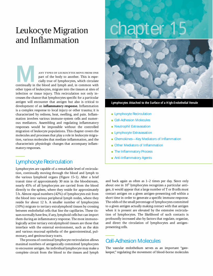
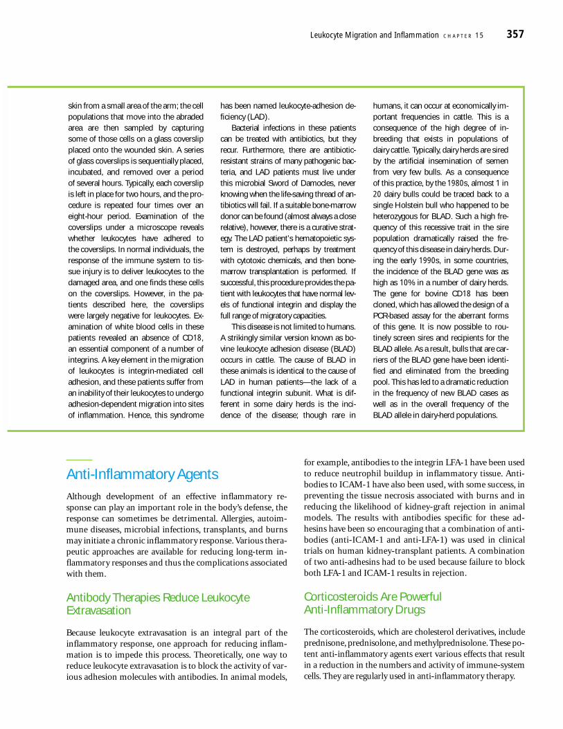
Lymphocytes Attached to the Surface of a High-Endothelial Venule
and leukocytes into the tissues. In order for circulating leuko-cytes to enter inflamed tissue or peripheral lymphoid organs,the cells must adhere to and pass between the endothelialcells lining the walls of blood vessels, a process called extra-vasation. Endothelial cells express leukocyte-specific cell-adhesion molecules (CAMs). Some of these membrane pro-teins are expressed constitutively; others are expressed only in response to local concentrations of cytokines producedduring an inflammatory response. Recirculating lympho-cytes, monocytes, and granulocytes bear receptors that bindto CAMs on the vascular endothelium, enabling these cells toextravasate into the tissues.
In addition to their role in leukocyte adhesion to vascularendothelial cells, CAMs on leukocytes also serve to increasethe strength of the functional interactions between cells ofthe immune system. Various adhesion molecules have beenshown to contribute to the interactions between TH cells andAPCs, TH and B cells, and CTLs and target cells.
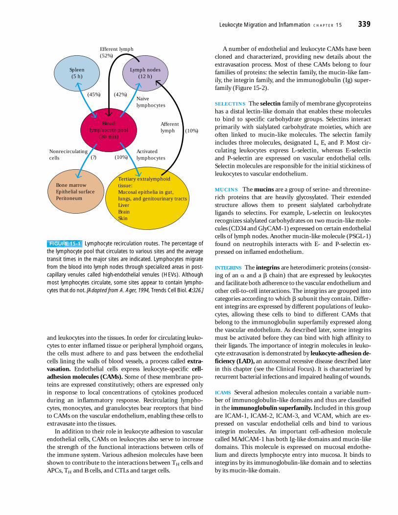
A number of endothelial and leukocyte CAMs have beencloned and characterized, providing new details about theextravasation process. Most of these CAMs belong to fourfamilies of proteins: the selectin family, the mucin-like fam-ily, the integrin family, and the immunoglobulin (Ig) super-family (Figure 15-2).
SELECTINS The selectin family of membrane glycoproteinshas a distal lectin-like domain that enables these molecules to bind to specific carbohydrate groups. Selectins interactprimarily with sialylated carbohydrate moieties, which areoften linked to mucin-like molecules. The selectin familyincludes three molecules, designated L, E, and P. Most cir-culating leukocytes express L-selectin, whereas E-selectinand P-selectin are expressed on vascular endothelial cells.Selectin molecules are responsible for the initial stickiness ofleukocytes to vascular endothelium.
MUCINS The mucins are a group of serine- and threonine-rich proteins that are heavily glycosylated. Their extendedstructure allows them to present sialylated carbohydrate ligands to selectins. For example, L-selectin on leukocytesrecognizes sialylated carbohydrates on two mucin-like mole-cules (CD34 and GlyCAM-1) expressed on certain endothelialcells of lymph nodes. Another mucin-like molecule (PSGL-1)found on neutrophils interacts with E- and P-selectin ex-pressed on inflamed endothelium.
INTEGRINS The integrins are heterodimeric proteins (consist-ing of an � and a � chain) that are expressed by leukocytesand facilitate both adherence to the vascular endothelium andother cell-to-cell interactions. The integrins are grouped intocategories according to which � subunit they contain. Differ-ent integrins are expressed by different populations of leuko-cytes, allowing these cells to bind to different CAMs thatbelong to the immunoglobulin superfamily expressed alongthe vascular endothelium. As described later, some integrinsmust be activated before they can bind with high affinity totheir ligands. The importance of integrin molecules in leuko-cyte extravasation is demonstrated by leukocyte-adhesion de-ficiency (LAD), an autosomal recessive disease described laterin this chapter (see the Clinical Focus). It is characterized byrecurrent bacterial infections and impaired healing of wounds.
ICAMS Several adhesion molecules contain a variable num-ber of immunoglobulin-like domains and thus are classifiedin the immunoglobulin superfamily. Included in this groupare ICAM-1, ICAM-2, ICAM-3, and VCAM, which are ex-pressed on vascular endothelial cells and bind to variousintegrin molecules. An important cell-adhesion moleculecalled MAdCAM-1 has both Ig-like domains and mucin-likedomains. This molecule is expressed on mucosal endothe-lium and directs lymphocyte entry into mucosa. It binds to integrins by its immunoglobulin-like domain and to selectinsby its mucin-like domain.
Leukocyte Migration and Inflammation C H A P T E R 15 339
Spleen(5 h)
Bone marrowEpithelial surfacePeritoneum
Activatedlymphocytes
Nonrecirculatingcells
Afferentlymph
Naivelymphocytes
(45%) (42%)
Efferent lymph(52%)
Bloodlymphocyte pool
(30 min)
Lymph nodes(12 h)
(?) (10%)
(10%)
Tertiary extralymphoid tissue:Mucosal epithelia in gut, lungs, and genitourinary tractsLiverBrainSkin
FIGURE 15-1 Lymphocyte recirculation routes. The percentage ofthe lymphocyte pool that circulates to various sites and the averagetransit times in the major sites are indicated. Lymphocytes migratefrom the blood into lymph nodes through specialized areas in post-capillary venules called high-endothelial venules (HEVs). Althoughmost lymphocytes circulate, some sites appear to contain lympho-cytes that do not. [Adapted from A. Ager, 1994, Trends Cell Biol. 4:326.]
Neutrophil ExtravasationAs an inflammatory response develops, various cytokinesand other inflammatory mediators act upon the local bloodvessels, inducing increased expression of endothelial CAMs.The vascular endothelium is then said to be activated, orinflamed. Neutrophils are generally the first cell type to bindto inflamed endothelium and extravasate into the tissues. Toaccomplish this, neutrophils must recognize the inflamedendothelium and adhere strongly enough so that they are notswept away by the flowing blood. The bound neutrophilsmust then penetrate the endothelial layer and migrate intothe underlying tissue. Monocytes and eosinophils extravasateby a similar process, but the steps have been best establishedfor the neutrophil, so we focus on neutrophils here.
The process of neutrophil extravasation can be divided intofour sequential steps: (1) rolling, (2) activation by chemoat-tractant stimulus, (3) arrest and adhesion, and (4) transendo-thelial migration (Figure 15-3a). In the first step, neutrophilsattach loosely to the endothelium by a low-affinity selectin-carbohydrate interaction. During an inflammatory response,cytokines and other mediators act upon the local endothe-lium, inducing expression of adhesion molecules of the selec-tin family. These E- and P-selectin molecules bind to mucin-
like cell-adhesion molecules on the neutrophil membrane orwith a sialylated lactosaminoglycan called sialyl Lewisx (Figure15-3b). This interaction tethers the neutrophil briefly to theendothelial cell, but the shear force of the circulating bloodsoon detaches the neutrophil. Selectin molecules on anotherendothelial cell again tether the neutrophil; this process isrepeated so that the neutrophil tumbles end-over-end alongthe endothelium, a type of binding called rolling.
As the neutrophil rolls, it is activated by various chemoat-tractants; these are either permanent features of the endo-thelial cell surface or secreted locally by cells involved in theinflammatory response. Among the chemoattractants aremembers of a large family of chemoattractive cytokines calledchemokines. Two chemokines involved in the activationprocess are interleukin 8 (IL-8) and macrophage inflamma-tory protein (MIP-1�). However, not all chemoattractantsbelong to the chemokine group. Other chemoattractants areplatelet-activating factor (PAF), the complement split prod-ucts C5a, C3a, and C5b67 and various N-formyl peptides pro-duced by the breakdown of bacterial proteins during an infec-tion. Binding of these chemoattractants to receptors on theneutrophil membrane triggers an activating signal mediatedby G proteins associated with the receptor. This signal inducesa conformational change in the integrin molecules in the neu-
340 P A R T I I I Immune Effector Mechanisms
SS
SS
SS
SS
SS
Ig domains
Lectin domain
Mucin-like CAMs Integrins
βα
CHO sidechains
Ig-superfamily CAMsSelectins
(a) General structure of CAM families
Fibrinonectin-typedomains
(b) Selected CAMs belonging to each family
Mucin-like CAMs:GlyCAM-1CD34PSGL-1MAdCAM-1
Selectins:L-selectinP-selectinE-selectin
Ig-superfamily CAMs:ICAM-1, -2, -3VCAM-1LFA-2 (CD2)LFA-3 (CD58)MAdCAM-1
Integrins:α4β1 (VLA-4, LPAM-2)α4β7 (LPAM-1)α6β1 (VLA-6)αLβ2 (LFA-1)αMβ2 (Mac-1)αXβ2 (CR4, p150/95)
FIGURE 15-2 Schematic diagrams depicting the general structuresof the four families of cell-adhesion molecules (a) and a list of repre-sentative molecules in each family (b). The lectin domain in selectinsinteracts primarily with carbohydrate (CHO) moieties on mucin-likemolecules. Both component chains in integrin molecules contribute tothe binding site, which interacts with an Ig domain in CAMs belongingto the Ig superfamily. MAdCAM-1 contains both mucin-like and Ig-likedomains and can bind to both selectins and integrins.
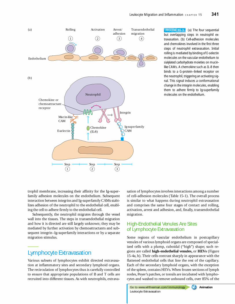
trophil membrane, increasing their affinity for the Ig-super-family adhesion molecules on the endothelium. Subsequentinteraction between integrins and Ig-superfamily CAMs stabi-lizes adhesion of the neutrophil to the endothelial cell, enabl-ing the cell to adhere firmly to the endothelial cell.
Subsequently, the neutrophil migrates through the vesselwall into the tissues. The steps in transendothelial migrationand how it is directed are still largely unknown; they may bemediated by further activation by chemoattractants and sub-sequent integrin–Ig-superfamily interactions or by a separatemigration stimulus.
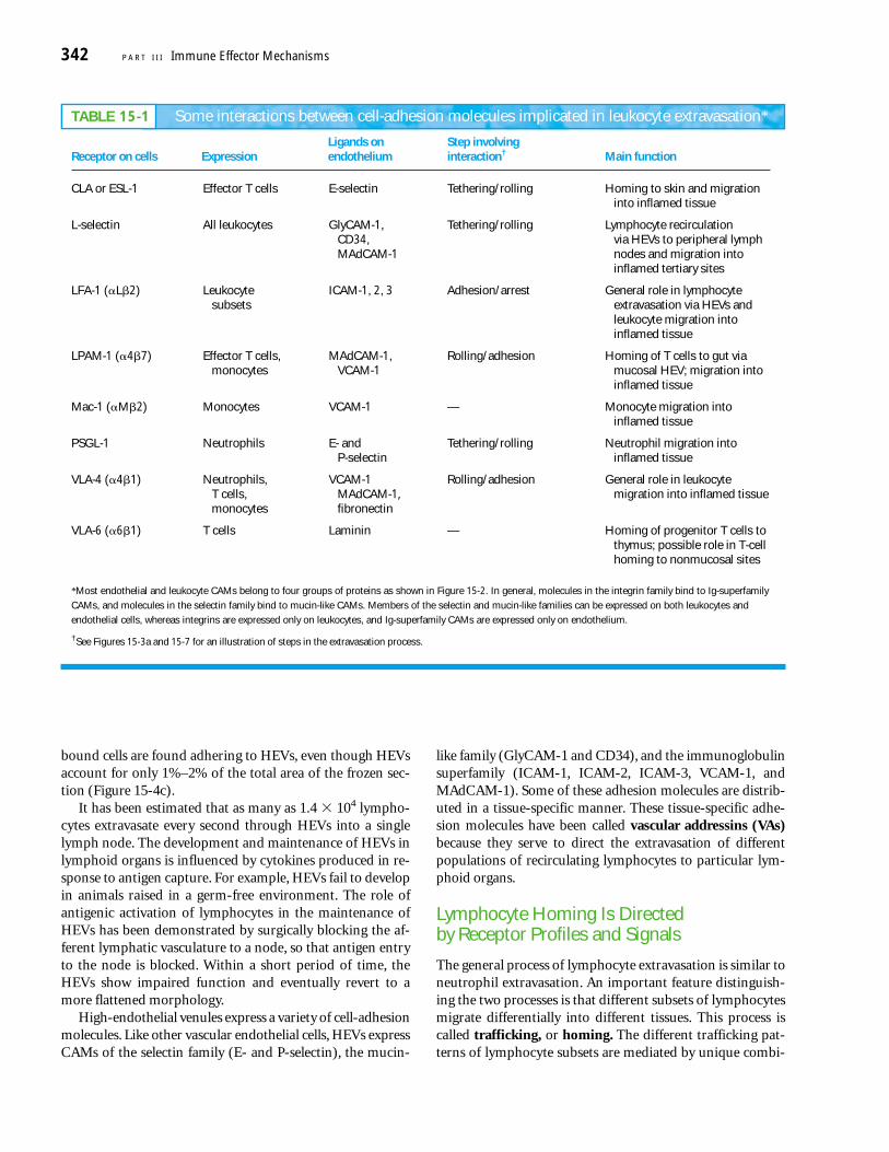
Lymphocyte ExtravasationVarious subsets of lymphocytes exhibit directed extravasa-tion at inflammatory sites and secondary lymphoid organs.The recirculation of lymphocytes thus is carefully controlledto ensure that appropriate populations of B and T cells arerecruited into different tissues. As with neutrophils, extrava-
sation of lymphocytes involves interactions among a numberof cell-adhesion molecules (Table 15-1). The overall processis similar to what happens during neutrophil extravasationand comprises the same four stages of contact and rolling,activation, arrest and adhesion, and, finally, transendothelialmigration.
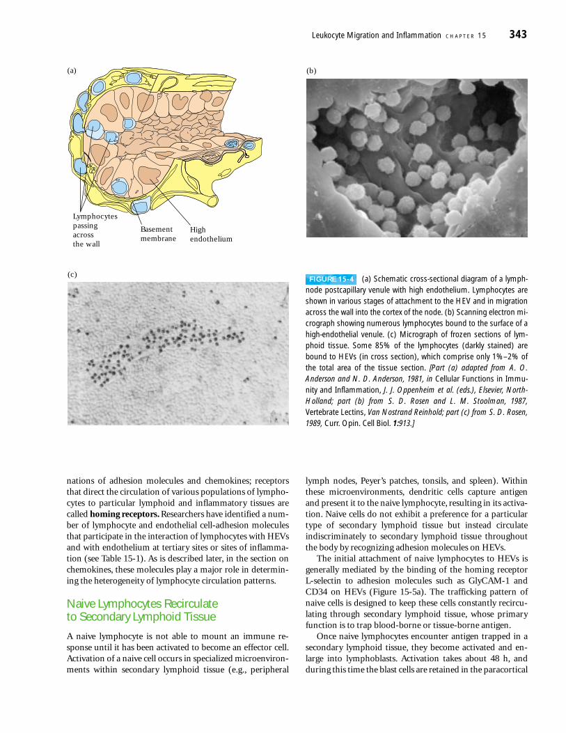
High-Endothelial Venules Are Sites of Lymphocyte Extravasation
Some regions of vascular endothelium in postcapillaryvenules of various lymphoid organs are composed of special-ized cells with a plump, cuboidal (“high”) shape; such re-gions are called high-endothelial venules, or HEVs (Figure15-4a, b). Their cells contrast sharply in appearance with theflattened endothelial cells that line the rest of the capillary.Each of the secondary lymphoid organs, with the exceptionof the spleen, contains HEVs. When frozen sections of lymphnodes, Peyer’s patches, or tonsils are incubated with lympho-cytes and washed to remove unbound cells, over 85% of the
Leukocyte Migration and Inflammation C H A P T E R 15 341
Endothelium
(a) Rolling Activation Arrest/adhesion
Transendothelialmigration
1 2 3 4
(b)
Step
2
Step
3
Step
1
Neutrophil
Integrin
Ig-superfamilyCAME-selectin
Chemokine orchemoattractantreceptor
Mucin-likeCAM
Chemokine(IL-8)
SSSS
SSSS
FIGURE 15-3 (a) The four sequentialbut overlapping steps in neutrophil ex-travasation. (b) Cell-adhesion moleculesand chemokines involved in the first threesteps of neutrophil extravasation. Initialrolling is mediated by binding of E-selectinmolecules on the vascular endothelium tosialylated carbohydrate moieties on mucin-like CAMs. A chemokine such as IL-8 thenbinds to a G-protein–linked receptor onthe neutrophil, triggering an activating sig-nal. This signal induces a conformationalchange in the integrin molecules, enablingthem to adhere firmly to Ig-superfamilymolecules on the endothelium.
Go to www.whfreeman.com/immunology AnimationLeukocyte Extravasation
bound cells are found adhering to HEVs, even though HEVsaccount for only 1%–2% of the total area of the frozen sec-tion (Figure 15-4c).
It has been estimated that as many as 1.4 � 104 lympho-cytes extravasate every second through HEVs into a singlelymph node. The development and maintenance of HEVs inlymphoid organs is influenced by cytokines produced in re-sponse to antigen capture. For example, HEVs fail to developin animals raised in a germ-free environment. The role ofantigenic activation of lymphocytes in the maintenance ofHEVs has been demonstrated by surgically blocking the af-ferent lymphatic vasculature to a node, so that antigen entryto the node is blocked. Within a short period of time, theHEVs show impaired function and eventually revert to amore flattened morphology.
High-endothelial venules express a variety of cell-adhesionmolecules. Like other vascular endothelial cells, HEVs expressCAMs of the selectin family (E- and P-selectin), the mucin-
like family (GlyCAM-1 and CD34), and the immunoglobulinsuperfamily (ICAM-1, ICAM-2, ICAM-3, VCAM-1, andMAdCAM-1). Some of these adhesion molecules are distrib-uted in a tissue-specific manner. These tissue-specific adhe-sion molecules have been called vascular addressins (VAs)because they serve to direct the extravasation of differentpopulations of recirculating lymphocytes to particular lym-phoid organs.
Lymphocyte Homing Is Directed by Receptor Profiles and Signals
The general process of lymphocyte extravasation is similar toneutrophil extravasation. An important feature distinguish-ing the two processes is that different subsets of lymphocytesmigrate differentially into different tissues. This process iscalled trafficking, or homing. The different trafficking pat-terns of lymphocyte subsets are mediated by unique combi-
342 P A R T I I I Immune Effector Mechanisms
TABLE 15-1 Some interactions between cell-adhesion molecules implicated in leukocyte extravasation*
Ligands on Step involvingReceptor on cells Expression endothelium interaction† Main function
CLA or ESL-1 Effector T cells E-selectin Tethering/rolling Homing to skin and migrationinto inflamed tissue
L-selectin All leukocytes GlyCAM-1, Tethering/rolling Lymphocyte recirculationCD34, via HEVs to peripheral lymphMAdCAM-1 nodes and migration into
inflamed tertiary sites
LFA-1 (�L�2) Leukocyte ICAM-1, 2, 3 Adhesion/arrest General role in lymphocytesubsets extravasation via HEVs and
leukocyte migration intoinflamed tissue
LPAM-1 (�4�7) Effector T cells, MAdCAM-1, Rolling/adhesion Homing of T cells to gut viamonocytes VCAM-1 mucosal HEV; migration into
inflamed tissue
Mac-1 (�M�2) Monocytes VCAM-1 — Monocyte migration intoinflamed tissue
PSGL-1 Neutrophils E- and Tethering/rolling Neutrophil migration intoP-selectin inflamed tissue
VLA-4 (�4�1) Neutrophils, VCAM-1 Rolling/adhesion General role in leukocyteT cells, MAdCAM-1, migration into inflamed tissuemonocytes fibronectin
VLA-6 (�6�1) T cells Laminin — Homing of progenitor T cells tothymus; possible role in T-cell homing to nonmucosal sites
*Most endothelial and leukocyte CAMs belong to four groups of proteins as shown in Figure 15-2. In general, molecules in the integrin family bind to Ig-superfamily
CAMs, and molecules in the selectin family bind to mucin-like CAMs. Members of the selectin and mucin-like families can be expressed on both leukocytes and
endothelial cells, whereas integrins are expressed only on leukocytes, and Ig-superfamily CAMs are expressed only on endothelium.
†See Figures 15-3a and 15-7 for an illustration of steps in the extravasation process.
nations of adhesion molecules and chemokines; receptorsthat direct the circulation of various populations of lympho-cytes to particular lymphoid and inflammatory tissues arecalled homing receptors. Researchers have identified a num-ber of lymphocyte and endothelial cell-adhesion moleculesthat participate in the interaction of lymphocytes with HEVsand with endothelium at tertiary sites or sites of inflamma-tion (see Table 15-1). As is described later, in the section onchemokines, these molecules play a major role in determin-ing the heterogeneity of lymphocyte circulation patterns.
Naive Lymphocytes Recirculate to Secondary Lymphoid Tissue
A naive lymphocyte is not able to mount an immune re-sponse until it has been activated to become an effector cell.Activation of a naive cell occurs in specialized microenviron-ments within secondary lymphoid tissue (e.g., peripheral
lymph nodes, Peyer’s patches, tonsils, and spleen). Withinthese microenvironments, dendritic cells capture antigenand present it to the naive lymphocyte, resulting in its activa-tion. Naive cells do not exhibit a preference for a particulartype of secondary lymphoid tissue but instead circulateindiscriminately to secondary lymphoid tissue throughoutthe body by recognizing adhesion molecules on HEVs.
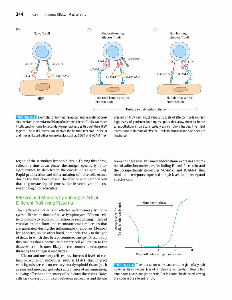
The initial attachment of naive lymphocytes to HEVs isgenerally mediated by the binding of the homing receptor L-selectin to adhesion molecules such as GlyCAM-1 andCD34 on HEVs (Figure 15-5a). The trafficking pattern ofnaive cells is designed to keep these cells constantly recircu-lating through secondary lymphoid tissue, whose primaryfunction is to trap blood-borne or tissue-borne antigen.
Once naive lymphocytes encounter antigen trapped in asecondary lymphoid tissue, they become activated and en-large into lymphoblasts. Activation takes about 48 h, andduring this time the blast cells are retained in the paracortical
Leukocyte Migration and Inflammation C H A P T E R 15 343
Lymphocytespassingacrossthe wall
Basementmembrane
Highendothelium
(a) (b)
(c)FIGURE 15-4 (a) Schematic cross-sectional diagram of a lymph-
node postcapillary venule with high endothelium. Lymphocytes areshown in various stages of attachment to the HEV and in migrationacross the wall into the cortex of the node. (b) Scanning electron mi-crograph showing numerous lymphocytes bound to the surface of ahigh-endothelial venule. (c) Micrograph of frozen sections of lym-phoid tissue. Some 85% of the lymphocytes (darkly stained) arebound to HEVs (in cross section), which comprise only 1%–2% ofthe total area of the tissue section. [Part (a) adapted from A. O. Anderson and N. D. Anderson, 1981, in Cellular Functions in Immu-nity and Inflammation, J. J. Oppenheim et al. (eds.), Elsevier, North-Holland; part (b) from S. D. Rosen and L. M. Stoolman, 1987,Vertebrate Lectins, Van Nostrand Reinhold; part (c) from S. D. Rosen,1989, Curr. Opin. Cell Biol. 1:913.]
region of the secondary lymphoid tissue. During this phase,called the shut-down phase, the antigen-specific lympho-cytes cannot be detected in the circulation (Figure 15-6).Rapid proliferation and differentiation of naive cells occursduring the shut-down phase. The effector and memory cellsthat are generated by this process then leave the lymphoid tis-sue and begin to recirculate.
Effector and Memory Lymphocytes AdoptDifferent Trafficking PatternsThe trafficking patterns of effector and memory lympho-cytes differ from those of naive lymphocytes. Effector cellstend to home to regions of infection by recognizing inflamedvascular endothelium and chemoattractant molecules thatare generated during the inflammatory response. Memorylymphocytes, on the other hand, home selectively to the typeof tissue in which they first encountered antigen. Presumablythis ensures that a particular memory cell will return to thetissue where it is most likely to reencounter a subsequentthreat by the antigen it recognizes.
Effector and memory cells express increased levels of cer-tain cell-adhesion molecules, such as LFA-1, that interactwith ligands present on tertiary extralymphoid tissue (suchas skin and mucosal epithelia) and at sites of inflammation,allowing effector and memory cells to enter these sites. Naivecells lack corresponding cell-adhesion molecules and do not
home to these sites. Inflamed endothelium expresses a num-ber of adhesion molecules, including E- and P-selectin andthe Ig-superfamily molecules VCAM-1 and ICAM-1, thatbind to the receptors expressed at high levels on memory andeffector cells.
344 P A R T I I I Immune Effector Mechanisms
(a)Naive T cell
L-selectinL-selectin
GlyCAM-1CD34
HEV
Tertiary extralymphoid tissue
LFA-1
LPAM-1
ICAM-1
E-selectin
CLA
(b)
L-selectinLFA-1
ICAM-1
(c)Mucosal-homingeffector T cell
Skin-homingeffector T cell
Intestinal lamina propriaendothelium
Skin dermal venuleendothelium
MAdCAM-1
SSSS
SSSSSS
SS
SSSS
SS
SSSS
SS
SS
FIGURE 15-5 Examples of homing receptors and vascular addres-sins involved in selective trafficking of naive and effector T cells. (a) NaiveT cells tend to home to secondary lymphoid tissues through their HEVregions. The initial interaction involves the homing receptor L-selectinand mucin-like cell-adhesion molecules such as CD34 or GlyCAM-1 ex-
pressed on HEV cells. (b, c) Various subsets of effector T cells expresshigh levels of particular homing receptors that allow them to home to endothelium in particular tertiary extralymphoid tissues. The initialinteractions in homing of effector T cells to mucosal and skin sites areillustrated.
2 4 6 8Days following antigen exposure
Shut-down phase
An
tige
n-s
pec
ific
T c
ells
in e
ffer
ent
lym
ph
FIGURE 15- 6 T-cell activation in the paracortical region of a lymphnode results in the brief loss of lymphocyte recirculation. During thisshut-down phase, antigen-specific T cells cannot be detected leavingthe node in the efferent lymph.
Unlike naive lymphocytes, subsets of the memory andeffector populations exhibit tissue-selective homing behavior.Such tissue specificity is imparted not by a single adhesionreceptor but by different combinations of adhesion molecules.For example, a mucosal homing subset of memory/effectorcells has high levels of the integrins LPAM-1 (��4�7) andLFA-1 (�Lb2), which bind to MAdCAM and various ICAMson intestinal lamina propria venules (see Figure 15-5b). How-ever, these cells avoid direction to secondary lymphoid tissuesbecause they have low levels of the L-selectin that would facil-itate their entry into secondary lymphoid tissue. A second sub-set of memory/effector cells displays preferential homing tothe skin. This subset also expresses low levels of L-selectin butdisplays high levels of cutaneous lymphocyte antigen (CLA)and LFA-1, which bind to E-selectin and ICAMs on dermalvenules of the skin (see Figure 15-5c). Although effector andmemory cells that express reduced levels of L-selectin do nottend to home through HEVs into peripheral lymph nodes,they can enter peripheral lymph nodes through the afferentlymphatic vessels.
Adhesion-Molecule Interactions Play Critical Roles in Extravasation
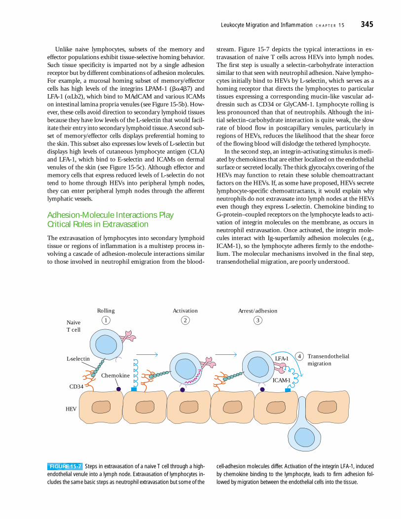
The extravasation of lymphocytes into secondary lymphoidtissue or regions of inflammation is a multistep process in-volving a cascade of adhesion-molecule interactions similarto those involved in neutrophil emigration from the blood-
stream. Figure 15-7 depicts the typical interactions in ex-travasation of naive T cells across HEVs into lymph nodes.The first step is usually a selectin-carbohydrate interactionsimilar to that seen with neutrophil adhesion. Naive lympho-cytes initially bind to HEVs by L-selectin, which serves as ahoming receptor that directs the lymphocytes to particulartissues expressing a corresponding mucin-like vascular ad-dressin such as CD34 or GlyCAM-1. Lymphocyte rolling isless pronounced than that of neutrophils. Although the ini-tial selectin-carbohydrate interaction is quite weak, the slowrate of blood flow in postcapillary venules, particularly inregions of HEVs, reduces the likelihood that the shear forceof the flowing blood will dislodge the tethered lymphocyte.
In the second step, an integrin-activating stimulus is medi-ated by chemokines that are either localized on the endothelialsurface or secreted locally. The thick glycocalyx covering of theHEVs may function to retain these soluble chemoattractantfactors on the HEVs. If, as some have proposed, HEVs secretelymphocyte-specific chemoattractants, it would explain whyneutrophils do not extravasate into lymph nodes at the HEVseven though they express L-selectin. Chemokine binding to G-protein–coupled receptors on the lymphocyte leads to acti-vation of integrin molecules on the membrane, as occurs inneutrophil extravasation. Once activated, the integrin mole-cules interact with Ig-superfamily adhesion molecules (e.g.,ICAM-1), so the lymphocyte adheres firmly to the endothe-lium. The molecular mechanisms involved in the final step,transendothelial migration, are poorly understood.
Leukocyte Migration and Inflammation C H A P T E R 15 345
L-selectin
NaiveT cell
Chemokine
LFA-1
ICAM-1CD34
HEV
Rolling
1
Activation
2
Arrest/adhesion
3
Transendothelialmigration
4
FIGURE 15-7 Steps in extravasation of a naive T cell through a high-endothelial venule into a lymph node. Extravasation of lymphocytes in-cludes the same basic steps as neutrophil extravasation but some of the
cell-adhesion molecules differ. Activation of the integrin LFA-1, inducedby chemokine binding to the lymphocyte, leads to firm adhesion fol-lowed by migration between the endothelial cells into the tissue.
Chemokines—Key Mediators of Inflammation Chemokines are a superfamily of small polypeptides, most ofwhich contain 90–130 amino acid residues. They selectively,and often specifically, control the adhesion, chemotaxis, andactivation of many types of leukocyte populations and sub-populations. Consequently, they are major regulators of leu-kocyte traffic. Some chemokines are primarily involved ininflammatory processes, others are constitutively expressedand play important homeostatic or developmental roles.“Housekeeping” chemokines are produced in lymphoid or-gans and tissues or in non-lymphoid sites such as skin, wherethey direct normal trafficking of lymphocytes, such as deter-mining the correct positioning of leukocytes newly generatedby hematopoiesis and arriving from bone marrow. The thy-mus constitutively expresses chemokines, and normal B celllymphopoiesis is also dependent on appropriate chemokineexpression. Chemokine-mediated effects are not limited tothe immune system. Mice that lack either the chemokineCXCL12 (also called SDF-1) or its receptor (see Table 15-2)show major defects in the development of the brain and theheart. Members of the chemokine family have also beenshown to play regulatory roles in the development of bloodvessels (angiogenesis), and wound healing.
The inflammatory chemokines are typically induced inresponse to infection. Contact with pathogens or the action ofproinflammatory cytokines, such as TNF-�, up-regulate theexpression of inflammatory cytokines at sites of developinginflammation. Chemokines cause leukocytes to move intovarious tissue sites by inducing the adherence of these cells tothe vascular endothelium. After migrating into tissues, leuko-cytes are attracted toward high localized concentrations ofchemokines resulting in the targeted recruitment of phago-cytes and effector lymphocyte populations to inflammatorysites. The assembly of leukocytes at sites of infection, orches-trated by chemokines, is an essential part of mounting anappropriately focused response to infection.
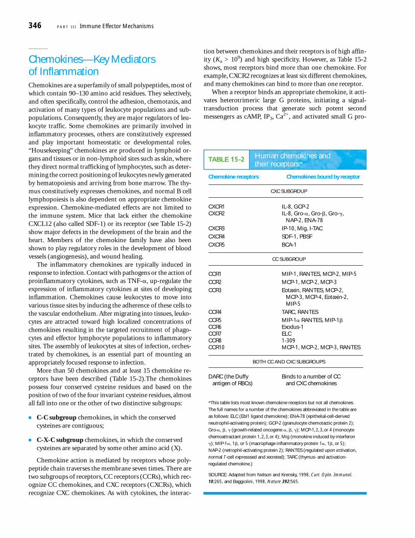
More than 50 chemokines and at least 15 chemokine re-ceptors have been described (Table 15-2).The chemokinespossess four conserved cysteine residues and based on theposition of two of the four invariant cysteine residues, almostall fall into one or the other of two distinctive subgroups:
■ C-C subgroup chemokines, in which the conservedcysteines are contiguous;
■ C-X-C subgroup chemokines, in which the conservedcysteines are separated by some other amino acid (X).
Chemokine action is mediated by receptors whose poly-peptide chain traverses the membrane seven times. There aretwo subgroups of receptors, CC receptors (CCRs), which rec-ognize CC chemokines, and CXC receptors (CXCRs), whichrecognize CXC chemokines. As with cytokines, the interac-
tion between chemokines and their receptors is of high affin-ity (Ka > 109) and high specificity. However, as Table 15-2shows, most receptors bind more than one chemokine. Forexample, CXCR2 recognizes at least six different chemokines,and many chemokines can bind to more than one receptor.
When a receptor binds an appropriate chemokine, it acti-vates heterotrimeric large G proteins, initiating a signal-transduction process that generate such potent second messengers as cAMP, IP3, Ca2+, and activated small G pro-
346 P A R T I I I Immune Effector Mechanisms
TABLE 15-2 Human chemokines and their receptors*
Chemokine receptors Chemokines bound by receptor
CXC SUBGROUP
CXCR1 IL-8, GCP-2CXCR2 IL-8, Gro-�, Gro-�, Gro-�,
NAP-2, ENA-78
CXCR3 IP-10, Mig, I-TAC
CXCR4 SDF-1, PBSF
CXCR5 BCA-1
CC SUBGROUP
CCR1 MIP-1, RANTES, MCP-2, MIP-5
CCR2 MCP-1, MCP-2, MCP-3
CCR3 Eotaxin, RANTES, MCP-2,MCP-3, MCP-4, Eotaxin-2,MIP-5
CCR4 TARC, RANTES
CCR5 MIP-1� RANTES, MIP-1�CCR6 Exodus-1CCR7 ELCCCR8 1-309CCR10 MCP-1, MCP-2, MCP-3, RANTES
BOTH CC AND CXC SUBGROUPS
DARC (the Duffy Binds to a number of CCantigen of RBCs) and CXC chemokines
*This table lists most known chemokine receptors but not all chemokines.
The full names for a number of the chemokines abbreviated in the table are
as follows: ELC (Ebl1 ligand chemokine); ENA-78 (epithelial-cell-derived
neutrophil-activating protein); GCP-2 (granulocyte chemotactic protein 2);
Gro-�, �, � (growth-related oncogene �, �, �); MCP-1, 2, 3, or 4 (monocyte
chemoattractant protein 1, 2, 3, or 4); Mig (monokine induced by interferon
�); MIP-1�, 1�, or 5 (macrophage inflammatory protein 1�, 1�, or 5);
NAP-2 (netrophil-activating protein 2); RANTES (regulated upon activation,
normal T-cell expresssed and secreted); TARC (thymus- and activation-
regulated chemokine.)
SOURCE: Adapted from Nelson and Krensky, 1998, Curr. Opin. Immunol.
10:265, and Baggiolini, 1998, Nature 392:565.
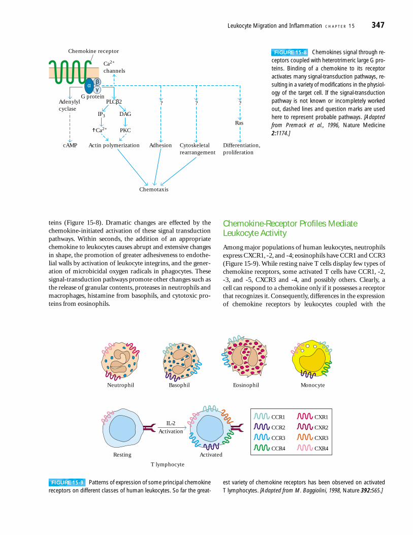
teins (Figure 15-8). Dramatic changes are effected by thechemokine-initiated activation of these signal transductionpathways. Within seconds, the addition of an appropriatechemokine to leukocytes causes abrupt and extensive changesin shape, the promotion of greater adhesiveness to endothe-lial walls by activation of leukocyte integrins, and the gener-ation of microbicidal oxygen radicals in phagocytes. Thesesignal-transduction pathways promote other changes such asthe release of granular contents, proteases in neutrophils andmacrophages, histamine from basophils, and cytotoxic pro-teins from eosinophils.
Chemokine-Receptor Profiles MediateLeukocyte Activity
Among major populations of human leukocytes, neutrophilsexpress CXCR1, -2, and -4; eosinophils have CCR1 and CCR3(Figure 15-9). While resting naive T cells display few types ofchemokine receptors, some activated T cells have CCR1, -2,-3, and -5, CXCR3 and -4, and possibly others. Clearly, a cell can respond to a chemokine only if it possesses a receptorthat recognizes it. Consequently, differences in the expressionof chemokine receptors by leukocytes coupled with the
Leukocyte Migration and Inflammation C H A P T E R 15 347
α βγ
Differentiation,proliferation
Cytoskeletalrearrangement
Adhesion
Chemotaxis
?
Ras
Ca2+ channels
Chemokine receptor
??
cAMP
Adenylylcyclase
G proteinPLCβ2
IP3 DAG
Ca2+ PKC
Actin polymerization
Neutrophil Basophil
Activation
T lymphocyte
Resting Activated
IL-2CCR1
Eosinophil Monocyte
CCR2
CCR3
CCR4
CXR1
CXR2
CXR3
CXR4
FIGURE 15-8 Chemokines signal through re-ceptors coupled with heterotrimeric large G pro-teins. Binding of a chemokine to its receptoractivates many signal-transduction pathways, re-sulting in a variety of modifications in the physiol-ogy of the target cell. If the signal-transductionpathway is not known or incompletely workedout, dashed lines and question marks are usedhere to represent probable pathways. [Adaptedfrom Premack et al., 1996, Nature Medicine2:1174.]
FIGURE 15-9 Patterns of expression of some principal chemokinereceptors on different classes of human leukocytes. So far the great-
est variety of chemokine receptors has been observed on activated T lymphocytes. [Adapted from M. Baggiolini, 1998, Nature 392:565.]
production of distinctive profiles of chemokines by destina-tion tissues and sites provide rich opportunities for the dif-ferential regulation of activities of different leukocyte popu-lations. Indeed, differences in patterns of chemokine-receptorexpression occur within leukocyte populations as well as be-tween different ones. Recall that TH1 and TH2 subsets of TH
cells can be distinguished by their different patterns of cyto-kine production. These subsets also display different profilesof chemokine receptors. TH2 cells express CCR3 and -4, anda number of other receptors not expressed by TH1 cells. Onthe other hand, TH1 cells express CCR1, -3, and -5, but mostTH2 cells do not.
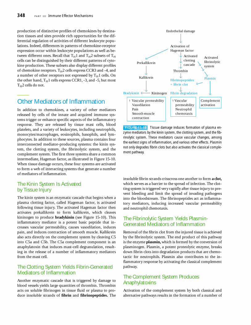
Other Mediators of InflammationIn addition to chemokines, a variety of other mediators released by cells of the innate and acquired immune sys-tems trigger or enhance specific aspects of the inflammatoryresponse. They are released by tissue mast cells, bloodplatelets, and a variety of leukocytes, including neutrophils,monocytes/macrophages, eosinophils, basophils, and lym-phocytes. In addition to these sources, plasma contains fourinterconnected mediator-producing systems: the kinin sys-tem, the clotting system, the fibrinolytic system, and thecomplement system. The first three systems share a commonintermediate, Hageman factor, as illustrated in Figure 15-10.When tissue damage occurs, these four systems are activatedto form a web of interacting systems that generate a numberof mediators of inflammation.
The Kinin System Is Activated by Tissue InjuryThe kinin system is an enzymatic cascade that begins when aplasma clotting factor, called Hageman factor, is activatedfollowing tissue injury. The activated Hageman factor thenactivates prekallikrein to form kallikrein, which cleaveskininogen to produce bradykinin (see Figure 15-10). Thisinflammatory mediator is a potent basic peptide that in-creases vascular permeability, causes vasodilation, inducespain, and induces contraction of smooth muscle. Kallikreinalso acts directly on the complement system by cleaving C5into C5a and C5b. The C5a complement component is ananaphylatoxin that induces mast-cell degranulation, result-ing in the release of a number of inflammatory mediatorsfrom the mast cell.
The Clotting System Yields Fibrin-GeneratedMediators of InflammationAnother enzymatic cascade that is triggered by damage toblood vessels yields large quantities of thrombin. Thrombinacts on soluble fibrinogen in tissue fluid or plasma to pro-duce insoluble strands of fibrin and fibrinopeptides. The
insoluble fibrin strands crisscross one another to form a clot,which serves as a barrier to the spread of infection. The clot-ting system is triggered very rapidly after tissue injury to pre-vent bleeding and limit the spread of invading pathogensinto the bloodstream. The fibrinopeptides act as inflamma-tory mediators, inducing increased vascular permeabilityand neutrophil chemotaxis.
The Fibrinolytic System Yields Plasmin-Generated Mediators of InflammationRemoval of the fibrin clot from the injured tissue is achievedby the fibrinolytic system. The end product of this pathway is the enzyme plasmin, which is formed by the conversion ofplasminogen. Plasmin, a potent proteolytic enzyme, breaksdown fibrin clots into degradation products that are chemo-tactic for neutrophils. Plasmin also contributes to the in-flammatory response by activating the classical complementpathway.
The Complement System ProducesAnaphylatoxinsActivation of the complement system by both classical andalternative pathways results in the formation of a number of
348 P A R T I I I Immune Effector Mechanisms
Bradykinin
Kallikrein
Kininogen
Prekallikrein
Activatedfibrinolyticsystem
Plasmin
Activation ofHageman factor
Fibrinopeptides+ fibrin clot
Fibrin degradation
Thrombin
Endothelial damage
Activated clotting cascade
↑ Vascular permeabilityVasodilationPainSmooth-musclecontraction
↑ VascularpermeabilityNeutrophilchemotaxis
Complementactivation
FIGURE 15-10 Tissue damage induces formation of plasma en-zyme mediators by the kinin system, the clotting system, and the fib-rinolytic system. These mediators cause vascular changes, amongthe earliest signs of inflammation, and various other effects. Plasminnot only degrades fibrin clots but also activates the classical comple-ment pathway.
complement split products that serve as important media-tors of inflammation (see Chapter 13). Binding of the ana-phylatoxins (C3a, C4a, and C5a) to receptors on the mem-brane of tissue mast cells induces degranulation with releaseof histamine and other pharmacologically active mediators.These mediators induce smooth-muscle contraction andincrease vascular permeability. C3a, C5a, and C5b67 acttogether to induce monocytes and neutrophils to adhere tovascular endothelial cells, extravasate through the endothe-lial lining of the capillary, and migrate toward the site ofcomplement activation in the tissues. Activation of the com-plement system thus results in influxes of fluid that carryantibody and phagocytic cells to the site of antigen entry.
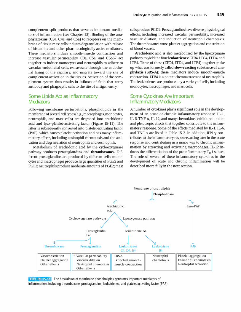
Some Lipids Act as Inflammatory MediatorsFollowing membrane perturbations, phospholipids in themembrane of several cell types (e.g., macrophages, monocytes,neutrophils, and mast cells) are degraded into arachidonicacid and lyso–platelet-activating factor (Figure 15-11). Thelatter is subsequently converted into platelet-activating factor(PAF), which causes platelet activation and has many inflam-matory effects, including eosinophil chemotaxis and the acti-vation and degranulation of neutrophils and eosinophils.
Metabolism of arachidonic acid by the cyclooxygenasepathway produces prostaglandins and thromboxanes. Dif-ferent prostaglandins are produced by different cells: mono-cytes and macrophages produce large quantities of PGE2 andPGF2; neutrophils produce moderate amounts of PGE2; mast
cells produce PGD2. Prostaglandins have diverse physiologicaleffects, including increased vascular permeability, increasedvascular dilation, and induction of neutrophil chemotaxis.The thromboxanes cause platelet aggregation and constrictionof blood vessels.
Arachidonic acid is also metabolized by the lipoxygenasepathway to yield the four leukotrienes: LTB4, LTC4, LTD4, andLTE4. Three of these (LTC4, LTD4, and LTE4) together makeup what was formerly called slow-reacting substance of ana-phylaxis (SRS-A); these mediators induce smooth-musclecontraction. LTB4 is a potent chemoattractant of neutrophils.The leukotrienes are produced by a variety of cells, includingmonocytes, macrophages, and mast cells.
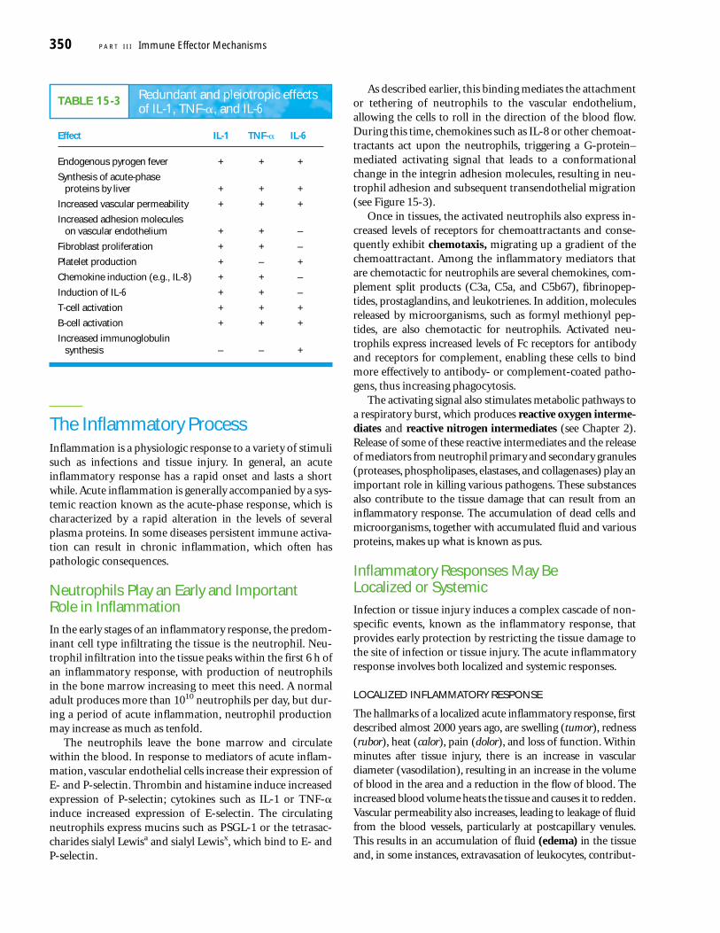
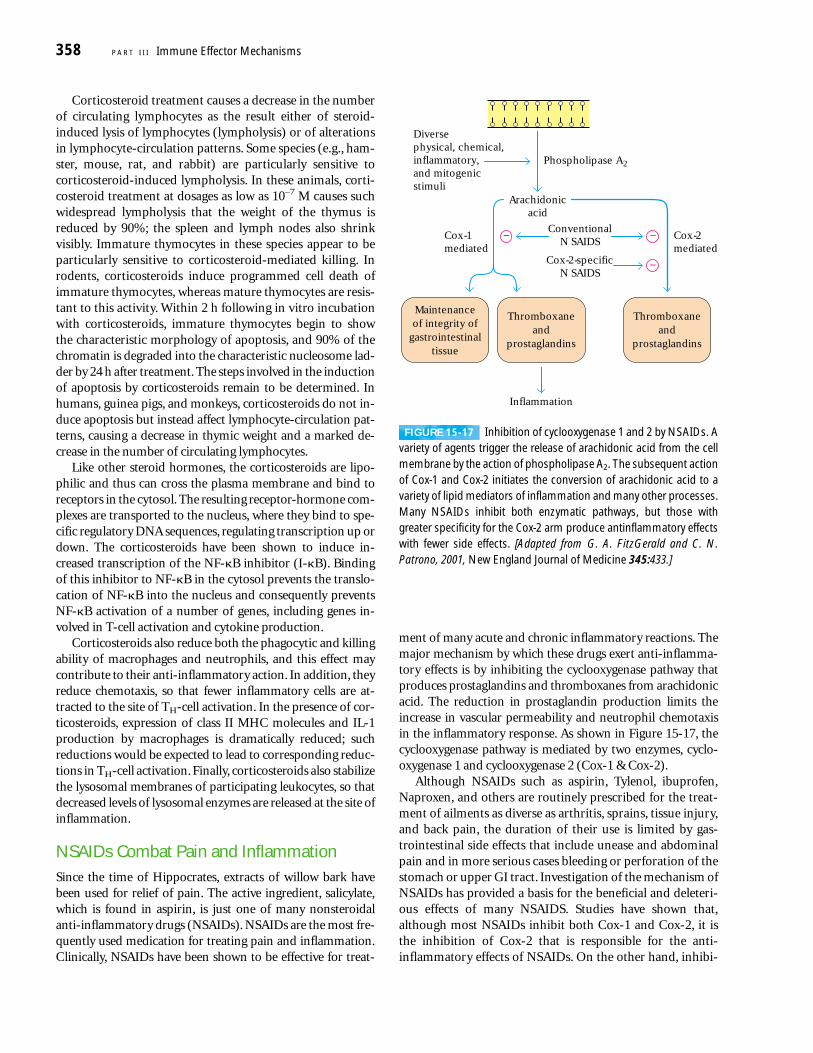
Some Cytokines Are Important Inflammatory MediatorsA number of cytokines play a significant role in the develop-ment of an acute or chronic inflammatory response. IL-1,IL-6, TNF-�, IL-12, and many chemokines exhibit redundantand pleiotropic effects that together contribute to the inflam-matory response. Some of the effects mediated by IL-1, IL-6,and TNF-� are listed in Table 15-3. In addition, IFN-� con-tributes to the inflammatory response, acting later in the acuteresponse and contributing in a major way to chronic inflam-mation by attracting and activating macrophages. IL-12 in-duces the differentiation of the proinflammatory TH1 subset.The role of several of these inflammatory cytokines in thedevelopment of acute and chronic inflammation will bedescribed more fully in the next section.
Leukocyte Migration and Inflammation C H A P T E R 15 349
Arachidonicacid
ProstaglandinG2
Leukotriene A4
Lipoxygenase pathwayCyclooxygenase pathways
Lyso-PAF
Membrane phospholipids
Phospholipase
Thromboxane Prostaglandins LeukotrienesC4, D4, E4
LeukotrieneB4
PAF
↑ Vascular permeabilityVascular dilationNeutrophil chemotaxisOther effects
VasoconstrictionPlatelet aggregationOther effects
SRS-ABronchial smooth-muscle contraction
Neutrophilchemotaxis
Platelet aggregationEosinophil chemotaxisNeutrophil activation
FIGURE 15-11 The breakdown of membrane phospholipids generates important mediators of inflammation, including thromboxane, prostaglandins, leukotrienes, and platelet-activating factor (PAF).
The Inflammatory ProcessInflammation is a physiologic response to a variety of stimulisuch as infections and tissue injury. In general, an acuteinflammatory response has a rapid onset and lasts a shortwhile. Acute inflammation is generally accompanied by a sys-temic reaction known as the acute-phase response, which ischaracterized by a rapid alteration in the levels of severalplasma proteins. In some diseases persistent immune activa-tion can result in chronic inflammation, which often haspathologic consequences.
Neutrophils Play an Early and Important Role in InflammationIn the early stages of an inflammatory response, the predom-inant cell type infiltrating the tissue is the neutrophil. Neu-trophil infiltration into the tissue peaks within the first 6 h ofan inflammatory response, with production of neutrophilsin the bone marrow increasing to meet this need. A normaladult produces more than 1010 neutrophils per day, but dur-ing a period of acute inflammation, neutrophil productionmay increase as much as tenfold.
The neutrophils leave the bone marrow and circulatewithin the blood. In response to mediators of acute inflam-mation, vascular endothelial cells increase their expression ofE- and P-selectin. Thrombin and histamine induce increasedexpression of P-selectin; cytokines such as IL-1 or TNF-�induce increased expression of E-selectin. The circulatingneutrophils express mucins such as PSGL-1 or the tetrasac-charides sialyl Lewisa and sialyl Lewisx, which bind to E- andP-selectin.
As described earlier, this binding mediates the attachmentor tethering of neutrophils to the vascular endothelium,allowing the cells to roll in the direction of the blood flow.During this time, chemokines such as IL-8 or other chemoat-tractants act upon the neutrophils, triggering a G-protein–mediated activating signal that leads to a conformationalchange in the integrin adhesion molecules, resulting in neu-trophil adhesion and subsequent transendothelial migration(see Figure 15-3).
Once in tissues, the activated neutrophils also express in-creased levels of receptors for chemoattractants and conse-quently exhibit chemotaxis, migrating up a gradient of thechemoattractant. Among the inflammatory mediators thatare chemotactic for neutrophils are several chemokines, com-plement split products (C3a, C5a, and C5b67), fibrinopep-tides, prostaglandins, and leukotrienes. In addition, moleculesreleased by microorganisms, such as formyl methionyl pep-tides, are also chemotactic for neutrophils. Activated neu-trophils express increased levels of Fc receptors for antibodyand receptors for complement, enabling these cells to bindmore effectively to antibody- or complement-coated patho-gens, thus increasing phagocytosis.
The activating signal also stimulates metabolic pathways toa respiratory burst, which produces reactive oxygen interme-diates and reactive nitrogen intermediates (see Chapter 2).Release of some of these reactive intermediates and the releaseof mediators from neutrophil primary and secondary granules(proteases, phospholipases, elastases, and collagenases) play animportant role in killing various pathogens. These substancesalso contribute to the tissue damage that can result from aninflammatory response. The accumulation of dead cells andmicroorganisms, together with accumulated fluid and variousproteins, makes up what is known as pus.
Inflammatory Responses May Be Localized or SystemicInfection or tissue injury induces a complex cascade of non-specific events, known as the inflammatory response, thatprovides early protection by restricting the tissue damage tothe site of infection or tissue injury. The acute inflammatoryresponse involves both localized and systemic responses.
LOCALIZED INFLAMMATORY RESPONSE
The hallmarks of a localized acute inflammatory response, firstdescribed almost 2000 years ago, are swelling (tumor), redness(rubor), heat (calor), pain (dolor), and loss of function. Withinminutes after tissue injury, there is an increase in vasculardiameter (vasodilation), resulting in an increase in the volumeof blood in the area and a reduction in the flow of blood. Theincreased blood volume heats the tissue and causes it to redden.Vascular permeability also increases, leading to leakage of fluidfrom the blood vessels, particularly at postcapillary venules.This results in an accumulation of fluid (edema) in the tissueand, in some instances, extravasation of leukocytes, contribut-
350 P A R T I I I Immune Effector Mechanisms
TABLE 15-3 Redundant and pleiotropic effectsof IL-1, TNF-�, and IL-6
Effect IL-1 TNF-� IL-6
Endogenous pyrogen fever + + +
Synthesis of acute-phase proteins by liver + + +
Increased vascular permeability + + +
Increased adhesion moleculeson vascular endothelium + + –
Fibroblast proliferation + + –
Platelet production + – +
Chemokine induction (e.g., IL-8) + + –
Induction of IL-6 + + –
T-cell activation + + +
B-cell activation + + +
Increased immunoglobulinsynthesis – – +
ing to the swelling and redness in the area. When fluid exudesfrom the bloodstream, the kinin, clotting, and fibrinolytic sys-tems are activated (see Figure 15-10). Many of the vascularchanges that occur early in a local response are due to the directeffects of plasma enzyme mediators such as bradykinin and fib-rinopeptides, which induce vasodilation and increased vascu-lar permeability. Some of the vascular changes are due to theindirect effects of the complement anaphylatoxins (C3a, C4a,and C5a), which induce local mast-cell degranulation with re-lease of histamine. Histamine is a potent mediator of inflam-mation, causing vasodilation and smooth-muscle contraction.The prostaglandins can also contribute to the vasodilation andincreased vascular permeability associated with the acute in-flammatory response.
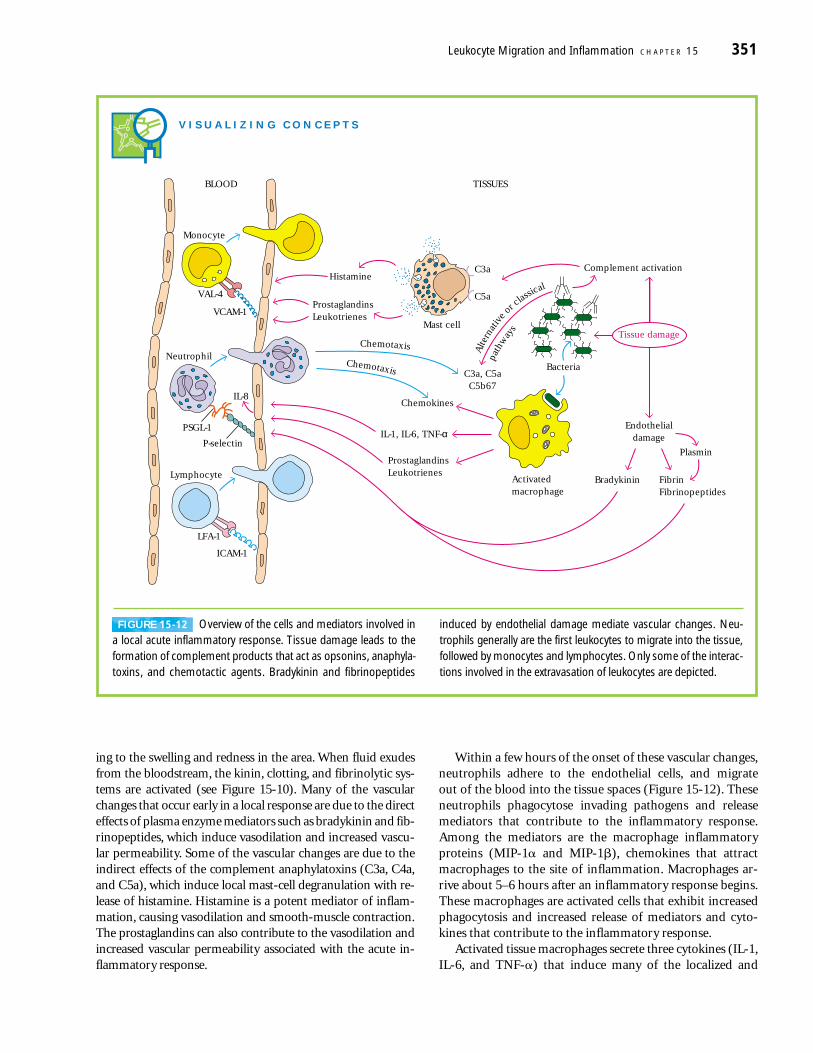
Within a few hours of the onset of these vascular changes,neutrophils adhere to the endothelial cells, and migrate out of the blood into the tissue spaces (Figure 15-12). Theseneutrophils phagocytose invading pathogens and release mediators that contribute to the inflammatory response.Among the mediators are the macrophage inflammatoryproteins (MIP-1� and MIP-1�), chemokines that attractmacrophages to the site of inflammation. Macrophages ar-rive about 5–6 hours after an inflammatory response begins.These macrophages are activated cells that exhibit increasedphagocytosis and increased release of mediators and cyto-kines that contribute to the inflammatory response.
Activated tissue macrophages secrete three cytokines (IL-1,IL-6, and TNF-�) that induce many of the localized and
Leukocyte Migration and Inflammation C H A P T E R 15 351
V I S U A L I Z I N G C O N C E P T S
Alte
rnat
ive
orcla
ssical
path
way
s
Chemotaxis
Chemotaxis
Mast cell
C3a, C5aC5b67
C3a
C5a
Histamine
ProstaglandinsLeukotrienes
ProstaglandinsLeukotrienes
FibrinFibrinopeptides
Bradykinin
IL-1, IL-6, TNF-α
Chemokines
Complement activation
Tissue damage
Endothelialdamage
BLOOD TISSUES
Bacteria
PlasminP-selectin
PSGL-1
IL-8
VAL-4
VCAM-1
Monocyte
LFA-1
ICAM-1
Lymphocyte
Neutrophil
Activatedmacrophage
FIGURE 15-12 Overview of the cells and mediators involved ina local acute inflammatory response. Tissue damage leads to theformation of complement products that act as opsonins, anaphyla-toxins, and chemotactic agents. Bradykinin and fibrinopeptides
induced by endothelial damage mediate vascular changes. Neu-trophils generally are the first leukocytes to migrate into the tissue,followed by monocytes and lymphocytes. Only some of the interac-tions involved in the extravasation of leukocytes are depicted.
systemic changes observed in the acute inflammatoryresponse (see Table 15-3). All three cytokines act locally,inducing coagulation and an increase in vascular permeabil-ity. Both TNF-� and IL-1 induce increased expression ofadhesion molecules on vascular endothelial cells. For in-stance, TNF-� stimulates expression of E-selectin, an endo-thelial adhesion molecule that selectively binds adhesionmolecules on neutrophils. IL-1 induces increased expressionof ICAM-1 and VCAM-1, which bind to integrins on lym-phocytes and monocytes. Circulating neutrophils, mono-cytes, and lymphocytes recognize these adhesion moleculeson the walls of blood vessels, adhere, and then move throughthe vessel wall into the tissue spaces. IL-1 and TNF-� also acton macrophages and endothelial cells to induce productionof the chemokines that contribute to the influx of neutro-phils by increasing their adhesion to vascular endothelialcells and by acting as potent chemotactic factors. In addition,IFN-� and TNF-� activate macrophages and neutrophils,promoting increased phagocytic activity and increased re-lease of lytic enzymes into the tissue spaces.
A local acute inflammatory response can occur withoutthe overt involvement of the immune system. Often, how-ever, cytokines released at the site of inflammation facilitateboth the adherence of immune-system cells to vascular en-dothelial cells and their migration through the vessel wallinto the tissue spaces. The result is an influx of lymphocytes,neutrophils, monocytes, eosinophils, basophils, and mastcells to the site of tissue damage, where these cells participatein clearance of the antigen and healing of the tissue.
The duration and intensity of the local acute inflamma-tory response must be carefully regulated to control tissuedamage and facilitate the tissue-repair mechanisms that arenecessary for healing. TGF-� has been shown to play an im-portant role in limiting the inflammatory response. It alsopromotes accumulation and proliferation of fibroblasts andthe deposition of an extracellular matrix that is required forproper tissue repair.
Clearly, the processes of leukocyte adhesion are of great im-portance in the inflammatory response. A failure of properleukocyte adhesion can result in disease, as exemplified byleukocyte-adhesion deficiency (see Clinical Focus on page 358).
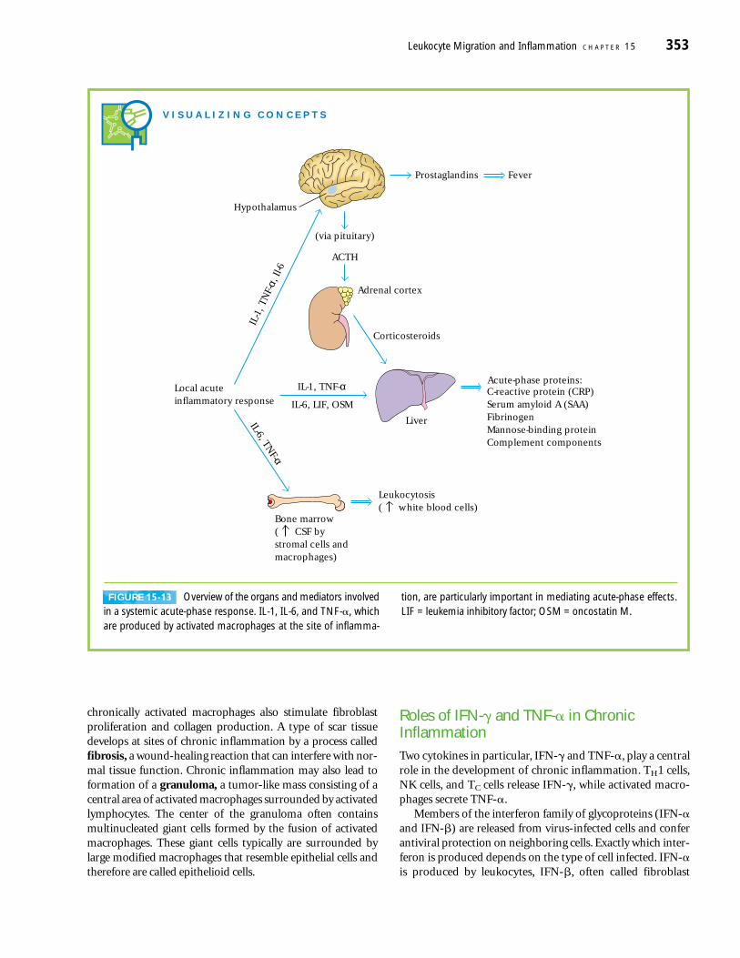
SYSTEMIC ACUTE-PHASE RESPONSE
The local inflammatory response is accompanied by a sys-temic response known as the acute-phase response (Figure15-13). This response is marked by the induction of fever,increased synthesis of hormones such as ACTH and hydro-cortisone, increased production of white blood cells (leuko-cytosis), and production of a large number of acute-phaseproteins in the liver. The increase in body temperatureinhibits the growth of a number of pathogens and appears toenhance the immune response to the pathogen.
C-reactive protein is a prototype acute-phase proteinwhose serum level increases 1000-fold during an acute-phase
response. It is composed of five identical polypeptides heldtogether by noncovalent interactions. C-reactive proteinbinds to a wide variety of microorganisms and activates com-plement, resulting in deposition of the opsonin C3b on thesurface of microorganisms. Phagocytic cells, which expressC3b receptors, can then readily phagocytose the C3b-coatedmicroorganisms.
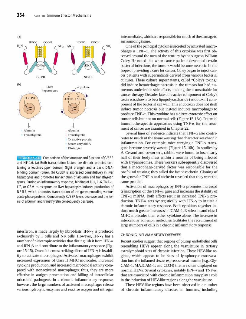
Many systemic acute-phase effects are due to the com-bined action of IL-1, TNF-� and IL-6 (see Figure 15-13).Each of these cytokines acts on the hypothalamus to induce afever response. Within 12–24 h of the onset of an acute-phaseinflammatory response, increased levels of IL-1, TNF-� andIL-6 (as well as leukemia inhibitory factor (LIF) and onco-statin M (OSM)) induce production of acute-phase proteinsby hepatocytes. TNF-� also acts on vascular endothelial cellsand macrophages to induce secretion of colony-stimulatingfactors (M-CSF, G-CSF, and GM-CSF). These CSFs stimulatehematopoiesis, resulting in transient increases in the numberof white blood cells needed to fight the infection.
The redundancy in the ability of at least five cytokines(TNF-�, IL-1, IL-6, LIF, and OSM) to induce production ofacute-phase proteins by the liver results from the inductionof a common transcription factor, NF-IL6, after each of thesecytokines interacts with its receptor. Amino-acid sequencingof cloned NF-IL6 revealed that it has a high degree ofsequence identity with C/EBP, a liver-specific transcriptionfactor (Figure 15-14a). Both NF-IL6 and C/EBP contain aleucine-zipper domain and a basic DNA-binding domain,and both proteins bind to the same nucleotide sequence inthe promoter or enhancer of the genes encoding various liverproteins. C/EBP, which stimulates production of albuminand transthyretin, is expressed constitutively by hepatocytes.As an inflammatory response develops and the cytokinesinteract with their respective receptors on liver hepatocytes,expression of NF-IL6 increases and that of C/EBP decreases(Figure 15-14b). The inverse relationship between these twotranscription factors accounts for the observation that serumlevels of proteins such as albumin and transthyretin declinewhile those of acute-phase proteins increase during an in-flammatory response.
Chronic Inflammation Develops When Antigen PersistsSome microorganisms are able to evade clearance by theimmune system, for example by possessing cell-wall compo-nents that enable them to resist phagocytosis. Such organ-isms often induce a chronic inflammatory response, result-ing in significant tissue damage. Chronic inflammation alsooccurs in a number of autoimmune diseases in which self-antigens continually activate T cells. Finally, chronic inflam-mation also contributes to the tissue damage and wastingassociated with many types of cancer.
The accumulation and activation of macrophages is thehallmark of chronic inflammation. Cytokines released by the
352 P A R T I I I Immune Effector Mechanisms
chronically activated macrophages also stimulate fibroblastproliferation and collagen production. A type of scar tissuedevelops at sites of chronic inflammation by a process calledfibrosis, a wound-healing reaction that can interfere with nor-mal tissue function. Chronic inflammation may also lead toformation of a granuloma, a tumor-like mass consisting of acentral area of activated macrophages surrounded by activatedlymphocytes. The center of the granuloma often containsmultinucleated giant cells formed by the fusion of activatedmacrophages. These giant cells typically are surrounded bylarge modified macrophages that resemble epithelial cells andtherefore are called epithelioid cells.
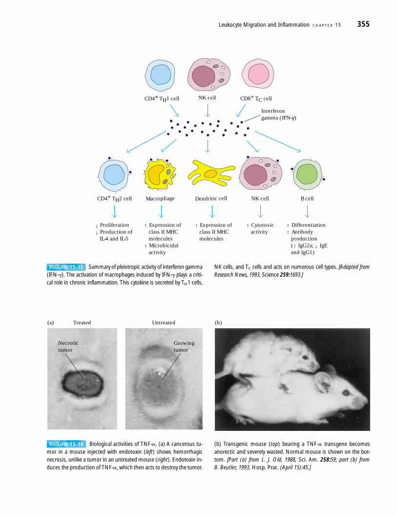
Roles of IFN-� and TNF-� in ChronicInflammationTwo cytokines in particular, IFN-� and TNF-�, play a centralrole in the development of chronic inflammation. TH1 cells,NK cells, and TC cells release IFN-�, while activated macro-phages secrete TNF-�.
Members of the interferon family of glycoproteins (IFN-�and IFN-�) are released from virus-infected cells and conferantiviral protection on neighboring cells. Exactly which inter-feron is produced depends on the type of cell infected. IFN-�is produced by leukocytes, IFN-�, often called fibroblast
Leukocyte Migration and Inflammation C H A P T E R 15 353
V I S U A L I Z I N G C O N C E P T S
Prostaglandins
Acute-phase proteins:C-reactive protein (CRP)Serum amyloid A (SAA)FibrinogenMannose-binding proteinComplement components
Fever
Bone marrow( CSF bystromal cells andmacrophages)
Leukocytosis( white blood cells)
ACTH
(via pituitary)
Local acuteinflammatory response
Corticosteroids
IL-1, TNF-α
IL-6, TNF-α
IL-6, LIF, OSM
IL-1
, TN
F-α,
Il-6
Liver
Hypothalamus
Adrenal cortex
FIGURE 15-13 Overview of the organs and mediators involvedin a systemic acute-phase response. IL-1, IL-6, and TNF-�, whichare produced by activated macrophages at the site of inflamma-
tion, are particularly important in mediating acute-phase effects.LIF = leukemia inhibitory factor; OSM = oncostatin M.
interferon, is made largely by fibroblasts. IFN-� is producedexclusively by T cells and NK cells. However, IFN-� has anumber of pleiotropic activities that distinguish it from IFN-�and IFN-� and contribute to the inflammatory response (Fig-ure 15-15). One of the most striking effects of IFN-� is its abil-ity to activate macrophages. Activated macrophages exhibitincreased expression of class II MHC molecules, increasedcytokine production, and increased microbicidal activity com-pared with nonactivated macrophages; thus, they are moreeffective in antigen presentation and killing of intracellularmicrobial pathogens. In a chronic inflammatory response,however, the large numbers of activated macrophages releasevarious hydrolytic enzymes and reactive oxygen and nitrogen
intermediates, which are responsible for much of the damage tosurrounding tissue.
One of the principal cytokines secreted by activated macro-phages is TNF-�. The activity of this cytokine was first ob-served around the turn of the century by the surgeon WilliamColey. He noted that when cancer patients developed certainbacterial infections, the tumors would become necrotic. In thehope of providing a cure for cancer, Coley began to inject can-cer patients with supernatants derived from various bacterialcultures. These culture supernatants, called “Coley’s toxins,”did induce hemorrhagic necrosis in the tumors but had nu-merous undesirable side effects, making them unsuitable forcancer therapy. Decades later, the active component of Coley’stoxin was shown to be a lipopolysaccharide (endotoxin) com-ponent of the bacterial cell wall. This endotoxin does not itselfinduce tumor necrosis but instead induces macrophages toproduce TNF-�. This cytokine has a direct cytotoxic effect ontumor cells but not on normal cells (Figure 15-16a). Potentialimmunotherapeutic approaches using TNF-� for the treat-ment of cancer are examined in Chapter 22.
Several lines of evidence indicate that TNF-� also contri-butes to much of the tissue wasting that characterizes chronicinflammation. For example, mice carrying a TNF-� trans-gene become severely wasted (Figure 15-16b). In studies byA. Cerami and coworkers, rabbits were found to lose nearlyhalf of their body mass within 2 months of being infectedwith trypanosomes. These workers subsequently discoveredthat a macrophage-derived factor was responsible for theprofound wasting; they called the factor cachetin. Cloning ofthe genes for TNF-� and cachetin revealed that they were thesame protein.
Activation of macrophages by IFN-� promotes increasedtranscription of the TNF-� gene and increases the stability ofTNF-� mRNA. Both effects result in increased TNF-� pro-duction. TNF-� acts synergistically with IFN-� to initiate achronic inflammatory response. Both cytokines together in-duce much greater increases in ICAM-1, E-selectin, and class IMHC molecules than either cytokine alone. The increase inintercellular adhesion molecules facilitates the recruitment oflarge numbers of cells in a chronic inflammatory response.
CHRONIC INFLAMMATORY DISEASES
Recent studies suggest that regions of plump endothelial cellsresembling HEVs appear along the vasculature in tertiary extralymphoid sites of chronic infection. These HEV-like re-gions, which appear to be sites of lymphocyte extravasa-tion into the inflamed tissue, express several mucins (e.g., Gly-CAM-1, MAdCAM-1, and CD34) that are often displayed onnormal HEVs. Several cytokines, notably IFN-� and TNF-�,that are associated with chronic inflammation may play a rolein the induction of HEV-like regions along the vasculature.
These HEV-like regions have been observed in a numberof chronic inflammatory diseases in humans, including
354 P A R T I I I Immune Effector Mechanisms
(a)
HOOC COOH
L LL L
L LL L
L LNH2
HOOC COOH
L LL L
L LL L
L LNH2H2NH2N
DNA DNA
C/EPB NF-IL6
(b)
↑C/EPB
↑ Albumin
↓C/EPB ↑NF-IL6
↑ Transthyretin
Cytokine
↓ Albumin
↓ Transthyretin
↑ C-reactive protein
↑ Serum amyloid A
↑ Fibrinogen
Liverhepatocytes
FIGURE 15-14 Comparison of the structure and function of C/EBPand NF-IL6. (a) Both transcription factors are dimeric proteins con-taining a leucine-zipper domain (light orange) and a basic DNA-binding domain (blue). (b) C/EBP is expressed constitutively in liver hepatocytes and promotes transcription of albumin and transthyretingenes. During an inflammatory response, binding of IL-1, IL-6, TNF-�,LIF, or OSM to receptors on liver hepatocytes induces production ofNF-IL6, which promotes transcription of the genes encoding variousacute-phase proteins. Concurrently, C/EBP levels decrease and the lev-els of albumin and transthyretin consequently decrease.
Leukocyte Migration and Inflammation C H A P T E R 15 355
↓ Proliferation
↓ Production of IL-4 and IL-5
↑ Expression of class II MHC molecules
↑ Microbicidal activity
↑ Cytotoxic activity
↑ Differentiation
↑ Antibody production (↑ IgG2a; ↓ IgE and IgG1)
CD4+ TH2 cell B cellMacrophage
↑ Expression of class II MHC molecules
Dendritic cell NK cell
NK cellCD4+ TH1 cell CD8+ TC cell
Interferongamma (IFN-γ)
FIGURE 15-15 Summary of pleiotropic activity of interferon gamma(IFN-�). The activation of macrophages induced by IFN-� plays a criti-cal role in chronic inflammation. This cytokine is secreted by TH1 cells,
NK cells, and TC cells and acts on numerous cell types. [Adapted fromResearch News, 1993, Science 259:1693.]
(a) Treated Untreated
Necrotictumor
Growingtumor
(b)
FIGURE 15-16 Biological activities of TNF-�. (a) A cancerous tu-mor in a mouse injected with endotoxin (left) shows hemorrhagicnecrosis, unlike a tumor in an untreated mouse (right). Endotoxin in-duces the production of TNF-�, which then acts to destroy the tumor.
(b) Transgenic mouse (top) bearing a TNF-� transgene becomesanorectic and severely wasted. Normal mouse is shown on the bot-tom. [Part (a) from L. J. Old, 1988, Sci. Am. 258:59; part (b) from B. Beutler, 1993, Hosp. Prac. (April 15):45.]
rheumatoid arthritis, Crohn’s disease, ulcerative colitis,Graves’ disease, Hashimoto’s thyroiditis, and diabetes mellitus(Table 15-4). Development of this HEV-like vasculature islikely to facilitate a large-scale influx of leukocytes, contribut-
ing to chronic inflammation. These observations suggest thatan effective approach for treating chronic inflammatory dis-eases may be to try to control the development of these HEV-like regions.
356 P A R T I I I Immune Effector Mechanisms
Once in, they are guided by gradients ofchemoattractants to the sites of theinflammatory responses and becomeparticipants in the process. The key play-ers in the adhesive interactions that arecentral to adhesion and extravasation areheterodimeric integrin molecules on thesurface of the migrating leukocytes.There are a number of integrins, amongwhich are LFA-1 (composed of CD11aand CD18); Mac-1, also called CR3 (com-ponents: CD11b and CD18); and p150/95 or CR4 (components: CD11c andCD18). When leukocytes encounter theappropriate chemokine or other chemo-attractant, their complement of mem-brane integrin molecules undergoes aconformational change that transformsthem from a slightly adhesive to a highlyadhesive state.
In 1979, a paper entitled “Delayed sep-aration of the umbilical cord, widespread
infections, and defective neutrophil mo-bility” appeared in Lancet, a British med-ical journal. This was the first in a seriesof reports that have appeared over theyears describing patients afflicted with a rare autosomal recessive disease inwhich the first indication is quite oftenomphalitis, a swelling and reddeningaround the stalk of the umbilical cord. Although no more susceptible to virus infections than normal controls, those afflicted with this disorder suffer recur-rent and often chronic bacterial infec-tions, and sites where one would expectto find pus are instead pus-free. This ob-servation is particularly striking becausethe patients are not deficient in granulo-cytes; in fact, they typically have greatly el-evated numbers of granulocytes in thecirculation. Detailed immunological work-ups of these patients showed that Ig levels were in the normal range and thatthey had nearly normal B-, T-, and NK-cellfunction. However, examination of leuko-cyte migration in response to tissue dam-age revealed the root cause of the diseasein these patients.
One method of evaluating leukocytemigration involves gently scraping the
The immune system uses in-flammation to assemble the componentsof an effective response and focus theseresources at the site of infection.Inflammation is complex, involvingvasodilation, increased vascular perme-ability, exudation of plasma proteins,and a gathering of inflammatory cells.Chemoattractants are key elements incalling leukocytes to sites of inflamma-tion. These include chemokines such asIL-8, monocyte chemoattractant protein1 (MCP-1), macrophage inflammatoryprotein 1 (MIP-1) and peptide fragments,such as C5a, generated during comple-ment fixation. Chemoattractants signalpassing leukocytes to adhere tightly tothe vascular surface, and, using adhesiveinteractions for traction, these cells pushtheir way between endothelial cells andgain entry into the surrounding tissue.
C L I N I C A L F O C U S
Leukocyte-Adhesion Deficiency(LAD) in Humans and Cattle
TABLE 15-4 Chronic inflammatory diseases associated with HEV-like vasculature
Plump Mucin-like CAMsDisease Affected organ endothelium on endothelium*
Crohn’s disease Gut + +Diabetes mellitus Pancreas + +Graves’ disease Thyroid + +Hashimoto’s thyroiditis Thyroid + +Rheumatoid arthritis Synovium + +Ulcerative colitis Gut + +
*Includes GlyCAM-1, MAdCAM-1, and CD34.
SOURCE: Adapted from J. P. Girard and T. A. Springer, 1995, Immunol. Today 16:449.
Anti-Inflammatory AgentsAlthough development of an effective inflammatory re-sponse can play an important role in the body’s defense, theresponse can sometimes be detrimental. Allergies, autoim-mune diseases, microbial infections, transplants, and burnsmay initiate a chronic inflammatory response. Various thera-peutic approaches are available for reducing long-term in-flammatory responses and thus the complications associatedwith them.
Antibody Therapies Reduce LeukocyteExtravasation
Because leukocyte extravasation is an integral part of theinflammatory response, one approach for reducing inflam-mation is to impede this process. Theoretically, one way toreduce leukocyte extravasation is to block the activity of var-ious adhesion molecules with antibodies. In animal models,
for example, antibodies to the integrin LFA-1 have been usedto reduce neutrophil buildup in inflammatory tissue. Anti-bodies to ICAM-1 have also been used, with some success, inpreventing the tissue necrosis associated with burns and inreducing the likelihood of kidney-graft rejection in animalmodels. The results with antibodies specific for these ad-hesins have been so encouraging that a combination of anti-bodies (anti-ICAM-1 and anti-LFA-1) was used in clinicaltrials on human kidney-transplant patients. A combinationof two anti-adhesins had to be used because failure to blockboth LFA-1 and ICAM-1 results in rejection.
Corticosteroids Are Powerful Anti-Inflammatory Drugs
The corticosteroids, which are cholesterol derivatives, includeprednisone, prednisolone, and methylprednisolone. These po-tent anti-inflammatory agents exert various effects that resultin a reduction in the numbers and activity of immune-systemcells. They are regularly used in anti-inflammatory therapy.
Leukocyte Migration and Inflammation C H A P T E R 15 357
has been named leukocyte-adhesion de-ficiency (LAD).
Bacterial infections in these patientscan be treated with antibiotics, but they recur. Furthermore, there are antibiotic-resistant strains of many pathogenic bac-teria, and LAD patients must live underthis microbial Sword of Damocles, neverknowing when the life-saving thread of an-tibiotics will fail. If a suitable bone-marrowdonor can be found (almost always a closerelative), however, there is a curative strat-egy. The LAD patient’s hematopoietic sys-tem is destroyed, perhaps by treatmentwith cytotoxic chemicals, and then bone-marrow transplantation is performed. Ifsuccessful, this procedure provides the pa-tient with leukocytes that have normal lev-els of functional integrin and display thefull range of migratory capacities.
This disease is not limited to humans.A strikingly similar version known as bo-vine leukocyte adhesion disease (BLAD)occurs in cattle. The cause of BLAD inthese animals is identical to the cause ofLAD in human patients—the lack of afunctional integrin subunit. What is dif-ferent in some dairy herds is the inci-dence of the disease; though rare in
humans, it can occur at economically im-portant frequencies in cattle. This is aconsequence of the high degree of in-breeding that exists in populations ofdairy cattle. Typically, dairy herds are siredby the artificial insemination of semenfrom very few bulls. As a consequence of this practice, by the 1980s, almost 1 in20 dairy bulls could be traced back to asingle Holstein bull who happened to beheterozygous for BLAD. Such a high fre-quency of this recessive trait in the sirepopulation dramatically raised the fre-quency of this disease in dairy herds. Dur-ing the early 1990s, in some countries,the incidence of the BLAD gene was ashigh as 10% in a number of dairy herds.The gene for bovine CD18 has beencloned, which has allowed the design of aPCR-based assay for the aberrant formsof this gene. It is now possible to rou-tinely screen sires and recipients for theBLAD allele. As a result, bulls that are car-riers of the BLAD gene have been identi-fied and eliminated from the breedingpool. This has led to a dramatic reductionin the frequency of new BLAD cases aswell as in the overall frequency of theBLAD allele in dairy-herd populations.
skin from a small area of the arm; the cellpopulations that move into the abradedarea are then sampled by capturingsome of those cells on a glass coverslipplaced onto the wounded skin. A seriesof glass coverslips is sequentially placed,incubated, and removed over a period of several hours. Typically, each coverslipis left in place for two hours, and the pro-cedure is repeated four times over aneight-hour period. Examination of thecoverslips under a microscope revealswhether leukocytes have adhered to the coverslips. In normal individuals, theresponse of the immune system to tis-sue injury is to deliver leukocytes to thedamaged area, and one finds these cellson the coverslips. However, in the pa-tients described here, the coverslipswere largely negative for leukocytes. Ex-amination of white blood cells in thesepatients revealed an absence of CD18,an essential component of a number ofintegrins. A key element in the migrationof leukocytes is integrin-mediated celladhesion, and these patients suffer froman inability of their leukocytes to undergoadhesion-dependent migration into sitesof inflammation. Hence, this syndrome
Corticosteroid treatment causes a decrease in the numberof circulating lymphocytes as the result either of steroid-induced lysis of lymphocytes (lympholysis) or of alterationsin lymphocyte-circulation patterns. Some species (e.g., ham-ster, mouse, rat, and rabbit) are particularly sensitive to corticosteroid-induced lympholysis. In these animals, corti-costeroid treatment at dosages as low as 10–7 M causes suchwidespread lympholysis that the weight of the thymus isreduced by 90%; the spleen and lymph nodes also shrink visibly. Immature thymocytes in these species appear to beparticularly sensitive to corticosteroid-mediated killing. Inrodents, corticosteroids induce programmed cell death ofimmature thymocytes, whereas mature thymocytes are resis-tant to this activity. Within 2 h following in vitro incubationwith corticosteroids, immature thymocytes begin to showthe characteristic morphology of apoptosis, and 90% of thechromatin is degraded into the characteristic nucleosome lad-der by 24 h after treatment. The steps involved in the inductionof apoptosis by corticosteroids remain to be determined. Inhumans, guinea pigs, and monkeys, corticosteroids do not in-duce apoptosis but instead affect lymphocyte-circulation pat-terns, causing a decrease in thymic weight and a marked de-crease in the number of circulating lymphocytes.
Like other steroid hormones, the corticosteroids are lipo-philic and thus can cross the plasma membrane and bind toreceptors in the cytosol. The resulting receptor-hormone com-plexes are transported to the nucleus, where they bind to spe-cific regulatory DNA sequences, regulating transcription up ordown. The corticosteroids have been shown to induce in-creased transcription of the NF-�B inhibitor (I-�B). Bindingof this inhibitor to NF-�B in the cytosol prevents the translo-cation of NF-�B into the nucleus and consequently preventsNF-�B activation of a number of genes, including genes in-volved in T-cell activation and cytokine production.
Corticosteroids also reduce both the phagocytic and killingability of macrophages and neutrophils, and this effect maycontribute to their anti-inflammatory action. In addition, theyreduce chemotaxis, so that fewer inflammatory cells are at-tracted to the site of TH-cell activation. In the presence of cor-ticosteroids, expression of class II MHC molecules and IL-1production by macrophages is dramatically reduced; suchreductions would be expected to lead to corresponding reduc-tions in TH-cell activation. Finally, corticosteroids also stabilizethe lysosomal membranes of participating leukocytes, so thatdecreased levels of lysosomal enzymes are released at the site ofinflammation.
NSAIDs Combat Pain and InflammationSince the time of Hippocrates, extracts of willow bark havebeen used for relief of pain. The active ingredient, salicylate,which is found in aspirin, is just one of many nonsteroidalanti-inflammatory drugs (NSAIDs). NSAIDs are the most fre-quently used medication for treating pain and inflammation.Clinically, NSAIDs have been shown to be effective for treat-
ment of many acute and chronic inflammatory reactions. Themajor mechanism by which these drugs exert anti-inflamma-tory effects is by inhibiting the cyclooxygenase pathway thatproduces prostaglandins and thromboxanes from arachidonicacid. The reduction in prostaglandin production limits theincrease in vascular permeability and neutrophil chemotaxisin the inflammatory response. As shown in Figure 15-17, thecyclooxygenase pathway is mediated by two enzymes, cyclo-oxygenase 1 and cyclooxygenase 2 (Cox-1 & Cox-2).
Although NSAIDs such as aspirin, Tylenol, ibuprofen,Naproxen, and others are routinely prescribed for the treat-ment of ailments as diverse as arthritis, sprains, tissue injury,and back pain, the duration of their use is limited by gas-trointestinal side effects that include unease and abdominalpain and in more serious cases bleeding or perforation of thestomach or upper GI tract. Investigation of the mechanism ofNSAIDs has provided a basis for the beneficial and deleteri-ous effects of many NSAIDS. Studies have shown that,although most NSAIDs inhibit both Cox-1 and Cox-2, it isthe inhibition of Cox-2 that is responsible for the anti-inflammatory effects of NSAIDs. On the other hand, inhibi-
358 P A R T I I I Immune Effector Mechanisms
Diversephysical, chemical,inflammatory,and mitogenicstimuli
Cox-1mediated
Cox-2mediated
Cox-2-specificN SAIDS
Arachidonicacid
Inflammation
ConventionalN SAIDS
Phospholipase A2
− −
−
Maintenanceof integrity of
gastrointestinaltissue
Thromboxaneand
prostaglandins
Thromboxaneand
prostaglandins
FIGURE 15-17 Inhibition of cyclooxygenase 1 and 2 by NSAIDs. Avariety of agents trigger the release of arachidonic acid from the cellmembrane by the action of phospholipase A2. The subsequent actionof Cox-1 and Cox-2 initiates the conversion of arachidonic acid to avariety of lipid mediators of inflammation and many other processes.Many NSAIDs inhibit both enzymatic pathways, but those withgreater specificity for the Cox-2 arm produce antinflammatory effectswith fewer side effects. [Adapted from G. A. FitzGerald and C. N. Patrono, 2001, New England Journal of Medicine 345:433.]
tion of Cox-1 by thses agents causes damage to the GI tractbut does not have significant anti-inflammatory benefits.This realization led to the design and development of a newgeneration of NSAIDs that specifically inhibit Cox-2 buthave little effect on Cox-1 activity. The action of these highlytargeted drugs is shown in Figure 15-17.
SUMMARY
■ Lymphocytes undergo constant recirculation between theblood, lymph, lymphoid organs, and tertiary extralymphoidtissues, increasing the chances that the small number oflymphocytes specific for a given antigen (about 1 in 105
cells) will actually encounter that antigen.
■ Migration of leukocytes into inflamed tissue or into lym-phoid organs requires interaction between cell-adhesionmolecules (CAMs) on the vascular endothelium and thoseon the circulating cells.
■ Most CAMs fall into one of four protein families: theselectins, the mucin-like family, integrins, or the Ig super-family. Selectins and mucin-like CAMs interact with eachother, and members of each family are expressed on bothleukocytes and endothelial cells. Integrins, expressed onleukocytes, interact with Ig-superfamily CAMs, expressedon endothelial cells.
■ Extravasation of both neutrophils and lymphocytes involvesfour steps: rolling, activation, arrest and adhesion, andtransendothelial migration. Neutrophils are generally thefirst cell type to move from the bloodstream into inflamma-tory sites.
■ Unlike neutrophils, various lymphocyte populations ex-hibit differential extravasation into various tissues. Hom-ing receptors on lymphocytes interact with tissue-specificadhesion molecules, called vascular addressins, on high-endothelial venules (HEVs) in lymphoid organs and onthe endothelium in tertiary extralymphoid tissues.
■ Naive lymphocytes home to secondary lymphoid organs,extravasating across HEVs, whereas effector lymphocytesselectively home to inflamed vascular endothelium.
■ Inflammation is a physiologic response to a variety of stimulisuch as tissue injury and infection. An acute inflammatoryresponse involves both localized and systemic effects. Thelocalized response begins when tissue and endothelial dam-age induces formation of plasma enzyme mediators that leadto vasodilation and increased vascular permeability.
■ Several types of mediators play a role in the inflammatoryresponse. Chemokines act as chemoattractants and acti-vating molecules during leukocyte extravasation. Plasmaenzyme mediators include bradykinin and fibrinopep-tides, which increase vascular permeability; plasmin is aproteolytic enzyme that degrades fibrin clots into chemo-
tactic products and activates complement; and variouscomplement products act as anaphylatoxins, opsonins,and chemotactic molecules for neutrophils and mono-cytes. Lipid inflammatory mediators include thrombox-anes, prostaglandins, leukotrienes, and platelet-activatingfactor. Three cytokines, IL-1, IL-6, and TNF-�, mediatemany of the local and systemic features of the acuteinflammatory response
■ Activation of tissue macrophages and degranulation of mastcells lead to release of numerous inflammatory mediators,some of which induce the acute-phase response, which in-cludes fever, leukocytosis, and production of corticosteroidsand acute-phase proteins.
■ A chronic inflammatory response may accompany allergies,autoimmune diseases, microbial infections, transplants, andburns. Drug-based therapies employing corticosteroidsand a variety of nonsteroidal anti-inflammatory drugs(NSAIDs) are the most commonly used medications forpain and inflammation.
ReferencesButcher, E., and L. J. Picker. 1996. Lymphocyte homing and
homeostasis. Science 272:60.
FitzGerald, G. A., and C. Patrono. 2002. The coxibs, selectiveinhibitors of cyclooxygenase-2. New England Journal of Medi-cine 345:433.
Gabay, C., and I. Kushner. 1999. Acute-phase proteins and othersystemic responses to inflammation. N. Engl. J. Med. 340:448.
Kuijpers, T. W., et al. 1997. Leukocyte adhesion deficiency type 1(LAD-1)/variant. A novel immunodeficiency syndrome char-acterized by dysfunctional beta2 integrins. Journal of ClinicalInvestigation 100:1725.
Kunkel, E. J., and E. C. Butcher. 2002. Chemokines and the tissue-specific migration of lymphocytes. Immunity 16:1.
Shuster, D. E., et al. 1992. Identification and prevalence of agenetic defect that causes leukocyte adhesion deficiency inHolstein cattle. Proceedings of the National Academy of Sciences(USA) 89:9225.
Springer, T. A. 1994. Traffic signals for lymphocyte recirculationand leukocyte emigration: the multistep paradigm. Cell 76:301.
Steel, D. M., and A. S. Whitehead. 1994. The major acute phasereactants: C-reactive protein, serum amyloid P componentand serum amyloid A protein. Immunol. Today 15:81.
USEFUL WEB SITES
http://www.mdsystems.com
The Cytokine Mini-Reviews Section (Site Map/Reviews &Technical Notes) of the R&D Systems site contains extensive,
Leukocyte Migration and Inflammation C H A P T E R 15 359
Go to www.whfreeman.com/immunology Self-TestReview and quiz of key terms
detailed, and well-illustrated reviews of many chemokinesand chemokine receptors.
http://www.ncbi.nlm.nih.gov/Omim/
Online Mendelian Inheritance in Man is a catalog of humangenes and genetic disorders. It contains pictures and refer-ences on many diseases, including LAD.
Study QuestionsCLINICAL FOCUS QUESTION Why does a defect in CD18 result in anincreased vulnerability to bacterial infection? Please address, asprecisely as you can, the cell biology of cell migration.
1. Indicate whether each of the following statements is true orfalse. If you think a statement is false, explain why.
a. Chemokines are chemoattractants for lymphocytes butnot other leukocytes.
b. Integrins are expressed on both leukocytes and endothe-lial cells.
c. Leukocyte extravasation involves multiple interactionsbetween cell-adhesion molecules.
d. Most secondary lymphoid organs contain high-endothe-lial venules (HEVs).
e. Mucin-like CAMs interact with selectins.
f. An acute inflammatory response involves only localizedeffects in the region of tissue injury or infection.
g. MAdCAM-1 is an endothelial adhesion molecule thatbinds to L-selectin and to several integrins.
h. Granuloma formation is a common symptom of localinflammation.
2. Various inflammatory mediators induce expression of ICAMson a wide variety of tissues. What effect might this inductionhave on the localization of immune cells?
3. Extravasation of neutrophils and of lymphocytes occurs bygenerally similar mechanisms, although some differences distin-guish the two processes.
a. List in order the four basic steps in leukocyte extravasation.
b. At which sites are neutrophils most likely to extravasate?Why?
c. Different lymphocyte subpopulations migrate preferen-tially into different tissues, a process called homing (ortrafficking). Discuss the roles of the three types of mole-cules that permit homing of lymphocytes.
4. Which three cytokines secreted by activated macrophagesplay a major role in mediating the localized and systemic effectsassociated with an acute inflammatory response?
5. An effective inflammatory response requires differentiationand proliferation of various nonlymphoid white blood cells.Explain how hematopoiesis in the bone marrow is induced by tis-sue injury or local infection.
6. For each pair of molecules listed below, indicate whether themolecules interact during the 1st, 2nd, 3rd, or 4th step in neu-trophil extravasation at an inflammatory site. Use N to indicateany molecules that do not interact.
a. Chemokine and L-selectinb. E-selectin and mucin-like CAMc. IL-8 and E-selectind. Ig-superfamily CAM and integrine. ICAM and chemokinef. Chemokine and G-protein–coupled receptorg. ICAM and integrin
7. Discuss the main effects of IFN-� and TNF-� during achronic inflammatory response.
8. Five cytokines (IL-1, IL-6, TNF-�, LIF, and OSM) induce pro-duction of C-reactive protein and other acute-phase proteins byhepatocytes. Briefly explain how these different cytokines canexert the same effect on hepatocytes.
9. For each inflammation-related term (a–h), select the descrip-tions listed below (1–11) that apply. Each description may beused once, more than once, or not at all; more than one descrip-tion may apply to some terms.
Terms
a. Tertiary extralymphoid tissueb. P- and E-selectinc. Prostaglandinsd. Nonsteroidal anti-inflammatory drugse. ICAM-1, -2, -3f. MAdCAMg. Bradykininh. Inflamed endothelium
Descriptions
(1) Bind to sialylated carbohydrate moieties(2) Inhibit cyclooxygenase pathway(3) Induce expression of NF-�B inhibitor(4) Has both Ig domains and mucin-like domains(5) Region of vascular endothelium found in postcapillary
venules(6) Expressed by inflamed endothelium(7) Exhibits HEV-like vasculature in chronic inflammation(8) Belong to Ig-superfamily of CAMs(9) Exhibits increased expression of CAMs
(10) Increase vascular permeability and induce fever(11) Induce fever
360 P A R T I I I Immune Effector Mechanisms