Embed Size (px)
Citation preview
This article appeared in a journal published by Elsevier. The attachedcopy is furnished to the author for internal non-commercial researchand education use, including for instruction at the authors institution
and sharing with colleagues.
Other uses, including reproduction and distribution, or selling orlicensing copies, or posting to personal, institutional or third party
websites are prohibited.
In most cases authors are permitted to post their version of thearticle (e.g. in Word or Tex form) to their personal website orinstitutional repository. Authors requiring further information
regarding Elsevier’s archiving and manuscript policies areencouraged to visit:
http://www.elsevier.com/copyright
Author's personal copy
Neuroscience Letters 445 (2008) 31–35
Contents lists available at ScienceDirect
Neuroscience Letters
journa l homepage: www.e lsev ier .com/ locate /neule t
Astrocyte response to Junín virus infection
Roberto G. Poznera,1, Soledad Colladob,1, Carolina Jaquenod de Giustib, Agustín E. Ureb,Marina E. Biedmab, Victor Romanowskib, Mirta Schattner a, Ricardo M. Gómezb,∗
a Thrombosis 1 Laboratory, Haematological Research Institute, National Academy of Medicine, Pacheco de Melo 3081, 1425 Buenos Aires, Argentinab Molecular Biology and Biotechnology Institute, CCT-La Plata, CONICET-UNLP, 49 and 115 Street, 1900 La Plata, Argentina
a r t i c l e i n f o
Article history:Received 14 June 2008Received in revised form 8 August 2008Accepted 19 August 2008
Keywords:Cultured astrocytesViral infectionGFAPiNOSNO
a b s t r a c t
In a previous study of experimental murine encephalitis induced by Junín virus (JV), an arenavirus, weshowed increased expression of iNOS by unidentified cells, concomitant with the astrocyte reaction. Thespecific inhibition of iNOS was associated with greater mortality but lower astrocytosis, suggesting thatthe protective role of nitric oxide (NO) synthesized by iNOS was related to enhanced astrocyte activa-tion, representing a beneficial cellular response to virus-induced central nervous system damage. In thepresent work, cultured astrocytes were used to study whether JV infection could trigger iNOS expressionand assess its eventual relationship with viral replication, glial fibrilary acidic protein (GFAP) expressionlevels and the presence of apoptosis. We found that JV infection of astrocytes did not induce apoptosis butproduced both increased iNOS synthesis, detected by immunocytochemistry and fluorescence activatedcell sorting (FACS) analysis, and increased NO, which was indirectly measured by nitrite/nitrate levels.These changes occurred early relative to the increases in GFAP expression, as detected by immunocyto-chemistry, FACS analysis and RT-PCR. The fact that iNOS inhibition abolished enhanced GFAP expressionin infected monolayers suggests that NO was directly involved. In addition, iNOS inhibition enhancedvirus replication. Together with data from confocal microscopy, these results suggest that JV induces iNOSexpression in infected astrocytes and that the resulting NO has an important role both in reducing viralreplication and in enhancing subsequent astrocyte activation.
© 2008 Elsevier Ireland Ltd. All rights reserved.
Astrocytes are the most numerous cell type in the central nervoussystem (CNS). They provide structural, trophic and metabolic sup-port to neurons, in addition to modulating synaptic plasticity andactivity, neurite outgrowth and neuron regeneration [8].
An important characteristic of astrocytes is their activation,characterized by enhanced expression of glial fibrilary acidic pro-tein (GFAP), in response to diverse CNS injuries such as trauma,infectious diseases or chemical insults. The prominence of thisreaction and its evolutionary conservation indicate that activatedastrocytes serve important functions in the injured CNS [9] by selec-tive expression of several proteins including inducible nitric oxidesynthase (iNOS) [14]. Remarkably, both beneficial and detrimentaleffects have been attributed to reactive astrocytes [8,18,19].
Argentine hemorrhagic fever (AHF) is a systemic febrile syn-drome characterized by several haematological alterations caused
∗ Corresponding author at: Instituto de Bioctecnología y Biología Molecular, CCT-La Plata, CONICET-UNLP, Calle 49 y 115, 1900 La Plata, Argentina.Tel.: +54 221 425 0497x32; fax: +54 221 422 6947.
E-mail address: [email protected] (R.M. Gómez).1 These authors contributed equally to this work.
by Junín virus (JV), a member of the Arenaviridae [26]. Patientswith AHF frequently exhibit neurological involvement in the acuteperiod [10,20]. In addition, although serum treatment reduces mor-tality from 30% to 1%, about 10–15% of patients who receive thistreatment present a delayed neurological syndrome [28]. Inter-estingly, the histopathological findings in both humans as wellas animal models do not reflect the severity of disease [20], andalthough it has been shown in animals that the virus can reachthe CNS via a neural route [17], its pathogenesis is poorly under-stood. When studied in experimental models, viral antigen canbe detected in the CNS during the acute period, widely dissemi-nated primarily in neurons and a few astrocytes [16]. The chronicstage is characterized by the gradual disappearance of viral antigenand a prominent astrocyte reaction [16]. In a previous study of JV-induced experimental murine encephalitis, we showed increasedexpression of iNOS by unidentified cells, concomitant with theastrocyte reaction. Specific inhibition of iNOS was associated withgreater mortality but lower astrocytosis. Still, similar infective titerswere observed, suggesting that the apparent protective role ofnitric oxide (NO) synthesized by iNOS was unrelated to reducedviral replication but was rather due to enhanced astrocyte activa-tion, representing a beneficial cellular response to viral-induced
0304-3940/$ – see front matter © 2008 Elsevier Ireland Ltd. All rights reserved.doi:10.1016/j.neulet.2008.08.059
Author's personal copy
32 R.G. Pozner et al. / Neuroscience Letters 445 (2008) 31–35
CNS damage [12]. Our present work is aimed to show if JV infec-tion could trigger expression of iNOS in astrocytes and whethersubsequently iNOS expression affects viral replication, astrocyteapoptosis or astrocyte activation. This study used an in vitro model,which affords the advantage of avoiding the complexity of CNSmicroenvironment and the host immune response. To this end,we used JV-infected cultured astrocytes to study the relationshipbetween viral replication and the expression levels of iNOS andGFAP.
The JV strain P3441 and the infectivity titration procedure havebeen previously described [11]. UV-irradiated JV was used as a neg-ative control.
Astroglial cell cultures were obtained from the brains of new-born Balb/c mice as previously described [2]. Briefly, corticalhemispheres were harvested, and neural tissues digested by 0.25%trypsin. Flasks were seeded with 6 × 10⁵ to 8 × 10⁵ cells/ml ofgrowth medium (D-MEM plus 10% fetal calf serum) twice for 1 hin order to eliminate fibroblast contamination. By changing thesupernatant within 24 h, the neurons were eliminated by means ofdifferential attachment, leaving an almost homogenous populationof glial cells. After 3 weeks at 37 ◦C, the already confluent primarycell culture was shaken for 2 h at 37 ◦C, and supernatant was dis-carded in order to detach contaminating oligodendrocytes growingon top of the remaining adherent astroglial cell monolayer, whichin turn were trypsinized. The resulting cells were then resuspended(3 × 10⁵ cells/ml of growth medium) and seeded as first subculturein 24 wells plates with 12 mm glass inserts or in 25 cm2 flasks. Sub-sequently, immunoperoxidase labeling for GFAP showed a highlyhomogeneous population of astrocytes (≥ 95%, data not shown).
After 2 days of seeding, growth medium was removed, andcell monolayers were infected with JV at a multiplicity of infec-tion of 1. At 1, 3, 7 and 14 days post-infection (PI), 3–4 sampleswere harvested for each experimental time point. Supernatantswere routinely frozen at −70 ◦C for later measurement of infectiv-ity titers and nitrate/nitrite levels. Monolayers were fixed with 2%paraformaldehyde plus 0.1% Triton X-100 at 37 ◦C for immunolabel-ing or trypsinized for cytofluorometry. In some studies, astrocyteswere treated with the iNOS specific inhibitor, N6-(1-Iminoethyl)-L-lysine hydrochloride (L-NIL) (5 �M) from Tocris (USA).
The DNA content of astrocytes was analyzed by flow cytom-etry using propidium iodide and histogram analysis to identifyhypodiploid nuclei as previously described [23]. Morphologicalchanges and viability were evaluated by staining cells with a mix-ture of fluorescent DNA-binding dyes: acridine orange (100 �g/ml)to determine the percentage of cells undergoing apoptosis andethidium bromide (100 �g/ml) to differentiate between viable andnonviable cells. At least 200 cells were scored in each experiment.Samples treated with H2O2 (400 mM) for 1 h served as a positivecontrol.
A pool of monoclonal antibodies against JV [22] was kindly pro-vided by Dr. C.J. Peters (Centers for Disease Control and Prevention,Atlanta, USA) and used as a primary antibody, as well as commer-cially available antibodies such as anti-GFAP (Dako Corporation, CA,USA) and anti-iNOS (Cayman Chem. Co., MI, USA). For immunoper-oxidase labeling, the second and third reagents were biotinylatedanti-species immunoglobulin and peroxidase-conjugated strep-tavidin (Dako), respectively. Development of the reaction wasdone with 0.03% DAB (Fluka, Buchs SG, Switzerland) plus 0.02%hydrogen peroxide. For fluorescence microscopy, the secondaryimmunoglobulins were FITC-conjugated anti-rabbit (Dako, USA)and Cy3-conjugated anti-mouse Ig (Zymed, USA). No primary anti-body was used as a negative control of the procedure. Analysis wasperformed using a confocal laser-scanning microscope (LSM 510;Carl Zeiss MicroImaging, Inc.) or an inverted microscope (EclipseTE2000-U; Nikon) equipped with an oil immersion plan Apo 60×
NA 1.4, 100× NA 1.4 or a dry plan Fluor 40× NA 0.6 objective (Nikon)and a CCD camera (MicroMAX RTE/CCD-1300Y; Princeton Instru-ments). Acquisition and analysis of still images were performedusing MetaMorph Imaging Software (version 6.1; Universal ImagingCorp.).
After trypsinization, astrocytes were centrifuged at 200 × g for10 min, resuspended in PBS/FBS and incubated with the appro-priate primary antibody and a FITC-conjugated fragment F(ab′)2anti-species IgG (Immunotech, Marseille, France). For non-specificbinding, FITC-conjugated or primary MoAbs were replaced by anirrelevant isotypic MoAb. After labeling, cells were washed, fixedwith 1% paraformaldehyde and analyzed by fluorescence activatedcell sorting (FACS) analysis.
Nitrite sample concentration was established by the Griessreaction as described [12]. Optical density at 550 nm was deter-mined on a spectrophotometer from a sodium nitrite (Sigma) curveperformed for each experiment. Medium absorbance alone wassubtracted from the value obtained for each sample.
Total RNA was isolated from cell pellets using Trizol (Invitro-gen) as recommended by the manufacturer. cDNA was synthesizedfrom 20 ng of total RNA using 15 �M random hexamers andSuperScript III reverse transcriptase (Invitrogen), according tothe manufacturer’s protocol. The cDNA samples were diluted 10-fold, and the PCR reaction was conducted with an annealingtemperature of 55 ◦C using the following primers (F/R): �ActinACTATTGGCAACGAGCGGTT/CAGGATTCCATACCCAAGAAGGA; GFAPAAAACCGCATCACCATTCC/CCTCTTCCCTTCCAATTCTAAC. All reac-tions were confirmed to be in the linear range of amplification.
All results are expressed as means ± S.E.M. Data analysis wasaccomplished using a Student’s t-test. Values of p lower than 0.05were considered statistically significant.
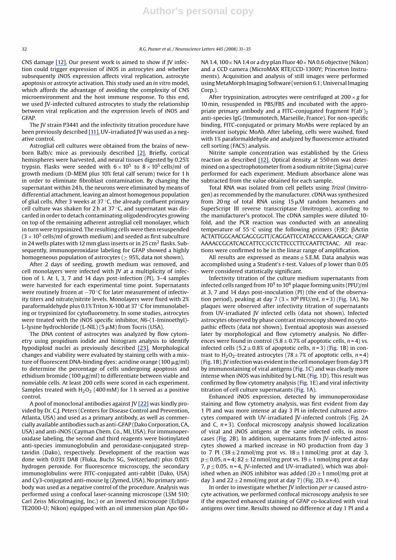
Infectivity titration of the culture medium supernatants frominfected cells ranged from 105 to 106 plaque forming units (PFU)/mlat 3, 7 and 14 days post-inoculation (PI) (the end of the observa-tion period), peaking at day 7 (3 × 10⁶ PFU/ml, n = 3) (Fig. 1A). Noplaques were observed after infectivity titration of supernatantsfrom UV-irradiated JV infected cells (data not shown). Infectedastrocytes observed by phase contrast microscopy showed no cyto-pathic effects (data not shown). Eventual apoptosis was assessedlater by morphological and flow cytometry analysis. No differ-ences were found in control (5.8 ± 0.7% of apoptotic cells, n = 4) vs.infected cells (5.2 ± 0.8% of apoptotic cells, n = 3) (Fig. 1B) in con-trast to H2O2-treated astrocytes (78 ± 7% of apoptotic cells, n = 4)(Fig. 1B). JV infection was evident in the cell monolayer from day 3 PIby immunostaining of viral antigens (Fig. 1C) and was clearly moreintense when iNOS was inhibited by L-NIL (Fig. 1D). This result wasconfirmed by flow cytometry analysis (Fig. 1E) and viral infectivitytitration of cell culture supernatants (Fig. 1A).
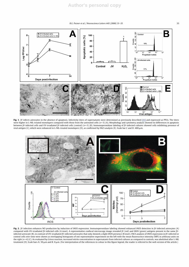
Enhanced iNOS expression, detected by immunoperoxidasestaining and flow cytometry analysis, was first evident from day1 PI and was more intense at day 3 PI in infected cultured astro-cytes compared with UV-irradiated JV-infected controls (Fig. 2Aand C, n = 3). Confocal microscopy analysis showed localizationof viral and iNOS antigens at the same infected cells, in mostcases (Fig. 2B). In addition, supernatants from JV-infected astro-cytes showed a marked increase in NO production from day 3to 7 PI (38 ± 2 nmol/mg prot vs. 18 ± 1 nmol/mg prot at day 3,p ≤ 0.05, n = 4; 82 ± 12 nmol/mg prot vs. 19 ± 1 nmol/mg prot at day7, p ≤ 0.05, n = 4, JV-infected and UV-irradiated), which was abol-ished when an iNOS inhibitor was added (20 ± 1 nmol/mg prot atday 3 and 22 ± 2 nmol/mg prot at day 7) (Fig. 2D, n = 4).
In order to investigate whether JV infection per se caused astro-cyte activation, we performed confocal microscopy analysis to seeif the expected enhanced staining of GFAP co-localized with viralantigens over time. Results showed no difference at day 1 PI and a
Author's personal copy
R.G. Pozner et al. / Neuroscience Letters 445 (2008) 31–35 33
Fig. 1. JV infects astrocytes in the absence of apoptosis. Infectivity titers of supernatants were determined as previously described [11] and expressed as PFUs. The titerswere higher in L-NIL-treated monolayers compared with those from the untreated cells (n = 3) (A). Morphology and cytometry analysis showed no differences in apoptosisbetween JV-infected cells and UV-irradiated JV-infected cells (control) (n = 3) (B). Immunoperoxidase labeling of JV-infected cultures showed cells exhibiting presence ofviral antigen (C), which were enhanced in L-NIL-treated monolayers (D), as confirmed by FACS analysis (E). Scale bar C and D: 400 �m.
Fig. 2. JV infection enhances NO production by induction of iNOS expression. Immunoperoxidase labeling showed enhanced iNOS detection in JV-infected astrocytes (A)compared with UV-irradiated JV-infected cells (A inset). A representative confocal microscopy image revealed JV (red) and iNOS (green) antigens present in the same JV-infected astrocyte (B), in contrast of UV-irradiated JV-infected astrocytes that only showed a slight iNOS presence (B inset). FACS analysis of iNOS expression in JV-infected orcontrol cells over time were shown as overlapping histograms of one representative experiment on the left with the mean fluorescence intensity (MFI) in arbitrary units onthe right (n = 4) (C). As evaluated by Griess reaction, increased nitrite concentration in supernatants from infected cultures as compared to controls, was abolished after L-NILtreatment (D). Scale bars A: 50 �m and B: 8 �m. (For interpretation of the references to colour in this figure legend, the reader is referred to the web version of the article.)
Author's personal copy
34 R.G. Pozner et al. / Neuroscience Letters 445 (2008) 31–35
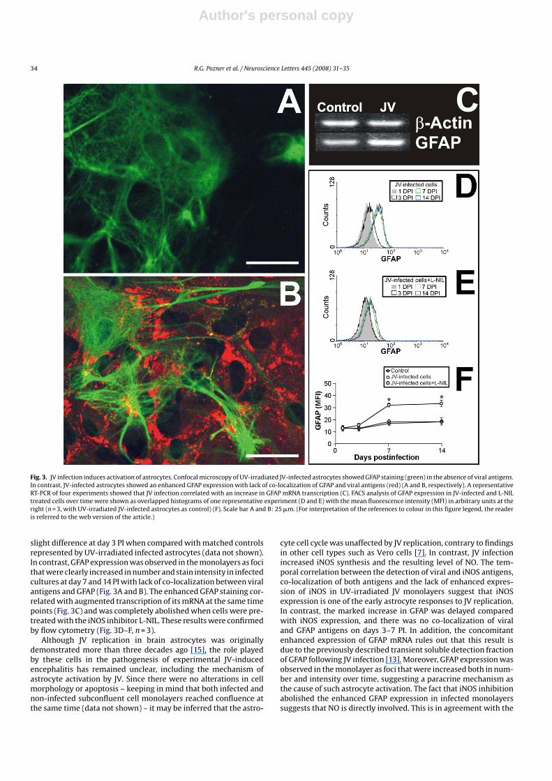
Fig. 3. JV infection induces activation of astrocytes. Confocal microscopy of UV-irradiated JV-infected astrocytes showed GFAP staining (green) in the absence of viral antigens.In contrast, JV-infected astrocytes showed an enhanced GFAP expression with lack of co-localization of GFAP and viral antigens (red) (A and B, respectively). A representativeRT-PCR of four experiments showed that JV infection correlated with an increase in GFAP mRNA transcription (C). FACS analysis of GFAP expression in JV-infected and L-NILtreated cells over time were shown as overlapped histograms of one representative experiment (D and E) with the mean fluorescence intensity (MFI) in arbitrary units at theright (n = 3, with UV-irradiated JV-infected astrocytes as control) (F). Scale bar A and B: 25 �m. (For interpretation of the references to colour in this figure legend, the readeris referred to the web version of the article.)
slight difference at day 3 PI when compared with matched controlsrepresented by UV-irradiated infected astrocytes (data not shown).In contrast, GFAP expression was observed in the monolayers as focithat were clearly increased in number and stain intensity in infectedcultures at day 7 and 14 PI with lack of co-localization between viralantigens and GFAP (Fig. 3A and B). The enhanced GFAP staining cor-related with augmented transcription of its mRNA at the same timepoints (Fig. 3C) and was completely abolished when cells were pre-treated with the iNOS inhibitor L-NIL. These results were confirmedby flow cytometry (Fig. 3D–F, n = 3).
Although JV replication in brain astrocytes was originallydemonstrated more than three decades ago [15], the role playedby these cells in the pathogenesis of experimental JV-inducedencephalitis has remained unclear, including the mechanism ofastrocyte activation by JV. Since there were no alterations in cellmorphology or apoptosis – keeping in mind that both infected andnon-infected subconfluent cell monolayers reached confluence atthe same time (data not shown) – it may be inferred that the astro-
cyte cell cycle was unaffected by JV replication, contrary to findingsin other cell types such as Vero cells [7]. In contrast, JV infectionincreased iNOS synthesis and the resulting level of NO. The tem-poral correlation between the detection of viral and iNOS antigens,co-localization of both antigens and the lack of enhanced expres-sion of iNOS in UV-irradiated JV monolayers suggest that iNOSexpression is one of the early astrocyte responses to JV replication.In contrast, the marked increase in GFAP was delayed comparedwith iNOS expression, and there was no co-localization of viraland GFAP antigens on days 3–7 PI. In addition, the concomitantenhanced expression of GFAP mRNA rules out that this result isdue to the previously described transient soluble detection fractionof GFAP following JV infection [13]. Moreover, GFAP expression wasobserved in the monolayer as foci that were increased both in num-ber and intensity over time, suggesting a paracrine mechanism asthe cause of such astrocyte activation. The fact that iNOS inhibitionabolished the enhanced GFAP expression in infected monolayerssuggests that NO is directly involved. This is in agreement with the
Author's personal copy
R.G. Pozner et al. / Neuroscience Letters 445 (2008) 31–35 35
demonstrated role of NO in the expression of GFAP by astrocytesvia GC-cGMP-PKG [4] and with the increased NO production andGFAP expression observed in influenza virus-infected mice [27].Using cytokine production as a parameter of astrocyte activation,it was recently shown that glial cells, including astrocytes, wereactivated by Lymphocytic Choriomeningitis Virus (LCMV) in a TLR2-MyD88/Mal-dependent manner [29]. Although LCMV and JV havea different pathogenesis, both are arenaviruses and share the samegenomic organization [21]; thus, it seems probable that JV acti-vate astrocytes in a similar manner. In contrast, a marked increasein the expression of GFAP in the absence of iNOS expression hasbeen found to be related to the neurovirulent properties of Sindbisvirus strains [5]. Thus, intense viral-induced astrocyte activationcan hardly be a stereotyped response to viral infection, as it may beselective with regard to concomitantly expressed molecules.
In the present study, we found that iNOS inhibition resultedin higher viral titers as well as viral antigen detection, in contrastto similar infectivity titers previously found in brain homogenatesfrom mice treated or untreated with iNOS inhibitors [12]. The rea-sons for this discrepancy may be that most viral replication in thebrain occurs in neurons instead of astrocytes, and it is probable thatneurons do not produce NO at the same level as astrocytes. Keepingin mind these in vivo and in vitro results, it is possible to specu-late that astrocytes respond early to JV infection with enhancedexpression of iNOS and subsequent NO production, resulting inboth reduced viral replication as well as higher astrocytosis, play-ing a protective role in the course of viral disease. Nevertheless,following infection of euthymic – but not athymic – mice by LCMV,a correlation between neuropathology and iNOS brain expressionwas described in euthymic mice, attributable to the presence ofiNOS in infiltrating inflammatory cells [6]. Moreover, in Venezue-lan equine virus-induced encephalitis, the inflammatory responseis partially mediated by iNOS and TNF-� and seems to contributeto neurodegeneration [24]. In fact, NO production by iNOS mightexert contrasting effects not only on virus infection but also onthe elicited immune response [3]. This is perhaps because NO isreported to switch type 1 helper T (Th1) cell-dependent responsesto Th2-biased immunological responses [1].
Further studies focusing on the role of astrocyte activation by NOshould be carried out, as an improved understanding of astrocytesin the pathogenesis of viral-induced neurologic disease offers thepotential to develop novel strategies to treat neurological disorders[25]. In this field, JV has confirmed its potential as an in vivo and invitro experimental model of astrocyte response to viral injury.
Acknowledgments
This work was partly supported by grants PIP 5102/04 CONICET,PICT 13768/04 ANPCyT, and CABBIO 19/04, Argentina. AEU, MEB yCJG hold a CONICET fellowship.
References
[1] T. Akaike, Role of free radicals in viral pathogenesis and mutation, Rev. Med.Virol. 11 (2001) 87–101.
[2] M.I. Berría, E.F. Lascano, Astrocyte differentiation induced by Junin virus in ratbrain cell cultures, Acta Neuropathol. (Berl.) 66 (1985) 233–238.
[3] C. Bogdan, Nitric oxide and the immune response, Nat. Immunol. 2 (2001)907–916.
[4] S. Brahmachari, Y.K. Fung, K. Pahan, Induction of glial fibrillary acidic proteinexpression in astrocytes by nitric oxide, J. Neurosci. 26 (2006) 4930–4939.
[5] C. Brodie, N. Weizman, A. Katzoff, S. Lustig, D. Kobiler, Astrocyte activation bySindbis virus: expression of GFAP, cytokines, and adhesion molecules, Glia 19(1997) 275–285.
[6] I.L. Campbell, A. Samimi, C.S. Chiang, Expression of the inducible nitric oxidesynthase. Correlation with neuropathology and clinical features in mice withlymphocytic choriomeningitis, J. Immunol. 153 (1994) 3622–3629.
[7] N.A. Candurra, L.A. Scolaro, S.E. Mersich, E.B. Damonte, C.E. Coto, A compari-son of Junin virus strains: growth characteristics, cytopathogenicity and viralpolypeptides, Res. Virol. 141 (1990) 505–515.
[8] Y. Chen, R.A. Swanson, Astrocytes and brain injury, J. Cereb. Blood Flow. Metab.23 (2003) 137–149.
[9] L.F. Eng, R.S. Ghirnikar, Y.L. Lee, Glial fibrillary acidic protein: GFAP-thirty-oneyears (1969–2000), Neurochem. Res. 25 (2000) 1439–1451.
[10] T.W. Geisbert, P.B. Jahrling, Exotic emerging viral diseases: progress and chal-lenges, Nat. Med. 10 (2004) S110–S121.
[11] R.M. Gómez, R.G. Pozner, M.A. Lazzari, L.P. D’Atri1, S. Negrotto, A.M. Chudzinski-Tavassi, M.I. Berría, M. Schattner, Endothelial cell function alteration after Juninvirus infection, Thromb. Hemos. 90 (2003) 326–333.
[12] R.M. Gómez, A. Yep, M. Schattner, M.I. Berria, Junin virus-induced astrocytosisis impaired by iNOS inhibition, J. Med. Virol. 69 (2003) 145–149.
[13] R.F. Iacono, A. Nessi de Avinon, F.A. Rosetti, M.I. Berria, Glial fibrillary acidic pro-tein (GFAP) immunochemical profile after junin virus infection of rat culturedastrocytes, Neurosci. Lett. 200 (1995) 175–178.
[14] T. Iwahashi, A. Inoue, C.S. Koh, T.K. Shin, B.S. Kim, Expression and potential roleof inducible nitric oxide synthase in the central nervous system of Theiler’smurine encephalomyelitis virus-induced demyelinating disease, Cell Immunol.194 (1999) 186–193.
[15] E.F. Lascano, M.I. Berría, Ultrastructure of Junin virus in mouse whole brain andmouse brain tissue cultures, J. Virol. 14 (1974) 965–974.
[16] E.F. Lascano, M.I. Berria, M.M. Avila, M.C. Weissenbacher, Astrocytic reactionpredominance in chronic encephalitis of Junin virus-infected rats, J. Med. Virol.29 (1989) 327–333.
[17] E.F. Lascano, G.D. Lerman, J.L. Blejer, R.L. Caccuri, M.I. Berria, Immunoperoxidasetracing of Junin virus neural route after footpad inoculation, Arch. Virol. 122(1992) 13–22.
[18] D.J. Myer, G.G. Gurkoff, S.M. Lee, D.A. Hovda, M.V. Sofroniew, Essential pro-tective roles of reactive astrocytes in traumatic brain injury, Brain 129 (2006)2761–2772.
[19] C.M. Norris, I. Kadish, E.M. Blalock, K.C. Chen, V. Thibault, N.M. Porter, P.W.Landfield, S.D. Kraner, Calcineurin triggers reactive/inflammatory processes inastrocytes and is upregulated in aging and Alzheimer’s models, J. Neurosci. 25(2005) 4649–4658.
[20] C.J. Peters, Human infection with Arenaviruses in the Americas, in: M.B.A. Old-stone (Ed.), Arenaviruses I, vol. 262, Springer–Verlag, Berlin Heidelberg, 2002,pp. 65–74.
[21] M.S. Salvato, J.C.S. Clegg, M.J. Buchmeier, R.N. Charrel, J.P. Gonzales, I.S. Lukashe-vich, C.J. Peters, R. Rico-Hesse, V. Romanowski, C.M. Fauquet, J. Maniloff, M.A.Mayo, U. Desselberger, L.A. Ball, Arenaviridae Virus Taxonomy. VIIIth Report ofthe ICTV, Elsevier/Academic Press, London, 2005, pp. 725–733.
[22] A. Sanchez, D.Y. Pifat, R.H. Kenyon, C.J. Peters, J.B. McCormick, M.P. Kiley, Juninvirus monoclonal antibodies: characterization and cross-reactivity with otherarenaviruses, J. Gen. Virol. 70 (1989) 1125–1132.
[23] M. Schattner, R.G. Pozner, I. Engelberger, A. Gorostizaga, N. Maugeri, R. Gomez,A. Pasqualini, O. Torres, M.A. Lazzari, Effect of nitric oxide on megakary-ocyte growth induced by thrombopoietin, J. Lab. Clin. Med. 137 (2001)261–269.
[24] B.A. Schoneboom, M.J. Fultz, T.H. Miller, L.C. McKinney, F.B. Grieder, Astrocytesas targets for Venezuelan equine encephalitis virus infection, J. Neurovirol. 5(1999) 342–354.
[25] G. Seifert, K. Schilling, C. Steinhauser, Astrocyte dysfunction in neurologi-cal disorders: a molecular perspective, Nat. Rev. Neurosci. 7 (2006) 194–206.
[26] B. Vainrub, R. Salas, Latin American hemorrhagic fever, Infect. Dis. Clin. NorthAm. 8 (1994) 47–59.
[27] C. Watanabe, H. Kawashima, K. Takekuma, A. Hoshika, Y. Watanabe, Increasednitric oxide production and GFAP expression in the brains of influenza A/NWSvirus infected mice, Neurochem. Res. 33 (2008) 1017–1023.
[28] M.C. Weissenbacher, R.P. Laguens, C.E. Coto, Argentine hemorrhagic fever, Curr.Top. Microbiol. Immunol. 134 (1987) 79–116.
[29] S. Zhou, A. Halle, E.A. Kurt-Jones, A.M. Cerny, E. Porpiglia, M. Rogers, D.T. Golen-bock, R.W. Finberg, Lymphocytic choriomeningitis virus (LCMV) infection ofCNS glial cells results in TLR2-MyD88/Mal-dependent inflammatory responses,J. Neuroimmunol. 194 (2008) 70–82.


