Embed Size (px)
Citation preview
This article appeared in a journal published by Elsevier. The attachedcopy is furnished to the author for internal non-commercial researchand education use, including for instruction at the authors institution
and sharing with colleagues.
Other uses, including reproduction and distribution, or selling orlicensing copies, or posting to personal, institutional or third party
websites are prohibited.
In most cases authors are permitted to post their version of thearticle (e.g. in Word or Tex form) to their personal website orinstitutional repository. Authors requiring further information
regarding Elsevier’s archiving and manuscript policies areencouraged to visit:
http://www.elsevier.com/authorsrights
Author's personal copy
Influence of ionic strength and soil characteristics on the behaviorof Cryptosporidium oocysts in saturated porous media
Ketty Balthazard-Accou a,b,⇑, Urbain Fifi a, Patrice Agnamey b,d, Justin André Casimir c, Philippe Brasseur e,Evens Emmanuel a
a Université Quisqueya – Laboratoire de Qualité de l’Eau de l’Environnement, 218 Ave Jean Paul II, Haut de Turgeau, Port-au-Prince, Haitib Parasitology Laboratory – Mycology, Amiens University Hospital, Avenue Laënnec, 80054 Amiens, Francec Université d’État d’Haïti – Unité de Recherche en Environnement, Faculté des Sciences, 270 rue Mgr Guilloux, Port-au-Prince, Haitid University of Picardie Jules Verne, URF Pharmacie, Equipe théra, Laboratoire des Glucides-FRE-CNRS 3517, 1, rue des Louvels, 80037 Amiens Cedex 1 Amiens, Francee Institut de Recherche pour le Développement (IRD), UMR 198, Centre de Hann, Dakar, Senegal
h i g h l i g h t s
�We study the behavior of Cryptosporidium oocysts with a soil sample from Les Cayes.� Batch tests are used to investigate the soil adsorption of electrolyte solutions.� The Debye–Hückel equation was used to describe ion activity.� The equilibrium adsorption mechanism is used to enumerate the oocysts in the soil.� Oocyst adsorption phenomenon is observed with chemical changes in the environment.
a r t i c l e i n f o
Article history:Received 5 June 2013Received in revised form 29 October 2013Accepted 8 November 2013Available online 19 December 2013
Keywords:Cryptosporidium oocystSoilBatch testsAdsorptionTransferGroundwater
a b s t r a c t
The physico-chemical behavior of Cryptosporidium oocysts was investigated during their transfer throughan alluvial formation from Les Cayes (Haiti) via batch tests. Five approximately 3 kg soil samples werecollected and combined prior to batch tests from the alluvial formations. The experiments were carriedout at soil pH by equilibrating different ranges of pure oocysts concentrations and soil samples with3 mM CaCl2 and 1 mM NaBr as electrolyte. We used the Debye–Hückel equation describing ion activityin a solution for a given ionic strength. The equilibrium adsorption mechanism is used to enumeratethe oocysts in the soil. The results suggest that the oocysts behavior in porous media depends on soilcharacteristics such as soil pH, the nature of the mineral and organic constituents of the soil and the ionicstrength and activities in solution. These results show that a total transfer in batch containing NaBr solu-tions against a partial one in batch containing CaCl2 solutions depends on the oocysts media concentra-tion. To confirm the oocysts number retained in soil, confocal microscopy was successfully used and theimages demonstrate that the majority of oocysts were retained at the range of concentrations tested. Thefindings from this study demonstrated that the retention of C. Parvum in soils may be influenced bychemical conditions and soils characteristics, which are important for groundwater risk assessment.
� 2013 Elsevier Ltd. All rights reserved.
1. Introduction
Cryptosporidium parvum is a protozoan parasite that infects theintestines of a variety of wild and domesticated animals (Kim et al.,2009). Some species infect humans (Kim et al., 2009; Liu, 2012).The scattering and resistant form of Cryptosporidium sp. is the oo-cysts, which are eliminated with feces (Craun and Calderon, 2006).
Cryptosporidium oocysts can enter in surface water systemsthrough runoff from areas with dense animal populations, includ-ing agricultural and wildlife populations, and from human popula-tions via wastewater treatment facilities and open defecation(Considine et al., 2002). They are ubiquitous in groundwater, publicwater supplies and surface water (Balthazard-Accou et al., 2009,2010; Liu, 2012; Their presence in water supplies a major publichealth concern. In order to protect water resources, informationabout retention and transport is essential to risk assessment anddevelopment of effective control practices. The morphology andbehavior of oocysts are reported in the literature (Kuznar andElimelech, 2004). C. parvum oocysts are small (4–6 lm diameter)
0045-6535/$ - see front matter � 2013 Elsevier Ltd. All rights reserved.http://dx.doi.org/10.1016/j.chemosphere.2013.11.045
⇑ Corresponding author at: Université Quisqueya – Laboratoire de Qualité del’Eau de l’Environnement, 218 Ave Jean Paul II, Haut de Turgeau, Port-au-Prince,Haiti. Tel.: +509 44 42 71 64.
E-mail address: [email protected] (K. Balthazard-Accou).
Chemosphere 103 (2014) 114–120
Contents lists available at ScienceDirect
Chemosphere
journal homepage: www.elsevier .com/locate /chemosphere
Author's personal copy
and have a low specific gravity similar to water (1.05 g cm�3), sotheir movement in surface waters is generally not considered tobe influenced by gravitational settling (Searcy et al., 2005). Oocystshave a negative surface charge under typical environmental condi-tions (Thomas et al., 2001; Hsu and Huang, 2002), likely due to thepresence of carboxylate, carboxylic, and phosphate groups on theoocyst surface (Karaman et al., 1999). Oocysts are resistant to anumber of environmental stresses, including chlorination duringdrinking water treatment (Carey et al., 2004).
Filtration methods are sometimes used to remove oocysts fromraw water (Bradford and Bettahar, 2005). The oocyst behavior atthe bench-scale has been investigated, using laboratory columnspacked with a wide range of different sediments or artificial gran-ular materials (Harter et al., 2000; Dai and Hozalski, 2003; Brad-ford and Bettahar, 2005; Tufenkji et al., 2006; Byrd and Walz,2007) or radial stagnation point flow (RSPF) cell (Liu et al., 2009;Janjaroen et al., 2010). Various aspects of C. parvum transportand removal in granular porous media have been also examined,such as the influence of solution chemistry, fluid flow rate, andsediment grain size (Tufenkji et al., 2006). These investigationsare mostly conducted using columns tests, and have limited oocystreactivity in soils. Batch equilibrium tests are also extremelyimportant to describe the partitioning of Cryptosporidium particlesbetween solid and liquid phases and play a significant role in theoocysts removal from the pore fluid.
Several factors, such as solution chemistry (ionic strength, ionvalence, and pH), flow rate and grain size/shape contribute to oo-cyst deposition and interaction mechanisms with porous media(Abudalo et al., 2010). Hsu et al. (2001) and Harter et al. (2000) re-ported that physico-chemical interactions, based particularly onoocysts surface properties, control oocyst deposition. Kuznar andElimelech (2005) and Dai et al. (2004) showed that the surfaceproperties depend on the ionic strength and pH dominating theinteraction force between the parasite surface adhesion and the fil-ter media. Despite the potent health impacts and difficulty of inac-tivation of C. parvum, incomplete data exist regarding the fate andtransport of these pathogens in soils and aquifers (Mohanramet al., 2010). Oocyst removal in porous media is still poorly under-stood. The objective of this study was to investigate the behavior ofCryptosporidium oocysts under chemical conditions and soil char-acteristics in saturated porous media. Equilibrium batch tests werecarried out to evaluate the chemical reactivity of C. parvum duringtheir movement in soils. All experiments were conducted at soil pHto mimic natural conditions using solutions concentrations of3 mM CaCl2 and 1 mM NaBr as electrolyte.
2. Materials and methods
2.1. Cryptosporidium oocyst source and preparation
Viable Cryptosporidium oocysts were obtained from the NationalInstitute of Agronomic Research (INRA) in Animal Infectious Dis-eases and Public Health. The oocysts were collected in fecal sam-ples from naturally infected dairy calves and kept in an aqueousmedium. The oocysts were purified (at INRA) using discontinuoussaccharose. They were stored (in the dark at 4 �C) in potassiumdichromate. The number of oocysts in the stock solution was about2.0 � 107 oocysts mL�1. They were used for laboratory experi-ments within 10 d of collection. According to Hsu and Huang(2002) and Considine et al. (2002) the oocyst purification and stor-age procedures could potentially alter the surface properties of theexperimental oocysts compared to those of oocysts found in theenvironment. However, these procedures are essential for experi-mental work and are commonly used in published research on C.
parvum oocysts. Fluorescence microscopy was used to determinethe concentration of Cryptosporidium oocysts before batch tests.
2.2. Chemical conditions
To study the effect of chemical conditions and soil characteris-tics on the behavior of Cryptosporidium oocysts in porous media,batch equilibrium experiments were used. These experimentswere conducted with two electrolyte solutions: 0.003 M CaCl2
and 0.001 M NaBr which have no effect on the oocysts (Searcyet al., 2005). These solutions were prepared by dissolving a knownamount of chemical products in deionized water to achieve desiredconcentrations. The Debye–Hückel equation was used Eq. (A.1) todescribe the activity of the ions in a solution for a given ionicstrength:
� log cx ¼ 0:51ffiffiIp=1þ 3:3ax
ffiffiIp� �� Z2
x ðA:1Þ
In this equation ax represents the diameter of the ion nm, Zx thecharge of the ion, the ion X is I and the ionic strength of the solu-tion. aCa = 0.6 ; aCl = 0.3
2.3. Soil samples and characteristics
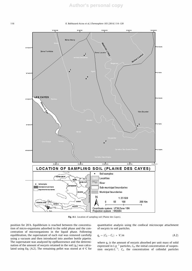
Soil samples were carried out under different emission sourcesof oocysts into the groundwater. Five points were selected. Eachpoint on a random sample has been taken. The samples were col-lected manually. Soil samples were collected at a depth between40 and 80 cm (Fig. A.1). The top 5 cm were excluded for not collect-ing surface debris. The samples were mixed to obtain a representa-tive sample of the study site. All the soil samples were air-dried atlocal temperature (35 �C), passed though a 2 mm sieve, homoge-nized and stored pending physico-chemical characteristics andbatch tests. Sedimentary grain size less than 2 mm is believed tobe more reactive than granular soils with larger particles (Plassardet al., 2000). The granular soils (>2 mm) generally have a lowertendency for adsorption of microorganisms than fine soils, whichoccurs most often during the adsorption phenomena (Balthazard-Accou, 2011).
The physico-chemical properties of soil, such as pH, organicmatter, clay, and CaCO3 were measured using standard analyticalmethods. Chemical treatments were deliberately avoided to keepthe soil in its natural state, including clay particles and organicmatter that are important in the adsorption process and can be ele-vated in the saturated aquifer environment. The analysis of someintrinsic soil parameters were performed at the National Labora-tory of Building and Public Works (LNBTP) Laboratory Analyzesof Soil Arras (INRA), the Institute for Radiological Protection andNuclear Safety (IRSN). Table A.1 summarizes the parameters mea-sured, laboratories and execution protocols.
2.4. Batch tests
The batch tests were conducted at soil pH balancing 4 g of soilwith a suspension of oocysts in twelve bottles of crystal polysty-rene sealed at a ratio of 1:10. To avoid changing the soil properties,the ionic strength of the solutions was adjusted by the addition of a3 mM solution of Calcium Chloride (CaCl2) in six of the twelve bot-tles and a 100 mM solution of Sodium Bromide (NaBr) in theremaining six. The suspension was prepared with a concentrationof approximately 40000 oocysts mL�1. Because oocysts heteroge-neously distributed in suspensions the volume of samples shouldbe large enough to obtain representative samples (Drozd andSchwartzbrod, 1996). These were stirred for 24 h using a shaker ta-ble at 220 shakes per minute room temperature (23 ± 2 �C). At theend of the stirring period, the tubes were placed in an upright
K. Balthazard-Accou et al. / Chemosphere 103 (2014) 114–120 115
Author's personal copy
position for 20 h. Equilibrium is reached between the concentra-tion of micro-organisms adsorbed to the solid phase and the con-centration of microorganisms in the liquid phase. Followingequilibration, the supernatant of each vial was removed carefullyusing a vacuum and then introduced into another bottle pipette.The supernatant was analyzed by epifluoresence and the determi-nation of the amount of oocysts retained in the soil (qe) was calcu-lated using Eq. (A.2). The remaining pellet was stored at 4 �C for
quantitative analysis using the confocal microscope attachmentof oocysts to soil particles.
qe ¼ ðC0 � CeÞ � V=m ðA:2Þ
where qe is the amount of oocysts absorbed per unit mass of solidexpressed in L g�1 particles, C0, the initial concentration of suspen-sion oocysts L�1; Ce, the concentration of colloidal particles
Fig. A.1. Location of sampling soil (Plaine des Cayes).
116 K. Balthazard-Accou et al. / Chemosphere 103 (2014) 114–120
Author's personal copy
balanced oocysts L�1; V, is the volume of solution used in L; m, is themass in grams of dry soil.
2.5. Enumeration of C. parvum oocysts
Each supernatant fraction tubes was analyzed respectively bythe method of concentration and counting AFNOR NFT90-455(AFNOR, 2001). Supernatant fraction was centrifuged at 3500g for30 min at 4 �C and Cryptosporidium oocysts were isolated byimmunomagnetic separation using magnetic beads coated withanti-Cryptosporidium oocyst monoclonal antibody (Dynabeads, Dy-nal, Norvège). The collection of beads was performed in a magneticfield. The oocysts/bead complex was then dissociated by a solutionof HCl 0.1 N. Isolated oocysts were marked by a monoclonal anti-body conjugated to a fluorochrome and specific to the genus Cryp-tosporidium that binds to surface antigens of oocysts. Directimmunofluorescence (IF) was used to enumerate the oocysts. Oo-cysts were counted using an epifluorescence microscope (UV exci-tation at 490 nm, emission 456 nm; BX41, Olympus). At least 30fields of view were counted in each 100 lL.
2.6. Confocal microscopic confirmation
The observation with a confocal microscope (Hitachi S80010 kV) of the samples after adsorption was analyzed by the methodof bypassing the critical technological Centre microstructures(EZUS Lyon 1-Claude Bernard University). The samples were fixedwith 2% glutaraldehyde dissolved in a 0.1 M sodium cacodylatebuffer for 4 h and 30 min at room temperature. Then the sampleswere rinsed three times with a 0.2 M sodium cacodylate solution.A post-fixation was made with a solution of 0.5% osmium tetroxidefor 45 min at room temperature. In addition, these samples werebathed in 20% acetone in a bell drying overnight, followed by an-other 1-h bath in 100% acetone. After adsorption, the samples weredeposited on substrates of SEM metallization with gold–palladium.The completion of the bypass of the critical point in the criticalapparatus and images were captured.
3. Results and discussion
3.1. Soils characteristics
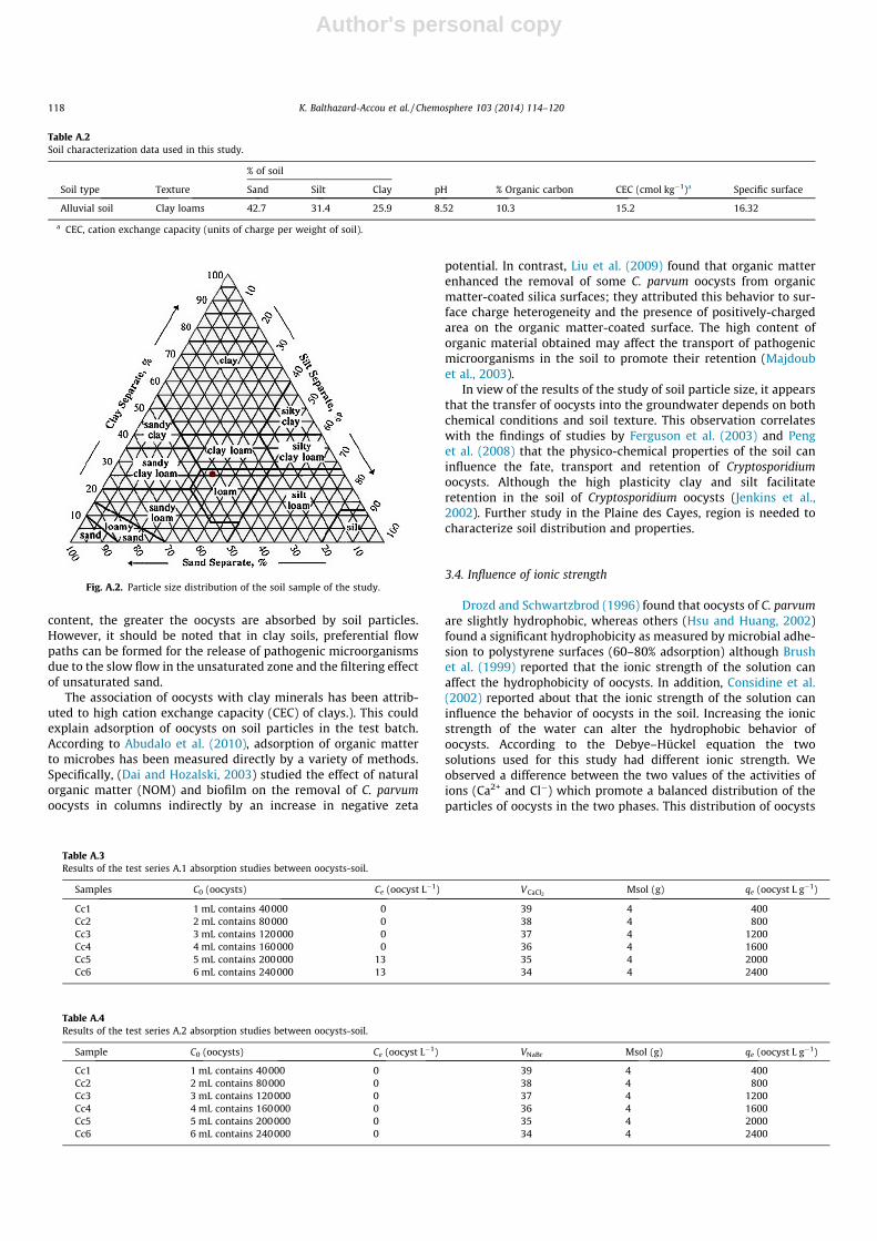
Table A.2 presents the results of physico-chemical analysis andparticle size distribution of the soil of the study. Particle size anal-ysis was conducted to determine the particle size distribution ofthe reconstituted soil. The results of particle size analysis showsthat the soil is characterized by fine particles, the size of the largestelement is 2.50 mm. This fraction represents about 25% of thesample (Fig. A.2).
The soil consisted of 35.6% coarse sand, 7.1% fine sands, 25.4%fine silt, 6% coarse silt, and 25.9% clay. This soil has an alkalinepH (8.52) probably due to the high calcium carbonate content(10.3 g kg�1). Total calcium (590 g kg�1) was high suggesting cal-cite is the dominant carbonate mineral present. The soil contentalso 15.2 (cmol kg�1) of cation exchange capacity (CEC). The soilhas a large surface area due to the presence of silts and clays there-fore a large number of binding sites (Hillel, 1998), and sometimes acapacity of ion exchange, which can promote the adsorption ofCryptosporidium oocysts.
3.2. Oocysts retention
The results for series A.1 and series A.2 are summarized inTables A.3 and A.4, respectively. These results provide evidenceof the physico-chemical interactions of oocysts during their trans-fer in soil. In one test series with the CaCl2 solution, it was foundthat the oocysts were removed from the suspension with a muchhigher rate when combined with soil particles. Whereas with the2 test series containing the solution of NaBr all oocysts were trans-ferred to the solid phase, suggesting a chemical environmentalfavorable for the retention of oocysts. This is due to the intrinsiccharacteristics of the soil used in the test batch. However, the timeallocated to both settling (20 h) and the ratio of 1:10, that is to say(4 g of dry soil in 40 mL suspension of oocysts) seem to signifi-cantly enhance the sedimentation oocysts. The results obtainedfrom adsorption studies seem to be in agreement with the workof Bradford and Bettahar (2005), Searcy et al. (2005).
3.3. Influence of soil characteristics
Transportation through soil has usually been considered aninsignificant pathway because soil is generally assumed to be aneffective filter inhibiting the transport of different pathogens(Petersen et al., 2012). For colloid-sized Cryptosporidium oocyststhe fate and transport processes depend much on the soil physicaland chemical properties (Peng et al., 2011). Soil texture, a staticproperty of the soil, also appears to affect oocyst survival in the soilenvironment (Peng et al., 2008). According to Peng et al. (2011) oo-cysts preferentially attach to fine particles, but soil organic carbonprevents some oocyst attachment to soil particles. Clay, fine siltand coarse silt represent 57.3% of the soils studied. Soils with clayand silt are less permeable, and have the ability to absorb and re-tain water. However, Cryptosporidium oocysts remain inactivelonger in loamy soils than in clay soils (Jenkins et al., 2002. Davieset al., 2005). Moreover, the values found in soil tests correspond tothe values encountered in loam soil. As a result, the absorptiveproperties of a soil depend in part on the composition of organicmatter and clay (Balthazard-Accou, 2011). The higher the clay
Table A.1Physico-chemical analysis of soil.
Parameters Laboratory Protocol excution
Sedimentometry National Laboratory of Building and Public Works – LNBTP NF X 31-107
Size NF X 31-107Water content NF ISO 11465Soil pH Water-pH National Institute of Agronomic Research – INRA NF ISO 10390
KCl-pHCarbonate content Total limestone NF ISO 10693
Active limestone NF X 31-106Organic carbon content
NF ISO 10694Clay content NF X 31-107CEC NF X 31-130
Specific soil surface Institute for Radiological Protection and Nuclear Safety – IRSN NF ISO 9277
K. Balthazard-Accou et al. / Chemosphere 103 (2014) 114–120 117
Author's personal copy
content, the greater the oocysts are absorbed by soil particles.However, it should be noted that in clay soils, preferential flowpaths can be formed for the release of pathogenic microorganismsdue to the slow flow in the unsaturated zone and the filtering effectof unsaturated sand.
The association of oocysts with clay minerals has been attrib-uted to high cation exchange capacity (CEC) of clays.). This couldexplain adsorption of oocysts on soil particles in the test batch.According to Abudalo et al. (2010), adsorption of organic matterto microbes has been measured directly by a variety of methods.Specifically, (Dai and Hozalski, 2003) studied the effect of naturalorganic matter (NOM) and biofilm on the removal of C. parvumoocysts in columns indirectly by an increase in negative zeta
potential. In contrast, Liu et al. (2009) found that organic matterenhanced the removal of some C. parvum oocysts from organicmatter-coated silica surfaces; they attributed this behavior to sur-face charge heterogeneity and the presence of positively-chargedarea on the organic matter-coated surface. The high content oforganic material obtained may affect the transport of pathogenicmicroorganisms in the soil to promote their retention (Majdoubet al., 2003).
In view of the results of the study of soil particle size, it appearsthat the transfer of oocysts into the groundwater depends on bothchemical conditions and soil texture. This observation correlateswith the findings of studies by Ferguson et al. (2003) and Penget al. (2008) that the physico-chemical properties of the soil caninfluence the fate, transport and retention of Cryptosporidiumoocysts. Although the high plasticity clay and silt facilitateretention in the soil of Cryptosporidium oocysts (Jenkins et al.,2002). Further study in the Plaine des Cayes, region is needed tocharacterize soil distribution and properties.
3.4. Influence of ionic strength
Drozd and Schwartzbrod (1996) found that oocysts of C. parvumare slightly hydrophobic, whereas others (Hsu and Huang, 2002)found a significant hydrophobicity as measured by microbial adhe-sion to polystyrene surfaces (60–80% adsorption) although Brushet al. (1999) reported that the ionic strength of the solution canaffect the hydrophobicity of oocysts. In addition, Considine et al.(2002) reported about that the ionic strength of the solution caninfluence the behavior of oocysts in the soil. Increasing the ionicstrength of the water can alter the hydrophobic behavior ofoocysts. According to the Debye–Hückel equation the twosolutions used for this study had different ionic strength. Weobserved a difference between the two values of the activities ofions (Ca2+ and Cl�) which promote a balanced distribution of theparticles of oocysts in the two phases. This distribution of oocysts
Table A.2Soil characterization data used in this study.
% of soil
Soil type Texture Sand Silt Clay pH % Organic carbon CEC (cmol kg�1)a Specific surface
Alluvial soil Clay loams 42.7 31.4 25.9 8.52 10.3 15.2 16.32
a CEC, cation exchange capacity (units of charge per weight of soil).
Fig. A.2. Particle size distribution of the soil sample of the study.
Table A.3Results of the test series A.1 absorption studies between oocysts-soil.
Samples C0 (oocysts) Ce (oocyst L�1) VCaCl2Msol (g) qe (oocyst L g�1)
Cc1 1 mL contains 40000 0 39 4 400Cc2 2 mL contains 80000 0 38 4 800Cc3 3 mL contains 120000 0 37 4 1200Cc4 4 mL contains 160000 0 36 4 1600Cc5 5 mL contains 200000 13 35 4 2000Cc6 6 mL contains 240000 13 34 4 2400
Table A.4Results of the test series A.2 absorption studies between oocysts-soil.
Sample C0 (oocysts) Ce (oocyst L�1) VNaBr Msol (g) qe (oocyst L g�1)
Cc1 1 mL contains 40000 0 39 4 400Cc2 2 mL contains 80000 0 38 4 800Cc3 3 mL contains 120000 0 37 4 1200Cc4 4 mL contains 160000 0 36 4 1600Cc5 5 mL contains 200000 0 35 4 2000Cc6 6 mL contains 240000 0 34 4 2400
118 K. Balthazard-Accou et al. / Chemosphere 103 (2014) 114–120
Author's personal copy
can be explained by the activity of calcium ions in the solution con-taining CaCl2 (Table A.3). While for NaBr, for the same ionicstrength, the activity of Na+ ions is almost identical to the activityof Br� ions, there is a total transfer of the oocysts of the aqueousphase to the solid phase (Table A.4).
Researche into oocyst adhesion to solid surfaces is rare (Kuznarand Elimelech, 2004). Techniques based on the principles of colloidand surface chemistry are providing new insights about their inter-actions during transport in granular porous media. The results ofadsorption equilibrium experiments showed a difference betweenthe two sets of analyses. Which appears to be influenced by theaddition of bromide ion Br�. Oocysts have a negative charge atpH values below 9 (Drozd and Schwartzbrod, 1996). Therefore,we can assume that the oocysts undergo electrostatic repulsionwhich could prevent their adsorption. The effectiveness of the bro-mide ion and inertia vis-à-vis the clay were observed by Schoenet al. (1999). However, the addition of calcium chloride promoteson one hand increasing the number of cations (Ca2+) and secondlythe ionic strength of the aqueous phase. However, the contributionof Na+, Ca2+and the initial presence of other ions (Ca2+, Mg2+, etc.)in soil can probably be the basis for the transfer. It is well knownthat divalent cations in aqueous media can also cause changes inthe surface properties of oocysts (Considine et al., 2002) and favor-ing the retention of oocysts in the soil. For example, Kuznar andElimelech (2004) demonstrated surface adhesion to a quartz filterin the presence of a divalent salt (CaCl2) with a pH of 5.5–5.7 mediawhere the ionic strength increases with a minimum of repulsiveforce. The divalent cations (CaCl2) will chemically adhere to thesurface and neutralize the negative charges. Attachment underunfavorable conditions is known to be highly dependent on solu-tion chemistry (Tufenkji et al., 2004). High rates of oocysts ob-served in the soil (Fig. A.3), which may explain why the additionof ions (Ca2+ could promote the retention of oocysts in the soil.Normally, two negative charges repel, but in the presence of solu-tions having high electrical loads, the electrostatic double layer iscompressed and the surfaces of contact between the microorgan-isms and the soil particles are strongly attached to each other bythe formation of Van der Waals bonds (Majdoub et al., 2003).Therefore, it appears that oocysts particle interactions were notdominated by electrostatic repulsive forces.
The additional research is need into the zeta potential of oo-cysts, especially in soils with naturally high concentrations of cal-cium and organic matter. The information reported in theliterature have shown that the adsorption of natural organic mat-ter on the surface of the oocyst causes a significant increase of thezeta potential, counteracting its negative charge (Considine et al.,2002; Kuznar and Elimelech, 2004). On the other hand Searcyet al. (2005) showed that the zeta potential of purified oocysts
becomes more negative with increasing solution pH. Furthermore,the ionic strength of the solution also impacts the oocyst zeta po-tential. Indeed, soil clays have amphoteric site promoting theappearance of positive and negative charges acid alkaline environ-ment (Fifi, 2010). In this context, the alkaline pH measured in thephysico-chemical analyzes of the soil, can increase the negativecharge of zeta potential of oocysts and ultimately reduce the reten-tion capacity of the soil. This could explain the presence of oocystsin groundwater. In addition, the quality of the fine fraction (clay,silt and organic matter, etc.) seems also to explain a large part ofthe phenomena observed after exposure of soil to oocysts.
3.5. Dispersion of oocysts by microscopic observation
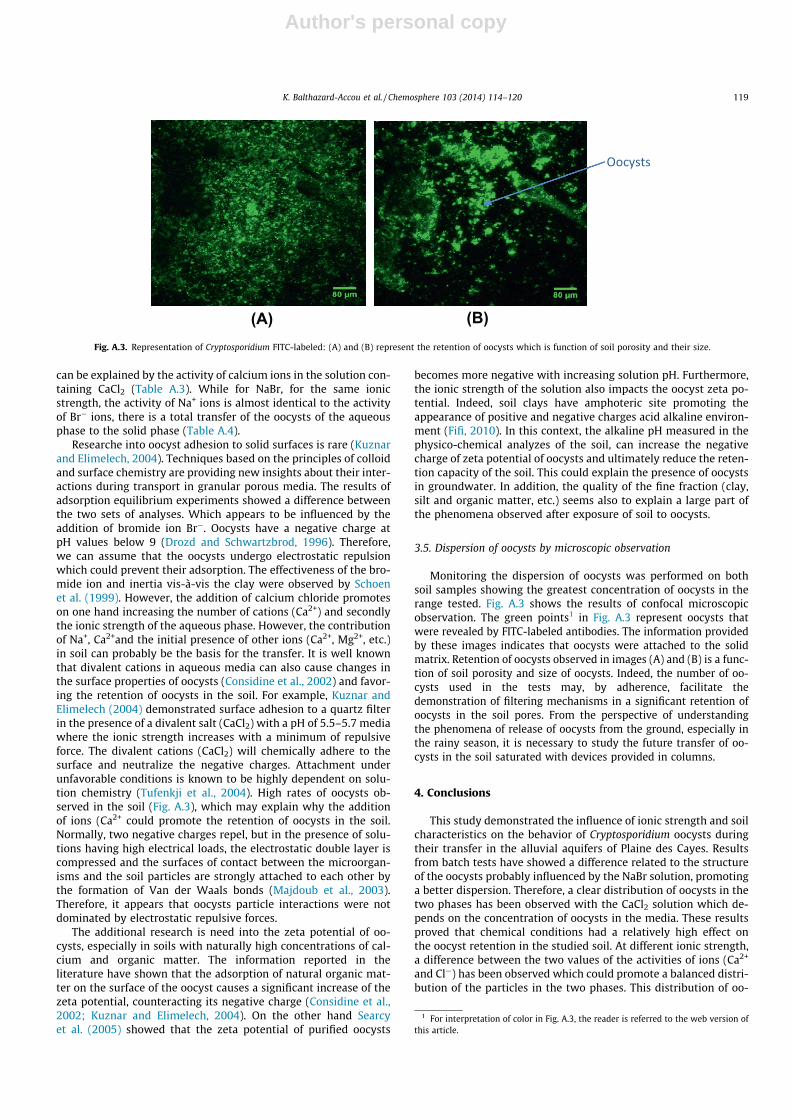
Monitoring the dispersion of oocysts was performed on bothsoil samples showing the greatest concentration of oocysts in therange tested. Fig. A.3 shows the results of confocal microscopicobservation. The green points1 in Fig. A.3 represent oocysts thatwere revealed by FITC-labeled antibodies. The information providedby these images indicates that oocysts were attached to the solidmatrix. Retention of oocysts observed in images (A) and (B) is a func-tion of soil porosity and size of oocysts. Indeed, the number of oo-cysts used in the tests may, by adherence, facilitate thedemonstration of filtering mechanisms in a significant retention ofoocysts in the soil pores. From the perspective of understandingthe phenomena of release of oocysts from the ground, especially inthe rainy season, it is necessary to study the future transfer of oo-cysts in the soil saturated with devices provided in columns.
4. Conclusions
This study demonstrated the influence of ionic strength and soilcharacteristics on the behavior of Cryptosporidium oocysts duringtheir transfer in the alluvial aquifers of Plaine des Cayes. Resultsfrom batch tests have showed a difference related to the structureof the oocysts probably influenced by the NaBr solution, promotinga better dispersion. Therefore, a clear distribution of oocysts in thetwo phases has been observed with the CaCl2 solution which de-pends on the concentration of oocysts in the media. These resultsproved that chemical conditions had a relatively high effect onthe oocyst retention in the studied soil. At different ionic strength,a difference between the two values of the activities of ions (Ca2+
and Cl�) has been observed which could promote a balanced distri-bution of the particles in the two phases. This distribution of oo-
(A) (B)Fig. A.3. Representation of Cryptosporidium FITC-labeled: (A) and (B) represent the retention of oocysts which is function of soil porosity and their size.
1 For interpretation of color in Fig. A.3, the reader is referred to the web version ofthis article.
K. Balthazard-Accou et al. / Chemosphere 103 (2014) 114–120 119
Author's personal copy
cysts can be explained by the activity of calcium ions in the solu-tion containing CaCl2. While for NaBr the activity of Na+ ions is al-most identical to the activity of Br� ions, there is a total transfer ofthe oocysts of the aqueous phase to the solid phase. The scanningelectron microscopy of the dispersion of the oocysts on soil parti-cles showed that the oocysts have been retained by the highestrange concentration used the study. These results denoted thatthe modification of chemical environmental conditions could havean impact on the transfer of oocysts in porous media, which couldmake possible an adsorption phenomenon. It appears that theirtransfer into soils and groundwater is probably influenced by bothsoils constituents and texture. For example, the fine fraction (clay,silt or organic matter) of the selected soil is probably contributingto the retention of oocysts in soils. Columns tests may provide abetter understanding of the behavior of the oocysts during theirtransfer in soils of Plaine des Cayes.
Acknowledgments
The authors would like to thank the following Institutions fortheir financial support: the Office of the Prime Minister of theRepublic of Haiti, the Cooperation and Cultural Action of the Em-bassy of France in Haiti, the AUF (Agence Universitaire de la Fran-cophonie) and the LEHNA (ENTPE) for their technical and financialassistance.
References
Abudalo, R.A., Ryan, J.N., Harvey, R.W., Metge, D.W., Landkamer, L., 2010. Influenceof organic matter on the transport of Cryptosporidium parvum oocysts in a ferricoxyhydroxide-coated quartz sand saturated porous medium. Water Res. 44,1104–1113.
AFNOR NFT90-455, 2001. Qualité de l’eau – Recherche et dénombrement d’oocystesde Cryptosporidium et de kystes de Giardia – Méthode de concentration et dedénombrement. AFNOR. p. 25.
Balthazard-Accou, K., Emmanuel, E., Agnamey, P., Brasseur, P., Lilite, O., Totet, A.,Raccurt, C., 2009. Presence of Cryptosporidium oocysts and Giardia cysts in thesurface water and groundwater in the City of Cayes, Haití. Aqua-LAC 1, 63–71.
Balthazard-Accou, K., Emmanuel, E., Agnamey, P., Brasseur, P., Totet, A., Raccurt, C.P.,2010. Cryptosporidium oocysts transmission in the aquatic environment of Haiti.In: Laboy-Nieves, E.N., Goosen, M.F.A., Emmanuel, E. (Eds.), Environmental andHuman Health. CRC Press. Taylor & Francis Group, pp. 201–214.
Balthazard-Accou, K., 2011. Contamination microbiologique des eaux souterrainesde la ville des Cayes, Haïti. Evaluation des risques pour la santé desconsommateurs. Thèse de doctorat. Université de Picardie Jules Verne &Université Quisqueya, p. 208.
Brush, C.F., Ghiorse, W.C., Anguish, L.J., 1999. Transport of Cryptosporidium parvumoocysts through saturated columns. J. Environ. Qual. 28, 809–815.
Bradford, S.A., Bettahar, M., 2005. Straining attachment and detachment ofCryptosporidium oocysts in saturated porous media. J. Environ. Qual. 34, 469–478.
Byrd, T.L., Walz, J.Y., 2007. Investigation of the interaction force betweenCryptosporidium parvum oocysts and solid surfaces. Langmuir 23, 7475–7483.
Carey, C.M., Lee, H., Trevors, J.T., 2004. Biology, persistence and detection ofCryptosporidium parvum and Cryptosporidium hominis oocyst. Water Res. 38,818–862.
Considine, R.F., Dixon, D.R., Drummond, C.J., 2002. Oocysts of Cryptosporidiumparvum and model sand surfaces in aqueous solutions: an atomic forcemicroscope (AFM) study. Water Res. 36, 3421–3428.
Craun, G.F., Calderon, R.L., 2006. Observational epidemiologic studies of endemicwaterborne risks: cohort, case-control, time-series, and ecologic studies. J.Water Health 4, 101–119.
Dai, X., Hozalski, R.M., 2003. Evaluation of microspheres as surrogates forCryptosporidium parvum oocysts in filtration experiments. Environ. Sci.Technol. 37, 1037–1042.
Dai, X., Boll, J., Hayes, M.E., Aston, D.E., 2004. Adhesion of Cryptosporidium parvumand Giardia lamblia to solid surfaces: the role of surface charge andhydrophobicity. Colloids Surf. B: Biointerfaces 34, 259–263.
Davies, C.M., Altavilla, N., Krogh, M., Ferguson, C.M., Deere, D.A., Ashbolt, N.J., 2005.Environmental inactivation of Cryptosporidium oocysts in catchment soils. J.Appl. Microbiol. 98, 308–317.
Drozd, C., Schwartzbrod, J., 1996. Hydrophobic and electrostatic cell surfaceproperties of Cryptosporidium parvum. Appl. Environ. Microbiol. 62, 1227–1232.
Ferguson, C., Husman, A.M.R., Altavilla, N., Deere, D., Ashbolt, N., 2003. Fate andtransport of surface water pathogens in watersheds. Crit. Rev. Env. Sci. Technol.33, 299–361.
Fifi, U., 2010. Impacts des eaux pluviales urbaines sur les eaux souterraines dans lespays en développement – mécanismes de transfert des métaux lourds à traversun sol modèle de Port-au-Prince, Haïti. Thèse de doctorat. Lyon: INSA de Lyon,p. 260.
Harter, T., Wagner, S., Atwill, E.R., 2000. Colloid transport and filtration ofCryptosporidium parvum in sandy soils and aquifer sediments. Environ. Sci.Technol. 34, 62–70.
Hillel, D., 1998. Environmental Soil Physics: Fundamentals, Applications andEnvironmental Considerations. Academic Press. p. 771.
Hsu, B.-M., Huang, C., Pan, J.R., 2001. Filtration behaviors of Giardia andCryptosporidium-ionic strength and pH effects. Water Res. 35, 3777–3782.
Hsu, B.M., Huang, C.P., 2002. Influence of ionic strength and pH on hydrophobicityand zeta potential of Giardia and Cryptosporidium. Colloids Surf. A: Phys. Chem.Eng. Aspects 3, 201–206.
Janjaroen, D., Liu, Y., Kuhlenschmidt, M.S., Kuhlenschmidt, T.B., Nguyen, T.H., 2010.Role of divalent cations on deposition of Cryptosporidium parvum oocysts onnatural organic matter surfaces. Environ. Sci. Technol. 44, 4519–4524.
Jenkins, M.B., Bowman, D.D., Fogarty, E.A., Ghiiorse, W.C., 2002. Cryptosporidiumparvum oocyst inactivation in three soil types at various temperatures andwater potentials. Soil Biol. Biochem. 34, 1101–1109.
Karaman, M.E., Pashley, R.M., Bustamante, H., Shanker, S.R., 1999.Microelectrophoresis of Cryptosporidium parvum oocysts in aqueous solutionsof inorganic and surfactant cations. Colloids Surf. A: Phys. Chem. Eng. Aspects146, 217–225.
Kim, H.N., Hong, Y., Lee, I., Bradford, S.A., Walker, S.L., 2009. Surface characteristicsand adhesion behavior of Escherichia coli. O157:H7: role of extracellularmacromolecules. Biomacromolecules 10, 2556–2564.
Kuznar, Z.A., Elimelech, M., 2004. Adhesion kinetics of viable Cryptosporidiumparvum oocysts to quartz surfaces. Environ. Sci. Technol. 38, 6839–6845.
Kuznar, Z.A., Elimelech, M., 2005. Role of surface proteins in the deposition kineticsof Cryptosporidium parvum oocysts. Langmuir 21, 710–716.
Liu, Y., Janjaroen, D., Kuhlenschmidt, M.S., Kuhlenschmidt, T.B., Nguyen, T.H., 2009.Deposition of Cryptosporidium parvum oocysts on natural organic mattersurfaces: microscopic evidence for secondary minimum deposition in a radialstagnation point flow cell. Langmuir 3, 1594–1605.
Liu, Y., 2012. Roles of Surface Interaction on Cryptosporidium parvum OocystsTransport in Subsurface Environment. Thèse de doctorat. University of Illinois atUrbana-Champaign, p. 131.
Majdoub, R., Caroline, C., Mohamed, L., Katline, G., Mylène, G., 2003. Impact del’utilisation des engrais de ferme sur la qualité microbiologique de l’eausouterraine. IRDA. p. 120.
Mohanram, A., Ray, C., Harvey, R.W., Metge, D.W., Ryan, J.N., Chorover, J., Eberl, D.D.,2010. Comparison of transport and attachment behaviors of Cryptosporidiumparvum oocysts and oocyst-sized microspheres being advected through threeminerologically different granular porous media. Water Res. 44, 5334–5344.
Petersen, Heidi H., Enemark, Heidi L., Olsen, Annette, Mostofa, M.G., AndersDalsgaarda, Amin, 2012. Transport of Cryptosporidium parvum Oocysts in soilcolumns following applications of raw and separated liquid slurries. Appl.Environ. Microbiol. 78, 5994–6000.
Peng, X., Macdonald, S., Murphy, T.M., Holden, N.M. 2011. The fate and transport ofCryptosporidium parvum oocysts in the Soil. In: Burcu E. Ozkaraova Gungor (Ed.),Principles, Application and Assessment in Soil Science, ISBN: 978-953-307-740-6, Rijeka: InTech, pp. 179–192. <http://www.intechopen.com/books/principlesapplication-and-assessment-in-soil-science/the-fate-and-transport-of-cryptosporidium-parvum-oocysts-inthesoil>.
Peng, X., Murphy, T.M., Holden, N.M., 2008. Evaluation of the effect of temperatureon the die-off rate for Cryptosporidium parvum oocysts in water, soils, and feces.Appl. Environ. Microbiol. 74, 7101–7107.
Plassard, F., Winiarski, T., Petit-Ramel, M., 2000. Retention and distribution of threeheavy metals in a carbonated soil: comparison between batch and unsaturatedcolumn studies. J. Contam. Hydrol. 42, 99–111.
Schoen, R., Gaudet, J.P., Bariac, T., 1999. Preferential flow and solute transport in alarge lysimeter under controlled boundary conditions. J. Hydrol. 215, 70–81.
Searcy, K.E., Packman, A.I., Atwill, E.R., Harter, T., 2005. Association ofCryptosporidium parvum with suspended particles: impact on oocystsedimentation. Appl. Environ. Microbiol. 71, 1072–1078.
Thomas, F., Bard, E., Rouillier, M.C., Prelot, B., Mathieu, L., 2001. Filtration-elution ofCryptosporidium oocysts assisted by electrostatic interactions. Colloids Surf. A:Phys. Chem. Eng. Aspects, 135–142.
Tufenkji, N., Miller, G.F., Ryan, J.N., Harvey, R.W., Elimelech, M., 2004. Transport ofCryptosporidium oocysts in porous media: role of straining and physicochemicalfiltration. Environ. Sci. Technol. 38, 5932–5938.
Tufenkji, N., Dixon, D.R., Considine, R.F., Drummond, C.J., 2006. Multi-scaleCryptosporidium/sand interactions in water treatment. Water Res. 40, 3315–3331.
120 K. Balthazard-Accou et al. / Chemosphere 103 (2014) 114–120


