Embed Size (px)
Citation preview
Immunity
Review
Innate Lymphoid Cell Interactions with Microbiota:Implications for Intestinal Health and Disease
Gregory F. Sonnenberg1,* and David Artis1,2,*1Department of Microbiology and Institute for Immunology, Perelman School of Medicine2Department of Pathobiology, School of Veterinary MedicineUniversity of Pennsylvania, Philadelphia, PA 19104, USA*Correspondence: [email protected] (G.F.S.), [email protected] (D.A.)http://dx.doi.org/10.1016/j.immuni.2012.10.003
The mammalian intestine harbors trillions of beneficial commensal bacteria that are essential for the devel-opment of the immune system and for maintenance of physiologic processes in multiple organs. However,numerous chronic infectious, inflammatory, and metabolic diseases in humans have been associated withalterations in the composition or localization of commensal bacteria that result in dysregulated host-commensal bacteria relationships. The mammalian immune system plays an essential role in regulatingthe acquisition, composition, and localization of commensal bacteria in the intestine. Emerging researchhas implicated innate lymphoid cells (ILCs) as a critical immune cell population that orchestrates some ofthese host-commensal bacteria relationships that can impact immunity, inflammation, and tissue homeo-stasis in the intestine. This review will discuss reciprocal interactions between intestinal commensal bacteriaand ILCs in the context of health and disease.
IntroductionThe mammalian gastrointestinal tract is colonized by an esti-
mated 100 trillion bacteria composed of thousands of different
species (Clemente et al., 2012; Dethlefsen et al., 2007; Ley
et al., 2008). In the healthy intestine, these bacterial communities
reside in defined anatomical locations and exist in a symbiotic
relationship with their hosts, promoting normal physiologic
processes and limiting colonization with potentially pathogenic
microbes (Hill and Artis, 2010; Honda and Littman, 2012; Hooper
et al., 2012; Ley et al., 2006a; Littman and Pamer, 2011). In
contrast, the pathogenesis and progression of numerous chronic
infectious, inflammatory, or metabolic diseases in humans,
including viral hepatitis, HIV-AIDS, inflammatory bowel disease
(IBD), cancer, diabetes, obesity and cardiovascular disease,
have been associated with alterations in the composition and/
or anatomical location of intestinal commensal bacteria (Brench-
ley and Douek, 2012; Chin et al., 2007; Hill and Artis, 2010; Ley
et al., 2006b; McGuckin et al., 2009; Ott et al., 2006; Sandler
et al., 2011). Therefore, understanding the mechanisms that
regulate healthy host-commensal bacteria relationships could
aid in the development of novel therapeutics to prevent or limit
chronic human diseases.
Commensal bacteria that reside in the intestinal lumen are
separated from the underlying connective tissues of the body
by a single layer of intestinal epithelial cells. The immune
system is a critical regulator of this epithelial barrier and asso-
ciated commensal bacteria (Hill and Artis, 2010; Honda and
Littman, 2012; Hooper et al., 2012; Littman and Pamer,
2011). For example, cytokines derived from CD4+ T helper cells
can profoundly influence the biology of intestinal epithelial cells
through regulating epithelial permeability, proliferation, repair,
and expression of critical factors including tight-junction and
antimicrobial proteins that control host interactions with intes-
tinal commensal bacteria (Hill and Artis, 2010; Hooper et al.,
2012; Littman and Pamer, 2011; Maloy and Powrie, 2011).
Conversely, commensal bacteria also have a profound influ-
ence on the development and homeostasis of the mammalian
immune system. In the absence of commensal bacteria, devel-
opment of the innate and adaptive immune system is impaired
(Hill and Artis, 2010; Honda and Littman, 2012; Hooper et al.,
2012; Littman and Pamer, 2011), and more recent studies
have reported selective regulation of CD4+ T cell subsets by
specific species of commensal bacteria (Atarashi et al., 2011;
Ivanov et al., 2009; Littman and Pamer, 2011; Round and
Mazmanian, 2010). These findings indicate that regulatory
pathways through which commensal bacteria influence the
mammalian immune system are sophisticated and perhaps
highly selective.
Innate lymphoid cells (ILCs) are an emerging population of
innate immune cells that share numerous developmental and
functional characteristics with CD4+ T cell populations, and
recent reports suggest ILCs also play a critical role in regulat-
ing epithelial cell responses and maintaining intestinal homeo-
stasis (Colonna, 2009; Sonnenberg et al., 2011a; Spits and
Cupedo, 2012; Spits and Di Santo, 2011; Veldhoen and Withers,
2010). Whereas recent comprehensive reviews have focused
on the development, heterogeneity, and function of ILCs in the
context of inflammation and infection (Colonna, 2009; Cua and
Tato, 2010; Monticelli et al., 2012; Sonnenberg et al., 2011a;
Spits and Cupedo, 2012; Spits and Di Santo, 2011; Veldhoen
and Withers, 2010), this review will provide a broad overview of
ILC populations and focus onmechanisms by which commensal
bacteria may directly and indirectly influence ILC development
and function. Next, the review will explore the reciprocal impact
of ILCs on the diversity and anatomical containment of intestinal
commensal bacteria. Lastly, ILC-commensal bacteria interac-
tions will be discussed in the context of human health and
disease.
Immunity 37, October 19, 2012 ª2012 Elsevier Inc. 601
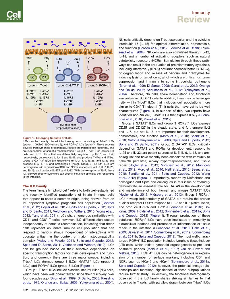
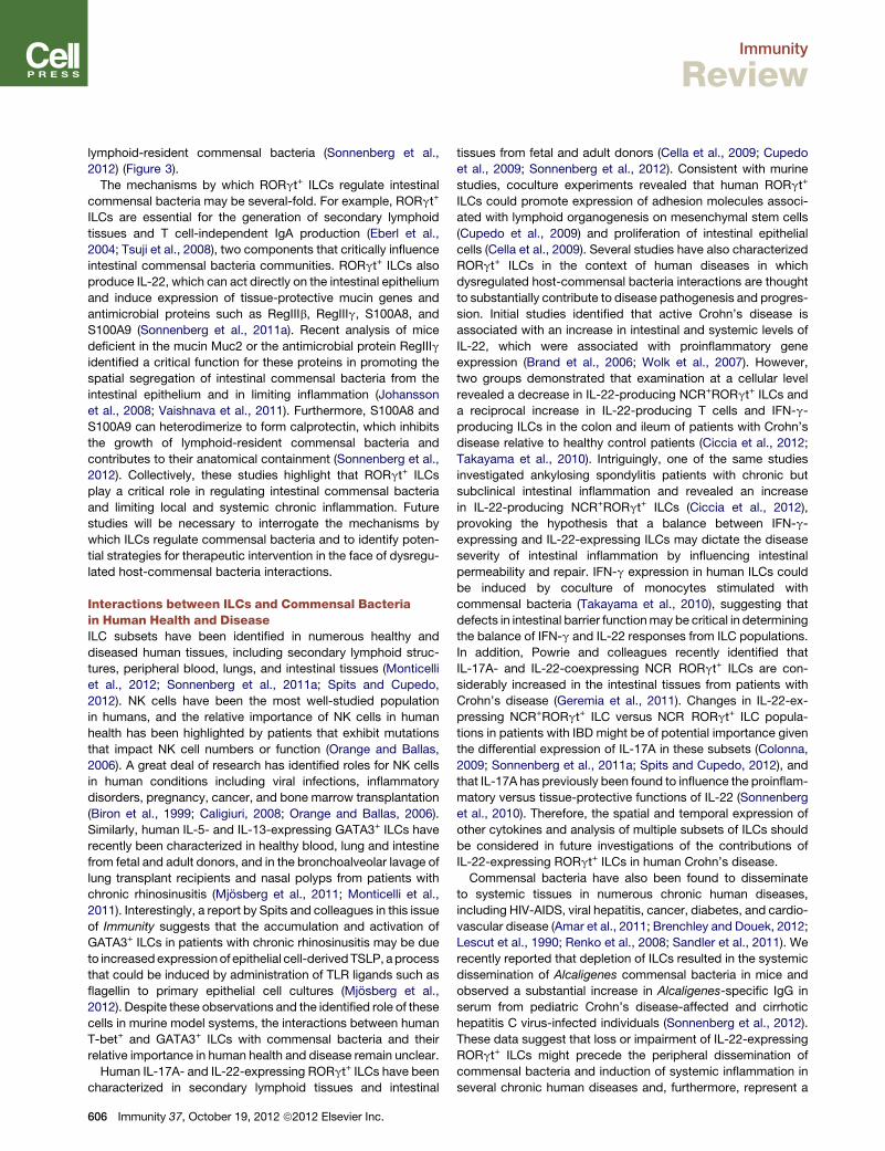
Figure 1. Emerging Subsets of ILCsILCs can be broadly placed into three groups, consisting of T-bet+ ILCs(group 1), GATA3+ ILCs (group 2), and RORgt+ ILCs (group 3). These subsetsdevelop from lymphoid progenitor(s), require the transcription factor Id2, andare independent of somatic recombination. Group 1 T-bet+ ILCs include NKcells and NCR� ILCs that are differentially regulated by IL-15 and IL-7,respectively, but respond to IL-12 and IL-18, and produce TNF-a and IFN-g.Group 2 GATA3+ ILCs are responsive to IL-2, IL-7, IL-25, and IL-33 andproduce IL-5, IL-13, and amphiregulin (Areg). Group 3 RORgt+ ILCs areheterogeneous in expression of CD4 andNCRs, responsive to IL-2, IL-7, IL-23,and IL-1b, and produce IL-17A and IL-22. With the exception of IL-5, theseILC-derived effector cytokines can directly influence epithelial cell responsesin the intestine.
Immunity
Review
The ILC FamilyThe term ‘‘innate lymphoid cell’’ refers to both well-established
and recently identified populations of innate immune cells
that appear to share a common origin, being derived from an
Id2-dependent lymphoid progenitor cell population (Cherrier
et al., 2012; Hoyler et al., 2012; Spits and Cupedo, 2012; Spits
and Di Santo, 2011; Veldhoen and Withers, 2010; Wong et al.,
2012; Yang et al., 2011). ILCs share numerous similarities with
CD4+ and CD8+ T cells; however, ILC differentiation occurs
independently of somatic recombination, indicating that these
cells represent an innate immune cell population that can
respond to various stimuli independent of interactions with
cognate antigen in the context of major histocompatibility
complex (Maloy and Powrie, 2011; Spits and Cupedo, 2012;
Spits and Di Santo, 2011; Veldhoen and Withers, 2010). ILCs
can be grouped based on their selective dependence on
specific transcription factors for their development and func-
tion, and currently there are three major groups, including
T-bet+ ILCs (termed group 1 ILCs), GATA3+ ILCs (group 2
ILCs) and RORgt+ ILCs (group 3 ILCs) (Figure 1).
Group 1 T-bet+ ILCs include classical natural killer (NK) cells,
which have been well characterized since their discovery over
four decades ago (Biron et al., 1999; Di Santo, 2008; Kiessling
et al., 1975; Orange and Ballas, 2006; Yokoyama et al., 2004).
602 Immunity 37, October 19, 2012 ª2012 Elsevier Inc.
NK cells critically depend on T-bet expression and the cytokine
interleukin-15 (IL-15) for optimal differentiation, homeostasis,
and function (Gordon et al., 2012; Lodolce et al., 1998; Town-
send et al., 2004). NK cells are also stimulated through IL-12,
IL-18, and a number of activating receptors, such as natural
cytotoxicity receptors (NCRs). Stimulation through these path-
ways can result in the production of proinflammatory cytokines,
including interferon-g (IFN-g) or tumor necrosis factor a (TNF-a),
or degranulation and release of perforin and granzymes for
inducing lysis of target cells, all of which are critical for tumor
suppression and immunity to some intracellular pathogens
(Biron et al., 1999; Di Santo, 2008; Ganal et al., 2012; Orange
and Ballas, 2006; Schulthess et al., 2012; Yokoyama et al.,
2004). Therefore, NK cells share homeostatic and functional
similarities with CD8+ T cells. In addition, there may be heteroge-
neity within T-bet+ ILCs that includes cell populations more
similar to CD4+ T helper 1 (Th1) cells that have yet to be well
characterized (Figure 1). In support of this, two reports have
identified non-NK cell, T-bet+ ILCs that express IFN-g (Buono-
core et al., 2010; Powell et al., 2012).
Group 2 GATA3+ ILCs and group 3 RORgt+ ILCs express
CD25 and CD127 in the steady state, and furthermore IL-2
and IL-7, but not IL-15, are important for their development,
homeostasis, and function (Moro et al., 2010; Saenz et al.,
2010; Satoh-Takayama et al., 2008; Spits and Cupedo, 2012;
Spits and Di Santo, 2011). Group 2 GATA3+ ILCs, critically
depend on GATA3 and RORa for development, respond to
IL-25 and IL-33; are potent sources of IL-5, IL-9, IL-13, and am-
phiregulin; and have recently been associated with immunity to
helminth parasites, airway hyperresponsiveness, and tissue
repair (Hoyler et al., 2012; Mjosberg et al., 2012; Monticelli
et al., 2012; Moro et al., 2010; Neill et al., 2010; Saenz et al.,
2010; Sandler et al., 2011; Spits and Cupedo, 2012; Wong
et al., 2012) (Figure 1). Importantly, reports by Diefenbach and
colleagues and Spits and colleagues in this issue of Immunity
demonstrate an essential role for GATA3 in the development
and maintenance of both human and mouse GATA3+ ILCs
(Hoyler et al., 2012; Mjosberg et al., 2012). Group 3 RORgt+
ILCs develop independently of GATA3 but require the orphan
nuclear receptor RORgt, respond to IL-23 and IL-1b stimulation,
and produce IL-17A and IL-22 (Buonocore et al., 2010; Co-
lonna, 2009; Hoyler et al., 2012; Sonnenberg et al., 2011a; Spits
and Cupedo, 2012) (Figure 1). Through production of these
cytokines, RORgt+ ILCs have been implicated in immunity to
extracellular bacteria and promotion of inflammation or tissue
repair in the intestine (Buonocore et al., 2010; Cella et al.,
2009; Sawa et al., 2011; Sonnenberg et al., 2011a; Sonnenberg
et al., 2011b; Spits and Cupedo, 2012). The most well-charac-
terized RORgt+ ILC population includes lymphoid tissue inducer
(LTi) cells, which initiate lymphoid organogenesis at pre- and
postnatal periods (Mebius et al., 1997; van de Pavert and
Mebius, 2010). RORgt+ ILCs can be heterogeneous in expres-
sion of a number of surface markers, including CD4 and
NCRs such as NKp46 and NKp44 (Sonnenberg et al., 2011a;
Spits and Cupedo, 2012); however, the potential lineage rela-
tionships and functional significance of these subpopulations
require further study. Collectively, the functional heterogeneity
observed in the ILC family shares striking similarities to that
observed in T cells, with parallels drawn between T-bet+ ILCs
Immunity
Review
and CD8+ T cells and CD4+ Th1 cells, GATA3+ ILCs and Th2
cells, and RORgt+ ILCs and Th17 cells.
Regulation of ILC Development, Maintenance, andFunction by Commensal BacteriaILCs have been found to be resident immune cell populations at
barrier surfaces of the mammalian body, including the skin,
airway, and intestinal tract (Spits and Cupedo, 2012; Spits and
Di Santo, 2011; Veldhoen and Withers, 2010). Through pro-
duction of soluble effector molecules including IFN-g, TNF-a,
IL-13, IL-17A, IL-22, and amphiregulin, ILCs can have a pro-
found impact on epithelial cells that are in direct contact with
commensal bacteria (Maloy and Powrie, 2011; Sonnenberg
et al., 2011a; Spits and Cupedo, 2012; Spits and Di Santo,
2011; Veldhoen and Withers, 2010) (Figure 1). Given the spatial
proximity of commensal bacteria, epithelial cells, and ILCs,
numerous groups have investigated the impact of commensal
bacteria on the development of ILCs. Studies employing
germ-free mice that lack live commensal bacteria identified
that NK cells and GATA3+ ILCs can develop in the absence of
commensal colonization (Ganal et al., 2012; Monticelli et al.,
2011). However, there have been conflicting reports on the
requirement of commensal bacteria on the development of
RORgt+ ILCs. Subsets of RORgt+ ILCs can develop indepen-
dently of commensal bacteria, as made evident by the presence
of LTi cells and the generation of secondary lymphoid structures
in the sterile environment of the fetus prior to birth (Mebius et al.,
1997; van de Pavert and Mebius, 2010). However, following
birth, the maturation of intestinal cyptopatches into isolated
lymphoid follicles is compromised in germ-free mice, suggesting
impairment in the function of some populations of LTi-like
RORgt+ ILCs (Bouskra et al., 2008; Tsuji et al., 2008). Direct
examination of RORgt+ ILCs by several groups identified normal
development of all RORgt+ ILC subsets in the absence of live
commensal bacteria in both germ-free and antibiotic-treated
mice (Reynders et al., 2011; Sawa et al., 2010; Sawa et al.,
2011; Sonnenberg et al., 2012). In contrast, three reports identi-
fied a substantial reduction in NCR+RORgt+ ILCs and a lack of
expression of Rorc or Il22 in the small intestine lamina propria
of germ-free or antibiotic-treated mice (Sanos et al., 2009;
Satoh-Takayama et al., 2008; Vonarbourg et al., 2010). Whereas
some of these differencesmay relate to host genetics or differen-
tial exposure to nonlive-bacteria- or diet-derived signals, it is
clear that further investigation will be necessary to clarify the
influence of commensal bacteria on the development of
NCR+RORgt+ ILCs.
Although commensal bacteria do not appear to be essential
for the development of most groups of ILCs, signals derived
from commensal bacteria may substantially impact the function
of ILCs. For example, mice devoid of live commensal bacteria
exhibit impaired NK cell cytolytic and IFN-g responses to poly-IC
stimulation or following infection with mouse cytomegalovirus
(Ganal et al., 2012), which is consistent with additional reports
demonstrating impaired antiviral immune responses in the
absence of commensal bacteria (Abt et al., 2012; Ichinohe
et al., 2011). These data indicate that commensal bacteria may
be essential for promoting optimal NK cell responses. In con-
trast, Eberl and colleagues reported that commensal bacteria
can suppress intestinal RORgt+ ILC production of IL-22 in
healthy mice, a process that could be reversed following exper-
imental damage to the intestinal epithelium (Sawa et al., 2011).
Furthermore, although it is clear that the absence of commensal
bacteria or dysbiosis is associatedwith altered NKT cell or baso-
phil function and exaggerated allergic inflammation (Hill and Ar-
tis, 2010; Hill et al., 2012; Olszak et al., 2012), whether
commensal bacteria influence the function of GATA3+ ILCs
that express Th2 cell-associated cytokines remains to be tested.
It is clear that additional studies will be required to comprehen-
sively define the impact of commensal bacteria on the develop-
ment, homeostasis, and function of ILCs. The following section
will discuss how commensal bacteria may influence the homeo-
stasis and function of ILC populations through direct and indirect
mechanisms.
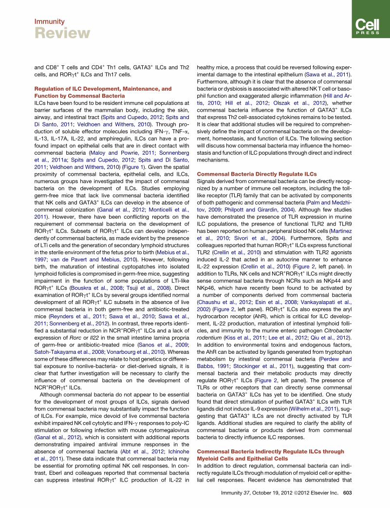
Commensal Bacteria Directly Regulate ILCsSignals derived from commensal bacteria can be directly recog-
nized by a number of immune cell receptors, including the toll-
like receptor (TLR) family that can be activated by components
of both pathogenic and commensal bacteria (Palm and Medzhi-
tov, 2009; Philpott and Girardin, 2004). Although few studies
have demonstrated the presence of TLR expression in murine
ILC populations, the presence of functional TLR2 and TLR9
has been reported on human peripheral blood NK cells (Martinez
et al., 2010; Sivori et al., 2004). Furthermore, Spits and
colleagues reported that human RORgt+ ILCs express functional
TLR2 (Crellin et al., 2010) and stimulation with TLR2 agonists
induced IL-2 that acted in an autocrine manner to enhance
IL-22 expression (Crellin et al., 2010) (Figure 2, left panel). In
addition to TLRs, NK cells and NCR+RORgt+ ILCs might directly
sense commensal bacteria through NCRs such as NKp44 and
NKp46, which have recently been found to be activated by
a number of components derived from commensal bacteria
(Chaushu et al., 2012; Esin et al., 2008; Vankayalapati et al.,
2002) (Figure 2, left panel). RORgt+ ILCs also express the aryl
hydrocarbon receptor (AhR), which is critical for ILC develop-
ment, IL-22 production, maturation of intestinal lymphoid folli-
cles, and immunity to the murine enteric pathogen Citrobacter
rodentium (Kiss et al., 2011; Lee et al., 2012; Qiu et al., 2012).
In addition to environmental toxins and endogenous factors,
the AhR can be activated by ligands generated from tryptophan
metabolism by intestinal commensal bacteria (Perdew and
Babbs, 1991; Stockinger et al., 2011), suggesting that com-
mensal bacteria and their metabolic products may directly
regulate RORgt+ ILCs (Figure 2, left panel). The presence of
TLRs or other receptors that can directly sense commensal
bacteria on GATA3+ ILCs has yet to be identified. One study
found that direct stimulation of purified GATA3+ ILCs with TLR
ligands did not induce IL-9 expression (Wilhelm et al., 2011), sug-
gesting that GATA3+ ILCs are not directly activated by TLR
ligands. Additional studies are required to clarify the ability of
commensal bacteria or products derived from commensal
bacteria to directly influence ILC responses.
Commensal Bacteria Indirectly Regulate ILCs throughMyeloid Cells and Epithelial CellsIn addition to direct regulation, commensal bacteria can indi-
rectly regulate ILCs throughmodulation of myeloid cell or epithe-
lial cell responses. Recent evidence has demonstrated that
Immunity 37, October 19, 2012 ª2012 Elsevier Inc. 603
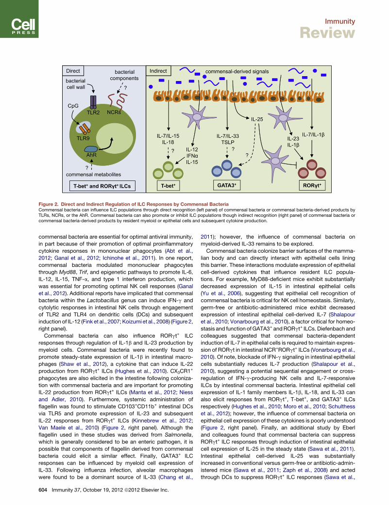
Figure 2. Direct and Indirect Regulation of ILC Responses by Commensal BacteriaCommensal bacteria can influence ILC populations through direct recognition (left panel) of commensal bacteria or commensal bacteria-derived products byTLRs, NCRs, or the AhR. Commensal bacteria can also promote or inhibit ILC populations though indirect recognition (right panel) of commensal bacteria orcommensal bacteria-derived products by resident myeloid or epithelial cells and subsequent cytokine production.
Immunity
Review
commensal bacteria are essential for optimal antiviral immunity,
in part because of their promotion of optimal proinflammatory
cytokine responses in mononuclear phagocytes (Abt et al.,
2012; Ganal et al., 2012; Ichinohe et al., 2011). In one report,
commensal bacteria modulated mononuclear phagocytes
through Myd88, Trif, and epigenetic pathways to promote IL-6,
IL-12, IL-15, TNF-a, and type 1 interferon production, which
was essential for promoting optimal NK cell responses (Ganal
et al., 2012). Additional reports have implicated that commensal
bacteria within the Lactobacillus genus can induce IFN-g and
cytolytic responses in intestinal NK cells through engagement
of TLR2 and TLR4 on dendritic cells (DCs) and subsequent
induction of IL-12 (Fink et al., 2007; Koizumi et al., 2008) (Figure 2,
right panel).
Commensal bacteria can also influence RORgt+ ILC
responses through regulation of IL-1b and IL-23 production by
myeloid cells. Commensal bacteria were recently found to
promote steady-state expression of IL-1b in intestinal macro-
phages (Shaw et al., 2012), a cytokine that can induce IL-22
production from RORgt+ ILCs (Hughes et al., 2010). CX3CR1+
phagocytes are also elicited in the intestine following coloniza-
tion with commensal bacteria and are important for promoting
IL-22 production from RORgt+ ILCs (Manta et al., 2012; Niess
and Adler, 2010). Furthermore, systemic administration of
flagellin was found to stimulate CD103+CD11b+ intestinal DCs
via TLR5 and promote expression of IL-23 and subsequent
IL-22 responses from RORgt+ ILCs (Kinnebrew et al., 2012;
Van Maele et al., 2010) (Figure 2, right panel). Although the
flagellin used in these studies was derived from Salmonella,
which is generally considered to be an enteric pathogen, it is
possible that components of flagellin derived from commensal
bacteria could elicit a similar effect. Finally, GATA3+ ILC
responses can be influenced by myeloid cell expression of
IL-33. Following influenza infection, alveolar macrophages
were found to be a dominant source of IL-33 (Chang et al.,
604 Immunity 37, October 19, 2012 ª2012 Elsevier Inc.
2011); however, the influence of commensal bacteria on
myeloid-derived IL-33 remains to be explored.
Commensal bacteria colonize barrier surfaces of the mamma-
lian body and can directly interact with epithelial cells lining
this barrier. These interactions modulate expression of epithelial
cell-derived cytokines that influence resident ILC popula-
tions. For example, MyD88-deficient mice exhibit substantially
decreased expression of IL-15 in intestinal epithelial cells
(Yu et al., 2006), suggesting that epithelial cell recognition of
commensal bacteria is critical for NK cell homeostasis. Similarly,
germ-free or antibiotic-administered mice exhibit decreased
expression of intestinal epithelial cell-derived IL-7 (Shalapour
et al., 2010; Vonarbourg et al., 2010), a factor critical for homeo-
stasis and function of GATA3+ andRORgt+ ILCs. Diefenbach and
colleagues suggested that commensal bacteria-dependent
induction of IL-7 in epithelial cells is required to maintain expres-
sion of RORgt in intestinal NCR+RORgt+ ILCs (Vonarbourg et al.,
2010). Of note, blockade of IFN-g signaling in intestinal epithelial
cells substantially reduces IL-7 production (Shalapour et al.,
2010), suggesting a potential sequential engagement or cross-
regulation of IFN-g-producing NK cells and IL-7-responsive
ILCs by intestinal commensal bacteria. Intestinal epithelial cell
expression of IL-1 family members IL-1b, IL-18, and IL-33 can
also elicit responses from RORgt+, T-bet+, and GATA3+ ILCs
respectively (Hughes et al., 2010; Moro et al., 2010; Schulthess
et al., 2012); however, the influence of commensal bacteria on
epithelial cell expression of these cytokines is poorly understood
(Figure 2, right panel). Finally, an additional study by Eberl
and colleagues found that commensal bacteria can suppress
RORgt+ ILC responses through induction of intestinal epithelial
cell expression of IL-25 in the steady state (Sawa et al., 2011).
Intestinal epithelial cell-derived IL-25 was substantially
increased in conventional versus germ-free or antibiotic-admin-
istered mice (Sawa et al., 2011; Zaph et al., 2008) and acted
through DCs to suppress RORgt+ ILC responses (Sawa et al.,
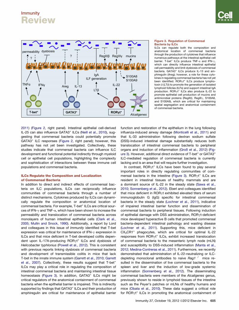
Figure 3. Regulation of CommensalBacteria by ILCsILCs can regulate both the composition andanatomical location of commensal bacteriathrough the production of cytokines that influencenumerous pathways at the intestinal epithelial cellbarrier. T-bet+ ILCs produce TNF-a and IFN-g,which can directly influence intestinal epithelialcell permeability and limit dysbiosis of commensalbacteria. GATA3+ ILCs produce IL-13 and am-phiregulin (Areg); however, a role for these cyto-kines in regulating commensal bacteria has not yetbeen identified. RORgt+ ILCs produce lympho-toxin b (LTb) to promote the generation of isolatedlymphoid follicles (ILFs) and support intestinal IgAproduction. RORgt+ ILCs also produce IL-22 topromote epithelial cell production of mucins andantimicrobial proteins (RegIIIb, RegIIIg, S100A8,and S100A9), which are critical for maintainingspatial segregation and anatomical containmentof commensal bacteria.
Immunity
Review
2011) (Figure 2, right panel). Intestinal epithelial cell-derived
IL-25 can also influence GATA3+ ILCs (Neill et al., 2010), sug-
gesting that commensal bacteria could potentially promote
GATA3+ ILC responses (Figure 2, right panel); however, this
pathway has not yet been investigated. Collectively, these
studies indicate that commensal bacteria can influence ILC
development and functional potential indirectly through myeloid
cell or epithelial cell populations, highlighting the complexity
and sophistication of interactions between these immune cell
populations and commensal bacteria.
ILCs Regulate the Composition and Localizationof Commensal BacteriaIn addition to direct and indirect effects of commensal bac-
teria on ILC populations, ILCs can reciprocally influence
communities of commensal bacteria through a number of
distinct mechanisms. Cytokines produced by ILCs can dynami-
cally regulate the composition or anatomical location of
commensal bacteria. For example, T-bet+ ILCs are critical sour-
ces of IFN-g and TNF-a, which have been shown to increase the
permeability and translocation of commensal bacteria across
monolayers of human intestinal epithelial cells (Clark et al.,
2005; Mullin and Snock, 1990). Additionally, a report by Lord
and colleagues in this issue of Immunity identified that T-bet
expression was critical for maintenance of IFN-g expression in
ILCs and that mice deficient in T-bet developed colitis depen-
dent upon IL-17A-producing RORgt+ ILCs and dysbiosis of
Helicobacter typhlonius (Powell et al., 2012). This is consistent
with previous reports linking dysbiosis of commensal bacteria
and development of transmissible colitis in mice that lack
T-bet in the innate immune system (Garrett et al., 2010; Garrett
et al., 2007). Collectively, these results suggest that T-bet+
ILCs may play a critical role in regulating the composition of
intestinal commensal bacteria and maintaining intestinal tissue
homeostasis (Figure 3). In addition, GATA3+ ILCs might be
critical regulators of the anatomical containment of commensal
bacteria when the epithelial barrier is impaired. This is indirectly
supported by findings that GATA3+ ILCs and their production of
amphiregulin are critical for maintenance of epithelial barrier
function and restoration of the epithelium in the lung following
influenza-induced airway damage (Monticelli et al., 2011) and
that IL-33 administration following dextran sodium sulfate
(DSS)-induced intestinal damage substantially reduces both
translocation of intestinal commensal bacteria to peripheral
organs and induction of inflammation (Groß et al., 2012) (Fig-
ure 3). However, additional direct evidence of T-bet+ or GATA3+
ILC-mediated regulation of commensal bacteria is currently
lacking and is an area that will require further investigation.
In contrast, RORgt+ ILCs have been found to play several
important roles in directly regulating communities of com-
mensal bacteria in the intestine (Figure 3). RORgt+ ILCs are
resident in intestinal tissues of healthy mammals and are
a dominant source of IL-22 in the steady state (Sawa et al.,
2010; Sonnenberg et al., 2012). Eberl and colleagues identified
that mice deficient in RORgt exhibited elevated titers of serum
immunoglobulin G (IgG) specific for intestinal commensal
bacteria in the steady state (Lochner et al., 2011), indicative
of impaired intestinal barrier function and dissemination of
commensal bacteria to peripheral tissues. Following induction
of epithelial damage with DSS administration, RORgt-deficient
mice developed hyperactive B cells that promoted commensal
bacteria-dependent intestinal pathology and wasting disease
(Lochner et al., 2011). Supporting this, mice deficient in
CX3CR1+ phagocytes, which are critical for optimal IL-22
responses from RORgt+ ILCs, exhibit increased translocation
of commensal bacteria to the mesenteric lymph node (mLN)
and susceptibility to DSS-induced inflammation (Manta et al.,
2012; Medina-Contreras et al., 2011). Furthermore, we recently
demonstrated that administration of IL-22-neutralizing or ILC-
depleting monoclonal antibodies to naive Rag1�/� mice re-
sulted in the dissemination of live commensal bacteria to the
spleen and liver and the induction of low-grade systemic
inflammation (Sonnenberg et al., 2012). The disseminating
commensal bacteria were members of the Alcaligenes genus,
previously shown to reside in lymphoid tissues of the intestine
such as the Peyer’s patches or mLNs of healthy humans and
mice (Obata et al., 2010). These data suggest a critical role
for RORgt+ ILCs in promoting the anatomical containment of
Immunity 37, October 19, 2012 ª2012 Elsevier Inc. 605
Immunity
Review
lymphoid-resident commensal bacteria (Sonnenberg et al.,
2012) (Figure 3).
The mechanisms by which RORgt+ ILCs regulate intestinal
commensal bacteria may be several-fold. For example, RORgt+
ILCs are essential for the generation of secondary lymphoid
tissues and T cell-independent IgA production (Eberl et al.,
2004; Tsuji et al., 2008), two components that critically influence
intestinal commensal bacteria communities. RORgt+ ILCs also
produce IL-22, which can act directly on the intestinal epithelium
and induce expression of tissue-protective mucin genes and
antimicrobial proteins such as RegIIIb, RegIIIg, S100A8, and
S100A9 (Sonnenberg et al., 2011a). Recent analysis of mice
deficient in the mucin Muc2 or the antimicrobial protein RegIIIg
identified a critical function for these proteins in promoting the
spatial segregation of intestinal commensal bacteria from the
intestinal epithelium and in limiting inflammation (Johansson
et al., 2008; Vaishnava et al., 2011). Furthermore, S100A8 and
S100A9 can heterodimerize to form calprotectin, which inhibits
the growth of lymphoid-resident commensal bacteria and
contributes to their anatomical containment (Sonnenberg et al.,
2012). Collectively, these studies highlight that RORgt+ ILCs
play a critical role in regulating intestinal commensal bacteria
and limiting local and systemic chronic inflammation. Future
studies will be necessary to interrogate the mechanisms by
which ILCs regulate commensal bacteria and to identify poten-
tial strategies for therapeutic intervention in the face of dysregu-
lated host-commensal bacteria interactions.
Interactions between ILCs and Commensal Bacteriain Human Health and DiseaseILC subsets have been identified in numerous healthy and
diseased human tissues, including secondary lymphoid struc-
tures, peripheral blood, lungs, and intestinal tissues (Monticelli
et al., 2012; Sonnenberg et al., 2011a; Spits and Cupedo,
2012). NK cells have been the most well-studied population
in humans, and the relative importance of NK cells in human
health has been highlighted by patients that exhibit mutations
that impact NK cell numbers or function (Orange and Ballas,
2006). A great deal of research has identified roles for NK cells
in human conditions including viral infections, inflammatory
disorders, pregnancy, cancer, and bone marrow transplantation
(Biron et al., 1999; Caligiuri, 2008; Orange and Ballas, 2006).
Similarly, human IL-5- and IL-13-expressing GATA3+ ILCs have
recently been characterized in healthy blood, lung and intestine
from fetal and adult donors, and in the bronchoalveolar lavage of
lung transplant recipients and nasal polyps from patients with
chronic rhinosinusitis (Mjosberg et al., 2011; Monticelli et al.,
2011). Interestingly, a report by Spits and colleagues in this issue
of Immunity suggests that the accumulation and activation of
GATA3+ ILCs in patients with chronic rhinosinusitis may be due
to increasedexpressionof epithelial cell-derived TSLP, aprocess
that could be induced by administration of TLR ligands such as
flagellin to primary epithelial cell cultures (Mjosberg et al.,
2012). Despite these observations and the identified role of these
cells in murine model systems, the interactions between human
T-bet+ and GATA3+ ILCs with commensal bacteria and their
relative importance in human health and disease remain unclear.
Human IL-17A- and IL-22-expressing RORgt+ ILCs have been
characterized in secondary lymphoid tissues and intestinal
606 Immunity 37, October 19, 2012 ª2012 Elsevier Inc.
tissues from fetal and adult donors (Cella et al., 2009; Cupedo
et al., 2009; Sonnenberg et al., 2012). Consistent with murine
studies, coculture experiments revealed that human RORgt+
ILCs could promote expression of adhesion molecules associ-
ated with lymphoid organogenesis on mesenchymal stem cells
(Cupedo et al., 2009) and proliferation of intestinal epithelial
cells (Cella et al., 2009). Several studies have also characterized
RORgt+ ILCs in the context of human diseases in which
dysregulated host-commensal bacteria interactions are thought
to substantially contribute to disease pathogenesis and progres-
sion. Initial studies identified that active Crohn’s disease is
associated with an increase in intestinal and systemic levels of
IL-22, which were associated with proinflammatory gene
expression (Brand et al., 2006; Wolk et al., 2007). However,
two groups demonstrated that examination at a cellular level
revealed a decrease in IL-22-producing NCR+RORgt+ ILCs and
a reciprocal increase in IL-22-producing T cells and IFN-g-
producing ILCs in the colon and ileum of patients with Crohn’s
disease relative to healthy control patients (Ciccia et al., 2012;
Takayama et al., 2010). Intriguingly, one of the same studies
investigated ankylosing spondylitis patients with chronic but
subclinical intestinal inflammation and revealed an increase
in IL-22-producing NCR+RORgt+ ILCs (Ciccia et al., 2012),
provoking the hypothesis that a balance between IFN-g-
expressing and IL-22-expressing ILCs may dictate the disease
severity of intestinal inflammation by influencing intestinal
permeability and repair. IFN-g expression in human ILCs could
be induced by coculture of monocytes stimulated with
commensal bacteria (Takayama et al., 2010), suggesting that
defects in intestinal barrier functionmay be critical in determining
the balance of IFN-g and IL-22 responses from ILC populations.
In addition, Powrie and colleagues recently identified that
IL-17A- and IL-22-coexpressing NCR�RORgt+ ILCs are con-
siderably increased in the intestinal tissues from patients with
Crohn’s disease (Geremia et al., 2011). Changes in IL-22-ex-
pressing NCR+RORgt+ ILC versus NCR�RORgt+ ILC popula-
tions in patients with IBD might be of potential importance given
the differential expression of IL-17A in these subsets (Colonna,
2009; Sonnenberg et al., 2011a; Spits and Cupedo, 2012), and
that IL-17A has previously been found to influence the proinflam-
matory versus tissue-protective functions of IL-22 (Sonnenberg
et al., 2010). Therefore, the spatial and temporal expression of
other cytokines and analysis of multiple subsets of ILCs should
be considered in future investigations of the contributions of
IL-22-expressing RORgt+ ILCs in human Crohn’s disease.
Commensal bacteria have also been found to disseminate
to systemic tissues in numerous chronic human diseases,
including HIV-AIDS, viral hepatitis, cancer, diabetes, and cardio-
vascular disease (Amar et al., 2011; Brenchley and Douek, 2012;
Lescut et al., 1990; Renko et al., 2008; Sandler et al., 2011). We
recently reported that depletion of ILCs resulted in the systemic
dissemination of Alcaligenes commensal bacteria in mice and
observed a substantial increase in Alcaligenes-specific IgG in
serum from pediatric Crohn’s disease-affected and cirrhotic
hepatitis C virus-infected individuals (Sonnenberg et al., 2012).
These data suggest that loss or impairment of IL-22-expressing
RORgt+ ILCs might precede the peripheral dissemination of
commensal bacteria and induction of systemic inflammation in
several chronic human diseases and, furthermore, represent a
Immunity
Review
potentially important therapeutic target. Supporting this, recent
studies have identified a loss of IL-22-expressing RORgt+ ILCs
in both Crohn’s disease in humans (Ciccia et al., 2012;
Takayama et al., 2010) and progressive simian immunodefi-
ciency virus infection in nonhuman primates (Klatt et al., 2012;
Reeves et al., 2011; Xu et al., 2012). Future studies will be
needed to further interrogate whether IL-22-expressing RORgt+
ILCs are lost across multiple chronic human diseases, to
determine the mechanisms by which this occurs, and to identify
novel therapeutic strategies for restoring normal ILC homeo-
stasis and preventing or limiting pathologic host-commensal
bacteria interactions.
Conversely, pathogenic ILC responses that have been asso-
ciated with mouse models of airway hyperresponsiveness,
asthma, intestinal inflammation, and psoriasis could also be
targeted in human disease (Buonocore et al., 2010; Chang
et al., 2011; Pantelyushin et al., 2012). Thismay be accomplished
in part with already-available humanized monoclonal antibodies
that target pathways including IL-23, IL-17, and CD25 (Ding
et al., 2008; Leonardi et al., 2012; Perry et al., 2012). As a proof
of principle, it was recently found that blockade of CD25 with
daclizumab in patients with multiple sclerosis shifted the
balance of circulating ILC subsets by decreasing RORgt+ ILCs
and increasing NK cells and was also associated with reduced
parameters of inflammation (Perry et al., 2012). Additional
studies will be necessary to determine the full effects of targeting
these pathways on ILC biology, and it is probable that additional
immunomodulatory agents will need to be developed that
selectively regulate the balance of ILC responses and restore
healthy host-commensal bacteria interactions.
Summary and Future PerspectivesEmerging evidence of the development, function, and heteroge-
neity of the ILC family represents an exciting advance in the field
of immunology. These cells appear to play important roles at
barrier surfaces of the body, such as the intestine, and are inti-
mately associated with commensal bacteria that colonize these
tissues. There is a need to develop new tools and models to
specifically target ILC responses, determine their roles in murine
model systems, and interrogate potential interactions of ILCs
with adaptive immune cell populations. Translational studies
examining ILCs in the context of human health and disease
with extensive analysis of cytokine and surface marker expres-
sion will be valuable for a better understanding of the functional
contributions of human ILCs. Given the recent appreciation
that dysregulated host-commensal bacteria relationships
are associated with the pathogenesis and progression of
numerous chronic human infectious, inflammatory, and meta-
bolic diseases, targeting interactions between ILCs and
commensal bacteria may offer new approaches for developing
preventative and therapeutic treatments for these diseases.
ACKNOWLEDGMENTS
We thank members of the Artis laboratory for discussions and critical readingof the manuscript. This work is supported by the National Institutes ofHealth (AI061570, AI087990, AI074878, AI083480, AI095466, AI095608, andAI097333 to D.A.; T32-AI055428 and DP5OD012116 to G.F.S.) and theBurroughs Wellcome Fund Investigator in the Pathogenesis of InfectiousDisease Award (D.A.).
REFERENCES
Abt, M.C., Osborne, L.C., Monticelli, L.A., Doering, T.A., Alenghat, T., Sonnen-berg, G.F., Paley, M.A., Antenus, M., Williams, K.L., Erikson, J., et al. (2012).Commensal bacteria calibrate the activation threshold of innate antiviralimmunity. Immunity 37, 158–170.
Amar, J., Serino, M., Lange, C., Chabo, C., Iacovoni, J., Mondot, S., Lepage,P., Klopp, C., Mariette, J., Bouchez, O., et al; D.E.S.I.R. Study Group. (2011).Involvement of tissue bacteria in the onset of diabetes in humans: evidence fora concept. Diabetologia 54, 3055–3061.
Atarashi, K., Tanoue, T., Shima, T., Imaoka, A., Kuwahara, T., Momose, Y.,Cheng, G., Yamasaki, S., Saito, T., Ohba, Y., et al. (2011). Induction of colonicregulatory T cells by indigenous Clostridium species. Science 331, 337–341.
Biron, C.A., Nguyen, K.B., Pien, G.C., Cousens, L.P., and Salazar-Mather, T.P.(1999). Natural killer cells in antiviral defense: function and regulation by innatecytokines. Annu. Rev. Immunol. 17, 189–220.
Bouskra, D., Brezillon, C., Berard, M., Werts, C., Varona, R., Boneca, I.G., andEberl, G. (2008). Lymphoid tissue genesis induced by commensals throughNOD1 regulates intestinal homeostasis. Nature 456, 507–510.
Brand, S., Beigel, F., Olszak, T., Zitzmann, K., Eichhorst, S.T., Otte, J.M.,Diepolder, H., Marquardt, A., Jagla, W., Popp, A., et al. (2006). IL-22 isincreased in active Crohn’s disease and promotes proinflammatory geneexpression and intestinal epithelial cell migration. Am. J. Physiol. Gastrointest.Liver Physiol. 290, G827–G838.
Brenchley, J.M., and Douek, D.C. (2012). Microbial translocation across theGI tract. Annu. Rev. Immunol. 30, 149–173.
Buonocore, S., Ahern, P.P., Uhlig, H.H., Ivanov, I.I., Littman, D.R., Maloy, K.J.,and Powrie, F. (2010). Innate lymphoid cells drive interleukin-23-dependentinnate intestinal pathology. Nature 464, 1371–1375.
Caligiuri, M.A. (2008). Human natural killer cells. Blood 112, 461–469.
Cella, M., Fuchs, A., Vermi, W., Facchetti, F., Otero, K., Lennerz, J.K., Doherty,J.M., Mills, J.C., and Colonna, M. (2009). A human natural killer cell subsetprovides an innate source of IL-22 formucosal immunity. Nature 457, 722–725.
Chang, Y.J., Kim, H.Y., Albacker, L.A., Baumgarth, N., McKenzie, A.N., Smith,D.E., Dekruyff, R.H., and Umetsu, D.T. (2011). Innate lymphoid cells mediateinfluenza-induced airway hyper-reactivity independently of adaptive immunity.Nat. Immunol. 12, 631–638.
Chaushu, S., Wilensky, A., Gur, C., Shapira, L., Elboim, M., Halftek, G., Polak,D., Achdout, H., Bachrach, G., and Mandelboim, O. (2012). Direct recognitionof Fusobacterium nucleatum by the NK cell natural cytotoxicity receptorNKp46 aggravates periodontal disease. PLoS Pathog. 8, e1002601.
Cherrier, M., Sawa, S., and Eberl, G. (2012). Notch, Id2, and RORgt se-quentially orchestrate the fetal development of lymphoid tissue inducer cells.J. Exp. Med. 209, 729–740.
Chin, K.F., Kallam, R., O’Boyle, C., and MacFie, J. (2007). Bacterial trans-location may influence the long-term survival in colorectal cancer patients.Dis. Colon Rectum 50, 323–330.
Ciccia, F., Accardo-Palumbo, A., Alessandro, R., Rizzo, A., Principe, S.,Peralta, S., Raiata, F., Giardina, A., De Leo, G., and Triolo, G. (2012).Interleukin-22 and interleukin-22-producing NKp44+ natural killer cells insubclinical gut inflammation in ankylosing spondylitis. Arthritis Rheum. 64,1869–1878.
Clark, E., Hoare, C., Tanianis-Hughes, J., Carlson, G.L., and Warhurst, G.(2005). Interferon gamma induces translocation of commensal Escherichiacoli across gut epithelial cells via a lipid raft-mediated process. Gastroenter-ology 128, 1258–1267.
Clemente, J.C., Ursell, L.K., Parfrey, L.W., and Knight, R. (2012). The impact ofthe gut microbiota on human health: an integrative view. Cell 148, 1258–1270.
Colonna, M. (2009). Interleukin-22-producing natural killer cells and lymphoidtissue inducer-like cells in mucosal immunity. Immunity 31, 15–23.
Crellin, N.K., Trifari, S., Kaplan, C.D., Satoh-Takayama, N., Di Santo, J.P., andSpits, H. (2010). Regulation of cytokine secretion in human CD127(+) LTi-likeinnate lymphoid cells by Toll-like receptor 2. Immunity 33, 752–764.
Immunity 37, October 19, 2012 ª2012 Elsevier Inc. 607
Immunity
Review
Cua, D.J., and Tato, C.M. (2010). Innate IL-17-producing cells: the sentinels ofthe immune system. Nat. Rev. Immunol. 10, 479–489.
Cupedo, T., Crellin, N.K., Papazian, N., Rombouts, E.J., Weijer, K., Grogan,J.L., Fibbe, W.E., Cornelissen, J.J., and Spits, H. (2009). Human fetal lymphoidtissue-inducer cells are interleukin 17-producing precursors to RORC+CD127+ natural killer-like cells. Nat. Immunol. 10, 66–74.
Dethlefsen, L., McFall-Ngai, M., and Relman, D.A. (2007). An ecological andevolutionary perspective on human-microbe mutualism and disease. Nature449, 811–818.
Di Santo, J.P. (2008). Natural killer cells: diversity in search of a niche. Nat.Immunol. 9, 473–475.
Ding, C., Xu, J., and Li, J. (2008). ABT-874, a fully human monoclonal anti-IL-12/IL-23 antibody for the potential treatment of autoimmune diseases.Curr. Opin. Investig. Drugs 9, 515–522.
Eberl, G., Marmon, S., Sunshine, M.J., Rennert, P.D., Choi, Y., and Littman,D.R. (2004). An essential function for the nuclear receptor RORgamma(t) inthe generation of fetal lymphoid tissue inducer cells. Nat. Immunol. 5, 64–73.
Esin, S., Batoni, G., Counoupas, C., Stringaro, A., Brancatisano, F.L., Colone,M., Maisetta, G., Florio, W., Arancia, G., and Campa, M. (2008). Direct bindingof human NK cell natural cytotoxicity receptor NKp44 to the surfaces ofmycobacteria and other bacteria. Infect. Immun. 76, 1719–1727.
Fink, L.N., Zeuthen, L.H., Christensen, H.R., Morandi, B., Frøkiaer, H., andFerlazzo, G. (2007). Distinct gut-derived lactic acid bacteria elicit divergentdendritic cell-mediated NK cell responses. Int. Immunol. 19, 1319–1327.
Ganal, S.C., Sanos, S.L., Kallfass, C., Oberle, K., Johner, C., Kirschning, C.,Lienenklaus, S., Weiss, S., Staeheli, P., Aichele, P., and Diefenbach, A.(2012). Priming of natural killer cells by nonmucosal mononuclear phago-cytes requires instructive signals from commensal microbiota. Immunity37, 171–186.
Garrett, W.S., Lord, G.M., Punit, S., Lugo-Villarino, G., Mazmanian, S.K., Ito,S., Glickman, J.N., and Glimcher, L.H. (2007). Communicable ulcerative colitisinduced by T-bet deficiency in the innate immune system. Cell 131, 33–45.
Garrett, W.S., Gallini, C.A., Yatsunenko, T., Michaud, M., DuBois, A., Delaney,M.L., Punit, S., Karlsson, M., Bry, L., Glickman, J.N., et al. (2010). Enterobac-teriaceae act in concert with the gut microbiota to induce spontaneous andmaternally transmitted colitis. Cell Host Microbe 8, 292–300.
Geremia, A., Arancibia-Carcamo, C.V., Fleming, M.P., Rust, N., Singh, B.,Mortensen, N.J., Travis, S.P., and Powrie, F. (2011). IL-23-responsive innatelymphoid cells are increased in inflammatory bowel disease. J. Exp. Med.208, 1127–1133.
Gordon, S.M., Chaix, J., Rupp, L.J., Wu, J., Madera, S., Sun, J.C., Lindsten, T.,and Reiner, S.L. (2012). The transcription factors T-bet and Eomes control keycheckpoints of natural killer cell maturation. Immunity 36, 55–67.
Groß, P., Doser, K., Falk, W., Obermeier, F., and Hofmann, C. (2012). IL-33attenuates development and perpetuation of chronic intestinal inflammation.Inflamm. Bowel Dis. 18, 1900–1909.
Hill, D.A., and Artis, D. (2010). Intestinal bacteria and the regulation of immunecell homeostasis. Annu. Rev. Immunol. 28, 623–667.
Hill, D.A., Siracusa, M.C., Abt, M.C., Kim, B.S., Kobuley, D., Kubo, M.,Kambayashi, T., Larosa, D.F., Renner, E.D., Orange, J.S., et al. (2012).Commensal bacteria-derived signals regulate basophil hematopoiesis andallergic inflammation. Nat. Med. 18, 538–546.
Honda, K., and Littman, D.R. (2012). The microbiome in infectious diseaseand inflammation. Annu. Rev. Immunol. 30, 759–795.
Hooper, L.V., Littman, D.R., and Macpherson, A.J. (2012). Interactionsbetween the microbiota and the immune system. Science 336, 1268–1273.
Hoyler, T., Klose, C.S.N., Souabni, A., Turqueti-Neves, A., Pfeifer, D., Rawlins,E.L., Voehringer, D., Busslinger, M., and Diefenbach, A. (2012). The transcrip-tion factor GATA3 controls cell fate andmaintenance of type 2 innate lymphoidcells. Immunity 37, this issue, 634–648.
Hughes, T., Becknell, B., Freud, A.G., McClory, S., Briercheck, E., Yu, J., Mao,C., Giovenzana, C., Nuovo, G., Wei, L., et al. (2010). Interleukin-1betaselectively expands and sustains interleukin-22+ immature human naturalkiller cells in secondary lymphoid tissue. Immunity 32, 803–814.
608 Immunity 37, October 19, 2012 ª2012 Elsevier Inc.
Ichinohe, T., Pang, I.K., Kumamoto, Y., Peaper, D.R., Ho, J.H., Murray, T.S.,and Iwasaki, A. (2011). Microbiota regulates immune defense against respira-tory tract influenza A virus infection. Proc. Natl. Acad. Sci. USA 108, 5354–5359.
Ivanov, I.I., Atarashi, K., Manel, N., Brodie, E.L., Shima, T., Karaoz, U., Wei, D.,Goldfarb, K.C., Santee, C.A., Lynch, S.V., et al. (2009). Induction of intestinalTh17 cells by segmented filamentous bacteria. Cell 139, 485–498.
Johansson, M.E., Phillipson, M., Petersson, J., Velcich, A., Holm, L., andHansson, G.C. (2008). The inner of the two Muc2 mucin-dependent mucuslayers in colon is devoid of bacteria. Proc. Natl. Acad. Sci. USA 105, 15064–15069.
Kiessling, R., Klein, E., Pross, H., andWigzell, H. (1975). ‘‘Natural’’ killer cells inthe mouse. II. Cytotoxic cells with specificity for mouse Moloney leukemiacells. Characteristics of the killer cell. Eur. J. Immunol. 5, 117–121.
Kinnebrew, M.A., Buffie, C.G., Diehl, G.E., Zenewicz, L.A., Leiner, I., Hohl,T.M., Flavell, R.A., Littman, D.R., and Pamer, E.G. (2012). Interleukin 23production by intestinal CD103(+)CD11b(+) dendritic cells in response tobacterial flagellin enhances mucosal innate immune defense. Immunity 36,276–287.
Kiss, E.A., Vonarbourg, C., Kopfmann, S., Hobeika, E., Finke, D., Esser, C., andDiefenbach, A. (2011). Natural aryl hydrocarbon receptor ligands controlorganogenesis of intestinal lymphoid follicles. Science 334, 1561–1565.
Klatt, N.R., Estes, J.D., Sun, X., Ortiz, A.M., Barber, J.S., Harris, L.D., Cervasi,B., Yokomizo, L.K., Pan, L., Vinton, C.L., et al. (2012). Loss of mucosal CD103+DCs and IL-17+ and IL-22+ lymphocytes is associated with mucosal damagein SIV infection. Mucosal Immunol. Published online May 30, 2012. http://dx.doi.org/10.1038/mi.2012.38.
Koizumi, S., Wakita, D., Sato, T., Mitamura, R., Izumo, T., Shibata, H., Kiso, Y.,Chamoto, K., Togashi, Y., Kitamura, H., and Nishimura, T. (2008). Essential roleof Toll-like receptors for dendritic cell and NK1.1(+) cell-dependent activationof type 1 immunity by Lactobacillus pentosus strain S-PT84. Immunol. Lett.120, 14–19.
Lee, J.S., Cella, M., McDonald, K.G., Garlanda, C., Kennedy, G.D., Nukaya,M., Mantovani, A., Kopan, R., Bradfield, C.A., Newberry, R.D., and Colonna,M. (2012). AHR drives the development of gut ILC22 cells and postnatallymphoid tissues via pathways dependent on and independent of Notch.Nat. Immunol. 13, 144–151.
Leonardi, C., Matheson, R., Zachariae, C., Cameron, G., Li, L., Edson-Heredia,E., Braun, D., and Banerjee, S. (2012). Anti-interleukin-17monoclonal antibodyixekizumab in chronic plaque psoriasis. N. Engl. J. Med. 366, 1190–1199.
Lescut, D., Colombel, J.F., Vincent, P., Cortot, A., Fournier, L., Quandalle, P.,Vankemmel, M., Triboulet, J.P., Wurtz, A., Paris, J.C., et al. (1990). Bacterialtranslocation in colorectal cancers. Gastroenterol. Clin. Biol. 14, 811–814.
Ley, R.E., Peterson, D.A., and Gordon, J.I. (2006a). Ecological and evolu-tionary forces shaping microbial diversity in the human intestine. Cell 124,837–848.
Ley, R.E., Turnbaugh, P.J., Klein, S., and Gordon, J.I. (2006b). Microbialecology: human gut microbes associated with obesity. Nature 444, 1022–1023.
Ley, R.E., Lozupone, C.A., Hamady, M., Knight, R., and Gordon, J.I. (2008).Worlds within worlds: evolution of the vertebrate gut microbiota. Nat. Rev.Microbiol. 6, 776–788.
Littman, D.R., and Pamer, E.G. (2011). Role of the commensal microbiotain normal and pathogenic host immune responses. Cell Host Microbe 10,311–323.
Lochner, M., Ohnmacht, C., Presley, L., Bruhns, P., Si-Tahar, M., Sawa, S.,and Eberl, G. (2011). Microbiota-induced tertiary lymphoid tissues aggravateinflammatory disease in the absence of RORgamma t and LTi cells. J. Exp.Med. 208, 125–134.
Lodolce, J.P., Boone, D.L., Chai, S., Swain, R.E., Dassopoulos, T., Trettin, S.,andMa, A. (1998). IL-15 receptor maintains lymphoid homeostasis by support-ing lymphocyte homing and proliferation. Immunity 9, 669–676.
Maloy, K.J., and Powrie, F. (2011). Intestinal homeostasis and its breakdown ininflammatory bowel disease. Nature 474, 298–306.
Immunity
Review
Manta, C., Heupel, E., Radulovic, K., Rossini, V., Garbi, N., Riedel, C.U., andNiess, J.H. (2012). CX(3)CR1(+) macrophages support IL-22 production byinnate lymphoid cells during infection with Citrobacter rodentium. MucosalImmunol. Published online August 1, 2012. http://dx.doi.org/10.1038/mi.2012.61.
Martinez, J., Huang, X., and Yang, Y. (2010). Direct TLR2 signaling is critical forNK cell activation and function in response to vaccinia viral infection. PLoSPathog. 6, e1000811.
McGuckin, M.A., Eri, R., Simms, L.A., Florin, T.H., and Radford-Smith, G.(2009). Intestinal barrier dysfunction in inflammatory bowel diseases. Inflamm.Bowel Dis. 15, 100–113.
Mebius, R.E., Rennert, P., andWeissman, I.L. (1997). Developing lymph nodescollect CD4+CD3- LTbeta+ cells that can differentiate to APC, NK cells, andfollicular cells but not T or B cells. Immunity 7, 493–504.
Medina-Contreras, O., Geem, D., Laur, O., Williams, I.R., Lira, S.A., Nusrat, A.,Parkos, C.A., and Denning, T.L. (2011). CX3CR1 regulates intestinal macro-phage homeostasis, bacterial translocation, and colitogenic Th17 responsesin mice. J. Clin. Invest. 121, 4787–4795.
Mjosberg, J.M., Trifari, S., Crellin, N.K., Peters, C.P., van Drunen, C.M., Piet,B., Fokkens, W.J., Cupedo, T., and Spits, H. (2011). Human IL-25- andIL-33-responsive type 2 innate lymphoid cells are defined by expression ofCRTH2 and CD161. Nat. Immunol. 12, 1055–1062.
Mjosberg, J., Bernink, J., Golebski, K., Karrich, J.J., Peters, C.P., Blom, B., teVelde, A.A., Fokkens, W., van Drunen, C.M., and Spits, H. (2012). Thetranscription factor GATA3 is essential for the function of human type 2 innatelymphoid cells. Immunity 37, this issue, 649–659.
Monticelli, L.A., Sonnenberg, G.F., Abt, M.C., Alenghat, T., Ziegler, C.G., Doer-ing, T.A., Angelosanto, J.M., Laidlaw, B.J., Yang, C.Y., Sathaliyawala, T., et al.(2011). Innate lymphoid cells promote lung-tissue homeostasis after infectionwith influenza virus. Nat. Immunol. 12, 1045–1054.
Monticelli, L.A., Sonnenberg, G.F., and Artis, D. (2012). Innate lymphoid cells:critical regulators of allergic inflammation and tissue repair in the lung. Curr.Opin. Immunol. 24, 284–289.
Moro, K., Yamada, T., Tanabe, M., Takeuchi, T., Ikawa, T., Kawamoto, H.,Furusawa, J., Ohtani, M., Fujii, H., and Koyasu, S. (2010). Innate productionof T(H)2 cytokines by adipose tissue-associated c-Kit(+)Sca-1(+) lymphoidcells. Nature 463, 540–544.
Mullin, J.M., and Snock, K.V. (1990). Effect of tumor necrosis factor on epithe-lial tight junctions and transepithelial permeability. Cancer Res. 50, 2172–2176.
Neill, D.R., Wong, S.H., Bellosi, A., Flynn, R.J., Daly, M., Langford, T.K., Bucks,C., Kane, C.M., Fallon, P.G., Pannell, R., et al. (2010). Nuocytes representa new innate effector leukocyte that mediates type-2 immunity. Nature 464,1367–1370.
Niess, J.H., and Adler, G. (2010). Enteric flora expands gut lamina propriaCX3CR1+ dendritic cells supporting inflammatory immune responses undernormal and inflammatory conditions. J. Immunol. 184, 2026–2037.
Obata, T., Goto, Y., Kunisawa, J., Sato, S., Sakamoto, M., Setoyama, H.,Matsuki, T., Nonaka, K., Shibata, N., Gohda, M., et al. (2010). Indigenousopportunistic bacteria inhabit mammalian gut-associated lymphoid tissuesand share a mucosal antibody-mediated symbiosis. Proc. Natl. Acad. Sci.USA 107, 7419–7424.
Olszak, T., An, D., Zeissig, S., Vera, M.P., Richter, J., Franke, A., Glickman,J.N., Siebert, R., Baron, R.M., Kasper, D.L., and Blumberg, R.S. (2012).Microbial exposure during early life has persistent effects on natural killerT cell function. Science 336, 489–493.
Orange, J.S., and Ballas, Z.K. (2006). Natural killer cells in human health anddisease. Clin. Immunol. 118, 1–10.
Ott, S.J., El Mokhtari, N.E., Musfeldt, M., Hellmig, S., Freitag, S., Rehman, A.,Kuhbacher, T., Nikolaus, S., Namsolleck, P., Blaut, M., et al. (2006). Detectionof diverse bacterial signatures in atherosclerotic lesions of patients with coro-nary heart disease. Circulation 113, 929–937.
Palm, N.W., and Medzhitov, R. (2009). Pattern recognition receptors andcontrol of adaptive immunity. Immunol. Rev. 227, 221–233.
Pantelyushin, S., Haak, S., Ingold, B., Kulig, P., Heppner, F.L., Navarini, A.A.,and Becher, B. (2012). Rorgt+ innate lymphocytes and gd T cells initiate psor-iasiform plaque formation in mice. J. Clin. Invest. 122, 2252–2256.
Perdew, G.H., and Babbs, C.F. (1991). Production of Ah receptor ligands in ratfecal suspensions containing tryptophan or indole-3-carbinol. Nutr. Cancer16, 209–218.
Perry, J.S., Han, S., Xu, Q., Herman, M.L., Kennedy, L.B., Csako, G., andBielekova, B. (2012). Inhibition of LTi cell development by CD25 blockade isassociated with decreased intrathecal inflammation in multiple sclerosis.Sci. Transl. Med. 4, 145ra106.
Philpott, D.J., and Girardin, S.E. (2004). The role of Toll-like receptors andNod proteins in bacterial infection. Mol. Immunol. 41, 1099–1108.
Powell, N., Walker, A.W., Stolarczyk, E., Canavan, J.B., Gokmen, M.R., Marks,E., Jackson, I., Hashim, A., Curtis, M.A., Howard, J.K., et al. (2012). The tran-scription factor T-bet regulates intestinal inflammation mediated by Inter-leukin-7 Receptor+ innate lymphoid cells. Immunity 37, this issue, 674–684.
Qiu, J., Heller, J.J., Guo, X., Chen, Z.M., Fish, K., Fu, Y.X., and Zhou, L. (2012).The aryl hydrocarbon receptor regulates gut immunity through modulation ofinnate lymphoid cells. Immunity 36, 92–104.
Reeves, R.K., Rajakumar, P.A., Evans, T.I., Connole, M., Gillis, J., Wong, F.E.,Kuzmichev, Y.V., Carville, A., and Johnson, R.P. (2011). Gut inflammation andindoleamine deoxygenase inhibit IL-17 production and promote cytotoxicpotential in NKp44+ mucosal NK cells during SIV infection. Blood 118,3321–3330.
Renko, J., Lepp, P.W., Oksala, N., Nikkari, S., and Nikkari, S.T. (2008).Bacterial signatures in atherosclerotic lesions represent human commensalsand pathogens. Atherosclerosis 201, 192–197.
Reynders, A., Yessaad, N., Vu Manh, T.P., Dalod, M., Fenis, A., Aubry, C.,Nikitas, G., Escaliere, B., Renauld, J.C., Dussurget, O., et al. (2011). Identity,regulation and in vivo function of gut NKp46+RORgt+ and NKp46+RORgt-lymphoid cells. EMBO J. 30, 2934–2947.
Round, J.L., and Mazmanian, S.K. (2010). Inducible Foxp3+ regulatory T-celldevelopment by a commensal bacterium of the intestinal microbiota. Proc.Natl. Acad. Sci. USA 107, 12204–12209.
Saenz, S.A., Noti, M., and Artis, D. (2010). Innate immune cell populationsfunction as initiators and effectors in Th2 cytokine responses. Trends Immunol.31, 407–413.
Sandler, N.G., Koh, C., Roque, A., Eccleston, J.L., Siegel, R.B., Demino, M.,Kleiner, D.E., Deeks, S.G., Liang, T.J., Heller, T., and Douek, D.C. (2011).Host response to translocated microbial products predicts outcomes ofpatients with HBV or HCV infection. Gastroenterology 141, 1220–1230.
Sanos, S.L., Bui, V.L., Mortha, A., Oberle, K., Heners, C., Johner, C., andDiefenbach, A. (2009). RORgammat and commensal microflora are requiredfor the differentiation of mucosal interleukin 22-producing NKp46+ cells.Nat. Immunol. 10, 83–91.
Satoh-Takayama, N., Vosshenrich, C.A., Lesjean-Pottier, S., Sawa, S., Loch-ner, M., Rattis, F., Mention, J.J., Thiam, K., Cerf-Bensussan, N., Mandelboim,O., et al. (2008). Microbial flora drives interleukin 22 production in intestinalNKp46+ cells that provide innate mucosal immune defense. Immunity 29,958–970.
Sawa, S., Cherrier, M., Lochner, M., Satoh-Takayama, N., Fehling, H.J.,Langa, F., Di Santo, J.P., and Eberl, G. (2010). Lineage relationship analysisof RORgammat+ innate lymphoid cells. Science 330, 665–669.
Sawa, S., Lochner, M., Satoh-Takayama, N., Dulauroy, S., Berard, M., Klein-schek, M., Cua, D., Di Santo, J.P., and Eberl, G. (2011). RORgt+ innatelymphoid cells regulate intestinal homeostasis by integrating negative signalsfrom the symbiotic microbiota. Nat. Immunol. 12, 320–326.
Schulthess, J., Meresse, B., Ramiro-Puig, E., Montcuquet, N., Darche, S.,Begue, B., Ruemmele, F., Combadiere, C., Di Santo, J.P., Buzoni-Gatel, D.,and Cerf-Bensussan, N. (2012). Interleukin-15-dependent NKp46+ innatelymphoid cells control intestinal inflammation by recruiting inflammatorymonocytes. Immunity 37, 108–121.
Shalapour, S., Deiser, K., Sercan, O., Tuckermann, J., Minnich, K., Willimsky,G., Blankenstein, T., Hammerling, G.J., Arnold, B., and Schuler, T. (2010).Commensal microflora and interferon-gamma promote steady-state inter-leukin-7 production in vivo. Eur. J. Immunol. 40, 2391–2400.
Immunity 37, October 19, 2012 ª2012 Elsevier Inc. 609
Immunity
Review
Shaw,M.H., Kamada, N., Kim, Y.G., andNunez, G. (2012). Microbiota-inducedIL-1b, but not IL-6, is critical for the development of steady-state TH17 cells inthe intestine. J. Exp. Med. 209, 251–258.
Sivori, S., Falco, M., Della Chiesa, M., Carlomagno, S., Vitale, M., Moretta, L.,and Moretta, A. (2004). CpG and double-stranded RNA trigger human NK cellsby Toll-like receptors: induction of cytokine release and cytotoxicity againsttumors and dendritic cells. Proc. Natl. Acad. Sci. USA 101, 10116–10121.
Sonnenberg, G.F., Nair, M.G., Kirn, T.J., Zaph, C., Fouser, L.A., and Artis, D.(2010). Pathological versus protective functions of IL-22 in airway inflammationare regulated by IL-17A. J. Exp. Med. 207, 1293–1305.
Sonnenberg, G.F., Fouser, L.A., and Artis, D. (2011a). Border patrol: regulationof immunity, inflammation and tissue homeostasis at barrier surfaces by IL-22.Nat. Immunol. 12, 383–390.
Sonnenberg, G.F., Monticelli, L.A., Elloso, M.M., Fouser, L.A., and Artis, D.(2011b). CD4(+) lymphoid tissue-inducer cells promote innate immunity inthe gut. Immunity 34, 122–134.
Sonnenberg, G.F., Monticelli, L.A., Alenghat, T., Fung, T.C., Hutnick, N.A.,Kunisawa, J., Shibata, N., Grunberg, S., Sinha, R., Zahm, A.M., et al. (2012).Innate lymphoid cells promote anatomical containment of lymphoid-residentcommensal bacteria. Science 336, 1321–1325.
Spits, H., and Cupedo, T. (2012). Innate lymphoid cells: emerging insights indevelopment, lineage relationships, and function. Annu. Rev. Immunol. 30,647–675.
Spits, H., and Di Santo, J.P. (2011). The expanding family of innate lymphoidcells: regulators and effectors of immunity and tissue remodeling. Nat. Immu-nol. 12, 21–27.
Stockinger, B., Hirota, K., Duarte, J., and Veldhoen, M. (2011). External influ-ences on the immune system via activation of the aryl hydrocarbon receptor.Semin. Immunol. 23, 99–105.
Takayama, T., Kamada, N., Chinen, H., Okamoto, S., Kitazume, M.T., Chang,J., Matuzaki, Y., Suzuki, S., Sugita, A., Koganei, K., et al. (2010). Imbalance ofNKp44(+)NKp46(-) and NKp44(-)NKp46(+) natural killer cells in the intestinalmucosa of patients with Crohn’s disease. Gastroenterology 139, 882–892.
Townsend, M.J., Weinmann, A.S., Matsuda, J.L., Salomon, R., Farnham, P.J.,Biron, C.A., Gapin, L., and Glimcher, L.H. (2004). T-bet regulates the terminalmaturation and homeostasis of NK and Valpha14i NKT cells. Immunity 20,477–494.
Tsuji, M., Suzuki, K., Kitamura, H., Maruya, M., Kinoshita, K., Ivanov, I.I., Itoh,K., Littman, D.R., and Fagarasan, S. (2008). Requirement for lymphoid tissue-inducer cells in isolated follicle formation and T cell-independent immunoglob-ulin A generation in the gut. Immunity 29, 261–271.
Vaishnava, S., Yamamoto, M., Severson, K.M., Ruhn, K.A., Yu, X., Koren, O.,Ley, R., Wakeland, E.K., and Hooper, L.V. (2011). The antibacterial lectinRegIIIgamma promotes the spatial segregation of microbiota and host in theintestine. Science 334, 255–258.
610 Immunity 37, October 19, 2012 ª2012 Elsevier Inc.
van de Pavert, S.A., and Mebius, R.E. (2010). New insights into the develop-ment of lymphoid tissues. Nat. Rev. Immunol. 10, 664–674.
Van Maele, L., Carnoy, C., Cayet, D., Songhet, P., Dumoutier, L., Ferrero, I.,Janot, L., Erard, F., Bertout, J., Leger, H., et al. (2010). TLR5 signaling stimu-lates the innate production of IL-17 and IL-22 by CD3(neg)CD127+ immunecells in spleen and mucosa. J. Immunol. 185, 1177–1185.
Vankayalapati, R., Wizel, B., Weis, S.E., Safi, H., Lakey, D.L., Mandelboim, O.,Samten, B., Porgador, A., and Barnes, P.F. (2002). The NKp46 receptorcontributes to NK cell lysis of mononuclear phagocytes infected with an intra-cellular bacterium. J. Immunol. 168, 3451–3457.
Veldhoen, M., and Withers, D.R. (2010). Immunology. Innate lymphoid cellrelations. Science 330, 594–595.
Vonarbourg, C., Mortha, A., Bui, V.L., Hernandez, P.P., Kiss, E.A., Hoyler, T.,Flach, M., Bengsch, B., Thimme, R., Holscher, C., et al. (2010). Regulatedexpression of nuclear receptor RORgt confers distinct functional fates toNK cell receptor-expressing RORgt(+) innate lymphocytes. Immunity 33,736–751.
Wilhelm, C., Hirota, K., Stieglitz, B., Van Snick, J., Tolaini, M., Lahl, K.,Sparwasser, T., Helmby, H., and Stockinger, B. (2011). An IL-9 fate reporterdemonstrates the induction of an innate IL-9 response in lung inflammation.Nat. Immunol. 12, 1071–1077.
Wolk, K., Witte, E., Hoffmann, U., Doecke, W.D., Endesfelder, S., Asadullah,K., Sterry, W., Volk, H.D., Wittig, B.M., and Sabat, R. (2007). IL-22 induceslipopolysaccharide-binding protein in hepatocytes: a potential systemic roleof IL-22 in Crohn’s disease. J. Immunol. 178, 5973–5981.
Wong, S.H., Walker, J.A., Jolin, H.E., Drynan, L.F., Hams, E., Camelo, A.,Barlow, J.L., Neill, D.R., Panova, V., Koch, U., et al. (2012). Transcription factorRORa is critical for nuocyte development. Nat. Immunol. 13, 229–236.
Xu, H., Wang, X., Liu, D.X., Moroney-Rasmussen, T., Lackner, A.A., andVeazey, R.S. (2012). IL-17-producing innate lymphoid cells are restricted tomucosal tissues and are depleted in SIV-infected macaques. Mucosal Immu-nol. Published online June 6, 2012. http://dx.doi.org/10.1038/mi.2012.39.
Yang, Q., Saenz, S.A., Zlotoff, D.A., Artis, D., and Bhandoola, A. (2011). Cuttingedge: Natural helper cells derive from lymphoid progenitors. J. Immunol. 187,5505–5509.
Yokoyama, W.M., Kim, S., and French, A.R. (2004). The dynamic life of naturalkiller cells. Annu. Rev. Immunol. 22, 405–429.
Yu, Q., Tang, C., Xun, S., Yajima, T., Takeda, K., and Yoshikai, Y. (2006).MyD88-dependent signaling for IL-15 production plays an important role inmaintenance of CD8 alpha alpha TCR alpha beta and TCR gamma deltaintestinal intraepithelial lymphocytes. J. Immunol. 176, 6180–6185.
Zaph, C., Du, Y., Saenz, S.A., Nair, M.G., Perrigoue, J.G., Taylor, B.C., Troy,A.E., Kobuley, D.E., Kastelein, R.A., Cua, D.J., et al. (2008). Commensal-dependent expression of IL-25 regulates the IL-23-IL-17 axis in the intestine.J. Exp. Med. 205, 2191–2198.


