Embed Size (px)
Citation preview

Journal of Chromatography B, 822 (2005) 304–310
Short communication
Immobilisation of oligo-peptidic probes for microarray implementation:Characterisation by FTIR, Atomic Force Microscopy and 2D
fluorescence�
S. Soultani-Vignerona, V. Dugasb, M.H. Rouillata, J. Fedollierea, M.C. Duclosc,E. Vnuka, M. Phaner-Goutorbea, V. Buloned, J.R. Martina,
J. Wallachc, J.P. Cloareca,∗a LEOM UMR CNRS 5512, Ecole Centrale de Lyon, 69134 Ecully, France
b RosaTech S.A., 36 avenue Guy de Collongue, 69130 Ecully, Francec LBASB Universit´e Claude Bernard Lyon 1, France
d EMB2 UMR 5013 Universit´e Claude Bernard Lyon 1, France
Received 15 June 2004; accepted 14 April 2005Available online 23 May 2005
This work is dedicated to the memory of Jean-Paul Chauvet, professor at Ecole Centrale de Lyon, who initiated the project.
A
in–proteini ious pro-t bilisationo functionals is organicl achievedt troscopy,A centration,s d orientedg ry amine.T ctivity oft©
K
1
m
m
gionsiciney to
at
ultn,
1d
bstract
Proteomic microarrays show a wide range of applications for the investigation of DNA–protein, enzyme–substrate as well as protenteractions. Among many challenges to build a viable “protein microarray”, the surface chemistry that will allow to immobilised vareins to retain their biological activity is of paramount importance. Here we report a chemical functionalisation method allowing immof oligo-peptides onto silica surface (porous silica, glass, thermal silicon dioxide). Substrates were first derivatised with a monoilane allowing the elaboration of dense and uniform monolayers in highly reproducible way. Prior to the oligo-peptides grafting, thayer was functionalised with an amino-polyethyleneglycol. The coupling step of oligo-peptides onto functionalised supports ishrough activation of the C-terminal function of the oligo-peptides. Chemical surface modifications were followed by FTIR specFM measurements and fluorescence scanning microscopy. A systematic study of the oligo-peptide grafting conditions (time, conolvent) was carried out to optimise this step. The oligo-peptides grafting strategy implemented in this work ensure a covalent anrafting of the oligo-peptides. This orientation is ensured through the use of fully protected peptide except the terminal primahe immobilized peptides will be then deprotected before biological recognition. This strategy is crucial to retain the biological a
housands of oligo-probes assessed on a microarray.2005 Elsevier B.V. All rights reserved.
eywords:Oligo-peptide immobilization; Microarray; Biotin; Streptavidin; FTIR; AFM; Fluorescence
. Introduction
Protein microarrays are becoming widely available andore broadly applied with a market estimated to grow up to
� This work was presented at the 9th International Symposium on Biochro-atography, ‘from Nanoseperations to Macropurifications’ (SBCN 2004).∗ Corresponding author. Tel.: +33 472186252; fax: +33 478331577.E-mail address:[email protected] (J.P. Cloarec).
US$ 500 million in 2006[1]. Indeed, they help identifyinproteins from different cells and under various conditallowing their use in basic research as well as biomed[2–4]. Therefore, they enable high throughput technologproteomics in order to understand protein interactions[5];catalytic specificities[6] and identify small molecules thregulate protein activities[7].
The complexity of protein structure, which may resin the loss of protein functionality after immobilizatio
570-0232/$ – see front matter © 2005 Elsevier B.V. All rights reserved.oi:10.1016/j.jchromb.2005.04.019
S. Soultani-Vigneron et al. / J. Chromatogr. B 822 (2005) 304–310 305
explains why proteomic microarrays still need strongimprovements compared to DNA chips[8].
Developing effective, rapid and non destructive immobil-isation protocols and detection methodologies applicable toprotein chips is a major challenge for proteomic researchers.
The most commonly used protocol of immobilisationis adsorption. This method has been widely developedin biosensor production due to its simplicity and cost-effectiveness. However, the adsorption technique relies onnon-specific electrostatic and hydrophobic interactions to im-mobilise proteins. Surfaces with adsorbed biomolecules insolution are susceptible to instability as desorption may oc-cur due to the reversible nature of non-covalent attachment[9]. As an alternative, covalent binding of proteins to solidsurfaces is increasingly investigated to increase the proteinstability, and the control of protein-binding site availability[10]. Proper surface chemistry will allow immobilised pro-teins of diverse types to retain their secondary and tertiarystructure, and thus their biological activity. Different strate-gies can be considered depending on the nature of the ligands(proteins, antibody, epitope, etc.,. . .). In all the cases, the aimof covalent binding is to create an oriented linkage betweensurface and ligands (protein, peptide, drugs, etc.,. . .) in or-der to homogenise and optimise the biological activity of thethousands different bio-molecules immobilised on the samesupport.
n inp on-v andpS d val-i ts etry,A t bothm sen-s
ell-d uni-f ds.S se-q tein,w nt im-m beu ino-s andg r coup har-a logy,a afteda IRs fluo-r singC den-s con-d ning.S
who immobilised G-protein-coupled receptors within a syn-thetic lipid bilayer membrane anchored to a self-assembledmonolayer by way of streptavidin–biotin interactions, andChapman-Smith et al.[14] who directly immobilised biotiny-lated proteins to supports modified with avidin.
2. Experimental
2.1. Materials
Peptide synthesis reagents, including Fmoc-Gly-Wangresin and Fmoc-Gly-OPfp, were supplied by Bachem(Bubendorf, Switzerland). Biotin O-Su was from Nov-abiochem (distributed by VWR, Fontenay sous Bois, France).10-(Carbomethoxy)decyldimethylchlorosilane (CDCS) andiodotrimethylsilane (ITMS) were purchased from Roth-Sochiel (Lauterbourg, France) and used as received. Sul-furic acid 99%, hydrogen peroxide 30%, tetrahydrofurane(THF) purum grade,N-hydroxysuccinimide (NHS) purumgrade and diisopropylcarbodiimide (DIPCDI) purum gradewere obtained from Fluka (St. Quentin Fallavier, France).Polyethyleneglycol bis(3-aminopropyl) terminated (PEG)was obtained from Sigma (St Quentin Fallavier, France).Dimethylformamide (DMF), dimethylsulfoxide (DMSO)and N-methyl pyrrolidone (NMP) were anhydrous grade,o ket.T andr DS.W wa-t Sili-c ara-tb asedf
2
ffer,0 ide,p ewedr ri-t on-j ces,a
2
d byt Gly-W yp u-p ors tion,b fromt r. It
The other key parameter to take into consideratiorotein chips development is the method of detection. Centional enzyme-linked immunosorbent assay (ELISA)robes labelling with fluorescent dyes were first used[11].ubsequently, other detection methods were studied an
dated. For instance, Volle et al.[12], who compared differenurface characterisation techniques of biochips: ellipsomFM, XPS and fluorescence spectroscopy, reported thaethodologies were applicable to a large variety of bio
ors.The approach reported herein consists of using w
efined surface chemical modifications to ensure a goodormity and thus the best activity of immobilised liganince protein activity is controlled by short amino-aciduences (epitopes) localized at the active site of proe propose, besides, a strategy based on the covaleobilisation of oligo-peptides. Such oligo-peptides willsed as probes to detect antibodies in further works. Amilanised porous silica, thermal silicon dioxide supportslass slides were prepared and used as solid supports foling biotinylated oligo-peptides. Silanisation step was ccterised by AFM measurements to estimate the morphond the homogeneity of the organic silane monolayer grt the Si/SiO2 surface. Coupling steps were followed by FTpectroscopy to validate the covalent coupling. Indirectescent labeling of immobilised biotinylated peptides, uy3-streptavidin, was accomplished to measure relativeities of peptides and to compare several immobilisationitions. Detection was performed by fluorescence scanimilar strategies were successfully used by Bieri et al.[13],
-
btained from Fluka and stored under dry gas blanween was obtained form Sigma. All other solventseagents were analytical grade and obtained from Sater was purified using an Elga Option 4 deionised
er system. Glass slides were obtained from Menzel.on/silicon dioxide supports used for porous silica prepion were n-doped (phosphorus: 1014–1015 atom/cm3) andearing a gold/chrome ohmic contact. They were purch
rom Tronic’s (Grenoble, France).
.2. Buffers and solutions
PBS buffer (Sigma) was 0.01 M phosphate bu.0027 M potassium chloride and 0.137 M sodium chlorH 7.4. Solution was stored at room temperature and renegularly. 1% Triton X-100 solution was made of 1 ml Ton X-100 diluted in 100 ml of water. Cy3-streptavidin cugate solution was supplied from Amersham Bioscienliquoted in Eppendorf tubes and stored at−20◦C.
.3. Synthesis and characterisation of oligo-peptides
Biotinylated hexa- and octaglycine were synthesizehe solid-phase method using Fmoc chemistry. Fmoc-
ang resin (140 mg; 110�mol) was first deprotected biperidine 20%. After washing with DMF, a cycle of coling of Fmoc-Gly-OPfp (excess 4) was performed fiveeven times in presence of HOBt. After the last deproteciotin-OSu was added (excess 4). The peptide was split
he resin by TFA 95%, and precipitated by ethylic ethe
306 S. Soultani-Vigneron et al. / J. Chromatogr. B 822 (2005) 304–310
was characterised by FAB-mass spectrometry and its puritychecked by HPLC (>95%).
2.4. Porous silica supports preparation
Supports of 1 cm2 area were prepared by anodic etchingof bare Si/SiO2 wafer in HF 45%/pure ethanol solution (1:1,v/v) during 6 min at 20 mA/cm2 under illumination (Tungstenlamp 60 W), thus forming porous silicon supports. Sampleswere washed with water, dried under nitrogen flow and heatedat 500◦C for 2 h under synthetic air to oxidise porous siliconto porous silica. Porous thickness was 9�m, and the size ofthe meso-pores was distributed between 0.5 and 1�m [15].Samples were then ready for silanisation.
2.5. Chemical functionalisation of porous silica,thermal Si/SiO2 supports and glass slides
Silanisation was performed following protocols previ-ously described[16]. Cleavage of methyl ester was carriedout under mild conditions with iodotrimethylsilane[17], sup-ports were left to react in 0.1 M ITMS solution in anhydrousdichloromethane overnight at room temperature. The gen-erated carboxylic acid terminal groups were then activatedo DIi es-t HFo d sur-f ouldt veralw
2
wasp eda ingi on-s of1 nm.
2
cidt ithN -l nsw d lefttr r, (2)w omt 40 mlo ateru
2.8. IR characterisation
Fourier Transform IR spectroscopy was performed onporous silica supports using an ATR universal Perkin-Elmerspectrophotometer. Spectrophotometer was purged with an-hydrous nitrogen for 15 min to remove traces of water vapour.Sixteen scans were recorded for each sample, with a wavenumber resolution of 4 cm−1.
2.9. Labelling of biotinylated peptides immobilised ontoglass slides with Cy3-streptavidin conjugates
An aliquot of Cy3-streptavidine conjugate solution wasleft to warm at room temperature, then diluted 1:100 withwater. Ten microliters of diluted solution were deposited perpeptide spot, and left to incubate at 22◦C in darkness and wa-ter saturated atmosphere for 5 h. Slides were then thoroughlyrinsed with water, and dried under gaseous nitrogen flow.
2.10. Fluorescence scanning
Slides were analysed using a fluorescence scanner devel-oped at LEOM (for more details see[18]). The laser wasfiltered (514 nm) and focused on the surface sample with theobjective of microscope. Fluorescent light emission was col-lected through the same objective, filtered (560 nm) and di-r
2
mades aticfl ignalsw ic “4s lgo-r antlyfl oundfl ies isc ge ofs
3
p fore . Im-m theirC car-b tivei o bet willo iva-t esisoa ed inf
vernight with an equimolar mixture of NHS and DIPCn THF, at room temperature to obtain NHS activateders. They were then left to react with 0.1 M PEG in Tvernight at room temperature to generate an aminateace, washed with THF, ethanol and water. The supports chen be stored at room temperature in darkness for seeeks.
.6. Atomic Force Microscopy characterisation
A topographical investigation of the silane surfaceerformed by Atomic Force Microscopy (AFM). We usstand alone SMENA (NT-MDT) microscope operat
n air using the amplitude modulation mode, spring ctants of 5–14 N m−1 driven near their resonant frequency50–300 kHz. Nominal tip curvatures were less than 10
.7. Oligo-peptide immobilisation
After solubilisation of the biomolecules, carboxylic aerminal moieties of oligo-peptides were activated wHS/DIPCDI overnight at 22◦C (peptide:NHS:DIPCDI mo
ar ratio of 1:2:2). 0.5�l of NHS activated peptide solutioere then deposited onto functionalised glass slides, an
o react under saturated solvent vapours at 22◦C. After theeaction, each slide was (1) rinsed with deionised wateashed in 40 ml of 1% Triton X-100 aqueous solution at ro
emperature 15 min under ultrasounds, (3) rewashed inf water, 5 min under ultrasounds, and (4) stored in wntil use.
ected to a photomultiplier.
.11. Fluorescence image analysis
Fluorescence scans were analysed using a homeoftware named “Target”, and dedicated to monochromuorescence image analysis. Relevant fluorescence sere extracted from each spot using a semi-automatigma” image analysis segmentation algorithm. This aithm was used to select pixels considered as significuorescent, and discard pixels corresponding to backgruorescence. The average pixel fluorescence intensitomputed for each spot. Results correspond to the averaeveral repetitions (minimum 3)
. Results and discussion
Primary amine was chosen as surface chemical grouither peptide immobilisation or peptide direct synthesisobilisation of peptides can be performed by reaction of-terminal end with amine-derivatised surface. Severaloxylic acid activation methods are available to yield reac
ntermediates. In our work NHS ester activated proved the most efficient for our application and present resultsnly deal with this activation method. Primary amine der
ized surface is also of interest for direct chemical synthf peptides on solid support, using Fmoc strategy[19]. Thispproach on our functionalised surfaces will be present
urther papers.
S. Soultani-Vigneron et al. / J. Chromatogr. B 822 (2005) 304–310 307
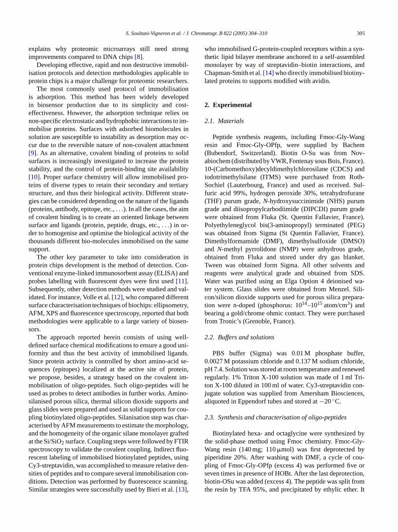
Fig. 1. Steps of immobilisation of peptides.
3.1. Functionalisation of solid supports
Steps allowing the immobilisation of peptides are de-scribed inFig. 1. (1) Silanisation of the support with CDCS;(2) capping of non reacted Si-OH, and hydrolysis of methylesters with iodotrimethylsilane; (3) activation of COOHgroups by formation of reactive NHS esters; (4) grafting ofan aminated polyethyleneglycol linker; (5) grafting of oligo-peptides after the formation of NHS ester at its C-terminalend. Currently microarrays are made on conventional glassslide supports. However, the glass slides are unsuitable forsurface characterisations. In order to validate each steps ofthe support functionalisation, various silica supports wereused.
Porous silica is a large specific surface area support[15]suitable for FTIR surface analysis spectroscopy. Thermal sili-con dioxide are well-defined supports characterised by a lowroot mean square roughness (RMS) which allows accurateAtomic Force Microscopy analysis. The surface of porous sil-ica and silicon dioxide substrates bear surface silanol groups.The organosilanes grafting on “silica” surface involves thereaction with these surface hydroxyl groups. The cross-analyses obtained by FTIR spectroscopy on porous silica andAFM measurements on silicon dioxide support are assumedto be well-correlated to the glass slide functionalisation.
Functionalisation of silica surface is currently done bys ievedi orms ilani-s ventp re-a
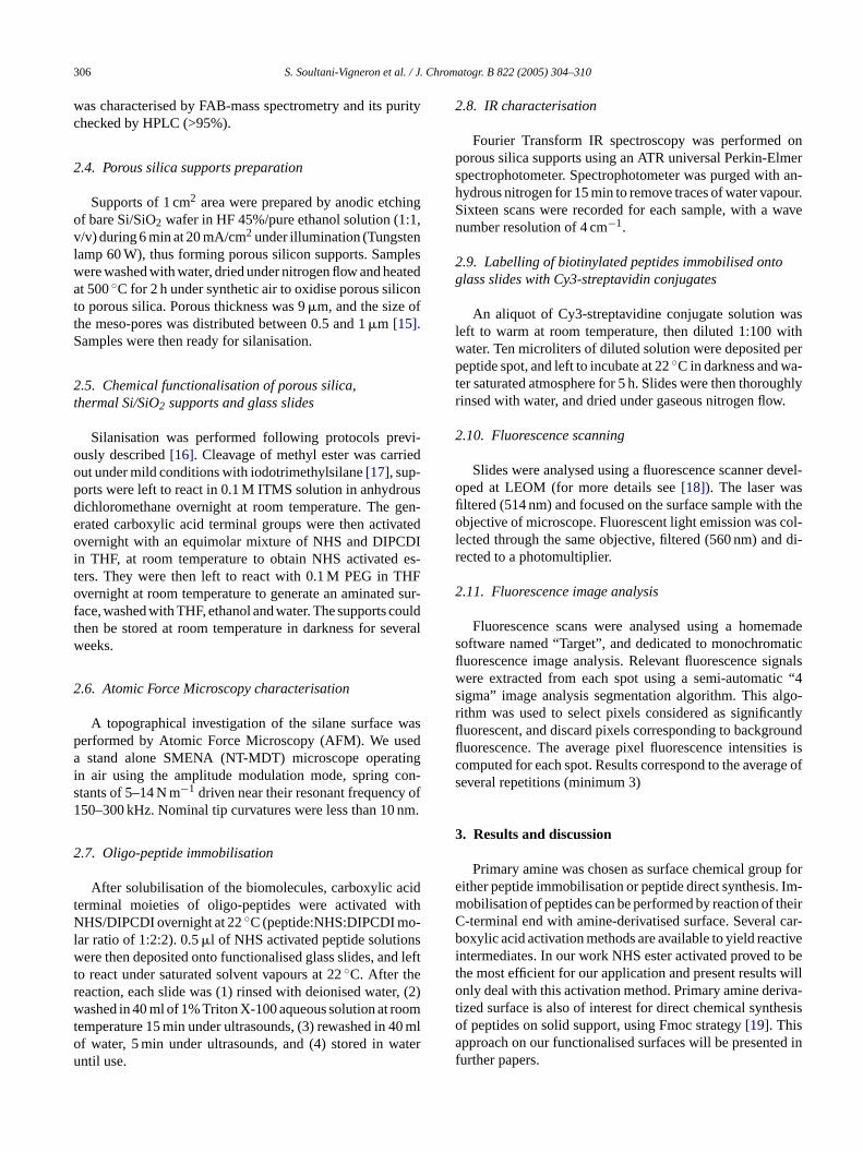
Fig. 2. IR characterisation of grafting of PEG linker onto silanised porous sil-ica. (1) Thin line: starting surface = carboxylic acid function; (2) dashed line:NHS ester activated; (3) bold line: PEG linker coupled to COOH throughamide bond.
Fig. 2shows the evolution of infra-red characteristics spec-tra of the solid supports along functionalisation steps. Aftersilanisation and ester-deprotection with ITMS, the surfaceexhibits terminal carboxylic acid groups characterised by aband at 1712 cm−1. Activation with NHS (spectrum 2) shiftsthe band towards 1740 cm−1 together with the appearanceof two bands at 1786 and 1816 cm−1 corresponding to NHSester[20]. Grafting of PEG linker via amide bond is shownby the decrease of NHS ester band and the appearance ofcharacteristic amide bands (1545 and 1643 cm−1).
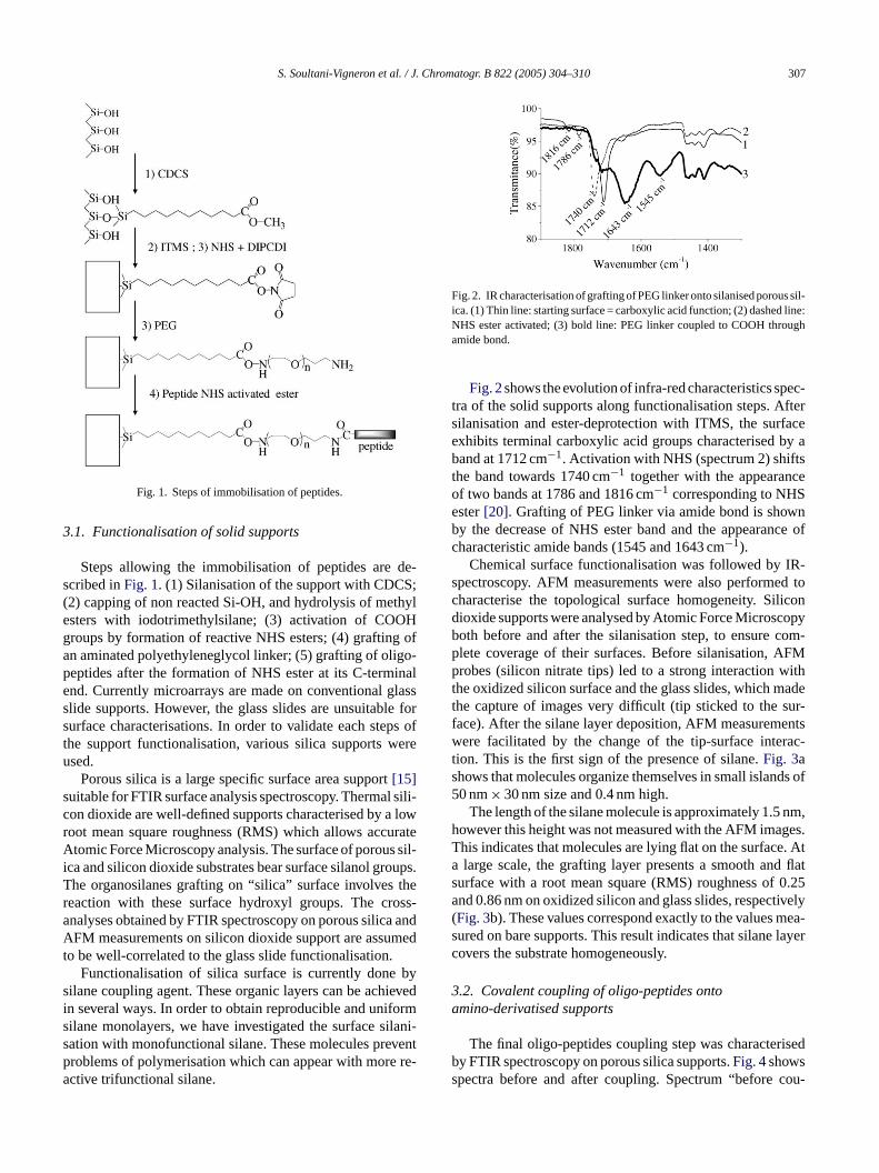
Chemical surface functionalisation was followed by IR-spectroscopy. AFM measurements were also performed tocharacterise the topological surface homogeneity. Silicondioxide supports were analysed by Atomic Force Microscopyboth before and after the silanisation step, to ensure com-plete coverage of their surfaces. Before silanisation, AFMprobes (silicon nitrate tips) led to a strong interaction withthe oxidized silicon surface and the glass slides, which madethe capture of images very difficult (tip sticked to the sur-face). After the silane layer deposition, AFM measurementswere facilitated by the change of the tip-surface interac-tion. This is the first sign of the presence of silane.Fig. 3ashows that molecules organize themselves in small islands of50 nm× 30 nm size and 0.4 nm high.
The length of the silane molecule is approximately 1.5 nm,however this height was not measured with the AFM images.T e. Ata d flats 0.25a tively( mea-s layerc
3a
isedbs cou-
ilane coupling agent. These organic layers can be achn several ways. In order to obtain reproducible and unifilane monolayers, we have investigated the surface sation with monofunctional silane. These molecules preroblems of polymerisation which can appear with morective trifunctional silane.
his indicates that molecules are lying flat on the surfaclarge scale, the grafting layer presents a smooth an
urface with a root mean square (RMS) roughness ofnd 0.86 nm on oxidized silicon and glass slides, respecFig. 3b). These values correspond exactly to the valuesured on bare supports. This result indicates that silaneovers the substrate homogeneously.
.2. Covalent coupling of oligo-peptides ontomino-derivatised supports
The final oligo-peptides coupling step was charactery FTIR spectroscopy on porous silica supports.Fig. 4showspectra before and after coupling. Spectrum “before
308 S. Soultani-Vigneron et al. / J. Chromatogr. B 822 (2005) 304–310
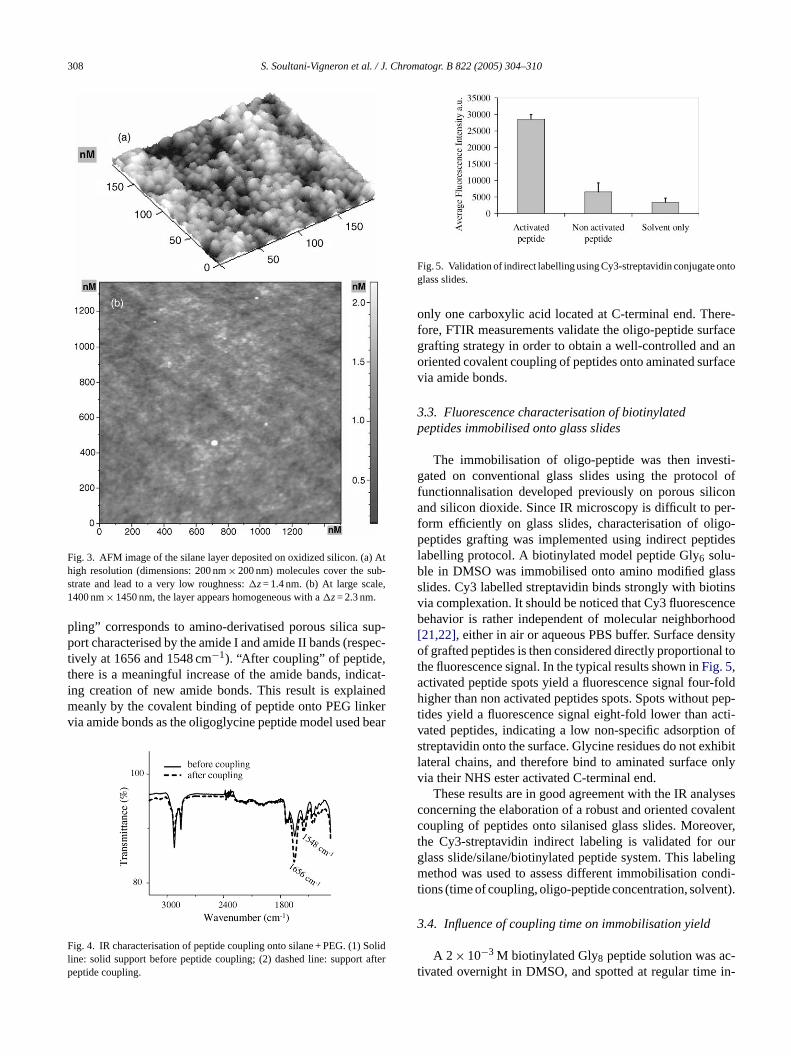
Fig. 3. AFM image of the silane layer deposited on oxidized silicon. (a) Athigh resolution (dimensions: 200 nm× 200 nm) molecules cover the sub-strate and lead to a very low roughness:�z= 1.4 nm. (b) At large scale,1400 nm× 1450 nm, the layer appears homogeneous with a�z= 2.3 nm.
pling” corresponds to amino-derivatised porous silica sup-port characterised by the amide I and amide II bands (respec-tively at 1656 and 1548 cm−1). “After coupling” of peptide,there is a meaningful increase of the amide bands, indicat-ing creation of new amide bonds. This result is explainedmeanly by the covalent binding of peptide onto PEG linkervia amide bonds as the oligoglycine peptide model used bear
Fig. 4. IR characterisation of peptide coupling onto silane + PEG. (1) Solidline: solid support before peptide coupling; (2) dashed line: support afterpeptide coupling.
Fig. 5. Validation of indirect labelling using Cy3-streptavidin conjugate ontoglass slides.
only one carboxylic acid located at C-terminal end. There-fore, FTIR measurements validate the oligo-peptide surfacegrafting strategy in order to obtain a well-controlled and anoriented covalent coupling of peptides onto aminated surfacevia amide bonds.
3.3. Fluorescence characterisation of biotinylatedpeptides immobilised onto glass slides
The immobilisation of oligo-peptide was then investi-gated on conventional glass slides using the protocol offunctionnalisation developed previously on porous siliconand silicon dioxide. Since IR microscopy is difficult to per-form efficiently on glass slides, characterisation of oligo-peptides grafting was implemented using indirect peptideslabelling protocol. A biotinylated model peptide Gly6 solu-ble in DMSO was immobilised onto amino modified glassslides. Cy3 labelled streptavidin binds strongly with biotinsvia complexation. It should be noticed that Cy3 fluorescencebehavior is rather independent of molecular neighborhood[21,22], either in air or aqueous PBS buffer. Surface densityof grafted peptides is then considered directly proportional tothe fluorescence signal. In the typical results shown inFig. 5,activated peptide spots yield a fluorescence signal four-foldhigher than non activated peptides spots. Spots without pep-t acti-v n ofs hibitl onlyv
lysesc alentc over,t ourg elingm ndi-t nt).
3
c-t in-
ides yield a fluorescence signal eight-fold lower thanated peptides, indicating a low non-specific adsorptiotreptavidin onto the surface. Glycine residues do not exateral chains, and therefore bind to aminated surfaceia their NHS ester activated C-terminal end.
These results are in good agreement with the IR anaoncerning the elaboration of a robust and oriented covoupling of peptides onto silanised glass slides. Morehe Cy3-streptavidin indirect labeling is validated forlass slide/silane/biotinylated peptide system. This labethod was used to assess different immobilisation co
ions (time of coupling, oligo-peptide concentration, solve
.4. Influence of coupling time on immobilisation yield
A 2 × 10⁻³ M biotinylated Gly8 peptide solution was aivated overnight in DMSO, and spotted at regular time
S. Soultani-Vigneron et al. / J. Chromatogr. B 822 (2005) 304–310 309
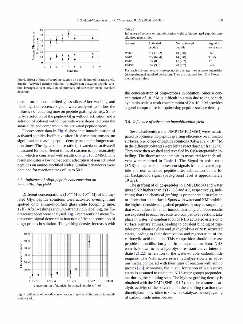
Fig. 6. Effect of time of coupling reaction on peptide immobilisation yield.Square: Activated peptide solution, (triangle) non activated peptide solu-tion, lozenge: solvent only. Lateral error bars indicate experimental standarddeviation.
tervals on amino modified glass slide. After washing andlabelling, fluorescence signals were analysed to follow theinfluence of coupling time on peptide grafting density. Simi-larly, a solution of the peptide Gly8 without activation and asolution of solvent without peptide were deposited onto thesame slide and compared to the activated peptide spots.
Fluorescence data inFig. 6 show that immobilisation ofactivated peptides is effective after 1 h of reaction time and nosignificant increase in peptide density occurs for longer reac-tion times. The signal to noise ratio (activated/non activated)measured for the different times of reaction is approximatelyof 5, which is consistent with results ofFig. 5for DMSO. Thisresult indicates a low non-specific adsorption of non activatedpeptides on amino-modified slides. Similar behaviours wereobtained for reaction times of up to 50 h.
3.5. Influence of oligo-peptide concentration onimmobilisation yield
Different concentrations (10−4 M to 10−2 M) of biotiny-lated Gly6 peptide solutions were activated overnight andspotted onto amino-modified glass slide (coupling time:12 h). After washings and Cy3-streptavidin labelling, the flu-orescence spots were analysed.Fig. 7represents the mean flu-orescence signal detected in function of the concentration ofoligo-probes in solution. The grafting density increases with
F obil-i
Table 1Influence of solvent on immobilisation yield of biotinylated peptides ontosilanised glass slides
Solvent Activatedpeptide
Non activatedpeptide
Signal tonoise ratio
Water 214 (13.2) 40 (9.0) 6.8NMP 377 (67.4) 14 (3.8) 91.75DMF 27 (8.9) 11 (2.2) 17DMSO 52 (9.5) 20 (7.7) 4.2
For each solvent, results correspond to average fluorescence intensities(± experimental standard deviation). They are obtained from 3 to 6 experi-mental data points.
the concentration of oligo-probes in solution. Since a con-centration of 10−2 M is difficult to attain due to the peptidesynthesis scale, a work concentration of 2× 10⁻³ M providesa good compromise for optimising peptide surface density.
3.6. Influence of solvent on immobilisation yield
Several solvents (water, NMP, DMF, DMSO) were investi-gated to optimise the peptide grafting efficiency on aminatedsurfaces. 5�l drops of peptide solutions (Gly8 at 2× 10⁻³ Min the different solvents) were left to react during 3 h at 22◦C.They were then washed and revealed by Cy3-streptavidin la-belling. The fluorescence intensities measured for each sol-vent were reported inTable 1. The Signal to noise ratio(SNR) compares the fluorescent signals from activated pep-tide and non activated peptide after subtraction of the lo-cal background signal (background level is approximately10± 2).
The grafting of oligo-peptides in DMF, DMSO and watergives SNR higher than 3 (17, 6.8 and 4.2, respectively), indi-cating that the chemical grafting is preponderant in relationto adsorption at interfaces. Spots with water and NMP exhibitthe highest densities of grafted peptides. It may be surprisingthat water allows for a fair immobilisation yield. Poor resultsare expected to occur because two competitive reactions takep ontos pep-t atede f thec asep HSe rme-d der que-o inog vee nder-a ity iso cat-a i.e.d tingo
ig. 7. Influence of peptide concentration in spotted solutions on immsation yield.
lace in water: (i) condensation of NHS activated estersurface primary amines, leading to covalent binding ofides onto silanised glass and (ii) hydrolysis of NHS activsters, leading to their deactivation and regeneration oarboxylic acid moieties. This competition should decreeptide immobilisation yield in an aqueous medium. Nster is known to be a hydrolysis-resistant active inteiate [21,22] in relation to the water-soluble carbodiimieagents. The NHS active esters hydrolyse slowly in aus media compared with their rates of reaction with amroups[23]. Moreover, the in situ formation of NHS actisters is assumed to retain the NHS-ester groups prepont during the coupling step. The highest grafting densbtained with the NMP (SNR = 91.7), it can be assume alytic activity of the solvent upon the coupling reaction (imethylaminopyridine is known to catalyze the conjugaf carbodiimide intermediate).
310 S. Soultani-Vigneron et al. / J. Chromatogr. B 822 (2005) 304–310
4. Conclusion
A three-step method for immobilising oligo-peptides ontoglass and Si/SiO2 was presented here. A systematic investiga-tion of the bio-functionalisation was realised on silicon diox-ide and porous silica support in order to define and controleach step of the oligo-peptides grafting. Topological AFMmeasurements have showed the formation of a dense andhomogeneous silane monolayer at the silica surface. FTIRanalysis proved attachments to be covalent at each step of theprocess. The oligo-peptides grafting step allow to attach pep-tide in a covalent and oriented manner. Indirect labelling ofbiotinylated peptides with Cy3-streptavidin conjugate provedto be a simple and efficient method to measure relative den-sities of peptides on glass slides, and to compare differentconditions of immobilisation.
Further papers will present biological recognition of anti-bodies using model peptide microarrays, and implementationof peptide microarrays by direct chemical synthesis.
Acknowledgments
This work was supported by Centre National de laRecherche Scientifique (programme «Proteomique et Geniedes Proteines») and Region Rhone-Alpes (thematique pri-o alf iaS
R
t.
[3] Y.S. Lee, M. Mrksich, Trends Biotechnol. 20 (2002) S14.[4] W.H. Robinson, L. Steinman, P.J. Utz, Arthritis Rheum. 46 (2002)
885.[5] H. Zhu, J.F. Klemic, S. Chang, P. Bertone, A. Casamayor, K.G.
Klemic, D. Smith, M. Gerstein, M.A. Reed, M. Snyder, Science 293(2001) 2101.
[6] G. MacBeath, S.L. Schreiber, Science 289 (2000) 1760.[7] H. Zhu, M. Snyder, Curr. Opin. Chem. Biol. 7 (2003) 55.[8] R. Aebersold, B.F. Cravatt, Trends Biotechnol. 20 (2002) 1.[9] B.T. Houseman, M. Mrksich, Trends Biotechnol. 20 (2002)
279.[10] M.F. Templin, D. Stoll, M. Schrenk, P.C. Traub, C.F. Vohringer, T.O.
Joos, Trends Biotechnol. 20 (2002) 160.[11] T. Kukar, S. Eckenrode, Y. Gu, W. Lian, M. Meggison, J.X. She, D.
Wu, Anal. Biochem. 306 (2002) 50.[12] J.N. Volle, G. Chambon, A. Sayah, C. Reymond, N. Fasel, M.A.M.
Gijs, Biosens. Bioelectron. 19 (2004) 457.[13] C. Bieri, O.P. Ernst, S. Heyse, K.P. Hofmann, H. Vogel, Nat. Biotech-
nol. 17 (1999) 1105.[14] A. Chapman-Smith, J.E. Cronan Jr., Trends Biochem. Sci. 24 (1999)
359.[15] F. Bessueille, V. Dugas, J.P. Cloarec, V. Vikulov, E. Souteyrand, J.R.
Martin, Biosens. Bioelectron. (2005), in press,doi:10.1016/j.bios.2005.02.007.
[16] V. Dugas, G. Depret, Y. Chevalier, X. Nesme, E. Souteyrand, Sens.Actuators 101 (2004) 112.
[17] T.L. Ho, G.A. Olah, Angew. Chem. Int. Ed. Engl. 15 (1976)774.
[18] M. Bras, V. Dugas, F. Bessueille, J.P. Cloarec, J.R. Martin, M. Cabr-era, J.P. Chauvet, E. Souteyrand, M. Garrigues, Biosens. Bioelectron.20 (2004) 796.
[ ao,
[ all,
[ , J.P.
[ 86)
[
ritaire «Sciences Analytiques Appliquees»). Post-doctorellowship from French Ministere de la Recherche for Samoultani-Vigneron is also gratefully acknowledged.
eferences
[1] P. Mitchell, Nat. Biotechnol. 20 (2002) 225.[2] R.M.T. De Wildt, C.R. Mundy, B.D. Gorick, I.M. Tomlinson, Na
Biotechnol. 18 (2000) 989.
19] J.P. Pellois, X. Zhou, O. Srivannavit, T. Zhou, E. Gulari, X. GNat. Biotechnol. 20 (2002) 922.
20] N.M. Grubor, R. Shinar, R. Jankowiak, M.D. Porter, G.J. SmBiosens. Bioelectron. 19 (2004) 547.
21] M. Bras, J.P. Cloarec, F. Bessueille, E. Souteyrand, J.R. MartinChauvet, J Fluoresc. 20 (2000) 247.
22] J.V. Staros, R.W. Wright, D.M. Swingle, Anal. Biochem. 156 (19220.
23] D. Sehgal, I.K. Vijay, Anal. Biochem. 218 (1994) 87.


