Embed Size (px)
Citation preview

A New Approach to Drug Delivery: Non-Peptidic, High Load Macrocyclic Alternatives to
Cell Penetrating Peptides
Zoe Karthauser
School of Pharmacy University of East Anglia
A thesis submitted for the degree of Doctor of Philosophy
June 2013
This copy of the thesis has been supplied on condition that anyone
who consults it is understood to recognise that its copyright rests with the author and that use of any information derived there from must be
in accordance with current UK Copyright Law. In addition, any quotation or extract must include full attribution.
Declaration
This thesis is submitted to the University of East Anglia for the Degree of Doctor
of Philosophy and has not been previously submitted at this or any other university for
assessment or for any other degree. Except where stated, and reference or
acknowledgement is given, this work is original and has been carried out by the author
alone.
Zoe Karthauser
Abstract
Calixarenes are versatile macrocycles formed from the condensation of para-tert-
butyl-phenol and formaldehyde. Chapter 1 describes the synthesis of these molecules
and how conformational control and selective functionalisation can give an array of
molecules with customised properties; this allows for various applications including
those of biological relevance. The copper catalysed alkyne-azide cycloaddition
(CuAAC) reaction is also introduced as a tool for functionalising calixarenes.
The phenomenon of cell penetration is of interest where a molecule has an
intracellular target, for example gene therapy, delivery of cytotoxic agents or cellular
imaging. Chapter 2 introduces the mechanisms of cell uptake and the design and
applications of cell penetrating peptides. Calixarenes are presented as alternatives to
cell penetrating peptides and the work published to date on intracellular delivery of
calixarenes is summarised. A synthetic route for calixarenes with variable fluorescent
dyes and different functionalities on the upper rim via a common intermediate is
presented. Synthesis of an analogue featuring guanidinium groups on the upper rim
was achieved using carboxybenzyl (Cbz) protecting groups as a less labile alternative
to butoxycarbonyl (Boc) groups. The syntheses of analogues with varied linkers for
attachment of the dye are also presented. Biological evaluation revealed that the
dynamics of cellular uptake and the intracellular localisation were sensitive to the
upper-rim functionalisation and the dye molecule. The linker attaching the dye had
less impact.
Chapter 3 describes the suitability of calixarenes as scaffolds to form
glycoconjugates. These can be used to target Pseudomonas aeruginosa; research
towards development of novel treatments of infections from this pathogen is
summarised. A route that has been developed towards bifunctional calixarenes
featuring a fluorescent tag and points of attachment for sugars via CuAAC reactions is
presented. The use of alkyne protecting groups to maintain the integrity of the scaffold
during transformations was found to be particularly important.
Table of Contents
Chapter 1: Introduction ......................................................................... 19
1.1 Calixarenes ...................................................................................................... 20
1.1.1 Synthesis ................................................................................................. 21
1.1.2 Conformation ......................................................................................... 21
1.1.3 Functionalisation .................................................................................... 22
1.1.3.1 Lower-rim functionalisation ........................................................... 23
1.1.3.2 Upper-rim functionalisation ............................................................ 23
1.1.4 Applications in biological systems ......................................................... 24
1.1.4.1 Artificial Receptors ......................................................................... 25
1.1.4.2 Artificial Transporters ..................................................................... 27
1.1.4.3 Artificial Enzymes .......................................................................... 29
1.1.4.4 Anticancer applications ................................................................... 30
1.1.4.5 Antimicrobial activity ..................................................................... 31
1.1.4.6 Other applications ........................................................................... 34
1.2 Click chemistry ............................................................................................... 34
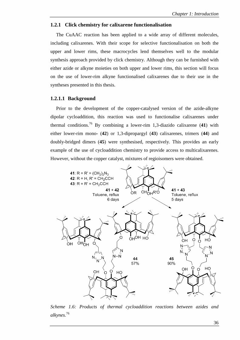
1.2.1 Click chemistry for calixarene functionalisation ................................... 36
1.2.1.1 Background ..................................................................................... 36
1.2.1.2 Preparation of Lower-Rim- Alkynes............................................... 38
1.2.1.3 Applications .................................................................................... 40
1.3 Overview of thesis ........................................................................................... 44
1.4 References ....................................................................................................... 44
Chapter 2: Calixarene-Based Cell-Penetration Agents ...................... 50
2.1 Introduction .................................................................................................... 51
2.1.1 Mechanisms of cell uptake ..................................................................... 51
2.1.1.1 Clathrin mediated endocytosis ........................................................ 51
2.1.1.2 Caveolae .......................................................................................... 52
2.1.1.3 Clathrin- and caveolin-independent pathways ................................ 53
2.1.2 Cell penetration agents ........................................................................... 54
2.1.2.1 Cell penetrating peptides ................................................................. 54
2.1.2.1.1 Cellular uptake mechanisms ...................................................... 54
2.1.2.1.2 Design of cell penetrating peptides ........................................... 56
2.1.2.1.3 Applications ............................................................................... 57

2.1.2.2 Non-peptidic cell penetration agents............................................... 58
2.1.2.2.1 Calixarenes as cell penetration agents ....................................... 60
2.2 Aims ................................................................................................................. 67
2.3 Results and Discussion ................................................................................... 69
2.3.1 Synthesis of water soluble click conjugates ........................................... 69
2.3.1.1 Synthesis of common intermediate ................................................. 69
2.3.1.2 Synthesis of dye molecules ............................................................. 74
2.3.1.3 Coumarin-appended neutral calixarene .......................................... 76
2.3.1.4 Coumarin-appended tetra-amino calixarene ................................... 78
2.3.1.5 Pyrene-appended tetra-amino calixarene ........................................ 80
2.3.1.6 Anthracene-appended tetra-amino calixarene ................................. 83
2.3.1.7 Coumarin appended tetra-glycine calixarene .................................. 84
2.3.1.8 Removal of Boc protecting groups ................................................. 87
2.3.2 Synthesis of the guanidinium click conjugate ........................................ 87
2.3.2.1 Synthesis of common intermediate – TBDMS route ...................... 88
2.3.2.2 Guanidinylation – Boc protection route .......................................... 90
2.3.2.3 Attempted CuAAC reaction with deprotected guanidinium
derivative….. ................................................................................... 94
2.3.2.4 Guanidinylation – Cbz protection route .......................................... 97
2.3.2.5 Coumarin appended tetra-guanidine ............................................... 98
2.3.2.6 Deprotection of Cbz protected guanidine derivative ...................... 99
2.3.3 Synthesis of dye-conjugates with variable linkers ............................... 103
2.3.3.1 Amine-linked coumarin via alkyl halide ....................................... 104
2.3.3.2 Amine-linked coumarin via amide ................................................ 109
2.3.3.2.1 Synthesis of NBD-conjugate with 4-carbon linker .................. 110
2.3.3.2.2 Synthesis of amide-linked coumarin ....................................... 115
2.3.3.2.3 Reduction of amide-linked coumarin ...................................... 118
2.3.3.2.4 Deprotection of amide-linked coumarin conjugate ................. 119
2.3.4 Biological analysis ............................................................................... 120
2.3.4.1 Toxicity assays .............................................................................. 120
2.3.4.2 Cellular uptake .............................................................................. 124
2.3.4.3 Mechanism of uptake .................................................................... 129
2.3.4.4 Intracellular localisation ................................................................ 134
2.4 Conclusions and further work .................................................................... 137
2.5 Experimental ................................................................................................. 139
2.5.1 General procedures - chemistry ............................................................ 139
2.5.2 Synthesis ............................................................................................... 140
2.5.3 General procedures - biology ............................................................... 173
2.5.4 Toxicity assays ..................................................................................... 173
2.5.5 Cellular uptake ..................................................................................... 173
2.5.6 Inhibition studies .................................................................................. 173
2.5.7 Co-localisation studies ......................................................................... 174
2.6 References ..................................................................................................... 174
Chapter 3: Calixarene-Based Glycoconjugates ....................... 182
3.1 Introduction .................................................................................................. 183
3.1.1 Calixarene-based glycoconjugates ....................................................... 183
3.1.2 Glycoconjugates against Pseudomonas aeruginosa ............................ 186
3.2 Aims ............................................................................................................... 191
3.3 Results and Discussion ................................................................................. 192
3.3.1 Design of the fluorescent glycoconjugate ............................................ 192
3.3.2 Route 1: Regioselective mono-nitration ............................................... 193
3.3.3 Route 2: Non-selective mono-nitration ................................................ 196
3.3.3.1 Dye synthesis ................................................................................ 198
3.3.3.2 Sugar synthesis .............................................................................. 198
3.3.3.3 Conjugation of the dye .................................................................. 199
3.3.3.4 Sugar conjugation ......................................................................... 201
3.3.3.5 Deprotection .................................................................................. 203
3.3.4 Route 3: Non-selective mono-nitration on tert-butyl calixarene.......... 204
3.3.5 Route 4: Synthesis of scaffold using TBDMS-protected alkynes ........ 208
3.4 Conclusions and further work .................................................................... 216
3.5 Experimental ................................................................................................. 217
3.6 References ..................................................................................................... 232
List of Figures
Figure 1.1: Conformations of calix[4]arene: cone (2), partial cone (3), 1,3-alternate (4)
and 1,2-alternate (5). .................................................................................................... 22
Figure 1.2: Designation of calixarene rims. ................................................................. 22
Figure 1.3: Calixarene based lectin mimetic (21),38 calixarene with
pseudocylcopentapeptide upper rim featuring AspGlyAspGly sequence (22)39 and
amphiphilic calixarenes with anionic (23) and cationic (24 and 25) upper rims.42...... 26
Figure 1.4: a) Tetraguanidinomethylcalixarene ligand (26); b) Interaction of 26 with
p53 tetramer (© 2008 by the National Academy of Sciences of the United States of
America).45 ................................................................................................................... 27
Figure 1.5: Artificial transporters based on ester (27),46 cholic acid (28),47 amide
(29)48 and spermidine (30)49 functionalised calixarenes. ............................................. 28
Figure 1.6: 1,2-difunctionalised artificial metallonucelease (31)51 and 1,3-diguanidine
functionalised calixarene catalyst (32) in the catalytically active form.53 ................... 30
Figure 1.7: CD69-binding carboxylated thiacalixarene (33)54 and multivalent folic
acid conjugate (34).55 ................................................................................................... 31
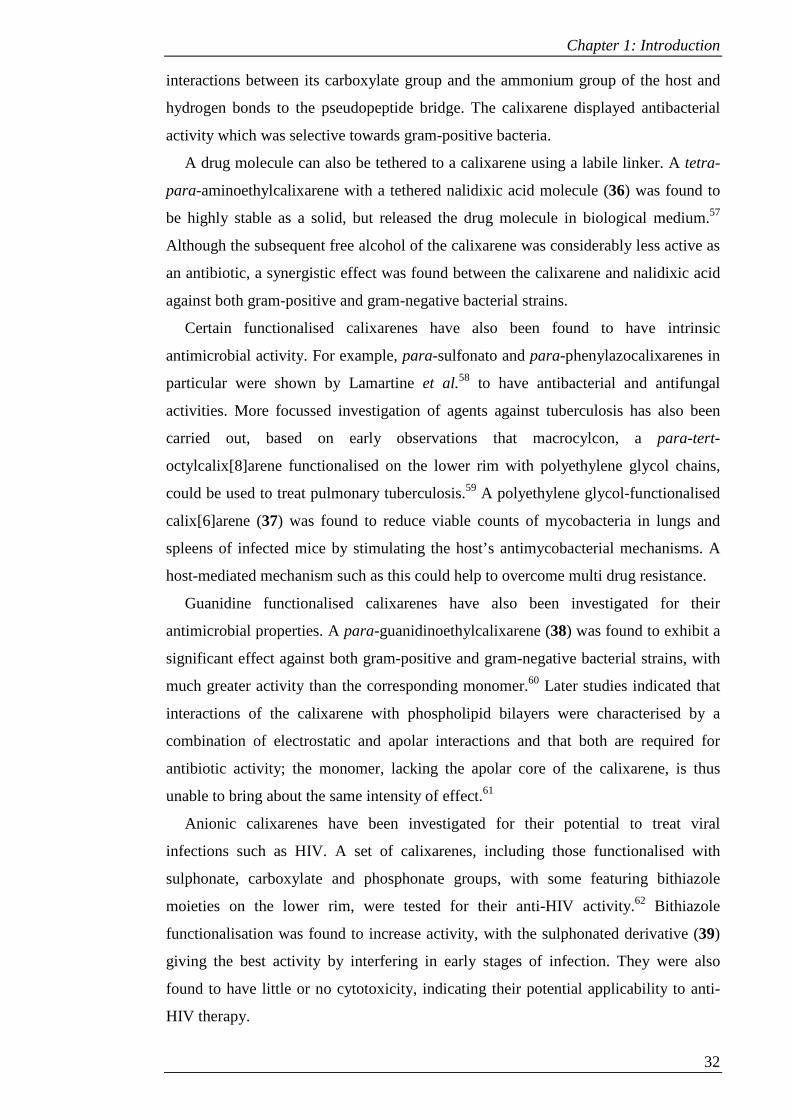
Figure 1.8: Antimicrobial calixarenes: general structure of vancomycin mimic (35)
where AA = amino acid,56 nalidixic acid prodrug (36),57 macrocyclon analogue (37),63
guanidinium antibacterial agent (38),60 anti-HIV agent (39),62 and dual function anti-
HIV and –HCV agent (40).64 ........................................................................................ 33
Figure 1.9: Lower-rim triazole linked ethylamine (50), calixarene-based cavitand (52),
calix-tube (51) and calixarene-centred copolymer (x = 25, y = 22) (53). .................... 41
Figure 1.10: Examples of ion sensors synthesised by Chung and coworkers (54)86 and
Pathak et al. (55).91 ....................................................................................................... 42
Figure 1.11: Example of glycoconjugate (56) functionalised with lactose moieties.101
...................................................................................................................................... 43
Figure 2.1: Mechanisms of endocytosis. Reprinted by permission from Macmillan
Publishers Ltd: Nature Reviews Molecular Cell Biology4 © 2007 .............................. 52
Figure 2.2: Scheme of key steps in clathrin mediated endocytosis. Reprinted by
permission from Macmillan Publishers Ltd: Nature7 © 2007 ...................................... 53
Figure 2.3: Oligoguanidinium compounds based on peptides (57), peptoids (58) and
carbamates (59).49,50 ..................................................................................................... 60
Figure 2.4: Cell penetration agents based on a dendrimer (60)51 and an inositol dimer
(61).52 FITC = fluorescein isothiocyanate. ................................................................... 60
Figure 2.5: Cell permeable cation binders based on pyridyl (62, 63), thiophenyl (64)
and dimethylamino (65) conjugates.56–58 ..................................................................... 61
Figure 2.6: Cell permeable thiacalixarene based Fe3+ sensor (66).59 ........................... 62
Figure 2.7: Glycine functionalised multicalixarene (67).61 .......................................... 63
Figure 2.8: Upper rim (68)60 and lower rim (69)54 guanidinium functionalised
calixarenes. ................................................................................................................... 65
Figure 2.9: Upper rim (70) and lower rim (71) arginine functionalised calixarenes.62 65
Figure 2.10: Amphiphilic dye-appended calixarene (72),63 amphiphilic micelle-
forming calixarene (73),64 vanadyl sulfonylcalixarene (74),65
nitrobenzoxadiazole(NBD)-appended cationic calixarene (75).55,66 ............................ 66
Figure 2.11: Intermediate in the synthesis of tetra-guanidinium calix[4]arene with
NBD labelled lower rim. .............................................................................................. 69
Figure 2.12: a) Two dimensional model of compound 78, showing rotational
symmetry plane, b) Positions of axial and equatorial methylene bridge hydrogens, c) 1H-NMR spectrum of 78 (CDCl3). ............................................................................... 72
Figure 2.13: 1H-NMR spectra of a) tBu (78), b) nitro (79) and c) amino (80)
functionalised calixarenes in CDCl3. Peaks corresponding to aromatic protons are
marked with an asterisk. ............................................................................................... 74
Figure 2.14: a 1H-NMR spectrum and b) 2D-HMBC of 87 (CDCl3). ........................ 78
Figure 2.15: 1H-NMR spectrum of 88 (CDCl3). .......................................................... 80
Figure 2.16: 1H-NMR spectrum of 89 (CDCl3). .......................................................... 81
Figure 2.17: 1H-NMR spectrum of 90 (CDCl3). .......................................................... 82
Figure 2.18: a) 2D-HSQC and b) 2D-HMBC of 90 (CDCl3). ...................................... 83
Figure 2.19: 1H-NMR spectrum of 91 (CDCl3). .......................................................... 85
Figure 2.20: 1H-NMR spectrum of 92 (CD3OD). ........................................................ 86
Figure 2.21: 1H-NMR spectrum of 93 (CD3OD). ........................................................ 87
Figure 2.22: 1H-NMR spectrum of a) 78 and b) 98 (CDCl3) showing loss of terminal
alkyne triplet (circled) and conversion of methylene doublet to singlet. ..................... 89
Figure 2.23: 1H-NMR spectrum of crude 102 (CDCl3). .............................................. 93
Figure 2.24: 1H-NMR-spectrum of a) 104 and b) crude 106 (CD3OD) showing loss of
terminal alkyne triplet (circled) and conversion of methylene doublet to a singlet. .... 96
Figure 2.25: 1H-NMR spectrum of 107 (CDCl3). ........................................................ 99
Figure 2.26: 1H-NMR spectrum of 108 (CDCl3). ...................................................... 100
Figure 2.27: 1H-NMR spectrum of 109 (CD3OD). .................................................... 104
Figure 2.28: Analogues with NBD and coumarin dyes linked by a) triazole rings and
b) secondary amines. .................................................................................................. 104
Figure 2.29: 1H-NMR spectrum of 110. ..................................................................... 106
Figure 2.30: 1H-NMR spectrum of 120. ..................................................................... 112
Figure 2.31: 2D-HSQC spectrum of 120. .................................................................. 113
Figure 2.32: 1H-NMR spectrum of 125. ..................................................................... 115
Figure 2.33: 1H-NMR spectrum of 132. ..................................................................... 118
Figure 2.34: Summary of compounds subjected to in vivo testing. ........................... 122
Figure 2.35: MTS assays performed on THP-1 cells. a) Compound 87; b) Compounds
94, 95 and 97; c) Compounds 96, 126 and 134; d) Compound 109. .......................... 123
Figure 2.36: Cellular uptake of 95 after 48 hours of incubation with CHO cells. ..... 125
Figure 2.37: Cellular uptake of 94 at given time intervals after addition of compound
to CHO cells. .............................................................................................................. 127
Figure 2.38: Cellular uptake of 97 at given time intervals after addition of compound
to CHO cells. .............................................................................................................. 127
Figure 2.39: Cellular uptake of 109 at given time intervals after addition of compound
to CHO cells. .............................................................................................................. 128
Figure 2.40: Cellular uptake of 126 at given time intervals after addition of compound
to CHO cells. .............................................................................................................. 128
Figure 2.41: Cellular uptake of 134 at given time intervals after addition of compound
to CHO cells. .............................................................................................................. 129
Figure 2.42: Uptake of compound 94 after incubation with specified inhibitors. ..... 131
Figure 2.43: Uptake of compound 97 after incubation with specified inhibitors. ..... 131
Figure 2.44: Uptake of compound 109 after incubation with specified inhibitors..... 132
Figure 2.45: Uptake of compound 126 after incubation with specified inhibitors..... 133
Figure 2.46: Uptake of compound 134 after incubation with specified inhibitors..... 134
Figure 2.47: Co-localisation of 94 with LysoTracker Red. White arrows indicate a
pink spot of overlaid fluorescence (a) and an area of blue fluorescence with poor
overlay (b). ................................................................................................................. 135
Figure 2.48: a) Co-localisation of 97 with LysoTracker Red; white arrow indicates a
pink spot of overlaid fluorescence. b) Cells showing limited overlay; white arrow
indicates spot of blue fluorescence with no overlay. ................................................. 136
Figure 2.49: Limited overlay of 109 with LysoTracker Red; white arrow indicates spot
of blue fluorescence with no overlay. ........................................................................ 137
Figure 2.50: Co-localisation of 126 with LysoTracker Red; white arrow indicates
yellow spot of overlaid fluorescence. ......................................................................... 137
Figure 2.51: Co-localisation of 134 with LysoTracker Red; white arrow indicates pink
spot of overlaid fluorescence. ..................................................................................... 138
Figure 3.1: Calixarene-based glycoconjugates for receptor binding (135)7, an artificial
antibody (136)8, antiviral activity (137)9 and toxin binding (138).10 ......................... 185
Figure 3.2: Crystal structures of a) PA-IL (PDB code: 1OKO) and b) PA-IIL (PDB
code: 1UZV) showing Ca2+ binding sites in yellow and ball and stick representations
of monosaccharides. ................................................................................................... 188
Figure 3.3: Calixarene-based glycoconjugates for targeting Pseudomonas aeruginosa
lectins: compounds 139,20 140,21 and 14122 and the variable linkers23 applied to 140.
.................................................................................................................................... 190
Figure 3.4: Nile Red derivative (NRD) 142. .............................................................. 193
Figure 3.5: a) Diagram of 144 showing diastereotopic hydrogens on the propargyl
groups adjacent to the nitrophenol group; b) 1H-NMR spectrum of 144 (CDCl3). ... 195
Figure 3.6: 1H-NMR spectrum of 151 (CDCl3). ........................................................ 201
Figure 3.7: 1H-NMR spectrum of 152 (CDCl3). ........................................................ 204
Figure 3.8: 1H-NMR spectrum of 156 (CDCl3). ........................................................ 208

Figure 3.9: 1H-NMR spectra of a) compound 154 and b) compound 159 showing loss
of terminal alkyne triplet (circled) and change of methylene doublet in a) to singlet in
b). ................................................................................................................................ 210
Figure 3.10: a) 1H-NMR spectrum and b) 2D-HMBC spectrum of 158. ................... 211
Figure 3.11: 1H-NMR spectrum of 163 (CDCl3) ....................................................... 214
Figure 3.12: 1H-NMR spectrum of 157 (CDCl3). ...................................................... 216
Figure 3.13: 1H-NMR spectrum of 164 (DMSO-d6). ................................................ 217
List of Schemes
Scheme 1.1: Condensation reactions of phenols with formaldehyde to give the cross-
linked polymer Bakelite (1) and the cyclic tetramer calix[4]arene (2). ....................... 20
Scheme 1.2: Routes for functionalisation of lower rim. .............................................. 23
Scheme 1.3: Routes to functionalisation of the upper rim. .......................................... 24
Scheme 1.4: Outcome of the 1,3-dipolar cycloaddition reaction between an azide and
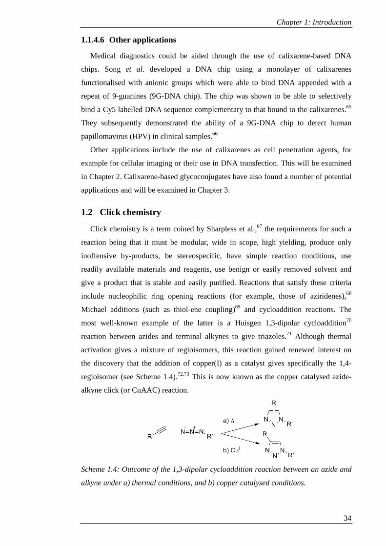
alkyne under a) thermal conditions, and b) copper catalysed conditions. .................... 34
Scheme 1.5: Proposed mechanism for the CuAAC reaction.75 .................................... 35
Scheme 1.6: Products of thermal cycloaddition reactions between azides and
alkynes.76 ...................................................................................................................... 36
Scheme 1.7: Lower-rim alkyne vs. lower-rim azide for CuAAC mediated synthesis of
water soluble calixarenes.77 .......................................................................................... 37
Scheme 1.8: Summary of main methods that have been used for accessing partially
and fully propargylated calixarenes, with the latter in different conformations. ......... 39
Scheme 2.1: Synthesis of tert-butyl-calix[4]arene (2).................................................. 71
Scheme 2.2: Selective tripropylation (77) and subsequent alkylation (78) of tert-butyl-
calix[4]arene (2). .......................................................................................................... 71
Scheme 2.3: Ipso-nitration of 78 and reduction to 80. ................................................. 73
Scheme 2.4: Synthesis of 3-acetamido7-acetoxy-coumarin (81) and 3-azido-7-hydroxy
coumarin (82). .............................................................................................................. 75
Scheme 2.5: Synthesis of 1-bromomethyl-pyrene (83) and 1-azidomethyl-pyrene (84)
...................................................................................................................................... 76
Scheme 2.6: Synthesis of 9-azidomethyl-10-bromo-anthracene (86). ......................... 77
Scheme 2.7: Click reaction of 78 with 82 to give conjugate 87. .................................. 77
Scheme 2.8: Protection of tetra amino calixarene (80). ............................................... 79
Scheme 2.9: Click reaction of 88 with 82. ................................................................... 80
Scheme 2.10: Click reaction of 88 with 84. ................................................................. 82
Scheme 2.11: Click reaction of 88 with 86. ................................................................. 84
Scheme 2.12: Coupling reaction of 80 with Boc glycine. ............................................ 86
Scheme 2.13: Click reaction of 92 with 82. ................................................................. 87
Scheme 2.14: Deprotection reactions on 89, 90, 91 and 93 to give 94, 95, 96 and 97. 88
Scheme 2.15: Synthesis of 80 from 78 via TBDMS protected route. .......................... 90
Scheme 2.16: Synthesis of 101 from 80. ...................................................................... 91
Scheme 2.17: Two possible routes for the synthesis of 104, via 102 followed by 103
or 105. ........................................................................................................................... 94
Scheme 2.18: Click reaction of 104 with 82. ............................................................... 96
Scheme 2.19: Principle of purification by ion-exchange chromatography (X = 106). 97
Scheme 2.20: Synthesis of 107 from 80. ...................................................................... 98
Scheme 2.21: Click reaction of 107 with 82. ............................................................. 100
Scheme 2.22: Deprotection of 108. ............................................................................ 102
Scheme 2.23: Alkylation of 77 to give 110. ............................................................... 105
Scheme 2.24: Conversion of 110 to 113 via nitration, reduction and Boc protection.
.................................................................................................................................... 107
Scheme 2.25: Synthesis of 115 from 81 using a hydrolysis-free method. ................. 108
Scheme 2.26: Silylation of 115 using TBDMSCl. ..................................................... 108
Scheme 2.27: Conditions for the attempted synthesis of 116 or 118 by reaction of 113
with 115 or 117. ......................................................................................................... 109
Scheme 2.28: Attempted synthesis of 119 with ethyl 3-bromopropanoate. ............... 110
Scheme 2.29: Attempted reaction of 77 with ethyl acrylate catalysed by KF/alumina.
.................................................................................................................................... 111
Scheme 2.30: Synthesis of compound 120. ................................................................ 112
Scheme 2.31: Conversion of 120 to 123 via nitration, reduction and Boc protection.
.................................................................................................................................... 114
Scheme 2.32: Synthesis of 126 from 123 via phthalimide cleavage, SEAr reaction and
removal of Boc groups. .............................................................................................. 115
Scheme 2.33: Conversion of 77 to 130 via alkylation, nitration, reduction and Boc
protection. ................................................................................................................... 117
Scheme 2.34: Synthesis of 132 from 130 via ester hydrolysis and amide bond
formation. ................................................................................................................... 118
Scheme 2.35: Attempted synthesis of 133 by reduction of 132. ................................ 120
Scheme 2.36: Boc-deprotection of 132 to give 134. .................................................. 121
Scheme 3.1: Synthesis of 144 via selective trialkylation and mono-nitration. .......... 195
Scheme 3.2: Attempted synthesis of 145 via a Mitsunobu reaction on 144. ............. 197
Scheme 3.3: Synthesis of 148 from 2 via de-tert-butylation, tetraalkyation and time-
controlled nitration. .................................................................................................... 198
Scheme 3.4: Synthesis of NRD 142. .......................................................................... 199
Scheme 3.5: Synthesis of 149 from pentaacetyl-β-D-galactose. ................................ 200
Scheme 3.6: Reduction of 148 and subsequent conjugation with 142 to give 151. ... 200
Scheme 3.7: CuAAC reaction between 149 and 151 to give 152. ............................. 203
Scheme 3.8: Attempted deacetylation of 152 to give 153. ......................................... 205
Scheme 3.9: Synthesis of 155 via alkylation, mono-nitration and reduction. ............ 207
Scheme 3.10: Synthesis of 156 by amide coupling reaction of 155 with Boc-glycine;
attempted deprotection to give 157. ........................................................................... 207
Scheme 3.11: Attempted TBDMS protection of 145 to give 158; analogous protection
of 154 to give 159. ...................................................................................................... 209
Scheme 3.12: Synthesis of 161 via mono-nitration, reduction and amide coupling to
Boc-glycine. ............................................................................................................... 212
Scheme 3.13: Synthesis of 163 via Boc-deprotection of 161 followed by conjugation
with 142 and attempted synthesis of 164 by removal of TBDMS groups. ................ 213
Scheme 3.14: Synthesis of 164 from 161 via Boc-deprotection, removal of TBDMS
groups and conjugation with 142. .............................................................................. 215
List of Tables
Table 2.1: Conditions used in the deprotection of 108 with TMSCl/NaI .................. 103
Table 3.1: Change in proportions of product 145 and recovered 154 over time. ....... 206
Table 3.2: Change in proportions of product 158 and recovered 159 over time. ....... 210
Abbreviations Ac Acetyl
ADP Adenosine diphosphate
AFM Atomic force microscopy
Ala Alanine
Apaf-1 Apoptotic protease activating factor 1
APCI Atmospheric-pressure chemical ionisation
Ar Aromatic
ARE Antioxidant response element
ARF6 ADP-ribosylation factor 6
Asc Ascorbate
Asp Aspartate
ATP Adenosine triphosphate
ATR Attenuated total reflectance
BAR Bin–Amphiphysin–Rvs
BKV BK virus
Bn Benzyl
Boc Butoxy carbonyl
Bz Benzoyl
Cbz Carboxybenzyl
CD34/69 Cluster of differentiation 34/69
cdc42 Cell division control protein 42
CHO cell Chinese hamster ovary cell
CLIC Clathrin-independent carriers
COSY Correlation spectroscopy
CuAAC Copper catalysed alkyne azide cycloaddition
DCC Dicyclohexylcarbodiimide
DCM Dichloromethane
DEAD Diethyl azodicarboxylate
DIAD Diisopropyl azodicarboxylate
DIPEA Diisopropyl ethylamine
DMAP Dimethylaminopyridine
DMEM Dulbecco's modified eagle medium
DMF Dimethylformamide
DMSO Dimethylsulfoxide
DNA Deoxyribonucleic acid
DOPE 1,2-Dioleoyl-sn-Glycero-3-Phosphoethanolamine
DTT Dithiothreitol
EDCI 1-Ethyl-3-(3-dimethylaminopropyl)carbodiimide
ESI Electrospray ionisation
FITC Fluorescein isothiocyanate
Flk1 Fetal liver kinase 1
GDP Guanosine diphosphate
GEEC GPI-enriched earl endosomal compartment
GFP Green fluorescent protein
Glc Glucosamine
Gly Glycine
GM1os Monosialotetrahexosylganglioside oligosaccharide
GRAF1 GTPase regulator associated with focal adhesion kinase
GTP Guanosine triphosphate
HA Hemagglutinin
HCV Hepatitis C virus
HEK-293 cell Human embryonic kidney 293 cell
HeLa Cervical cancer cell line from Henrietta Lacks
HIA Hemagglutination assay
HIV Human immunodeficiency virus
HL60 cell Human promyelocytic leukemia cell
HMBC Heteronuclear multiple-bond coherence spectroscopy
HOBt Hydroxybenzotriazole
HPNP Hydroxypropyl-p-nitrophenyl phosphate
HRMS High resolution mass spectrometry
HSPG Heparan sulphate proteoglycan
HSQC Heteronuclear single-quantum coherence spectroscopy
IR Infrared
ITC Isothermal calorimetry
J774.A1 cell Murine macrophage cell
Keap1 Kelch-like ECH associated protein 1
LiHMDS Lithium hexamethyldisilazane
LPS Lipopolysaccharide
MALDI-TOF Matrix-assisted laser desorption ionisation time of flight
MCD Methyl-b-cyclodextrin
MCF-7 cell Michigan Cancer Foundation breast cancer cell
MHC Major histocompatibility
Mp Melting point
MRI Magnetic resonance imaging
MS Mass spectrometry
MTS (3-(4,5-Dimethylthiazol-2-yl)-5-(3-carboxymethoxyphenyl)-
2-(4-sulfophenyl)-2H-tetrazolium)
NAz N-azido acetyl
NBD Nitrobenzoxadiazole
Neu Neuraminic acid
NIPAAM N-isopropylacrylamide
NKR-P1 Natural killer receptor-p1
NMM N-methylmorpholine
NMR Nuclear magnetic resonance
NRD Nile Red derivative
Nrf2 Nuclear factor erythroid 2-related factor 2
NSI Nanospray ionisation
P3CS Tripalmitoyl-S-glycerylcysteinylserine
p53 Tumour supressor protein
PA-IIL Pseudomonas aeruginosa lectin II (LecB)
PA-IL Pseudomonas aeruginosa lectin I (LecA)

PBS Phosphate buffered saline
PC-12 cell Rat pheochromocytoma cell
PC3 cell Human prostate cancer cell
PCL Polycaprolactone
PEG Polyethyleneglycol
PI3-K Phosphoinositide 3-kinase
PMS Phenazine methosulfate
Rac1 Ras-related C3 botulinum toxin substrate 1
RD-4 cell Rhabdomyosarcoma cell
RNA Ribonucleic acid
RPMI Roswell Park Memorial Institute medium
siRNA Small interfering RNA
Tat Transactivator of transcription
Tatp Protein transduction domain of Tat
TBAF Tetrabutylammonium fluoride
TBDMS Tert-butyldimethylsilyl
TBTA Tris[(1-benzyl-1H-1,2,3-triazol-4-yl)methyl]amine
TFA Trifluoroacetic acid
THF Tetrahydrofuran
THP-1 cell Human leukemic monocytic cell
TLC Thin layer chromatography
TMS Trimethylsilyl
TPP Triphenyl phosphine
UV Ultraviolet
VEGF Vascular endothelial growth factor

Acknowledgements First and foremost, I would like to thank Susan Matthews for offering me the PhD
position, for her support over the past few years, for all of her patience and for being
calm and optimistic at times when I was not. Likewise I would like to thank Anja
Mueller for her help and patience with the biological side of things.
Thanks go to other members of the Matthews lab, past and present, for their
company, discussions about chemistry and for putting up with my taste in music. To
the rest of the Medicinal Chemistry group, thank you to anyone who has been kind
enough to say ‘yes’ on any occasion when I asked ‘can I pick your brain about
something?’ or ‘can you help me with this?’ Thank you in particular to Zoe Waller for
long chats (when you probably had much better things to be doing with your time) and
for taking the time to give feedback on the thesis.
Thank you to Sebastien Vidal for looking after me during my time in Lyon, for
help and advice with the sugar chemistry and for patience with the long process of
refining the synthesis of the final scaffold. Thank you also to Shuai, Niko, Karine and
everybody else who made my time in France pleasant.
Thank you to all of the technical and teaching lab staff for being helpful over the
years.
Last but by no means least, I owe big thanks for my family for encouragement and
support over the past few years and for putting up with my bad moods when I was
having a bad day (or week, or month). Thank you to Sean for always being there,
listening to my various rants and helping me to keep going. I could not have done this
without you.

Chapter 1: Introduction
Chapter 1: Introduction
20
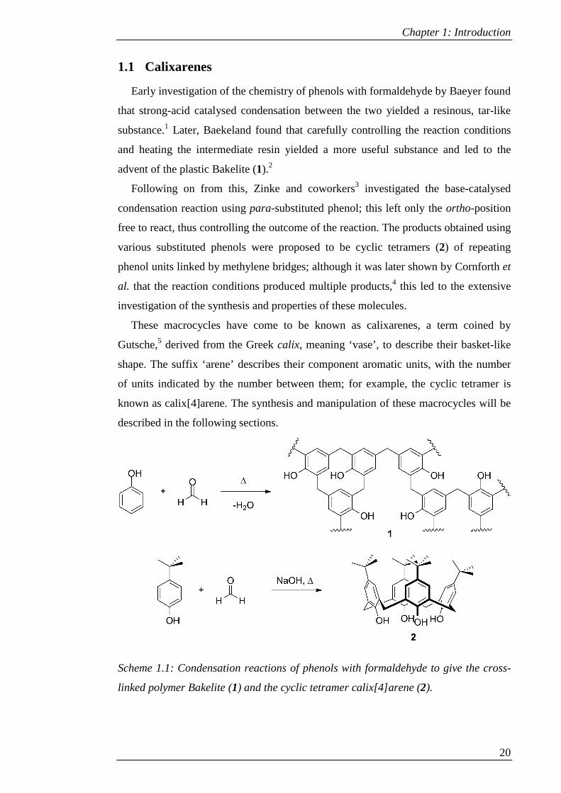
1.1 Calixarenes
Early investigation of the chemistry of phenols with formaldehyde by Baeyer found
that strong-acid catalysed condensation between the two yielded a resinous, tar-like
substance.1 Later, Baekeland found that carefully controlling the reaction conditions
and heating the intermediate resin yielded a more useful substance and led to the
advent of the plastic Bakelite (1).2
Following on from this, Zinke and coworkers3 investigated the base-catalysed
condensation reaction using para-substituted phenol; this left only the ortho-position
free to react, thus controlling the outcome of the reaction. The products obtained using
various substituted phenols were proposed to be cyclic tetramers (2) of repeating
phenol units linked by methylene bridges; although it was later shown by Cornforth et
al. that the reaction conditions produced multiple products,4 this led to the extensive
investigation of the synthesis and properties of these molecules.
These macrocycles have come to be known as calixarenes, a term coined by
Gutsche,5 derived from the Greek calix, meaning ‘vase’, to describe their basket-like
shape. The suffix ‘arene’ describes their component aromatic units, with the number
of units indicated by the number between them; for example, the cyclic tetramer is
known as calix[4]arene. The synthesis and manipulation of these macrocycles will be
described in the following sections.
Scheme 1.1: Condensation reactions of phenols with formaldehyde to give the cross-
linked polymer Bakelite (1) and the cyclic tetramer calix[4]arene (2).
Chapter 1: Introduction
21
1.1.1 Synthesis
The problem of multiple products from the procedure used by Zinke et al. was
approached by investigating the effect of varying the reaction conditions including the
temperature and the amount of base used. It was found that the latter was a critical
factor, with 0.03-0.04 equivalents of sodium hydroxide base giving the optimal yield
of calix[4]arene,6 whilst 0.3 equivalents of base gave calix[6]arene as the major
product.7 This led to the modified one-pot Zinke-Cornforth procedure for the synthesis
of tert-butyl-calix[4]arene,8 which is commonly used for the synthesis of this
derivative due to its high yield and brevity of synthesis (in comparison to multi-step
procedures; see below).
Modifications of this procedure have been developed to give other macrocycle
sizes. High-yielding methods for the synthesis of calix[6]-9 and calix[8]arene10 are
available; however, the syntheses for calix[5]-11 and calix[7]arene12 are less efficient.
Larger macrocycles, i.e. calix[9]- up to calix[12]arene have also been synthesised
using a base-catalysed procedure,13 but acid-catalysis is more effective in producing
the larger calixarenes, up to calix[20]arene.14 Modified procedures have also yielded
calixarenes in which the methylene bridge has been replaced with a heteroatom,
including oxa-,15 aza-16 and thiacalixarenes.17
As an alternative to the one-pot methods, calixarenes can also be synthesised in a
multi-step manner via linear intermediates. The non-convergent stepwise synthesis of
Hayes and Hunter18 had an early role in the confirmation of the cyclic tetramer
structure and was improved upon by Kammerer.19 Convergent synthesis was later
developed by Böhmer and co-workers,20 using a fragment condensation approach.
However, although stepwise synthesis offers the opportunity for diversification by
using different building blocks in each step, these syntheses are longer and generally
give poorer yields. The one-pot methods are therefore normally preferred.
1.1.2 Conformation
Calixarenes are conformationally mobile due to the ability of individual rings to
undergo transannular rotation. Although larger calixarenes can adopt a wide range of
potential conformers, calix[4]arene can adopt just four distinct conformations. These
are the cone (2), where all of the hydroxyl groups are pointing in the same direction;
partial cone (3), where one phenol has rotated to point one hydroxyl group towards the
upper rim; 1,3-alternate (4), where two opposite phenols have rotated; and 1,2-
alternate (5), where two adjacent phenols have rotated (see Figure 1.1).

Chapter 1: Introduction
22
Figure 1.1: Conformations of calix[4]arene: cone (2), partial cone (3), 1,3-alternate
(4) and 1,2-alternate (5).
These conformations are interchangeable in solution;21 however the calixarene can
be locked into a particular conformation by preventing transannular rotation. This can
be accomplished by functionalisation of the lower rim, either by blocking the
calixarene cavity with a bridging chain between two phenol groups, or by sterically
hindering the movement of the phenol through the annulus. The latter can be achieved
by forming an ether with any aliphatic chain longer than ethyl.22,23 Calixarenes can
therefore be synthesised in specific conformations by alkylating the lower rim, with
the base utilised influencing the final conformation via metal templating effects.24,25
However other factors such as solvent and reaction time can also have an effect.26
1.1.3 Functionalisation
Calixarenes can be independently functionalised at either of their rims, the side
bearing the phenol groups being termed the lower, narrow or endo rim, whilst the
opposite is referred to as either the upper, wide or exo rim. This, combined with the
ability to control the conformation, allows for dramatic diversification of the simple
core molecule.
Figure 1.2: Designation of calixarene rims.

Chapter 1: Introduction
23
1.1.3.1 Lower-rim functionalisation
As noted previously, the lower rim can be functionalised by alkyl or aryl ether (6)
formation. Selective partial alkylation is possible by utilising a specific base or
limiting the amount of alkylating agent available.25 Whilst alkyl chains can be used to
simply lock the conformation, various functional groups can also be introduced by use
of suitably functionalised alkylating agents. Furthermore, multiple alkylation events
can be used to give diverse function on the lower rim.
Alternatively, the hydroxyl groups of the lower rim can be removed (i.e. replaced
with H, as in 7) via reductive cleavage of their phosphonate esters.27 The hydroxyl
groups can also be replaced with a thiol (7),28 or, via a monospirodienone intermediate
(8), be selectively replaced with amine, azide and halogen functionalities (9).29,30
Scheme 1.2: Routes for functionalisation of lower rim.
1.1.3.2 Upper-rim functionalisation
The main method for upper-rim functionalisation involves electrophilic aromatic
substitution. Nitro groups (10) can be introduced directly by using ipso-nitration to
replace the tert-butyl groups,31 which can easily be reduced (11) to give access to
other functionalities, including amines, amides and guanidines (12). Sulphonate
groups (13) can also be introduced in a single step.32 Other functionalities require the
prior removal of the tert-butyl groups using a reverse Friedel-Crafts reaction with
phenol.33 The free positions on the upper rim (14) can subsequently be converted to,
for example, bromo, cyano and carboxyl functionalities (15).34 Also,
chloromethylation (15)35 is possible and can be followed by nucleophilic substitution
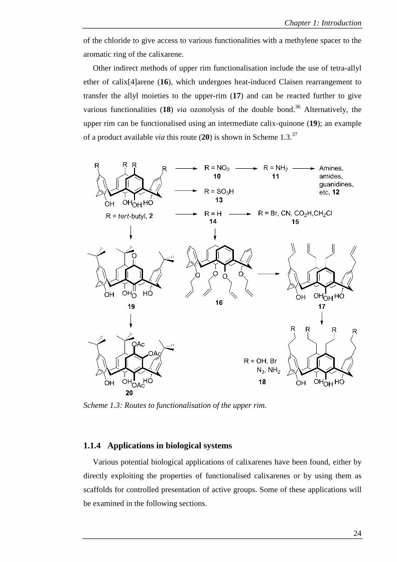
Chapter 1: Introduction
24
of the chloride to give access to various functionalities with a methylene spacer to the
aromatic ring of the calixarene.
Other indirect methods of upper rim functionalisation include the use of tetra-allyl
ether of calix[4]arene (16), which undergoes heat-induced Claisen rearrangement to
transfer the allyl moieties to the upper-rim (17) and can be reacted further to give
various functionalities (18) via ozonolysis of the double bond.36 Alternatively, the
upper rim can be functionalised using an intermediate calix-quinone (19); an example
of a product available via this route (20) is shown in Scheme 1.3.37
Scheme 1.3: Routes to functionalisation of the upper rim.
1.1.4 Applications in biological systems
Various potential biological applications of calixarenes have been found, either by
directly exploiting the properties of functionalised calixarenes or by using them as
scaffolds for controlled presentation of active groups. Some of these applications will
be examined in the following sections.

Chapter 1: Introduction
25
1.1.4.1 Artificial Receptors
Calixarenes can be used to present a binding surface for a target molecule and so
can mimic receptors. For example, a synthetic lectin (21) was synthesised38 using a
macrobicyclic calixarene with a peptide bridge between opposite aromatic units and a
phosphate group in the middle of this bridge to cooperate with the hydrogen bonding
of the peptide (see Figure 1.3). The lower rim was ester functionalised to provide a
means to immobilise the scaffold or alternatively could be deprotected to reveal
charged moieties to increase solubility in water.
Lipophilic sugar derivatives were tested for binding to 21 in organic media. The
receptor displayed good selectivity for β-octylglucoside over its α- form and also
selectivity over β-octylgalactoside. Replacing the phosphate with an acid or methyl
ester reduced the association. It was concluded that the phosphate group was most
important for strength of binding, with hydrogen bonding and steric factors governing
the selectivity.
Hamilton and co-workers constructed synthetic receptors for proteins by targeting
the protein surface to modulate protein-substrate interactions. Protein targets were
selected that had active sites that were formed of a hydrophobic region surrounded by
polar areas. This pattern could be matched to a calixarene with polar arms surrounding
its hydrophobic cavity. These arms took the form of pseudocyclopentapeptides, which
formed a stable hairpin loop structure, whose residues could be varied to give different
activities.39–41
With an anionic GlyAspGlyAsp sequence, the calixarene (22) bound to
Cytochrome C, complementing a hydrophobic region surrounded by cationic lysine
residues. This was found to disrupt the interaction of Cytochrome C with reducing
agents.39 Further investigation found that the interaction was strong enough to
compete with cytochrome C peroxidase and may be able to disrupt the interaction
with Apaf-1 (apoptosis protease activating factor-1).40
By changing Asp to Lys, a peptidomimetic capable of binding to a patch of anionic
residues in vascular endothelial growth factor (VEGF) was synthesised.41 This
prevents VEGF from binding to its receptor, Flk-1 (fetal liver kinase 1), thus halting
the VEGF stimulated tyrosine phosphorylation of the receptor and subsequent
activation of other protein kinases. Antiangiogenesis, antitumorigenesis and
antimetastasis activities were found in vivo. Binding to VEGF was highly selective,
with other growth factors remaining unaffected.

Chapter 1: Introduction
26
Figure 1.3: Calixarene based lectin mimetic (21),38 calixarene with
pseudocylcopentapeptide upper rim featuring AspGlyAspGly sequence (22)39 and
amphiphilic calixarenes with anionic (23) and cationic (24 and 25) upper rims.42
Synthetic receptors can also be constructed using multiple calixarenes incorporated
into lipid monolayers, which can self-organise to optimise binding of a protein. An
amphiphilic calixarene with butyl groups on the lower rim and charged phosphonate
groups at the upper rim (23) was synthesised and incorporated into a lipid monolayer
in a concentration dependent fashion.43 The calixarenes were distributed evenly in the
monolayer, but addition of poly-arginine or larger peptides containing arginine or
lysine residues stimulated self-assembly and multi-point binding.
Similarly, by adorning the upper rim with cationic moieties (24 and 25), acidic
proteins could be attracted to the monolayer.42 In both cases it is proposed that binding
occurs with excess calixarene in the aqueous phase, followed by embedding of the
ligand-protein complex in the monolayer.
It has been shown that cationic and anionic ligands can be combined in the
monolayer to give a combined response to a given protein. A fingerprint can be
obtained by measuring the response to anionic, cationic, polar and mixed bilayers,
allowing selective detection at nanomolar concentrations.42 By constructing vesicles
also containing polydiacetylene, a chromatic polymer, a colorimetric response was
Chapter 1: Introduction
27
observed on protein binding and likewise a fingerprint could be obtained for a given
protein.44
Calixarenes can also bind to stabilise protein structures. For example, the protein
p53, which protects against tumours by either inducing DNA repair or cell apoptosis,
contains a tetramerisation domain which is mutated in some cancers. The R337H
mutation destabilises the tetramer, but the integrity can be recovered by using a ‘clip’
in the form of a cone calixarene with four guanidinomethyl groups on the upper rim
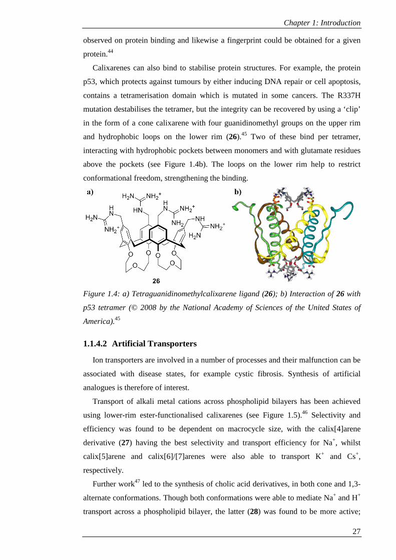
and hydrophobic loops on the lower rim (26).45 Two of these bind per tetramer,
interacting with hydrophobic pockets between monomers and with glutamate residues
above the pockets (see Figure 1.4b). The loops on the lower rim help to restrict
conformational freedom, strengthening the binding.
Figure 1.4: a) Tetraguanidinomethylcalixarene ligand (26); b) Interaction of 26 with
p53 tetramer (© 2008 by the National Academy of Sciences of the United States of
America).45
1.1.4.2 Artificial Transporters
Ion transporters are involved in a number of processes and their malfunction can be
associated with disease states, for example cystic fibrosis. Synthesis of artificial
analogues is therefore of interest.
Transport of alkali metal cations across phospholipid bilayers has been achieved
using lower-rim ester-functionalised calixarenes (see Figure 1.5).46 Selectivity and
efficiency was found to be dependent on macrocycle size, with the calix[4]arene
derivative (27) having the best selectivity and transport efficiency for Na+, whilst
calix[5]arene and calix[6]/[7]arenes were also able to transport K+ and Cs+,
respectively.
Further work47 led to the synthesis of cholic acid derivatives, in both cone and 1,3-
alternate conformations. Though both conformations were able to mediate Na+ and H+
transport across a phospholipid bilayer, the latter (28) was found to be more active;

Chapter 1: Introduction
28
this, combined with the similarity in the length of the calixarene and the thickness of
the vesicle, led to the conclusion that the 1,3-alternate calixarene was able to span the
bilayer.
Anion transport has also been achieved. A calixarene in the 1,3-alternate
conformation with a tetrabutyl amide functionalised lower rim (29)48 was found to
mediate Cl- transport in liposomes, planar lipid bilayers and in HEK-293 cells, with a
concomitant change in pH. This may occur by H+/Cl- symport or Cl-/OH- antiport.
Though this calixarene was not large enough to span the bilayer, it may have been
able to form aggregates (as observed with a tetramethyl amide derivative in HCl) to
allow it to form ion channels.
Figure 1.5: Artificial transporters based on ester ((27),46 cholic acid (28),47 amide
(29)48 and spermidine (30)49 functionalised calixarenes.
It was also found that the activity of the corresponding partial cone derivatives of
these tetra-amide calixarenes was influenced by the upper rim functionalisation.50 The
para-tert-butyl and unfunctionalised calixarenes had different crystal packing, which
was proposed to be the reason for the inactivity of the former compared with the latter.
Chapter 1: Introduction
29
The non-linear concentration dependence of the transport suggested that the calixarene
forms aggregates which are responsible for transport. Co-aggregates with the inactive
tert-butyl derivative inhibited transport. Although it was determined that the single
inverted amide of the partial cone was not required for transport function, it was
suggested that this could be exploited for synthesis of dimers or oligomers.
Another 1,3-alternate calixarene, this time functionalised at the lower rim with
spermidine (30) was able to span the entire bilayer without forming an aggregate.49 It
displayed some selectivity towards I- and Br- over Cl-, whilst oxo-anions showed poor
transport. This calixarene also showed a moderate antiproliferative effect against
murine monocyte/macrophage J774.A1 cancer cells.
1.1.4.3 Artificial Enzymes
The potential for calixarenes to act as enzyme mimics by organising catalytic
moieties has also been investigated. For example, artificial metallonucleases, which
cleave phosphodiester bonds in DNA and RNA, can be synthesised by pre-organising
catalytic Cu(II).
To investigate this, cone calixarenes with one, two or three copper centres tethered
to the upper rim via [12]-ane azamacrocyles were synthesised.51 A 104 fold rate
enhancement was achieved for the 1,2-di- (31) and tri-copper derivatives and it was
concluded that there was cooperativity between the metal centres in the cleavage of
the phosphodiester bond; however, the third metal gave no additional rate
enhancement, excluding simultaneous cooperation between all three. The 1,3-di-
copper species showed no cooperativity, indicating a sensitivity to the distance
between the metal centres. It was proposed that one metal centre tethers the non-
reacting part of the molecule, whilst the other activates the phosphoryl group for
cleavage.
For diribonucleoside monophosphates, it was found that substrates containing a
uracil base gave the best activity with these catalysts.52 It was proposed that the uracil
may be deprotonated, providing an electrostatic interaction between the base and the
metal centre. By contrast, for longer oligonucleoside monophosphates, greater
selectivity for bonds adjacent to adenosine and cytosine was found, with the most
scissile bond being 5’-pCpA. This mimics the activity of ribonuclease A. In this case
the interaction with the terminal phosphate seems to dominate, resulting in the
preferential cleavage of the naturally more labile CpA bond.
Chapter 1: Introduction
30
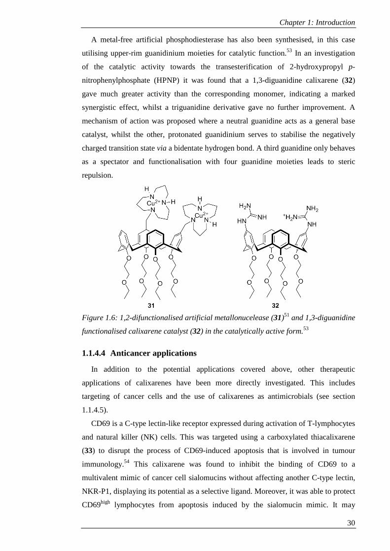
A metal-free artificial phosphodiesterase has also been synthesised, in this case
utilising upper-rim guanidinium moieties for catalytic function.53 In an investigation
of the catalytic activity towards the transesterification of 2-hydroxypropyl p-
nitrophenylphosphate (HPNP) it was found that a 1,3-diguanidine calixarene (32)
gave much greater activity than the corresponding monomer, indicating a marked
synergistic effect, whilst a triguanidine derivative gave no further improvement. A
mechanism of action was proposed where a neutral guanidine acts as a general base
catalyst, whilst the other, protonated guanidinium serves to stabilise the negatively
charged transition state via a bidentate hydrogen bond. A third guanidine only behaves
as a spectator and functionalisation with four guanidine moieties leads to steric
repulsion.
Figure 1.6: 1,2-difunctionalised artificial metallonucelease (31)51 and 1,3-diguanidine
functionalised calixarene catalyst (32) in the catalytically active form.53
1.1.4.4 Anticancer applications
In addition to the potential applications covered above, other therapeutic
applications of calixarenes have been more directly investigated. This includes
targeting of cancer cells and the use of calixarenes as antimicrobials (see section
1.1.4.5).
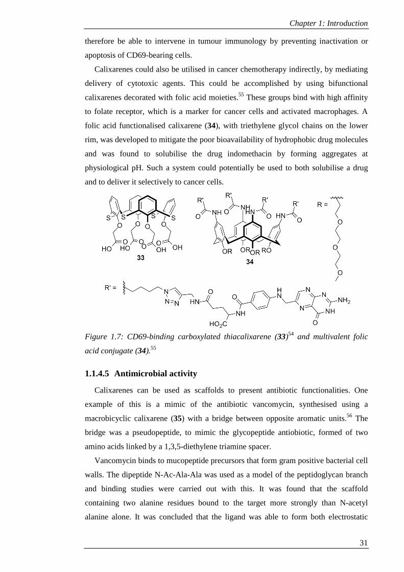
CD69 is a C-type lectin-like receptor expressed during activation of T-lymphocytes
and natural killer (NK) cells. This was targeted using a carboxylated thiacalixarene
(33) to disrupt the process of CD69-induced apoptosis that is involved in tumour
immunology.54 This calixarene was found to inhibit the binding of CD69 to a
multivalent mimic of cancer cell sialomucins without affecting another C-type lectin,
NKR-P1, displaying its potential as a selective ligand. Moreover, it was able to protect
CD69high lymphocytes from apoptosis induced by the sialomucin mimic. It may
Chapter 1: Introduction
31
therefore be able to intervene in tumour immunology by preventing inactivation or
apoptosis of CD69-bearing cells.
Calixarenes could also be utilised in cancer chemotherapy indirectly, by mediating
delivery of cytotoxic agents. This could be accomplished by using bifunctional
calixarenes decorated with folic acid moieties.55 These groups bind with high affinity
to folate receptor, which is a marker for cancer cells and activated macrophages. A
folic acid functionalised calixarene (34), with triethylene glycol chains on the lower
rim, was developed to mitigate the poor bioavailability of hydrophobic drug molecules
and was found to solubilise the drug indomethacin by forming aggregates at
physiological pH. Such a system could potentially be used to both solubilise a drug
and to deliver it selectively to cancer cells.
Figure 1.7: CD69-binding carboxylated thiacalixarene (33)54 and multivalent folic
acid conjugate (34).55
1.1.4.5 Antimicrobial activity
Calixarenes can be used as scaffolds to present antibiotic functionalities. One
example of this is a mimic of the antibiotic vancomycin, synthesised using a
macrobicyclic calixarene (35) with a bridge between opposite aromatic units.56 The
bridge was a pseudopeptide, to mimic the glycopeptide antiobiotic, formed of two
amino acids linked by a 1,3,5-diethylene triamine spacer.
Vancomycin binds to mucopeptide precursors that form gram positive bacterial cell
walls. The dipeptide N-Ac-Ala-Ala was used as a model of the peptidoglycan branch
and binding studies were carried out with this. It was found that the scaffold
containing two alanine residues bound to the target more strongly than N-acetyl
alanine alone. It was concluded that the ligand was able to form both electrostatic
Chapter 1: Introduction
32
interactions between its carboxylate group and the ammonium group of the host and
hydrogen bonds to the pseudopeptide bridge. The calixarene displayed antibacterial
activity which was selective towards gram-positive bacteria.
A drug molecule can also be tethered to a calixarene using a labile linker. A tetra-
para-aminoethylcalixarene with a tethered nalidixic acid molecule (36) was found to
be highly stable as a solid, but released the drug molecule in biological medium.57
Although the subsequent free alcohol of the calixarene was considerably less active as
an antibiotic, a synergistic effect was found between the calixarene and nalidixic acid
against both gram-positive and gram-negative bacterial strains.
Certain functionalised calixarenes have also been found to have intrinsic
antimicrobial activity. For example, para-sulfonato and para-phenylazocalixarenes in
particular were shown by Lamartine et al.58 to have antibacterial and antifungal
activities. More focussed investigation of agents against tuberculosis has also been
carried out, based on early observations that macrocylcon, a para-tert-
octylcalix[8]arene functionalised on the lower rim with polyethylene glycol chains,
could be used to treat pulmonary tuberculosis.59 A polyethylene glycol-functionalised
calix[6]arene (37) was found to reduce viable counts of mycobacteria in lungs and
spleens of infected mice by stimulating the host’s antimycobacterial mechanisms. A
host-mediated mechanism such as this could help to overcome multi drug resistance.
Guanidine functionalised calixarenes have also been investigated for their
antimicrobial properties. A para-guanidinoethylcalixarene (38) was found to exhibit a
significant effect against both gram-positive and gram-negative bacterial strains, with
much greater activity than the corresponding monomer.60 Later studies indicated that
interactions of the calixarene with phospholipid bilayers were characterised by a
combination of electrostatic and apolar interactions and that both are required for
antibiotic activity; the monomer, lacking the apolar core of the calixarene, is thus
unable to bring about the same intensity of effect.61
Anionic calixarenes have been investigated for their potential to treat viral
infections such as HIV. A set of calixarenes, including those functionalised with
sulphonate, carboxylate and phosphonate groups, with some featuring bithiazole
moieties on the lower rim, were tested for their anti-HIV activity.62 Bithiazole
functionalisation was found to increase activity, with the sulphonated derivative (39)
giving the best activity by interfering in early stages of infection. They were also
found to have little or no cytotoxicity, indicating their potential applicability to anti-
HIV therapy.
Chapter 1: Introduction
33
Figure 1.8: Antimicrobial calixarenes: general structure of vancomycin mimic (35)
where AA = amino acid,56 nalidixic acid prodrug (36),57 macrocyclon analogue
(37),63 guanidinium antibacterial agent (38),60 anti-HIV agent (39),62 and dual
function anti-HIV and –HCV agent (40).64
The problem of co-infection of HIV and hepatitis C virus (HCV) has led to the
search for anti-virals with activity towards both of these targets. A calixarene-based
antiviral agent (40)64 has been found which meets this goal, displaying antiviral
activity in multiple cell lines. The isophthalic acid head groups were found to be
important for activity, along with the locking into the cone conformation provided by
either butyl or benzyl ethers on the lower rim.
Chapter 1: Introduction
34
1.1.4.6 Other applications
Medical diagnostics could be aided through the use of calixarene-based DNA
chips. Song et al. developed a DNA chip using a monolayer of calixarenes
functionalised with anionic groups which were able to bind DNA appended with a
repeat of 9-guanines (9G-DNA chip). The chip was shown to be able to selectively
bind a Cy5 labelled DNA sequence complementary to that bound to the calixarenes.65
They subsequently demonstrated the ability of a 9G-DNA chip to detect human
papillomavirus (HPV) in clinical samples.66
Other applications include the use of calixarenes as cell penetration agents, for
example for cellular imaging or their use in DNA transfection. This will be examined
in Chapter 2. Calixarene-based glycoconjugates have also found a number of potential
applications and will be examined in Chapter 3.
1.2 Click chemistry
Click chemistry is a term coined by Sharpless et al.,67 the requirements for such a
reaction being that it must be modular, wide in scope, high yielding, produce only
inoffensive by-products, be stereospecific, have simple reaction conditions, use
readily available materials and reagents, use benign or easily removed solvent and
give a product that is stable and easily purified. Reactions that satisfy these criteria
include nucleophilic ring opening reactions (for example, those of aziridenes),68
Michael additions (such as thiol-ene coupling)69 and cycloaddition reactions. The
most well-known example of the latter is a Huisgen 1,3-dipolar cycloaddition70
reaction between azides and terminal alkynes to give triazoles.71 Although thermal
activation gives a mixture of regioisomers, this reaction gained renewed interest on
the discovery that the addition of copper(I) as a catalyst gives specifically the 1,4-
regioisomer (see Scheme 1.4).72,73 This is now known as the copper catalysed azide-
alkyne click (or CuAAC) reaction.
Scheme 1.4: Outcome of the 1,3-dipolar cycloaddition reaction between an azide and
alkyne under a) thermal conditions, and b) copper catalysed conditions.
Chapter 1: Introduction
35
The copper catalyst can be added directly using a copper salt such as
copper(I)iodide in combination with a base such as diisopropylethylamine (DIPEA),73
or can be generated in situ from copper(II) sulphate by adding sodium ascorbate as a
reducing agent.72 Addition of amine ligands can accelerate the reaction by stabilising
the active copper(I) species and improving its solubility in organic solvent, with
tris(benzyltriazolyl)methyl amine (TBTA)74 being particularly effective for this
purpose.
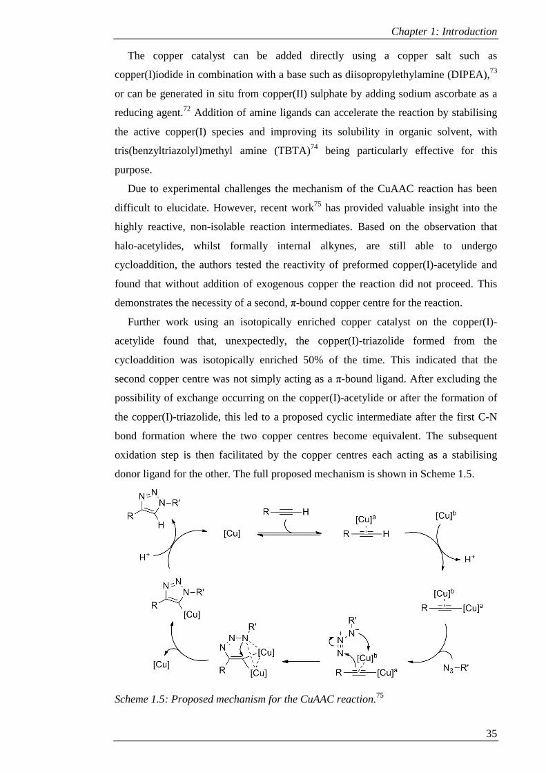
Due to experimental challenges the mechanism of the CuAAC reaction has been
difficult to elucidate. However, recent work75 has provided valuable insight into the
highly reactive, non-isolable reaction intermediates. Based on the observation that
halo-acetylides, whilst formally internal alkynes, are still able to undergo
cycloaddition, the authors tested the reactivity of preformed copper(I)-acetylide and
found that without addition of exogenous copper the reaction did not proceed. This
demonstrates the necessity of a second, π-bound copper centre for the reaction.
Further work using an isotopically enriched copper catalyst on the copper(I)-
acetylide found that, unexpectedly, the copper(I)-triazolide formed from the
cycloaddition was isotopically enriched 50% of the time. This indicated that the
second copper centre was not simply acting as a π-bound ligand. After excluding the
possibility of exchange occurring on the copper(I)-acetylide or after the formation of
the copper(I)-triazolide, this led to a proposed cyclic intermediate after the first C-N
bond formation where the two copper centres become equivalent. The subsequent
oxidation step is then facilitated by the copper centres each acting as a stabilising
donor ligand for the other. The full proposed mechanism is shown in Scheme 1.5.
Scheme 1.5: Proposed mechanism for the CuAAC reaction.75
Chapter 1: Introduction
36
1.2.1 Click chemistry for calixarene functionalisation
The CuAAC reaction has been applied to a wide array of different molecules,
including calixarenes. With their scope for selective functionalisation on both the
upper and lower rims, these macrocycles lend themselves well to the modular
synthesis approach provided by click chemistry. Although they can be furnished with
either azide or alkyne moieties on both upper and lower rims, this section will focus
on the use of lower-rim alkyne functionalised calixarenes due to their use in the
syntheses presented in this thesis.
1.2.1.1 Background
Prior to the development of the copper-catalysed version of the azide-alkyne
dipolar cycloaddition, this reaction was used to functionalise calixarenes under
thermal conditions.76 By combining a lower-rim 1,3-diazido calixarene (41) with
either lower-rim mono- (42) or 1,3-dipropargyl (43) calixarenes, trimers (44) and
doubly-bridged dimers (45) were synthesised, respectively. This provides an early
example of the use of cycloaddition chemistry to provide access to multicalixarenes.
However, without the copper catalyst, mixtures of regioisomers were obtained.
Scheme 1.6: Products of thermal cycloaddition reactions between azides and
alkynes.76

Chapter 1: Introduction
37
Interestingly, although all possible isomers of 45 were synthesised, only two
regiosomers of 44 were obtained. In the latter case the isomers could therefore be
easily separated.
Several years later, after the advent of the CuAAC reaction, Ryu and Zhao77
investigated the potential of this reaction. Their aim was to synthesise water-soluble
calixarenes through a modular approach that would provide a route to various
functionalised calixarenes without the need for protection/deprotection steps.
Although they were able to introduce a variety of functionalities (including sulfonates
and quaternary amines) to the lower rim via an azide-functionalised calixarene (46),
attempts to furnish the lower rim with carboxylic acid moieties using tetra-propargyl
calixarene (47) were unsuccessful (see Scheme 1.7, compounds 48 and 49,
respectively). The authors suggested that side-reactions between alkyne groups were
responsible for the observed complex mixture of products and that for this reason, and
due to the greater ease and safety of storing the various alkyne-based small-molecule
precursors compared with azides, the azide-functionalised calixarenes were more
effective precursors.
Scheme 1.7: Lower-rim alkyne vs. lower-rim azide for CuAAC mediated synthesis of
water soluble calixarenes.77

Chapter 1: Introduction
38
However, in 2006 Chen et al.78 demonstrated the successful use of tetra-propargyl
calixarene in CuAAC chemistry by functionalising it with triazole-linked aminoethyl
moieties, showing the applicability of the reaction to lower-rim alkynes. Such
molecules have since been widely utilised in the CuAAC reaction.
1.2.1.2 Preparation of Lower-Rim- Alkynes
Lower-rim alkynes are easily accessible by utilising a Williamson ether formation
between the phenols of the calixarene and propargyl bromide. The degree of
functionalisation and the conformation of the calixarene can be controlled to give
different arrangements of the functionalised scaffold.25 The main methods of
accessing partially and fully propargylated calixarenes, as described below, are
summarised in Scheme 1.8.
Chetcuti et al.79 synthesised several different alkyne-functionalised calixarenes,
although they did not utilise them in a CuAAC reaction. Mono, 1,3-di and tetra-alkyne
derivatives were synthesised by refluxing in acetone with K2CO3 as a base to control
the conformation by forming electrostatic interactions between K+ and the oxygens of
the lower rim. The degree of functionalisation was controlled by varying the amount
of alkylating agent, using 1, 3 or 6 equivalents of propargyl bromide respectively, and
increasing the reaction time. The 1,3-alternate conformation of the tetra-propargyl
derivative was obtained in a stepwise manner by using the 1,3-dipropargyl calixarene
and treating it with excess propargyl bromide in the presence of Cs2CO3 as a base.
Matthews and coworkers80 used a different approach to access partially-
propargylated derivatives. Instead of leaving free phenols, the calixarenes were first
partially alkylated with propyl bromide. Using a mixture of Ba(OH)2 and BaO as a
base afforded the tripropyl, whilst the 1,3-dipropyl was accessible using K2CO3. The
1,2-dipropyl was synthesised using NaH as the base with 2.2 equivalents of propyl
bromide. In contrast with the method used by Chetcuti et al., the mono-propyl
derivative was obtained using CsF as a weak base and 1.1 equivalents of alkylating
agent. All of these derivatives were subsequently treated with an excess of propargyl
bromide in the presence of NaH to exhaustively alkylate the lower rim and lock the
final calixarene into the cone conformation, with the desired number of alkyne groups
present.
Tetra-propargylated derivatives were also synthesised.80 The cone conformer was
synthesised using the method of Ryu and Zhao,77 utilising NaH as a base instead of
the K2CO3 used by Chetcuti et al.; the calixarene is held in the cone conformation in a

Chapter 1: Introduction
39
similar manner. The 1,3-alternate conformer was synthesised using the same method
as Chetcuti; however in this case a mixture of products was obtained, and therefore
also gave access to the partial cone conformer. Similarly, Puddephatt et al.,81 although
using K2CO3 as a base, also obtained a mixture of partial cone and 1,3-alternate, in
ratios of 2:1 and 4:1 after 24 and 48 hours, respectively. This suggests that the 1,3-
alternate conformation can convert into the more stable partial cone with prolonged
heating.
Scheme 1.8: Summary of main methods that have been used for accessing partially
and fully propargylated calixarenes, with the latter in different conformations.
Chapter 1: Introduction
40
An alternative method for mono-alkylation was provided by Bonnamour et al.,82 in
which the 1,3-dialkyl derivative is treated with TiCl4 to furnish the mono-alkylated
calixarene. This was demonstrated for a number of different functionalities, including
but-3-ynyl.
These alkyne-functionalised scaffolds have found a number of applications, which
will be examined in the following section.
1.2.1.3 Applications
As noted in section 1.2.1.1, the work of Chen et al.78 led to the successful
functionalisation of lower-rim tetra-propargyl calixarene. The triazole-linked
aminoethyl functionalised calixarene (50) was synthesised in cone and partial cone
conformers as part of a library of molecules. This was aimed at creating topomimetics
of amphiphilic α-helices and β-sheets for neutralisation of lipopolysaccharide (LPS)
endotoxin from Gram negative bacteria. Administration of the cone conformer to mice
given a lethal dose of LPS from Salmonella resulted in 100% survival, although it was
less effective against LPS from other strains of bacteria.
Click chemistry has also been applied to the formation of calixarene-based
cavitands and nanotubes. Morales Sanfrutos et al.,83 following on from the previous
work using thermal cycloaddition conditions,76 investigated the use of the CuAAC
reaction to react dipropargyl-functionalised calixarenes with diazide-functionalised
calixarenes or bis-azidomethylbenzenes. In the former case only the tube-like dimer
(51) was formed. With the latter, in addition to the desired benzene-capped
calixarenes (52) a doubly-bridged dimer was formed. However, a preference was
found for the product that formed by the second click reaction being intramolecular,
such that the doubly-bridged dimer was always the minor product.
Lower-rim propargylated calixarenes have also been incorporated into co-
polymers. For example, conjugation of 1,3-dipropargyl calixarene to a cyclodextrin
gave a product that was able to form a complex with adamantyl units on poly-N-
isopropylacrylamide (poly(NIPAAM)).84 In another case the bifunctionality was
centred on the calixarene itself (53) by using ring-opening polymerisation with ϵ-
caprolactone to give polycaprolactone (PCL) chains followed by click chemistry to
introduce polyethylene glycol (PEG) chains.85
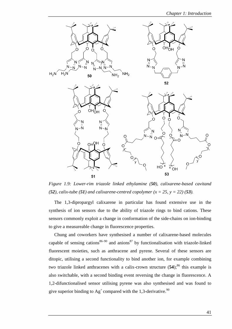
Chapter 1: Introduction
41
Figure 1.9: Lower-rim triazole linked ethylamine (50), calixarene-based cavitand
(52), calix-tube (51) and calixarene-centred copolymer (x = 25, y = 22) (53).
The 1,3-dipropargyl calixarene in particular has found extensive use in the
synthesis of ion sensors due to the ability of triazole rings to bind cations. These
sensors commonly exploit a change in conformation of the side-chains on ion-binding
to give a measureable change in fluorescence properties.
Chung and coworkers have synthesised a number of calixarene-based molecules
capable of sensing cations86–90 and anions87 by functionalisation with triazole-linked
fluorescent moieties, such as anthracene and pyrene. Several of these sensors are
ditopic, utilising a second functionality to bind another ion, for example combining
two triazole linked anthracenes with a calix-crown structure (54);86 this example is
also switchable, with a second binding event reversing the change in fluorescence. A
1,2-difunctionalised sensor utilising pyrene was also synthesised and was found to
give superior binding to Ag+ compared with the 1,3-derivative.90

Chapter 1: Introduction
42
Figure 1.10: Examples of ion sensors synthesised by Chung and coworkers (54)86 and
Pathak et al. (55).91
Kim and coworkers have also synthesised dipyrene functionalised calixarenes,
capable of binding Cd2+, Zn2+ and I-.92,93 During the course of developing the latter, a
mono-pyrene derivative was synthesised with a quaternary amine on the opposite
calixarene ring; the amine was noted to behave as an intramolecular ligand during the
CuAAC reaction, enhancing the reactivity.93
Other work on sensors includes that of Li and coworkers. A calixarene with a
bridging anthraquinone on the lower rim, linked via triethyleneglycol chains and
triazole rings, gave a switchable fluorescence response with selective binding of Ca2+
and subsequent quenching by interaction of the complex with F-.94 Other ion binding
calixarenes have also been synthesised that have no fluorescent moiety, but have the
ability to extract cations from aqueous to organic phase in a biphasic mixture.95,96
The work of Pathak et al. on calixarene-based sensors has had more of a biological
focus. The sensors developed by this group are based on a common motif: the 1,3-
dipropargyl calixarene is conjugated to benzaldehyde moieties, which are then used to
form a Schiff-base to various molecules to diversify the scaffold (for example
compound 55).91,97–100 This has yielded sensors which bind cations using not the
triazole moieties as noted previously, but the Schiff-base groups.
A Zn2+ binding calixarene featuring imine-linked thiophene units has been
developed99 which gives a switchable response, with increase in fluorescence on Zn2+
binding being reversed by addition of cysteine or dithiothreitrol (DTT) due to ligand
displacement. Release of Zn2+ from its complex with cysteine or DTT by addition of

Chapter 1: Introduction
43
Cd2+ or Hg2+ and concomitant restoration of fluorescence was also demonstrated.
These two processes mimic metallothionein and metal-detoxification, respectively.
Similarly, a Zn2+ complex with a pyridyl-imine derivative provided the means for
selective recognition of cysteine by fluorescence switching. This was shown to be
applicable to both the free amino acid and protein-bound cysteine.91 This amino-acid
sensing application was extended further with a triazole-linked o-iminophenol
functionalised calixarene.100 The function of this molecule was diversified by forming
complexes with different metal cations, which were shown to display selective sensing
properties towards different amino acids depending on the incorporated metal, with
similarities to amino acid-metal ion pairs found in metalloproteins.
Other work by Pathak et al. has demonstrated the applicability of switchable
sensors inside living cells. This will be examined in Chapter 2.
Click chemistry also provides easy access to glycoconjugates. Galante et al.101
synthesised lower-rim tetrapropargyl calixarene in cone and partial cone
conformations as part of a larger library including upper- and lower-rim azide
functionalised calixarenes, which were then used to form glycoconjugates with
suitably functionalised lactose moieties for testing as trypanocidal agents. Although
the partial cone showed little activity, the cone conformer (56) showed activity
comparable to the established drug benznidazole.
Figure 1.11: Example of glycoconjugate (56) functionalised with lactose moieties.101
Chinta and Rao102 used 1,3-dipropargyl calixarene to synthesise galactosyl and
lactosyl functionalised calixarenes. After testing binding to amino acids, which
interestingly showed that the triazole ring was involved in the interaction with
tryptophan, binding to the target protein, Jacilin, was confirmed.
The various propargylated calixarenes synthesised by Matthews and coworkers80
were also conjugated to sugars. By performing a click reaction between the calixarene
Chapter 1: Introduction
44
scaffolds and azide-functionalised galactose, linked by triethylene glycol, water-
soluble glycoconjugates were synthesised with different valencies and conformations
for targeting the PA-IL lectin of Pseudomonas aeruginosa. These, and other
glycoconjugates, will be examined in more detail in Chapter 3.
1.3 Overview of thesis
Chapter 1 has provided an overview of the key themes that are present throughout
the thesis: calixarenes, their potential biological applications and the use of click
chemistry in combination with these scaffolds.
Chapter 2 will examine in more detail the concept of cell penetration, cell
penetration agents and the use of calixarenes in this context. The present research
undertaken to add to this body of work will then be presented, including the synthesis
and biological evaluation of novel calixarene-based cell penetration agents.
Chapter 3 will explain the glycoside-cluster effect and how multivalent
glycocongugates can be used to exploit this. The use of calixarenes as a scaffold to
this end will be examined, particularly in the context of targeting Pseudomonas
aeruginosa. A synthetic strategy towards bi-functional fluorescent calixarene-based
glycoconjugates will be presented.
1.4 References
(1) Baeyer, A. Ber. Dtsch. Chem. Ges. 1872, 5, 1094–1100.
(2) Baekeland, L. H. Method of Making Insoluble Product of Phenol and Formaldehyde. US942699 1908.
(3) Zinke, A.; Kretz, R.; Leggewie, E.; Hössinger, K.; Hoffmann, G.; Ostwalden, P. W. v.; Wiesenberger, E.; Sobotka, M. Monatshefte für Chemie 1952, 83, 1213–1227.
(4) Cornforth, J. W.; D’arcy Hart, P.; Nicholls, G. A.; Rees, R. J. W.; Stock, J. A. Br. J. Pharmacol. Chemother. 1955, 10, 73–86.
(5) Gutsche, C. D.; Muthukrishnan, R. J. Org. Chem. 1978, 43, 4905–4906.
(6) Gutsche, C. D.; Iqbal, M.; Stewart, D. J. Org. Chem. 1986, 51, 742–745.
(7) Gutsche, C. D.; Dhawan, B.; No, K. H.; Muthukrishnan, R. J. Am. Chem. Soc. 1981, 103, 3782–3792.
(8) Gutsche, C. D.; Iqbal, M. Org. Synth. 1990, 68, 234–236.
(9) Gutsche, C. D.; Dhawan, B.; Leonis, M.; Stewart, D. Org. Synth. 1990, 68, 238.
Chapter 1: Introduction
45
(10) Munch, J. H.; Gutsche, C. D. Org. Synth. 1990, 68, 243.
(11) Stewart, D. R.; Gutsche, C. D. Org. Prep. Proced. Int. 1993, 25, 137–139.
(12) Vocanson, F.; Lamartine, R.; Lanteri, P.; Longeray, R.; Gauvrit, J. Y. New J. Chem. 1995, 19, 825–829.
(13) Dumazet, I.; Regnouf-de-Vains, J.-B.; Lamartine, R. Synth. Commun. 1997, 27, 2547–2555.
(14) Gutsche, C. D.; Rogers, J. S.; Stewart, D.; See, K.-A. Pure Appl. Chem. 1990, 62, 485–491.
(15) Gilbert, E. E. J. Heterocycl. Chem. 1974, 11, 899–904.
(16) Tsue, H.; Ono, K.; Tokita, S.; Ishibashi, K.; Matsui, K.; Takahashi, H.; Miyata, K.; Takahashi, D.; Tamura, R. Org. Lett. 2011, 13, 490–493.
(17) Kumagai, H.; Hasegawa, M.; Miyanari, S.; Sugawa, Y.; Sato, Y.; Hori, T.; Ueda, S.; Kamiyama, H.; Miyano, S. Tetrahedron Lett. 1997, 38, 3971–3972.
(18) Hayes, B. T.; Hunter, R. F. J. Appl. Chem. 2007, 8, 743–748.
(19) Kämmerer, H.; Happel, G.; Caesar, F. Die Makromolekulare Chemie 1972, 162, 179–197.
(20) Böhmer, V.; Chhim, P.; Kämmerer, H. Die Makromolekulare Chemie 1979, 180, 2503–2506.
(21) Gutsche, C. D. Calixarenes: An Introduction, 2nd Edition; The Royal Society of Chemistry, 2008.
(22) Groenen, L. C.; Van Loon, J. D.; Verboom, W.; Harkema, S.; Casnati, A.; Ungaro, R.; Pochini, A.; Ugozzoli, F.; Reinhoudt, D. N. J. Am. Chem. Soc. 1991, 113, 2385–2392.
(23) Iwamoto, K.; Araki, K.; Shinkai, S. J. Org. Chem. 1991, 56, 4955–4962.
(24) Iwamoto, K.; Fujimoto, K.; Matsuda, T.; Shinkai, S. Tetrahedron Lett. 1990, 31, 7169–7172.
(25) Iwamoto, K.; Araki, K.; Shinkai, S. Tetrahedron 1991, 47, 4325–4342.
(26) Iwamoto, K.; Shinkai, S. J. Org. Chem. 1992, 57, 7066–7073.
(27) Goren, Z.; Biali, S. E. J. Chem. Soc. Perkin Trans. 1 1990, 1484–1487.
(28) Gibbs, C. G.; Sujeeth, P. K.; Rogers, J. S.; Stanley, G. G.; Krawiec, M.; Watson, W. H.; Gutsche, C. D. J. Org. Chem. 1995, 60, 8394–8402.
(29) Aleksiuk, O.; Cohen, S.; Biali, S. E. J. Am. Chem. Soc. 1995, 117, 9645–9652.
Chapter 1: Introduction
46
(30) Van Gelder, J. M.; Aleksiuk, O.; Biali, S. E. J. Org. Chem. 1996, 61, 8419–8424.
(31) Verboom, W.; Durie, A.; Egberink, R. J. M.; Asfari, Z.; Reinhoudt, D. N. J. Org. Chem. 1992, 57, 1313–1316.
(32) Kumar, S.; Chawla, H. M.; Varadarajan, R. Indian J. Chem. Sect. B 2003, 42B, 2863–2865.
(33) Gutsche, C. D.; Levine, J. A. J. Am. Chem. Soc. 1982, 104, 2652–2653.
(34) Gutsche, C. D.; Pagoria, P. F. J. Org. Chem. 1985, 50, 5795–5802.
(35) Almi, M.; Arduini, A.; Casnati, A.; Pochini, A.; Ungaro, R. Tetrahedron 1989, 45, 2177–2182.
(36) Gutsche, C. D.; Levine, J. A.; Sujeeth, P. K. J. Org. Chem. 1985, 50, 5802–5806.
(37) Reddy, P. A.; Gutsche, C. D. J. Org. Chem. 1993, 58, 3245–3251.
(38) Segura, M.; Bricoli, B.; Casnati, A.; Munoz, E. M.; Sansone, F.; Ungaro, R.; Vicent, C. J. Org. Chem. 2003, 68, 6296–6303.
(39) Hamuro, Y.; Calama, M. C.; Park, H. S.; Hamilton, A. D. Angew. Chem., Int. Ed. 1997, 36, 2680–2683.
(40) Wei, Y.; McLendon, G. L.; Hamilton, A. D.; Case, M. A.; Purring, C. B.; Lin, Q.; Park, H. S.; Lee, C. S.; Yu, T. N. Chem. Commun. 2001, 1580–1581.
(41) Sun, J. Z.; Blaskovich, M. A.; Jain, R. K.; Delarue, F.; Paris, D.; Brem, S.; Wotoczek-Obadia, M.; Lin, Q.; Coppola, D.; Choi, K. H.; Mullan, M.; Hamilton, A. D.; Sebti, S. M. Cancer Res. 2004, 64, 3586–3592.
(42) Zadmard, R.; Schrader, T. J. Am. Chem. Soc. 2005, 127, 904–915.
(43) Zadmard, R.; Arendt, M.; Schrader, T. J. Am. Chem. Soc. 2004, 126, 7752–7753.
(44) Kolusheva, S.; Zadmard, R.; Schrader, T.; Jelinek, R. J. Am. Chem. Soc. 2006, 128, 13592–8.
(45) Gordo, S.; Martos, V.; Santos, E.; Menendez, M.; Bo, C.; Giralt, E.; de Mendoza, J.; Menéndez, M. Proc. Natl. Acad. Sci. U. S. A. 2008, 105, 16426–16431.
(46) Jin, T.; Kinjo, M.; Kobayashi, Y.; Hirata, H. J. Chem. Soc. Faraday Trans. 1998, 94, 3135–3140.
(47) Maulucci, N.; De Riccardis, F.; Botta, C. B.; Casapullo, A.; Cressina, E.; Fregonese, M.; Tecilla, P.; Izzo, I. Chem. Commun. 2005, 1354–1356.
Chapter 1: Introduction
47
(48) Sidorov, V.; Kotch, F. W.; Abdrakhmanova, G.; Mizani, R.; Fettinger, J. C.; Davis, J. T. J. Am. Chem. Soc. 2002, 124, 2267–2278.
(49) Izzo, I.; Licen, S.; Maulucci, N.; Autore, G.; Marzocco, S.; Tecilla, P.; De Riccardis, F. Chem. Commun. 2008, 26, 2986–8.
(50) Seganish, J. L.; Santacroce, P. V; Salimian, K. J.; Fettinger, J. C.; Zavalij, P.; Davis, J. T. Angew. Chem., Int. Ed. 2006, 45, 3334–8.
(51) Cacciapaglia, R.; Casnati, A.; Mandolini, L.; Reinhoudt, D. N.; Salvio, R.; Sartori, A.; Ungaro, R. J. Am. Chem. Soc. 2006, 128, 12322–12330.
(52) Cacciapaglia, R.; Casnati, A.; Mandolini, L.; Peracchi, A.; Reinhoudt, D. N.; Salvio, R.; Sartori, A.; Ungaro, R. J. Am. Chem. Soc. 2007, 129, 12512–12520.
(53) Baldini, L.; Cacciapaglia, R.; Casnati, A.; Mandolini, L.; Salvio, R.; Sansone, F.; Ungaro, R. J. Org. Chem. 2012, 77, 3381–9.
(54) Bezouska, K.; Snajdrová, R.; Krenek, K.; Vancurová, M.; Kádek, A.; Adámek, D.; Lhoták, P.; Kavan, D.; Hofbauerová, K.; Man, P.; Bojarová, P.; Kren, V. Bioorg. Med. Chem. 2010, 18, 1434–40.
(55) Consoli, G. M. L.; Granata, G.; Geraci, C. Org. Biomol. Chem. 2011, 9, 6491–5.
(56) Casnati, A.; Fabbi, M.; Pelizzi, N.; Pochini, A.; Sansone, F.; Ungaro, R.; DiModugno, E.; Tarzia, G. Bioorg. Med. Chem. Lett. 1996, 6, 2699–2704.
(57) Dibama, H. M.; Clarot, I.; Fontanay, S.; Salem, A. Ben; Mourer, M.; Finance, C.; Duval, R. E.; Regnouf-de-Vains, J.-B. Bioorg. Med. Chem. Lett. 2009, 19, 2679–82.
(58) Lamartine, R.; Tsukada, M.; Wilson, D.; Shirata, A. Comptes Rendus Chim. 2002, 5, 163–169.
(59) Boyd, D. H. A.; Stewart, S. M.; Somner, A. R.; Crofton, J. W.; Rees, R. J. W. Tubercle 1959, 40, 369–376.
(60) Mourer, M.; Duval, R. E.; Finance, C.; Regnouf-de-Vains, J.-B. Bioorg. Med. Chem. Lett. 2006, 16, 2960–3.
(61) Sautrey, G.; Orlof, M.; Korchowiec, B.; Regnouf-de-Vains, J.-B.; Rogalska, E. J. Phys. Chem. B 2011, 115, 15002–12.
(62) Mourer, M.; Psychogios, N.; Laumond, G.; Aubertin, A.-M.; Regnouf-de-Vains, J.-B. Bioorg. Med. Chem. 2010, 18, 36–45.
(63) Colston, M.; Hailes, H. Infect. Immun. 2004, 72, 6318–6323.
(64) Tsou, L. K.; Dutschman, G. E.; Gullen, E. A.; Telpoukhovskaia, M.; Cheng, Y.-C.; Hamilton, A. D. Bioorg. Med. Chem. Lett. 2010, 20, 2137–9.
Chapter 1: Introduction
48
(65) Song, K.-S.; Nimse, S. B.; Kim, J.; Kim, J.; Nguyen, V.-T.; Ta, V.-T.; Kim, T. Chem. Commun. 2011, 47, 7101–7103.
(66) Nimse, S. B.; Song, K.-S.; Kim, J.; Ta, V.-T.; Nguyen, V.-T.; Kim, T. Chem. Commun. 2011, 47, 12444–6.
(67) Kolb, H. C.; Finn, M. G.; Sharpless, K. B. Angew. Chem., Int. Ed. 2001, 40, 2004–2021.
(68) Ferraris, D.; Drury, W. J.; Cox, C.; Lectka, T. J. Org. Chem. 1998, 63, 4568–4569.
(69) Hoyle, C. E.; Bowman, C. N. Angew. Chem., Int. Ed. 2010, 49, 1540–1573.
(70) Huisgen, R. In 1,3-Dipolar Cycloaddition Chemistry, Volume 1; Padwa, A., Ed.; Wiley, 1984; pp. 1–176.
(71) Huisgen, R.; Szeimies, G.; Möbius, L. Chem. Ber. 1967, 100, 2494–2507.
(72) Rostovtsev, V. V; Green, L. G.; Fokin, V. V; Sharpless, K. B. Angew. Chem., Int. Ed. 2002, 41, 2596–9.
(73) Tornøe, C. W.; Christensen, C.; Meldal, M. J. Org. Chem. 2002, 67, 3057–3064.
(74) Chan, T. R.; Hilgraf, R.; Sharpless, K. B.; Fokin, V. V Org. Lett. 2004, 6, 2853–2855.
(75) Worrell, B. T.; Malik, J. A.; Fokin, V. V. Science 2013, 340, 457–460.
(76) Calvo-Flores, F. G.; Isac-García, J.; Hernández-Mateo, F.; Pérez-Balderas, F.; Calvo-Asín, J. a; Sanchéz-Vaquero, E.; Santoyo-González, F. Org. Lett. 2000, 2, 2499–2502.
(77) Ryu, E. H.; Zhao, Y. Org. Lett. 2005, 7, 1035–1037.
(78) Chen, X. M.; Dings, R. P. M.; Nesmelova, I.; Debbert, S.; Haseman, J. R.; Maxwell, J.; Hoye, T. R.; Mayo, K. H. J. Med. Chem. 2006, 49, 7754–7765.
(79) Chetcuti, M. J.; Devoille, A. M. J.; Othman, A. Ben; Souane, R.; Thuéry, P.; Vicens, J. Dalton Trans. 2009, 2999–3008.
(80) Cecioni, S.; Lalor, R.; Blanchard, B.; Praly, J.-P.; Imberty, A.; Matthews, S. E.; Vidal, S. Chem.--Eur. J. 2009, 15, 13232–13240.
(81) Xu, W.; Vittal, J. J.; Puddephatt, R. J. Can. J. Chem. 1996, 74, 766–774.
(82) Bois, J.; Espinas, J.; Darbost, U.; Felix, C.; Duchamp, C.; Bouchu, D.; Taoufik, M.; Bonnamour, I. J. Org. Chem. 2010, 75, 7550–7558.
(83) Morales-Sanfrutos, J.; Ortega-Muñoz, M.; Lopez-Jaramillo, J.; Hernández-Mateo, F.; Santoyo-González, F. J. Org. Chem. 2008, 73, 7768–7771.

Chapter 1: Introduction
49
(84) Garska, B.; Tabatabai, M.; Ritter, H. Beilstein J. Org. Chem. 2010, 6, 784–788.
(85) Gou, P.; Zhu, W.; Shen, Z. J. Polym. Sci., Part A: Polym. Chem. 2010, 48, 5643–5651.
(86) Chang, K.-C.; Su, I.-H.; Senthilvelan, A.; Chung, W.-S. Org. Lett. 2007, 9, 3363–3366.
(87) Chang, K.-C.; Su, I.-H.; Wang, Y.-Y.; Chung, W.-S. European J. Org. Chem. 2010, 2010, 4700–4704.
(88) Ho, I.-T.; Haung, K.-C.; Chung, W.-S. Chem. Asian J. 2011, 6, 2738–2746.
(89) Wang, N.-J.; Sun, C.-M.; Chung, W.-S. Tetrahedron 2011, 67, 8131–8139.
(90) Wang, N.-J.; Sun, C.-M.; Chung, W.-S. Sensors Actuators B Chem. 2012, 171-172, 984–993.
(91) Pathak, R. K.; Tabbasum, K.; Hinge, V. K.; Rao, C. P. Chem.--Eur. J. 2011, 17, 13999–14003.
(92) Park, S. Y.; Yoon, J. H.; Hong, C. S.; Souane, R.; Kim, J. S.; Matthews, S. E.; Vicens, J. J. Org. Chem. 2008, 73, 8212–8218.
(93) Kim, J.-S.; Park, S.-Y.; Kim, S.-H.; Thuery, P.; Souane, R.; Matthews, S. E.; Vicens, J. Bull. Korean Chem. Soc. 2010, 31, 624–629.
(94) Zhan, J.; Fang, F.; Tian, D.; Li, H. Supramol. Chem. 2012, 24, 272–278.
(95) Li, H.; Zhan, J.; Chen, M.; Tian, D.; Zou, Z. J. Incl. Phenom. Macrocycl. Chem. 2010, 66, 43–47.
(96) Zhan, J.; Tian, D.; Li, H. New J. Chem. 2009, 33, 725–728.
(97) Pathak, R. K.; Ibrahim, S. M.; Rao, C. P. Tetrahedron Lett. 2009, 50, 2730–2734.
(98) Pathak, R. K.; Dikundwar, A. G.; Row, T. N. G.; Rao, C. P. Chem. Commun. 2010, 46, 4345–4347.
(99) Pathak, R. K.; Hinge, V. K.; Mondal, M.; Rao, C. P. J. Org. Chem. 2011, 76, 10039–10049.
(100) Pathak, R. K.; Dessingou, J.; Rao, C. P. Anal. Chem. 2012, 84, 8294–8300.
(101) Galante, E.; Geraci, C.; Sciuto, S.; Campo, V. L.; Carvalho, I.; Sesti-Costa, R.; Guedes, P. M. M.; Silva, J. S.; Hill, L.; Nepogodiev, S. A.; Field, R. A. Tetrahedron 2011, 67, 5902–5912.
(102) Chinta, J. P.; Rao, C. P. Carbohydr. Res. 2013, 369, 58–62.
Chapter 2: Calixarene-Based Cell-Penetration Agents
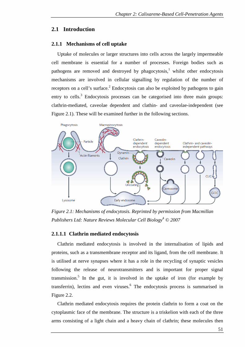
Chapter 2: Calixarene-Based Cell-Penetration Agents
51
2.1 Introduction
2.1.1 Mechanisms of cell uptake
Uptake of molecules or larger structures into cells across the largely impermeable
cell membrane is essential for a number of processes. Foreign bodies such as
pathogens are removed and destroyed by phagocytosis,1 whilst other endocytosis
mechanisms are involved in cellular signalling by regulation of the number of
receptors on a cell’s surface.2 Endocytosis can also be exploited by pathogens to gain
entry to cells.3 Endocytosis processes can be categorised into three main groups:
clathrin-mediated, caveolae dependent and clathin- and caveolae-independent (see
Figure 2.1). These will be examined further in the following sections.
Figure 2.1: Mechanisms of endocytosis. Reprinted by permission from Macmillan
Publishers Ltd: Nature Reviews Molecular Cell Biology4 © 2007
2.1.1.1 Clathrin mediated endocytosis
Clathrin mediated endocytosis is involved in the internalisation of lipids and
proteins, such as a transmembrane receptor and its ligand, from the cell membrane. It
is utilised at nerve synapses where it has a role in the recycling of synaptic vesicles
following the release of neurotransmitters and is important for proper signal
transmission.5 In the gut, it is involved in the uptake of iron (for example by
transferrin), lectins and even viruses.6 The endocytosis process is summarised in
Figure 2.2.
Clathrin mediated endocytosis requires the protein clathrin to form a coat on the
cytoplasmic face of the membrane. The structure is a triskelion with each of the three
arms consisting of a light chain and a heavy chain of clathrin; these molecules then

Chapter 2: Calixarene-Based Cell-Penetration Agents
52
form the lattice that coats the vesicle.7 A number of accessory proteins are also
required. These include adaptor proteins, such as AP2, which links the clathrin to the
cell membrane. For example, AP2 recognises the cytoplasmic face of transferrin and
links it to the nucleating clathrin.8
The membrane becomes deformed at the site of clathrin nucleation, creating a
clathrin coated pit. More accessory proteins aid in this deformation of the membrane.
For example, epsin type proteins partially insert an amphipathic helix into the cell
membrane, displacing lipid residues in a wedge-like manner to create a curve.9 Further
polymerisation leads to constriction of the vesicle neck. Dynamin, a GTPase, forms a
helical polymer around the neck, such that subsequent GTP hydrolysis results in
scission of the vesicle from the cell membrane to form a clathrin coated vesicle.10
Recruitment of dynamin is effected by amphiphysin, which also binds to clathrin and
AP2.11 The protein coat is then removed by auxilin and heat shock cognate protein 70
kD (HSC70).12 The vesicle can then undergo intracellular trafficking followed by
fusion to the target membrane.
Figure 2.2: Scheme of key steps in clathrin mediated endocytosis. Reprinted by
permission from Macmillan Publishers Ltd: Nature7 © 2007
2.1.1.2 Caveolae
Caveolae (‘little caves’) are flask-shaped invaginations in the cell membrane,
forming a microdomain that is a type of lipid raft. They are particularly abundant in
smooth muscle, fibroblasts, adipocytes and epithelial cells13, but some cells such as
lymphocytes lack caveolae.14 Previously associated only with non-specific bulk
transfer of fluid-phase cargoes, caveolae dependent endocytosis is now implicated in a
number of processes. These include a role in calcium mediated signalling in
endothelial cells, both negative and positive regulation of membrane bound receptors
and cholesterol homeostasis.15
Caveolae are enriched with caveolin type proteins. Caveolin1 (or caveolin3 in
muscle cells) is crucial for their formation. Caveolin1 has an intramembrane domain
that inserts into the cell membrane, forming a hairpin loop with both N- and C-
Chapter 2: Calixarene-Based Cell-Penetration Agents
53
terminal domains in the cytoplasm. It is palmitoylated and can bind cholesterol and
fatty acids to stabilise its oligomers.16 It is thought that caveolin itself effects the
curvature of the membrane in the formation of the caveolae. The amphipathic
structure can be seen as analogous to those involved in clathrin mediated endocytosis,
with the inserted loop deforming the membrane. In addition, the enrichment of
cholesterol within the membrane envelope may enhance the curvature.9
Although dynamin is required for scission, it has been shown that not all caveolae
pinch off from the membrane. Caveosomal structures have been found distant from
the cell membrane that were still connected to it,17 suggesting that some caveolae are
stable structures that are not involved in endocytosis.
2.1.1.3 Clathrin- and caveolin-independent pathways
It has been found that cells in which clathrin and caveolin have been depleted can
still undergo cholesterol dependent endocytosis. GPI-anchored proteins (GPI-AP) are
not usually concentrated into clathrin coated pits or caveolae although the latter play a
role. Instead, clathrin-independent carriers (CLICs) can form and transfer the
structures to GPI-AP-enriched early endosomal compartments (GEECs), more tubular
in shape than typical vesicles. This pathway is regulated by the G-protein cdc42.18
This is a lipid raft associated process, with the clustering of the GPI-APs dependent on
cholesterol.19 This does not create sufficient membrane curvature so other proteins
with Bin–Amphiphysin–Rvs (BAR)-type domains such as GRAF1 are recruited to the
curved membrane and induce further curvature.9
Another lipid-raft associated process is macropinocytosis. This occurs when
protrusions in the cell membrane fuse back to the lipid bilayer to form large, fluid-
filled macropinosomes, which is associated with extensive actin rearrangement to give
characteristic ruffles.20 It has been shown to be important in class II MHC-positive
cells in the uptake of antigens.21 A number of enzymes have been shown to be
important for this process, including phosphoinositide 3-kinase (PI3-K), the GTPase
Rac1 and ARF6.20,22 The localisation of Rac1 and ARF6 to the membrane seems to be
cholesterol dependent and cholesterol depletion inhibits macropinocytosis.23 However,
cholesterol depletion is a non-specific inhibitor. It may be that the observed inhibition
is a secondary effect, for example due to interference with the actin cytoskeleton at the
cell membrane.20
Chapter 2: Calixarene-Based Cell-Penetration Agents
54
2.1.2 Cell penetration agents
Many therapeutic targets lie within cells. As such, it is essential for drugs or other
therapeutic agents to be able to cross the cell membrane. In some cases this barrier
prevents the agent being available within the cell, for example in the delivery of
nucleic acids in gene therapy. Therefore it is important to investigate ways to facilitate
the delivery of various molecules into cells.
2.1.2.1 Cell penetrating peptides
Some naturally occurring peptides are able to cross the cell membrane. One early
observation of this was the Antennapedia homeodomain, the DNA binding portion of
a transcription factor from Drosophila, which was found to penetrate cells in culture.24
Further examples have also been found, including the transactivator of transcription
(Tat) protein from HIV, from which the protein transduction domain (Tatp) was
isolated.25
The potential usage of such peptides as cell penetration agents to deliver cargos
into cells has led to extensive investigation of the mechanisms of their uptake into
cells and the essential features that allow them to do so. This has informed the design
of synthetic cell penetrating peptides and the investigation of potential applications.
These investigations will be examined in the following sections.
2.1.2.1.1 Cellular uptake mechanisms
In order to facilitate the design of cell penetrating peptides, their uptake
mechanisms into cells have been investigated. Early work seemed to indicate an
energy-independent, non-endocytotic mechanism of uptake due to the observation that
at 4 °C there was no inhibition of uptake into cells.24,25 However, subsequent work has
suggested that this was an artefact of the procedures used to fix the cells.26 Fixation
using paraformaldehyde resulted in the cellular membranes becoming more leaky,
leading to release of the peptides from endosomes to give the observed diffuse
staining. Furthermore, excess peptide associated with the external surface of the
membrane was able to leak into the cell even at low temperature.
Following this, many studies have been carried out on live cells. Nakase et al.27
suggested that macropinocytosis was the dominant mechanism of uptake based on the
observation that uptake of Tatp and other arginine rich peptides was associated with
actin rearrangement. Also, inhibitors of macropinocytosis such as
ethylisopropylamiloride inhibited their uptake. However, penetratin, a cell penetrating
Chapter 2: Calixarene-Based Cell-Penetration Agents
55
peptide derived from the third helix Antennapedia homeodomain, was not affected by
the same inhibitors except at high concentrations, suggesting that multiple
mechanisms may be used by the same peptide.
They also suggested an important role of heparan sulphate proteoglycan (HSPG).
Deficiency of this cell surface structure led to decreased uptake efficiency and was
associated with a lack of induction of macropinocytosis by arginine-rich peptides,
although this effect was less prominent for the 8 residue arginine oligomer, R8. They
suggested that HSPG may act as a receptor for macropinocytosis by concentrating the
cargo at the cell surface and stimulating the endocytosis.
However, other studies of Tatp have shown colocalisation with transferrin in HeLa
cells, implicating clathrin mediated processes.28 Consistent with this, treatment with
chlorpromazine, an inhibitor of clathrin mediated endocytosis, gave 50% inhibition of
uptake.29 Other studies in the same cell line also found colocalisation with caveolin1,30
although inhibitors of caveolae such as filipin did not have a significant impact on
uptake.29
It has also been shown that a direct translocation mechanism may play a role, even
in live cells. Fretz et al.31 studied the uptake of octaarginine into cells under varying
conditions in CD34+ leukaemia cells. It was found that at low temperature there was
diffuse staining of the cytoplasm and localisation to the nucleus, consistent with the
original assumptions about endocytosis-independent uptake. At increasing
temperature, localisation to endocytotic vesicles occurred, whilst at 37 °C this was the
only staining present. A concentration threshold was implicated by the observation
that at higher concentrations of peptide diffuse staining became evident once more.
Cholesterol depletion also gave diffuse staining. This suggests that either cholesterol
inhibits direct translocation, or that disruption of endocytosis necessitates an
alternative mechanism.
Cumulatively, the data suggests that multiple mechanisms may be important,
perhaps even operating in a concerted manner. Mechanistic studies have been
complicated by the fact that a number of factors influence the mechanism that a
peptide is taken up by. The type of cell, cell penetrating peptide and cargo all have an
effect on the mechanism.32 An example of the latter has been demonstrated by
Tünnemann et al.33 who found that varying the size of a peptide-based cargo attached
to Tat from a peptide to a globular protein led to differences in uptake. The globular
protein cargo was taken up only by endocytosis, whereas the peptide was also taken
up by an unknown, rapid mechanism of translocation.

Chapter 2: Calixarene-Based Cell-Penetration Agents
56
In summary, the dependence of uptake on a number of variables precludes the use
of a single universal cell penetrating peptide. On the other hand, it may allow fine
tuning for selective applications.
2.1.2.1.2 Design of cell penetrating peptides
Much work has been carried out to determine which features are essential for
cellular uptake in order to design optimised cell penetrating peptides. Studies of
individual segments of the Antennapedia homeodomain found that a 16 amino acid
stretch corresponding to the third α-helix without its N-terminal glutamate (now
known as penetratin) was the essential component for its translocation.24 However, the
α-helical structure is not essential for uptake, since introduction of 1-3 proline residues
to interrupt the secondary structure did not prevent uptake.34
In the case of Tatp, studies to determine the key features of the peptide determined
that translocation was due to a cluster of basic amino acids (six arginines and two
lysines), whilst the amphipathic α-helix, initially thought to be a key determinant for
uptake, was not essential.25 It has also been found that deletion of arginine in Tatp is
more detrimental to its cellular uptake efficiency than deletion of lysine.32
The difference in acidities between lysine and guanidinium cations has been
proposed as a reason for this effect.35 Lysine is sufficiently acidic to release a proton
to water in order to reduce charge repulsion with neighbouring cationic groups;
however, the guanidinium group of arginine is not sufficiently acidic to do the same.
Therefore in order to stabilise the positive charge arginine may instead act as an anion
scavenger. This may increase its tendency to bind to the negatively charged phosphate
groups on the cell membrane.
It has also been proposed that arginine can create Gaussian negative curvature in
the cell membrane, which is associated with the formation of pores, invaginations and
protrusions. Arginine can form both electrostatic interactions and multiple hydrogen
bonds, giving a bidentate mode of binding to multiple polar head groups of the cell
membrane, whereas lysine can only form a hydrogen bond. This allows poly-arginine
to give Gaussian negative curvature.36 The ability of poly-arginine to do this would
depend on the degree of intrinsic curvature in the membrane and so depend on the
composition of the lipid bilayer, including the amount of cholesterol, accounting for
some differences between cell types.
The length of an arginine oligomer is also important. Oligomers of 5-20 residues
are taken up into cells, with maximum efficiency at 15 arginine residues. However the

Chapter 2: Calixarene-Based Cell-Penetration Agents
57
R8 peptide has been most extensively studied as it gives a good compromise between
uptake efficiency and ease of synthesis.32,37
Further studies into the effects of modifications of cell penetrating peptides found
that both D- and L-isomers were efficiently taken up into cells, indicating that the
backbone conformation of the peptide was not important. This suggests that enzyme
interactions at the cell surface, which would depend on chirality, are not responsible
for uptake. The D-isomer of an oligoarginine seemed to have superior uptake, perhaps
due to its greater stability to proteolysis and longer in vivo lifetime.32
These observations led to the study of non-peptidic guanidinium-rich transporters,
such as those based on peptoids and carbamates, which lack secondary structure and
have greater flexibility. These will be examined further in section 2.1.2.2.
2.1.2.1.3 Applications
Cell penetrating peptides have been used to deliver a variety of cargoes into cells
which would otherwise have poor uptake efficiency, or no uptake at all.
Peptide-based therapies have great potential but are hampered by the inability of
most peptides to cross the cell membrane. However, peptides lend themselves to
improvement with cell penetrating peptides as their incorporation can be included
during the peptide synthesis. An example of this is the targeting of Nuclear factor
(erythroid-derived 2)-like 2 (Nrf2).38 This binds to the anti-oxidant response element
(ARE) when not bound by its regulator Keap1 and so this interaction provides a target
for treatment of inflammation. A 14-mer peptide, designed to interact with Keap1,
when combined with Tatp was able to disrupt this interaction in vivo, giving
downstream expression of heme oxygenase and supressing expression of tumor
necrosis factor, a pro-inflammatory cytokine.
A further example is provided by Psorban, a cell penetrating peptide conjugate that
is currently undergoing clinical trials. It is based on cyclosporine-A, a cyclic peptide
with anti-inflammatory effects that is used to treat psoriasis. It was conjugated to R7,
which allowed it to cross cutaneous barriers and penetrate into the target T-cells in
inflamed skin, giving an anti-inflammatory response when used topically and avoiding
the side-effects associated with systemic use.39
Nucleic acids can also be delivered, with potential applications in transfection and
gene therapy. One such cargo is siRNA, small sections of double-stranded RNA that
are capable of affecting the expression of target complementary genes by post-
transcriptional silencing. Both covalent40,41 and non-covalent42 conjugates of cell
Chapter 2: Calixarene-Based Cell-Penetration Agents
58
penetrating peptides with siRNA have been investigated and gave successful gene
silencing, though vesicular accumulation was identified as a barrier in the latter case.
This led to the development of PepFect6.43 This transportan 10-based peptide features
several trifluoromethylquinoline moieties, whose pH buffering capability leads to
osmotic swelling and subsequent endosomal release. It was internalised into multiple
cell lines with low toxicity and gave in vivo gene silencing in mouse models. However
a more simple poly-arginine R15 was also able to internalise siRNA and give gene
silencing, with cellular localisation suggesting endosomal escape, and gave reduction
in tumor growth in a mouse model.44
Another example of their use is aiding in the delivery of small drug molecules by
improving their properties. For example, polyarginine can be used to improve the
water solubility and uptake of paclitaxel.45 A different facet of this is improving
availability of a drug by circumventing the action of efflux pumps which can give
resistance. To this end a doxorubicin-Tatp conjugate was synthesised, which
successfully exerted a cytotoxic effect on resistant cell lines.46
Cell penetrating peptides also have uses in cellular imaging. This is important
therapeutically for visualising diseased tissue and also for improving understanding of
biological processes. Jiang et al.47 exploited the increased concentration of
extracellular proteases in tumour tissue by conjugating a fluorescein peptide hairpin to
a polyarginine cell penetrating peptide with an associated polyanionic segment.
Proteolytic cleavage of the anionic domain was most prominent in the tumour tissue,
allowing internalisation of the probe and selective visualisation of the tumour cells.
MRI can also be used to visualise cells, relying on the incorporation of MRI
contrast agents which enhance the magnetic spin coupling of protons in water. Tat-
iron oxide nanoparticles allow delivery of the contrast agents into cells and have been
used for in vivo, real time tracking of T-cells with no effect on the normal immune
function.48
2.1.2.2 Non-peptidic cell penetration agents
As noted in section 2.1.2.1.2, the ability of a cell penetrating peptide to resist
proteolysis influences the efficacy of its uptake. The desire to find alternatives that
will remain stable for longer periods in living cells has led to the investigation of non-
peptidic systems.
Oligoarginine peptoids have the same 1,4-spacing for their side chains as their
peptidic counterparts, but instead of the side chain being attached to carbon, it is

Chapter 2: Calixarene-Based Cell-Penetration Agents
59
attached to the nitrogen (see Figure 2.3). This eliminates the secondary structure
formed by N-H hydrogen bonding and increases flexibility. It was found that these
structures were superior to Tatp in their uptake and that increasing the chain length
between the backbone and the guanidinium moieties further increased uptake
efficiency. This suggests that greater flexibility is important in uptake.49
Oligocarbamates have also been investigated, which are characterised by 1,6-
spacing between their side chains (see Figure 2.3). Superior uptake was found relative
to oligoarginine peptides. This shows that 1,4-spacing between side chains is not
required for cell penetration.50
Figure 2.3: Oligoguanidinium compounds based on peptides (57), peptoids (58) and
carbamates (59).49,50
Figure 2.4: Cell penetration agents based on a dendrimer (60)51 and an inositol dimer
(61).52 FITC = fluorescein isothiocyanate.
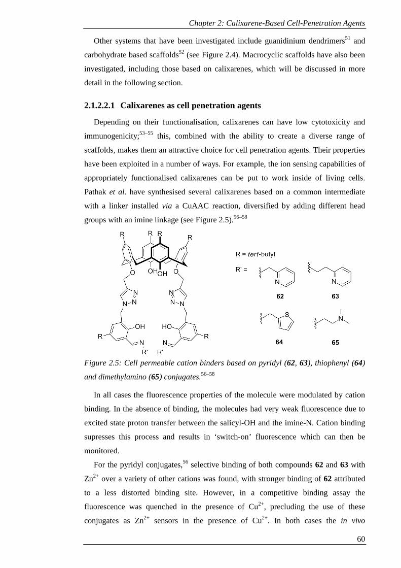
Chapter 2: Calixarene-Based Cell-Penetration Agents
60
Other systems that have been investigated include guanidinium dendrimers51 and
carbohydrate based scaffolds52 (see Figure 2.4). Macrocyclic scaffolds have also been
investigated, including those based on calixarenes, which will be discussed in more
detail in the following section.
2.1.2.2.1 Calixarenes as cell penetration agents
Depending on their functionalisation, calixarenes can have low cytotoxicity and
immunogenicity;53–55 this, combined with the ability to create a diverse range of
scaffolds, makes them an attractive choice for cell penetration agents. Their properties
have been exploited in a number of ways. For example, the ion sensing capabilities of
appropriately functionalised calixarenes can be put to work inside of living cells.
Pathak et al. have synthesised several calixarenes based on a common intermediate
with a linker installed via a CuAAC reaction, diversified by adding different head
groups with an imine linkage (see Figure 2.5).56–58
Figure 2.5: Cell permeable cation binders based on pyridyl (62, 63), thiophenyl (64)
and dimethylamino (65) conjugates.56–58
In all cases the fluorescence properties of the molecule were modulated by cation
binding. In the absence of binding, the molecules had very weak fluorescence due to
excited state proton transfer between the salicyl-OH and the imine-N. Cation binding
supresses this process and results in ‘switch-on’ fluorescence which can then be
monitored.
For the pyridyl conjugates,56 selective binding of both compounds 62 and 63 with
Zn2+ over a variety of other cations was found, with stronger binding of 62 attributed
to a less distorted binding site. However, in a competitive binding assay the
fluorescence was quenched in the presence of Cu2+, precluding the use of these
conjugates as Zn2+ sensors in the presence of Cu2+. In both cases the in vivo
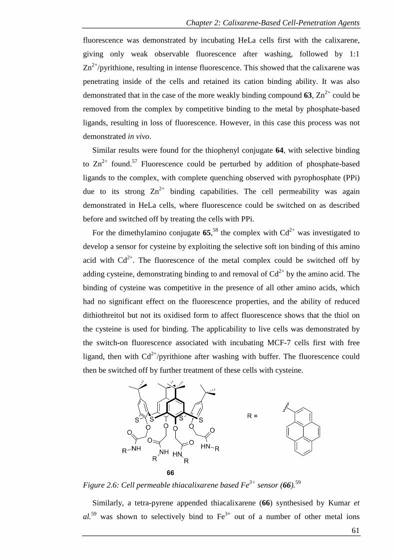
Chapter 2: Calixarene-Based Cell-Penetration Agents
61
fluorescence was demonstrated by incubating HeLa cells first with the calixarene,
giving only weak observable fluorescence after washing, followed by 1:1
Zn2+/pyrithione, resulting in intense fluorescence. This showed that the calixarene was
penetrating inside of the cells and retained its cation binding ability. It was also
demonstrated that in the case of the more weakly binding compound 63, Zn2+ could be
removed from the complex by competitive binding to the metal by phosphate-based
ligands, resulting in loss of fluorescence. However, in this case this process was not
demonstrated in vivo.
Similar results were found for the thiophenyl conjugate 64, with selective binding
to Zn2+ found.57 Fluorescence could be perturbed by addition of phosphate-based
ligands to the complex, with complete quenching observed with pyrophosphate (PPi)
due to its strong Zn2+ binding capabilities. The cell permeability was again
demonstrated in HeLa cells, where fluorescence could be switched on as described
before and switched off by treating the cells with PPi.
For the dimethylamino conjugate 65,58 the complex with Cd2+ was investigated to
develop a sensor for cysteine by exploiting the selective soft ion binding of this amino
acid with Cd2+. The fluorescence of the metal complex could be switched off by
adding cysteine, demonstrating binding to and removal of Cd2+ by the amino acid. The
binding of cysteine was competitive in the presence of all other amino acids, which
had no significant effect on the fluorescence properties, and the ability of reduced
dithiothreitol but not its oxidised form to affect fluorescence shows that the thiol on
the cysteine is used for binding. The applicability to live cells was demonstrated by
the switch-on fluorescence associated with incubating MCF-7 cells first with free
ligand, then with Cd2+/pyrithione after washing with buffer. The fluorescence could
then be switched off by further treatment of these cells with cysteine.
Figure 2.6: Cell permeable thiacalixarene based Fe3+ sensor (66).59
Similarly, a tetra-pyrene appended thiacalixarene (66) synthesised by Kumar et
al.59 was shown to selectively bind to Fe3+ out of a number of other metal ions
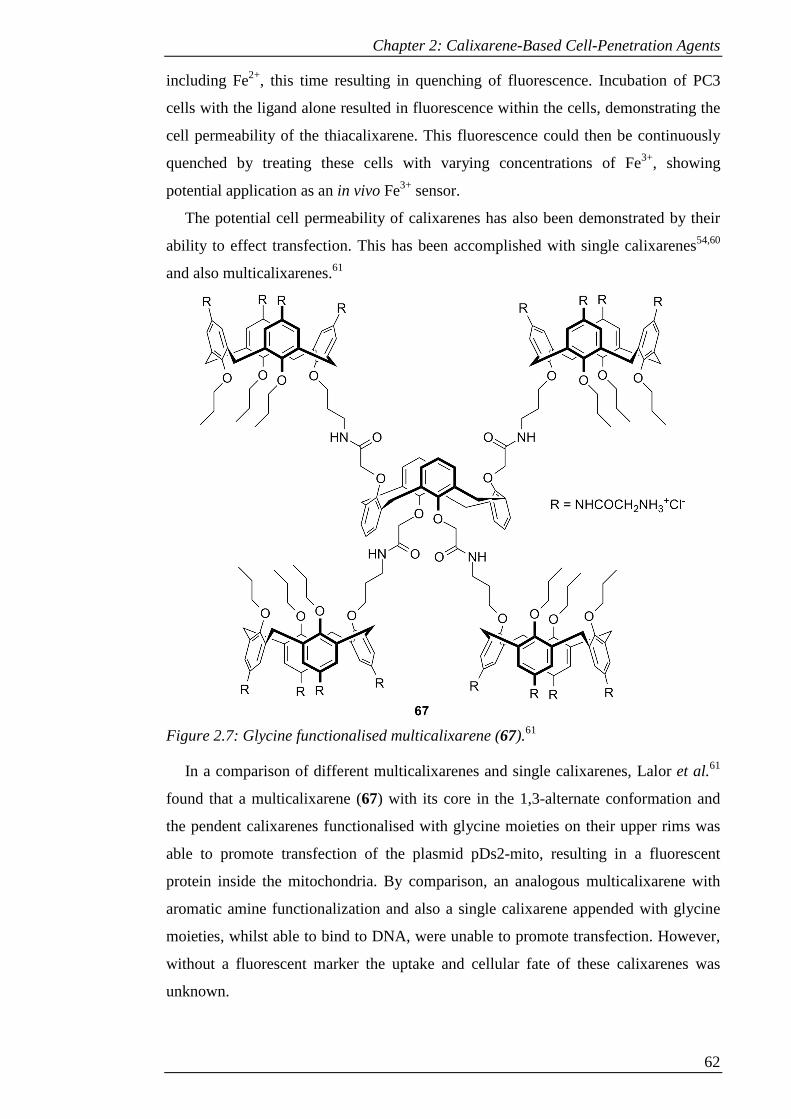
Chapter 2: Calixarene-Based Cell-Penetration Agents
62
including Fe2+, this time resulting in quenching of fluorescence. Incubation of PC3
cells with the ligand alone resulted in fluorescence within the cells, demonstrating the
cell permeability of the thiacalixarene. This fluorescence could then be continuously
quenched by treating these cells with varying concentrations of Fe3+, showing
potential application as an in vivo Fe3+ sensor.
The potential cell permeability of calixarenes has also been demonstrated by their
ability to effect transfection. This has been accomplished with single calixarenes54,60
and also multicalixarenes.61
Figure 2.7: Glycine functionalised multicalixarene (67).61
In a comparison of different multicalixarenes and single calixarenes, Lalor et al.61
found that a multicalixarene (67) with its core in the 1,3-alternate conformation and
the pendent calixarenes functionalised with glycine moieties on their upper rims was
able to promote transfection of the plasmid pDs2-mito, resulting in a fluorescent
protein inside the mitochondria. By comparison, an analogous multicalixarene with
aromatic amine functionalization and also a single calixarene appended with glycine
moieties, whilst able to bind to DNA, were unable to promote transfection. However,
without a fluorescent marker the uptake and cellular fate of these calixarenes was
unknown.
Chapter 2: Calixarene-Based Cell-Penetration Agents
63
In other work on single calixarenes, Sansone et al.60 investigated the ability of
calixarenes functionalised with aromatic guanidinium groups on the upper rim to
effect transfection. They found that the rigid scaffold provided by calix[4]arene
coupled with hexyl or octyl chains on the lower rim (68) gave a suitable, pre-
organised amphiphilic molecule capable of driving DNA condensation by a
combination of electrostatic and hydrophobic interactions, and promoting transfection.
However, the transfection efficiency of pEGFP-C1, a plasmid coding for green
fluorescent protein (GFP), was low and the cytotoxicity too high, with cell death
occurring at a concentration of 40 µM. Further investigation of cell uptake was not
carried out on this compound.
Further work54 was carried out on calixarenes with the guanidinium moieties
placed on the lower rim via a C3 spacer. A calixarene of this form with no upper rim
functionalization (69) exhibited low cytotoxicity, with 70-75% viable cells at 10 µM
concentration. However, the presence of tert-butyl or hexyl groups on the upper rim
gave molecules that, despite having similarly low toxicity to 69 when simply
incubated with cells, suffered increased cytotoxicity under transfection conditions.
In contrast with the poor transfection ability of 68, when used with the helper lipid
DOPE (dioleoylphosphatidylethanolamine) 69 was able to efficiently transfect
pEGFP-C1 into RD-4 cells. It was rationalised that the greater flexibility of the
cationic head groups when on the lower rim may improve DNA binding. Also, the
hydrophobic cavity of 69 may give better interactions with DOPE than its aromatic
guanidinium counterpart 68 (supported by the lack of any effect of DOPE on
transfection with the latter), giving an assembly with superior transfection properties
and lower toxicity.
The mechanism of uptake of the assembly was investigated by using inhibitors of
different pathways of endocytosis. It was found that inhibitors of macropinocytosis
(wortmannin and amiloride) and caveolae (filipin) had the most effect on supressing
transfection, suggesting that these are the primary and secondary uptake mechanisms,
respectively. No suppression of transfection was found with inhibitors of clathrin
mediated endocytosis.
It was also found that replacing the guanidinium groups with amino moieties gave
poor DNA binding properties, an inability to condense DNA and a loss of transfection
capabilities. However the cellular uptake of this derivative is unknown.
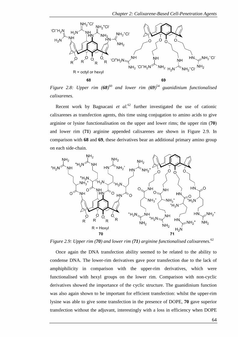
Chapter 2: Calixarene-Based Cell-Penetration Agents
64
Figure 2.8: Upper rim (68)60 and lower rim (69)54 guanidinium functionalised
calixarenes.
Recent work by Bagnacani et al.62 further investigated the use of cationic
calixarenes as transfection agents, this time using conjugation to amino acids to give
arginine or lysine functionalisation on the upper and lower rims; the upper rim (70)
and lower rim (71) arginine appended calixarenes are shown in Figure 2.9. In
comparison with 68 and 69, these derivatives bear an additional primary amino group
on each side-chain.
Figure 2.9: Upper rim (70) and lower rim (71) arginine functionalised calixarenes.62
Once again the DNA transfection ability seemed to be related to the ability to
condense DNA. The lower-rim derivatives gave poor transfection due to the lack of
amphiphilicity in comparison with the upper-rim derivatives, which were
functionalised with hexyl groups on the lower rim. Comparison with non-cyclic
derivatives showed the importance of the cyclic structure. The guanidinium function
was also again shown to be important for efficient transfection: whilst the upper-rim
lysine was able to give some transfection in the presence of DOPE, 70 gave superior
transfection without the adjuvant, interestingly with a loss in efficiency when DOPE

Chapter 2: Calixarene-Based Cell-Penetration Agents
65
was used. Remarkably, 70 was superior or at least comparable with commercially
available LTX lipofectamine in most cell lines tested, with similar impact on cell
viability under transfection conditions.
Another example of a cell permeable calixarene is an amphiphilic cationic
calixarene that was synthesised as part of a synthetic membrane.63 A combination of
calixarenes functionalised at the upper rim with trimethylamine (cationic) or
phosphate (anionic) groups, with dodecyl chains on the lower rim, were used to form
vesicles that were stabilised by electrostatic interactions. Although the cationic
calixarenes on their own formed only micelles, a dye appended analogue (72, see
Figure 2.10), with one cationic group replaced with a triazole linked dye, was
synthesised and the cell permeability evaluated. It was found to penetrate into PC-12
cells efficiently and localise within the cytoplasm. However, the mechanism of uptake
was not investigated. The cytotoxicity of both the cationic and anionic forms of these
calixarenes was relatively high. It was therefore decided that the less toxic octyl-
appended derivatives would be more suitable, although the toxicity of the dodecyl
derivatives could potentially be exploited if the vesicles stayed intact until reaching
their target.
Figure 2.10: Amphiphilic dye-appended calixarene (72),63 amphiphilic micelle-
forming calixarene (73),64 vanadyl sulfonylcalixarene (74),65 nitrobenzoxadiazole-
(NBD)-appended cationic calixarene (75).55,66
Chapter 2: Calixarene-Based Cell-Penetration Agents
66
A similar calixarene was synthesised by Rodik et al.64 with octyl chains on the
lower rim and hydroxylethylammonium moieties on the upper rim (73). These, too,
formed micelles, but in this case the utility of the cationic micelles as transfection
agents was demonstrated. Octyl chains gave superior micelle formation as well as
better DNA binding and condensation properties. When DNA was bound, the micelles
were preserved, even in the presence of DOPE. It was proposed that nanoparticles are
formed with the DNA molecules wrapped around and within the micelles. These
nanoparticles were able to transfect DNA and the process was followed with a
fluorescent DNA marker, showing slow adhesion to the cell membrane, followed by
internalisation and ultimately accumulation around the nucleus. The slow uptake and
relatively low transection efficiency however showed a need for optimisation of the
structure in the future.
Vanadyl calixarenes have also been investigated due to the potential apoptotic and
anti-tumour activities of vanadium complexes.65 A fluorescent vanadyl
sulfonylcalixarene complex (74) was found to penetrate into CHO cells with slow
uptake over 3-4 hours. Incubation with various endocytotic inhibitors (sucrose,
nystatin, monensin and methyl-β-cyclodextrin) found no inhibition of uptake,
suggesting direct translocation across the cell membrane. The complex localised
within the cytoplasm. This particular complex showed low toxicity in all cell types
screened, but other complexes investigated displayed different cytotoxicities in
different cell lines, suggesting that vanadyl calixarenes may have potential use as
selective cytotoxic agents.
A more detailed analysis of cell uptake and localisation was carried out in the case
of 75, a dye-appended cationic calixarene derivative synthesised as a mimic for cell
penetrating peptides with the cationic moieties clustered on a single face of a rigid
scaffold. Preliminary work55 on the this molecule, named NBDCalAm, investigated
the cellular uptake of the calixarene in formaldehyde-fixed cells. It was found that
cellular uptake was rapid, with images available in 10 minutes, and the fluorescence
from the probe was stable, with images still available after 60 minutes. Furthermore
there was no cytotoxic effect from the probe at the concentrations required for the
study.
The mechanism of cellular uptake was investigated with inhibitors of caveolae-
linked endocytosis and lipid raft processes (filipin, β-cyclodextrin) and clathrin
mediated endocytosis (sucrose). None of these gave inhibition of uptake, indicating
that the main endocytotic pathways were not responsible for uptake. Using a
Chapter 2: Calixarene-Based Cell-Penetration Agents
67
counterstain against a receptor localised on the cell membrane, it was found that the
probe accumulates in the cytoplasm and not in the cell membrane. It was also shown
that there was no accumulation in the nucleus.
Following on from this, studies were carried out on live cells to ensure that the
uptake mechanism and localisation were not artefacts of the fixing process.66 The
rapid uptake (1-3 minutes), stable fluorescence (even after 72 hours) and low
cytotoxity were confirmed with live cells. Again inhibitors of clathrin mediated
endocytosis (sucrose) and caveolae mediated processes (filipin, methyl-β-
cyclodextrin) failed to inhibit uptake, implicating direct translocation across the cell
membrane.
Co-localisation studies with Golgi apparatus and lysosome stains showed that the
probe initially localised to the Golgi apparatus (after 2-3 hours) then showed
localisation to the lysosomes (from 2-3 hours up to 72 hours). The route of transfer to
the lysosomes via the Golgi apparatus was confirmed by the inhibition of the process
by brefeldin A and its lack of effect when the probe was already localised within the
lysosomes prior to brefeldin A treatment. Further studies with monensin (which
affects endosomal pH), bafilomycin (an inhibitor of vacuolar ATPases) and sodium
azide (which depletes the available ATP pool) both prevented localisation in the
lysosomes and stimulated release of the probe from within them. This indicates that
these three factors are important both for the transfer to and the retention within the
lysosomes.
To further this work, the effect of using different cationic groups on the upper rim
and introducing different dye molecules was investigated. This will be the subject of
the remainder of Chapter 2.
2.2 Aims
Based on the previous work of our research group on NBDCalAm (as described in
Section 2.2.2.1), further investigation was required as to the effects of different
functionalization of the calixarene scaffold, both in the cationic groups on the upper
rim and in the cargo attached to the lower rim.
To alter the properties of the upper rim, two targets were proposed in addition to
the original aromatic amine: an aliphatic amine, accessible by furnishing the upper rim
with glycine residues, and a guanidinium derivative as a mimic of poly-arginine cell
penetrating peptides. Although the syntheses of both were previously attempted (R.
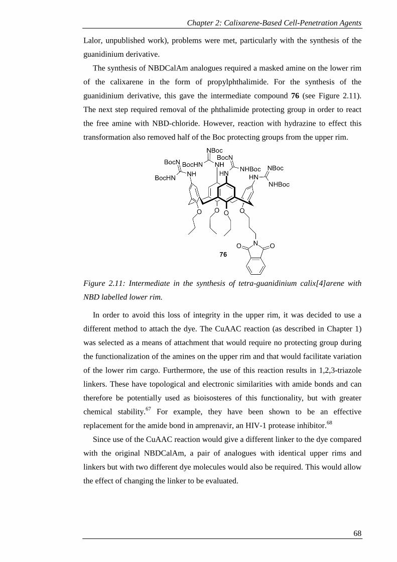
Chapter 2: Calixarene-Based Cell-Penetration Agents
68
Lalor, unpublished work), problems were met, particularly with the synthesis of the
guanidinium derivative.
The synthesis of NBDCalAm analogues required a masked amine on the lower rim
of the calixarene in the form of propylphthalimide. For the synthesis of the
guanidinium derivative, this gave the intermediate compound 76 (see Figure 2.11).
The next step required removal of the phthalimide protecting group in order to react
the free amine with NBD-chloride. However, reaction with hydrazine to effect this
transformation also removed half of the Boc protecting groups from the upper rim.
Figure 2.11: Intermediate in the synthesis of tetra-guanidinium calix[4]arene with
NBD labelled lower rim.
In order to avoid this loss of integrity in the upper rim, it was decided to use a
different method to attach the dye. The CuAAC reaction (as described in Chapter 1)
was selected as a means of attachment that would require no protecting group during
the functionalization of the amines on the upper rim and that would facilitate variation
of the lower rim cargo. Furthermore, the use of this reaction results in 1,2,3-triazole
linkers. These have topological and electronic similarities with amide bonds and can
therefore be potentially used as bioisosteres of this functionality, but with greater
chemical stability.67 For example, they have been shown to be an effective
replacement for the amide bond in amprenavir, an HIV-1 protease inhibitor.68
Since use of the CuAAC reaction would give a different linker to the dye compared
with the original NBDCalAm, a pair of analogues with identical upper rims and
linkers but with two different dye molecules would also be required. This would allow
the effect of changing the linker to be evaluated.
Chapter 2: Calixarene-Based Cell-Penetration Agents
69
In summary, the aims for this project were as follows:
• To synthesise three analogues with variation in the upper rim, i.e. aromatic
amine, aliphatic amine and guanidine functionalization, with a dye
molecule added to the lower rim by click chemistry;
• To synthesise analogues of the aromatic amine with different dye molecules
added to the lower rim by click chemistry;
• To synthesise a pair of analogues with aromatic amine functionalised upper
rims, with an identical linker on the lower rim but with two different dye
molecules, one of which would preferably be the original NBD dye to allow
for comparison with NBDCalAm.
2.3 Results and Discussion
2.3.1 Synthesis of water soluble click conjugates
In order to add a dye molecule by the CuAAC reaction, either an alkyne or an azide
was required on the calixarene to react with either an azide or alkyne functionalised
dye. Since an alkyne could be easily added to the calixarene in a single step using
readily available propargyl bromide and azide derivatives of several dye molecules
could be synthesised,69–71 this is the route that was taken.
2.3.1.1 Synthesis of common intermediate
To introduce the required single propargyl group to the lower rim of calix[4]arene,
two routes could have been followed. The first is to mono-propargylate the lower rim,
then add the three propyl chains to lock the conformation. However, this method is not
ideal as some of the 1,3-dialkyl species can be formed.72 Therefore it was decided to
use a selective tri-alkylation method, followed by introduction of the propargyl
group.73 For both alkylations, the Williamson ether synthesis can be used. This
involves treating an alcohol with a base to produce an alkoxide anion, then addition of
an alkyl halide, which reacts via an SN2 type reaction.
The first step was to form the calix[4]arene starting material using the method of
Gutsche et al.74 A mixture of para-tert-butyl-phenol, formaldehyde solution in water
and sodium hydroxide were heated to 120 °C and mechanically stirred until the
intermediate polymer was formed. This was dissolved in diphenyl ether, the water of
condensation driven off and the mixture heated to reflux to form the cracked product.
Precipitation with ethyl acetate gave 2 as off-white crystals in 67% yield, which was
used without further purification.
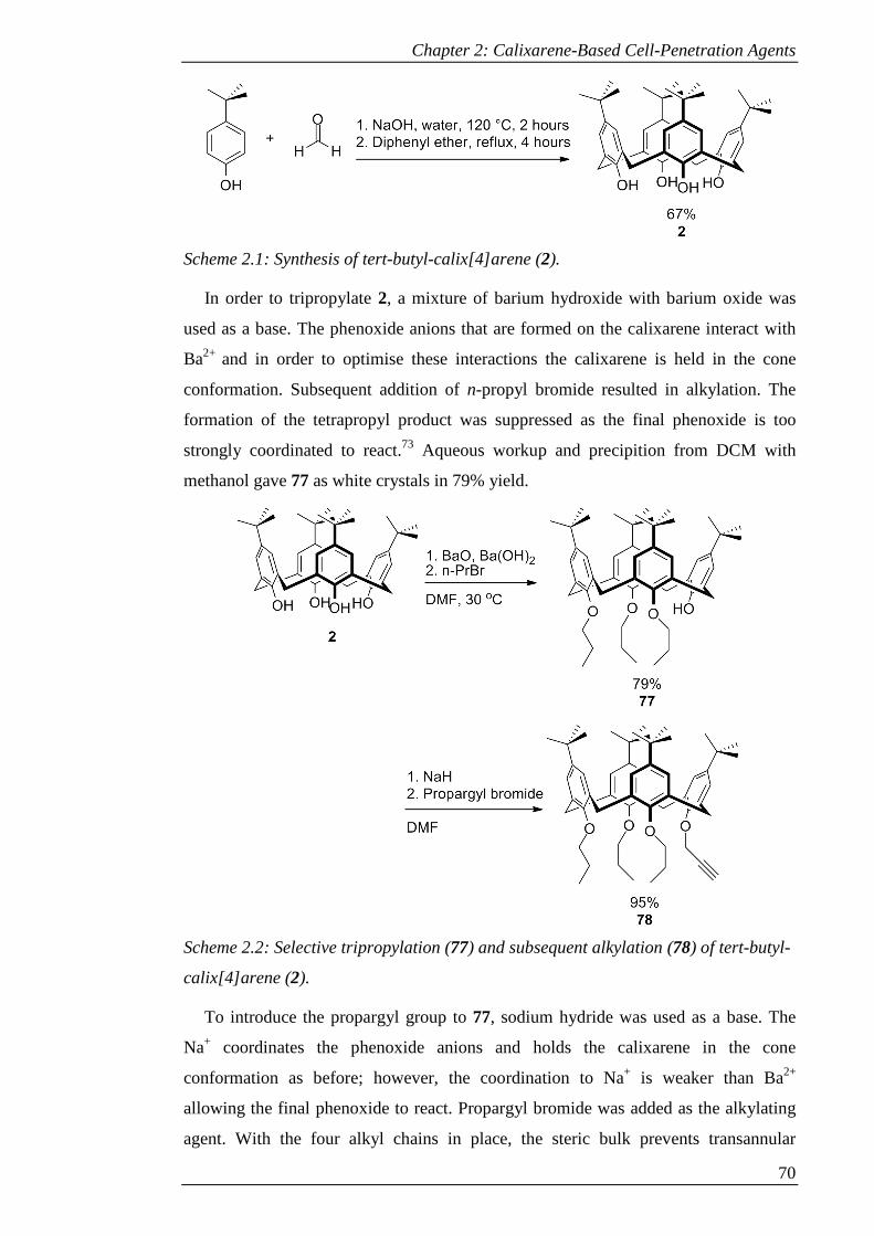
Chapter 2: Calixarene-Based Cell-Penetration Agents
70
Scheme 2.1: Synthesis of tert-butyl-calix[4]arene (2).
In order to tripropylate 2, a mixture of barium hydroxide with barium oxide was
used as a base. The phenoxide anions that are formed on the calixarene interact with
Ba2+ and in order to optimise these interactions the calixarene is held in the cone
conformation. Subsequent addition of n-propyl bromide resulted in alkylation. The
formation of the tetrapropyl product was suppressed as the final phenoxide is too
strongly coordinated to react.73 Aqueous workup and precipition from DCM with
methanol gave 77 as white crystals in 79% yield.
Scheme 2.2: Selective tripropylation (77) and subsequent alkylation (78) of tert-butyl-
calix[4]arene (2).
To introduce the propargyl group to 77, sodium hydride was used as a base. The
Na+ coordinates the phenoxide anions and holds the calixarene in the cone
conformation as before; however, the coordination to Na+ is weaker than Ba2+
allowing the final phenoxide to react. Propargyl bromide was added as the alkylating
agent. With the four alkyl chains in place, the steric bulk prevents transannular
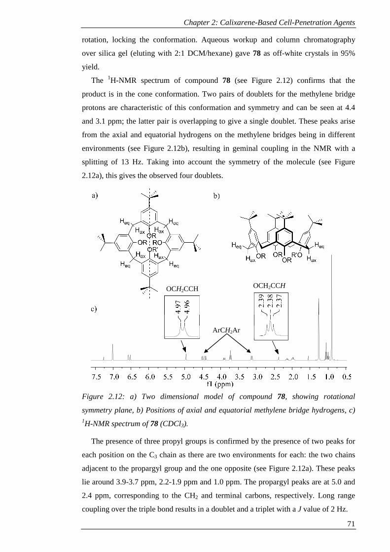
Chapter 2: Calixarene-Based Cell-Penetration Agents
71
rotation, locking the conformation. Aqueous workup and column chromatography
over silica gel (eluting with 2:1 DCM/hexane) gave 78 as off-white crystals in 95%
yield.
The 1H-NMR spectrum of compound 78 (see Figure 2.12) confirms that the
product is in the cone conformation. Two pairs of doublets for the methylene bridge
protons are characteristic of this conformation and symmetry and can be seen at 4.4
and 3.1 ppm; the latter pair is overlapping to give a single doublet. These peaks arise
from the axial and equatorial hydrogens on the methylene bridges being in different
environments (see Figure 2.12b), resulting in geminal coupling in the NMR with a
splitting of 13 Hz. Taking into account the symmetry of the molecule (see Figure
2.12a), this gives the observed four doublets.
Figure 2.12: a) Two dimensional model of compound 78, showing rotational
symmetry plane, b) Positions of axial and equatorial methylene bridge hydrogens, c) 1H-NMR spectrum of 78 (CDCl3).
The presence of three propyl groups is confirmed by the presence of two peaks for
each position on the C3 chain as there are two environments for each: the two chains
adjacent to the propargyl group and the one opposite (see Figure 2.12a). These peaks
lie around 3.9-3.7 ppm, 2.2-1.9 ppm and 1.0 ppm. The propargyl peaks are at 5.0 and
2.4 ppm, corresponding to the CH2 and terminal carbons, respectively. Long range
coupling over the triple bond results in a doublet and a triplet with a J value of 2 Hz.
OCH2CCH OCH2CCH
ArCH2Ar
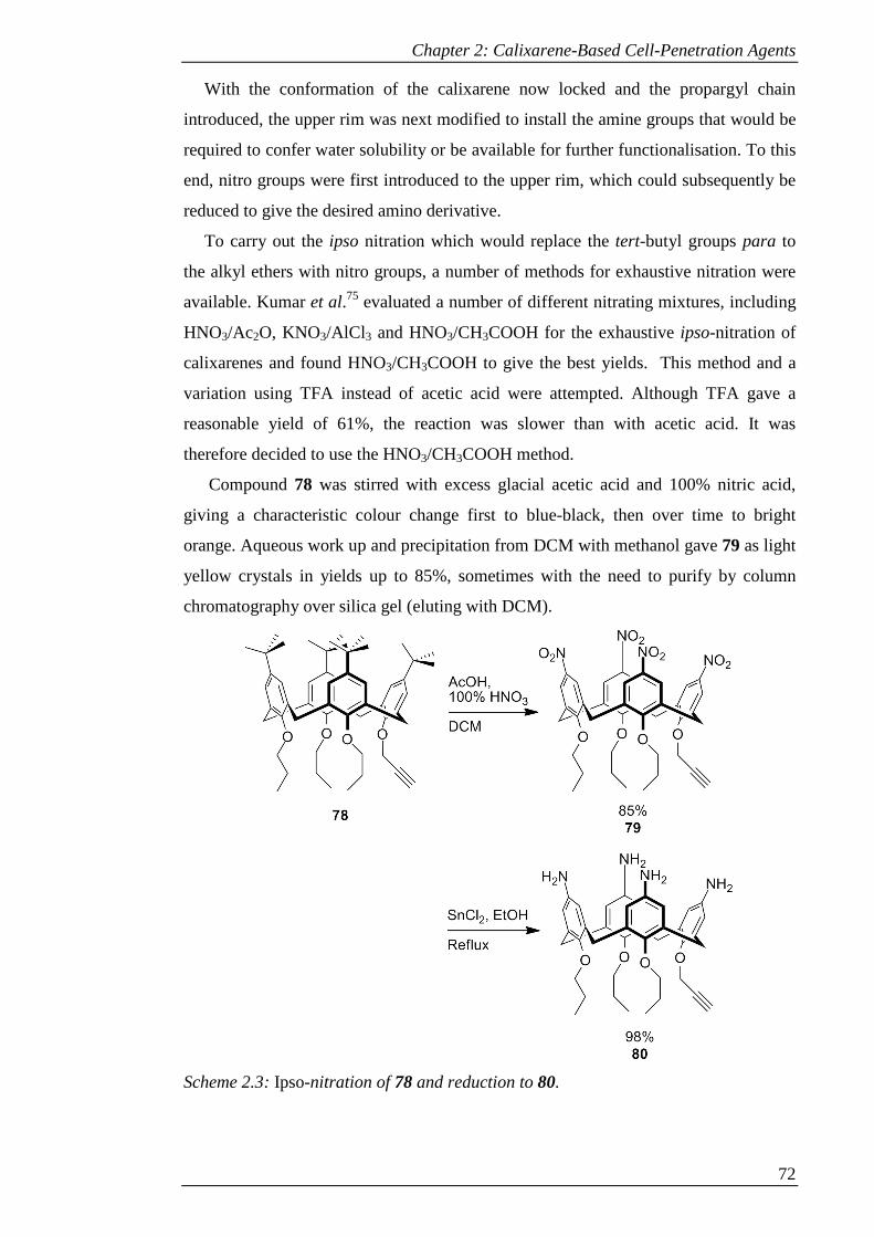
Chapter 2: Calixarene-Based Cell-Penetration Agents
72
With the conformation of the calixarene now locked and the propargyl chain
introduced, the upper rim was next modified to install the amine groups that would be
required to confer water solubility or be available for further functionalisation. To this
end, nitro groups were first introduced to the upper rim, which could subsequently be
reduced to give the desired amino derivative.
To carry out the ipso nitration which would replace the tert-butyl groups para to
the alkyl ethers with nitro groups, a number of methods for exhaustive nitration were
available. Kumar et al.75 evaluated a number of different nitrating mixtures, including
HNO3/Ac2O, KNO3/AlCl 3 and HNO3/CH3COOH for the exhaustive ipso-nitration of
calixarenes and found HNO3/CH3COOH to give the best yields. This method and a
variation using TFA instead of acetic acid were attempted. Although TFA gave a
reasonable yield of 61%, the reaction was slower than with acetic acid. It was
therefore decided to use the HNO3/CH3COOH method.
Compound 78 was stirred with excess glacial acetic acid and 100% nitric acid,
giving a characteristic colour change first to blue-black, then over time to bright
orange. Aqueous work up and precipitation from DCM with methanol gave 79 as light
yellow crystals in yields up to 85%, sometimes with the need to purify by column
chromatography over silica gel (eluting with DCM).
Scheme 2.3: Ipso-nitration of 78 and reduction to 80.
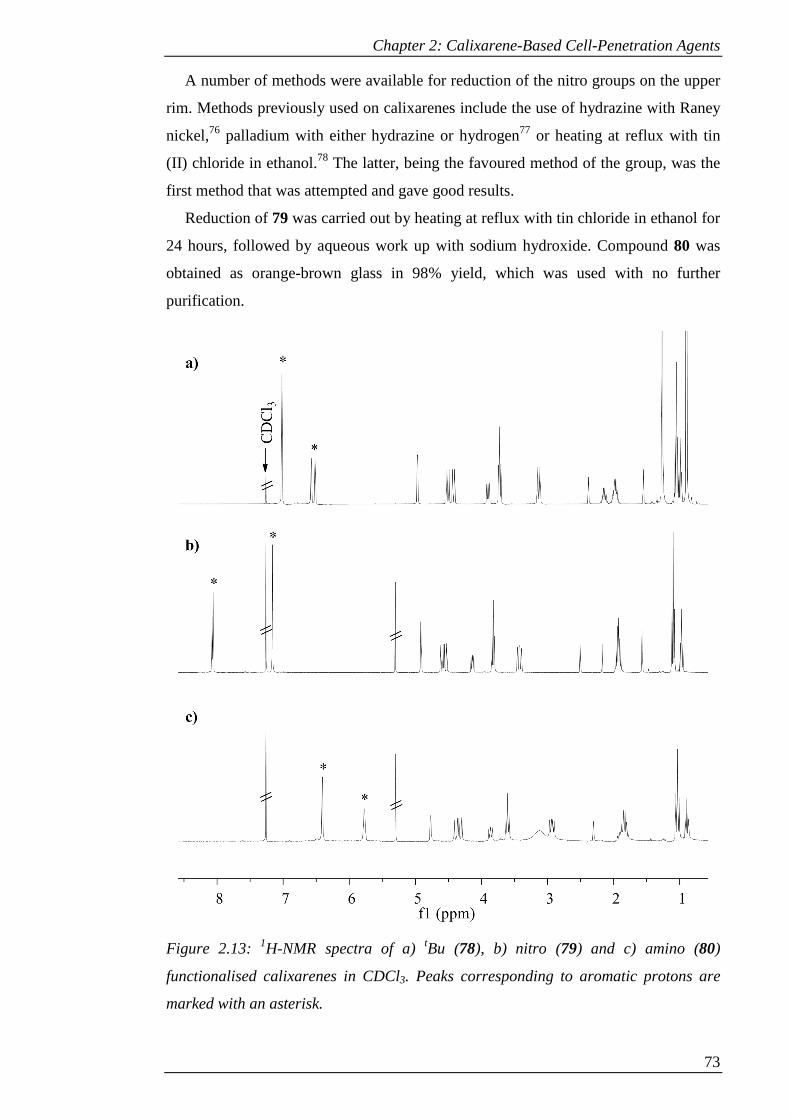
Chapter 2: Calixarene-Based Cell-Penetration Agents
73
A number of methods were available for reduction of the nitro groups on the upper
rim. Methods previously used on calixarenes include the use of hydrazine with Raney
nickel,76 palladium with either hydrazine or hydrogen77 or heating at reflux with tin
(II) chloride in ethanol.78 The latter, being the favoured method of the group, was the
first method that was attempted and gave good results.
Reduction of 79 was carried out by heating at reflux with tin chloride in ethanol for
24 hours, followed by aqueous work up with sodium hydroxide. Compound 80 was
obtained as orange-brown glass in 98% yield, which was used with no further
purification.
Figure 2.13: 1H-NMR spectra of a) tBu (78), b) nitro (79) and c) amino (80)
functionalised calixarenes in CDCl3. Peaks corresponding to aromatic protons are
marked with an asterisk.
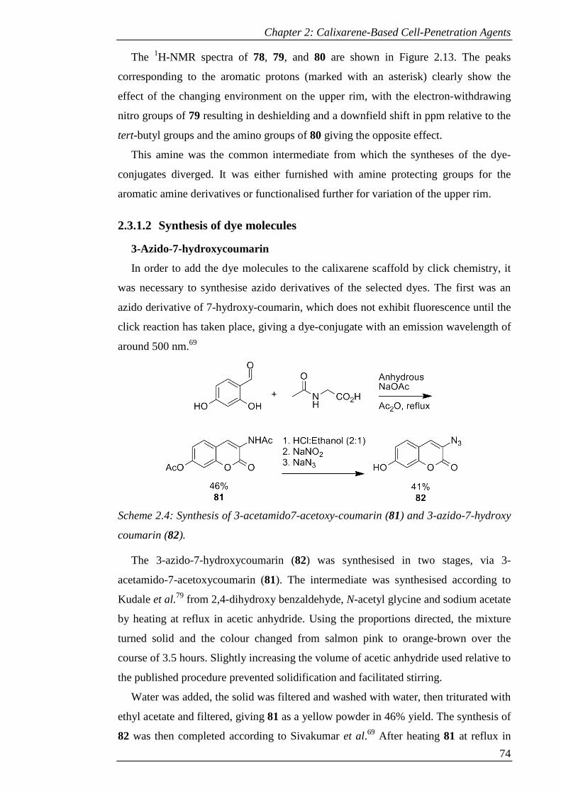
Chapter 2: Calixarene-Based Cell-Penetration Agents
74
The 1H-NMR spectra of 78, 79, and 80 are shown in Figure 2.13. The peaks
corresponding to the aromatic protons (marked with an asterisk) clearly show the
effect of the changing environment on the upper rim, with the electron-withdrawing
nitro groups of 79 resulting in deshielding and a downfield shift in ppm relative to the
tert-butyl groups and the amino groups of 80 giving the opposite effect.
This amine was the common intermediate from which the syntheses of the dye-
conjugates diverged. It was either furnished with amine protecting groups for the
aromatic amine derivatives or functionalised further for variation of the upper rim.
2.3.1.2 Synthesis of dye molecules
3-Azido-7-hydroxycoumarin
In order to add the dye molecules to the calixarene scaffold by click chemistry, it
was necessary to synthesise azido derivatives of the selected dyes. The first was an
azido derivative of 7-hydroxy-coumarin, which does not exhibit fluorescence until the
click reaction has taken place, giving a dye-conjugate with an emission wavelength of
around 500 nm.69
Scheme 2.4: Synthesis of 3-acetamido7-acetoxy-coumarin (81) and 3-azido-7-hydroxy
coumarin (82).
The 3-azido-7-hydroxycoumarin (82) was synthesised in two stages, via 3-
acetamido-7-acetoxycoumarin (81). The intermediate was synthesised according to
Kudale et al.79 from 2,4-dihydroxy benzaldehyde, N-acetyl glycine and sodium acetate
by heating at reflux in acetic anhydride. Using the proportions directed, the mixture
turned solid and the colour changed from salmon pink to orange-brown over the
course of 3.5 hours. Slightly increasing the volume of acetic anhydride used relative to
the published procedure prevented solidification and facilitated stirring.
Water was added, the solid was filtered and washed with water, then triturated with
ethyl acetate and filtered, giving 81 as a yellow powder in 46% yield. The synthesis of
82 was then completed according to Sivakumar et al.69 After heating 81 at reflux in
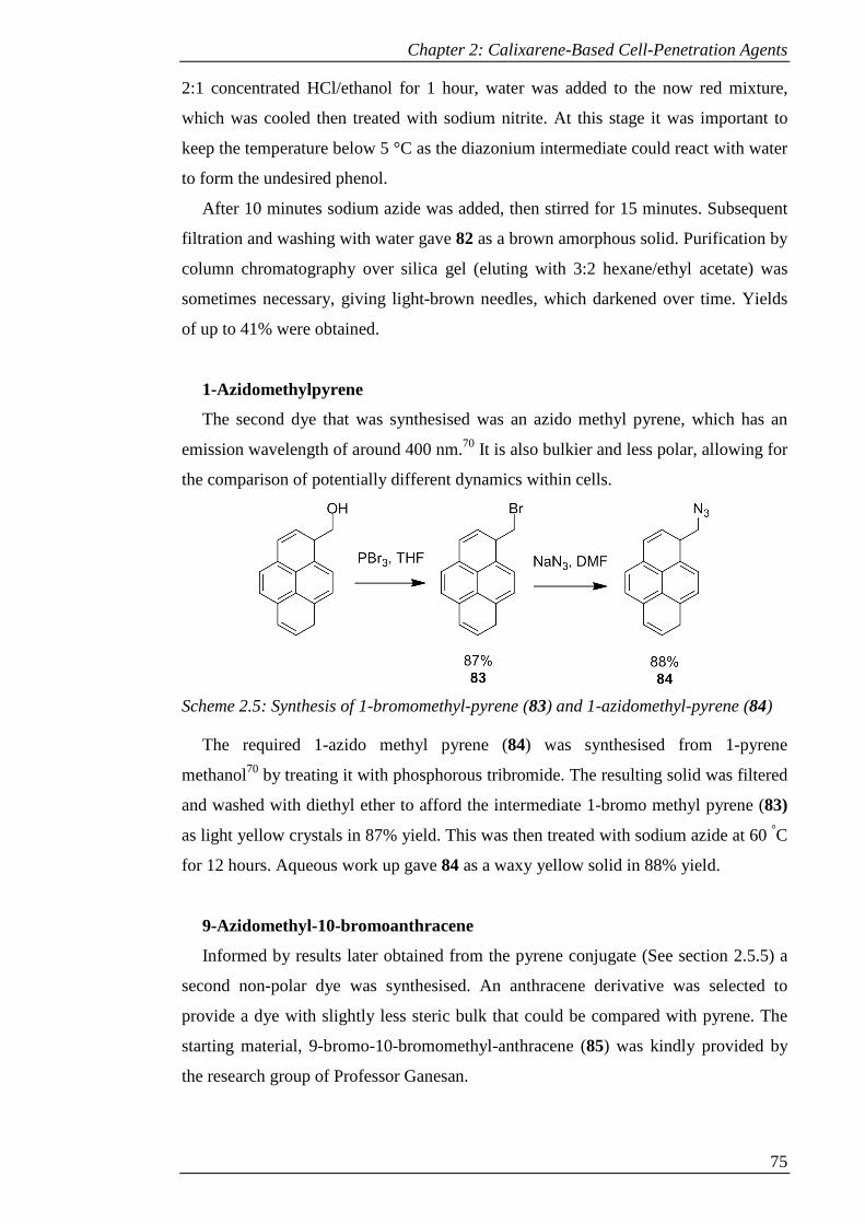
Chapter 2: Calixarene-Based Cell-Penetration Agents
75
2:1 concentrated HCl/ethanol for 1 hour, water was added to the now red mixture,
which was cooled then treated with sodium nitrite. At this stage it was important to
keep the temperature below 5 °C as the diazonium intermediate could react with water
to form the undesired phenol.
After 10 minutes sodium azide was added, then stirred for 15 minutes. Subsequent
filtration and washing with water gave 82 as a brown amorphous solid. Purification by
column chromatography over silica gel (eluting with 3:2 hexane/ethyl acetate) was
sometimes necessary, giving light-brown needles, which darkened over time. Yields
of up to 41% were obtained.
1-Azidomethylpyrene
The second dye that was synthesised was an azido methyl pyrene, which has an
emission wavelength of around 400 nm.70 It is also bulkier and less polar, allowing for
the comparison of potentially different dynamics within cells.
Scheme 2.5: Synthesis of 1-bromomethyl-pyrene (83) and 1-azidomethyl-pyrene (84)
The required 1-azido methyl pyrene (84) was synthesised from 1-pyrene
methanol70 by treating it with phosphorous tribromide. The resulting solid was filtered
and washed with diethyl ether to afford the intermediate 1-bromo methyl pyrene (83)
as light yellow crystals in 87% yield. This was then treated with sodium azide at 60 °C
for 12 hours. Aqueous work up gave 84 as a waxy yellow solid in 88% yield.
9-Azidomethyl-10-bromoanthracene
Informed by results later obtained from the pyrene conjugate (See section 2.5.5) a
second non-polar dye was synthesised. An anthracene derivative was selected to
provide a dye with slightly less steric bulk that could be compared with pyrene. The
starting material, 9-bromo-10-bromomethyl-anthracene (85) was kindly provided by
the research group of Professor Ganesan.
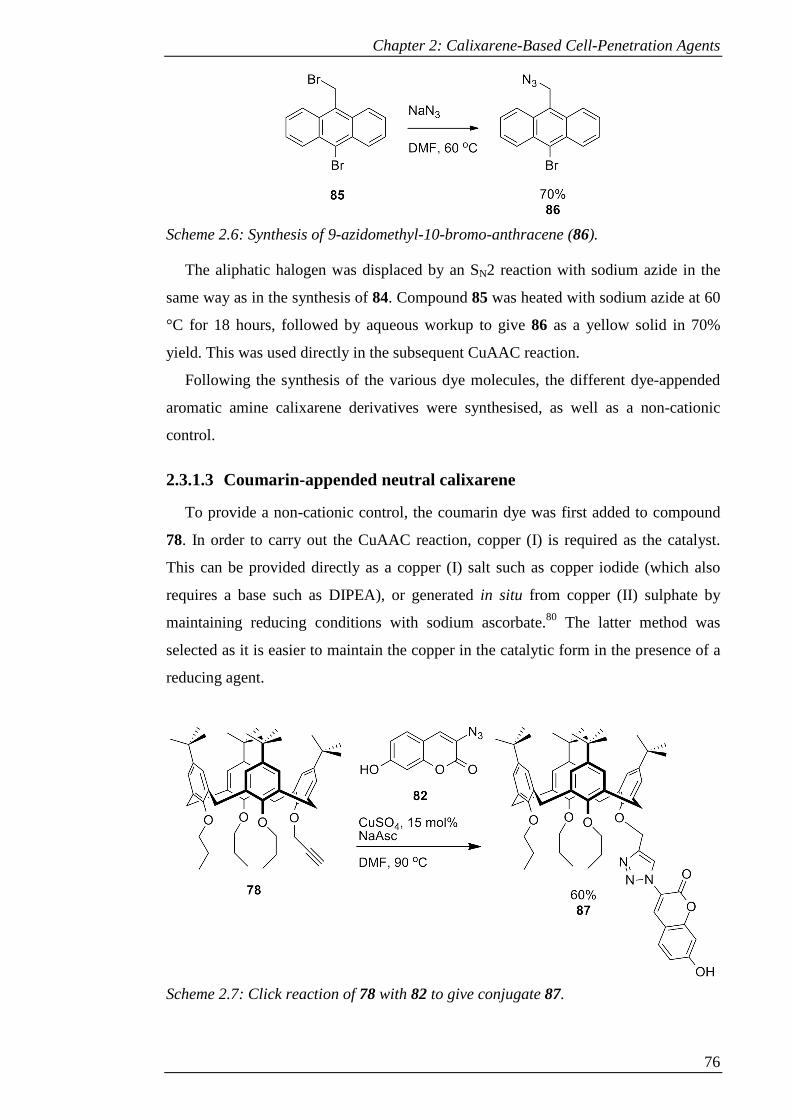
Chapter 2: Calixarene-Based Cell-Penetration Agents
76
Scheme 2.6: Synthesis of 9-azidomethyl-10-bromo-anthracene (86).
The aliphatic halogen was displaced by an SN2 reaction with sodium azide in the
same way as in the synthesis of 84. Compound 85 was heated with sodium azide at 60
°C for 18 hours, followed by aqueous workup to give 86 as a yellow solid in 70%
yield. This was used directly in the subsequent CuAAC reaction.
Following the synthesis of the various dye molecules, the different dye-appended
aromatic amine calixarene derivatives were synthesised, as well as a non-cationic
control.
2.3.1.3 Coumarin-appended neutral calixarene
To provide a non-cationic control, the coumarin dye was first added to compound
78. In order to carry out the CuAAC reaction, copper (I) is required as the catalyst.
This can be provided directly as a copper (I) salt such as copper iodide (which also
requires a base such as DIPEA), or generated in situ from copper (II) sulphate by
maintaining reducing conditions with sodium ascorbate.80 The latter method was
selected as it is easier to maintain the copper in the catalytic form in the presence of a
reducing agent.
Scheme 2.7: Click reaction of 78 with 82 to give conjugate 87.
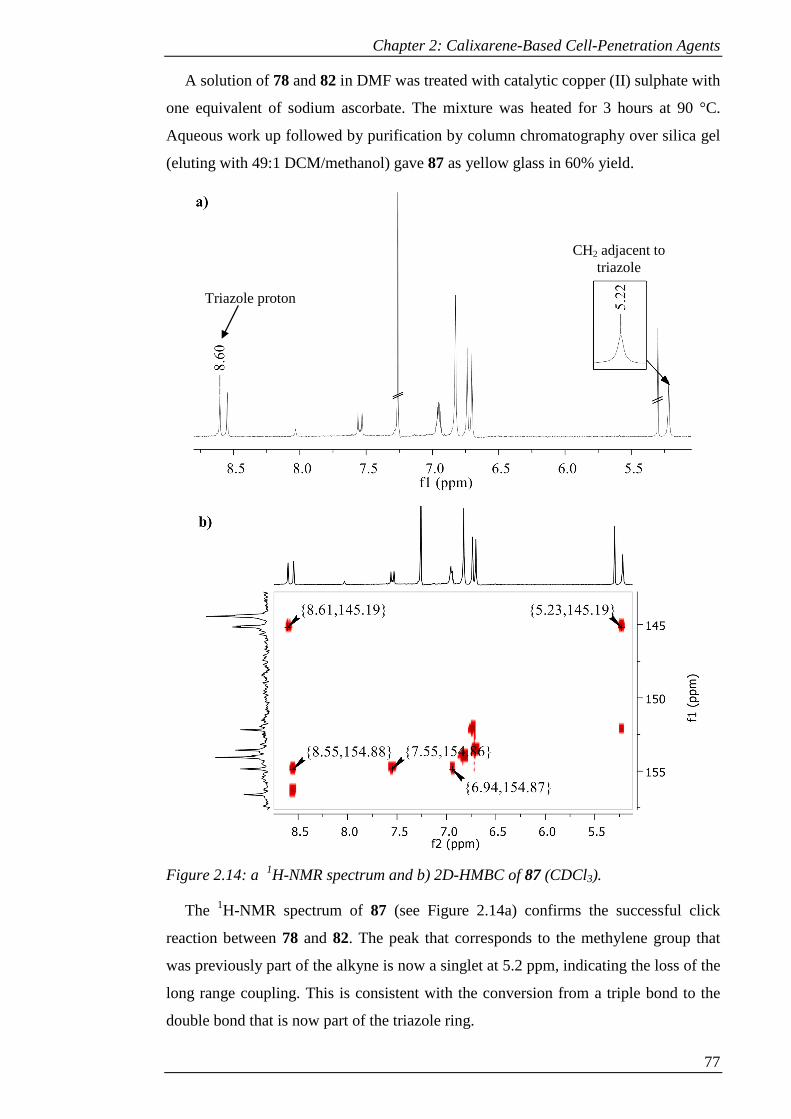
Chapter 2: Calixarene-Based Cell-Penetration Agents
77
A solution of 78 and 82 in DMF was treated with catalytic copper (II) sulphate with
one equivalent of sodium ascorbate. The mixture was heated for 3 hours at 90 °C.
Aqueous work up followed by purification by column chromatography over silica gel
(eluting with 49:1 DCM/methanol) gave 87 as yellow glass in 60% yield.
Figure 2.14: a 1H-NMR spectrum and b) 2D-HMBC of 87 (CDCl3).
The 1H-NMR spectrum of 87 (see Figure 2.14a) confirms the successful click
reaction between 78 and 82. The peak that corresponds to the methylene group that
was previously part of the alkyne is now a singlet at 5.2 ppm, indicating the loss of the
long range coupling. This is consistent with the conversion from a triple bond to the
double bond that is now part of the triazole ring.
Triazole proton
CH2 adjacent to triazole
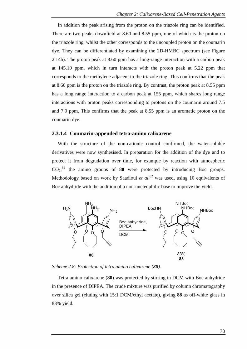
Chapter 2: Calixarene-Based Cell-Penetration Agents
78
In addition the peak arising from the proton on the triazole ring can be identified.
There are two peaks downfield at 8.60 and 8.55 ppm, one of which is the proton on
the triazole ring, whilst the other corresponds to the uncoupled proton on the coumarin
dye. They can be differentiated by examining the 2D-HMBC spectrum (see Figure
2.14b). The proton peak at 8.60 ppm has a long-range interaction with a carbon peak
at 145.19 ppm, which in turn interacts with the proton peak at 5.22 ppm that
corresponds to the methylene adjacent to the triazole ring. This confirms that the peak
at 8.60 ppm is the proton on the triazole ring. By contrast, the proton peak at 8.55 ppm
has a long range interaction to a carbon peak at 155 ppm, which shares long range
interactions with proton peaks corresponding to protons on the coumarin around 7.5
and 7.0 ppm. This confirms that the peak at 8.55 ppm is an aromatic proton on the
coumarin dye.
2.3.1.4 Coumarin-appended tetra-amino calixarene
With the structure of the non-cationic control confirmed, the water-soluble
derivatives were now synthesised. In preparation for the addition of the dye and to
protect it from degradation over time, for example by reaction with atmospheric
CO2,81 the amino groups of 80 were protected by introducing Boc groups.
Methodology based on work by Saadioui et al.82 was used, using 10 equivalents of
Boc anhydride with the addition of a non-nucleophilic base to improve the yield.
Scheme 2.8: Protection of tetra amino calixarene (80).
Tetra amino calixarene (80) was protected by stirring in DCM with Boc anhydride
in the presence of DIPEA. The crude mixture was purified by column chromatography
over silica gel (eluting with 15:1 DCM/ethyl acetate), giving 88 as off-white glass in
83% yield.
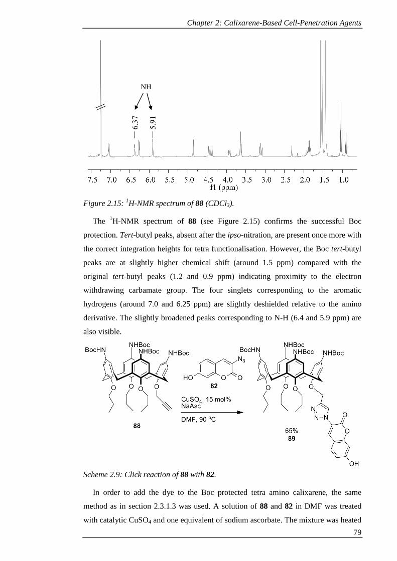
Chapter 2: Calixarene-Based Cell-Penetration Agents
79
Figure 2.15: 1H-NMR spectrum of 88 (CDCl3).
The 1H-NMR spectrum of 88 (see Figure 2.15) confirms the successful Boc
protection. Tert-butyl peaks, absent after the ipso-nitration, are present once more with
the correct integration heights for tetra functionalisation. However, the Boc tert-butyl
peaks are at slightly higher chemical shift (around 1.5 ppm) compared with the
original tert-butyl peaks (1.2 and 0.9 ppm) indicating proximity to the electron
withdrawing carbamate group. The four singlets corresponding to the aromatic
hydrogens (around 7.0 and 6.25 ppm) are slightly deshielded relative to the amino
derivative. The slightly broadened peaks corresponding to N-H (6.4 and 5.9 ppm) are
also visible.
Scheme 2.9: Click reaction of 88 with 82.
In order to add the dye to the Boc protected tetra amino calixarene, the same
method as in section 2.3.1.3 was used. A solution of 88 and 82 in DMF was treated
with catalytic CuSO4 and one equivalent of sodium ascorbate. The mixture was heated
5.9
1
6.3
7
NH

Chapter 2: Calixarene-Based Cell-Penetration Agents
80
for 20 hours at 90 °C. Aqueous work up followed by purification by column
chromatography over silica gel (eluting with 49:1 DCM/methanol) gave 89 as light
yellow glass in 65% yield.
Figure 2.16: 1H-NMR spectrum of 89 (CDCl3).
The 1H-NMR spectrum of 89 (see Figure 2.16) confirms the successful click
reaction between 88 and 82. The peak that corresponds to the methylene group that
was previously part of the alkyne is now a singlet at 5.2 ppm and the peak arising
from the proton on the triazole ring can be seen at 8.50 ppm, whilst the dye peaks lie
at 8.48, 7.5 and 6.8 ppm.
2.3.1.5 Pyrene-appended tetra-amino calixarene
For the CuAAC reaction to add the pyrene dye to 88, the same conditions as in
section 2.3.1.3 were used. A solution of 88 and 84 in DMF was treated with catalytic
CuSO4 and one equivalent of sodium ascorbate. The mixture was heated for 3 hours at
90 °C. Aqueous work up followed by purification by column chromatography over
silica gel (eluting with 97:3 DCM/acetone) gave 90 as light yellow glass in 32% yield.
Some of the starting materials were also recovered, with approximately 20% of the
yield lost due to incomplete reaction.
5.2
0
8.5
0
Triazole proton CH2 adjacent to
triazole
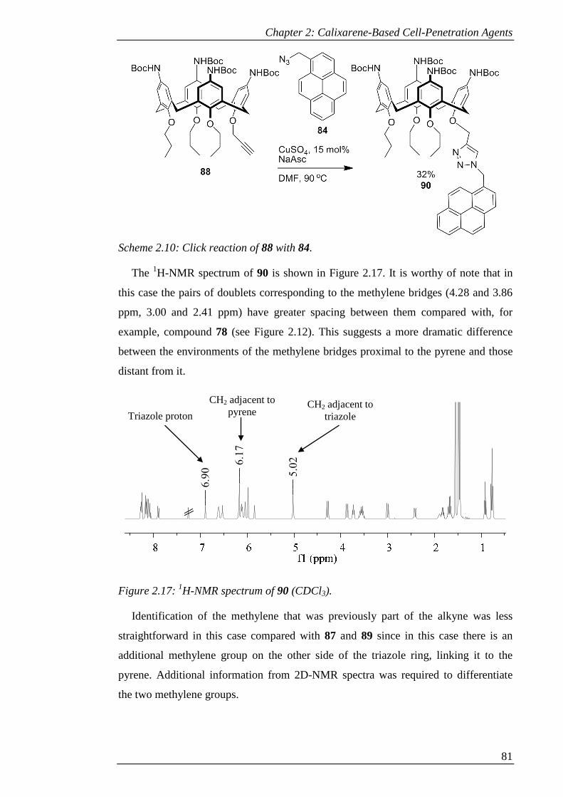
Chapter 2: Calixarene-Based Cell-Penetration Agents
81
Scheme 2.10: Click reaction of 88 with 84.
The 1H-NMR spectrum of 90 is shown in Figure 2.17. It is worthy of note that in
this case the pairs of doublets corresponding to the methylene bridges (4.28 and 3.86
ppm, 3.00 and 2.41 ppm) have greater spacing between them compared with, for
example, compound 78 (see Figure 2.12). This suggests a more dramatic difference
between the environments of the methylene bridges proximal to the pyrene and those
distant from it.
Figure 2.17: 1H-NMR spectrum of 90 (CDCl3).
Identification of the methylene that was previously part of the alkyne was less
straightforward in this case compared with 87 and 89 since in this case there is an
additional methylene group on the other side of the triazole ring, linking it to the
pyrene. Additional information from 2D-NMR spectra was required to differentiate
the two methylene groups.
5.0
26.1
7
6.9
0
CH2 adjacent to triazole
CH2 adjacent to pyrene Triazole proton
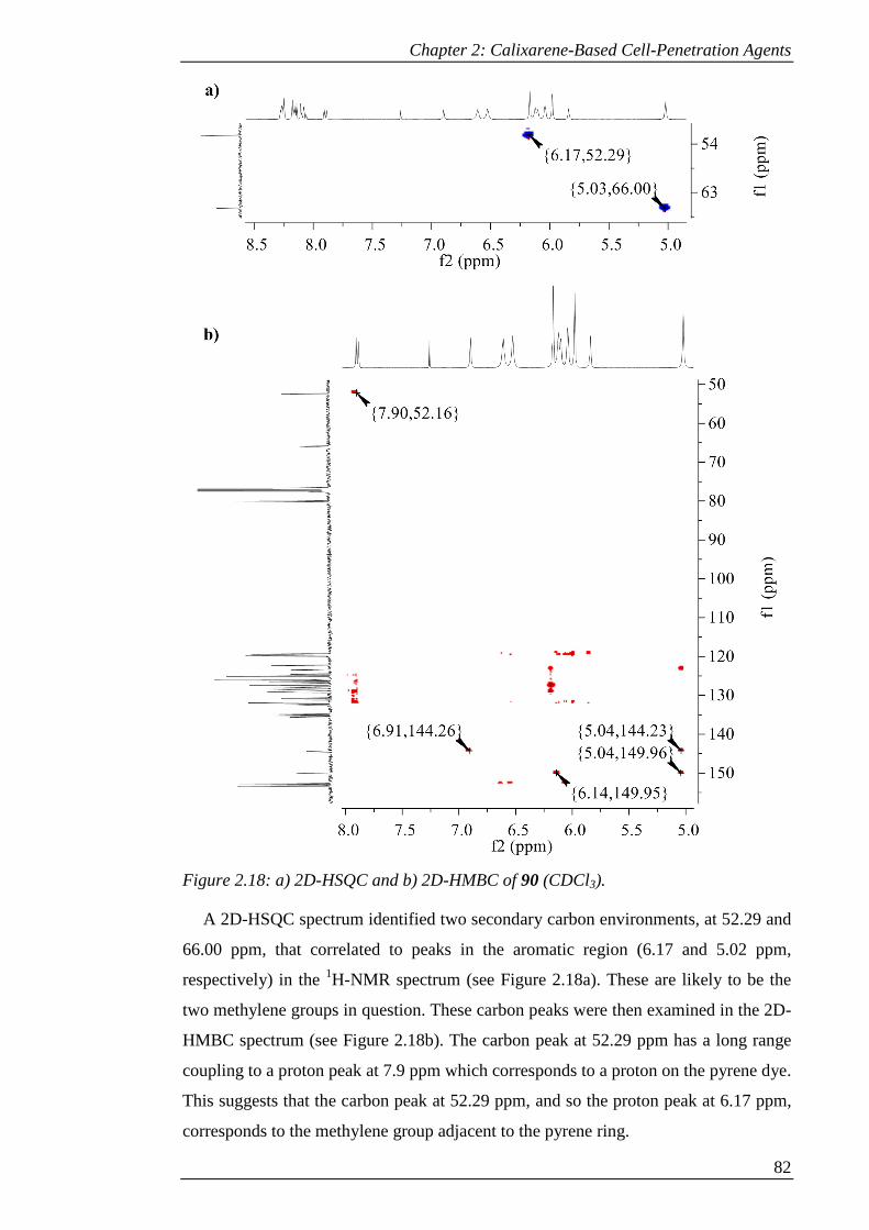
Chapter 2: Calixarene-Based Cell-Penetration Agents
82
Figure 2.18: a) 2D-HSQC and b) 2D-HMBC of 90 (CDCl3).
A 2D-HSQC spectrum identified two secondary carbon environments, at 52.29 and
66.00 ppm, that correlated to peaks in the aromatic region (6.17 and 5.02 ppm,
respectively) in the 1H-NMR spectrum (see Figure 2.18a). These are likely to be the
two methylene groups in question. These carbon peaks were then examined in the 2D-
HMBC spectrum (see Figure 2.18b). The carbon peak at 52.29 ppm has a long range
coupling to a proton peak at 7.9 ppm which corresponds to a proton on the pyrene dye.
This suggests that the carbon peak at 52.29 ppm, and so the proton peak at 6.17 ppm,
corresponds to the methylene group adjacent to the pyrene ring.
Chapter 2: Calixarene-Based Cell-Penetration Agents
83
The proton peak at 5.02 ppm should, by elimination, be the methylene group
between the calixarene core and the triazole ring. Further information from the 2D-
HMBC shows a long range interaction between this proton environment and a carbon
at 149.96 ppm, which in turn has an interaction with a proton peak at 6.14, which
corresponds to an aromatic proton on the calixarene. This mutual interaction supports
this assignment of the peak at 5.02 ppm.
Finally, the 2D-HMBC also allows the peak corresponding to the proton on the
triazole ring to be confirmed. The proton peak at 6.90 ppm which corresponds to a
single proton is a likely candidate. This peak has a long range interaction to the carbon
peak at 144.26 ppm. Since the proton peak at 5.02 ppm also has an interaction with
this carbon, the peak at 6.90 ppm must correspond to the triazole proton.
2.3.1.6 Anthracene-appended tetra-amino calixarene
For the CuAAC reaction to add the anthracene dye to 88, the same conditions as in
section 2.3.1.3 were used. A solution of 88 and 86 in DMF was treated with sodium
ascorbate and catalytic CuSO4. The mixture was heated for 3 hours at 90 °C. Aqueous
work up followed by purification by column chromatography over silica gel (eluting
with 2:1 hexane/ethyl acetate) gave 91 as yellow glass in 36% yield.
Scheme 2.11: Click reaction of 88 with 86.
The 1H-NMR spectrum of 91 (see Figure 2.19) shows that, as with the pyrene
derivative (90), the methylene bridges of the calixarene core give pairs of doublets
that are relatively widely spaced (4.3 and 3.7 ppm, 3.0 and 2.5 ppm). This suggests
that this feature arises from having a non-polar aromatic moiety on the lower rim,
perhaps resulting from π-stacking interactions between the dye and the calixarene
core.
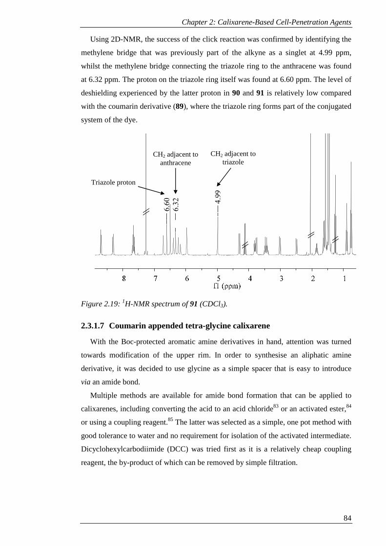
Chapter 2: Calixarene-Based Cell-Penetration Agents
84
Using 2D-NMR, the success of the click reaction was confirmed by identifying the
methylene bridge that was previously part of the alkyne as a singlet at 4.99 ppm,
whilst the methylene bridge connecting the triazole ring to the anthracene was found
at 6.32 ppm. The proton on the triazole ring itself was found at 6.60 ppm. The level of
deshielding experienced by the latter proton in 90 and 91 is relatively low compared
with the coumarin derivative (89), where the triazole ring forms part of the conjugated
system of the dye.
Figure 2.19: 1H-NMR spectrum of 91 (CDCl3).
2.3.1.7 Coumarin appended tetra-glycine calixarene
With the Boc-protected aromatic amine derivatives in hand, attention was turned
towards modification of the upper rim. In order to synthesise an aliphatic amine
derivative, it was decided to use glycine as a simple spacer that is easy to introduce
via an amide bond.
Multiple methods are available for amide bond formation that can be applied to
calixarenes, including converting the acid to an acid chloride83 or an activated ester,84
or using a coupling reagent.85 The latter was selected as a simple, one pot method with
good tolerance to water and no requirement for isolation of the activated intermediate.
Dicyclohexylcarbodiimide (DCC) was tried first as it is a relatively cheap coupling
reagent, the by-product of which can be removed by simple filtration.
4.9
9
6.3
2
6.6
0
CH2 adjacent to triazole
CH2 adjacent to anthracene
Triazole proton
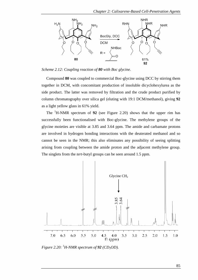
Chapter 2: Calixarene-Based Cell-Penetration Agents
85
Scheme 2.12: Coupling reaction of 80 with Boc glycine.
Compound 80 was coupled to commercial Boc-glycine using DCC by stirring them
together in DCM, with concomitant production of insoluble dicyclohexylurea as the
side product. The latter was removed by filtration and the crude product purified by
column chromatography over silica gel (eluting with 19:1 DCM/methanol), giving 92
as a light yellow glass in 61% yield.
The 1H-NMR spectrum of 92 (see Figure 2.20) shows that the upper rim has
successfully been functionalised with Boc-glycine. The methylene groups of the
glycine moieties are visible at 3.85 and 3.64 ppm. The amide and carbamate protons
are involved in hydrogen bonding interactions with the deuterated methanol and so
cannot be seen in the NMR; this also eliminates any possibility of seeing splitting
arising from coupling between the amide proton and the adjacent methylene group.
The singlets from the tert-butyl groups can be seen around 1.5 ppm.
Figure 2.20: 1H-NMR spectrum of 92 (CD3OD).
3.6
4
3.8
5
Glycine CH2

Chapter 2: Calixarene-Based Cell-Penetration Agents
86
In order to add the dye to 92, the same method as in section 2.3.1.3 was used. A
solution of 92 and 82 in DMF was treated with catalytic CuSO4 and one equivalent of
sodium ascorbate, heating to 90 °C for 18 hours. Aqueous work up in DCM/methanol
followed by purification by column chromatography over silica gel (eluting with 97:3
DCM/methanol) gave 93 as light yellow glass in 33% yield.
The 1H-NMR of 93 (see Figure 2.21) confirms the successful click reaction. The
peak that corresponds to the methylene group that was previously part of the alkyne is
now a singlet around 5.2 ppm. In this spectrum, the peaks corresponding to the dye are
more clearly resolved. The spectrum of the dye features, starting from high ppm, a
singlet, a doublet, a doublet of doublets and another doublet. These can be seen at
8.54, 7.67, 6.87 and 6.79 ppm, respectively. The singlet at 8.55 ppm corresponds to
the triazole proton. The relatively broad peaks that overlap with the dye peaks
correspond to the aromatic protons on the calixarene itself.
Scheme 2.13: Click reaction of 92 with 82.
Figure 2.21: 1H-NMR spectrum of 93 (CD3OD).
8.5
4
8.5
5
6.7
9
6.8
6
6.8
8
Triazole proton
Coumarin protons
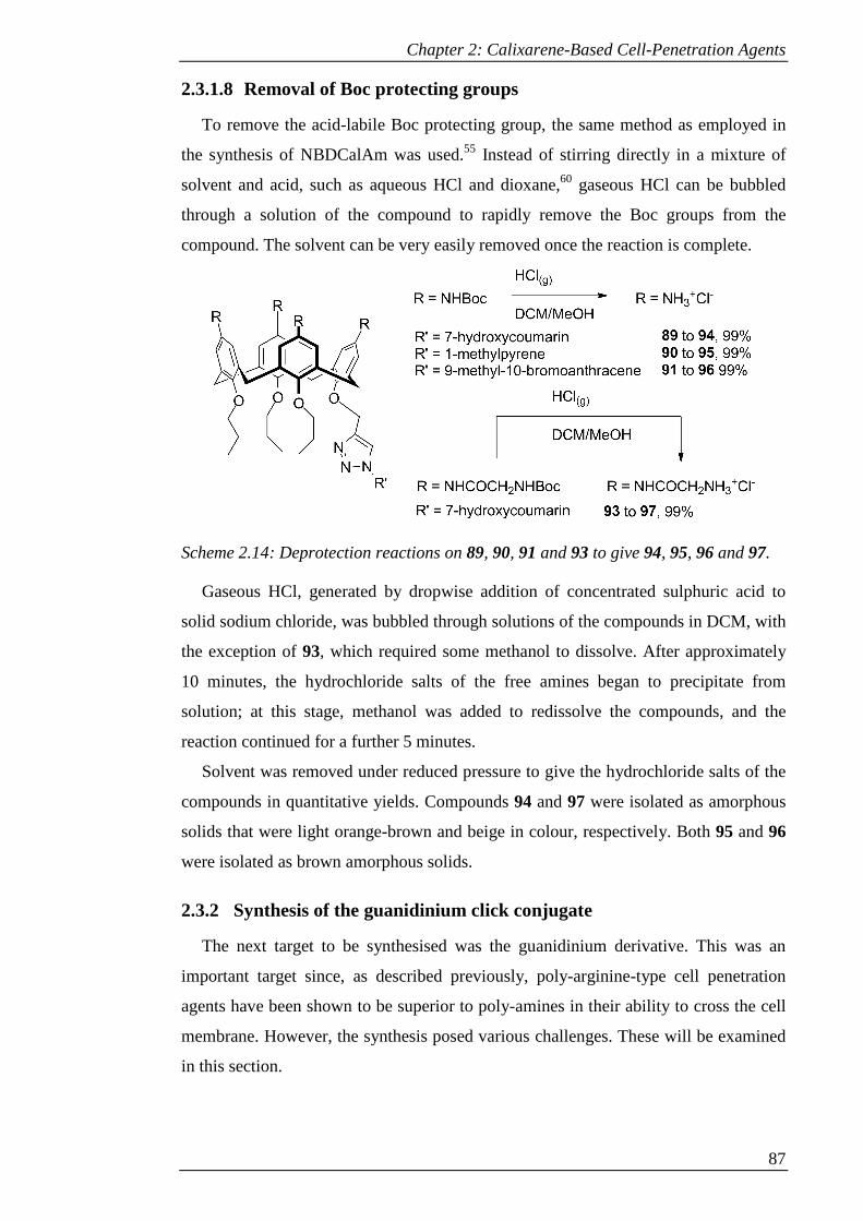
Chapter 2: Calixarene-Based Cell-Penetration Agents
87
2.3.1.8 Removal of Boc protecting groups
To remove the acid-labile Boc protecting group, the same method as employed in
the synthesis of NBDCalAm was used.55 Instead of stirring directly in a mixture of
solvent and acid, such as aqueous HCl and dioxane,60 gaseous HCl can be bubbled
through a solution of the compound to rapidly remove the Boc groups from the
compound. The solvent can be very easily removed once the reaction is complete.
Scheme 2.14: Deprotection reactions on 89, 90, 91 and 93 to give 94, 95, 96 and 97.
Gaseous HCl, generated by dropwise addition of concentrated sulphuric acid to
solid sodium chloride, was bubbled through solutions of the compounds in DCM, with
the exception of 93, which required some methanol to dissolve. After approximately
10 minutes, the hydrochloride salts of the free amines began to precipitate from
solution; at this stage, methanol was added to redissolve the compounds, and the
reaction continued for a further 5 minutes.
Solvent was removed under reduced pressure to give the hydrochloride salts of the
compounds in quantitative yields. Compounds 94 and 97 were isolated as amorphous
solids that were light orange-brown and beige in colour, respectively. Both 95 and 96
were isolated as brown amorphous solids.
2.3.2 Synthesis of the guanidinium click conjugate
The next target to be synthesised was the guanidinium derivative. This was an
important target since, as described previously, poly-arginine-type cell penetration
agents have been shown to be superior to poly-amines in their ability to cross the cell
membrane. However, the synthesis posed various challenges. These will be examined
in this section.

Chapter 2: Calixarene-Based Cell-Penetration Agents
88
2.3.2.1 Synthesis of common intermediate – TBDMS route
The first issue to be addressed was a problem that was found in the synthesis of the
tetra-amine intermediate. At the ipso-nitration stage, although the desired product
could normally be isolated in reasonable yields, there was sporadically an impurity
present that was very difficult to separate by column chromatography. This reduced
the yield and was costly in terms of time spent on this step. In addition, the purity of
the product obtained in the reduction step was inconsistent.
Other work within the group on tetra-alkyne derivatives turned up similar issues
with the ipso-nitration, although in this case the efficiency of the transformation was
even lower. The problem was addressed by use of alkyne protecting groups. It was
therefore decided to apply this to the current synthesis.
The mono-alkyne (78) was synthesised as before. At this stage, the protecting
group was installed to improve the efficiency of the following steps. Tert-butyl-
dimethylsilane (TBDMS) was selected for its resistance to hydrolysis. It can be put in
place using TBDMS-chloride via an SN2 reaction by removing the mildly acidic
terminal proton on the alkyne with a strong base. Compound 78 was stirred with
lithium hexamethyldisilazane (LiHMDS) in THF at -70 °C to remove the terminal
proton. TBDMSCl was added and the reaction continued at room temperature for 18
hours. Aqueous work-up followed by trituration of the residue with methanol to
remove excess reagent gave the protected form 98 as white crystals in 93% yield.
Successful protection of the alkyne was confirmed by the 1H-NMR spectrum (see
Figure 2.22). The doublet that was previously generated by the methylene group of
the alkyne has changed to a singlet with the loss of the long-range coupling, whilst the
triplet from the terminal proton is absent. In addition, the integrals of the TBDMS
peaks (not shown) are of the correct proportion for a single protecting group.
Figure 2.22: 1H-NMR spectrum of a) 78 and b) 98 (CDCl3) showing loss of terminal
alkyne triplet (circled) and conversion of methylene doublet to singlet.
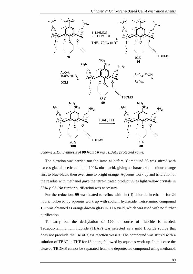
Chapter 2: Calixarene-Based Cell-Penetration Agents
89
Scheme 2.15: Synthesis of 80 from 78 via TBDMS protected route.
The nitration was carried out the same as before. Compound 98 was stirred with
excess glacial acetic acid and 100% nitric acid, giving a characteristic colour change
first to blue-black, then over time to bright orange. Aqueous work up and trituration of
the residue with methanol gave the tetra-nitrated product 99 as light yellow crystals in
86% yield. No further purification was necessary.
For the reduction, 99 was heated to reflux with tin (II) chloride in ethanol for 24
hours, followed by aqueous work up with sodium hydroxide. Tetra-amino compound
100 was obtained as orange-brown glass in 90% yield, which was used with no further
purification.
To carry out the desilylation of 100, a source of fluoride is needed.
Tetrabutylammonium fluoride (TBAF) was selected as a mild fluoride source that
does not preclude the use of glass reaction vessels. The compound was stirred with a
solution of TBAF in THF for 18 hours, followed by aqueous work-up. In this case the
cleaved TBDMS cannot be separated from the deprotected compound using methanol,

Chapter 2: Calixarene-Based Cell-Penetration Agents
90
since both are soluble in this solvent. The broad solubility of TBDMS was exploited
instead. By triturating with hexane, the TBDMS was separated from the amine. The
deprotected compound, now identical to 80, was isolated as light brown powder in
quantitative yield.
2.3.2.2 Guanidinylation – Boc protection route
A number of methods for guanidinylation of amines are available which have been
utilised in synthesis of guanidinium calixarenes. These include: the use of Boc-
protected triflyl guanidine on its own,86 relying on a good leaving group to drive the
reaction; Boc-protected thiourea with an activating reagent such as Mukaiyama
reagent (2-chloro-1-methylpyridinium iodide) or mercury chloride;60 or the use of a
Boc-protected isothiourea with mercury chloride.87 The latter method was selected for
the commercial availability and relatively low cost of the reagents.
Compound 80 was stirred with a mixture of N,N’-di-Boc-S-methylisothiourea,
HgCl2 and triethylamine in dry DMF for 48 hours. The byproduct, HgSMe, appeared
as a white precipitate over this time and was subsequently removed by filtration.
Recrystallisation from hexane allowed the majority of the excess reagent to be
removed prior to purification by column chromatography over silica gel.
However, the purification of this compound revealed a problem: the Boc protecting
groups were surprisingly labile, leading to degradation on the mildly acidic silica. This
could be mitigated by use of triethylamine in the eluent (8:1:1 hexane/diethyl
ether/triethylamine), allowing 101 to be isolated as white solid, but in poor yield
(13%).
Scheme 2.16: Synthesis of 101 from 80.
With the addition of the coumarin dye, an acidic phenolic proton is introduced into
the molecule. Triethylamine would therefore not be suitable as a component of the
eluent for purification of the dye conjugate as it would form a salt with the dye and
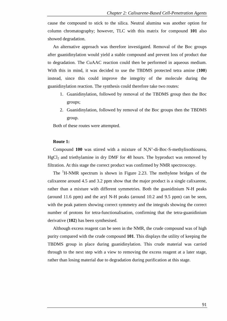
Chapter 2: Calixarene-Based Cell-Penetration Agents
91
cause the compound to stick to the silica. Neutral alumina was another option for
column chromatography; however, TLC with this matrix for compound 101 also
showed degradation.
An alternative approach was therefore investigated. Removal of the Boc groups
after guanidinylation would yield a stable compound and prevent loss of product due
to degradation. The CuAAC reaction could then be performed in aqueous medium.
With this in mind, it was decided to use the TBDMS protected tetra amine (100)
instead, since this could improve the integrity of the molecule during the
guanidinylation reaction. The synthesis could therefore take two routes:
1. Guanidinylation, followed by removal of the TBDMS group then the Boc
groups;
2. Guanidinylation, followed by removal of the Boc groups then the TBDMS
group.
Both of these routes were attempted.
Route 1:
Compound 100 was stirred with a mixture of N,N’-di-Boc-S-methylisothiourea,
HgCl2 and triethylamine in dry DMF for 48 hours. The byproduct was removed by
filtration. At this stage the correct product was confirmed by NMR spectroscopy.
The 1H-NMR spectrum is shown in Figure 2.23. The methylene bridges of the
calixarene around 4.5 and 3.2 ppm show that the major product is a single calixarene,
rather than a mixture with different symmetries. Both the guanidinium N-H peaks
(around 11.6 ppm) and the aryl N-H peaks (around 10.2 and 9.5 ppm) can be seen,
with the peak pattern showing correct symmetry and the integrals showing the correct
number of protons for tetra-functionalisation, confirming that the tetra-guanidinium
derivative (102) has been synthesised.
Although excess reagent can be seen in the NMR, the crude compound was of high
purity compared with the crude compound 101. This displays the utility of keeping the
TBDMS group in place during guanidinylation. This crude material was carried
through to the next step with a view to removing the excess reagent at a later stage,
rather than losing material due to degradation during purification at this stage.
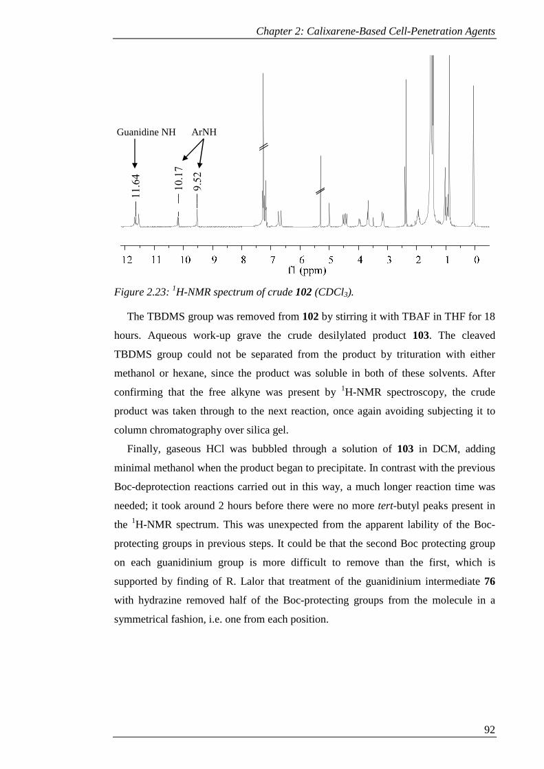
Chapter 2: Calixarene-Based Cell-Penetration Agents
92
Figure 2.23: 1H-NMR spectrum of crude 102 (CDCl3).
The TBDMS group was removed from 102 by stirring it with TBAF in THF for 18
hours. Aqueous work-up grave the crude desilylated product 103. The cleaved
TBDMS group could not be separated from the product by trituration with either
methanol or hexane, since the product was soluble in both of these solvents. After
confirming that the free alkyne was present by 1H-NMR spectroscopy, the crude
product was taken through to the next reaction, once again avoiding subjecting it to
column chromatography over silica gel.
Finally, gaseous HCl was bubbled through a solution of 103 in DCM, adding
minimal methanol when the product began to precipitate. In contrast with the previous
Boc-deprotection reactions carried out in this way, a much longer reaction time was
needed; it took around 2 hours before there were no more tert-butyl peaks present in
the 1H-NMR spectrum. This was unexpected from the apparent lability of the Boc-
protecting groups in previous steps. It could be that the second Boc protecting group
on each guanidinium group is more difficult to remove than the first, which is
supported by finding of R. Lalor that treatment of the guanidinium intermediate 76
with hydrazine removed half of the Boc-protecting groups from the molecule in a
symmetrical fashion, i.e. one from each position.
9.5
2
10.1
7
11.6
4Guanidine NH ArNH

Chapter 2: Calixarene-Based Cell-Penetration Agents
93
Scheme 2.17: Two possible routes for the synthesis of 104, via 102 followed by 103 or
105.
Due to the high polarity of this compound, even in neutral form, it is unsuitable for
purification by column chromatography over silica gel. Therefore to remove the
excess thiourea reagent (now also deprotected) and the cleaved TBDMS, reverse-
phase column chromatography was used, eluting with a gradient of methanol in water.
Pure fully deprotected compound 104 was obtained as off-white solid in 21% yield
over 3 steps. This is favourable when compared with the synthesis of compound 101,
which was isolated pure in only 13% yield in a single step.
Chapter 2: Calixarene-Based Cell-Penetration Agents
94
Route 2:
The first step in this route required removal of the Boc groups. Gaseous HCl was
bubbled through a solution of compound 102 in DCM, with methanol added to
redissolve any precipitate that was formed. As with the Boc-deprotection of
intermediate 103, complete removal of all Boc groups to give 105 took around 2
hours. Although the compound could be subjected to reverse-phase chromatography at
this stage to remove the deprotected reagent, the compound was otherwise pure, with
only a single calixarene species apparent. It was decided therefore to limit the
purification to a single step to maximise the efficiency of the process.
Crude 105 was stirred with TBAF in THF for 18 hours. After quenching the
reaction with saturated ammonium chloride, some of the ammonium chloride
precipitated and could be removed by filtration. However, at this stage the presence of
the water-soluble guanidinium compound precluded the use of aqueous work-up to
remove excess reagent. The crude residue was therefore subjected to reverse-phase
column chromatography to purify the product and remove all excess reagents and
cleaved TBDMS, eluting with a gradient of methanol in water. Pure fully deprotected
compound 104 was obtained as off-white solid in 23% yield over 3 steps.
Although this is a higher yield than obtained from route 1, such a small difference
in yield may not be significant. Route 2 posed more problems, particularly with not
being able to remove the excess tetrabutylammonium except by reverse phase column
chromatography. However, the crude final product by this route was more pure than
that of route 1. Therefore both routes have both problems and merits, but
fundamentally both gave the desired product pure and in better yield than could be
obtained by attempting to manipulate the Boc-protected form.
2.3.2.3 Attempted CuAAC reaction with deprotected guanidinium
derivative
In order to add the dye to 104, it was hoped to use a similar strategy to the previous
CuAAC reactions. A mixture of 104 and 82 was stirred with sodium ascorbate and
catalytic copper sulphate at 90 °C in several different solvent systems to test which
was most effective. Ethanol, DMF, tert-butanol and 1:1 ethanol/water were tested; the
latter gave the best conversion to product 106 according to the crude NMR (see Figure
2.24). Confirmation of the reaction was given by the loss of the alkyne triplet around 3
ppm and the conversion of the alkyne doublet around 5 ppm to the singlet of the
methylene adjacent to the triazole ring. Dye peaks can also be seen downfield.

Chapter 2: Calixarene-Based Cell-Penetration Agents
95
Scheme 2.18: Click reaction of 104 with 82.
Figure 2.24: 1H-NMR-spectrum of a) 104 and b) crude 106 (CD3OD) showing loss of
terminal alkyne triplet (circled) and conversion of methylene doublet to a singlet.
At this stage there was brown, amorphous solid present that was poorly soluble in
both organic solvent and water unless treated with dilute HCl. Reverse-phase column
chromatography was attempted, but solubility problems led to blockage of the
column. No fractions were recovered that resembled the product.
Attention was turned to an alternative method for purifying the product. Since there
should only be a single product from the CuAAC reaction, the presence of multiple
products in the NMR could be attributed to the potential of the guanidinium groups to

Chapter 2: Calixarene-Based Cell-Penetration Agents
96
interact with copper in the reaction, as well as the different anions present, resulting in
a mixture of different species. The use of an anionic resin to bind the guanidinium
product would allow the excess dye and the ascorbic acid to be readily removed as
these would not bind to the resin. The product could then be separated from the copper
catalyst, which would also bind to the matrix, by exploiting their different binding
strengths. The compound would be eluted from the column in a single salt form with
only one type of counter-anion (see Scheme 2.19).
Scheme 2.19: Principle of purification by ion-exchange chromatography (X = 106).
Commercially available cation-binding resins include those featuring strongly
acidic groups such as sulphonic acid and weakly acidic groups such as carboxylic
acid. Since four guanidinium moieties are clustered on each molecule, it would be
expected that a strong-cation binder such as a sulphonic acid resin would bind the
compound too strongly leading to harsh conditions to elute it. Therefore it was
decided to use a carboxylic acid resin (Amberlite IRC-50) as a weak cation binder.
The crude product was applied to the sodium form of the resin. After washing with
water, increasing concentrations of HCl solution were applied to the column to
displace cations from the resin. However, even 2 M HCl failed to elute the product.
Increasing concentrations of first ammonium chloride, then sodium chloride, up to
saturated solutions of each, also failed to displace the product. The strength in binding
could be attributed to a combination of the inherent tendency of guanidinium moieties
to bind anions, as described previously, along with the clustering of four of these
groups onto a single face of a molecule, with the potential to give binding that is
greater than the sum of its parts.
Since applying cations to displace the product was not sufficient, it was reasoned
that if the guanidinium groups of the compound could be deprotonated, then the
binding strength could be lessened, allowing the product to be eluted. By using dilute
sodium hydroxide, fluorescent fractions were indeed eluted from the column;
however, the material recovered was a complex mixture by 1H-NMR with no major
species, suggesting significant degradation of the product. At this stage an alternative
route to the product was sought.
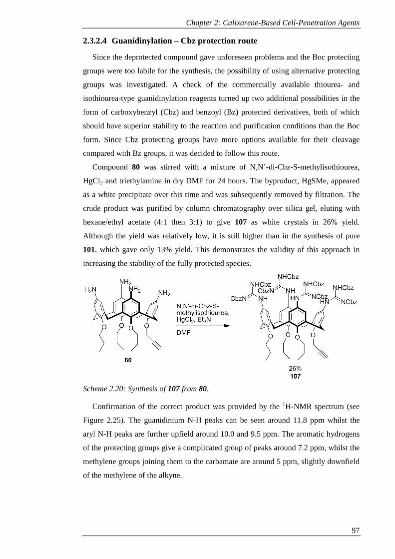
Chapter 2: Calixarene-Based Cell-Penetration Agents
97
2.3.2.4 Guanidinylation – Cbz protection route
Since the deprotected compound gave unforeseen problems and the Boc protecting
groups were too labile for the synthesis, the possibility of using alternative protecting
groups was investigated. A check of the commercially available thiourea- and
isothiourea-type guanidinylation reagents turned up two additional possibilities in the
form of carboxybenzyl (Cbz) and benzoyl (Bz) protected derivatives, both of which
should have superior stability to the reaction and purification conditions than the Boc
form. Since Cbz protecting groups have more options available for their cleavage
compared with Bz groups, it was decided to follow this route.
Compound 80 was stirred with a mixture of N,N’-di-Cbz-S-methylisothiourea,
HgCl2 and triethylamine in dry DMF for 24 hours. The byproduct, HgSMe, appeared
as a white precipitate over this time and was subsequently removed by filtration. The
crude product was purified by column chromatography over silica gel, eluting with
hexane/ethyl acetate (4:1 then 3:1) to give 107 as white crystals in 26% yield.
Although the yield was relatively low, it is still higher than in the synthesis of pure
101, which gave only 13% yield. This demonstrates the validity of this approach in
increasing the stability of the fully protected species.
Scheme 2.20: Synthesis of 107 from 80.
Confirmation of the correct product was provided by the 1H-NMR spectrum (see
Figure 2.25). The guanidinium N-H peaks can be seen around 11.8 ppm whilst the
aryl N-H peaks are further upfield around 10.0 and 9.5 ppm. The aromatic hydrogens
of the protecting groups give a complicated group of peaks around 7.2 ppm, whilst the
methylene groups joining them to the carbamate are around 5 ppm, slightly downfield
of the methylene of the alkyne.

Chapter 2: Calixarene-Based Cell-Penetration Agents
98
Figure 2.25: 1H-NMR spectrum of 107 (CDCl3).
2.3.2.5 Coumarin appended tetra-guanidine
With the stable guanidine compound in hand, the CuAAC reaction was
implemented. A solution of 107 and 82 in DMF was treated with sodium ascorbate
and catalytic CuSO4, heating at 70 °C for 2 hours. Aqueous work up followed by
purification by column chromatography over silica gel (eluting with 5-10% ethyl
acetate in DCM) gave 108 as light yellow solid in 32% yield.
It was thought that the low yield was due to degradation of the product, so the
reaction was repeated at room temperature for 24 hours. A modified work-up was
used. Following removal of the DMF under reduced pressure, the solid was triturated
with water and filtered to remove the catalyst, followed by washing with methanol to
remove excess dye, exploiting the poor solubility of the Cbz protected compound in
this solvent. After verifying that no product had been lost in the methanol wash, the
product was purified by column chromatography over silica gel, eluting with 19:1
DCM/ethyl acetate to give 108 in 74% yield.
The correct product was confirmed by 1H-NMR (see Figure 2.26). The methylene
group adjacent to the triazole ring can no longer be differentiated from those of the
Cbz groups around 5.1 ppm. However, the proton on the triazole ring itself can be
seen at 8.54 ppm, confirmed by 2D-NMR. The dye peaks are also in evidence around
8.35 and 6.6 ppm, with the remaining dye peak obscured by the aromatic protons of
the Cbz groups. In this case the guanidinium N-H protons give rise to one broad
singlet, in contrast with 107, possibly due to intermolecular interactions with the
coumarin dye.
123456789101112f1 (ppm)
9.4
8
10.0
7
11.6
9
Guanidine NH ArNH

Chapter 2: Calixarene-Based Cell-Penetration Agents
99
Scheme 2.21: Click reaction of 107 with 82.
Figure 2.26: 1H-NMR spectrum of 108 (CDCl3).
2.3.2.6 Deprotection of Cbz protected guanidine derivative
A number of methods are available for cleavage of Cbz protecting groups. For
aromatic Cbz-protected guanidines, catalytic hydrogenolysis with palladium on
carbon88 is the dominant literature method. However, this is unsuitable in this case due
to the presence of the coumarin dye, which can be reduced under these conditions.89
For aliphatic Cbz-protected guanidines, methods that have been applied for
removal of the protecting groups include the use of HBr in acetic acid90 or thioanisole
in TFA,91 both of which utilise a combination of acidic conditions to protonate the
carbonyl of the carbamate and a nucleophile to attack the methylene of the benzyl
group. Another method simply heated the protected compound to reflux in 6 M HCl to
remove the Cbz groups;92 however it was decided to avoid such harsh conditions.
8.5
4
9.8
0
11
.81
Guanidine NH ArNH Triazole proton

Chapter 2: Calixarene-Based Cell-Penetration Agents
100
The first attempt at removal of the protecting groups from 108 was made by
stirring the compound with HBr in acetic acid. After 18 hours, DCM was added to
precipitate the product to separate it from the cleaved benzyl groups. However, the
solid that precipitated was found to be a complex mixture of products by 1H-NMR,
suggesting considerable degradation of the product.
Following this, deprotection was attempted by stirring 108 with thioanisole in TFA.
The colour of the solution immediately changed from yellow to green. After 24 hours
the solvent was removed under reduced pressure and the residue triturated with diethyl
ether. The progress of the reaction was assessed by 1H-NMR, which showed a mixture
of products with benzyl peaks still present, suggesting incomplete deprotection. After
a further 48 hours under the same conditions the reaction still seemed to be
incomplete, so the reaction mixture was heated to 40 °C for 48 hours, during which
time the colour changed to grey-blue. This time the 1H-NMR spectrum showed
decomposition of the product.
At this stage, an alternative method was investigated which uses
iodotrimethylsilane (TMSI), where TMS acts as a Lewis acid instead of a Brønsted
acid, with iodide as a nucleophile.93 Although TMSI is commercially available, it is
commonly stabilised with copper, which could bind to the free guanidinium groups
and cause problems with purification. TMSI can however be easily generated in situ
from TMSCl and anhydrous sodium iodide,94 with concomitant generation of sodium
chloride. Anhydrous conditions are desirable for this reaction as TMSI is readily
hydrolysed, generating HI, which could lead to undesired side-reactions.
The reaction was first attempted on 108 by stirring the compound with sodium
iodide in acetonitrile under argon, followed by addition of TMSCl, resulting in a
colour change to yellow-orange and precipitation of sodium chloride. After stirring at
room temperature for 4 hours, methanol and dilute aqueous HCl were added to cleave
the TMS still bound to the carbamate to allow decarboxylation to take place.
After removing the solvent under reduced pressure and triturating with diethyl
ether, the 1H-NMR spectrum showed that the integration height of the benzyl peaks
had been halved, whilst the symmetry of the molecule was the same, suggesting that
one Cbz had been removed from each guanidine. This is consistent with previous
observations suggesting that the half-deprotected species is more stable than the fully
protected compound.

Chapter 2: Calixarene-Based Cell-Penetration Agents
101
Scheme 2.22: Deprotection of 108.
In an attempt to push the deprotection through to completion, two modified
procedures were used. First, thioanisole was added to the reaction mixture to provide
an additional nucleophile and a cation scavenger. However, after stirring overnight at
room temperature the reaction still proceeded only halfway.
Second, the reaction was heated to 60 °C for 24 hours; this gave a complex mixture
of products by NMR, suggesting decomposition of the product. This is likely due to
side-reactions stemming from the variety of potential reactions that TMSI can carry
out, including cleavage of lactones and ethers.93 The cleavage of ethers on calixarenes
by TMSI has in fact previously been investigated as a route towards mono-alkyl
derivatives.95 Therefore more caution is required to prevent undesired side-reactions.
Although it has been reasoned that acetonitrile participates in the reaction by
forming a complex with the TMSI,93 resulting in the observed colour-change, it was
decided to investigate DMF as an alternative solvent. The deprotection was attempted
at different temperatures and varying reaction times. Although reaction at room
temperature gave only half-deprotected product after 24 hours, heating to 50 °C gave
the fully-deprotected product that could, for the first time in this synthesis, be
distinguished in the 1H-NMR spectrum.
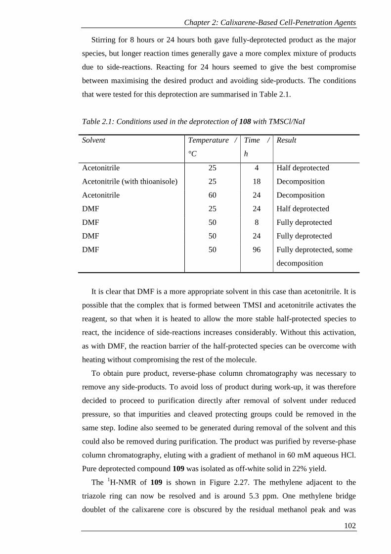
Chapter 2: Calixarene-Based Cell-Penetration Agents
102
Stirring for 8 hours or 24 hours both gave fully-deprotected product as the major
species, but longer reaction times generally gave a more complex mixture of products
due to side-reactions. Reacting for 24 hours seemed to give the best compromise
between maximising the desired product and avoiding side-products. The conditions
that were tested for this deprotection are summarised in Table 2.1.
Table 2.1: Conditions used in the deprotection of 108 with TMSCl/NaI
Solvent Temperature /
°C
Time /
h
Result
Acetonitrile 25 4 Half deprotected
Acetonitrile (with thioanisole) 25 18 Decomposition
Acetonitrile 60 24 Decomposition
DMF 25 24 Half deprotected
DMF 50 8 Fully deprotected
DMF 50 24 Fully deprotected
DMF 50 96 Fully deprotected, some
decomposition
It is clear that DMF is a more appropriate solvent in this case than acetonitrile. It is
possible that the complex that is formed between TMSI and acetonitrile activates the
reagent, so that when it is heated to allow the more stable half-protected species to
react, the incidence of side-reactions increases considerably. Without this activation,
as with DMF, the reaction barrier of the half-protected species can be overcome with
heating without compromising the rest of the molecule.
To obtain pure product, reverse-phase column chromatography was necessary to
remove any side-products. To avoid loss of product during work-up, it was therefore
decided to proceed to purification directly after removal of solvent under reduced
pressure, so that impurities and cleaved protecting groups could be removed in the
same step. Iodine also seemed to be generated during removal of the solvent and this
could also be removed during purification. The product was purified by reverse-phase
column chromatography, eluting with a gradient of methanol in 60 mM aqueous HCl.
Pure deprotected compound 109 was isolated as off-white solid in 22% yield.
The 1H-NMR of 109 is shown in Figure 2.27. The methylene adjacent to the
triazole ring can now be resolved and is around 5.3 ppm. One methylene bridge
doublet of the calixarene core is obscured by the residual methanol peak and was

Chapter 2: Calixarene-Based Cell-Penetration Agents
103
found using 2D-COSY NMR to be at 3.3 ppm. In this case the peaks around 8.6 ppm
corresponding to the proton on the triazole ring and one of the dye protons can barely
be resolved as separate peaks and so cannot be differentiated due to the insufficient
resolution of the 2D-NMR experiments.
Figure 2.27: 1H-NMR spectrum of 109 (CD3OD).
2.3.3 Synthesis of dye-conjugates with variable linkers
In order to investigate the potential effect of changing the linkage method to a
triazole ring, two compounds that were identical except for the type of linker were
required. This gave two immediate possibilities as shown in Figure 2.28.
Figure 2.28: Analogues with NBD and coumarin dyes linked by a) triazole rings and
b) secondary amines.
In order to make an analogue to a previously synthesised molecule with a different
dye but the same linker, a triazole-linked NBD could be synthesised to compare with
compound 94, or an amine-linked coumarin to compare with NBDCalAm.
5.3
2
CH2 adjacent to triazole
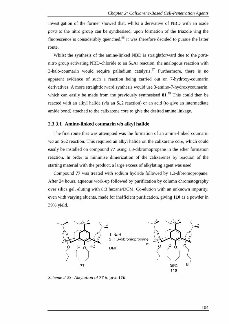
Chapter 2: Calixarene-Based Cell-Penetration Agents
104
Investigation of the former showed that, whilst a derivative of NBD with an azide
para to the nitro group can be synthesised, upon formation of the triazole ring the
fluorescence is considerably quenched.96 It was therefore decided to pursue the latter
route.
Whilst the synthesis of the amine-linked NBD is straightforward due to the para-
nitro group activating NBD-chloride to an SNAr reaction, the analogous reaction with
3-halo-coumarin would require palladium catalysis.97 Furthermore, there is no
apparent evidence of such a reaction being carried out on 7-hydroxy-coumarin
derivatives. A more straightforward synthesis would use 3-amino-7-hydroxycoumarin,
which can easily be made from the previously synthesised 81.79 This could then be
reacted with an alkyl halide (via an SN2 reaction) or an acid (to give an intermediate
amide bond) attached to the calixarene core to give the desired amine linkage.
2.3.3.1 Amine-linked coumarin via alkyl halide
The first route that was attempted was the formation of an amine-linked coumarin
via an SN2 reaction. This required an alkyl halide on the calixarene core, which could
easily be installed on compound 77 using 1,3-dibromopropane in the ether formation
reaction. In order to minimise dimerization of the calixarenes by reaction of the
starting material with the product, a large excess of alkylating agent was used.
Compound 77 was treated with sodium hydride followed by 1,3-dibromopropane.
After 24 hours, aqueous work-up followed by purification by column chromatography
over silica gel, eluting with 8:3 hexane/DCM. Co-elution with an unknown impurity,
even with varying eluents, made for inefficient purification, giving 110 as a powder in
39% yield.
Scheme 2.23: Alkylation of 77 to give 110.
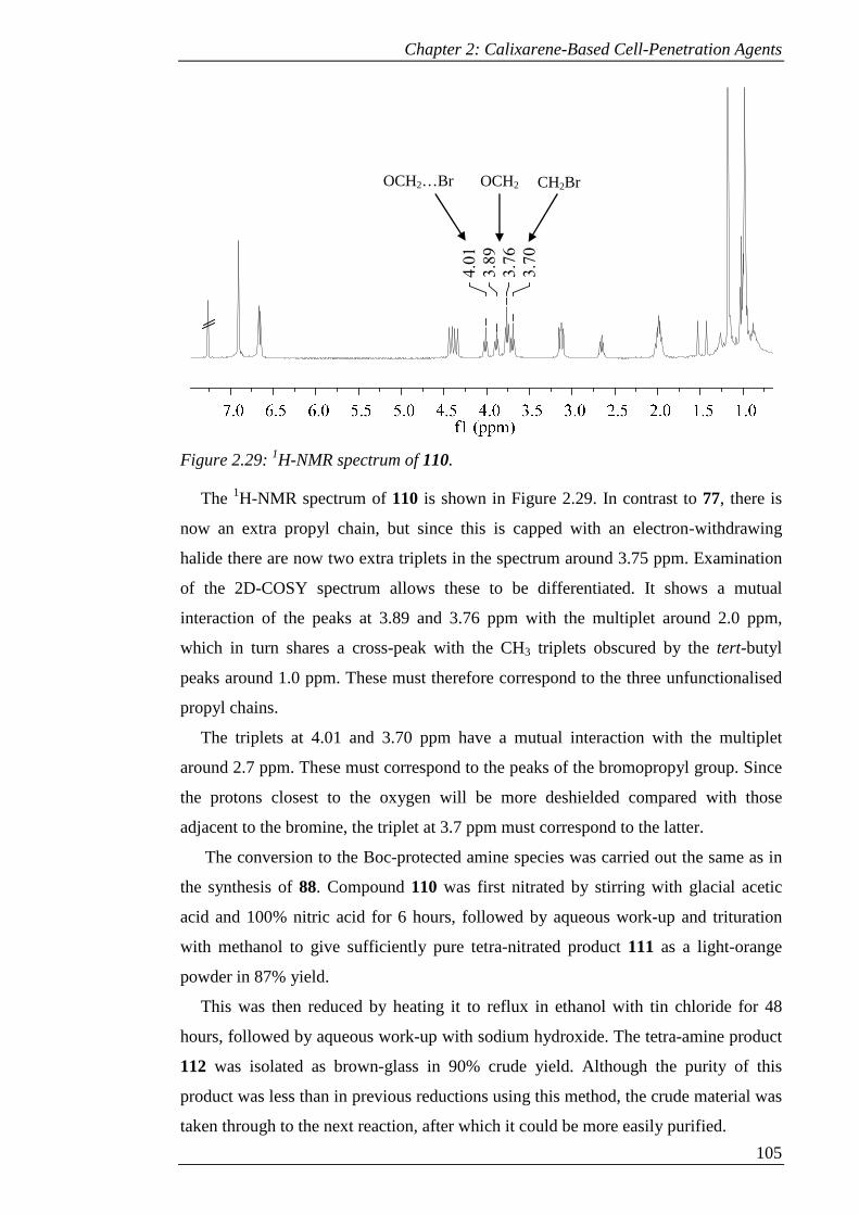
Chapter 2: Calixarene-Based Cell-Penetration Agents
105
Figure 2.29: 1H-NMR spectrum of 110.
The 1H-NMR spectrum of 110 is shown in Figure 2.29. In contrast to 77, there is
now an extra propyl chain, but since this is capped with an electron-withdrawing
halide there are now two extra triplets in the spectrum around 3.75 ppm. Examination
of the 2D-COSY spectrum allows these to be differentiated. It shows a mutual
interaction of the peaks at 3.89 and 3.76 ppm with the multiplet around 2.0 ppm,
which in turn shares a cross-peak with the CH3 triplets obscured by the tert-butyl
peaks around 1.0 ppm. These must therefore correspond to the three unfunctionalised
propyl chains.
The triplets at 4.01 and 3.70 ppm have a mutual interaction with the multiplet
around 2.7 ppm. These must correspond to the peaks of the bromopropyl group. Since
the protons closest to the oxygen will be more deshielded compared with those
adjacent to the bromine, the triplet at 3.7 ppm must correspond to the latter.
The conversion to the Boc-protected amine species was carried out the same as in
the synthesis of 88. Compound 110 was first nitrated by stirring with glacial acetic
acid and 100% nitric acid for 6 hours, followed by aqueous work-up and trituration
with methanol to give sufficiently pure tetra-nitrated product 111 as a light-orange
powder in 87% yield.
This was then reduced by heating it to reflux in ethanol with tin chloride for 48
hours, followed by aqueous work-up with sodium hydroxide. The tetra-amine product
112 was isolated as brown-glass in 90% crude yield. Although the purity of this
product was less than in previous reductions using this method, the crude material was
taken through to the next reaction, after which it could be more easily purified.
3.7
0
3.7
6
3.8
9
4.0
1
OCH2 OCH2…Br CH2Br
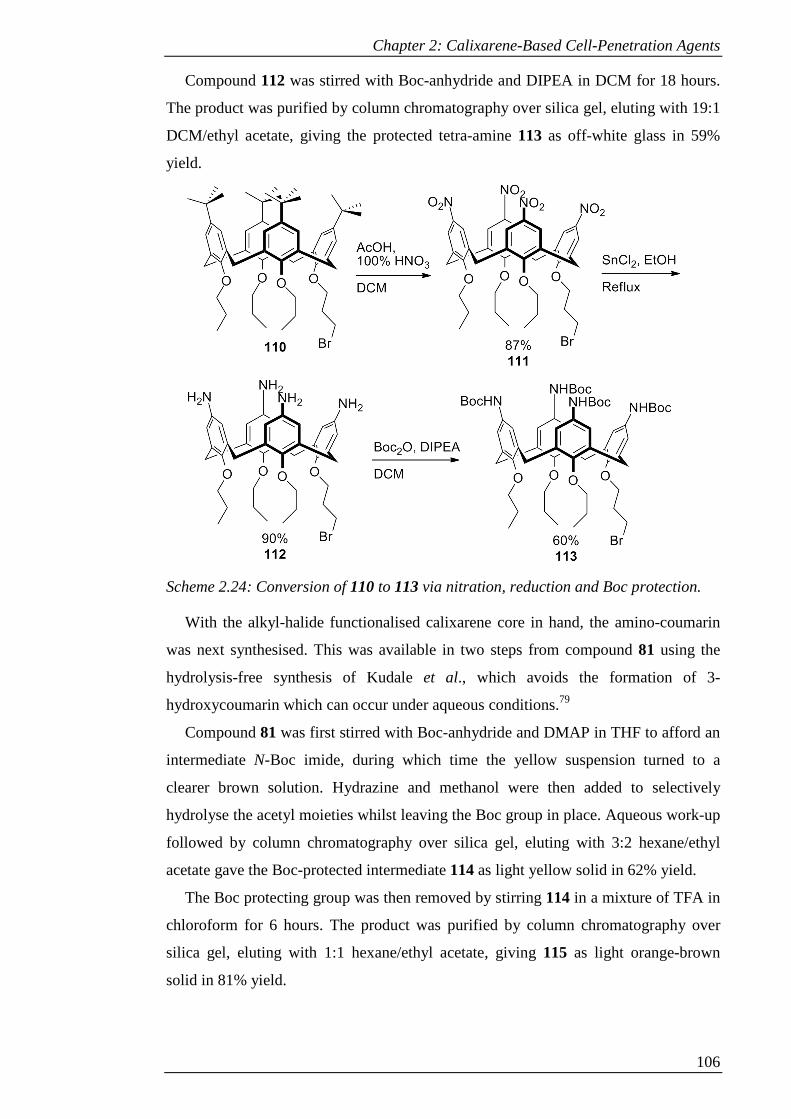
Chapter 2: Calixarene-Based Cell-Penetration Agents
106
Compound 112 was stirred with Boc-anhydride and DIPEA in DCM for 18 hours.
The product was purified by column chromatography over silica gel, eluting with 19:1
DCM/ethyl acetate, giving the protected tetra-amine 113 as off-white glass in 59%
yield.
Scheme 2.24: Conversion of 110 to 113 via nitration, reduction and Boc protection.
With the alkyl-halide functionalised calixarene core in hand, the amino-coumarin
was next synthesised. This was available in two steps from compound 81 using the
hydrolysis-free synthesis of Kudale et al., which avoids the formation of 3-
hydroxycoumarin which can occur under aqueous conditions.79
Compound 81 was first stirred with Boc-anhydride and DMAP in THF to afford an
intermediate N-Boc imide, during which time the yellow suspension turned to a
clearer brown solution. Hydrazine and methanol were then added to selectively
hydrolyse the acetyl moieties whilst leaving the Boc group in place. Aqueous work-up
followed by column chromatography over silica gel, eluting with 3:2 hexane/ethyl
acetate gave the Boc-protected intermediate 114 as light yellow solid in 62% yield.
The Boc protecting group was then removed by stirring 114 in a mixture of TFA in
chloroform for 6 hours. The product was purified by column chromatography over
silica gel, eluting with 1:1 hexane/ethyl acetate, giving 115 as light orange-brown
solid in 81% yield.
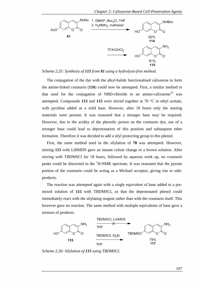
Chapter 2: Calixarene-Based Cell-Penetration Agents
107
Scheme 2.25: Synthesis of 115 from 81 using a hydrolysis-free method.
The conjugation of the dye with the alkyl-halide functionalised calixarene to form
the amine-linked coumarin (116) could now be attempted. First, a similar method to
that used for the conjugation of NBD-chloride to an amino-calixarene55 was
attempted. Compounds 113 and 115 were stirred together at 70 °C in ethyl acetate,
with pyridine added as a mild base. However, after 18 hours only the starting
materials were present. It was reasoned that a stronger base may be required.
However, due to the acidity of the phenolic proton on the coumarin dye, use of a
stronger base could lead to deprotonation of this position and subsequent ether
formation. Therefore it was decided to add a silyl protecting group to this phenol.
First, the same method used in the silylation of 78 was attempted. However,
stirring 115 with LiHMDS gave an instant colour change to a brown solution. After
stirring with TBDMSCl for 18 hours, followed by aqueous work up, no coumarin
peaks could be discerned in the 1H-NMR spectrum. It was reasoned that the pyrone
portion of the coumarin could be acting as a Michael acceptor, giving rise to side-
products.
The reaction was attempted again with a single equivalent of base added to a pre-
mixed solution of 115 with TBDMSCl, so that the deprotonated phenol could
immediately react with the silylating reagent rather than with the coumarin itself. This
however gave no reaction. The same method with multiple equivalents of base gave a
mixture of products.
Scheme 2.26: Silylation of 115 using TBDMSCl.
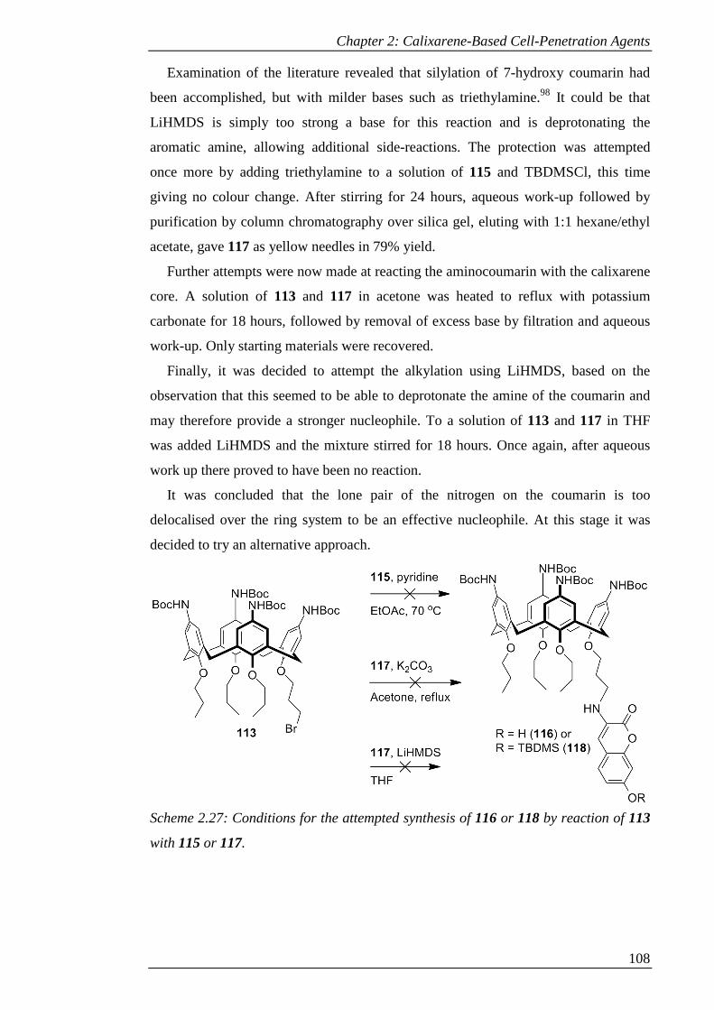
Chapter 2: Calixarene-Based Cell-Penetration Agents
108
Examination of the literature revealed that silylation of 7-hydroxy coumarin had
been accomplished, but with milder bases such as triethylamine.98 It could be that
LiHMDS is simply too strong a base for this reaction and is deprotonating the
aromatic amine, allowing additional side-reactions. The protection was attempted
once more by adding triethylamine to a solution of 115 and TBDMSCl, this time
giving no colour change. After stirring for 24 hours, aqueous work-up followed by
purification by column chromatography over silica gel, eluting with 1:1 hexane/ethyl
acetate, gave 117 as yellow needles in 79% yield.
Further attempts were now made at reacting the aminocoumarin with the calixarene
core. A solution of 113 and 117 in acetone was heated to reflux with potassium
carbonate for 18 hours, followed by removal of excess base by filtration and aqueous
work-up. Only starting materials were recovered.
Finally, it was decided to attempt the alkylation using LiHMDS, based on the
observation that this seemed to be able to deprotonate the amine of the coumarin and
may therefore provide a stronger nucleophile. To a solution of 113 and 117 in THF
was added LiHMDS and the mixture stirred for 18 hours. Once again, after aqueous
work up there proved to have been no reaction.
It was concluded that the lone pair of the nitrogen on the coumarin is too
delocalised over the ring system to be an effective nucleophile. At this stage it was
decided to try an alternative approach.
Scheme 2.27: Conditions for the attempted synthesis of 116 or 118 by reaction of 113
with 115 or 117.

Chapter 2: Calixarene-Based Cell-Penetration Agents
109
2.3.3.2 Amine-linked coumarin via amide
Although carrying out a simple SN2 reaction with the aminocoumarin proved to be
problematic, the formation of amide bonds with this type of compound has been
accomplished.99,100 This could be reacted with an acid moiety on the calixarene core
and the intermediate amide bond could then be reduced to the desired secondary
amine.
To form an end product with the correct length of linker, a C3-acid was required on
the calixarene. Initial attempts at installing this group were made using ethyl 3-
bromopropanoate. Compound 77 was stirred with sodium hydride in DMF for 1 hour
before adding the alkylating agent; at this stage, effervescence was observed. After 4
hours only starting material was present in the reaction mixture and the odour of ethyl
acrylate could be detected. It was concluded that an acidic hydrogen adjacent to the
carbonyl in the alkylating reagent was being removed by the sodium hydride, with
evolution of hydrogen gas, followed by elimination of bromide to give ethyl acrylate.
To try to overcome this problem different reaction conditions were tested. Stirring
with potassium carbonate as a weaker base in acetone gave no reaction, either at room
temperature or at reflux. Using sodium hydride, but carrying out the addition of
alkylating reagent at 0 °C, still gave evolution of hydrogen and likewise there was no
ether formation.
Scheme 2.28: Attempted synthesis of 119 with ethyl 3-bromopropanoate.
Since some of the base would be consumed in the deprotonation of the phenol, it
was expected that some of the ethyl 3-bromopropanoate would remain intact to react
with the calixarene. The lack of any reaction at all could be due to the phenoxide
anion generated in the first stage of the reaction being sufficiently basic to deprotonate
the alkylating agent rather than giving the desired SN2 reaction.
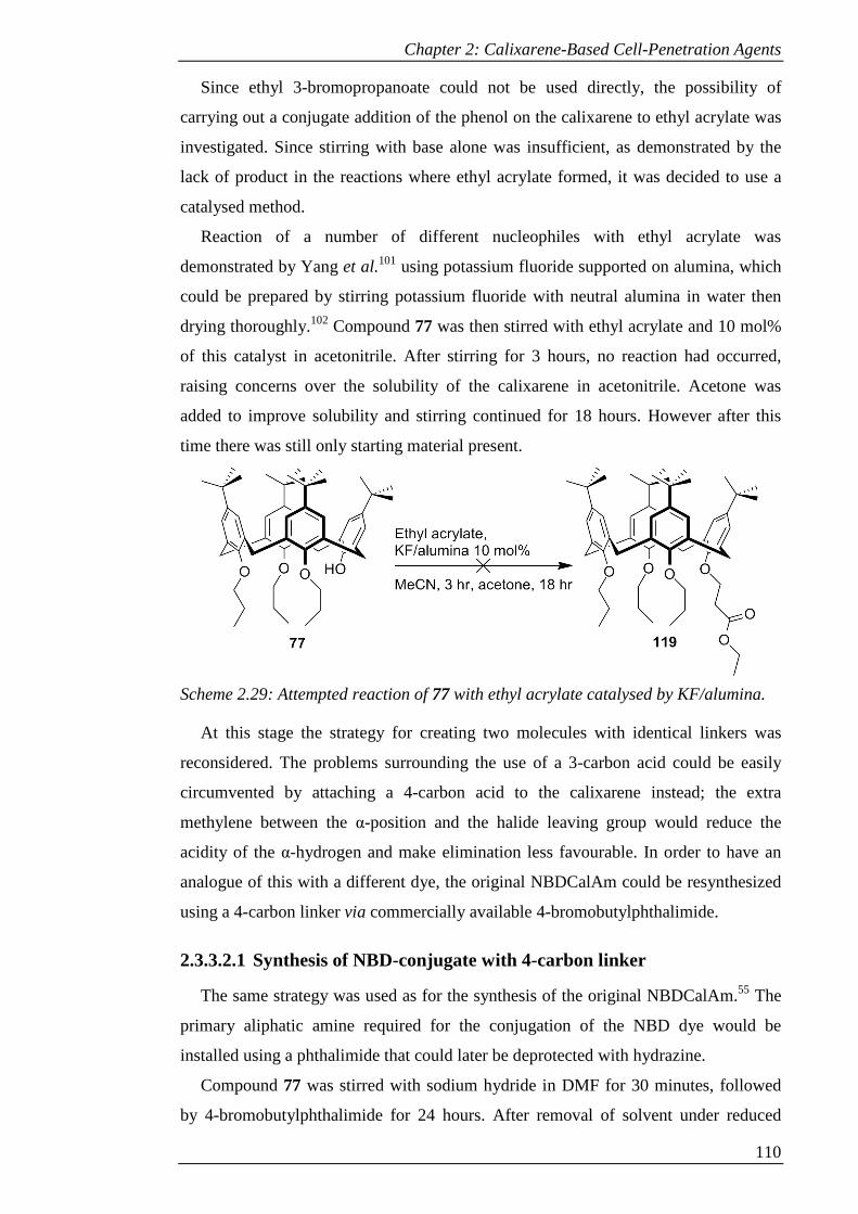
Chapter 2: Calixarene-Based Cell-Penetration Agents
110
Since ethyl 3-bromopropanoate could not be used directly, the possibility of
carrying out a conjugate addition of the phenol on the calixarene to ethyl acrylate was
investigated. Since stirring with base alone was insufficient, as demonstrated by the
lack of product in the reactions where ethyl acrylate formed, it was decided to use a
catalysed method.
Reaction of a number of different nucleophiles with ethyl acrylate was
demonstrated by Yang et al.101 using potassium fluoride supported on alumina, which
could be prepared by stirring potassium fluoride with neutral alumina in water then
drying thoroughly.102 Compound 77 was then stirred with ethyl acrylate and 10 mol%
of this catalyst in acetonitrile. After stirring for 3 hours, no reaction had occurred,
raising concerns over the solubility of the calixarene in acetonitrile. Acetone was
added to improve solubility and stirring continued for 18 hours. However after this
time there was still only starting material present.
Scheme 2.29: Attempted reaction of 77 with ethyl acrylate catalysed by KF/alumina.
At this stage the strategy for creating two molecules with identical linkers was
reconsidered. The problems surrounding the use of a 3-carbon acid could be easily
circumvented by attaching a 4-carbon acid to the calixarene instead; the extra
methylene between the α-position and the halide leaving group would reduce the
acidity of the α-hydrogen and make elimination less favourable. In order to have an
analogue of this with a different dye, the original NBDCalAm could be resynthesized
using a 4-carbon linker via commercially available 4-bromobutylphthalimide.
2.3.3.2.1 Synthesis of NBD-conjugate with 4-carbon linker
The same strategy was used as for the synthesis of the original NBDCalAm.55 The
primary aliphatic amine required for the conjugation of the NBD dye would be
installed using a phthalimide that could later be deprotected with hydrazine.
Compound 77 was stirred with sodium hydride in DMF for 30 minutes, followed
by 4-bromobutylphthalimide for 24 hours. After removal of solvent under reduced
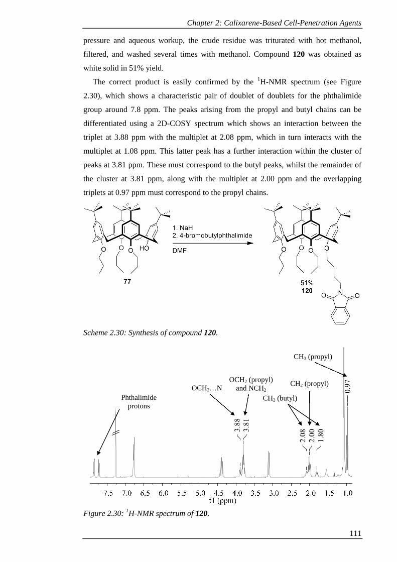
Chapter 2: Calixarene-Based Cell-Penetration Agents
111
pressure and aqueous workup, the crude residue was triturated with hot methanol,
filtered, and washed several times with methanol. Compound 120 was obtained as
white solid in 51% yield.
The correct product is easily confirmed by the 1H-NMR spectrum (see Figure
2.30), which shows a characteristic pair of doublet of doublets for the phthalimide
group around 7.8 ppm. The peaks arising from the propyl and butyl chains can be
differentiated using a 2D-COSY spectrum which shows an interaction between the
triplet at 3.88 ppm with the multiplet at 2.08 ppm, which in turn interacts with the
multiplet at 1.08 ppm. This latter peak has a further interaction within the cluster of
peaks at 3.81 ppm. These must correspond to the butyl peaks, whilst the remainder of
the cluster at 3.81 ppm, along with the multiplet at 2.00 ppm and the overlapping
triplets at 0.97 ppm must correspond to the propyl chains.
Scheme 2.30: Synthesis of compound 120.
Figure 2.30: 1H-NMR spectrum of 120.
0.9
7
1.8
0
2.0
0
2.0
83.8
1
3.8
8
OCH2…N OCH2 (propyl)
and NCH2 CH2 (propyl)
CH2 (butyl)
CH3 (propyl)
Phthalimide protons

Chapter 2: Calixarene-Based Cell-Penetration Agents
112
Elucidation of which peak corresponds to which methylene in the butyl chain
requires a 2D-HSQC spectrum (see Figure 2.31). Examination of the peaks around 3.8
ppm reveals that within the cluster at 3.81 ppm there is an interaction with a secondary
carbon that has a much lower chemical shift compared with the other carbon peaks
that have interactions with this cluster. This suggests that this carbon is adjacent to a
nitrogen, whereas the others around 75 ppm are adjacent to oxygen. This identifies the
peak at 3.88 ppm as the methylene adjacent to the oxygen in the butyl chain, which in
turn allows the differentiation of the peaks 2.08 and 1.80 ppm as the centre two
methylene groups closest to the oxygen and the nitrogen, respectively.
Figure 2.31: 2D-HSQC spectrum of 120.
Conversion to the Boc-protected amine was carried out as previously. Compound
120 was stirred with 100% nitric acid and glacial acetic acid in DCM for 4 hours
before quenching with water. Aqueous work-up followed by trituration with methanol
gave sufficiently pure tetra-nitrated product 121 as light-yellow solid in 87% yield.
This product was heated to reflux in ethanol with tin chloride for 48 hours,
followed by aqueous work-up with sodium hydroxide. The tetra-amine product 122
was isolated as brown glass in 88% yield

Chapter 2: Calixarene-Based Cell-Penetration Agents
113
Scheme 2.31: Conversion of 120 to 123 via nitration, reduction and Boc protection.
Compound 122 was stirred with Boc-anhydride and DIPEA in DCM for 24 hours.
The product was purified by column chromatography over silica gel, eluting with 19:1
DCM/ethyl acetate, giving the protected tetra-amine 123 as off-white glass in 67%
yield.
The required lower-rim amine was next unmasked by cleaving the phthalimide.
Compound 123 was stirred with hydrazine in ethanol for 18 hours. The resulting
precipitate was removed by filtration and the solvent removed from the filtrate under
reduced pressure. Due to the poor solubility of the product in organic solvent, aqueous
work-up was avoided by precipitating the product from minimum methanol with
water, filtering and drying under vacuum. Some of the solid was too fine to separate
from the solvent by filtration so was salvaged by extracting from water with 10%
methanol in ethyl acetate. The free-amine (124) was isolated as off-white solid in 72%
yield and was carried forward as the crude product.
This product could now be reacted with the dye via a nucleophilic aromatic
substitution reaction. Compound 124 was stirred with sodium hydrogencarbonate in
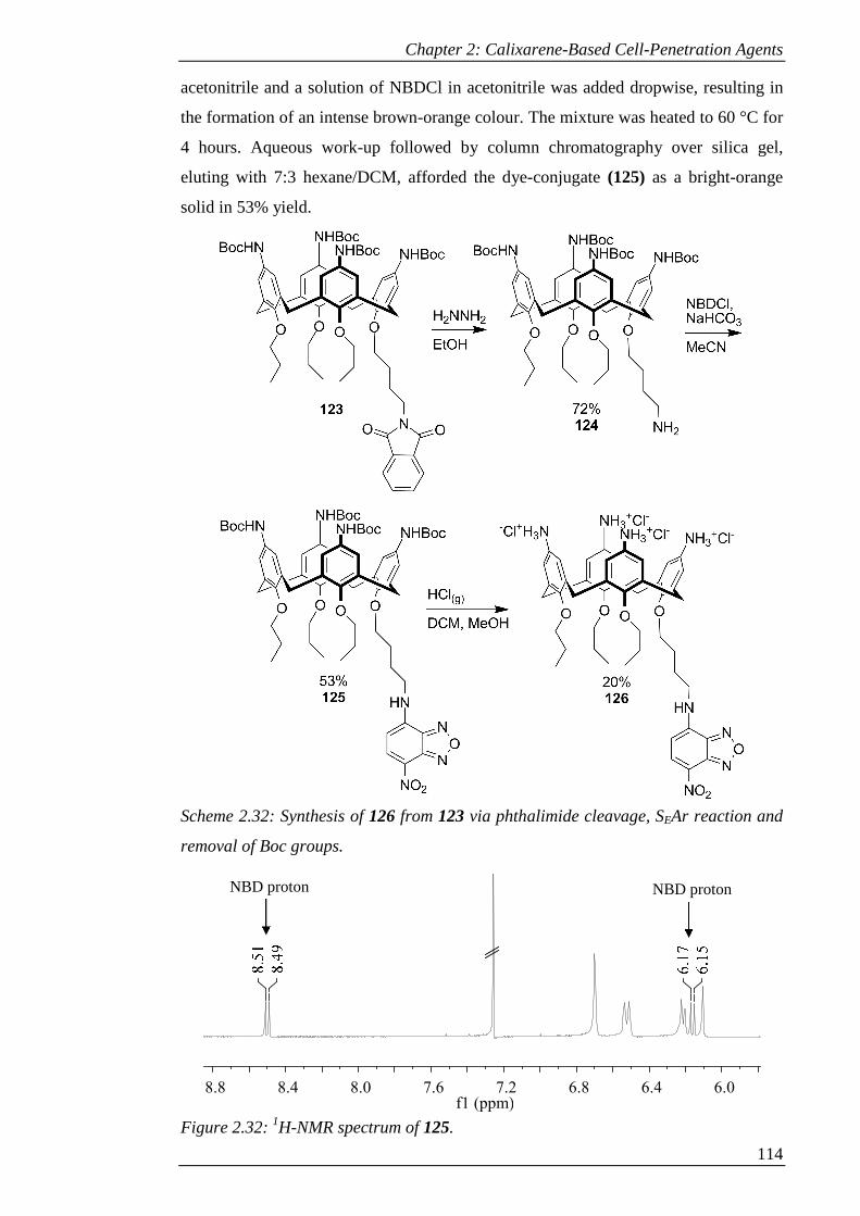
Chapter 2: Calixarene-Based Cell-Penetration Agents
114
acetonitrile and a solution of NBDCl in acetonitrile was added dropwise, resulting in
the formation of an intense brown-orange colour. The mixture was heated to 60 °C for
4 hours. Aqueous work-up followed by column chromatography over silica gel,
eluting with 7:3 hexane/DCM, afforded the dye-conjugate (125) as a bright-orange
solid in 53% yield.
Scheme 2.32: Synthesis of 126 from 123 via phthalimide cleavage, SEAr reaction and
removal of Boc groups.
Figure 2.32: 1H-NMR spectrum of 125.
6.06.46.87.27.68.08.48.8f1 (ppm)
NBD proton NBD proton
Chapter 2: Calixarene-Based Cell-Penetration Agents
115
The 1H-NMR spectrum of 125 (see Figure 2.32) confirms the incorporation of the
NBD dye. At 8.50 ppm there is a doublet arising from one of the two aromatic protons
on the dye. The 2D-COSY spectrum shows that the second doublet is at 6.16 ppm.
The former, being more deshielded, must be the one adjacent to the nitro group.
Finally, the water soluble tetra-amine salt was formed by bubbling HCl gas through
a solution of 125 in DCM for 10 minutes, followed by addition of methanol to
dissolve the resulting precipitate and continuing the reaction for a further 20 minutes.
The product was purified by reverse-phase column chromatography over C18, eluting
with a gradient of 0-100% methanol in 60 mM aqueous HCl. Compound 126 was
isolated as a bright-orange solid. Due to losses during purification, the final yield was
just 20%.
2.3.3.2.2 Synthesis of amide-linked coumarin
The required butanoic acid group on the calixarene was put in place using ethyl 4-
bromobutanoate. This ester can be hydrolysed once the upper rim has been
functionalised with Boc-protected amines to expose the acid for the amide coupling.
The synthesis of a para-nitro functionalised calixarene with a single C4-acid on the
lower rim has been previously reported.103
Compound 77 was stirred with sodium hydride in DMF for 30 minutes, followed
by addition of ethyl 4-bromobutanoate. After 18 hours, the solvent was removed under
reduced pressure. Aqueous work-up followed by trituration with methanol gave 127 as
white powder in 68% yield.
This was then converted to the Boc-protected amine in the same manner as before.
Compound 127 was stirred with 100% nitric acid and glacial acetic acid in DCM for 4
hours before quenching with water. Following aqueous work-up, the residue was
triturated with methanol to give 128 as light-orange powder in 92% yield.
Compound 128 was heated to reflux in ethanol with tin chloride for 48 hours. After
aqueous work-up with sodium hydroxide, the tetra-amine product 129 was isolated as
light-brown solid in 79% yield.
Finally, 129 was stirred with Boc anhydride and DIPEA in DCM for 72 hours.
Purification by column chromatography over silica gel, eluting with 19:1 DCM/ethyl
acetate gave 130 as off-white glass in 78% yield.
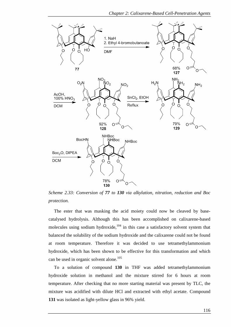
Chapter 2: Calixarene-Based Cell-Penetration Agents
116
Scheme 2.33: Conversion of 77 to 130 via alkylation, nitration, reduction and Boc
protection.
The ester that was masking the acid moiety could now be cleaved by base-
catalysed hydrolysis. Although this has been accomplished on calixarene-based
molecules using sodium hydroxide,104 in this case a satisfactory solvent system that
balanced the solubility of the sodium hydroxide and the calixarene could not be found
at room temperature. Therefore it was decided to use tetramethylammonium
hydroxide, which has been shown to be effective for this transformation and which
can be used in organic solvent alone.105
To a solution of compound 130 in THF was added tetramethylammonium
hydroxide solution in methanol and the mixture stirred for 6 hours at room
temperature. After checking that no more starting material was present by TLC, the
mixture was acidified with dilute HCl and extracted with ethyl acetate. Compound
131 was isolated as light-yellow glass in 96% yield.
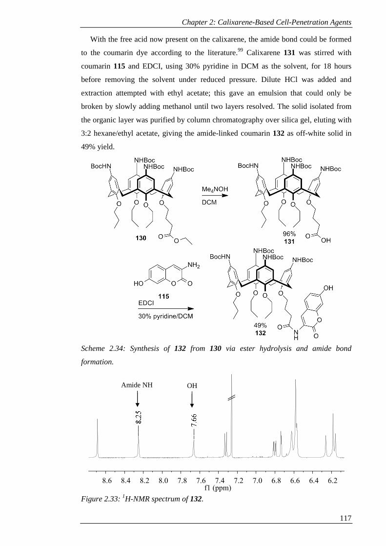
Chapter 2: Calixarene-Based Cell-Penetration Agents
117
With the free acid now present on the calixarene, the amide bond could be formed
to the coumarin dye according to the literature.99 Calixarene 131 was stirred with
coumarin 115 and EDCI, using 30% pyridine in DCM as the solvent, for 18 hours
before removing the solvent under reduced pressure. Dilute HCl was added and
extraction attempted with ethyl acetate; this gave an emulsion that could only be
broken by slowly adding methanol until two layers resolved. The solid isolated from
the organic layer was purified by column chromatography over silica gel, eluting with
3:2 hexane/ethyl acetate, giving the amide-linked coumarin 132 as off-white solid in
49% yield.
Scheme 2.34: Synthesis of 132 from 130 via ester hydrolysis and amide bond
formation.
Figure 2.33: 1H-NMR spectrum of 132.
6.26.46.66.87.07.27.47.67.88.08.28.48.6f1 (ppm)
Amide NH OH
Chapter 2: Calixarene-Based Cell-Penetration Agents
118
The 1H-NMR spectrum (see Figure 2.33) confirms that the coumarin is present and
joined by an amide bond. The characteristic peaks of the coumarin can be seen: a
downfield singlet around 8.7 ppm, a doublet around 7.3 ppm, and a doublet of
doublets next to a further doublet around 6.8 ppm. Two peaks are present at 8.25 and
7.66 ppm that give rise to no peaks in the 2D-HSQC, suggesting that these are the NH
of the amide bond and the OH of the coumarin. According to the 2D-HMBC
spectrum, the proton peak at 8.25 ppm and the proton peaks corresponding to the butyl
chain share a long-range interaction with a highly deshielded carbon. This suggests
that this carbon is the carbonyl of the amide and so the proton peak at 8.25 ppm would
be the NH of the amide. By elimination, the peak at 7.66 ppm must be the OH of the
coumarin.
2.3.3.2.3 Reduction of amide-linked coumarin
The reduction of an amide to an amine could theoretically be accomplished using a
strong nucleophilic reducing agent such as lithium aluminium hydride. However,
amides are more difficult to reduce in this way than esters due to the increased
electron density at the carbonyl; therefore in the presence of an ester, it will be the
latter that will be reduced first. The presence of the lactone in the coumarin dye
precludes the use of such a method in this case.
Multiple methods exist for the selective reduction of amides in the presence of
other functional groups, including esters. These include transition metal catalysed
hydrosilation,106 treatment with tetrabutylammonium hydride,107 conversion to a
thioamide prior to reduction108 and reduction with borane.109 The latter selectively
delivers hydride to amide bonds in preference to esters by forming a coordination
complex to the more electron-rich amide.110 Borane reduction has been applied to a
coumarin, albeit for the reduction of a carboxylic acid instead of an amide, without
reducing the lactone.111 For this reason, this reagent was selected for the reduction of
the coumarin-amide.
A solution of 132 in dry THF was cooled to 0 °C and stirred under argon whilst
borane-THF complex was added. This was then heated to 60 °C for 4 hours before
quenching with methanol. At this stage the 1H-NMR spectrum showed a complex
mixture of products; this was also the case when the reaction was allowed to proceed
at room temperature for 18 hours.
A mixture of products could arise from excess borane forming a complex with the
carbamates on the upper rim. A mild method for the cleavage of borane adducts was

Chapter 2: Calixarene-Based Cell-Penetration Agents
119
found which utilises palladium on carbon or Raney nickel as a catalyst for
methanolysis, which can otherwise require a very long period of stirring in
methanol.112 The crude product was therefore stirred in methanol with 5 mol% of
Raney nickel for 18 hours, followed by removal of the catalyst by filtration. The
borate now present in the product was removed by azeotroping several times with
methanol. However, the light-yellow glass that was obtained still proved to be a
complex mixture by 1H-NMR.
Scheme 2.35: Attempted synthesis of 133 by reduction of 132.
Although examples are present in the literature of amide reductions being carried
out in this way in the presence of Boc protecting groups,113 it would appear in this
case that the carbamates are not stable to these conditions. However, if these were
removed then the resulting free amines would necessitate the use of a greater excess of
borane, due to their propensity to form adducts with the reagent, and subsequently
would poison the catalyst used to effect methanolysis. An alternative method of
reduction would therefore be more suitable.
However, at this stage due to time constraints it was decided to proceed with the
removal of the Boc protecting groups from 132 to provide a cationic molecule with an
amide-linked coumarin. The cell-uptake properties could then be investigated to
provide a comparison with the original triazole-linked coumarin.
2.3.3.2.4 Deprotection of amide-linked coumarin conjugate
Removal of the Boc protecting groups from 132 was initially attempted using the
same procedure as in section 2.3.1.8. HCl(g) was bubbled through a solution of 132 in
DCM for 10 minutes, followed by addition of methanol and stirring for a further 5
minutes. However, the solid obtained was a mixture of two distinct products by 1H-
NMR; when the reaction was continued for a further 15 minutes, the minor component
increased in concentration. This led to the conclusion that the deprotection conditions
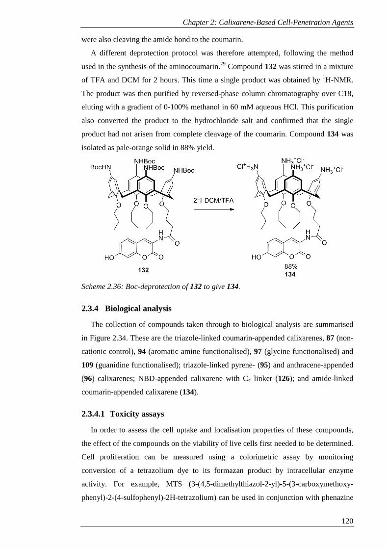
Chapter 2: Calixarene-Based Cell-Penetration Agents
120
were also cleaving the amide bond to the coumarin.
A different deprotection protocol was therefore attempted, following the method
used in the synthesis of the aminocoumarin.79 Compound 132 was stirred in a mixture
of TFA and DCM for 2 hours. This time a single product was obtained by 1H-NMR.
The product was then purified by reversed-phase column chromatography over C18,
eluting with a gradient of 0-100% methanol in 60 mM aqueous HCl. This purification
also converted the product to the hydrochloride salt and confirmed that the single
product had not arisen from complete cleavage of the coumarin. Compound 134 was
isolated as pale-orange solid in 88% yield.
Scheme 2.36: Boc-deprotection of 132 to give 134.
2.3.4 Biological analysis
The collection of compounds taken through to biological analysis are summarised
in Figure 2.34. These are the triazole-linked coumarin-appended calixarenes, 87 (non-
cationic control), 94 (aromatic amine functionalised), 97 (glycine functionalised) and
109 (guanidine functionalised); triazole-linked pyrene- (95) and anthracene-appended
(96) calixarenes; NBD-appended calixarene with C4 linker (126); and amide-linked
coumarin-appended calixarene (134).
2.3.4.1 Toxicity assays
In order to assess the cell uptake and localisation properties of these compounds,
the effect of the compounds on the viability of live cells first needed to be determined.
Cell proliferation can be measured using a colorimetric assay by monitoring
conversion of a tetrazolium dye to its formazan product by intracellular enzyme
activity. For example, MTS (3-(4,5-dimethylthiazol-2-yl)-5-(3-carboxymethoxy-
phenyl)-2-(4-sulfophenyl)-2H-tetrazolium) can be used in conjunction with phenazine

Chapter 2: Calixarene-Based Cell-Penetration Agents
121
methosulphate (PMS). The activity of oxidoreductase enzymes, which is indicative of
live cells, converts MTS to its formazan product to give an absorbance increase at
490-500 nm that is proportional to the number of live cells present.114
Selected cell-lines were incubated with increasing concentrations of the compounds
to be tested for 72 hours before carrying out the MTS assays. Compounds 94, 95, 96,
97, 126 and 134 were compared to a control where only sterile water was added. Due
to their poor solubility in water, compounds 87 and 109 were dissolved in DMSO and
therefore this was used as the control.
The results of the MTS assays in THP-1 cells are shown in Figure 2.35 and are
representative of the other cell lines tested.
Figure 2.34: Summary of compounds subjected to in vivo testing.

Chapter 2: Calixarene-Based Cell-Penetration Agents
122
Figure 2.35: MTS assays performed on THP-1 cells. a) Compound 87; b) Compounds
94, 95 and 97; c) Compounds 96, 126 and 134; d) Compound 109.
Ab
sorb
an
ce
Ab
sorb
an
ce
-9 -8 -7 -6 -5 -4 -3
0.0
0.2
0.4
0.6
0.8
1.0
Control
134
96
126
Compound concentration log[M]
c)
Ab
sorb
an
ce
Chapter 2: Calixarene-Based Cell-Penetration Agents
123
In comparison with NBDCalAm,55 which was only toxic at high concentrations
(around 100 mM), most of the compounds began to inhibit cell proliferation at much
lower concentrations (of the order of 10-6 to 10-5 M) . One explanation for this could
be the change in dye molecule: compounds 94, 95 and 96, with a coumarin, pyrene
and anthracene dye respectively, all began to display cytotoxic effects at around 10-6
M. However, the dye molecule clearly is not the only factor, as exemplified by the
lower apparent toxicity of compound 97, which in particular displayed no toxicity
towards HeLa cells at the concentrations tested, and the lack of dose-dependent
behaviour of compounds 87 and 109 at the concentrations used. These all bear a
coumarin moiety, but the difference in functionality in the upper rim compared with
the aromatic amine of 94 seems to influence the toxicity; compound 87 features only
tert-butyl groups, whilst 97 and 109 are functionalised with aliphatic amine and
aromatic guanidine groups, respectively.
The linker could also be a factor, since compounds 94, 95 and 96 all have triazole-
linked dyes, compared with the amine-linked dye of NBDCalAm. Compound 134,
which has an amide-linked coumarin, in comparison with 94, begins to inhibit cell
proliferation at a higher concentration in THP-1 cells (of the order of 10-5 M compared
with 10-6 M), although this effect was not observed with HeLa cells. The sensitivity to
the linker is particularly apparent in the case of 126, which provides an interesting
comparison with NBDCalAm. Although the only structural difference between these
two compounds is an increase in the length of linker from C3 to C4, 126 displays
cytotoxic effects at much lower concentrations (of the order of 10-6 M). The increased
length of linker may allow the calixarene to form interactions that could not be made
by NBDCalAm, resulting in increased impact on cell viability.
It is important to note that full characterisation of the cytotoxicity of these
compounds would require collection of data at a larger number of concentrations, in
order to determine IC50 values. Nevertheless, this data does reveal that at sub-
micromolar concentrations the cells remained viable with no significant change in
morphology (see section 2.3.4.2). Investigation of cell uptake and localisation could
therefore be carried out with low concentrations of the compounds.
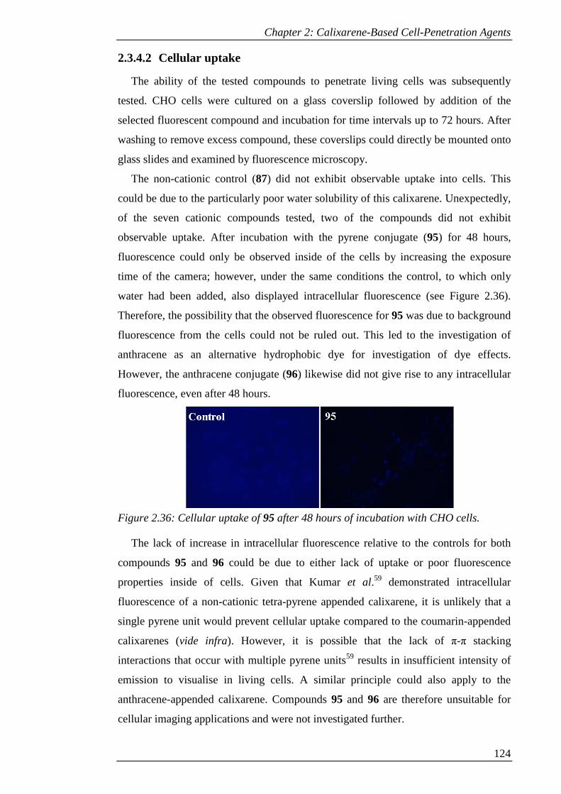
Chapter 2: Calixarene-Based Cell-Penetration Agents
124
2.3.4.2 Cellular uptake
The ability of the tested compounds to penetrate living cells was subsequently
tested. CHO cells were cultured on a glass coverslip followed by addition of the
selected fluorescent compound and incubation for time intervals up to 72 hours. After
washing to remove excess compound, these coverslips could directly be mounted onto
glass slides and examined by fluorescence microscopy.
The non-cationic control (87) did not exhibit observable uptake into cells. This
could be due to the particularly poor water solubility of this calixarene. Unexpectedly,
of the seven cationic compounds tested, two of the compounds did not exhibit
observable uptake. After incubation with the pyrene conjugate (95) for 48 hours,
fluorescence could only be observed inside of the cells by increasing the exposure
time of the camera; however, under the same conditions the control, to which only
water had been added, also displayed intracellular fluorescence (see Figure 2.36).
Therefore, the possibility that the observed fluorescence for 95 was due to background
fluorescence from the cells could not be ruled out. This led to the investigation of
anthracene as an alternative hydrophobic dye for investigation of dye effects.
However, the anthracene conjugate (96) likewise did not give rise to any intracellular
fluorescence, even after 48 hours.
Figure 2.36: Cellular uptake of 95 after 48 hours of incubation with CHO cells.
The lack of increase in intracellular fluorescence relative to the controls for both
compounds 95 and 96 could be due to either lack of uptake or poor fluorescence
properties inside of cells. Given that Kumar et al.59 demonstrated intracellular
fluorescence of a non-cationic tetra-pyrene appended calixarene, it is unlikely that a
single pyrene unit would prevent cellular uptake compared to the coumarin-appended
calixarenes (vide infra). However, it is possible that the lack of π-π stacking
interactions that occur with multiple pyrene units59 results in insufficient intensity of
emission to visualise in living cells. A similar principle could also apply to the
anthracene-appended calixarene. Compounds 95 and 96 are therefore unsuitable for
cellular imaging applications and were not investigated further.
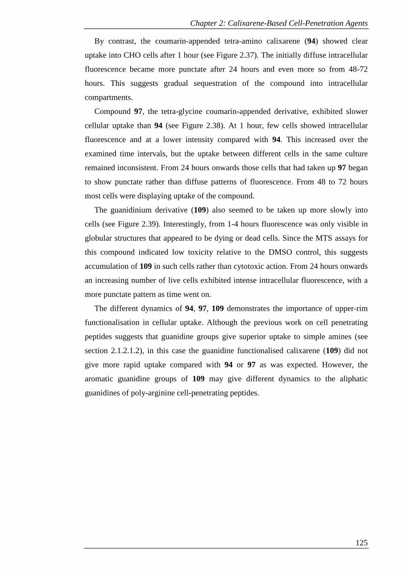
Chapter 2: Calixarene-Based Cell-Penetration Agents
125
By contrast, the coumarin-appended tetra-amino calixarene (94) showed clear
uptake into CHO cells after 1 hour (see Figure 2.37). The initially diffuse intracellular
fluorescence became more punctate after 24 hours and even more so from 48-72
hours. This suggests gradual sequestration of the compound into intracellular
compartments.
Compound 97, the tetra-glycine coumarin-appended derivative, exhibited slower
cellular uptake than 94 (see Figure 2.38). At 1 hour, few cells showed intracellular
fluorescence and at a lower intensity compared with 94. This increased over the
examined time intervals, but the uptake between different cells in the same culture
remained inconsistent. From 24 hours onwards those cells that had taken up 97 began
to show punctate rather than diffuse patterns of fluorescence. From 48 to 72 hours
most cells were displaying uptake of the compound.
The guanidinium derivative (109) also seemed to be taken up more slowly into
cells (see Figure 2.39). Interestingly, from 1-4 hours fluorescence was only visible in
globular structures that appeared to be dying or dead cells. Since the MTS assays for
this compound indicated low toxicity relative to the DMSO control, this suggests
accumulation of 109 in such cells rather than cytotoxic action. From 24 hours onwards
an increasing number of live cells exhibited intense intracellular fluorescence, with a
more punctate pattern as time went on.
The different dynamics of 94, 97, 109 demonstrates the importance of upper-rim
functionalisation in cellular uptake. Although the previous work on cell penetrating
peptides suggests that guanidine groups give superior uptake to simple amines (see
section 2.1.2.1.2), in this case the guanidine functionalised calixarene (109) did not
give more rapid uptake compared with 94 or 97 as was expected. However, the
aromatic guanidine groups of 109 may give different dynamics to the aliphatic
guanidines of poly-arginine cell-penetrating peptides.
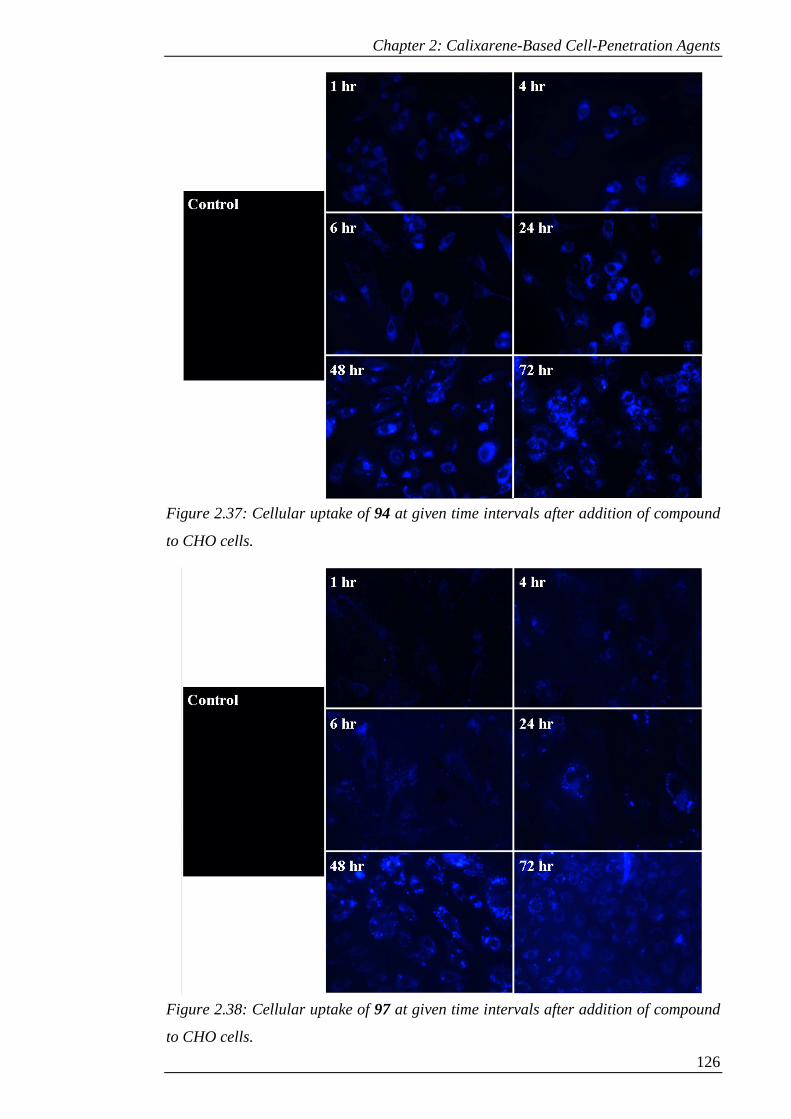
Chapter 2: Calixarene-Based Cell-Penetration Agents
126
Figure 2.37: Cellular uptake of 94 at given time intervals after addition of compound
to CHO cells.
Figure 2.38: Cellular uptake of 97 at given time intervals after addition of compound
to CHO cells.
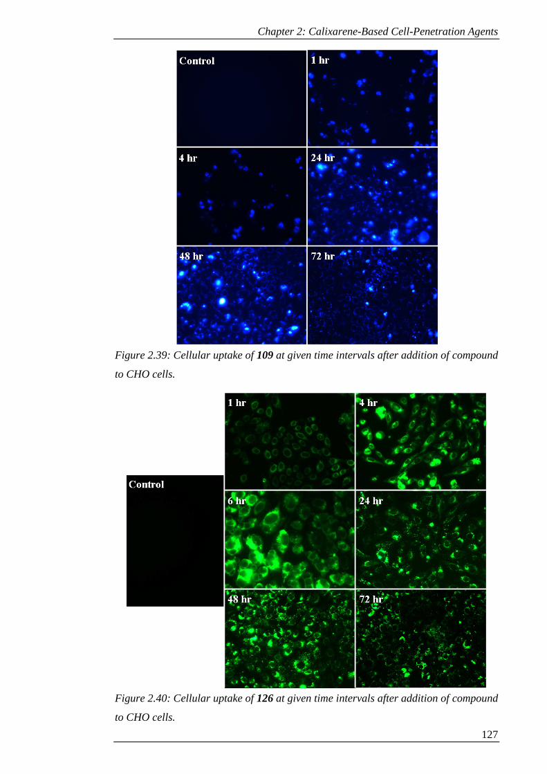
Chapter 2: Calixarene-Based Cell-Penetration Agents
127
Figure 2.39: Cellular uptake of 109 at given time intervals after addition of compound
to CHO cells.
Figure 2.40: Cellular uptake of 126 at given time intervals after addition of compound
to CHO cells.

Chapter 2: Calixarene-Based Cell-Penetration Agents
128
Figure 2.41: Cellular uptake of 134 at given time intervals after addition of compound
to CHO cells.
Compound 126 allowed the effect of an increased length of linker to be tested
compared with its previously investigated counterpart, NBDCalAm.66 Although the
increase in linker length resulted in higher cytotoxicity, the cellular uptake seemed to
be similar: uptake was rapid, with intracellular fluorescence clearly visible after 1
hour and intense fluorescence exhibited after 4 hours, and increasingly punctate
patterns visible from 24 hours onwards (see Figure 2.40).
Compound 134, despite the change of linker from triazole to amide, displayed
similar uptake to 94 (see Figure 2.41). Some intracellular fluorescence was visible
after 1 hour, increasing in intensity over time, with the diffuse pattern changing to
more punctate from 24 hours onwards. This suggests that the more rapid uptake
exhibited by the NBD-appended compounds 126 and NBDCalAm compared with 94
could be due to the effect of the dye rather than that of the triazole ring.
It is important to note that these uptake studies give only qualitative information
about the relative rate of uptake. In order to obtain accurate measurements of uptake
kinetics, flow cytometry experiments would be required. However, the above
experiments do reveal that an incubation time of 48 hours gives good uptake of all
compounds and is therefore a suitable amount of time to allow in further experiments.

Chapter 2: Calixarene-Based Cell-Penetration Agents
129
2.3.4.3 Mechanism of uptake
Incubation of cells with different inhibitors of endocytosis followed by incubation
with the compounds under investigation allows the mechanism of their uptake to be
investigated. As discussed in section 2.1.1, the two major pathways of endocytosis are
via clathrin-coated pits or caveolae. Processes involving the latter can be inhibited by
interfering with the function of cholesterol in the membrane. Suitable inhibitors
include methyl-β-cyclodextrin (MCD),115 which forms soluble inclusion complexes
with cholesterol and so extracts it from the membrane, and the polyene antibiotics
filipin 116 and nystatin,117 which perturb the function of membrane domains by
sequestering cholesterol within the membrane.
Clathrin-mediated processes can be inhibited by hypertonic sucrose, which
disperses clathrin lattices.118 The antibiotic monensin can also be used. This ionophore
dissipates the proton gradient in lysosomes and interferes with transfer of compounds
to these intracellular compartments; it therefore inhibits receptor-mediated uptake by
interfering with receptor recycling.119,120
Investigations were carried out with these five inhibitors by incubating CHO cells
grown on coverslips with each inhibitor followed by addition of the calixarene
compounds under investigation and incubation for a further 48 hours. Excess inhibitor
and calixarene was removed by washing before mounting on slides for imaging. In
some cases incubation with monensin or MCD for 48 hours reduced cell viability and
gave rounded dead or dying cells. In these cases the results are omitted since the
possibility that uptake was due to the loss of viability could not be excluded.
The results for compound 94 are shown in Figure 2.42. Both MCD and nystatin
resulted in a lower level of intracellular fluorescence, whilst filipin gave greatly
diminished fluorescence. Conversely, monensin and sucrose had no inhibitory effect.
This suggests that clathrin-mediated processes are not involved and that pathways
involving caveolae are dominant in the uptake of 94. Interestingly, although monensin
and sucrose did not prevent uptake, both resulted in altered distribution of the
calixarene within the cell. Whilst the cells treated with monensin displayed widely
distributed fluorescence as in the control, the fluorescence seemed to be beginning to
form into more aggregated structures. This can be attributed to perturbed intracellular
trafficking due to dispersion of proton gradients within the cell.120 Treatment with
sucrose on the other hand gave a dramatically more punctate pattern of fluorescence;
this could be due to side effects of hypertonic sucrose which include potential
stimulation of rearrangement of the actin cytoskeleton.121
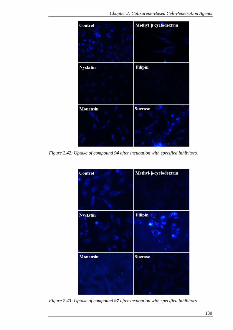
Chapter 2: Calixarene-Based Cell-Penetration Agents
130
Figure 2.42: Uptake of compound 94 after incubation with specified inhibitors.
Figure 2.43: Uptake of compound 97 after incubation with specified inhibitors.
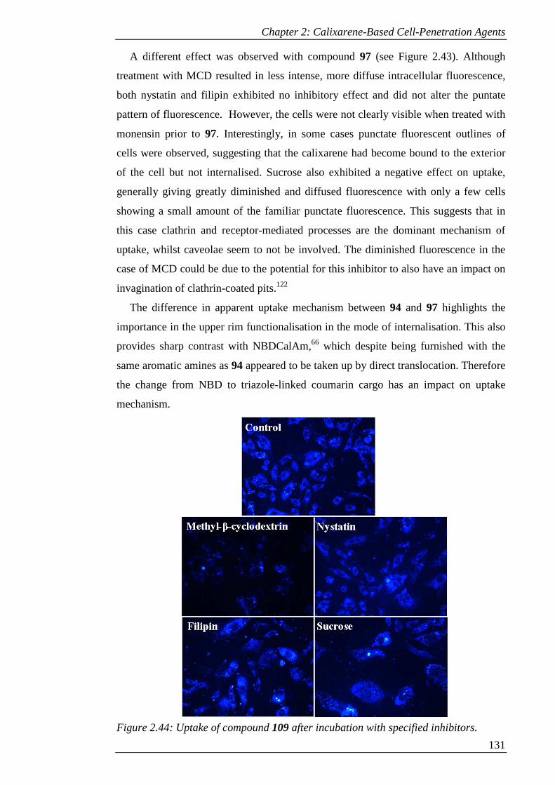
Chapter 2: Calixarene-Based Cell-Penetration Agents
131
A different effect was observed with compound 97 (see Figure 2.43). Although
treatment with MCD resulted in less intense, more diffuse intracellular fluorescence,
both nystatin and filipin exhibited no inhibitory effect and did not alter the puntate
pattern of fluorescence. However, the cells were not clearly visible when treated with
monensin prior to 97. Interestingly, in some cases punctate fluorescent outlines of
cells were observed, suggesting that the calixarene had become bound to the exterior
of the cell but not internalised. Sucrose also exhibited a negative effect on uptake,
generally giving greatly diminished and diffused fluorescence with only a few cells
showing a small amount of the familiar punctate fluorescence. This suggests that in
this case clathrin and receptor-mediated processes are the dominant mechanism of
uptake, whilst caveolae seem to not be involved. The diminished fluorescence in the
case of MCD could be due to the potential for this inhibitor to also have an impact on
invagination of clathrin-coated pits.122
The difference in apparent uptake mechanism between 94 and 97 highlights the
importance in the upper rim functionalisation in the mode of internalisation. This also
provides sharp contrast with NBDCalAm,66 which despite being furnished with the
same aromatic amines as 94 appeared to be taken up by direct translocation. Therefore
the change from NBD to triazole-linked coumarin cargo has an impact on uptake
mechanism.
Figure 2.44: Uptake of compound 109 after incubation with specified inhibitors.

Chapter 2: Calixarene-Based Cell-Penetration Agents
132
In the case of 109, none of the inhibitors seemed to have any impact on
internalisation. MCD, filipin, nystatin and sucrose all gave comparable fluorescence to
the control (see Figure 2.39), although in some cases capturing the true level of
fluorescence was difficult due to rapid photobleaching. This suggests a lack of
involvement of endocytotic mechanisms in the uptake of this calixarene and
potentially a direct translocation mechanism as was observed with NBDCalAm. This
was unexpected, since the triazole-linked coumarin had resulted in uptake by
endocytosis for 94 and 97. The clustered guanidine residues therefore seem to allow
this cargo to be directly translocated, although with slower apparent uptake than
NBDCalAm.
As was observed for NBDCalAm,66 internalisation of compound 126 was not
significantly affected by the inhibitors tested, suggesting a similar direct translocation
across the membrane. As with compound 94, monensin and sucrose altered the
distribution of the probe; this was particularly dramatic for the former, which gave the
appearance of network-like structures within the cells. This could be due to inhibition
of transfer to lysosomes from other cellular compartments such as the Golgi
apparatus, which was previously observed for NBDCalAm on treatment with
monensin.66
Figure 2.45: Uptake of compound 126 after incubation with specified inhibitors.
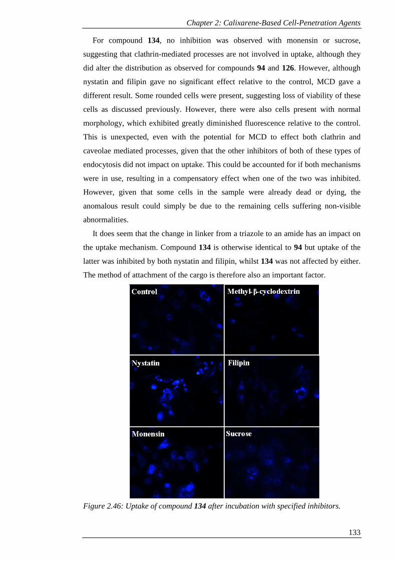
Chapter 2: Calixarene-Based Cell-Penetration Agents
133
For compound 134, no inhibition was observed with monensin or sucrose,
suggesting that clathrin-mediated processes are not involved in uptake, although they
did alter the distribution as observed for compounds 94 and 126. However, although
nystatin and filipin gave no significant effect relative to the control, MCD gave a
different result. Some rounded cells were present, suggesting loss of viability of these
cells as discussed previously. However, there were also cells present with normal
morphology, which exhibited greatly diminished fluorescence relative to the control.
This is unexpected, even with the potential for MCD to effect both clathrin and
caveolae mediated processes, given that the other inhibitors of both of these types of
endocytosis did not impact on uptake. This could be accounted for if both mechanisms
were in use, resulting in a compensatory effect when one of the two was inhibited.
However, given that some cells in the sample were already dead or dying, the
anomalous result could simply be due to the remaining cells suffering non-visible
abnormalities.
It does seem that the change in linker from a triazole to an amide has an impact on
the uptake mechanism. Compound 134 is otherwise identical to 94 but uptake of the
latter was inhibited by both nystatin and filipin, whilst 134 was not affected by either.
The method of attachment of the cargo is therefore also an important factor.
Figure 2.46: Uptake of compound 134 after incubation with specified inhibitors.
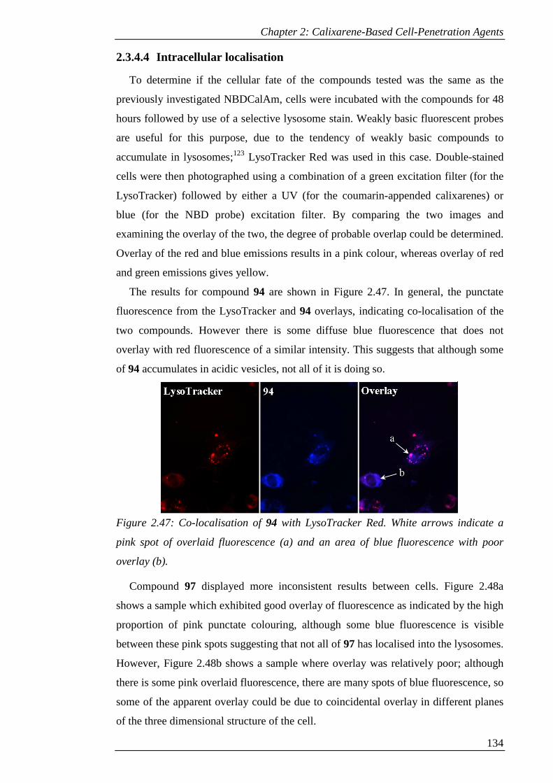
Chapter 2: Calixarene-Based Cell-Penetration Agents
134
2.3.4.4 Intracellular localisation
To determine if the cellular fate of the compounds tested was the same as the
previously investigated NBDCalAm, cells were incubated with the compounds for 48
hours followed by use of a selective lysosome stain. Weakly basic fluorescent probes
are useful for this purpose, due to the tendency of weakly basic compounds to
accumulate in lysosomes;123 LysoTracker Red was used in this case. Double-stained
cells were then photographed using a combination of a green excitation filter (for the
LysoTracker) followed by either a UV (for the coumarin-appended calixarenes) or
blue (for the NBD probe) excitation filter. By comparing the two images and
examining the overlay of the two, the degree of probable overlap could be determined.
Overlay of the red and blue emissions results in a pink colour, whereas overlay of red
and green emissions gives yellow.
The results for compound 94 are shown in Figure 2.47. In general, the punctate
fluorescence from the LysoTracker and 94 overlays, indicating co-localisation of the
two compounds. However there is some diffuse blue fluorescence that does not
overlay with red fluorescence of a similar intensity. This suggests that although some
of 94 accumulates in acidic vesicles, not all of it is doing so.
Figure 2.47: Co-localisation of 94 with LysoTracker Red. White arrows indicate a
pink spot of overlaid fluorescence (a) and an area of blue fluorescence with poor
overlay (b).
Compound 97 displayed more inconsistent results between cells. Figure 2.48a
shows a sample which exhibited good overlay of fluorescence as indicated by the high
proportion of pink punctate colouring, although some blue fluorescence is visible
between these pink spots suggesting that not all of 97 has localised into the lysosomes.
However, Figure 2.48b shows a sample where overlay was relatively poor; although
there is some pink overlaid fluorescence, there are many spots of blue fluorescence, so
some of the apparent overlay could be due to coincidental overlay in different planes
of the three dimensional structure of the cell.
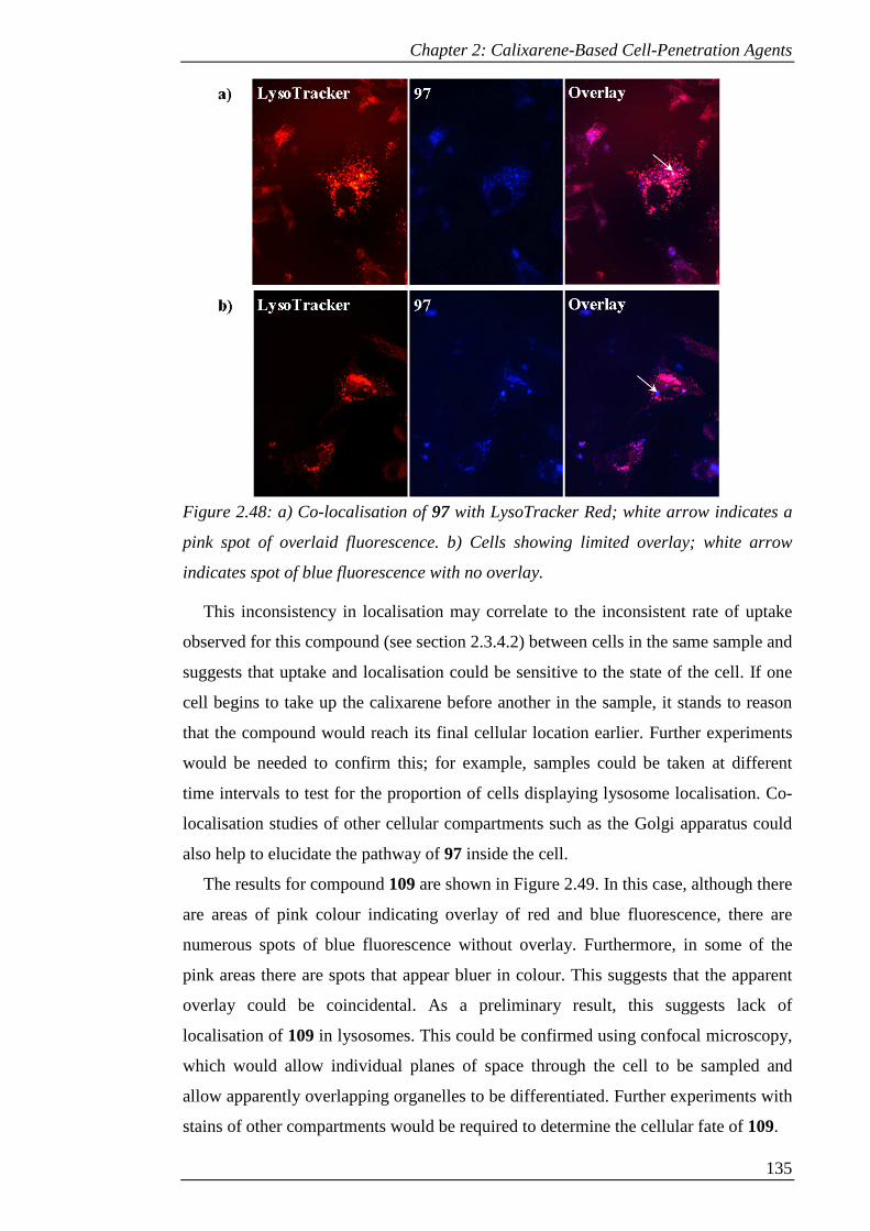
Chapter 2: Calixarene-Based Cell-Penetration Agents
135
Figure 2.48: a) Co-localisation of 97 with LysoTracker Red; white arrow indicates a
pink spot of overlaid fluorescence. b) Cells showing limited overlay; white arrow
indicates spot of blue fluorescence with no overlay.
This inconsistency in localisation may correlate to the inconsistent rate of uptake
observed for this compound (see section 2.3.4.2) between cells in the same sample and
suggests that uptake and localisation could be sensitive to the state of the cell. If one
cell begins to take up the calixarene before another in the sample, it stands to reason
that the compound would reach its final cellular location earlier. Further experiments
would be needed to confirm this; for example, samples could be taken at different
time intervals to test for the proportion of cells displaying lysosome localisation. Co-
localisation studies of other cellular compartments such as the Golgi apparatus could
also help to elucidate the pathway of 97 inside the cell.
The results for compound 109 are shown in Figure 2.49. In this case, although there
are areas of pink colour indicating overlay of red and blue fluorescence, there are
numerous spots of blue fluorescence without overlay. Furthermore, in some of the
pink areas there are spots that appear bluer in colour. This suggests that the apparent
overlay could be coincidental. As a preliminary result, this suggests lack of
localisation of 109 in lysosomes. This could be confirmed using confocal microscopy,
which would allow individual planes of space through the cell to be sampled and
allow apparently overlapping organelles to be differentiated. Further experiments with
stains of other compartments would be required to determine the cellular fate of 109.

Chapter 2: Calixarene-Based Cell-Penetration Agents
136
Figure 2.49: Limited overlay of 109 with LysoTracker Red; white arrow indicates spot
of blue fluorescence with no overlay.
Compound 126 showed consistently good overlay with LysoTracker (see Figure
2.50). Although the staining from the LysoTracker was not as efficient in this case,
resulting in lower intensity of red fluorescence and consequently a less dramatic
yellow colour in the overlay, comparison of the fluorescence images from
LysoTracker and 126 shows an almost identical pattern of staining. This suggests that
like its analogue, NBDCalAm, 126 localises within acidic vesicles.
Figure 2.50: Co-localisation of 126 with LysoTracker Red; white arrow indicates
yellow spot of overlaid fluorescence.
The results for compound 134 (see Figure 2.51) are similar to those of 94. The
fluorescence from the calixarene generally overlays with that of the LysoTracker,
although some areas of diffuse blue fluorescence do not overlay with red fluorescence
of a similar intensity. This suggests that most, but not all, of 134 has localised in
acidic vesicles and that the change in linker from triazole to amide does not have a
noticeable impact on localisation after the 48 hour incubation time.
As noted previously, the three dimensional nature of the cell means that the
possibility of coincidental overlay cannot be ruled out. To confirm the obtained results
and to obtain a quantitative measure of the degree of actual co-localisation of the
compounds with LysoTracker, confocal microscopy would be required. From these
preliminary results, it seems that the change in cargo from NBD to coumarin, whether
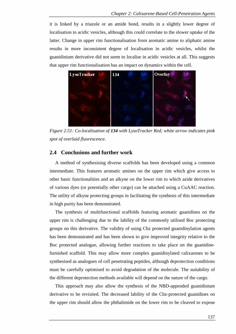
Chapter 2: Calixarene-Based Cell-Penetration Agents
137
it is linked by a triazole or an amide bond, results in a slightly lower degree of
localisation to acidic vesicles, although this could correlate to the slower uptake of the
latter. Change in upper rim functionalisation from aromatic amine to aliphatic amine
results in more inconsistent degree of localisation in acidic vesicles, whilst the
guanidinium derivative did not seem to localise in acidic vesicles at all. This suggests
that upper rim functionalisation has an impact on dynamics within the cell.
Figure 2.51: Co-localisation of 134 with LysoTracker Red; white arrow indicates pink
spot of overlaid fluorescence.
2.4 Conclusions and further work
A method of synthesising diverse scaffolds has been developed using a common
intermediate. This features aromatic amines on the upper rim which give access to
other basic functionalities and an alkyne on the lower rim to which azide derivatives
of various dyes (or potentially other cargo) can be attached using a CuAAC reaction.
The utility of alkyne protecting groups in facilitating the synthesis of this intermediate
in high purity has been demonstrated.
The synthesis of multifunctional scaffolds featuring aromatic guanidines on the
upper rim is challenging due to the lability of the commonly utilised Boc protecting
groups on this derivative. The validity of using Cbz protected guanidinylation agents
has been demonstrated and has been shown to give improved integrity relative to the
Boc protected analogue, allowing further reactions to take place on the guanidine-
furnished scaffold. This may allow more complex guanidinylated calixarenes to be
synthesised as analogues of cell penetrating peptides, although deprotection conditions
must be carefully optimised to avoid degradation of the molecule. The suitability of
the different deprotection methods available will depend on the nature of the cargo.
This approach may also allow the synthesis of the NBD-appended guanidinium
derivative to be revisited. The decreased lability of the Cbz-protected guanidines on
the upper rim should allow the phthalimide on the lower rim to be cleaved to expose
Chapter 2: Calixarene-Based Cell-Penetration Agents
138
the amine required for coupling the NBD dye. A set of analogues to compare with the
coumarin-appended calixarenes could be therefore be synthesised to determine if
upper-rim functionalisation also effects the uptake and localisation of the NBD
derivative.
Since the uptake and intracellular dynamics of these probes seems to be sensitive to
the upper rim functionalisation, it is of interest to create an expanded library of
compounds to test. Using the common intermediate, the amines on the upper rim
could be coupled to alternative amino acids to glycine, for example lysine; such upper
rim functionalisation has been accomplished by Bagnacani et al.62 Similarly, coupling
to arginine would provide a calixarene with aliphatic guanidine groups.
Alternatively, functionalisation with aliphatic guanidines could be achieved via
chloromethylation of the upper rim followed by conversion to methylamines and
ultimately guanidines. Previous work on this route within the group has encountered
difficulties, with side reactions occurring during chloromethylation on the lower-rim
functionalised calixarene and also if alkylation of the lower rim is attempted after
chloromethylation. However, this synthesis could be achieved using the method of
Mourer et al.,86 which involves completing the guanidinylation of the upper rim prior
to any functionalisation of the lower rim.
Biological tests of the compounds showed that the non-cationic control was not
taken up into the cells tested, presumably due to the poor water solubility. The pyrene
and anthracene derivatives proved to be unsuitable for cellular imaging, although it is
possible that installation of multiple dye units on the lower rim may improve the
fluorescence properties by allowing π-stacking between them.59
The coumarin derivatives were visibly taken up by cells and revealed sensitivity of
the uptake, mechanism and localisation to the upper rim functionalisation. All of these
derivatives were taken up more slowly than the NBD-appended calixarenes, but
unexpectedly the guanidinium derivative seemed to be taken up most slowly, with
probe only accumulating within dead or dying cells within the first 24 hours. The
aliphatic amine derivative gave inconsistent uptake between cells in the same sample
within this time frame, whilst the aromatic amine exhibited the fastest uptake of the
three.
Caveolae mediated processes were implicated in the uptake of the aromatic amine,
whilst the aliphatic amine seemed to be taken up via clathrin coated pits and the
guanidinium derivative did not seem to be taken up by either endocytotic pathway.
Localisation of the calixarenes seemed to be mostly within the lysosomes in the case
Chapter 2: Calixarene-Based Cell-Penetration Agents
139
of the aromatic amine, whilst localisation of the aliphatic amine seemed to be more
inconsistent. The guanidinium derivative did not seem to localise within acidic
vesicles.
Comparison of the C4-linked NBD derivative with NBDCalAm revealed no
considerable difference in uptake or localisation, although toxicity was increased. This
suggests that increasing the length of the linker does not affect uptake mechanism or
localisation and therefore allows the amide-linked coumarin, featuring a C4 length
linker, to be compared with the triazole-linked coumarin, which has a C3 length linker.
The triazole linker seemed to have no effect on relative rate of uptake or cellular
localisation, since the amide-linked derivative was taken up in a similar time-frame
and also seemed to localise within acidic vesicles; however, it resulted in uptake that
was apparently not by endocytosis. The impact of the triazole ring was therefore
limited. This suggests that the rapid uptake of NBDCalAm may have been due to the
NBD dye, since the coumarin derivatives were taken up more slowly whether they
featured a triazole or amide linker. The comparison of the triazole- and amide-linked
compounds is also valuable in that it suggests that the triazole may be a suitable amide
bioisostere in these compounds, although it can influence the mechanism of uptake.
To gain more information about the dynamics of these probes, flow cytometry
experiments will be required to allow comparison of the quantitative uptake kinetics.
Further investigation of the cellular localisation is needed for those probes that did not
fully localise in the lysosomes, for example by using a Golgi apparatus stain such as
BODIPY TR ceramide.66 For those that did colocalise with LysoTracker, confocal
microscopy experiments will be required to quantify the true degree of colocalisation.
In general, the current progress has elucidated some of the factors that influence the
uptake and intracellular dynamics of calixarene-based cell-penetration agents.
Synthesis of additional derivatives may allow more of the subtleties of the behaviour
of such molecules to be determined. This in turn will inform the design of cell-
penetration agents based on these scaffolds featuring other cargos such as antiviral or
anticancer agents, or development of novel cellular imaging agents.
2.5 Experimental
2.5.1 General procedures - chemistry
All chemicals were purchased from Sigma-Aldrich, Alfa Aesar, Acrôs Organics or
Novabiochem and were used without further purification. Deuterated solvents for
NMR use were purchased from Cambridge Isotope Laboratories or Sigma-Aldrich.

Chapter 2: Calixarene-Based Cell-Penetration Agents
140
Dry solvents were purchased from previously listed suppliers. Analytical thin layer
chromatography (TLC) was performed using Merck or Machery-Nagel Alugram silica
gel 60 F254 plates, with visualisation by UV light (254 nm). Column chromatography
was run using silica gel 60 (230-400 mesh). NMR spectra were acquired using a
Varian Unity Plus spectrometer (operating at 400 MHz for proton and 100 MHz for
carbon), a Gemini 2000 spectrometer (operating at 300 MHz for proton and 75 MHz
for carbon) or a Bruker Avance 400 Ultrashield spectrometer (operating at 400 MHz
for proton or 100 MHz for carbon). Chemical shifts are referenced relative to
tetramethylsilane (TMS) as the internal reference standard and expressed in parts per
million (ppm or δ) downfield from the standard. Coupling constants (J) are expressed
in Hz. Multiplicities are abbreviated as follows: s (singlet), br s (broad singlet), d
(doublet), t (triplet), m (multiplet), app (apparent). MALDI-TOF mass spectra were
recorded using a Kratos Axima CFR MALDI Mass Spectrometer using α-cyano-4-
hydroxycinnamic acid as matrix. APCI and ESI or NSI mass spectra were recorded by
the EPSRC National Mass Spectrometry Service, Swansea. Melting points were
measured using an electrothermal Mel-temp® melting point apparatus and are
reported uncorrected. Infrared spectra were recorded using a Perkin Elmer Spectrum
BX or Spectrum 65 spectrometer fitted with an ATR attachment and are expressed in
wavenumbers (cm-1).
2.5.2 Synthesis
5,11,17,23-p-tert-Butylcalix[4]arene (2)74
In a 3 L flange flask, p-tert-butyl phenol (200.00 g, 1.33 mol), NaOH (2.40 g, 60
mmol) and 37 wt% formaldehyde (130 mL, 1.74 mol) were heated to 120 °C with
mechanical stirring over 2 h. The resultant yellow solid was dissolved in diphenyl
ether (980 mL). Water of condensation (100 mL) was removed by distillation under a
stream of air, following which the solution was heated to reflux for 3 hours. After
cooling, ethyl acetate (750 mL) was added to precipitate the crude product, which was
collected by filtration, washed with ethyl acetate (2 x 150 mL), acetic acid (2 x 150
mL), water (2 x 150 mL) and acetone (1 x 150 mL) to give 2 as off-white crystals
(143.63 g, 67%). Mp 338-340 °C; IR ν 3158.5, 3052, 3028.5, 2952.5, 2903.5, 2864.5,
1738, 1604, 1592, 1586, 1481.5, 1463, 1428, 1405.5, 1392, 1361.5, 1306, 1299,
1285.5, 1240.5, 1229.5, 1199, 1158.5, 1124, 1102.5, 1039 cm-1; 1H-NMR (400 MHz,
CDCl3) δ 10.34 (s, 4H, OH), 7.05 (s, 8H, ArH), 4.26 (d, J = 13 Hz, 4H, ArCH2Ar),
3.49 (d, J = 13 Hz, 4H, ArCH2Ar), 1.21 (s, 36H, C(CH3)3).
Chapter 2: Calixarene-Based Cell-Penetration Agents
141
5,11,17,23-Tetra-tert-butyl-25,26,27-tripropoxy-28-hydroxy-calix[4]arene
(77)73
p-tert-Butylcalix[4]arene (2) (20.11 g, 31.03 mmol) was dissolved in DMF (300
mL) and heated to 30 °C. BaO (7.15 g, 46.78 mmol) and Ba(OH)2.8H2O (33.82 g,
107.35 mmol) were added and the mixture stirred for 30 mins. n-Propyl bromide (60
mL, 660 mmol) was added and the mixture stirred for 18 hrs. The solution was diluted
with water (200 mL) and the product extracted with DCM (3 x 100 mL). After
washing with water (2 x 200 mL) then brine (200 mL) the organic layer was dried
(MgSO4) and the solvent removed under reduced pressure. Precipitation from DCM
with methanol and filtration gave 77 as white crystals (18.37 g, 79%). Mp 172-174 oC; IR ν 3479, 2956, 2898, 2684, 1584, 1480, 1462, 1431, 1385, 1360, 1295, 1239,
1194, 1121 cm-1; 1H-NMR (400 MHz, CDCl3) δ 7.07 (s, 2 H, ArH), 6.99 (s, 2 H,
ArH), 6.45 (s, 4 H, ArH), 4.30 (d, J = 13 Hz, 2 H, ArCH2Ar), 4.27 (d, J = 13 Hz, 2 H,
ArCH2Ar), 3.78 (t, J = 8 Hz, 2 H, OCH2CH2CH3), 3.69 (t, J = 7 Hz, 4 H,
OCH2CH2CH3), 3.17 (d, J = 13 Hz, 2 H, ArCH2Ar), 3.10 (d, J = 13 Hz, 2 H,
ArCH2Ar), 2.36-2.26 (m, 2H, OCH2CH2CH3), 1.99-1.78 (m, 4 H, OCH2CH2CH3),
1.28 (s, 9 H, C(CH3)3), 1.27 (s, 9 H, CH3), 1.03 (t, J = 7 Hz, 6 H, OCH2CH2CH3), 0.89
(t, J = 8 Hz, 3 H, OCH2CH2CH3), 0.76 (s, 18 H, C(CH3)3).
5,11,17,23-Tetra-tert-butyl-25,26,27-tripropoxy-28-propargyloxy-calix[4]arene
(78)124
Compound 77 (12.51 g, 16.42 mmol) was dissolved in DMF (330 mL). NaH (1.54
g, 64.17 mmol) was added and the mixture stirred for 1 hour before addition of n-
propargyl bromide (8.06 g, 80% w/w in toluene, 54.20 mmol). After stirring for 72
hours, the solution was diluted with water (200 mL) and the product extracted with
DCM (3 x 100 mL). After washing with water (2 x 200 mL) then brine (200 mL), the
organic layer was dried (MgSO4) and the solvent removed under reduced pressure.
Purification by column chromatography over silica (eluent: 1:1 DCM/hexane) gave 78
as off-white crystals (12.60 g, 95%). Mp 203-205 oC; IR ν 3539, 3285, 3261, 2950,
2900, 2860, 2123, 1585, 1582, 1477, 1391, 1360, 1299, 1258, 1237, 1193, 1120 cm-1;
1H-NMR (400 MHz, CDCl3): δ 7.02 (s, 4 H, ArH), 6.58 (d, J = 2 Hz, 2 H, ArH), 6.52
(d, J = 2 Hz, 2 H, ArH), 4.97 (d, J = 2 Hz, 2 H, OCH2CCH), 4.51 (d, J = 12 Hz, 2 H,
ArCH2Ar), 4.43 (d, J = 13 Hz, 2 H, ArCH2Ar), 3.90 (t, J = 8 Hz, 2 H, OCH2CH2CH3),
3.73 (t, J = 7 Hz, 4 H, OCH2CH2CH3), 3.14 (d, J = 13 Hz, 2 H, ArCH2Ar), 3.13 (d, J =
12 Hz, 2 H, ArCH2Ar), 2.38 (t, J = 2 Hz, 1 H, OCH2CCH), 2.14 (sextet, J = 8 Hz, 2H,
Chapter 2: Calixarene-Based Cell-Penetration Agents
142
OCH2CH2CH3), 2.04-1.91 (m, 4 H, OCH2CH2CH3), 1.27 (s, 9 H, C(CH3)3), 1.26 (s, 9
H, C(CH3)3), 1.05 (t, J = 7 Hz, 6 H, OCH2CH2CH3), 0.99 (t, J = 8 Hz, 3 H,
OCH2CH2CH3), 0.99 (s, 18 H, C(CH3)3).
5,11,17,23-Tetranitro-25,26,27-tripropoxy-28-propargyloxy-calix[4]arene (79)
Compound 78 (3.25 g, 4.00 mmol) was dissolved in 130 mL DCM. Glacial acetic
acid (21.5 mL) and 100% nitric acid (21.5 mL) were added and the resulting blue-
black solution was stirred until the colour changed to bright orange. Iced H2O (45 mL)
was added to quench. The product was extracted with DCM (3 x 100 mL) then
washed with water (3 x 200 mL) then brine (200 mL). After drying (MgSO4) the
solvent was removed under reduced pressure and the product precipitated from DCM
with methanol to give 79 as light yellow crystals (2.61 g, 85%). Mp > 180 °C
(decomp.); IR ν 3285, 3069, 2959, 2921, 2867, 1722, 1638, 1597, 1583, 1514, 1449,
1338, 1301, 1262, 1205, 1156 cm-1; 1H-NMR (400 MHz, CDCl3): δ 8.07 (s, 2 H,
ArH), 8.06 (s, 2 H, ArH), 7.16 (s, 4 H, ArH), 4.92 (d, J = 2 Hz, 2 H, OCH2CCH), 4.60
(d, J = 14 Hz, 2 H, ArCH2Ar), 4.55 (d, J = 14 Hz, 2 H, ArCH2Ar), 4.14 (t, J = 8 Hz, 2
H, OCH2CH2CH3), 3.82 (t, J = 7 Hz, 4 H, OCH2CH2CH3), 3.44 (d, J = 14 Hz, 2 H,
ArCH2Ar), 3.42 (d, J = 14 Hz, 2 H, ArCH2Ar), 2.51 (t, J = 2 Hz, 1 H, OCH2CCH),
1.98-1.87 (m, 6 H, OCH2CH2CH3), 1.10 (t, J = 8 Hz, 6 H, OCH2CH2CH3), 0.97 (t, J =
7 Hz, 3 H, OCH2CH2CH3); 13C-NMR (100 MHz, CDCl3) δ 162.8, 160.9, 160.2,
143.8, 142.9, 137.9, 136.6, 134.5, 134.2, 124.8, 124.7, 123.4, 123.3, 78.2, 78.2, 78.0,
77.1, 60.7, 31.5, 31.1, 23.5, 23.4, 10.6, 10.0; ESI-MS m/z: [M]+ 768.2.
5,11,17,23-Tetraamino-25,26,27-tripropoxy-28-propargyloxy-calix[4]arene
(80)
Compound 79 (2.4 g, 3.13 mmol) and SnCl2·2H2O (18.42 g, 81.64 mmol) were
heated to reflux in ethanol (215 mL) for 48 hours. After removing the ethanol under
reduced pressure, 10% NaOH (300 mL) was added, then the product extracted with
DCM (3 x 100 mL). The organic layer was washed with water (200 mL) and brine
(200 mL). After drying (MgSO4) and removing the solvent under reduced pressure, 80
was obtained as an orange-brown glass (1.98 g, 98%). Mp >180 °C (decomp.); IR ν
3310, 2966, 2934, 2878, 1737, 1608, 1469, 1387, 1310, 1285, 1215, 1158, 1129 cm-1; 1H-NMR (300 MHz, CDCl3): δ 6.41 (s, 4 H, ArH), 5.78 (s, 2 H, ArH), 5.77 (s, 2 H,
ArH), 4.77 (d, J = 2 Hz, 2 H, OCH2CCH), 4.39 (d, J = 14 Hz, 2 H, ArCH2Ar), 4.32
(d, J = 13 Hz, 2 H, ArCH2Ar), 3.86 (t, J = 8 Hz, 2 H, OCH2CH2CH3), 3.61 (t, J = 7
Chapter 2: Calixarene-Based Cell-Penetration Agents
143
Hz, 4 H, OCH2CH2CH3), 2.95 (d, J = 14 Hz, 2 H, ArCH2Ar), 2.92 (d, J = 13 Hz, 2 H,
ArCH2Ar), 2.31 (t, J = 2.4 Hz, 1 H, OCH2CCH), 1.88 (sextet, J = 8 Hz, 2 H,
OCH2CH2CH3), 1.84 (sextet, J = 7 Hz, 4 H, OCH2CH2CH3), 1.04 (t, J = 7 Hz, 6 H,
OCH2CH2CH3), 0.97 (t, J = 8 Hz, 3 H, OCH2CH2CH3); 13C-NMR (100 MHz,
CDCl3): δ 150.6, 149.6, 147.9, 141.4, 140.4, 139.2, 138.1, 137.1, 134.3, 134.1, 116.2,
116.0, 115.8, 81.4, 74.2, 59.5, 31.6, 31.0, 23.5, 23.1, 10.9, 10.1; HRMS (NSI) m/z:
[M+H] + Calcd for C40H49N4O4 649.3748; Found 649.3745.
3-acetamido-7-acetoxy-chromen-2-one (81)79
4-Hydroxysalicylaldehyde (6.91 g, 50.01 mmol), N-acetyl glycine (5.85 g, 50.00
mmol), anhydrous sodium acetate (16.4 g, 19.99 mmol) and acetic anhydride (40 mL)
were heated to 120 °C for 5 hours. Ice-cold H2O was added and any solid broken up.
After vacuum filtration and washing 3 times with cold water, the solid was air dried
then triturated with ethyl acetate. Filtration under vacuum and air drying gave 81 as a
yellow powder (6.33 g, 49%). Mp > 240 °C (decomp.); IR ν 3339, 3088, 1757, 1716,
1676, 1611, 1531, 1430, 1352, 1283, 1251, 1199, 1154, 1117 cm-1; 1H-NMR (400
MHz, CDCl3): δ 8.67 (s, 1 H, ArH), 8.04 (s, 1 H, NH), 8.51 (d, J = 8 Hz, 1 H, ArH),
7.12 (d, J = 2.3 Hz, 1 H, ArH), 7.07 (dd, J = 8, 2.3 Hz, 1 H, ArH), 2.34 (s, 3 H, CH3),
2.25 (s, 3 H, CH3).
3-Azido-7-hydroxy-chromen-2-one (82)69
Compound 81 (1 g, 3.83 mmol) was heated to reflux in a solution of 37% HCl and
ethanol (2:1, 11 mL) for 1 hour. Iced H2O (15 mL) was added and the mixture cooled
in an ice bath. NaNO2 (1 g, 14.5 mmol) was added in small portions then stirred for 10
minutes. After adding NaN3 (1.4 g, 21.5 mmol) in small portions the mixture was
stirred for a further 15 minutes then vacuum filtered. Washing with water several
times then air drying gave crude 82 as a brown powder (0.64 g, 83%). Purification by
column chromatography over silica (eluent: 3:2 hexane/ethyl acetate) gave pure 82 as
yellow-orange needles (0.18 g, 23%). Mp 100-102 °C; IR ν 3276, 3047, 2105, 1679,
1609, 1593, 1449, 1370, 1340, 1315, 1256, 1217, 1153, 1119 cm-1; 1H-NMR (400
MHz, DMSO) δ 10.52 (s, 1H, ArOH), 7.59 (s, 1H, ArH), 7.48 (d, J = 8.5 Hz, 1H,
ArH), 6.81 (dd, J = 8.5, 2.3 Hz, 1H, ArH), 6.76 (d, J = 2.3 Hz, 1H, ArH).
Chapter 2: Calixarene-Based Cell-Penetration Agents
144
1-Bromomethylpyrene (83)70
To a stirred suspension of 1-pyrene methanol (1.6 g, 6.83 mmol) in THF (8 mL)
was added PBr3 (2.77 g, 10.25 mmol). After 30 minutes the solid was filtered and
washed with diethyl ether to give 83 as light yellow crystals (1.76 g, 87%). Mp 150-
152 °C; IR ν 3333.5, 3037, 2892.5, 2836, 1602, 1588.5, 1497.5, 1464, 1435.5, 1418.5,
1386.5, 1373, 1312.5, 1269, 1243, 1222.5, 1204.5, 1180, 1138, 1107, 1093.5, 1083,
1027.5 cm-1; 1H-NMR (300 MHz, CDCl3): δ 8.41 (d, J = 9 Hz, 1 H, ArH), 8.27-8.21
(m, 3 H, ArH), 8.14-8.01 (m, 5 H, ArH), 5.26 (s, 2 H, CH2).
1-Azidomethylpyrene (84)70
To a stirred suspension of 83 in DMF (9 mL), NaN3 (0.49 g, 7.54 mmol) was added
and the mixture heated at 60 °C for 16 hours. The product was extracted into ether and
washed with water 3 times. After drying and removing the solvent under reduced
pressure, 84 was obtained as a waxy yellow solid (1.1 g, 88%). Mp 63-65 °C; IR ν
3041, 2992, 2942, 2888.5, 2507, 2482, 2455.5, 2425, 2099, 2079.5, 1925.5, 1901.5,
1875.5, 1861, 1793.5, 1724, 1678, 1652.5, 1603, 1596, 1589, 1509.5, 1489, 1456.5,
1435.5, 1418.5, 1392.5, 1372, 1340, 1314.5, 1270.5, 1252.5, 1245, 1226.5, 1194.5,
1181, 1169.5, 1150.5, 1136.5, 1100, 1059.5, 1026 cm-1; 1H-NMR (300 MHz, CDCl3):
δ 8.31-8.17 (m, 5 H, ArH), 8.13-7.98 (m, 4 H, ArH), 5.05 (s, 2 H, CH2).
Coumarin appended tert-butyl calix[4]arene (87)
A solution of 78 (0.5 g, 0.62 mmol), 82 (125 mg, 0.62 mmol), sodium ascorbate
(132 mg, 0.67 mmol) and CuSO4.5H2O (0.024 g, 0.095 mmol) in DMF (19 mL) was
heated to 90 °C and stirred for 3 hours. After cooling, water (25 mL) was added and
the product extracted with DCM (3 x 25 mL), washed with water (2 x 25 mL) then
brine (25 mL), dried and the solvent removed under reduced pressure. The product
was purified by column chromatography over silica (eluent: 49:1 DCM/methanol) to
give 87 as a light yellow glass (0.38 g, 60%). Mp 175-180 °C; IR ν 2955, 2867, 1736,
1610, 1514, 1480, 1387, 1360, 1298, 1233, 1196, 1121 cm-1; 1H-NMR (300 MHz,
CDCl3) δ 8.60 (s, 1H, ArHTriazole), 8.55 (s, 1H, ArHCoumarin), 7.55 (d, J = 9.2 Hz, 1H,
ArHCoumarin), 6.97-6.93 (m, 2H, ArHCoumarin), 6.83 (s, 4H, ArH), 6.74 (s 2H, ArH), 6.70
(s, 2H, ArH), 5.22 (s, 2H, OCH2C), 4.39 (d, J = 12.4 Hz, 2H, ArCH2Ar), 4.36 (d, J =
12.4 Hz, 2H, ArCH2Ar), 3.83-3.73 (m, 6 H, OCH2CH2CH3), 3.10 (d, J = 12.4 Hz, 4H,
ArCH2Ar), 2.06-1.85 (m, 4 H, OCH2CH2CH3), 1.11 (s, 18H, C(CH3)3), 1.03 (s, 9H,
C(CH3)3), 1.01(s, 9H, C(CH3)3), 0.88-0.81 (m, 9 H, OCH2CH2CH3); 13C-NMR (100
Chapter 2: Calixarene-Based Cell-Penetration Agents
145
MHz, CDCl3): δ 162.8, 162.2, 159.8, 157.0, 155.0, 154.1, 154.0, 153.6, 152.2, 152.0,
151.5, 145.6, 145.4, 145.2, 145.2, 144.6, 144.5, 144.4, 135.1, 134.4, 134.1, 134.0,
133.8, 130.6, 125.3, 125.1, 125.0, 119.4, 115.6, 114.5, 110.9, 103.2, 102.8, 77.1, 66.5,
62.8, 34.1, 34.0, 32.0, 31.7, 31.6, 31.6, 31.5, 31.3, 29.3, 23.7, 23.7, 23.4, 23.4, 10.7,
10.6, 10.4, 10.3, 10.3; MALDI-TOF m/z: [M+Na]+ 1039.95.
5,11,17,23-Tetra-Boc-amino-25,26,27-tripropoxy-28-propargyloxy-
calix[4]arene (88)
To a solution of 80 (0.58 g, 0.89 mmol) in DCM (25 mL), Boc anhydride (1.99 g,
9.12 mmol) and DIPEA (0.33 mL, 2.00 mmol) were added. After stirring for 24 hours,
the solvent was removed under reduced pressure and the product purified by column
chromatography over silica (eluent: 15:1 DCM/ethyl acetate) to give 88 as off white
crystals (0.78 g, 83%). Mp 179-181 °C; IR ν 3433, 3294, 2967, 2924, 2870, 1693,
1595, 1514, 1467, 1413, 1375, 1364, 1289, 1226, 1211, 1146 cm-1; 1H-NMR (400
MHz, CDCl3) δ 7.05 (s, 2 H, ArH), 7.04 (s, 2 H, ArH), 6.37 (s, 2 H, NH), 6.26 (s, 2 H,
ArH), 6.24 (s, 2 H, ArH), 5.90 (s, 2 H, NH), 4.85 (d, J = 3 Hz, 2 H, OCH2CCH), 4.43
(d, J = 13 Hz, 2 H, ArCH2Ar), 4.36 (d, J = 13 Hz, 2 H, ArCH2Ar), 3.92 (t, J = 8 Hz, 2
H, OCH2CH2CH3), 3.63 (t, J = 6 Hz, 4 H, OCH2CH2CH3), 3.10 (d, J = 13 Hz, 2 H,
ArCH2Ar), 3.08 (d, J = 13 Hz, 2 H, ArCH2Ar), 2.31 (t, J = 3, 1 H, OCH2CCH), 1.96-
1.81 (m, 6 H, OCH2CH2CH3), 1.53 (s, 9 H, C(CH3)3), 1.53 (s, 9 H, C(CH3)3), 1.43 (s,
18 H, C(CH3)3), 1.02 (t, J = 7 Hz, 6 H, OCH2CH2CH3), 0.90 (t, J = 7 Hz, 3 H,
OCH2CH2CH3); 13C NMR (100 MHz, CDCl3): δ 153.4, 153.3, 153.1, 152.3, 150.8,
137.7, 136.6, 133.9, 133.6, 133.3, 132.4, 131.9, 120.3, 120.2, 119.5, 119.0, 80.9, 80.3,
79.9, 77.2, 76.0, 74.7, 59.5, 31.7, 31.0, 28.5, 23.5, 23.1, 10.8, 10.1; HRMS (NSI) m/z:
[M+NH4]+ Calcd for C60H84N5O12 1066.6111; Found 1066.6101.
Coumarin appended tetra-Boc-amino calix[4]arene (89)
A solution of 88 (0.5 g, 0.48 mmol), 82 (0.15 g, 7.43 mmol), sodium ascorbate
(0.10 g, 0.52 mmol) and CuSO4.5H2O (0.018 g, 0.074 mmol) in DMF (15 mL) was
heated to 90 °C and stirred for 20 hours. Water (20 mL) was added then the product
extracted with DCM (3 x 25 mL), washed with water (2 x 25 mL) then brine (25 mL)
before drying and removing the solvent under reduced pressure. The product was
purified by column chromatography over silica gel (eluent: 49:1 DCM/methanol) to
give 89 as a light yellow glass (0.39 g, 65%). Mp 170-172 °C; IR ν 2962, 2919, 2865,
1689, 1602, 1514, 1466, 1412, 1365, 1290, 1219, 1213, 1149 cm-1; 1H-NMR (300
Chapter 2: Calixarene-Based Cell-Penetration Agents
146
MHz, CDCl3) δ 8.51 (s, 1H, ArHTriazole), 8.49 (s, 1H, ArHCoumarin), 7.50 (d, J = 8 Hz,
1H, ArHCoumarin), 6.89-6.84 (m, 2H, ArHCoumarin), 6.71 (s, 4H, ArH), 6.52 (s, 4H, ArH),
6.28 (s, 2H, NH), 6.21 (s, 2H, NH), 6.13 (s, 4H, NH), 5.21 (s, 2H, OCH2C), 4.37 (d, J
= 13 Hz, 2H, ArCH2Ar), 4.25 (d, J = 13 Hz, 2H, ArCH2Ar), 3.85 (t, J = 8 Hz, 2H,
OCH2CH2CH3), 3.67 (t, J = 7 Hz, 4H, OCH2CH2CH3), 3.07 (d, J = 13 Hz, 2H,
ArCH2Ar), 3.02 (d, J = 13 Hz, 2H, ArCH2Ar), 1.90 (sextet, J = 8 Hz, 2H,
OCH2CH2CH3), 1.81-1.70 (m, 4H, OCH2CH2CH3), 1.50 (s, 9H, C(CH3)3), 1.49 (s,
9H, C(CH3)3), 1.47 (s, 18H, C(CH3)3), 0.96 (t, J = 7 Hz, 3H, OCH2CH2CH3), 0.86 (t, J
= 8 Hz, 6H, OCH2CH2CH3); 13C-NMR (100 MHz, CDCl3): δ 163.1, 162.8, 156.6,
155.0, 153.9, 153.8, 153.7, 153.1, 153.0, 151.2, 144.6, 136.1, 135.7, 135.1, 135.0,
134.7, 132.8, 132.1, 132.0, 130.4, 124.7, 120.4, 120.3, 120.1, 119.4, 115.2, 110.7,
103.2, 80.4, 77.3, 77.1, 77.0, 66.0, 37.0, 31.9, 31.6, 31.3, 28.6, 23.4, 23.2, 10.5, 10.5;
MALDI-TOF m/z: [M+Na]+ 1274.96.
Pyrene appended tetra-Boc-amino calix[4]arene (90)
A solution of 88 (0.69 g, 0.66 mmol), 84 (0.18 g, 0.69 mmol), sodium ascorbate
(0.13 g, 0.66 mmol) and CuSO4.5H2O (0.025 g, 0.099 mmol) in DMF (20 mL) were
heated to 90 °C and stirred for 3 hours. After removal of solvent under reduced
pressure, water (20 mL) was added then the product extracted with DCM (3 x 25 mL),
washed with water (2 x 25 mL) then brine (25 mL) before drying and removing the
solvent under reduced pressure. The product was purified by column chromatography
over silica gel (eluent: 19:1 DCM/acetone) to give 90 as light yellow crystals (0.28 g,
32%). Mp 175-177 °C; IR ν 3499, 3480, 3445.5, 3416, 3323.5, 3292, 2973.5, 2932,
2872.5, 1698.5, 1694, 1605.5, 1595.5, 1519.5, 1475, 1456, 1415, 1390, 1365.5, 1293,
1241, 1214.5, 1150.5, 1059, 1045, 1000.5; 1H-NMR (400 MHz, CDCl3) δ 8.28-8.25
(m, 3H, ArHPyrene), 8.17-8.14 (m, 3H, ArHPyrene), 8.11-8.06 (m, 2H, ArHPyrene), 7.90 (d,
J = 8 Hz, 1H, ArHPyrene), 6.90 (s, 1H, ArHTriazole), 6.58 (s, 2H, ArH), 6.53 (s, 2H,
ArH), 6.17 (s, 2H, CH2), 6.12 (s, 2H, ArH), 6.10 (s, 1H, NH), 6.04 (s, 2H, ArH), 5.98
(s, 2H, NH), 5.84 (s, 1H, NH), 5.02 (s, 2H, OCH2C), 4.28 (d, J = 13 Hz, 2H,
ArCH2Ar), 3.86 (d, J = 13 Hz, 2H, ArCH2Ar), 3.73 (t, J = 7 Hz, 2H, OCH2CH2CH3),
3.61-3.49 (m, 4H, OCH2CH2CH3), 3.00 (d, J = 13 Hz, 2H, ArCH2Ar), 2.41 (d, J = 13
Hz, 2H, ArCH2Ar), 1.82 (sextet, J = 7 Hz, 2H, OCH2CH2CH3), 1.68 (sextet, J = 7 Hz,
4H, OCH2CH2CH3), 1.55 (s, 9H, C(CH3)3), 1.48 (s, 18H, C(CH3)3), 1.45 (s, 9H,
C(CH3)3), 0.91 (t, J = 7 Hz, 3H, OCH2CH2CH3), 0.77 (t, J = 7 Hz, 6H,
OCH2CH2CH3). 13C-NMR (100 MHz, CDCl3) δ 153.43, 153.36, 153.22, 152.77,

Chapter 2: Calixarene-Based Cell-Penetration Agents
147
150.13, 144.43, 135.66, 135.19, 135.16, 134.91, 132.37, 132.13, 131.99, 131.94,
131.24, 130.77, 129.29, 129.17, 128.43, 127.86, 127.48, 126.67, 126.08, 125.13,
124.58, 123.45, 122.29, 119.87, 119.81, 119.49, 119.40, 80.07, 80.05, 80.01, 77.36,
76.93, 76.72, 65.94, 52.40, 31.26, 31.14, 28.65, 28.56, 28.52, 23.26, 22.98, 22.98,
10.47, 10.30, 10.30; MALDI-TOF m/z: [M+Na]+ 1329.41.
Anthracene appended tetra-Boc-amino calix[4]arene (91)
A solution of 9-bromomethyl-10-bromoanthracene (0.19 g, 0.70 mmol) and sodium
azide (0.23 g, 3.50 mmol) in DMF (1.26 mL) was stirred under argon at 60 °C for 18
hours. After removal of the solvent under reduced pressure, water (20 mL) was added
and the product extracted with ethyl acetate (3 x 10 mL). The combined organic
extracts were washed with water (20 mL) then brine (20 mL), dried over MgSO4 and
the solvent removed under reduced pressure to give 86 as a yellow solid (0.12 g,
70%). 1H-NMR (400 MHz, CDCl3) δ 8.67 – 8.63 (m, 2H, ArH), 8.33 – 8.29 (m, 2H,
ArH), 7.66 – 7.62 (m, 4H, ArH), 5.32 (s, 2H, CH2). A solution of 88 (0.22 g, 0.21
mmol), 86 (0.054 g, 0.17 mmol), sodium ascorbate (0.032 g, 0.16 mmol) and
CuSO4.5H2O (0.008 g, 0.032 mmol) in DMF (3 mL) were heated to 90 °C and stirred
for 3 hours. After removal of solvent under reduced pressure, water (20 mL) was
added then the product extracted with DCM (3 x 25 mL), washed with water (2 x 25
mL) then brine (25 mL) before drying over MgSO4 and removing the solvent under
reduced pressure. The product was purified by column chromatography over silica gel
(eluent: 2:1 hexane/ethyl acetate) to give 91 as light orange crystals (0.096 g, 47%).
Mp 182-184 °C; IR ν 3416, 3325, 2970.5, 2930.5, 2874.5, 1698.5, 1595.5, 1519,
1470.5, 1415, 1390, 1365.5, 1324.5, 1292.5, 1242, 1215, 1151, 1062, 1001 cm-1; 1H-
NMR (400 MHz, CDCl3) δ 8.70-8.68 (m, 2H, ArHAnthracene), 8.32-8.30 (m, 2H,
ArHAnthracene), 7.69-7.62 (m, 4H, ArHAnthracene), 6.71 (s, 2H, ArH), 6.60 (s, 1H,
ArHTriazole), 6.49 (s, 2H, ArH), 6.40 (s, 2H, ArH), 6.32 (s, 2H, CH2), 6.24 (s, 2H,
ArH), 6.17 (s, 1H, NH), 5.97 (s, 3H, NH), 4.99 (s, 2H, OCH2C), 4.30 (d, J = 13 Hz,
2H, ArCH2Ar), 3.81 (t, J = 8 Hz, 2H, OCH2CH2CH3), 3.76 (d, J = 13 Hz, 2H,
ArCH2Ar), 3.52-3.39 (m, 4H, OCH2CH2CH3), 3.02 (d, J = 13 Hz, 2H, ArCH2Ar), 2.49
(d, J = 13 Hz, 2H, ArCH2Ar), 1.90-1.90 (m, 2H, OCH2CH2CH3), 1.66-1.55 (m, 4H,
OCH2CH2CH3), 1.57 (s, 9H, C(CH3)3), 1.49 (s, 9H, C(CH3)3), 1.44 (s, 18H, C(CH3)3),
0.90 (t, J = 7 Hz, 3H, OCH2CH2CH3), 0.75 (t, J = 7 Hz, 6H, OCH2CH2CH3). 13C-
NMR (100 MHz, CDCl3) δ 153.37, 153.25, 152.87, 152.83, 152.43, 149.92, 144.10,
136.07, 135.51, 134.47, 134.26, 132.40, 132.00, 131.76, 130.99, 130.22, 128.81,

Chapter 2: Calixarene-Based Cell-Penetration Agents
148
127.83, 127.23, 126.03, 124.78, 123.29, 122.60, 119.91, 119.76, 119.58, 118.70,
79.89, 79.85, 79.74, 76.64, 76.56, 65.31, 60.34, 46.01, 31.05, 30.97, 28.48, 28.36,
23.01, 22.85, 10.27, 10.21; MALDI-TOF m/z: [M+Na]+ 1383.67.
5,11,17,23-Tetra-Boc-glycine-25,26,27-tripropoxy-28-propargyloxy-
calix[4]arene (92)
A solution of DCC (1.71 g, 8.29 mmol) in DCM (8 mL) was added to a solution of
Boc-glycine (1.45 g, 8.29 mmol) in DCM (8 mL). This mixture was added to a stirred
solution of 80 (0.6 g, 0.92 mmol) in DCM (8 mL). After 48 hours the precipitate was
removed by filtration and the solvent removed under reduced pressure. The product
was purified by column chromatography over silica gel (eluent: 19:1 DCM/methanol)
to give 92 as a light orange glass (0.71 g, 61%). Mp 158-160 °C; IR ν 3292, 2968,
2925, 2869, 1668, 1599, 1501, 1465, 1416, 1389, 1364, 1268, 1242, 1211, 1157 cm-1; 1H-NMR (300 MHz, DMSO) δ 9.77 (s, 2 H, ArNH), 9.31 (s, 2 H, ArNH), 7.29 (s, 4
H, ArH), 7.05 (t, J = 6 Hz, 2 H, CH2NH), 6.80 (s, J = 6 Hz, 2 H, CH2NH), 6.60 (s, 4
H, ArH), 4.94 (d, J = 2 Hz, 2 H, OCH2CCH), 4.41 (d, J = 13 Hz, 2 H, ArCH2Ar), 4.35
(d, J = 13 Hz, 2 H, ArCH2Ar), 3.92 (t, J = 8 Hz, 2 H, OCH2CH2CH3), 3.69-3.61 (m, 8
H, CH2NH), 3.55-3.50 (m, 4 H, CH2NH), 3.09 (d, J = 13 Hz, 4 H, ArCH2Ar), 1.99-
1.81 (m, 6 H, OCH2CH2CH3), 1.40 (s, 18 H, C(CH3)3), 1.36 (s, 18 H, C(CH3)3), 1.03
(t, J = 7 Hz, 6 H, OCH2CH2CH3), 0.91 (t, J = 7 Hz, 3 H, OCH2CH2CH3); 13C NMR
(100 MHz, CD3OD): δ 168.9, 168.5, 157.3, 152.6, 151.5, 137.5, 136.3, 133.7, 133.5,
133.3, 132.5, 132.0, 120.9, 120.5, 120.4, 80.3, 79.5, 79.3, 77.3, 77.0, 75.4, 59.5, 53.7,
43.8, 43.5, 31.5, 30.8, 27.6, 27.5, 23.4, 23.3, 10.1, 9.3; HRMS (NSI) m/z: [M+NH4]+
Calcd for C68H96N9O16 1294.6970; Found 1294.6967.
Coumarin appended tetra-Boc-glycine calix[4]arene (93)
A solution of 92 (0.39 g, 0.31 mmol), 82 (0.13 g, 0.64 mmol), sodium ascorbate
(0.06 g, 0.30 mmol) and CuSO4.5H2O (0.012 g, 0.04 mmol) in DMF (10 mL) were
heated to 90 °C and stirred for 18 hours. After removal of solvent under reduced
pressure, water (20 mL) was added. The product was extracted with 4:1
DCM/methanol before drying and removing the solvent under reduced pressure. The
product was purified by column chromatography over silica gel (eluent: 97:3
DCM/methanol) to give 93 as a light yellow glass (0.15 g, 33%). Mp >250 °C
(decomp.); IR ν 3301, 2981, 2939, 2883, 1682, 1609, 1519, 1475, 1423, 1394, 1368,
1226, 1164 cm-1-; 1H-NMR (400 MHz, CD3OD) δ 8.58 (s, 1 H, ArHTriazole), 8.57 (s, 1

Chapter 2: Calixarene-Based Cell-Penetration Agents
149
H, ArHCoumarin), 7.70 (d, J = 9 Hz, 1 H, ArHCoumarin), 6.96-6.85 (m, 10 H, ArH), 5.28
(s, 2 H, OCH2C), 4.49 (d, J = 13 Hz, 2 H, ArCH2Ar), 4.40 (d, J = 13 Hz, 2 H,
ArCH2Ar), 3.93 (t, J = 7 Hz, 2 H, OCH2CH2CH3), 3.83 (t, J = 8 Hz, 4 H,
OCH2CH2CH3), 3.77 (s, 8 H, CH2NH), 3.14 (d, J = 13 Hz, 2 H, ArCH2Ar), 3.10 (d, J
= 13 Hz, 2 H, ArCH2Ar), 2.04 (sextet, J = 7 Hz, 2 H, OCH2CH2CH3), 1.97-1.85 (m, 4
H, OCH2CH2CH3), 1.49 (s, 36 H, C(CH3)3), 1.08 (t, J = 7 Hz, 3 H, OCH2CH2CH3),
0.96 (t, J = 8 Hz, 6 H, OCH2CH2CH3); 13C-NMR (100 MHz, CD3OD): δ 168.7,
163.1, 157.3, 156.7, 155.2, 153.4, 153.2, 151.6, 144.2, 135.6, 135.0, 132.8, 132.1,
130.9, 125.3, 120.6, 119.3, 114.5, 110.7, 102.2, 80.3, 79.6, 77.0, 66.1, 43.7, 31.3,
30.9, 29.6, 27.7, 23.4, 23.1, 9.9, 9.6; MALDI-TOF m/z: [M+Na]+ 1503.38.
Coumarin appended tetra-amino calix[4]arene (·4HCl) (94)
HCl(g) was bubbled through a solution of 89 (0.16 g, 0.13 mmol) in DCM (12 mL).
After 15 minutes, methanol (5 mL) was added to dissolve the precipitate and after a
further 5 minutes the solvent was evaporated to give 94 as light orange-brown powder
(0.12 g, 99%). Mp 280 °C (decomp.); IR ν 3391, 3064, 2958.5, 2927.5, 2872.5, 2589,
1729, 1699, 1608, 1575, 1516, 1466.5, 1386, 1310.5, 1281.5, 1228, 1217, 1148.5,
1124.5, 1042, 1000.5 cm-1; 1H-NMR (400 MHz, CD3OD) δ 8.54 (s, 2H, ArHCoumarin
and ArHTriazole), 7.67 (d, J = 9 Hz, 1H, ArHCoumarin), 6.92 (dd, J = 9, 2 Hz, 1H,
ArHCoumarin), 6.84-6.79 (m, 9H, ArH and ArHCoumarin), 5.30 (s, 2H, OCH2C), 4.54 (d, J
= 13 Hz, 2H, ArCH2Ar), 4.45 (d, J = 14 Hz, 2H, ArCH2Ar), 3.98 (t, J = 7 Hz, 2H,
OCH2CH2CH3), 3.88 (t, J = 7 Hz, 4H, OCH2CH2CH3), 3.36 (d, J = 13 Hz, 2H,
ArCH2Ar), 3.32 (d, J = 14 Hz, 2H, ArCH2Ar), 2.01 (sextet, J = 7 Hz, 2H,
OCH2CH2CH3), 1.94-1.85 (m, 4H, OCH2CH2CH3), 1.07 (t, J = 7 Hz, 3H,
OCH2CH2CH3), 0.95 (t, J = 7 Hz, 6H, OCH2CH2CH3); 13C-NMR (100 MHz, CD3OD)
δ 164.53, 158.16, 158.08, 157.96, 156.57, 156.17, 144.46, 138.37, 137.70, 137.58,
137.23, 132.03, 126.86, 126.47, 125.87, 125.82, 124.54, 124.44, 124.40, 120.59,
115.72, 111.91, 103.45, 78.70, 78.63, 67.43, 31.99, 31.62, 24.53, 24.30, 10.77, 10.59;
HRMS (NSI) m/z: [M+H]+ Calcd for C49H54N7O7 852.4079; Found 852.4055.
Pyrene appended tetra-amino calix[4]arene (·4HCl) (95)
HCl(g) was bubbled through a solution of 90 (0.16 g, 0.12 mmol) in DCM (12 mL).
After 15 minutes, methanol (5 mL) was added to dissolve the precipitate and after a
further 5 minutes the solvent was evaporated to give 95 as a brown solid (0.13 g,
99%). Mp 280 °C (decomp); IR ν 3382, 2958, 2919.5, 2870, 2594.5, 1603.5, 1586.5,

Chapter 2: Calixarene-Based Cell-Penetration Agents
150
1523, 1465.5, 1385, 1309.5, 1280, 1249, 1216.5, 1147.5, 1129.5, 1062.5, 1041 cm-1; 1H-NMR (400 MHz, CD3OD) δ 8.38 (d, J = 9 Hz, 1H, ArHPyrene), 8.30-8.03 (m, 8H,
ArHPyrene), 7.84 (s, 1H, ArHTriazole), 6.81 (d, J = 2 Hz, 2H, ArH), 6.75 (d, J = 2 Hz, 2H,
ArH), 6.69 (s, 2H, ArH), 6.67 (s, 2H, ArH), 6.39 (s, 2H, CH2), 5.06 (s, 2H, OCH2C),
4.36 (d, J = 13.5 Hz, 2H, ArCH2Ar), 4.11 (d, J = 13.5 Hz, 2H, ArCH2Ar), 3.79 (t, J =
7 Hz, 2H, OCH2CH2CH3), 3.54-3.41 (m, 4H, OCH2CH2CH3), 3.21 (d, J = 13.5 Hz,
2H, ArCH2Ar), 2.95 (d, J = 13.5 Hz, 2H, ArCH2Ar), 1.83 (sextet, J = 7 Hz 2H,
OCH2CH2CH3), 1.52-1.42 (m, 4H, OCH2CH2CH3), 0.94 (t, J = 7 Hz, 3H,
OCH2CH2CH3), 0.52 (t, J = 7 Hz, 6H, OCH2CH2CH3); 13C-NMR (100 MHz,
CD3OD) δ 157.92, 157.44, 155.80, 144.63, 137.67, 137.60, 137.51, 137.08, 133.30,
132.40, 131.73, 130.22, 129.85, 129.19, 129.02, 128.95, 128.27, 127.47, 126.91,
126.74, 126.20, 126.10, 125.96, 125.68, 125.64, 125.40, 124.45, 124.34, 124.15,
123.23, 78.43, 78.13, 67.31, 53.17, 31.73, 31.33, 24.31, 23.85, 10.75, 10.23; HRMS
(NSI) m/z: [M+H]+ Calcd for C57H60N7O4 906.4701; Found 906.4691.
Anthracene appended tetra-amino calix[4]arene (·4HCl) (96)
HCl(g) was bubbled through a solution of 91 (0.096 g, 0.075 mmol) in DCM (2
mL). After 15 minutes, methanol (5 mL) to dissolve the precipitate and after a further
5 minutes the solvent was evaporated to give 96 as a brown solid (0.077 g, 99%). Mp
240 °C (decomp.); IR ν 3390, 2958.5, 2921, 2871.5, 2592.5, 2356.5, 2343, 1583.5,
1526, 1465.5, 1445, 1385, 1323, 1310, 1280, 1257.5, 1216.5, 1147.5, 1129.5, 1117.5,
1063, 1038 cm-1; 1H-NMR (400 MHz, CD3OD) δ 8.69-8.66 (m, 2H, ArHAnthracene),
8.59-8.57 (m, 2H, ArHAnthracene), 7.74-7.71 (m, 5H, ArHAnthracene), 7.59 (s, 1H,
ArHTriazole), 6.80 (d, J = 2.0 Hz, 3H, ArH), 6.73 (s, 2H ArH), 6.71 (d, J = 1.9 Hz, 2H,
ArH), 6.67 (s, 2H, CH2), 6.59 (s, 2H, ArH), 5.00 (s, 2H, OCH2C), 4.38 (d, J = 13.5
Hz, 2H, ArCH2Ar), 4.02 (d, J = 13.5 Hz, 2H, ArCH2Ar), 3.79 (t, J = 7 Hz, 2H,
OCH2CH2CH3), 3.58-3.41 (m, 4H, OCH2CH2CH3), 3.23 (d, J = 13.5 Hz, 2H,
ArCH2Ar), 2.83 (d, J = 13.5 Hz, 2H, ArCH2Ar), 1.84 (sextet, J = 7 Hz, 2H,
OCH2CH2CH3), 1.50-1.40 (m, 4H, OCH2CH2CH3), 0.96 (t, J = 7 Hz, 3H,
OCH2CH2CH3), 0.53 (t, J = 7 Hz, 6H, OCH2CH2CH3); 13C-NMR (100 MHz,
CD3OD) δ 157.96, 157.58, 155.75, 144.60, 137.75, 137.62, 137.50, 137.25, 132.55,
131.69, 129.78, 129.04, 128.58, 127.07, 126.76, 125.81, 125.27, 125.17, 124.42,
124.32, 124.18, 78.53, 78.23, 67.24, 47.37, 31.74, 31.45, 24.41, 23.94, 10.76, 10.41;
HRMS (NSI) m/z: [M+H]+ Calcd for C55H59BrN7O4 960.3806; Found 960.3810.

Chapter 2: Calixarene-Based Cell-Penetration Agents
151
Coumarin appended tetra-glycine calix[4]arene (·4HCl) (97)
HCl(g) was bubbled through a solution of 93 (0.088 g, 0.059 mmol) in DCM (6
mL). After 15 minutes, methanol (5 mL) to dissolve the precipitate and after a further
5 minutes the solvent was evaporated to give 97 as beige powder (0.072 g, 99%). Mp
>300 °C (decomp.); 1H-NMR (400 MHz, CD3OD) δ 8.56 (s, 1H, ArHTriazole), 8.56 (s,
1H, ArHCoumarin), 7.67 (d, J = 9 Hz, 1H, ArHCoumarin), 6.97 (s, 2H, ArH), 6.95 (s, 2H,
ArH), 6.91 (dd, J = 9, 2 Hz, 1H, ArHCoumarin), 6.91 (s, 2H, ArH), 6.90 (s, 2H, ArH),
6.83 (d, J = 2 Hz, 1H, ArHCoumarin), 5.27 (s, 2H, OCH2C), 4.49 (d, J = 13 Hz, 2H,
ArCH2Ar), 4.41 (d, J = 13 Hz, 2H, ArCH2Ar), 3.91 (t, J = 8 Hz, 2H, OCH2CH2CH3),
3.82 (t, J = 8 Hz, 4H, OCH2CH2CH3), 3.76 (s, 8H, CH2NH3+), 3.14 (d, J = 13 Hz, 2H,
ArCH2Ar), 3.11 (d, J = 13 Hz, 2H, ArCH2Ar), 2.06-1.97 (m, 2H, OCH2CH2CH3),
1.94-1.83 (m, 4H, OCH2CH2CH3), 1.05 (t, J = 7 Hz, 3H, OCH2CH2CH3), 0.93 (t, J =
7 Hz, 6H, OCH2CH2CH3); 13C-NMR (100 MHz, CD3OD) δ 164.93, 164.39, 157.98,
156.48, 154.75, 154.62, 152.94, 145.22, 137.00, 136.82, 136.40, 136.26, 136.21,
133.59, 132.90, 131.99, 126.57, 121.78, 120.61, 115.65, 111.92, 103.42, 78.30, 67.20,
42.12, 32.43, 32.01, 24.53, 24.29, 10.86, 10.69; MALDI-TOF m/z: [M+H]+ 1081.17,
[M+Na]+ 1103.16.
5,11,17,23-Tetra-tert-butyl-25,26,27-tripropoxy-28-[tert-butyl(dimethyl)silyl]-
propargyloxy-calix[4]arene (98)
To a stirred solution of 78 (7.31 g, 9.00 mmol) in THF (63 mL) cooled to -80 °C
was added 1 M LiHMDS in THF (9.45 mL, 9.45 mmol). After 5 minutes, TBDMSCl
(2.03 g, 13.50 mmol) in 3 mL of THF was added dropwise. The reaction mixture was
allowed to warm to room temperature and stirred for 18 hours. The reaction was
quenched with saturated ammonium chloride, extracted with DCM (3 x 100 mL) and
the combined organic extracts washed with water (200 mL) then brine (200 mL).
After drying over MgSO4 the solution was concentrated under reduced pressure to an
orange oil and 98 was precipitated with methanol as white crystals (7.77 g, 93%). Mp
152-154 °C; IR ν 2957.5, 2933.5, 2902.5, 2873, 2860, 2175.5, 1601.5, 1583.5, 1505,
1480, 1470.5, 1415, 1389.5, 1361, 1300, 1279, 1248, 1239.5, 1200, 1121, 1105.5,
1070.5, 1045, 1032.5, 1010 cm-1; 1H-NMR (400 MHz, CDCl3) δ 7.42 (s, 2H, ArH),
7.36 (s, 2H, ArH), 6.89 (d, J = 2 Hz, 2H, ArH), 6.82 (d, J = 2 Hz, 2H, ArH), 5.42 (s,
2H, OCH2C), 4.83 (d, J = 12.5 Hz, 2H, ArCH2Ar), 4.77 (d, J = 12.5 Hz, 2H,
ArCH2Ar), 4.30 – 4.26 (m, 2H, OCH2CH2CH3), 4.09 – 4.00 (m, 4H, OCH2CH2CH3),
3.48 (d, J = 12.5 Hz, 2H, ArCH2Ar), 3.46 (d, J = 12.5 Hz, 2H, ArCH2Ar), 2.52– 2.42

Chapter 2: Calixarene-Based Cell-Penetration Agents
152
(m, 2H, OCH2CH2CH3), 2.39 – 2.24 (m, 4H, OCH2CH2CH3), 1.65 (s, 9H, C(CH3)3),
1.64 (s, 9H, C(CH3)3), 1.42 (t, J = 7.5 Hz, 6H, OCH2CH2CH3), 1.32 (t, J = 7.5 Hz, 3H,
OCH2CH2CH3), 1.22 (s, 18H, C(CH3)3), 1.17 (s, 9H, SiC(CH3)3), 0.37 (s, 6H, SiCH3); 13C-NMR (100 MHz, CDCl3) δ 154.56, 153.23, 151.74, 145.34, 144.75, 144.13,
136.69, 135.59, 132.62, 132.27, 125.46, 125.11, 124.64, 124.47, 104.31, 89.73, 77.54,
59.48, 34.17, 33.73, 32.37, 31.87, 31.81, 31.70, 31.55, 31.49, 31.35, 31.11, 26.13,
23.78, 23.63, 16.56, 10.90, 10.21, -4.57; HRMS (NSI) m/z: [M+NH4]+ Calcd for
C62H94O4SiN 944.6947; Found 944.6936.
5,11,17,23-Tetranitro-25,26,27-tripropoxy-28-[tert-butyl(dimethyl)silyl]-
propargyloxy-calix[4]arene (99)
Compound 98 (4.08 g, 4.40 mmol) was dissolved in 100 mL DCM. Glacial acetic
acid (23 mL) and 100% nitric acid (23 mL) were added and the resulting blue-black
solution was stirred until the colour changed to bright orange. Water (100 mL) was
added to quench. The product was extracted with DCM (3 x 100 mL) then washed
with water (3 x 200 mL) and brine (200 mL). After drying over MgSO4 the solvent
was removed under reduced pressure and the product triturated with methanol to give
99 as light yellow powder (3.22 g, 83%). Mp 238-240 °C; IR ν 3079.5, 2958.5, 2936,
2927.5, 2877.5, 2857, 2170, 1609, 1586.5, 1523.5, 1455.5, 1343.5, 1299.5, 1286.5,
1263.5, 1233, 1209.5, 1202, 1159, 1096.5, 1060, 1041.5, 1018.5 cm-1; 1H-NMR (300
MHz, CDCl3) δ 8.08 (s, 2H, ArH), 8.07 (s, 2H, ArH), 7.16 (s, 4H, ArH), 5.00 (s, 2H,
OCH2C), 4.57 (d, J = 14 Hz, 2H, ArCH2Ar), 4.54 (d, J = 14 Hz, 2H, ArCH2Ar), 4.15
– 4.10 (m, 2H, OCH2CH2CH3), 3.87 – 3.75 (m, 4H, OCH2CH2CH3), 3.41 (d, J = 14
Hz, 2H, ArCH2Ar), 3.41 (d, J = 14 Hz, 2H, ArCH2Ar), 2.00 – 1.84 (m, 6H,
OCH2CH2CH3), 1.10 (t, J = 7 Hz, 6H, OCH2CH2CH3), 0.97 (t, J = 7 Hz, 3H,
OCH2CH2CH3), 0.80 (s, 9H, SiC(CH3)3), 0.01 (s, 6H, SiCH3); 13C-NMR (100 MHz,
CDCl3) δ 162.81, 160.84, 159.81, 143.75, 143.08, 138.34, 136.61, 134.62, 134.08,
124.85, 124.46, 123.45, 123.32, 99.91, 93.45, 78.17, 78.02, 60.79, 31.64, 31.07,
25.84, 23.53, 23.34, 16.36, 10.63, 10.02, -4.82; HRMS (NSI) m/z: [M+H]+ Calcd for
C46H55N4O12Si 883.3580, Found 883.3586.
5,11,17,23-Tetraamino-25,26,27-tripropoxy-28-[tert-butyl(dimethyl)silyl]-
propargyloxy-calix[4]arene (100)
A solution of 99 (2.86 g, 3.24 mmol) and SnCl2·2H2O (19.01 g, 84.24 mmol) in
ethanol (216 mL) were heated to reflux for 48 hours. After removing the solvent under

Chapter 2: Calixarene-Based Cell-Penetration Agents
153
reduced pressure, 10% NaOH (200 mL) was added, then the product extracted with
DCM (3 x 100 mL). The organic layer was washed with water (200 mL) and brine
(200 mL). After drying over MgSO4 and removing the solvent under reduced pressure,
100 was obtained as an orange-brown glass (2.22 g, 90%). Mp 157-159 °C; IR ν
3415.5, 3349, 3215.5, 2999.5, 2958.5, 2926, 2900, 2874, 2853.5, 2171, 1608, 1467.5,
1384.5, 1361.5, 1304.5, 1281, 1247, 1212, 1164.5, 1152, 1130.5, 1106.5, 1068,
1035.5, 1026, 1005.5 cm-1; 1H-NMR (400 MHz, CDCl3) δ 6.37 (s, 2H, ArH), 6.33 (s,
2H, ArH), 5.80 (s, 2H, ArH), 5.80 (s, 2H, ArH), 4.81 (s, 2H, OCH2C), 4.36 (d, J = 13
Hz, 2H, ArCH2Ar), 4.32 (d, J = 13 Hz, 2H, ArCH2Ar), 3.86 – 3.82 (m, 2H,
OCH2CH2CH3), 3.61 (t, J = 7 Hz, 4H, OCH2CH2CH3), 2.92 (d, J = 13 Hz, 4H,
ArCH2Ar), 1.95 – 1.81 (m, 6H, OCH2CH2CH3), 1.02 (t, J = 7 Hz, 6H, OCH2CH2CH3),
0.90 (t, J = 7.5 Hz, 3H, OCH2CH2CH3), 0.85 (s, 9H, SiC(CH3)3), 0.03 (s, 6H, SiCH3); 13C-NMR (100 MHz, CDCl3) δ 150.63, 149.37, 147.76, 141.10, 140.46, 140.40,
138.19, 137.07, 134.48, 133.97, 115.95, 115.67, 115.58, 115.44, 104.19, 89.26, 77.12,
76.84, 59.85, 31.86, 31.10, 26.05, 23.51, 23.13, 16.50, 10.84, 10.23, -4.61; HRMS
(NSI) m/z: [M+H]+ Calcd for C46H63N4O4Si 763.4613; Found 763.4613.
5,11,17,23-Tetraamino-25,26,27-tripropoxy-28-propargyloxy-calix[4]arene
(80)
To a stirred solution of 100 (1.05 g, 1.38 mmol) in THF (10 mL) was added 1 M
TBAF in THF (6.9 mL, 6.9 mmol) and the mixture stirred for 18 hours. The reaction
was quenched with saturated ammonium chloride, extracted with DCM (3 x 100 mL)
and the combined organic extracts washed with water (200 mL) then brine (200 mL).
After drying over MgSO4 the solvent was removed under reduced pressure and the
residue triturated with hexane. After filtration and air drying 80 was obtained as light
brown powder (0.7 g, 79%). Mp >180 °C (decomp.); IR ν 3310, 2966, 2934, 2878,
1737, 1608, 1469, 1387, 1310, 1285, 1215, 1158, 1129 cm-1; 1H-NMR (300 MHz,
CDCl3): δ 6.41 (s, 4 H, ArH), 5.78 (s, 2 H, ArH), 5.77 (s, 2 H, ArH), 4.77 (d, J = 2
Hz, 2 H, OCH2CCH), 4.39 (d, J = 14 Hz, 2 H, ArCH2Ar), 4.32 (d, J = 13 Hz, 2 H,
ArCH2Ar), 3.86 (t, J = 8 Hz, 2 H, OCH2CH2CH3), 3.61 (t, J = 7 Hz, 4 H,
OCH2CH2CH3), 2.95 (d, J = 14 Hz, 2 H, ArCH2Ar), 2.92 (d, J = 13 Hz, 2 H,
ArCH2Ar), 2.31 (t, J = 2.4 Hz, 1 H, OCH2CCH), 1.88 (sextet, J = 8 Hz, 2 H,
OCH2CH2CH3), 1.84 (sextet, J = 7 Hz, 4 H, OCH2CH2CH3), 1.04 (t, J = 7 Hz, 6 H,
OCH2CH2CH3), 0.97 (t, J = 8 Hz, 3 H, OCH2CH2CH3); 13C-NMR (100 MHz,
CDCl3): δ 150.6, 149.6, 147.9, 141.4, 140.4, 139.2, 138.1, 137.1, 134.3, 134.1, 116.2,
Chapter 2: Calixarene-Based Cell-Penetration Agents
154
116.0, 115.8, 81.4, 74.2, 59.5, 31.6, 31.0, 23.5, 23.1, 10.9, 10.1; HRMS (NSI) m/z:
[M+H] + Calcd for C40H49N4O4 649.3748; Found 649.3745.
5,11,17,23-Tetrakis[(N,N′-di-Boc)guanidine]-25,26,27-tripropoxy-28-
propargyloxy-calix[4]arene (101)
A mixture of 80 (0.30 g, 0.46 mmol), bis-Boc-methylthiopseudourea (0.64 g, 2.21
mmol) and HgCl2 (0.6 g, 2.21 mmol) were stirred under argon in dry DMF (10 mL).
Triethylamine (0.77 mL, 5.52 mmol) was added and the mixture stirred for 48 hours.
The solvent was removed under reduced pressure and the product dissolved in DCM
(10 mL). The HgSMe was removed by filtration, washed with DCM and the filtrate
washed with water (100 mL) and brine (100 mL). After drying over MgSO4, the
solvent was removed under reduced pressure and the solid recrystallized from
minimum hexane, the excess reagent filtered off, then the filtrate purified by column
chromatography over silica gel (eluent: 8:1:1 hexane/diethyl ether/triethylamine) to
give 101 as white crystals (0.10 g, 13%). IR ν 3263, 2972, 2878, 1716, 1621, 1455,
1409, 1393, 1366, 1249, 1214, 1142, 1102, 1057, 1003; 1H-NMR (400 MHz, CDCl3)
δ 11.62 (s, 1H, NHGuanidine), 11.60 (s, 1H, NHGuanidine), 11.46 (s, 2H, NHGuanidine), 10.22
(s, 1H, ArNH), 10.14 (s, 1H, ArNH), 9.41 (s, 2H, ArNH), 7.20 (s, 2H, ArH), 7.19 (s,
2H, ArH), 6.63 (d, J = 2.4 Hz, 2H, ArH), 6.56 (d, J = 2.4 Hz, 2H, ArH), 4.94 (d, J = 2
Hz, 2H, OCH2CCH), 4.44 (d, J = 13 Hz, 2H, ArCH2Ar), 4.36 (d, J = 13 Hz, 2H,
ArCH2Ar), 3.96 – 3.90 (m, 2H, OCH2CH2CH3), 3.60 (t, J = 7 Hz, 4H, OCH2CH2CH3),
3.12 (d, J = 13 Hz, 2H, ArCH2Ar), 3.11 (d, J = 13 Hz, 2H, ArCH2Ar), 2.27 (t, J = 2
Hz, 1H, OCH2CCH), 2.00 – 1.79 (m, 6H, OCH2CH2CH3), 1.46 (s, 18H, C(CH3)3),
1.44 (s, 9H, C(CH3)3), 1.43 (s, 9H, C(CH3)3), 1.39 (s, 18H, C(CH3)3), 1.36 (s, 18H,
C(CH3)3), 0.97 (t, J = 7 Hz, 6H, OCH2CH2CH3), 0.87 (t, J = 7 Hz, 3H,
OCH2CH2CH3); 13C-NMR (100 MHz, CDCl3) δ 163.64, 163.47, 154.43, 153.66,
153.39, 153.33, 153.19, 152.87, 151.88, 137.02, 135.89, 133.22, 133.12, 131.91,
130.93, 130.67, 123.35, 122.90, 122.30, 83.28, 83.22, 82.77, 81.07, 79.33, 79.26,
78.93, 77.44, 76.87, 74.83, 59.38, 31.95, 31.21, 28.22, 28.15, 28.06, 23.37, 23.04,
10.63, 9.96; MALDI-TOF m/z: [M+NH4]+ 1634.9.
5,11,17,23-Tetraguanidine-25,26,27-tripropoxy-28-propargyloxy-calix[4]arene
(104)
Route 1:
A mixture of 100 (0.50 g, 0.66 mmol), bis-Boc-methylthiopseudourea (0.92 g, 3.17
Chapter 2: Calixarene-Based Cell-Penetration Agents
155
mmol) and HgCl2 (0.86 g, 3.17 mmol) were stirred under argon in dry DMF (9 mL).
Triethylamine (1.10 mL, 7.92 mmol) was added and the mixture stirred for 24 hours.
The solvent was removed under reduced pressure and the product dissolved in DCM
(10 mL). The HgSMe was removed by filtration, washed with DCM and the filtrate
washed with water (2 x 100 mL) and brine (100 mL). After drying over MgSO4, the
solvent was removed under reduced pressure to give a light orange solid, the identity
of which was verified by 1H-NMR as 102 with residual reagent. 1H-NMR (400 MHz,
CDCl3) δ 11.68 (s, 1H, NHGuanidine), 11.64 (s, 1H, NHGuanidine), 11.53 (s, 2H,
NHGuanidine), 10.20 (s, 1H, ArNH), 10.17 (s, 1H, ArNH), 9.52 (s, 2H, ArNH), 7.28 (s,
2H, ArH), 7.21 (s, 2H, ArH), 6.74 (d, J = 2 Hz, 2H, ArH), 6.65 (d, J = 2 Hz, 2H,
ArH), 4.99 (s, 2H, OCH2C), 4.51 (d, J = 13 Hz, 2H, ArCH2Ar), 4.42 (d, J = 13 Hz,
2H, ArCH2Ar), 3.99 – 3.95 (m, 2H, OCH2CH2CH3), 3.67 (t, J = 7 Hz, 4H,
OCH2CH2CH3), 3.17 (d, J = 13 Hz, 2H, ArCH2Ar), 3.16 (d, J = 13 Hz, 2H,
ArCH2Ar), 2.01 – 1.88 (m, 6H, OCH2CH2CH3), 1.52 (s, 9H, C(CH3)3), 1.51 (s, 9H,
C(CH3)3), 1.49 (s, 9H, C(CH3)3), 1.49 (s, 9H, C(CH3)3), 1.45 (s, 18H, C(CH3)3), 1.43
(s, 18H, C(CH3)3), 1.02 (t, J = 7 Hz, 6H, OCH2CH2CH3), 0.93 (t, J = 7 Hz, 3H,
OCH2CH2CH3), 0.87 (s, 9H, Si(CH3)3), 0.05 (s, 6H, SiCH3). Half of this material was
dissolved in THF (3 mL) and stirred with TBAF (3.3 mL, 1 M in THF, 3.3 mmol) for
18 hours. The reaction was quenched with saturated aqueous ammonium chloride and
the product extracted with diethyl ether (3 x 50 mL). After washing with water (100
mL) then brine (100 mL) and drying with MgSO4, the solvent was removed under
reduced pressure. 1H-NMR verified the reappearance of the terminal alkyne. The
residue was redissolved in DCM (10 mL) and HCl(g) bubbled through the solution for
30 minutes. Methanol was added to dissolve the resulting precipitate and the solution
stirred for a further 1.5 hours. The solvent was removed under reduced pressure and
the product purified by column chromatography over C18 (eluent: methanol in water,
5-65%, 65%, then 65-100%) to give 104 as off white solid (0.07 g, 21% over three
steps).
Route 2:
A mixture of 100 (0.70 g, 0.92 mmol), bis-Boc-methylthiopseudourea (1.20 g, 4.14
mmol) and HgCl2 (1.12 g, 4.14 mmol) were stirred under argon in dry DMF (25 mL).
Triethylamine (1.53 mL, 11.04 mmol) was added and the mixture stirred for 24 hours.
The solvent was removed under reduced pressure and the product dissolved in DCM
(10 mL). The HgSMe was removed by filtration, washed with DCM and the filtrate
washed with water (2 x 100 mL) and brine (100 mL). After drying over MgSO4, the
Chapter 2: Calixarene-Based Cell-Penetration Agents
156
solvent was removed under reduced pressure to give a light orange solid, the identity
of which was verified by 1H-NMR as 102 with residual reagent. The solid was
dissolved in DCM (30 mL) and HCl(g) bubbled through the solution for 30 minutes.
Methanol was added to dissolve the resulting precipitate and the solution stirred for 1
hour. Removal of solvent under reduced pressure gave a light grey-green solid.
Complete removal of Boc groups was verified by 1H-NMR. The solid was suspended
in THF (9 mL) and stirred with TBAF (9.2 mL, 1 M in THF, 9.2 mmol) for 18 hours.
The reaction was quenched with saturated aqueous ammonium chloride and the
precipitate filtered and washed with THF. The solvent was removed from the filtrate
under reduced pressure and the residue purified by column chromatography over C18,
0-100% methanol in water to give 104 as off-white solid (0.21 g, 24% over three
steps). Mp 280 °C (decomp.); IR ν 3310.5, 3144.5, 2961, 2934.5, 2874.5, 1667, 1651,
1644.5, 1633.5, 1621.5, 1615, 1581.5, 1463.5, 1386, 1286, 1218.5, 1175.5, 1123.5,
1104.5, 1065, 1042, 1001 cm-1; 1H-NMR (400 MHz, CD3OD) δ 7.07 (s, 2H, ArH),
7.06 (s, 2H, ArH), 6.36 (s, 4H, ArH), 4.99 (d, J = 2 Hz, 2H, OCH2CCH), 4.62 (d, J =
13 Hz, 2H, ArCH2Ar), 4.54 (d, J = 13 Hz, 2H, ArCH2Ar), 4.11 – 4.07 (m, 2H,
OCH2CH2CH3), 3.82 (t, J = 7 Hz, 4H, OCH2CH2CH3), 2.95 (t, J = 2 Hz, 1H,
OCH2CCH), 2.12 – 1.95 (m, 6H, OCH2CH2CH3), 1.13 (t, J = 7 Hz, 6H,
OCH2CH2CH3), 1.02 (t, J = 7 Hz, 3H, OCH2CH2CH3); 13C-NMR (100 MHz,
CD3OD) δ 157.97, 157.86, 157.78, 157.49, 156.19, 155.42, 139.76, 138.63, 136.49,
136.24, 131.09, 130.18, 129.84, 127.32, 126.87, 125.61, 125.56, 80.87, 78.60, 78.42,
77.35, 61.23, 32.09, 31.47, 24.49, 24.44, 11.14, 10.38; HRMS (NSI) m/s: [M+H]+
Calcd for C44H57N12O4 817.4620; Found 817.4603.
Coumarin appended tetraguanidine calix[4]arene (106)
A mixture of 104 (0.050 g, 0.052 mmol), 82 (0.013 g, 0.062 mmol), CuSO4.5H2O
(2 mg, 8 µmol) and sodium ascorbate (7 mg, 0.036 mmol) were stirred in 1:1
ethanol/water (2 mL) at 90 °C for 4 hours. The mixture was allowed to cool to room
temperature before filtering off the brown insoluble material and washing it with
ethanol. Solvent was removed under reduced pressure from the filtrate. 1H-NMR
confirmed reaction had taken place. Attempted purification by column
chromatography over C18 (eluent: 0-100% methanol in water) and by ion exchange
chromatography over Amberlite IRC-50 resin failed to give product.

Chapter 2: Calixarene-Based Cell-Penetration Agents
157
5,11,17,23-Tetrakis[(N,N′-di-CBz)guanidine]-25,26,27-tripropoxy-28-
propargyloxy-calix[4]arene (107)
A mixture of 80 (0.60 g, 0.92 mmol), bis-CBz-methylthiopseudourea (1.58 g, 4.42
mmol) and HgCl2 (1.20 g, 4.42 mmol) were stirred under argon in dry DMF (13 mL).
Triethylamine (1.53 mL, 4.42 mmol) was added and the mixture stirred for 24 hours.
The solvent was removed under reduced pressure and the product dissolved in DCM
(10 mL). The HgSMe was removed by filtration, washed with DCM and the filtrate
washed with water (2 x100 mL) and brine (100 mL). After drying over MgSO4, the
solvent was removed under reduced pressure and the residue purified by column
chromatography over silica gel (eluent: 4:1 to 3:1 hexane/ethyl acetate) to give 107 as
white crystals (0.5 g, 28%). Mp 105-107 °C; IR ν 3280.5, 3164, 3092, 3064.5, 3034,
2960, 2936, 2875, 1724.5, 1622, 1575, 1497.5, 1454, 1422.5, 1380, 1367.5, 1335.5,
1309.5, 1250.5, 1234.5, 1204.5, 1167, 1103, 1081.5, 1053.5, 1028.5, 1003 cm-1; 1H-
NMR (400 MHz, CDCl3) δ 11.79 (s, 2H, NHGuanidine), 11.69 (s, 2H, NHGuanidine), 10.07
(s, 1H, ArNH), 10.03 (s, 1H, ArNH), 9.48 (s, 2H, ArNH), 7.35 – 7.14 (m, 44H, ArH
and OCH2ArH), 6.55 (s, 2H, ArH), 6.52 (s, 2H, ArH), 5.14 – 5.12 (m, 8H, OCH2Ar),
5.03 (s, 4H, OCH2Ar), 4.93 (s, 4H, OCH2Ar), 4.83 (d, J = 2 Hz, 2H, OCH2CCH), 4.47
(d, J = 13.5 Hz, 2H, ArCH2Ar), 4.40 (d, J = 13 Hz, 2H, ArCH2Ar), 3.98 – 3.94 (m,
2H, OCH2CH2CH3), 3.66 (t, J = 7 Hz, 4H, OCH2CH2CH3), 3.10 (d, J = 13.5 Hz, 2H,
ArCH2Ar), 3.08 (d, J = 13 Hz, 2H, ArCH2Ar), 2.31 (app. s, 1H, OCH2CCH), 1.95 –
1.82 (m, 6H, OCH2CH2CH3), 1.03 (t, J = 7 Hz, 6H, OCH2CH2CH3), 0.89 (t, J = 7 Hz,
3H, OCH2CH2CH3); 13C-NMR (100 MHz, CDCl3) δ 164.07, 163.92, 155.11, 153.85,
153.51, 153.45, 153.39, 153.27, 153.10, 152.58, 137.35, 137.13, 136.90, 136.23,
134.64, 134.53, 133.79, 133.59, 131.35, 130.48, 130.37, 128.88, 128.76, 128.63,
128.48, 128.40, 128.31, 127.97, 127.95, 127.85, 127.64, 127.43, 123.25, 122.79,
122.47, 122.43, 80.73, 77.36, 77.24, 76.99, 74.88, 68.34, 68.32, 68.17, 67.45, 67.37,
67.08, 59.82, 31.71, 31.64, 31.22, 23.47, 23.14, 10.79, 10.05; ESI-MS m/z: [M+H]+
1889.8.
Coumarin appended tetrakis[(N,N′-di-CBz)guanidine-calix[4]arene (108)
A mixture of 107 (1.02 g, 0.54 mmol), 82 (0.13 g, 0.65 mmol), CuSO4.5H2O
(0.027 g, 0.11 mmol) and sodium ascorbate (0.075 g, 0.38 mmol) were stirred in DMF
(20 mL) for 24 hours at room temperature. Solvent was removed under reduced
pressure and the residue triturated with water before filtering. The solid was washed
with H2O (2 x 10 mL) and methanol (10 mL) then purified by column
Chapter 2: Calixarene-Based Cell-Penetration Agents
158
chromatography over silica gel (eluent: 19:1 DCM/ethyl acetate) to give 108 as a light
yellow solid (0.83 g, 74%). Mp 133-135 °C; IR ν 3275.5, 3164.5, 3087.5, 3065,
3033.5, 2961, 2934.5, 2874, 1727.5, 1621.5, 1574.5, 1497, 1455, 1417.5, 1380, 1367,
1335, 1311, 1292, 1234, 1205, 1167.5, 1155.5, 1103.5, 1081.5, 1053.5, 1028.5,
1001.5 cm-1; 1H-NMR (400 MHz, CDCl3) δ 11.81 (br s, 4H, NHGuanidine), 9.80 (s, 2H,
ArNH), 9.74 (s, 2H, ArNH), 8.54 (s, 1H, ArHTriazole), 8.35 (s, 1H, ArHCoumarin), 7.32 –
7.17 (m, 41H, ArHCoumarin and OCH2ArH), 6.84 (s, 2H, ArH), 6.81 (s, 2H, ArH), 6.75
(s, 2H, ArH), 6.72 (s, 2H, ArH), 6.63 (dd, J = 8, 2 Hz, 1H, ArHCoumarin), 6.58 (d, J = 2
Hz, 1H, ArHCoumarin), 5.09 – 5.05 (m, 18H, OCH2Ar and OCH2C), 4.33 (d, J = 13 Hz,
2H, ArCH2Ar), 4.25 (d, J = 13 Hz, 2H, ArCH2Ar), 3.79 (t, J = 7 Hz, 2H,
OCH2CH2CH3), 3.64 – 3.53 (m, 4H, OCH2CH2CH3), 3.05 (d, J = 13 Hz, 2H,
ArCH2Ar), 3.02 (d, J = 13 Hz, 2H, ArCH2Ar), 1.86 (sextet, J = 7 Hz, 2H,
OCH2CH2CH3), 1.75 – 1.57 (m, 4H, OCH2CH2CH3), 0.95 (t, J = 7 Hz, 3H,
OCH2CH2CH3), 0.74 (t, J = 7 Hz, 6H, OCH2CH2CH3); 13C-NMR (100 MHz, CDCl3)
δ 163.96, 163.84, 162.69, 156.34, 154.61, 154.37, 153.89, 153.68, 153.57, 153.04,
144.29, 136.70, 136.57, 135.44, 135.13, 135.04, 134.86, 134.49, 134.35, 130.67,
130.07, 129.84, 129.67, 128.83, 128.73, 128.58, 128.32, 127.96, 127.89, 127.83,
124.23, 123.61, 123.47, 123.20, 119.00, 115.17, 110.16, 103.22, 77.36, 76.89, 68.37,
67.37, 60.50, 31.43, 31.22, 23.25, 23.03, 21.11, 14.26, 10.38, 10.12; ESI-MS m/z:
[M+H] + 2092.8.
Coumarin appended tetraguanidine-calix[4]arene (·4HCl) (109)
A mixture of 108 (25 mg, 12 µmol) and NaI (57 mg, 380 µmol) was stirred under
argon in dry DMF (0.5 mL). To this was added dry chlorotrimethylsilane (48 µL, 380
µmol) and the mixture heated to 50 °C for 24 hours. Methanol (1 mL) and dilute HCl
(5 mL) were added and the mixture stirred for 15 minutes. Solvent was removed under
reduced pressure and the major impurities removed by column chromatography over
C18 (eluent: methanol in 60 mM HCl(aq), 30-70%) to give crude product (40 mg,
93%). This was purified by further column chromatography over C18 (eluent:
methanol in 60 mM HCl(aq), 0-50%) to give pure 109 as off-white powder (9.3 mg,
22%). Mp 280 °C (decomp.); IR ν 3312, 3142, 2965.5, 2934, 2874, 1733, 1668.5,
1606.5, 1582.5, 1516.5, 1464, 1429, 1385.5, 1312, 1285, 1219, 1170.5, 1154.5, 1116,
1043 cm-1; 1H-NMR (400 MHz, CD3OD) δ 8.59-8.58 (2s, 2H, ArHCoumarin and
ArHTriazole), 7.68 (d, J = 8.5 Hz, 1H, ArHCoumarin), 6.92 (dd, J = 8.5, 2 Hz, 1H,
ArHCoumarin), 6.84 (d, J = 2 Hz, 1H, ArHCoumarin), 6.79 (s, 2H, ArH), 6.77 (s, 2H, ArH),
Chapter 2: Calixarene-Based Cell-Penetration Agents
159
6.60 (s, 2H, ArH), 6.57 (s, 2H, ArH), 5.32 (s, 2H, OCH2C), 4.52 (d, J = 13 Hz, 2H,
ArCH2Ar), 4.31 (d, J = 13 Hz, 2H, ArCH2Ar), 4.02 (t, J = 7 Hz, 2H, OCH2CH2CH3),
3.86 (t, J = 7 Hz, 4H, OCH2CH2CH3), 3.32 (d, J = 13 Hz, 2H, ArCH2Ar), 3.18 (d, J =
13 Hz, 2H, ArCH2Ar), 2.09 (sextet, J = 7 Hz, 2H, OCH2CH2CH3), 1.91 (sextet, J = 7
Hz, 4H, OCH2CH2CH3), 1.10 (t, J = 7 Hz, 3H, OCH2CH2CH3), 0.97 (t, J = 7 Hz, 6H,
OCH2CH2CH3). 13C-NMR (100 MHz, CD3OD) δ 164.56, 158.19, 158.07, 157.91,
157.79, 157.32, 156.97, 156.58, 155.53, 144.73, 138.56, 137.92, 137.32, 137.23,
132.10, 130.55, 129.89, 129.76, 127.39, 127.07, 126.96, 126.53, 126.38, 120.59,
115.73, 111.92, 103.43, 78.59, 78.46, 67.17, 32.01, 31.73, 24.63, 24.37, 10.75, 10.68;
MALDI-TOF m/z: [M+H]+ 1020.78.
5,11,17,23-Tetra-tert-butyl-25,26,27-tripropoxy-28-bromopropoxy-
calix[4]arene (110)125
To a stirred solution of 77 (2.00 g, 2.58 mmol) in DMF (60 mL) was added NaH
(0.25 g, 10.32 mmol) and the mixture stirred for 30 minutes before addition of 1,3-
dibromopropane (5.26 mL, 51.60 mmol). The mixture was stirred for 24 hours then
the solvent was removed under reduced pressure. Water (200 mL) was added and the
solid filtered, washed with water (3 x 50 mL) and air dried. The product was purified
by column chromatography over silica gel (eluent: 8:3 hexane/DCM) to give 110 as
white powder (0.91 g, 39%). Mp 209-211 °C; IR ν 3039, 2959, 2903.5, 2873, 1601.5,
1578, 1480, 1463, 1434, 1415, 1388, 1360, 1299, 1279, 1255.5, 1248, 1237, 1198.5,
1122.5, 1107.5, 1068.5, 1037.5, 1009 cm-1; 1H-NMR (400 MHz, CDCl3) δ 6.90 (s,
2H, ArH), 6.90 (s, 2H, ArH), 6.67 (d, J = 2.5 Hz, 2H, ArH), 6.64 (d, J = 2.5 Hz, 2H,
ArH), 4.42 (d, J = 12.5 Hz, 2H, ArCH2Ar), 4.35 (d, J = 12.5 Hz, 2H, ArCH2Ar), 4.01
(t, J = 7 Hz, 2H, OCH2CH2CH2Br), 3.91 – 3.87 (m, 2H, OCH2CH2CH3), 3.78 – 3.74
(m, 4H, OCH2CH2CH3), 3.69 (t, J = 7 Hz, 2H, OCH2CH2CH2Br), 3.14 (d, J = 12.5
Hz, 2H, ArCH2Ar), 3.12 (d, J = 12.5 Hz, 2H, ArCH2Ar), 2.65 (quintet, J = 7 Hz, 2H,
OCH2CH2CH2Br), 2.02 – 1.93 (m, 6H, OCH2CH2CH3), 1.18 (s, 9H, C(CH3)3), 1.17 (s,
9H, C(CH3)3), 1.03 – 0.97 (m, 27H, OCH2CH2CH3 and C(CH3)3).
5,11,17,23-Tetra-nitro-25,26,27-tripropoxy-28-bromopropoxy-calix[4]arene
(111)
To a stirred solution of 110 (0.58 g, 0.68 mmol) in DCM (40 mL) was added
glacial acetic acid (4 mL) and 100% nitric acid (4 mL). After 6 hours water (100 mL)
was added to quench. The organic layer was separated and washed with water (2 x
Chapter 2: Calixarene-Based Cell-Penetration Agents
160
200 mL) and brine (200 mL). After drying over MgSO4, the solvent was removed
under reduced pressure and the residue triturated with methanol. The product was
filtered and air dried to give 111 as light orange powder (0.50 g, 87%). Mp 270 °C;
IR ν 3075, 2968.5, 2932, 2877, 1607, 1585, 1513.5, 1463, 1450.5, 1435, 1385.5,
1341, 1303, 1287, 1262.5, 1221.5, 1211, 1160.5, 1094, 1060, 1034.5 cm-1; 1H-NMR
(400 MHz, CDCl3) δ 7.58 (s, 2H, ArH), 7.58 (s, 2H, ArH), 7.57 (s, 4H, ArH), 4.53 (d,
J = 14 Hz, 2H, ArCH2Ar), 4.50 (d, J = 14 Hz, 2H, ArCH2Ar), 4.17 (t, J = 7 Hz, 2H,
OCH2CH2CH2Br), 4.01 – 3.93 (m, 6H, OCH2CH2CH3), 3.57 (t, J = 6.5 Hz, 2H,
OCH2CH2CH2Br), 3.44 (d, J = 14 Hz, 2H, ArCH2Ar), 3.41 (d, J = 14 Hz, 2H,
ArCH2Ar), 2.43 (quintet, J = 6.5 Hz, 2H, OCH2CH2CH2Br), 1.96 – 1.85 (m, 6H,
OCH2CH2CH3), 1.03 (t, J = 7 Hz, 6H, OCH2CH2CH3), 1.02 (t, J = 7 Hz, 3H,
OCH2CH2CH3); 13C-NMR (100 MHz, CDCl3) δ 161.74, 161.71, 161.28, 143.17,
142.95, 135.60, 135.55, 135.39, 124.20, 124.17, 124.09, 124.06, 77.88, 73.95, 32.52,
31.20, 29.11, 23.40, 23.29, 10.29, 10.23; HRMS (APCI) m/z: [M]+ Calcd for
C40H43BrN4O12 850.2055; Found 850.2058; [M+H]+ Calcd for C40H44BrN4O12
851.2134; Found 851.2129.
5,11,17,23-Tetra-Boc-amino-25,26,27-tripropoxy-28-bromopropoxy-
calix[4]arene (113)
A mixture of 111 (0.45 g, 0.50 mmol) and SnCl2·2H2O (2.93 g, 13.00 mmol) was
heated to reflux in ethanol (30 mL) for 48 hours. The solvent was removed under
reduced pressure and 10% NaOH (100 mL) added. The product was extracted with
DCM (3 x 100 mL) then washed with water (2 x 200 mL) and brine (200 mL). After
drying over MgSO4, the solvent was removed under reduced pressure to give the
intermediate tetra-amine (112) as a brown glass (crude yield: 0.33 g, 90%). 1H-NMR
(400 MHz, CDCl3) δ 6.15 (s, 2H, ArH), 6.14 (s, 2H, ArH), 5.98 (app. s, 2H, ArH),
5.97 ( app. s, 2H, ArH), 4.30 (d, J = 13 Hz, 2H, ArCH2Ar), 4.25 (d, J = 13 Hz, 2H,
ArCH2Ar), 3.92 (t, J = 6.5 Hz, 2H, OCH2CH2CH2Br), 3.77 – 3.74 (m, 2H,
OCH2CH2CH3), 3.69 (t, J = 7 Hz, 4H, OCH2CH2CH3), 3.60 (t, J = 6.5 Hz, 2H,
OCH2CH2CH2Br), 2.94 (d, J = 13 Hz, 2H, ArCH2Ar), 2.92 (d, J = 13 Hz, 2H,
ArCH2Ar), 2.42 (quintet, J = 6.5 Hz, 2H, OCH2CH2CH2Br), 1.90 – 1.79 (m, 6H,
OCH2CH2CH3), 0.97 (t, J = 7 Hz, 6H, OCH2CH2CH3), 0.93 (t, J = 7 Hz, 3H,
OCH2CH2CH3). A solution of crude 112 (0.28 g, 0.38 mmol), Boc-anhydride (0.66 g,
3.04 mmol) and DIPEA (0.25 mL, 3.04 mmol) in DCM (10 mL) was stirred for 48
hours. After diluting with DCM (20 mL), the product was washed with water (2 x50
Chapter 2: Calixarene-Based Cell-Penetration Agents
161
mL) then brine (50 mL) before drying over MgSO4. The solvent was removed under
reduced pressure and the product purified by column chromatography over silica gel
(eluent: 19:1 DCM/ethyl acetate) to give 113 as off-white glass (0.26 g, 60%). Mp
231-233 °C; IR ν 3425.5, 3340, 3318.5, 3253, 3164, 2972.5, 2930.5, 2874, 1698,
1595.5, 1532, 1511, 1470.5, 1416.5, 1389.5, 1365, 1294.5, 1241, 1214.5, 1151,
1060.5, 1036.5, 1003.5 cm-1; 1H-NMR (400 MHz, CDCl3) δ 6.71 (s, 4H, ArH), 6.51
(s, 4H, ArH), 6.18 (s, 2H, NH), 6.06 (s, 2H, NH), 4.36 (d, J = 13 Hz, 2H, ArCH2Ar),
4.32 (d, J = 13 Hz, 2H, ArCH2Ar), 3.97 (t, J = 7 Hz, 2H, OCH2CH2CH2Br), 3.82 –
3.78 (m, 2H, OCH2CH2CH3), 3.73 (t, J = 7 Hz, 4H, OCH2CH2CH3), 3.57 (t, J = 7 Hz,
2H, OCH2CH2CH2Br), 3.11 (d, J = 13 Hz, 2H, ArCH2Ar), 3.09 (d, J = 13 Hz, 2H,
ArCH2Ar), 2.42 (quintet, J = 7 Hz, 2H, OCH2CH2CH2Br), 1.96 – 1.76 (m, 6H,
OCH2CH2CH3), 1.50 (s, 18H, C(CH3)3), 1.47 (s, 18H, C(CH3)3), 0.97 (t, J = 7 Hz, 6H,
OCH2CH2CH3), 0.93 (t, J = 7 Hz, 3H, OCH2CH2CH3); 13C-NMR (100 MHz, CDCl3)
δ 153.50, 153.41, 153.37, 152.88, 152.52, 152.48, 135.59, 135.49, 134.90, 134.67,
132.54, 132.18, 132.09, 120.12, 119.99, 119.89, 119.77, 80.10, 80.04, 79.98, 76.89,
76.72, 72.83, 32.97, 31.12, 30.36, 28.48, 23.23, 22.99, 10.45, 10.31; MALDI-TOF
m/z: [M+Na] 1154.49.
(7-Hydroxy-2-oxo-2H-chromen-3-yl)carbamic acid t-butyl ester (114)79
To a stirred solution of 81 (3.00 g, 11.49 mmol) and DMAP (0.28 g, 2.30 mmol) in
THF (60 mL) was added Boc anhydride (10.53 g. 48.26 mmol). After stirring for 30
minutes, hydrazine hydrate (7.02 mL, 114.90 mmol) and methanol (50 mL) were
added and stirring continued for 20 minutes. Solvent was removed under reduced
pressure and the residue dissolved in DCM (100 mL). The product was washed with
dilute HCl (100 mL), 1 M CuSO4 (100 mL), saturated NaHCO3 (100 mL) then brine
(100 mL). After drying over MgSO4, the solvent was removed under reduced pressure
and the product purified by column chromatography over silica gel (eluent: 3:2
hexane/ethyl acetate) to give 114 as light yellow solid (0.36 g, 62%). Mp 163-165 °C;
IR 3314.5, 3080, 2999, 2978.5, 2933, 1719.5, 1701.5, 1681, 1651, 1633, 1607.5,
1534, 1510.5, 1455, 1392.5, 1366, 1341, 1288, 1268, 1242, 1224, 1198.5, 1159, 1124,
1045, 1020.5 cm-1; 1H NMR (400 MHz, CDCl3) δ 8.23 (s, 1H, OH), 7.33 (d, J = 8 Hz,
1H, ArH), 7.28 (s, 1H, NH), 6.83 (d, J = 2 Hz, 1H, ArH), 6.81 (dd, J = 8, 2 Hz, 1H,
ArH), 5.73 (s, 1H, ArH), 1.53 (s, 9H, C(CH3)3).
Chapter 2: Calixarene-Based Cell-Penetration Agents
162
3-Amino-7-hydroxy-2H-chromen-2-one (115)79
A solution of 114 (1.29 g, 4.65 mmol) in 15% TFA/chloroform (57 mL) was stirred
for 6 hours. The solvent was removed under reduced pressure and the product purified
by column chromatography over silica gel (eluent: 1:1 hexane/ethyl acetate) to give
115 as light orange-brown solid (0.67 g, 81%). Mp 250-252 °C; IR ν 3435, 3343.5,
3210.5, 1678.5, 1651, 1615, 1605.5, 1556.5, 1508, 1455.5, 1416.5, 1360, 1330.5,
1280.5, 1241.5, 1197.5, 1152, 1124.5 cm-1; 1H-NMR (400 MHz, DMSO) δ 9.80 (s,
1H, OH), 7.23 (d, J = 8 Hz, 1H, ArH), 6.70 – 6.65 (m, 3H, ArH), 5.22 (s, 2H, NH2).
5,11,17,23-Tetra-Boc-amino-25,26,27-tripropoxy-28-[(7’-hydroxy-2’-oxo-2H-
chromen-3’-ylamino)propoxy-calix[4]arene (116)
A stirred solution of 113 (0.05 g, 0.044 mmol), 115 (0.016 g, 0.088 mmol) and
pyridine (3.9 µL, 0.044 mmol) in ethyl acetate (1 mL) was heated at 70 °C for 18
hours. Removal of solvent under reduced pressure gave a mixture of the starting
materials with no product.
3-Amino-7-(tert-butyl-dimethyl)silylether-2H-chromen-2-one (117)
To a stirred solution of 115 (0.5 g, 2.82 mmol) and TBDMSCl (0.64 g, 4.23 mmol)
in THF (14 mL) was added triethylamine (0.59 mL, 4.23 mmol) and the mixture
stirred for 24 hours. Saturated ammonium chloride (25 mL) was added and the
product extracted with ethyl acetate (3 x 50 mL) then washed with water (150 mL)
and brine (150 mL). After drying over MgSO4, the solvent was removed under
reduced pressure and the product purified by column chromatography over silica gel
(eluent: 1:1 hexane/ethyl acetate) to give 117 as yellow needles (0.65 g, 79%). Mp
109-110 °C; IR ν 3452.5, 3405, 3354, 2954, 2929, 2883.5, 2858.5, 1689, 1634,
1615.5, 1593.5, 1504.5, 1471, 1432, 1408, 1389, 1361, 1335.5, 1309.5, 1298, 1269.5,
1258, 1250.5, 1199.5, 1159, 1118.5, 1008.5, 1003 cm-1; 1H-NMR (400 MHz,
CD3OD) δ 7.23 (d, J = 8 Hz, 1H, ArH), 6.78 – 6.73 (m, 3H, ArH), 1.00 (s, 9H,
SiC(CH3)3), 0.22 (s, 6H, SiCH3); 13C-NMR (100 MHz, CD3OD) δ 161.34, 155.91,
150.99, 132.23, 126.92, 118.63, 117.21, 112.30, 108.08, 26.08, 19.06, -4.38; HRMS
(NSI) m/z: [M+H]+ Calcd for C15H22NO3Si 292.1363; Found 292.1364.
5,11,17,23-Tetra-Boc-amino-25,26,27-tripropoxy-28-[(7’-(tert-butyl-
dimethyl)silylether-2’-oxo-2H-chromen-3’-yl)amino]propoxy-calix[4]arene (118)
Method A: A stirred solution of 113 (0.046 g, 0.041 mmol), 117 (0.012 g, 0.041
Chapter 2: Calixarene-Based Cell-Penetration Agents
163
mmol) and K2CO3 (0.017 g, 0.12 mmol) in acetone (1 mL) was heated at reflux for 18
hours. After cooling to room temperature, excess K2CO3 was removed by filtration.
Water (20 mL) was added and the product extracted with ethyl acetate (3 x 20 mL)
then washed with water (50 mL) and brine (50 mL). After drying over MgSO4, the
solvent was removed under reduced pressure to give a mixture of starting materials
and no product.
Method B: To a stirred solution of 113 (0.030 g, 0.027 mmol) and 117 (0.015 g,
0.053 mmol) in THF (1 mL) was added 1 M LiHMDS in THF (0.053 mL g, 0.053
mmol) and the mixture stirred for 18 hours. The reaction was quenched with saturated
ammonium chloride (20 mL) and the product extracted with ethyl acetate (3 x 20 mL)
then washed with water (50 mL) and brine (50 mL). After drying over MgSO4, the
solvent was removed under reduced pressure to give a mixture of starting materials
and no product.
5,11,17,23-Tetra-tert-butyl-25,26,27-tripropoxy-28-[(ethoxycarbonyl)propoxy-
calix[4]arene (119)
Method A:
To a stirred solution of 77 (5.00 g, 6.45 mmol) in DMF (150 mL) was added NaH
(0.62 g, 25.80 mmol) and the mixture stirred for 1 hour before addition of ethyl 4-
bromopropionate (3.30 mL, 25.80 mmol). The mixture was stirred for 6 hours before
addition of water (150 mL). The resulting precipitate was filtered and washed with
methanol. 1H-NMR showed starting material only.
Method B:
A stirred solution of 77 (5.00 g, 6.45 mmol) in DMF (150 mL) was cooled to 0 °C
before adding NaH (0.62 g, 25.80 mmol). After 30 mins ethyl 4-bromopropionate
(3.30 mL, 25.80 mmol) was added. The mixture was stirred for 6 hours before
addition of water (150 mL). The resulting precipitate was filtered and washed with
methanol. 1H-NMR showed starting material only.
Method C:
A mixture of 77 (5.00 g, 6.45 mmol) and K2CO3 (3.57 g, 25.80 mmol) was heated
to reflux in acetone (100 mL). After 30 mins ethyl 4-bromopropionate (3.30 mL,
25.80 mmol) was added. The mixture was stirred for 72 hours then cooled to room
temperature. After removal of base by filtration, the product was precipitated with
water (100 mL), filtered and washed with methanol. 1H-NMR showed starting
material only.
Chapter 2: Calixarene-Based Cell-Penetration Agents
164
Method D:
A mixture of 77 (1.00 g, 1.29 mmol), KF/Al2O3 (21 mg, 0.13 mmol) and ethyl
acrylate (0.28 mL, 2.58 mmol) was stirred in acetonitrile for 3 hours. Acetone (5 mL)
was added to improve solubility and the mixture stirred for a further 18 hours. The
catalyst was removed by filtration and the product precipitated with water (20 mL),
filtered and washed with methanol. 1H-NMR showed starting material only.
5,11,17,23-Tetra-tert-butyl-25,26,27-tripropoxy-28-phthalimidobutoxy-
calix[4]arene (120)
To a stirred solution of 77 (5.00 g, 6.45 mmol) in DMF (200 mL) was added NaH
(0.62 g, 25.80 mmol) and the mixture stirred for 30 minutes before addition of 4-
bromobutylphthalimide (4.55 g, 16.13 mmol). The mixture was stirred for 24 hours
then the solvent was removed under reduced pressure. The residue was dissolved in
DCM (100 mL) and washed with water (3 x 200 mL) then brine (200 mL). After
drying over MgSO4, the solvent was removed under reduced pressure and the solid
triturated with hot methanol, filtered, washed with methanol and air dried to give 120
as white powder (3.19 g, 51%). Mp 108-110 °C; IR ν 2958, 2904, 2871.5, 1773.5,
1714.5, 1480.5, 1467, 1437, 1394.5, 1360, 1299, 1278.5, 1247.5, 1196, 1122.5, 1108,
1088, 1069, 1043, 1009 cm-1; 1H-NMR (400 MHz, CDCl3) δ 7.86 (dd, J = 5.5, 3.0
Hz, 2H, ArHPhthalimide), 7.72 (dd, J = 5.5, 3.0 Hz, 2H, ArHPhthalimide), 6.79 – 6.76 (m,
8H, ArH), 4.41 (d, J = 12.5 Hz, 2H, ArCH2Ar), 4.38 (d, J = 12.5 Hz, 2H, ArCH2Ar),
3.88 (t, J = 7.5 Hz, 2H, OCH2CH2CH2CH2N), 3.83 – 3.76 (m, 8H, OCH2CH2CH3 and
OCH2CH2CH2CH2N), 3.11 (d, J = 12.5 Hz, 4H, ArCH2Ar), 2.12 – 1.96 (m, 8H,
OCH2CH2CH2CH2N and OCH2CH2CH3), 1.84 – 1.76 (m, 2H, OCH2CH2CH2CH2N),
1.08 (s, 18H, C(CH3)3), 1.07 (s, 9H, C(CH3)3), 1.06 (s, 9H, C(CH3)3), 1.02 – 0.96 (m,
9H, OCH2CH2CH3). 13C-NMR (100 MHz, CDCl3) δ 168.34, 153.74, 153.69, 153.50,
144.38, 144.20, 133.90, 133.85, 133.79, 132.26, 124.96, 124.94, 124.90, 123.22,
77.42, 77.10, 77.02, 76.78, 74.55, 38.07, 33.85, 31.53, 31.13, 27.59, 25.46, 23.42,
23.36, 10.41, 10.37; HRMS (APCI) m/z: [M+H]+ Calcd for C65H86NO6 976.6450;
Found 976.6447.
5,11,17,23-Tetranitro-25,26,27-tripropoxy-28-phthalimidobutoxy-
calix[4]arene (121)
To a stirred solution of 120 (2.00 g, 2.05 mmol) in DCM (300 mL) was added
glacial acetic acid (11 mL) and 100% nitric acid (11 mL). After 4 hours water (200

Chapter 2: Calixarene-Based Cell-Penetration Agents
165
mL) was added to quench. The organic layer was separated and washed with water (3
x 300 mL) and brine (300 mL). After drying over MgSO4, the solvent was removed
under reduced pressure and the residue triturated with methanol. The product was
filtered and air dried to give 121 as light yellow powder (1.67 g, 87%). Mp 164-166
°C; IR ν 3075, 2963, 2934.5, 2876, 1770, 1710.5, 1607.5, 1585, 1518, 1448.5, 1434.5,
1396, 1340.5, 1302.5, 1286.5, 1262.5, 1209.5, 1170.5, 1159, 1092, 1038 cm-1; 1H-
NMR (400 MHz, CDCl3) δ 7.84 (dd, J = 5.5, 3.0 Hz, 2H, ArHPhthalimide), 7.74 (dd, J =
5.5, 3.0 Hz, 2H, ArHPhthalimide), 7.66 – 7.64 (m, 4H, ArH), 7.49 (s, 2H, ArH), 7.46 (s,
2H, ArH), 4.51 (d, J = 14.0 Hz, 2H, ArCH2Ar), 4.48 (d, J = 14.0 Hz, 2H, ArCH2Ar),
4.01 (t, J = 7.0 Hz, 2H, OCH2CH2CH2CH2N), 3.98 – 3.90 (m, 6H, OCH2CH2CH3),
3.75 (t, J = 7.0 Hz, 2H, OCH2CH2CH2CH2N), 3.40 (d, J = 14.0 Hz, 2H, ArCH2Ar),
3.39 (d, J = 14.0 Hz, 2H, ArCH2Ar), 1.97 – 1.77 (m, 10H, OCH2CH2CH3 and
OCH2CH2CH2CH2N), 1.03 (t, J = 7.5 Hz, 3H, OCH2CH2CH3), 0.97 (t, J = 7.5 Hz, 6H,
OCH2CH2CH3); 13C-NMR (100 MHz, CDCl3) δ 168.35, 162.00, 161.61, 161.30,
142.91, 142.84, 142.82, 135.72, 135.67, 135.29, 134.26, 131.96, 124.20, 124.17,
123.87, 123.85, 123.32, 77.87, 77.80, 75.33, 37.48, 31.19, 31.15, 27.40, 25.14, 23.34,
23.29, 10.28, 10.07; HRMS (APCI) m/z: [M+H]+ Calcd for C49H50N5O14 932.3349;
Found 932.3348.
5,11,17,23-Tetraamino-25,26,27-tripropoxy-28-phthalimidobutoxy-
calix[4]arene (122)
A mixture of 121 (1.50 g, 1.61 mmol) and SnCl2·2H2O (9.44 g, 41.86 mmol) was
heated to reflux in ethanol (90 mL) for 48 hours. The solvent was removed under
reduced pressure and 10% NaOH (300 mL) added. The product was extracted with
DCM (3 x 100 mL) then washed with water (200 mL) and brine (200 mL). After
drying over MgSO4, the solvent was removed under reduced pressure to give 122 as
brown glass (1.15 g, 88%). Mp 154-156 °C; IR ν 3418.5, 3347.5, 3210.5, 2956.5,
2930.5, 2873, 1769, 1707, 1607.5, 1465.5, 1396, 1370.5, 1333, 1306, 1279, 1260,
1213.5, 1188.5, 1169, 1149, 1130, 1106, 1088.5, 1067, 1040.5, 1007 cm-1; 1H-NMR
(400 MHz, CDCl3) δ 7.85 – 7.83 (m, 2H, ArHPhthalimide), 7.72 – 7.69 (m, 2H,
ArHPhthalimide), 6.07 (s, 4H, ArH), 6.03 – 6.02 (m, 4H, ArH), 4.30 (d, J = 13.0 Hz, 2H,
ArCH2Ar), 4.27 (d, J = 13.0 Hz, 2H, ArCH2Ar), 3.79 (t, J = 7.0 Hz, 2H,
OCH2CH2CH2CH2N), 3.75 – 3.70 (m, 8H, OCH2CH2CH3 and OCH2CH2CH2CH2N),
2.91 (d, J = 13.0 Hz, 4H, ArCH2Ar), 1.95 – 1.73 (m, 10H, OCH2CH2CH3 and
OCH2CH2CH2CH2N), 0.98 – 0.89 (m, 9H, OCH2CH2CH3); 13C-NMR (100 MHz,
Chapter 2: Calixarene-Based Cell-Penetration Agents
166
CDCl3) δ 168.29, 150.07, 150.01, 149.74, 140.21, 140.02, 135.75, 135.69, 135.57,
135.53, 133.87, 132.15, 123.16, 115.90, 76.68, 74.19, 38.00, 31.14, 27.48, 25.50,
23.13, 10.43, 10.34; HRMS (NSI) m/z: [M+H]+ Calcd for C49H58N5O6 812.4382;
Found 812.4380.
5,11,17,23-Tetra-Boc-amino-25,26,27-tripropoxy-28-phthalimidobutoxy-
calix[4]arene (123)
A solution of 122 (1.00 g, 1.23 mmol), Boc anhydride (2.15 g, 9.84 mmol) and
DIPEA (0.81 mL, 4.92 mmol) in DCM (34 mL) was stirred for 24 hours. The solvent
was removed under reduced pressure and the product purified by column
chromatography over silica gel (eluent: 19:1 DCM/ethyl acetate) to give 123 as light-
yellow glass (0.99 g, 67%). Mp 146-148 °C; IR ν 3332, 2975, 2931, 2874, 1772,
1708.5, 1596, 1519, 1468, 1415, 1392, 1365.5, 1291.5, 1241, 1214.5, 1151, 1061.5,
1042.5, 1002 cm-1; 1H-NMR (400 MHz, CDCl3) δ 7.84 (dd, J = 5.5, 3.0 Hz, 2H,
ArHPhthalimide), 7.70 (dd, J = 5.5, 3.0 Hz, 2H, ArHPhthalimide), 6.63 (s, 2H, ArH), 6.62 (s,
2H, ArH), 6.57 (s, 2H, ArH), 6.56 (s, 2H, ArH), 6.16 (s, 2H, NH), 6.13 (s, 1H, NH),
6.12 (s, 1H, NH), 4.35 (d, J = 13 Hz, 2H, ArCH2Ar), 4.32 (d, J = 13 Hz, 2H,
ArCH2Ar), 3.82 (t, J = 7 Hz, 2H, OCH2CH2CH2CH2N), 3.77 – 3.71 (m, 8H,
OCH2CH2CH3 and OCH2CH2CH2CH2N), 3.07 (d, J = 13 Hz, 4H, ArCH2Ar), 1.95 –
1.73 (m, 10H, OCH2CH2CH3 and OCH2CH2CH2CH2N), 1.48 (s, 18H, C(CH3)3), 1.48
(s, 18H, C(CH3)3), 0.95 (t, J = 7.5 Hz, 3H, OCH2CH2CH3), 0.91 (t, J = 7.5 Hz, 6H,
OCH2CH2CH3); 13C-NMR (100 MHz, CDCl3) δ 168.40, 153.52, 153.47, 152.98,
152.90, 152.63, 135.45, 135.37, 135.24, 135.19, 134.00, 132.25, 132.09, 123.31,
119.98, 119.94, 80.09, 76.84, 74.40, 38.02, 31.23, 28.56, 27.53, 25.53, 23.24, 23.21,
10.48, 10.36; MALDI-TOF m/z: [M+Na]+ 1235.48, [M+K]+ 1251.52.
NBD appended tetra-Boc-amino calix[4]arene (125)
To a solution of 123 (0.9 g, 0.74 mmol) in ethanol (5 mL) was added hydrazine
hydrate (0.14 mL, 80%, 2.22 mmol) and the mixture stirred for 24 hours. The
resulting precipitate was removed by filtration. Solvent was removed from the filtrate
under reduced pressure and the residue dissolved in minimum methanol followed by
precipitation of the product with water. After separation of the product by filtration,
the filtrate was extracted with 10% methanol in ethyl acetate (3 x 10 mL). After
removal of solvent from the organic extracts under reduced pressure, the solid was
combined with the previously isolated product and the two dried under reduced

Chapter 2: Calixarene-Based Cell-Penetration Agents
167
pressure to give crude 124 as off-white solid (0.57 g, 72%). To a stirred mixture of
this intermediate and NaHCO3 (45 mg, 0.53 mmol) in acetonitrile (4 mL), NBDCl
(0.12 g, 0.58 mmol) in acetonitrile (2 mL) was added dropwise. The mixture was
stirred at 60 °C for 4 hours. Water (20 mL) was added and the product extracted with
DCM (3 x 20 mL), then washed with brine (20 mL). After removal of solvent under
reduced pressure, the product was purified by column chromatography over silica gel
(eluent: 7:3 hexane/DCM) to give 125 as bright orange solid (0.35 g, 53%). Mp 154-
156 °C; IR ν 3328, 2975, 2926, 2875, 2854, 1726.5, 1696, 1623.5, 1576.5, 1517.5,
1472, 1414.5, 1388, 1365.5, 1349.5, 1296.5, 1242, 1214.5, 1151.5, 1063.5, 1035 cm-1; 1H-NMR (400 MHz, CDCl3) δ 8.50 (d, J = 8.5 Hz, 1H, ArHNBD), 6.70 (s, 4H, ArH),
6.53 (s, 2H, ArH), 6.51 (s, 2H, ArH), 6.22 (s, 2H, NH), 6.20 (s, 1H, NH), 6.16 (d, J =
8.5 Hz, 1H, ArHNBD), 6.11 (s, 2H, NH), 4.34 (d, J = 13 Hz, 2H, ArCH2Ar), 4.32 (d, J
= 13 Hz, 2H, ArCH2Ar), 3.95 (t, J = 7 Hz, 2H, OCH2CH2CH2CH2N), 3.81 – 3.67 (m,
6H, OCH2CH2CH3), 3.52 (q, J = 7 Hz, 2H, OCH2CH2CH2CH2N), 3.11 (d, J = 13 Hz,
2H, ArCH2Ar), 3.09 (d, J = 13 Hz, 2H, ArCH2Ar), 2.06 – 1.99 (m, 2H,
CH2CH2CH2CH2), 1.91 – 1.79 (m, 8H, CH2CH2CH2CH2 and OCH2CH2CH3), 1.50 (s,
18H, C(CH3)3), 1.48 (s, 18H, C(CH3)3), 0.96 – 0.91 (m, 9H, OCH2CH2CH3); 13C-
NMR (100 MHz, CDCl3) δ 153.55, 153.50, 152.78, 152.57, 152.46, 144.21, 143.97,
136.61, 135.25, 135.21, 134.97, 134.67, 132.34, 132.12, 132.10, 123.39, 120.10,
119.94, 119.86, 98.68, 80.09, 80.01, 76.69, 73.86, 44.07, 31.15, 31.05, 28.41, 27.37,
25.23, 23.16, 23.06, 10.39, 10.37; MALDI-TOF m/z: [M+Na]+ 1268.99, [M+K]+
1285.02.
NBD appended tetra-amino calix[4]arene (·4HCl) (126)
HCl(g) was bubbled through a solution of 125 (0.22 g, 0.18 mmol) in DCM (16
mL). After 10 minutes, methanol was added to dissolve the precipitate and stirring
continued for 30 minutes. After removing solvent under reduced pressure the product
was purified by reverse-phase column chromatography over C18 (eluent: 0-100%
methanol in 60 mM HCl) to give 126 as bright orange solid (30 mg, 20%). Mp 245 °C
(decomp.); IR ν 3379, 3196, 3134, 2921, 2873, 2592, 1617, 1576.5, 1529, 1507,
1496.5, 1405, 1385, 1349.5, 1293, 1253.5, 1217, 1185.5, 1146.5, 1131, 1103, 1065.5,
1035.5 cm-1; 1H-NMR (400 MHz, CD3OD) δ 8.51 (d, J = 9 Hz, 1H, ArHNBD), 6.86 (s,
2H, ArH), 6.85 (s, 2H, ArH), 6.77 (s, 4H, ArH), 6.40 (d, J = 9 Hz, 1H, ArHNBD), 4.50
(d, J = 13 Hz, 2H, ArCH2Ar), 4.47 (d, J = 13 Hz, 2H, ArCH2Ar), 4.03 (t, J = 7 Hz,
2H, OCH2CH2CH2CH2N), 3.91 – 3.86 (m, 6H, OCH2CH2CH3), 3.66 (app br s, 2H,
Chapter 2: Calixarene-Based Cell-Penetration Agents
168
OCH2CH2CH2CH2N), 2.12 – 2.05 (m, 2H, CH2CH2CH2CH2), 1.98-1.85 (m, 8H,
CH2CH2CH2CH2 and OCH2CH2CH3), 1.02 (t, J = 7 Hz, 3H, OCH2CH2CH3), 0.94 (t, J
= 7 Hz, 6H, OCH2CH2CH3); 13C-NMR (100 MHz, CD3OD) δ 157.98, 157.78, 157.67,
146.52, 145.84, 145.53, 138.47, 137.75, 137.64, 137.42, 125.93, 125.89, 125.83,
124.54, 124.49, 124.42, 124.37, 122.99, 99.68, 78.57, 78.51, 76.42, 44.61, 31.69,
31.57, 28.74, 26.03, 24.41, 24.36, 10.79, 10.63; HRMS (NSI) m/z: [M+H]+ Calcd for
C47H57N8O7 845.4345; Found 845.4345.
5,11,17,23-Tetra-tert-butyl-25,26,27-tripropoxy-28-[(ethoxycarbonyl)butoxy-
calix[4]arene (127)103
To a solution of 77 (5.00 g, 6.45 mmol) in DMF (200 mL) was added NaH (0.62 g,
25.80 mmol) and the mixture stirred for 1 hour before addition of ethyl 4-
bromobutyrate (3.7 mL, 25.80 mmol). The mixture was stirred for 18 hours then the
solvent was removed under reduced pressure. The residue was dissolved in DCM (100
mL) and washed with water (200 mL) then brine (200 mL). After drying over MgSO4,
the solvent removed under reduced pressure and the residue repeatedly triturated with
methanol until a solid was obtained, which was filtered and air dried to give 127 as
white powder (3.92 g, 68%). Mp 122-124 °C; IR ν 2959, 2903.5, 2872.5, 1737.5,
1604, 1582, 1480, 1390.5, 1373, 1360.5, 1299, 1278, 1247.5, 1197.5, 1175.5, 1123,
1109, 1090, 1069.5, 1036, 1009 cm-1; 1H-NMR (400 MHz, CDCl3) δ 6.80 (s, 2H,
ArH), 6.79 (s, 2H, ArH), 6.75 – 6.75 (m, 4H, ArH), 4.42 (d, J = 13 Hz, 2H,
ArCH2Ar), 4.38 (d, J = 13 Hz, 2H, ArCH2Ar), 4.17 (q, J = 7 Hz, 2H, CO2CH2CH3),
3.89 (t, J = 7.5 Hz, 2H, OCH2CH2CH2CO2), 3.85 – 3.79 (m, 6H, OCH2CH2CH3), 3.12
(d, J = 13 Hz, 2H, ArCH2Ar), 3.11 (d, J = 13 Hz, 2H, ArCH2Ar), 2.52 (t, J = 7.5 Hz,
2H, OCH2CH2CH2CO2), 2.33 (quintet, J = 7.5 Hz, 2H, OCH2CH2CH2CO2), 2.06 –
1.95 (m, 6H, OCH2CH2CH3), 1.28 (t, J = 7 Hz, 3H, CO2CH2CH3), 1.10 (s, 9H,
C(CH3)3), 1.09 (s, 9H, C(CH3)3), 1.06 (s, 18H, C(CH3)3), 0.99 (t, J = 7.5 Hz, 6H,
OCH2CH2CH3), 0.99 (t, J = 7.5 Hz, 3H, OCH2CH2CH3).
5,11,17,23-Tetra-nitro-25,26,27-tripropoxy-28-[(ethoxycarbonyl)butoxy-
calix[4]arene (128)103
To a stirred solution of 127 (2.36 g, 2.65 mmol) in DCM (300 mL) was added
glacial acetic acid (15 mL) and 100% nitric acid (15 mL). After 4 hours water (200
mL) was added to quench. The organic layer was separated and washed with water (3
x 300 mL) and brine (300 mL). After drying over MgSO4, the solvent was removed
Chapter 2: Calixarene-Based Cell-Penetration Agents
169
under reduced pressure and the residue triturated with methanol. The product was
filtered and air dried to give 128 as light orange powder (2.06 g, 92%). Mp 240-241
°C; IR ν 3078, 2969.5, 2934.5, 2877, 1731, 1609, 1585.5, 1519, 1451, 1386, 1341.5,
1301.5, 1286, 1262.5, 1210, 1180, 1092, 1061.5, 1034 cm-1; 1H-NMR (400 MHz,
CDCl3) δ 7.62 (s, 4H, ArH), 7.54 (s, 4H, ArH), 4.52 (d, J = 14 Hz, 2H, ArCH2Ar),
4.50 (d, J = 14 Hz, 2H, ArCH2Ar), 4.16 (q, J = 7 Hz, 2H, CO2CH2CH3), 4.02 (t, J = 7
Hz, 2H, OCH2CH2CH2CO2), 3.99 – 3.93 (m, 6H, OCH2CH2CH3), 3.41 (d, J = 14 Hz,
2H, ArCH2Ar), 3.41 (d, J = 14 Hz, 2H, ArCH2Ar), 2.45 (t, J = 7 Hz, 2H,
OCH2CH2CH2CO2), 2.21 (quintet, J = 7 Hz, 2H, OCH2CH2CH2CO2), 1.95 – 1.85 (m,
6H, OCH2CH2CH3), 1.26 (t, J = 7 Hz, 3H, CO2CH2CH3), 1.03 (t, J = 8 Hz, 3H,
OCH2CH2CH3), 1.01 (t, J = 8 Hz, 6H, OCH2CH2CH3).
5,11,17,23-Tetra-amino-25,26,27-tripropoxy-28-[(ethoxycarbonyl)butoxy-
calix[4]arene (129)
A mixture of 128 (2.06 g, 2.44 mmol) and SnCl2·2H2O (14.31 g, 63.44 mmol) was
heated to reflux in ethanol (140 mL) for 48 hours. The solvent was removed under
reduced pressure and 10% NaOH (300 mL) added. The product was extracted with
DCM (3 x 100 mL) then washed with water (200 mL) and brine (200 mL). After
drying over MgSO4, the solvent was removed under reduced pressure to give 129 as
light brown solid (1.39 g, 79%). Mp 139 °C; IR ν 3418.5, 3344.5, 3214.5, 2958.5,
2930.5, 2873.5, 2736, 1730, 1607, 1465, 1383, 1373.5, 1346, 1302.5, 1279.5, 1258.5,
1213.5, 1175.5, 1131.5, 1106.5, 1096, 1067, 1038.5, 1007 cm-1; 1H-NMR (400 MHz,
CDCl3) δ 6.07 (s, 4H, ArH), 6.04 (s, 2H, ArH), 6.03 (s, 2H, ArH), 4.30 (d, J = 13.5
Hz, 2H, ArCH2Ar), 4.27 (d, J = 13.5 Hz, 2H, ArCH2Ar), 4.14 (q, J = 7 Hz, 2H,
CO2CH2CH3), 3.79 (t, J = 7 Hz, 2H, OCH2CH2CH2CO2), 3.74 – 3.70 (m, 6H,
OCH2CH2CH3), 2.92 (d, J = 13.5 Hz, 2H, ArCH2Ar), 2.91 (d, J = 13.5 Hz, 2H,
ArCH2Ar), 2.46 (t, J = 8 Hz, 2H, OCH2CH2CH2CO2), 2.20 – 2.12 (m, 2H,
OCH2CH2CH2CO2), 1.90 – 1.79 (m, 6H, OCH2CH2CH3), 1.25 (t, J = 7 Hz, 3H,
CO2CH2CH3), 0.95 (t, J = 7 Hz, 3H, OCH2CH2CH3), 0.94 (t, J = 7 Hz, 6H,
OCH2CH2CH3); 13C-NMR (100 MHz, CDCl3) δ 173.45, 149.94, 149.64, 140.53,
140.29, 135.70, 135.63, 135.54, 135.51, 115.85, 115.82, 115.78, 76.74, 76.70, 73.83,
60.26, 31.29, 31.14, 30.93, 25.52, 23.13, 14.25, 10.41, 10.32; HRMS (NSI) m/z:
[M+H] + Calcd for C43H57N4O6 725.4273; Found 725.4282.
Chapter 2: Calixarene-Based Cell-Penetration Agents
170
5,11,17,23-Tetra-Boc-amino-25,26,27-tripropoxy-28-[(ethoxycarbonyl)butoxy-
calix[4]arene (130)
A solution of 129 (1.39 g, 1.92 mmol), Boc anhydride (6.70 g, 30.72 mmol) and
DIPEA (1.27 mL, 7.68 mmol) in DCM (50 mL) was stirred for 72 hours. The solvent
was removed under reduced pressure and the product purified by column
chromatography over silica gel (eluent: 19:1 DCM/ethyl acetate) to give 130 as an off-
white glass (1.68 g, 78%). Mp 144-146 °C; IR ν 3446, 3328, 2975, 2930.5, 2875.5,
1698.5, 1597.5, 1519.5, 1470.5, 1415.5, 1390, 1365.5, 1291.5, 1241.5, 1214.5,
1150.5, 1061.5, 1037, 1004 cm-1; 1H-NMR (400 MHz, CDCl3) δ 6.62 (s, 4H, ArH),
6.59 (s, 4H, ArH), 6.16 (s, 2H, NH), 6.15 (s, 2H, NH), 4.36 (d, J = 13 Hz, 2H,
ArCH2Ar), 4.33 (d, J = 13 Hz, 2H, ArCH2Ar), 4.14 (q, J = 7 Hz, 2H, CO2CH2CH3),
3.83 (t, J = 7.5 Hz, 2H, OCH2CH2CH2CO2), 3.48 – 3.74 (m, 6H, OCH2CH2CH3), 3.09
(d, J = 13 Hz, 2H), 3.08 (d, J = 13 Hz, 2H), 2.45 (t, J = 7.5 Hz, 2H,
OCH2CH2CH2CO2), 2.17 (quintet, J = 7.5 Hz, 2H, OCH2CH2CH2CO2), 1.85 (sextet, J
= 7 Hz, 6H, OCH2CH2CH3), 1.48 (s, 18H C(CH3)3), 1.48 (s, 18H, C(CH3)3), 1.25 (t, J
= 7 Hz, 3H, CO2CH2CH3), 0.95 (t, J = 7 Hz, 3H, OCH2CH2CH3), 0.94 (t, J = 7 Hz,
6H, OCH2CH2CH3); 13C-NMR (100 MHz, CDCl3) δ 172.95, 153.42, 153.39, 152.47,
152.22, 134.97, 134.90, 134.80, 132.21, 132.01, 119.78, 119.72, 79.66, 76.53, 73.68,
60.10, 30.98, 30.92, 28.28, 25.28, 22.89, 14.04, 10.17, 10.09; MALDI-TOF m/z:
[M+Na]+ 1148.15, [M+K]+ 1164.17.
5,11,17,23-Tetra-Boc-amino-25,26,27-tripropoxy-28-[(hydroxycarbonyl)-
propoxycalix[4]arene (131)
A solution of 130 (1.30 g, 1.06 mmol) and tetramethylammonium hydroxide (2.23
mL, 2.2 5.30) in THF (10 mL) was stirred for 6 hours. Water (50 mL) was added and
the pH adjusted to < 7 by addition of dilute HCl. The product was extracted with ethyl
acetate (3 x 50 mL) then washed with water (100 mL) and brine (100 mL). After
drying over MgSO4, the solvent was removed under reduced pressure to give 131 as a
light yellow glass (1.12 g, 96%). Mp 165-167 °C; IR ν 3444, 3327.5, 2975, 2930.5,
2875.5, 1698, 1596.5, 1518, 1470, 1414.5, 1390, 1365.5, 1292.5, 1242, 1214.5,
1150.5, 1063, 1036.5, 1002 cm-1; 1H-NMR (400 MHz, CDCl3) δ 6.63 (s, 4H, ArH),
6.60 (s, 2H, ArH), 6.55 (s, 2H, ArH), 6.30 (s, 1H, NH), 6.29 (s, 1H, NH), 6.22 (s, 2H,
NH), 4.36 (d, J = 13 Hz, 2H, ArCH2Ar), 4.33 (d, J = 13 Hz, 2H, ArCH2Ar), 3.88 (t, J
= 7.5 Hz, 2H, OCH2CH2CH2CO2), 3.79 – 3.73 (m, 6H, OCH2CH2CH3), 3.09 (d, J =
13 Hz, 2H, ArCH2Ar), 3.08 (d, J = 13 Hz, 2H, ArCH2Ar), 2.50 (t, J = 7.5 Hz, 2H,

Chapter 2: Calixarene-Based Cell-Penetration Agents
171
OCH2CH2CH2CO2), 2.20 (quintet, J = 7.5 Hz, 2H, OCH2CH2CH2CO2), 1.90 – 1.81
(m, 6H, OCH2CH2CH3), 1.48 (s, 9H, C(CH3)3), 1.48 (s, 27H, C(CH3)3), 0.94 (t, J = 7
Hz, 6H, OCH2CH2CH3), 0.94 (t, J = 7 Hz, 3H, OCH2CH2CH3); 13C-NMR (100 MHz,
CDCl3) δ 178.42, 153.64, 152.74, 152.71, 152.45, 135.21, 135.17, 134.98, 132.36,
132.06, 119.88, 80.11, 76.78, 76.70, 73.84, 31.12, 30.98, 28.44, 25.22, 23.11, 23.06,
10.32, 10.29; MALDI-TOF m/z: [M+Na]+ 1120.45.
5,11,17,23-Tetra-Boc-amino-25,26,27-tripropoxy-28-[(7-hydroxy-2-oxo-2H-
chromen-3-yl)carbamoyl]propoxy-calix[4]arene (132)
A solution of 131 (0.84 g, 0.77 mmol), 115 (0.32 g, 1.82 mmol) and EDCI·HCl
(0.34 g, 1.82 mmol) in 30% pyridine in DCM (43 mL) was stirred for 18 hours.
Solvent was removed under reduced pressure and dilute HCl (100 mL) was added
followed by ethyl acetate (200 mL). Methanol was then added in small portions, with
shaking, until the mixture resolved into two layers. The organic layer was collected,
the solvent removed under reduced pressure and the product purified by column
chromatography over silica gel (eluent: 3:2 hexane/ethyl acetate) to give 132 as off-
white solid (0.48 g, 49%). Mp 175-177 °C; IR ν 3327.5, 2975, 2930.5, 2875.5, 1685,
1607.5, 1521.5, 1476.5, 1465.5, 1414.5, 1388, 1365.5, 1319, 1293, 1242.5, 1214.5,
1151.5, 1063.5, 1035.5, 1002 cm-1; 1H-NMR (400 MHz, CDCl3) δ 8.69 (s, 1H,
ArHCoumarin), 8.25 (s, 1H, OH), 7.66 (s, 1H, NHCoumarin), 7.32 (d, J = 8.5 Hz, 1H,
ArHCoumarin), 6.80 (dd, J = 8.5, 2 Hz, 1H, ArHCoumarin), 6.73 (d, J = 2 Hz, 1H,
ArHCoumarin), 6.62 (s, 2H, ArH), 6.58 (s, 4H, ArH), 6.57 (s, 2H, ArH), 6.27 (s, 1H,
NH), 6.19 (s, 2H, NH), 6.17 (s, 1H, NH), 4.34 (d, J = 13 Hz, 4H, ArCH2Ar), 3.90 (t, J
= 7.5 Hz, 2H, OCH2CH2CH2CO2), 3.82 – 3.64 (m, 6H, OCH2CH2CH3), 3.06 (d, J =
13 Hz, 4H, ArCH2Ar), 2.54 (t, J = 7.5 Hz, 2H, OCH2CH2CH2CO2), 2.26 (quintet, J =
7.5 Hz, 2H, OCH2CH2CH2CO2), 1.94 – 1.72 (m, 6H, OCH2CH2CH3), 1.49 (s, 9H,
C(CH3)3), 1.49 (s, 27H, C(CH3)3), 0.95 (t, J = 7 Hz, 3H, OCH2CH2CH3), 0.92 (t, J = 7
Hz, 6H, OCH2CH2CH3). 13C-NMR (100 MHz, CDCl3) δ 171.94, 159.28, 159.22,
153.87, 153.74, 153.69, 152.85, 152.67, 151.47, 135.25, 135.21, 135.02, 132.02,
131.91, 131.87, 128.73, 125.00, 120.99, 120.41, 120.21, 120.09, 119.97, 114.29,
112.25, 102.89, 80.40, 80.24, 73.99, 34.32, 31.11, 28.45, 25.83, 23.13, 23.07, 10.39,
10.32; MALDI-TOF m/z: [M+Na]+ 1279.74, [M+K]+ 1295.77.
Chapter 2: Calixarene-Based Cell-Penetration Agents
172
5,11,17,23-Tetra-amino-25,26,27-tripropoxy-28-[(7-hydroxy-2-oxo-2H-
chromen-3-yl)amino]butoxy-calix[4]arene
A solution of 132 (20 mg, 0.016 mmol) in THF (1 mL) was cooled to 0 °C before
addition of BH3.THF (0.16 mL, 1 M in THF, 0.16 mmol) under argon. The mixture
was heated to 60 °C and stirred for 4 hours. After cooling to room temperature, the
reaction was quenched with methanol and the solvent removed under reduced
pressure. Methanol (1 mL) was added followed by Raney nickel (1 mg, 0.8 µmol) and
the mixture stirred for 18 hours. After removal of the catalyst by filtration, solvent was
removed from the filtrate under reduced pressure and residual borate removed by co-
evaporation with methanol three times. 1H-NMR showed a complex mixture of
products.
5,11,17,23-Tetra-amino-25,26,27-tripropoxy-28-[(7-hydroxy-2-oxo-2H-
chromen-3-ylcarbamoyl)butoxy-calix[4]arene (·4HCl) (134)
A solution of 132 (0.20 g, 0.16 mmol) in 2:1 DCM/TFA (3 mL) was stirred for 2
hours. The solvent was removed under reduced pressure and the product purified by
column chromatography over C18 (eluent: 0-100% methanol in 60mM HCl) to give
134 as pale orange solid (0.14 g, 88%). Mp 260 °C (decomp.); IR ν 3390, 2958.5,
2920.5, 2870.5, 2847, 2584.5, 1685, 1607.5, 1576.5, 1523, 1465, 1374, 1309.5,
1263.5, 1249.5, 1216.5, 1180, 1148.5, 1127.5, 1063.5, 1035.5, 1000 cm-1; 1H-NMR
(400 MHz, CD3OD) δ 8.56 (s, 1H, ArHCoumarin), 7.40 (d, J = 8.5 Hz, 1H, ArHCoumarin),
6.91 (s, 2H, ArH), 6.90 (s, 2H, ArH), 6.82 (dd, J = 8.5, 2 Hz, 1H, ArHCoumarin), 6.79 (s,
4H, ArH), 6.73 (d, J = 2 Hz, 1H, ArHCoumarin), 4.55 (d, J = 13.5 Hz, 2H, ArCH2Ar),
4.54 (d, J = 13.5 Hz, 2H, ArCH2Ar), 4.09 (t, J = 7 Hz, 2H, OCH2CH2CH2CO2), 3.97
(t, J = 7 Hz, 2H, OCH2CH2CH3), 3.91 (t, J = 7 Hz, 4H, OCH2CH2CH3), 3.36 (d, J =
13.5 Hz, 2H, ArCH2Ar), 3.34 (d, J = 13.5 Hz, 2H, ArCH2Ar), 2.61 (t, J = 7 Hz, 2H,
OCH2CH2CH2CO2), 2.33 (quintet, J = 7 Hz, 2H, OCH2CH2CH2CO2), 2.02 – 1.91 (m,
6H, OCH2CH2CH3), 1.04 (t, J = 7 Hz, 9H, OCH2CH2CH3); 13C-NMR (100 MHz,
CD3OD) δ 173.97, 161.48, 160.19, 158.02, 157.86, 157.83, 153.42, 137.89, 137.86,
137.47, 137.43, 130.07, 127.83, 126.09, 125.90, 125.84, 124.66, 124.57, 124.46,
124.44, 122.21, 114.99, 113.18, 103.33, 78.74, 78.57, 75.92, 34.06, 31.73, 31.63,
26.98, 24.49, 24.38, 10.85, 10.80; HRMS (NSI) m/z: [M+H]+ Calcd for C50H58N5O8
856.4280; Found 856.4268.
Chapter 2: Calixarene-Based Cell-Penetration Agents
173
2.5.3 General procedures - biology
THP-1, HL60, HeLa RC-49 and CHO.CCR5 cells have been described
previously.55,126,127 CHO-K1 cells were purchased from ATCC. THP-1 cells were
cultured in RPMI medium (Invitrogen) with 10% bovine calf serum, 100 units of
penicillin/mL, 100 µg of streptomycin/mL and 2 mM L-glutamine at pH 7.4. The
remaining cell lines were cultured in DMEM medium (Invitrogen) with 10% bovine
calf serum, 100 units of penicillin/mL, 100 µg of streptomycin/mL and 2mM L-
glutamine at pH 7.4. All cell lines were incubated at 37 °C in a water-saturated 5%
CO2 atmosphere. Nystatin, filipin, sucrose and methyl-β-cyclodextrin were purchased
from Sigma-Aldrich. Monensin was purchased from Calbiochem. LysoTracker® Red
(Invitrogen) was purchased from Molecular Probes. Remaining chemicals and
reagents were purchased from Fisher Scientific. Fluorescence images were obtained
using a Leica DM IL HC microscope.
2.5.4 Toxicity assays
MTS assays were performed using a CellTiter 96® AQueous Non-Radioactive Cell
Proliferation Assay (Promega). THP-1 cells were suspended in RPMI medium and
HL60, HeLa RC-49 and CHO.CCR5 cells were suspended in DMEM at a
concentration of 1 x 10⁵ cells/mL. Cell suspension (100 µL) was incubated with
calixarene (2 µL) dissolved in sterile water or DMSO (for 87 and 109) at
concentrations of 200 µM, 20 µM, 2 µM, 0.2 µM and 0.02 µM. After 72 hours, 10 µL
(for compounds 87, 96, 109, 126 and 134) or 20 µL (for compounds 94, 95 and 97) of
MTS was added followed by incubation for 4 hours. Absorbance readings were taken
at 492 nm using a BMG Labtech Fluostar Galaxy 96 well plate reader.
2.5.5 Cellular uptake
CHO.CCR5 cells were grown on coverslips in 3 mL of DMEM for 24 hours then
incubated with calixarene (10 µM) for time periods of 1, 4, 6, 24, 48 or 72 hours.
After removal of medium, the cells were washed with phosphate buffered saline
(PBS) (3 x 1 mL) and applied to glass slides with DPX mounting medium for
imaging.
2.5.6 Inhibition studies
CHO.CCR5 or CHO-K1 cells were grown on coverslips in 3 mL of DMEM for 24
hours then incubated with monensin (50 µg/mL), sucrose (0.4 M), methyl-β-
cyclodextrin (10 mM), filipin (5 µg/mL) or nystatin (5 µg/mL) for 1 hour. This was
Chapter 2: Calixarene-Based Cell-Penetration Agents
174
followed by incubation with calixarene (10 µM) for 48 hours. After removal of
medium, the cells were washed with PBS (3 x 1 mL) and applied to glass slides with
DPX mounting medium for imaging.
2.5.7 Co-localisation studies
CHO.CCR5 or CHO-K1 cells were grown on coverslips in 3 mL of DMEM for 24
hours then incubated with calixarene (10 µM) for 48 hours. The cells were then
incubated with LysoTracker® Red (50 nM) for 15 minutes. After removal of medium,
the cells were washed with PBS (3 x 1 mL) and applied to glass slides with DPX
mounting medium for imaging.
2.6 References
(1) Underhill, D. M.; Ozinsky, A.; Hajjar, a M.; Stevens, A.; Wilson, C. B.; Bassetti, M.; Aderem, A. Nature 1999, 401, 811–5.
(2) Hupalowska, A.; Miaczynska, M. Traffic 2012, 13, 9–18.
(3) Bonazzi, M.; Cossart, P. FEBS Lett. 2006, 580, 2962–7.
(4) Mayor, S.; Pagano, R. E. Nat. Rev. Mol. Cell Biol. 2007, 8, 603–612.
(5) Brodin, L.; Low, P.; Shupliakov, O. Curr. Opin. Neurobiol. 2000, 10, 312–320.
(6) Russell-Jones, G. J. Adv. Drug Deliv. Rev. 2001, 46, 59–73.
(7) Schmid, E. M.; McMahon, H. T. Nature 2007, 448, 883–888.
(8) Collawn, J. F.; Stangel, M.; Kuhn, L. A.; Esekogwu, V.; Jing, S. Q.; Trowbridge, I. S.; Tainer, J. A. Cell 1990, 63, 1061–1072.
(9) Lundmark, R.; Carlsson, S. R. Semin. Cell Dev. Biol. 21, 363–370.
(10) Schmid, S. L.; McNiven, M. A.; De Camilli, P. Curr. Opin. Cell Biol. 1998, 10, 504–512.
(11) Wigge, P.; Vallis, Y.; McMahon, H. T. Curr. Biol. 1997, 7, 554–560.
(12) Ungewickell, E. Proc. Natl. Acad. Sci. U. S. A. 1999, 96, 8809–8810.
(13) Parton, R. G.; Simons, K. Nat. Rev. Mol. Cell Biol. 2007, 8, 185–194.
(14) Fra, A. M.; Williamson, E.; Simons, K.; Parton, R. G. J. Biol. Chem. 1994, 269, 30745–30748.
(15) Kurzchalia, T. V; Parton, R. G. Curr. Opin. Cell Biol. 1999, 11, 424–431.
Chapter 2: Calixarene-Based Cell-Penetration Agents
175
(16) Parton, R. G.; Hanzal-Bayer, M.; Hancock, J. F. J. Cell Sci. 2006, 119, 787–796.
(17) Sandvig, K.; Torgersen, M. L.; Raa, H. A.; Van Deurs, B. Histochem. Cell Biol. 2008, 129, 267–276.
(18) Sabharanjak, S.; Sharma, P.; Parton, R. G.; Mayor, S. Dev. Cell 2002, 2, 411–423.
(19) Sharma, D. K.; Brown, J. C.; Choudhury, A.; Peterson, T. E.; Holicky, E.; Marks, D. L.; Simari, R.; Parton, R. G.; Pagano, R. E. Mol. Biol. Cell 2004, 15, 3114–3122.
(20) Kirkham, M.; Parton, R. G. Biochim. Biophys. Acta, Mol. Cell Res. 2005, 1746, 349–363.
(21) Watts, C. Annu. Rev. Immunol. 1997, 15, 821–850.
(22) Johannes, L.; Lamaze, C. Traffic 2002, 3, 443–451.
(23) Grimmer, S.; van Deurs, B.; Sandvig, K. J. Cell Sci. 2002, 115, 2953–2962.
(24) Derossi, D.; Joliot, A. H.; Chassaing, G.; Prochiantz, A. J. Biol. Chem. 1994, 269, 10444–10450.
(25) Vives, E.; Brodin, P.; Lebleu, B. J. Biol. Chem. 1997, 272, 16010–16017.
(26) Richard, J. P.; Melikov, K.; Vives, E.; Ramos, C.; Verbeure, B.; Gait, M. J.; Chernomordik, L. V; Lebleu, B. J. Biol. Chem. 2003, 278, 585–590.
(27) Nakase, I.; Takeuchi, T.; Tanaka, G.; Futaki, S. Adv. Drug Deliv. Rev. 2008, 60, 598–607.
(28) Potocky, T. B.; Menon, A. K.; Gellman, S. H. J. Biol. Chem. 2003, 278, 50188–50194.
(29) Richard, J. P.; Melikov, K.; Brooks, H.; Prevot, P.; Lebleu, B.; Chernomordik, L. V J. Biol. Chem. 2005, 280, 15300–15306.
(30) Fittipaldi, A.; Ferrari, A.; Zoppe, M.; Arcangeli, C.; Pellegrini, V.; Beltram, F.; Giacca, M. J. Biol. Chem. 2003, 278, 34141–34149.
(31) Fretz, M. M.; Penning, N. A.; Al-Taei, S.; Futaki, S.; Takeuchi, T.; Nakase, I.; Storm, G.; Jones, A. T. Biochem. J. 2007, 403, 335–342.
(32) Wender, P. A.; Galliher, W. C.; Goun, E. A.; Jones, L. R.; Pillow, T. H. Adv. Drug Deliv. Rev. 2008, 60, 452–472.
(33) Tünnemann, G.; Martin, R. M.; Haupt, S.; Patsch, C.; Edenhofer, F.; Cardoso, M. C. FASEB J. 2006, 20, 1775–84.
(34) Derossi, D.; Calvet, S.; Trembleu, A.; Brunissen, A.; Chassaing, G.; Prochiantz,

Chapter 2: Calixarene-Based Cell-Penetration Agents
176
A. J. Biol. Chem. 1996, 271, 18188–18193.
(35) Sakai, N.; Futaki, S.; Matile, S. Soft Matter 2006, 2, 636–641.
(36) Schmidt, N.; Mishra, A.; Lai, G. H.; Wong, G. C. L. FEBS Lett. 2010, 584, 1806–1813.
(37) Wender, P. A.; Jessop, T. C.; Pattabiraman, K.; Pelkey, E. T.; VanDeusen, C. L. Org. Lett. 2001, 3, 3229–3232.
(38) Steel, R.; Cowan, J.; Payerne, E.; O’Connell, M. A.; Searcey, M. ACS Med. Chem. Lett. 2012, 3, 407–410.
(39) Rothbard, J. B.; Garlington, S.; Lin, Q.; Kirschberg, T.; Kreider, E.; McGrane, P. L.; Wender, P. A.; Khavari, P. A. Nat. Med. 2000, 6, 1253–7.
(40) Chiu, Y. L.; Ali, A.; Chu, C. Y.; Cao, H.; Rana, T. M. Chem. Biol. 2004, 11, 1165–1175.
(41) Moschos, S. A.; Jones, S. W.; Perry, M. M.; Williams, A. E.; Erjefalt, J. S.; Turner, J. J.; Barnes, P. J.; Sproat, B. S.; Gait, M. J.; Lindsay, M. A. Bioconjug. Chem. 2007, 18, 1450–1459.
(42) Veldhoen, S.; Laufer, S. D.; Trampe, A.; Restle, T. Nucleic Acids Res. 2006, 34, 6561–6573.
(43) Andaloussi, S. E. L.; Lehto, T.; Mäger, I.; Rosenthal-Aizman, K.; Oprea, I. I.; Simonson, O. E.; Sork, H.; Ezzat, K.; Copolovici, D. M.; Kurrikoff, K.; Viola, J. R.; Zaghloul, E. M.; Sillard, R.; Johansson, H. J.; Said Hassane, F.; Guterstam, P.; Suhorutšenko, J.; Moreno, P. M. D.; Oskolkov, N.; Hälldin, J.; Tedebark, U.; Metspalu, A.; Lebleu, B.; Lehtiö, J.; Smith, C. I. E.; Langel, U. Nucleic Acids Res. 2011, 39, 3972–87.
(44) Kim, S. W.; Kim, N. Y.; Choi, Y. Bin; Park, S. H.; Yang, J. M.; Shin, S. J. Controlled Release 2010, 143, 335–43.
(45) Kirschberg, T. A.; VanDeusen, C. L.; Rothbard, J. B.; Yang, M.; Wender, P. A. Org. Lett. 2003, 5, 3459–3462.
(46) Liang, J. F.; Yang, V. C. Bioorg. Med. Chem. Lett. 2005, 15, 5071–5075.
(47) Jiang, T.; Olson, E. S.; Nguyen, Q. T.; Roy, M.; Jennings, P. A.; Tsien, R. Y. Proc. Natl. Acad. Sci. U. S. A. 2004, 101, 17867–17872.
(48) Dodd, C. H.; Hsu, H. C.; Chu, W. J.; Yang, P. G.; Zhang, H. G.; Mountz, J. D.; Zinn, K.; Forder, J.; Josephson, L.; Weissleder, R.; Mountz, J. M. J. Immunol. Methods 2001, 256, 89–105.
(49) Wender, P. A.; Mitchell, D. J.; Pattabiraman, K.; Pelkey, E. T.; Steinman, L.; Rothbard, J. B. Proc. Natl. Acad. Sci. U. S. A. 2000, 97, 13003–13008.
(50) Wender, P. A.; Rothbard, J. B.; Jessop, T. C.; Kreider, E. L.; Wylie, B. L. J.
Chapter 2: Calixarene-Based Cell-Penetration Agents
177
Am. Chem. Soc. 2002, 124, 13382–13383.
(51) Huang, K.; Voss, B.; Kumar, D.; Hamm, H. E.; Harth, E. Bioconjug. Chem. 2007, 18, 403–9.
(52) Maiti, K. K.; Jeon, O.-Y.; Lee, W. S.; Chung, S.-K. Chem.--Eur. J. 2007, 13, 762–75.
(53) Paclet, M.-H.; Rousseau, C. F.; Yannick, C.; Morel, F.; Coleman, A. W. J. Incl. Phenom. Macrocycl. Chem. 2006, 55, 353–357.
(54) Bagnacani, V.; Franceschi, V.; Fantuzzi, L.; Casnati, A.; Donofrio, G.; Sansone, F.; Ungaro, R. Bioconjug. Chem. 2012.
(55) Lalor, R.; Baillie-Johnson, H.; Redshaw, C.; Matthews, S. E.; Mueller, A. J. Am. Chem. Soc. 2008, 130, 2892–2893.
(56) Pathak, R. K.; Hinge, V. K.; Rai, A.; Panda, D.; Rao, C. P. Inorg. Chem. 2012, 51, 4994–5005.
(57) Pathak, R. K.; Tabbasum, K.; Rai, A.; Panda, D.; Rao, C. P. Anal. Chem. 2012, 84, 5117–23.
(58) Pathak, R. K.; Hinge, V. K.; Mahesh, K.; Rai, A.; Panda, D.; Rao, C. P. Anal. Chem. 2012, 84, 6907–13.
(59) Kumar, M.; Kumar, R.; Bhalla, V.; Sharma, P. R.; Kaur, T.; Qurishi, Y. Dalton Trans. 2012, 41, 408–12.
(60) Sansone, F.; Dudic, M.; Donofrio, G.; Rivetti, C.; Baldini, L.; Casnati, A.; Cellai, S.; Ungaro, R. J. Am. Chem. Soc. 2006, 128, 14528–14536.
(61) Lalor, R.; DiGesso, J. L.; Mueller, A.; Matthews, S. E. Chem. Commun. 2007, 4907.
(62) Bagnacani, V.; Franceschi, V.; Bassi, M.; Lomazzi, M.; Donofrio, G.; Sansone, F.; Casnati, A.; Ungaro, R. Nat. Commun. 2013, 4, 1721.
(63) Eggers, P. K.; Becker, T.; Melvin, M. K.; Boulos, R. A.; James, E.; Morellini, N.; Harvey, A. R.; Dunlop, S. A.; Fitzgerald, M.; Stubbs, K. A.; Raston, C. L. RSC Adv. 2012, 2, 6250.
(64) Rodik, R. V; Klymchenko, A. S.; Jain, N.; Miroshnichenko, S. I.; Richert, L.; Kalchenko, V. I.; Mély, Y. Chem.--Eur. J. 2011, 17, 5526–38.
(65) Redshaw, C.; Elsegood, M. R. J.; Wright, J. A.; Baillie-Johnson, H.; Yamato, T.; De Giovanni, S.; Mueller, A. Chem. Commun. 2012, 48, 1129–31.
(66) Mueller, A.; Lalor, R.; Cardaba, C. M.; Matthews, S. E. Cytometry, Part A 2011, 79, 126–36.
(67) Kolb, H. C.; Sharpless, K. B. Drug Discov. Today 2003, 8, 1128–37.
Chapter 2: Calixarene-Based Cell-Penetration Agents
178
(68) Brik, A.; Alexandratos, J.; Lin, Y.-C.; Elder, J. H.; Olson, A. J.; Wlodawer, A.; Goodsell, D. S.; Wong, C.-H. Chembiochem 2005, 6, 1167–9.
(69) Sivakumar, K.; Xie, F.; Cash, B. M.; Long, S.; Barnhill, H. N.; Wang, Q. Org. Lett. 2004, 6, 4603–4606.
(70) Park, S. Y.; Yoon, J. H.; Hong, C. S.; Souane, R.; Kim, J. S.; Matthews, S. E.; Vicens, J. J. Org. Chem. 2008, 73, 8212–8218.
(71) Xie, F.; Sivakumar, K.; Zeng, Q.; Bruckman, M. A.; Hodges, B.; Wang, Q. Tetrahedron 2008, 64, 2906–2914.
(72) Groenen, L. C.; Ruel, B. H. M.; Casnati, A. Tetrahedron 1991, 41, 8379–8384.
(73) Iwamoto, K.; Araki, K.; Shinkai, S. Tetrahedron 1991, 47, 4325–4342.
(74) Gutsche, C. D.; Iqbal, M. Org. Synth. 1990, 68, 234–236.
(75) Kumar, S.; Varadarajan, R.; Chawla, H. M. M.; Hundal, G.; Hundal, M. S. S. Tetrahedron 2004, 60, 1001–1005.
(76) Klimentová, J.; Vojtíšek, P. J. Mol. Struct. 2007, 826, 48–63.
(77) Sansone, F.; Chierici, E.; Casnati, A.; Ungaro, R. Org. Biomol. Chem. 2003, 1, 1802–1809.
(78) Budka, J.; Lhotak, P.; Michlova, V.; Stibor, I. Tetrahedron Lett. 2001, 42, 1583–1586.
(79) Kudale, A. A.; Kendall, J.; Warford, C. C.; Wilkins, N. D.; Bodwell, G. J. Tetrahedron Lett. 2007, 48, 5077–5080.
(80) Rostovtsev, V. V; Green, L. G.; Fokin, V. V; Sharpless, K. B. Angew. Chem., Int. Ed. 2002, 41, 2596–9.
(81) Baldini, L.; Melegari, M.; Bagnacani, V.; Casnati, A.; Dalcanale, E.; Sansone, F.; Ungaro, R. J. Org. Chem. 2011, 76, 3720–32.
(82) Saadioui, M.; Shivanyuk, A.; Böhmer, V.; Vogt, W.; Bohmer, V. J. Org. Chem. 1999, 64, 3774–3777.
(83) Boyko, V.; Rodik, R.; Danylyuk, O.; Tsymbal, L.; Lampeka, Y.; Suwinska, K.; Lipkowski, J.; Kalchenko, V. Tetrahedron 2005, 61, 12282–12287.
(84) Casnati, A.; Della Ca, N.; Fontanella, M.; Sansone, F.; Ugozzoli, F.; Ungaro, R.; Liger, K.; Dozol, J. F. European J. Org. Chem. 2005, 2338–2348.
(85) Mecca, T.; Cunsolo, F. Tetrahedron 2007, 63, 10764–10767.
(86) Mourer, M.; Dibama, H. M.; Fontanay, S.; Grare, M.; Duval, R. E.; Finance, C.; Regnouf-de-Vains, J.-B. Bioorg. Med. Chem. 2009, 17, 5496–509.

Chapter 2: Calixarene-Based Cell-Penetration Agents
179
(87) Chen, X. M.; Dings, R. P. M.; Nesmelova, I.; Debbert, S.; Haseman, J. R.; Maxwell, J.; Hoye, T. R.; Mayo, K. H. J. Med. Chem. 2006, 49, 7754–7765.
(88) Itoh, K.; Sekizaki, H.; Toyota, E.; Tanizawa, K. Bioorg. Chem. 1997, 25, 307–319.
(89) Lee, J. H.; Bang, H. B.; Han, S. Y.; Jun, J.-G. Tetrahedron Lett. 2007, 48, 2889–2892.
(90) Kaur, K.; Jain, M.; Khan, S. I.; Jacob, M. R.; Tekwani, B. L.; Singh, S.; Singh, P. P.; Jain, R. Eur. J. Med. Chem. 2012, 52, 230–41.
(91) Białas, A.; Grembecka, J.; Krowarsch, D.; Otlewski, J.; Potempa, J.; Mucha, A. J. Med. Chem. 2006, 49, 1744–53.
(92) Le Corre, L.; Kizirian, J.-C.; Levraud, C.; Boucher, J.-L.; Bonnet, V.; Dhimane, H. Org. Biomol. Chem. 2008, 6, 3388–98.
(93) Olah, G. A.; Narang, S. C. Tetrahedron 1982, 38, 2225–2277.
(94) Olah, G. A.; Narang, S. C.; Gupta, B. G. B.; Malhotra, R. J. Org. Chem. 1979, 44, 1247–1251.
(95) Casnati, A.; Arduini, A.; Ghidini, E.; Pochini, A.; Ungaro, R. Tetrahedron 1991, 47, 2221–2228.
(96) Key, J. A.; Cairo, C. W. Dyes Pigm. 2011, 88, 95–102.
(97) Soussi, M. A.; Audisio, D.; Messaoudi, S.; Provot, O.; Brion, J.-D.; Alami, M. European J. Org. Chem. 2011, 5077–5088.
(98) Carta, F.; Maresca, A.; Scozzafava, A.; Supuran, C. T. Bioorg. Med. Chem. 2012, 20, 2266–73.
(99) Burlison, J. a; Blagg, B. S. J. Org. Lett. 2006, 8, 4855–8.
(100) Wan, X.; Joullié, M. M. J. Am. Chem. Soc. 2008, 130, 17236–7.
(101) Yang, L.; Xu, L.-W.; Xia, C.-G. Tetrahedron Lett. 2005, 46, 3279–3282.
(102) Schmittling, E.; Sawyer, J. Tetrahedron Lett. 1991, 32, 7207–7210.
(103) Gattuso, G.; Grasso, G.; Marino, N.; Notti, A.; Pappalardo, A.; Pappalardo, S.; Parisi, M. F. European J. Org. Chem. 2011, 2011, 5696–5703.
(104) Kim, H. J.; Bok, J. H.; Vicens, J.; Suh, I.-H.; Ko, J.; Kim, J. S. Tetrahedron Lett. 2005, 46, 8765–8768.
(105) Bu, J.-H.; Zheng, Q.-Y.; Chen, C.-F.; Huang, Z.-T. Tetrahedron 2005, 61, 897–902.
(106) Das, S.; Addis, D.; Junge, K.; Beller, M. Chem.--Eur. J. 2011, 17, 12186–92.
Chapter 2: Calixarene-Based Cell-Penetration Agents
180
(107) Narasimhan, S.; Swarnalakshmi, S.; Balakumar, R.; Velmathi, S. Synlett 1999, 1321–1322.
(108) Raucher, S.; Klein, P. Tetrahedron Lett. 1980, 21, 4061–4064.
(109) Brown, H. C.; Heim, P. J. Org. Chem. 1973, 38, 912–916.
(110) Alcântara, A. F. de C.; Barroso, H. dos S.; Piló-Veloso, D. Quim. Nova 2002, 25, 300–311.
(111) Pisani, L.; Muncipinto, G.; Miscioscia, T. F.; Nicolotti, O.; Leonetti, F.; Catto, M.; Caccia, C.; Salvati, P.; Soto-Otero, R.; Mendez-Alvarez, E.; Passeleu, C.; Carotti, A. J. Med. Chem. 2009, 52, 6685–706.
(112) Couturier, M.; Tucker, J. L.; Andresen, B. M.; Dubé, P.; Negri, J. T. Org. Lett. 2001, 3, 465–7.
(113) Mochizuki, A.; Nagata, T.; Kanno, H.; Suzuki, M.; Ohta, T. Bioorg. Med. Chem. 2011, 19, 1623–42.
(114) Cory, A. H.; Owen, T. C.; Barltrop, J. A. Cory, J. G. Cancer Commun. 1991, 3, 207–212.
(115) Kilsdonk, E. P. C.; Yancey, P. G.; Stoudt, G. W.; Bangerter, F. W.; Johnson, W. J.; Phillips, M. C.; Rothblat, G. H. J. Biol. Chem. 1995, 270, 17250–17256.
(116) Orlandi, P. a; Fishman, P. H. J. Cell Biol. 1998, 141, 905–15.
(117) Kitajima, Y.; Sekiya, T.; Nozawa, Y. Biochim. Biophys. Acta, Biomembr. 1976, 455, 452–465.
(118) Hansen, S. H.; Sandvig, K.; van Deurs, B. J. Cell Biol. 1993, 121, 61–72.
(119) Dickson, R. B.; Willingham, M. C.; Pastan, I. H. Ann. N. Y. Acad. Sci. 1982, 401, 38–49.
(120) Stenseth, K.; Thyberg, J. Eur. J. Cell Biol. 1989, 49, 326–333.
(121) Malek, A. M.; Xu, C.; Kim, E. S.; Alper, S. L. Am. J. Physiol. Cell Physiol. 2007, 292, C1645–59.
(122) Rodal, S. K.; Skretting, G.; Garred, O.; Vilhardt, F.; van Deurs, B.; Sandvig, K. Mol. Biol. Cell 1999, 10, 961–74.
(123) De Duve, C.; De Barsy, T.; Poole, B.; Trouet, A.; Tulkens, P.; Van Hoof, F. Biochem. Pharmacol. 1974, 23, 2495–2531.
(124) Cecioni, S.; Lalor, R.; Blanchard, B.; Praly, J.-P.; Imberty, A.; Matthews, S. E.; Vidal, S. Chem.--Eur. J. 2009, 15, 13232–13240.
(125) Lhotak, P.; Shinkai, S. Tetrahedron 1995, 51, 7681–7696.

Chapter 2: Calixarene-Based Cell-Penetration Agents
181
(126) Mueller, A.; Kelly, E.; Strange, P. G. Blood 2002, 99, 785–791.
(127) Mueller, A.; Strange, P. G. FEBS Lett. 2004, 570, 126–32.
Chapter 3: Calixarene-Based Glycoconjugates

Chapter 3: Calixarene-Based Glycoconjugates
183
3.1 Introduction
Protein-carbohydrate interactions are involved in a range of biological processes,1
including binding of bacterial toxins to cells, bacterial adhesion, viral infection,
leukocyte trafficking and the response of the immune system to pathogens. Although
the interactions of individual carbohydrate moieties can be weak, when they are
clustered on a single scaffold the increase in binding strength can be much greater
than that associated with an equivalent increase in concentration of the monomer; this
is termed the glycoside cluster effect.2 This occurs due to pre-arrangement of multiple
binding moieties so that they are optimally presented to a multivalent target.
Due to the biological relevance of such interactions, there has been much interest in
producing synthetic glycoconjugates to target specific biological processes. Various
types of scaffold have been exploited to this end, including proteins,3 polymers,4
dendrimers5 and fullerenes.6 Calixarenes are also an attractive choice as a scaffold
since the conformation and the type and degree of functionalisation can be controlled,
allowing the synthesis of glycoconjugates with selected valency and finely tuned
orientation and flexibility of the presented sugar moieties in space.
The use of these scaffolds as multivalent glycoconjugates will be examined in the
following sections.
3.1.1 Calixarene-based glycoconjugates
A number of different biological targets have been pursued using calixarene-based
glycoconjugates. For example, C-type lectin-like receptors found on the surface of NK
cells have been targeted as they are involved in the activation of the immune response
against tumours and intracellular pathogens, such as viruses. N-acetylglucosamine
(GlcNAc) based glycoconjugates were synthesised7 and tested against NKR-P1 and
CD69 receptors. Although binding was observed in both cases, a glycoside cluster
effect was only observed with the latter. The conjugate with the greatest binding
strength, compound 135 (see Figure 3.1), was also able to stimulate NK-mediated
cytotoxicity against tumor cells.

Chapter 3: Calixarene-Based Glycoconjugates
184
Figure 3.1: Calixarene-based glycoconjugates for receptor binding (135)7, an
artificial antibody (136)8, antiviral activity (137)9 and toxin binding (138).10
Chapter 3: Calixarene-Based Glycoconjugates
185
Certain cell-surface glycosyltransferases are involved in cell adhesion and
mediation of cell-matrix interactions and have enhanced expression on metastatic
cells, their activity being associated with increased invasiveness. A calix[8]arene
analogue of compound 135 was tested against rat glioma cells and was found to
inhibit wound closure in a scratch-wound model, with greater efficacy than
monomeric GlcNAc even at high concentrations of the latter.11 Although this
conjugate was found to affect proliferation of the cells as well as migration,
comparison to a butyl-ureido control suggested that the sugar moieties were not
responsible for this inhibition.
A further potential anti-cancer application is the construction of an artificial
antigen. A tumor associated antigen, Tn, is overexpressed on epithelial cancer cells
due to incomplete glycosylation on the cell surface; however, it is a weak immunogen.
To overcome this, glycoconjugate 136 (see Figure 3.1), featuring Tn moieties on the
upper rim and tripalmitoyl-S-glycerylcysteinylserine (P3CS) on the lower rim as an
immunoadjuvant, was synthesised.8 This combination resulted in stimulation of
antibody production in mice with an apparent glycoside cluster effect when compared
with a monovalent analogue.
The use of glycoconjugates as antiviral agents against BK virus (BKV) and
influenza A has also been investigated.9 BKV is known to bind to the sialic acid 5-
acetylneuraminic acid (Neu5Ac) on host cells. Influenza A also forms interactions
with this epitope via hemagluttinin (HA) and a glycosidase (NA), allowing the virus to
bind to host cells and proliferate. To interfere with these processes, tetrameric
sialoside clusters were synthesised; the lower-rim tetravalent cluster 137 is shown in
Figure 3.1.
Although these conjugates displayed binding towards BKV, the opposite of the
glycoside cluster effect was achieved, with a loss of activity per sugar unit relative to
the monomer. This was attributed to a low concentration of viral hemagglutinin on the
virus surface. However, a moderate glycoside cluster effect was apparent for the
binding to influenza A. For both BKV and influenza A, infection of cells could be
prevented by pre-incubation with the glycoconjugate. This demonstrates that the
infection process can be interfered with by competing with the natural ligands of the
viral proteins.
Common targets for glycoconjugates are lectins. These are proteins that are
characterised by binding to specific sugars with a lack of catalytic activity and are not
the product of an immune response.12 For example, the cholera toxin, a pentavalent
Chapter 3: Calixarene-Based Glycoconjugates
186
protein-based toxin, presents sugar-binding sites on a single face of the molecule
which it uses to bind to host cells. This process is mediated by the ganglioside GM1,
which interacts via terminal galactose and sialic acid groups. To target this interaction,
a GM1 oligosaccharide (GM1os) mimic (138) was synthesised10 with two
pseudotrisaccharide moieties presented at the upper rim of a calixarene core, using
long linkers to give the necessary flexibility (see Figure 3.1). This divalent
glycoconjugate demonstrated superior binding to cholera toxin compared with
GM1os, and was also better able to inhibit binding of cholera toxin to its natural
target. Such a conjugate could potentially be applied to modulating the effect of such
toxins.
The targeting of lectins could potentially have applications in treatment of bacterial
infections, for example Pseudomonas aeruginosa. This will be examined in more
detail in the following section.
3.1.2 Glycoconjugates against Pseudomonas aeruginosa
Pseudomonas aeruginosa is an opportunistic pathogen that is capable of infecting
most tissues when their defences are compromised.13 It is the predominant cause of
nosocomial pneumonia14 and lung infections in cystic fibrosis patients.15 It is a highly
adaptable pathogen and multiple drug-resistant strains have developed.13 This, along
with its ability to form biofilms,16 makes treatment of infections with antibiotics
difficult. This has led to the investigation of alternative means of treatment.
Amongst the virulence factors of P. aeruginosa are two lectins, PA-IL and PA-IIL
(sometimes referred to as LecA and LecB, respectively). Both are homo-tetramers
featuring one Ca2+ dependent carbohydrate binding site per monomer (see Figure 3.2).
PA-IL is selective for D-galactose and its derivatives, which it binds with moderate
affinity, whereas PA-IIL has wider specificity, though it binds most strongly to L-
fucose and its derivatives. Both of these lectins have hemagglutination activity.17
These lectins have inherent cytotoxicity and can inhibit ciliary beating. However,
they are primarily implicated in the early stages of infection by binding to
glycoconjugates on host cells, allowing other virulence factors to reach their targets.
They have been shown to contribute to pathogenicity when acting in synergy with
other factors.18 PA-IIL in particular has also been shown to play a role in the
formation of protective biofilms.16
Chapter 3: Calixarene-Based Glycoconjugates
187
a)
b)
Figure 3.2: Crystal structures of a) PA-IL (PDB code: 1OKO) and b) PA-IIL (PDB
code: 1UZV) showing Ca2+ binding sites in yellow and ball and stick representations
of monosaccharides.
Their contribution to pathogenicity makes these lectins attractive targets. Early
investigation showed the potential to inhibit the interaction of the lectins with the host
cells using their natural ligands. In vivo tests with lung epithelial cells showed reduced
cytotoxicity in the presence of the preferred ligand, whilst tests in a mouse model
showed a reduction in bacterial load and lung injury, along with improved survival.18
Investigations were also carried out with cystic fibrosis patients, who were treated by
inhalation of fucose or galactose; each of these resulted in reduction of bacterial load,
although combination with antibiotic therapy did not give any further improvement,
possibly due to antibiotic-related suppression of lectin synthesis.19
Together, these studies showed that there was potential to treat infections by P.
aeruginosa by competitive inhibition of lectin binding. However, the binding

Chapter 3: Calixarene-Based Glycoconjugates
188
strengths of the individual monosaccharides would make this an inefficient process.
This led to the investigation of the use of artificial glycoconjugates to exploit the
glycoside cluster effect. Those clusters utilising calixarene scaffolds will be the focus
of the remainder of this chapter.
An early attempt at exploiting the glycoside cluster effect with a multivalent
calixarene was made with a scaffold functionalised at the upper rim with galactose
using click chemistry.20 The calixarene (139) was conformationally locked using three
propyl chains on the lower rim with the remaining phenolic position providing a linker
to an oligonucleotide, which in turn was used to bind the scaffold to a DNA-based
microarray (see Figure 3.3). A fluorescent dye (Cy3) on the oligonucleotide was used
to confirm the presence of the glycoconjugates, whilst a different fluorescent dye
(Alexa947) on PA-IL was used to measure the degree of binding between the lectin
and the glycoconjugates. However, no binding of PA-IL was observed with the
calixarene-based scaffold. It was concluded that the steric hindrance caused by the
short linkers to the sugars was responsible for this lack of binding to the lectin.
Following on from this, lower rim functionalised calixarenes were synthesised
using propargylated scaffolds and sugars with triethyleneglycol linkers, to give greater
flexibility whilst improving water solubility.21 A variety of scaffolds were synthesised
with different valencies (mono-, 1,2-di-, 1,3-di-, tri- and tetra-functionalised) and
geometries (tetra-functionalised in cone, partial cone and 1,3-alternate conformations).
The 1,3-alternate tetravalent scaffold (140) is shown in Figure 3.3. The monovalent
calixarene was insufficiently water-soluble for testing. Out of the remainder, the
tetravalent conjugates gave the best binding to PA-IL (measured by isothermal
titration calorimetry, ITC) and displayed a glycoside cluster effect when compared to
the corresponding monomer. A mannose-functionalised analogue gave no binding,
confirming that there was no non-specific binding from the calixarene core.
The 1,3-alternate compound gave the strongest binding of all and it was found that
all four galactose moieties were each binding to one monomer. Since the geometry of
the lectin would not allow for the simultaneous binding of all four sugars to a single
lectin and the aggregation that would be expected if they were binding to four
different lectins was not observed, an aggregative chelate binding mode was proposed
where the two pairs of sugars were each binding to one face of two different lectins.
Molecular modelling supported this binding mode.21
Chapter 3: Calixarene-Based Glycoconjugates
189
Figure 3.3: Calixarene-based glycoconjugates for targeting Pseudomonas aeruginosa
lectins: compounds 139,20 140,21 and 14122 and the variable linkers23 applied to 140.
Chapter 3: Calixarene-Based Glycoconjugates
190
An analogue of the cone conformer of 140 based on calix[6]arene was also
synthesised.24 Although this gave superior binding to PA-IL according to ITC and
surface plasmon resonance (SPR), a hemagglutination inhibition assay (HIA)
suggested that this hexavalent glycoconjugate was no better than a porphyrin-based
tetravalent cluster that was also tested. This hexameric scaffold also lacks the
conformational control that is possible with calix[4]arene.
Further investigation of the binding mode of 140 was carried out using atomic
force microscopy (AFM).25 A 1:1 mixture of PA-IL and 140 with CaCl2 was deposited
on mica and dried. This revealed long filaments with occasional branch-points. It was
proposed that these arose from rare defects in the symmetry of the conjugate, giving
one free galactose that could bind to a third lectin. Molecular modelling confirmed
that this was possible.
Finally, work was carried out to attempt to optimise the interactions of this
conjugate with PA-IL by varying the linker arms to the sugars.23 It was postulated that
greater rigidity would reduce the entropic cost during multivalent binding and also
provide additional contacts for binding. A series of four linkers were tested: the
original triethyleneglycol (EG3), diethyleneglycol with an amide bond (EG2-NAz,
where NAz is N-azidoacetyl), ethyleneglycol with two amide bonds (EG-Gly-NAz)
and a phenyl linker with an amide bond (Ph-NAz). The structures of these linkers are
shown in Figure 3.3.
Although the monosaccharide featuring the Ph-NAz linker gave the strongest
binding out of the set of monomers, the calixarene conjugates featuring this linker
resulted in haemolysis in HIA and were insufficiently soluble for ITC studies. In some
cases, for example with the partial cone and 1,3-alternate scaffolds with the EG-Gly-
NAz linker, there was a change in binding stoichiometry, with binding to 2-3
monomers instead of 4 as with the EG3 linker. However, with the 1,3-alternate
scaffold with the EG2-NAz linker, the 1:4 stoichiometry was retained; this was also
the highest affinity glycol-conjugate to be synthesised thus far, with a KD of 90 nM.
Contrary to the objective of the investigation, the entropic cost of binding had greatly
increased relative to 140; however this failed to counterbalance the increase in the
enthalpic contribution observed with this conjugate, postulated to be due to hydrogen
bonds between the amide of the linker with the backbone of the lectin.
Calix[4]arene glycoconjugates based on the propargylated scaffold are currently
subject to a patent.26 The scope of this covers variation of the sugars, the linkers, the

Chapter 3: Calixarene-Based Glycoconjugates
191
functionalisation of the calixarene upper-rim and also the substitution of the
calixarene methylene bridges with heteroatomic groups.
Although PA-IIL has received less attention within the scope of calixarene-based
glycoconjugates, some investigation has taken place with a fucose-functionalised
calixarene (141).22 The sugars were installed via an amide bond to the upper rim, with
glycine as a spacer. The calixarene was conformationally locked by exhaustive
propylation of the lower rim. Although the conjugate had no direct antimicrobial
effect, it was able to inhibit biofilm formation in a dose-dependent manner. With only
the free amines on the upper rim, inhibition only occurred at higher concentrations,
and to a lesser degree than the glycoconjugate. This research suggests that PA-IIL is a
valid target, as well as PA-IL.
It might be assumed that targeting both lectins would give an even more effective
treatment. However, the experiments with the free sugars in the mouse model showed
no improvement when both galactose and fucose were used. Although a multivalent
scaffold could behave differently, there is little evidence of heteroglycoconjugates
targeting PA-IL and PA-IIL in the literature. Non-calixarene based examples of
glycoconjugates bearing both fucose and galactose show that although such a
glycoconjugate can simultaneously bind both lectins, there is in fact a slight loss of
binding affinity.27,28
3.2 Aims
Although the probable binding mode of compound 140 to free PA-IL in solution
has been determined, and the ability of a fucose glycoconjugate (141) to inhibit P.
aeruginosa biofilm formation has been demonstrated, no direct observation of the
binding of a calixarene-based glycoconjugate to the bacterium has been made. To
confirm the binding of a glycoconjugate to cell-surface exposed lectins, a bifunctional
conjugate featuring a fluorescent tag in addition to the sugar units would be desirable.
This would allow binding to bacterium, instead of just the free lectins, to be observed.
The aim of this chapter was to synthesise a calixarene core featuring attachment
points for sugars via a CuAAC reaction, preferably by propargylation of the
calixarene, and to also attach a fluorescent dye, preferably on the opposite face to
avoid interference with the binding of the sugars to the target lectin, PA-IL. The
sugars could then be attached to the scaffold in collaboration with the research group
of Sebastien Vidal in Lyon.
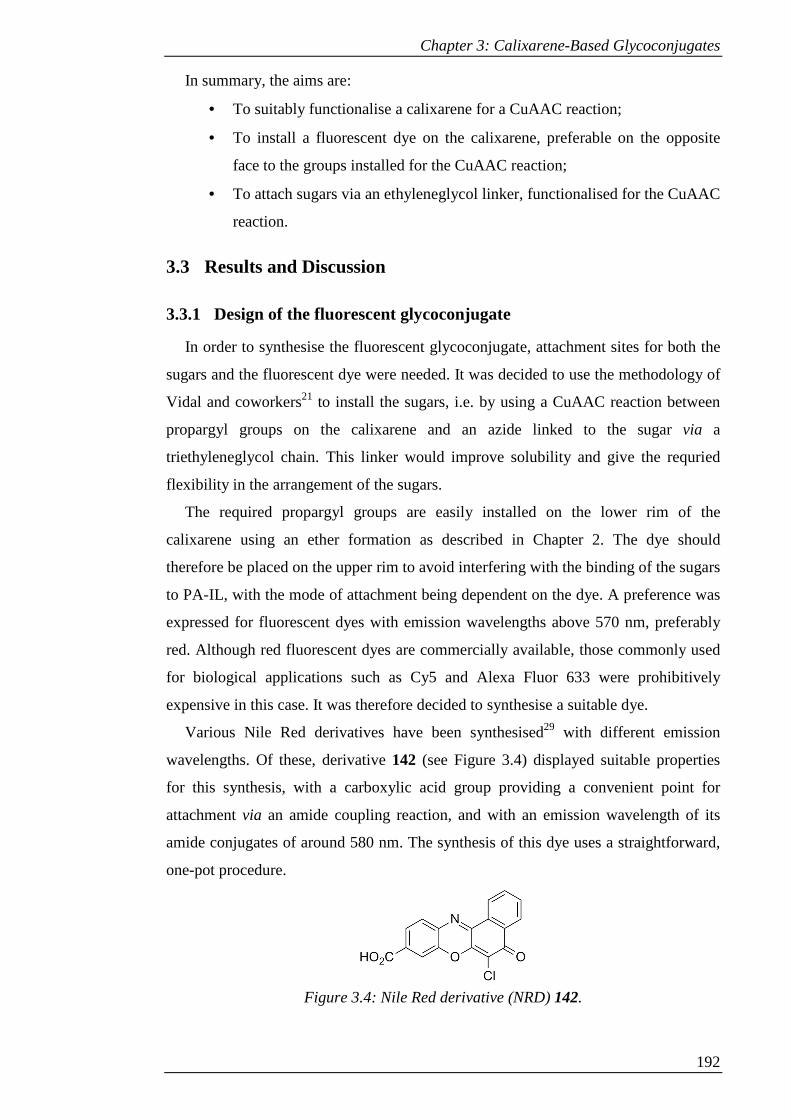
Chapter 3: Calixarene-Based Glycoconjugates
192
In summary, the aims are:
• To suitably functionalise a calixarene for a CuAAC reaction;
• To install a fluorescent dye on the calixarene, preferable on the opposite
face to the groups installed for the CuAAC reaction;
• To attach sugars via an ethyleneglycol linker, functionalised for the CuAAC
reaction.
3.3 Results and Discussion
3.3.1 Design of the fluorescent glycoconjugate
In order to synthesise the fluorescent glycoconjugate, attachment sites for both the
sugars and the fluorescent dye were needed. It was decided to use the methodology of
Vidal and coworkers21 to install the sugars, i.e. by using a CuAAC reaction between
propargyl groups on the calixarene and an azide linked to the sugar via a
triethyleneglycol chain. This linker would improve solubility and give the requried
flexibility in the arrangement of the sugars.
The required propargyl groups are easily installed on the lower rim of the
calixarene using an ether formation as described in Chapter 2. The dye should
therefore be placed on the upper rim to avoid interfering with the binding of the sugars
to PA-IL, with the mode of attachment being dependent on the dye. A preference was
expressed for fluorescent dyes with emission wavelengths above 570 nm, preferably
red. Although red fluorescent dyes are commercially available, those commonly used
for biological applications such as Cy5 and Alexa Fluor 633 were prohibitively
expensive in this case. It was therefore decided to synthesise a suitable dye.
Various Nile Red derivatives have been synthesised29 with different emission
wavelengths. Of these, derivative 142 (see Figure 3.4) displayed suitable properties
for this synthesis, with a carboxylic acid group providing a convenient point for
attachment via an amide coupling reaction, and with an emission wavelength of its
amide conjugates of around 580 nm. The synthesis of this dye uses a straightforward,
one-pot procedure.
Figure 3.4: Nile Red derivative (NRD) 142.
Chapter 3: Calixarene-Based Glycoconjugates
193
With the selection of this dye, an amine is required on the upper rim of the
calixarene. This is easily accessible by reduction of the corresponding nitro group.
The requirement for a single nitro group on the upper rim leads to two possibilities:
regioselective nitration, using the functionalisation of the lower rim to control the
substitution reaction, or non-selective nitration controlled by reaction time. Both of
these routes will be examined.
3.3.2 Route 1: Regioselective mono-nitration
By selectively alkylating the lower-rim of the calixarene, the subsequent
electrophilic aromatic substitution reaction can be controlled by the greater electron
density donated by a free phenol compared with alkyl ethers. This has been
demonstrated for the selective mono-nitration of calixarenes using a multistep process
involving temporary installation of benzoyl groups on the lower rim,30 and also more
directly by nitration31 or ipso-nitration32 of the desired alkyl ether. The latter
procedure, containing the fewest synthetic steps, was selected.
The required tripropargyl ether for the selective ipso-nitration was available from
tetra-tert-butylcalix[4]arene (2) using a selective trialkylation as described in Chapter
2. Compound 2 was stirred with a mixture of barium oxide and barium hydroxide,
followed by addition of propargyl bromide. The mixture was stirred for 18 hours.
Aqueous work up followed by column chromatography over silica (eluting with 2:1
DCM/Hexane) gave 143 in the cone conformation as a white powder in 28% yield.
Compound 143 was then reacted with a mixture of glacial acetic acid and 65%
nitric acid in proportions used in the literature.32 The nitric acid was added dropwise
over 2 minutes, then the solution stirred for a further 4 minutes, during which time the
reaction mixture gained a blue-black colour, before quenching. Aqueous work up
followed by purification by column chromatography over silica (eluting with 2:1
DCM/Hexane) gave 144 as yellow crystals in 44% yield.
The 1H-NMR spectrum of 144 (see Figure 3.5b) confirms the correct symmetry and
the successful mono nitration. The single plane of symmetry results in a pair of triplets
around 2.5 ppm corresponding to the terminal hydrogens of the alkynes, one for the
propargyl groups adjacent to the phenol ring and one for the distal propargyl group.
However, the same pattern is not observed for the methylene groups of the propargyl
ethers.
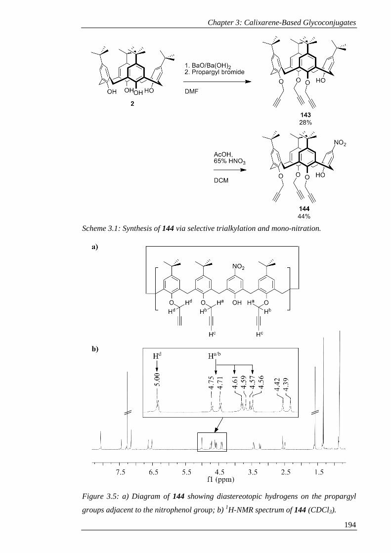
Chapter 3: Calixarene-Based Glycoconjugates
194
Scheme 3.1: Synthesis of 144 via selective trialkylation and mono-nitration.
Figure 3.5: a) Diagram of 144 showing diastereotopic hydrogens on the propargyl
groups adjacent to the nitrophenol group; b) 1H-NMR spectrum of 144 (CDCl3).
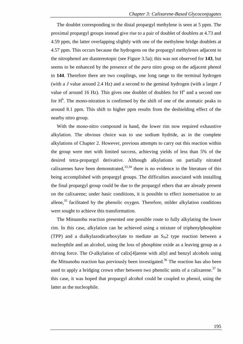
Chapter 3: Calixarene-Based Glycoconjugates
195
The doublet corresponding to the distal propargyl methylene is seen at 5 ppm. The
proximal propargyl groups instead give rise to a pair of doublet of doublets at 4.73 and
4.59 ppm, the latter overlapping slightly with one of the methylene bridge doublets at
4.57 ppm. This occurs because the hydrogens on the propargyl methylenes adjacent to
the nitrophenol are diastereotopic (see Figure 3.5a); this was not observed for 143, but
seems to be enhanced by the presence of the para nitro group on the adjacent phenol
in 144. Therefore there are two couplings, one long range to the terminal hydrogen
(with a J value around 2.4 Hz) and a second to the geminal hydrogen (with a larger J
value of around 16 Hz). This gives one doublet of doublets for Ha and a second one
for Hb. The mono-nitration is confirmed by the shift of one of the aromatic peaks to
around 8.1 ppm. This shift to higher ppm results from the deshielding effect of the
nearby nitro group.
With the mono-nitro compound in hand, the lower rim now required exhaustive
alkylation. The obvious choice was to use sodium hydride, as in the complete
alkylations of Chapter 2. However, previous attempts to carry out this reaction within
the group were met with limited success, achieving yields of less than 5% of the
desired tetra-propargyl derivative. Although alkylations on partially nitrated
calixarenes have been demonstrated,33,34 there is no evidence in the literature of this
being accomplished with propargyl groups. The difficulties associated with installing
the final propargyl group could be due to the propargyl ethers that are already present
on the calixarene; under basic conditions, it is possible to effect isomerisation to an
allene,35 facilitated by the phenolic oxygen. Therefore, milder alkylation conditions
were sought to achieve this transformation.
The Mitsunobu reaction presented one possible route to fully alkylating the lower
rim. In this case, alkylation can be achieved using a mixture of triphenylphosphine
(TPP) and a dialkylazodicarboxylate to mediate an SN2 type reaction between a
nucleophile and an alcohol, using the loss of phosphine oxide as a leaving group as a
driving force. The O-alkylation of calix[4]arene with allyl and benzyl alcohols using
the Mitsunobu reaction has previously been investigated.36 The reaction has also been
used to apply a bridging crown ether between two phenolic units of a calixarene.37 In
this case, it was hoped that propargyl alcohol could be coupled to phenol, using the
latter as the nucleophile.
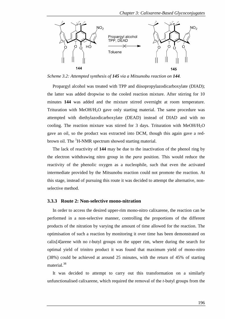
Chapter 3: Calixarene-Based Glycoconjugates
196
Scheme 3.2: Attempted synthesis of 145 via a Mitsunobu reaction on 144.
Propargyl alcohol was treated with TPP and diisopropylazodicarboxylate (DIAD);
the latter was added dropwise to the cooled reaction mixture. After stirring for 10
minutes 144 was added and the mixture stirred overnight at room temperature.
Trituration with MeOH/H2O gave only starting material. The same procedure was
attempted with diethylazodicarboxylate (DEAD) instead of DIAD and with no
cooling. The reaction mixture was stirred for 3 days. Trituration with MeOH/H2O
gave an oil, so the product was extracted into DCM, though this again gave a red-
brown oil. The 1H-NMR spectrum showed starting material.
The lack of reactivity of 144 may be due to the inactivation of the phenol ring by
the electron withdrawing nitro group in the para position. This would reduce the
reactivity of the phenolic oxygen as a nucleophile, such that even the activated
intermediate provided by the Mitsunobu reaction could not promote the reaction. At
this stage, instead of pursuing this route it was decided to attempt the alternative, non-
selective method.
3.3.3 Route 2: Non-selective mono-nitration
In order to access the desired upper-rim mono-nitro calixarene, the reaction can be
performed in a non-selective manner, controlling the proportions of the different
products of the nitration by varying the amount of time allowed for the reaction. The
optimisation of such a reaction by monitoring it over time has been demonstrated on
calix[4]arene with no t-butyl groups on the upper rim, where during the search for
optimal yield of trinitro product it was found that maximum yield of mono-nitro
(38%) could be achieved at around 25 minutes, with the return of 45% of starting
material.38
It was decided to attempt to carry out this transformation on a similarly
unfunctionalised calixarene, which required the removal of the t-butyl groups from the

Chapter 3: Calixarene-Based Glycoconjugates
197
previously synthesised 2. This could be accomplished via a reverse Friedel-Crafts
reaction with AlCl3 and phenol.39
To a stirred mixture of 2 and phenol in toluene was added AlCl3 in small portions.
After 4 hours the cloudy suspension had turned brown. The reaction mixture was
poured onto ice followed by extraction of the product with DCM. After removal of
solvent under reduced pressure, trituration of the residue with methanol gave 146 as
off-white powder in 79% yield.
This calixarene could be exhaustively alkylated at the lower rim with sodium
hydride as described in Chapter 2, using sodium hydride followed by propargyl
bromide. After stirring for 72 hours, aqueous work-up followed by column
chromatography over silica gel (eluting with 2:1 hexane/toluene) gave 147 as white
powder in 37%.
The mono-nitration reaction was then tested. After stirring 147 in DCM with
glacial acetic acid and 65% nitric acid for 30 minutes, the reaction was quenched.
Aqueous work up gave only the starting material.
It was decided to use a stronger nitrating mixture and monitor the reaction over
time by TLC. To a solution of 147 in DCM, 100% nitric acid and a few drops of 95%
sulphuric acid were added, giving an immediate colour change to purple-black. After
stirring for 1 hour the reaction was quenched with water; aqueous work up followed
by purification by column chromatography over silica (eluting with 1:1 DCM/hexane)
gave 148 as light yellow solid and returned 147 in 60% yield.
Scheme 3.3: Synthesis of 148 from 2 via de-tert-butylation, tetraalkyation and time-
controlled nitration.

Chapter 3: Calixarene-Based Glycoconjugates
198
Purification in this way was complicated by an unknown impurity that co-eluted
with the product. This problem could be mitigated by re-crystallising impure column
fractions from DCM/methanol, but it still led to a poor yield (total 21%) and made the
synthesis less efficient. Once again, in the 1H-NMR spectrum the pattern of two
doublets of doublets for the alkyne methylenes on the positions adjacent to the nitrated
ring was observed.
Although the overall yield over the three steps was lower than that achieved so far
from the first route, the latter synthesis was incomplete. The final alkylation would
have needed to have a yield of at least 50% to compare favourably, assuming that a
successful method could be found. The second route could potentially be improved if
the number of steps was reduced by carrying out an ipso-nitration on the tert-butyl
derivative. However, at this stage, the synthesis was continued with product 148.
3.3.3.1 Dye synthesis
The selected NRD was synthesised according to the literature.29 A mixture of 2,3-
dichloro-1,4-napthoquinone, 4-amino-3-hydroxybenzoic acid and sodium acetate in
methanol was heated to reflux for 24 hours, during which time the suspension turned
from yellow to red. After cooling in ice, the solid was filtered and washed with water.
The solid was triturated with hot methanol, filtered and washed with further methanol
to give 142 as brick-red solid in 66% yield.
Scheme 3.4: Synthesis of NRD 142.
3.3.3.2 Sugar synthesis
To selectively synthesise the target β-anomer of the azide-derivatised sugar,
neighbouring group participation can be exploited. Lewis-acid promoted elimination
of the leaving group at the anomeric centre can be accomplished with sugar-halides,
for example using silver or mercury salts;40 however, for the installation of the azido-
triethyleneglycol linker, it has been found that the use of peracetylated sugar in
combination with tin (IV) chloride and silver trifluoroacetate gave superior yield of
the desired anomer with short reaction times and on large scales.41
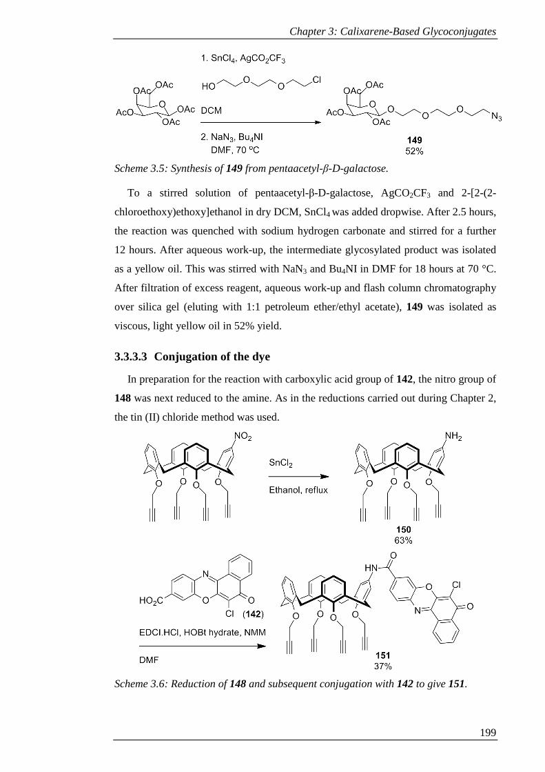
Chapter 3: Calixarene-Based Glycoconjugates
199
Scheme 3.5: Synthesis of 149 from pentaacetyl-β-D-galactose.
To a stirred solution of pentaacetyl-β-D-galactose, AgCO2CF3 and 2-[2-(2-
chloroethoxy)ethoxy]ethanol in dry DCM, SnCl4 was added dropwise. After 2.5 hours,
the reaction was quenched with sodium hydrogen carbonate and stirred for a further
12 hours. After aqueous work-up, the intermediate glycosylated product was isolated
as a yellow oil. This was stirred with NaN3 and Bu4NI in DMF for 18 hours at 70 °C.
After filtration of excess reagent, aqueous work-up and flash column chromatography
over silica gel (eluting with 1:1 petroleum ether/ethyl acetate), 149 was isolated as
viscous, light yellow oil in 52% yield.
3.3.3.3 Conjugation of the dye
In preparation for the reaction with carboxylic acid group of 142, the nitro group of
148 was next reduced to the amine. As in the reductions carried out during Chapter 2,
the tin (II) chloride method was used.
Scheme 3.6: Reduction of 148 and subsequent conjugation with 142 to give 151.

Chapter 3: Calixarene-Based Glycoconjugates
200
A mixture of 148 and SnCl2 was heated to reflux in ethanol for 48 hours. After
removal of solvent under reduced pressure, aqueous work-up with sodium hydroxide
gave the mono-amine 150 of moderate purity as brown glass in 63% yield. This crude
product was taken through to the next step without further purification to minimise
loss of product.
Procedures that have been applied to the conjugation of 142 to amines include the
use of amide coupling reagents and a one-pot acid-chloride mediated coupling using
Ghosez’s reagent.29 Due to availability of reagents, amide coupling using EDCI was
used.
A mixture of 150, 142, EDCI, HOBt and N-methylmorpholine (NMM) were stirred
in DMF for 18 hours. Aqueous work-up followed by column chromatography over
silica gel (eluting with DCM) gave 151 as brick-red solid in 37% yield.
The correct product was confirmed by 1H-NMR spectroscopy. The protons on the
dye give rise to two multiplets around 8.5 ppm and a cluster of peaks around 8.0 ppm.
The N-H of the amide bond can be observed at 7.59 ppm, and the aromatic protons
adjacent to this on the calixarene are visible as a singlet at 7.03 ppm, separated from
the cluster corresponding to the other aromatic protons around 6.75 ppm.
Interestingly, in the 1H-NMR spectra of both the amine and the amide, the peaks
corresponding to the alkyene methylene groups do not give rise to the previously
observed doublets of doublets; this indicates a lack of inequivalence between the
protons on the methylene bridges adjacent to the dye-appended ring.
Figure 3.6: 1H-NMR spectrum of 151 (CDCl3).
2.53.03.54.04.55.05.56.06.57.07.58.08.5f1 (ppm)
NH
ArH adjacent to NH
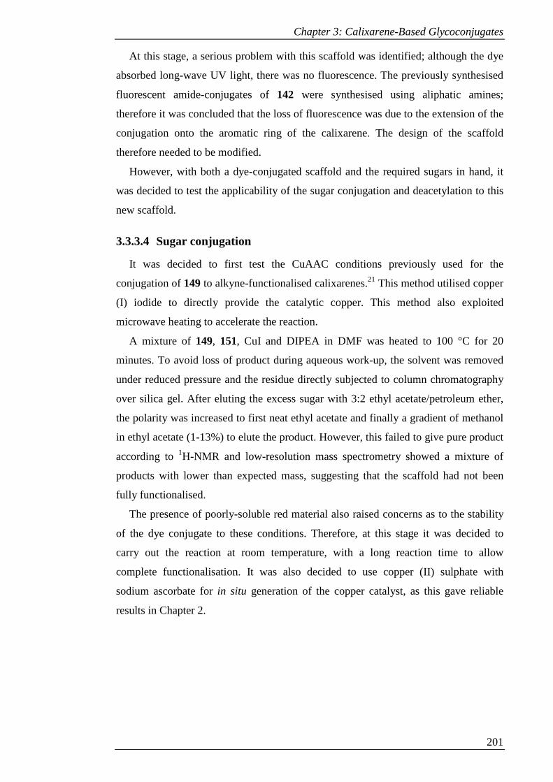
Chapter 3: Calixarene-Based Glycoconjugates
201
At this stage, a serious problem with this scaffold was identified; although the dye
absorbed long-wave UV light, there was no fluorescence. The previously synthesised
fluorescent amide-conjugates of 142 were synthesised using aliphatic amines;
therefore it was concluded that the loss of fluorescence was due to the extension of the
conjugation onto the aromatic ring of the calixarene. The design of the scaffold
therefore needed to be modified.
However, with both a dye-conjugated scaffold and the required sugars in hand, it
was decided to test the applicability of the sugar conjugation and deacetylation to this
new scaffold.
3.3.3.4 Sugar conjugation
It was decided to first test the CuAAC conditions previously used for the
conjugation of 149 to alkyne-functionalised calixarenes.21 This method utilised copper
(I) iodide to directly provide the catalytic copper. This method also exploited
microwave heating to accelerate the reaction.
A mixture of 149, 151, CuI and DIPEA in DMF was heated to 100 °C for 20
minutes. To avoid loss of product during aqueous work-up, the solvent was removed
under reduced pressure and the residue directly subjected to column chromatography
over silica gel. After eluting the excess sugar with 3:2 ethyl acetate/petroleum ether,
the polarity was increased to first neat ethyl acetate and finally a gradient of methanol
in ethyl acetate (1-13%) to elute the product. However, this failed to give pure product
according to 1H-NMR and low-resolution mass spectrometry showed a mixture of
products with lower than expected mass, suggesting that the scaffold had not been
fully functionalised.
The presence of poorly-soluble red material also raised concerns as to the stability
of the dye conjugate to these conditions. Therefore, at this stage it was decided to
carry out the reaction at room temperature, with a long reaction time to allow
complete functionalisation. It was also decided to use copper (II) sulphate with
sodium ascorbate for in situ generation of the copper catalyst, as this gave reliable
results in Chapter 2.
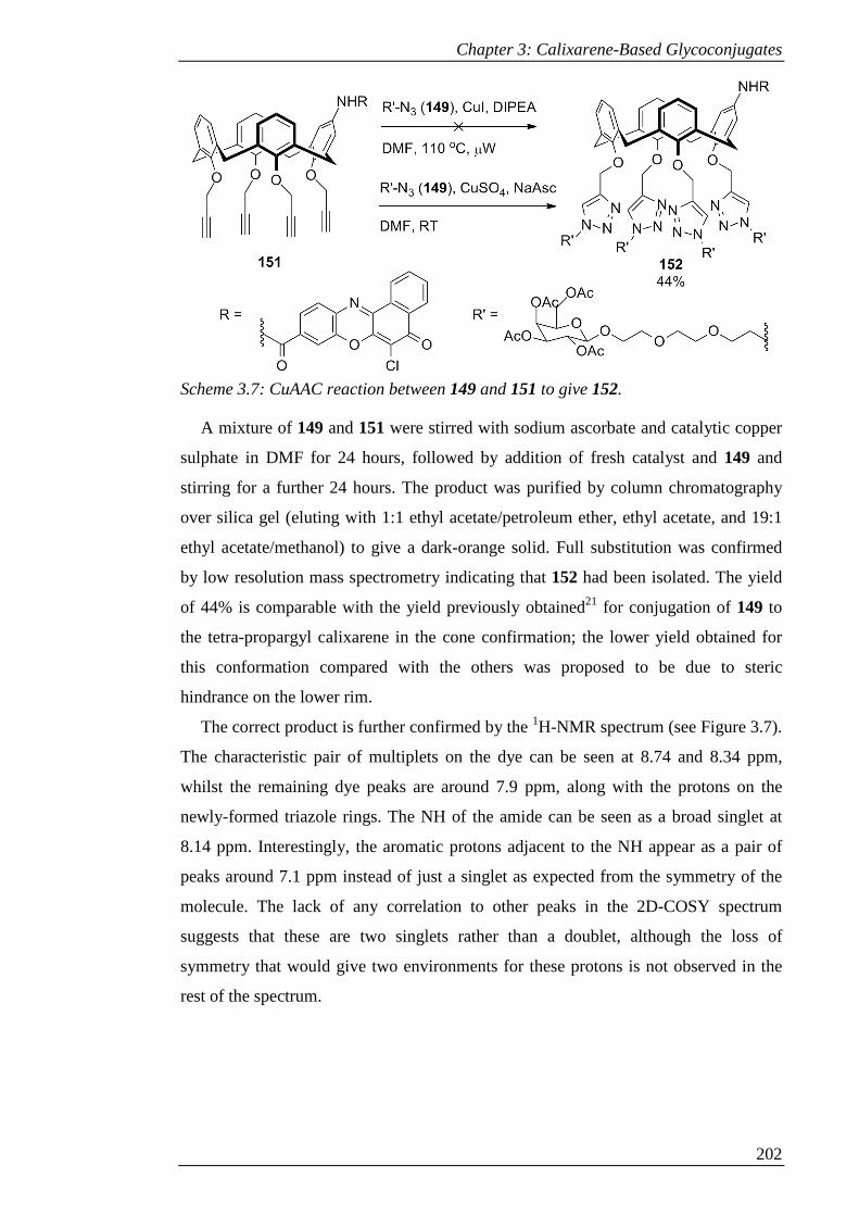
Chapter 3: Calixarene-Based Glycoconjugates
202
Scheme 3.7: CuAAC reaction between 149 and 151 to give 152.
A mixture of 149 and 151 were stirred with sodium ascorbate and catalytic copper
sulphate in DMF for 24 hours, followed by addition of fresh catalyst and 149 and
stirring for a further 24 hours. The product was purified by column chromatography
over silica gel (eluting with 1:1 ethyl acetate/petroleum ether, ethyl acetate, and 19:1
ethyl acetate/methanol) to give a dark-orange solid. Full substitution was confirmed
by low resolution mass spectrometry indicating that 152 had been isolated. The yield
of 44% is comparable with the yield previously obtained21 for conjugation of 149 to
the tetra-propargyl calixarene in the cone confirmation; the lower yield obtained for
this conformation compared with the others was proposed to be due to steric
hindrance on the lower rim.
The correct product is further confirmed by the 1H-NMR spectrum (see Figure 3.7).
The characteristic pair of multiplets on the dye can be seen at 8.74 and 8.34 ppm,
whilst the remaining dye peaks are around 7.9 ppm, along with the protons on the
newly-formed triazole rings. The NH of the amide can be seen as a broad singlet at
8.14 ppm. Interestingly, the aromatic protons adjacent to the NH appear as a pair of
peaks around 7.1 ppm instead of just a singlet as expected from the symmetry of the
molecule. The lack of any correlation to other peaks in the 2D-COSY spectrum
suggests that these are two singlets rather than a doublet, although the loss of
symmetry that would give two environments for these protons is not observed in the
rest of the spectrum.
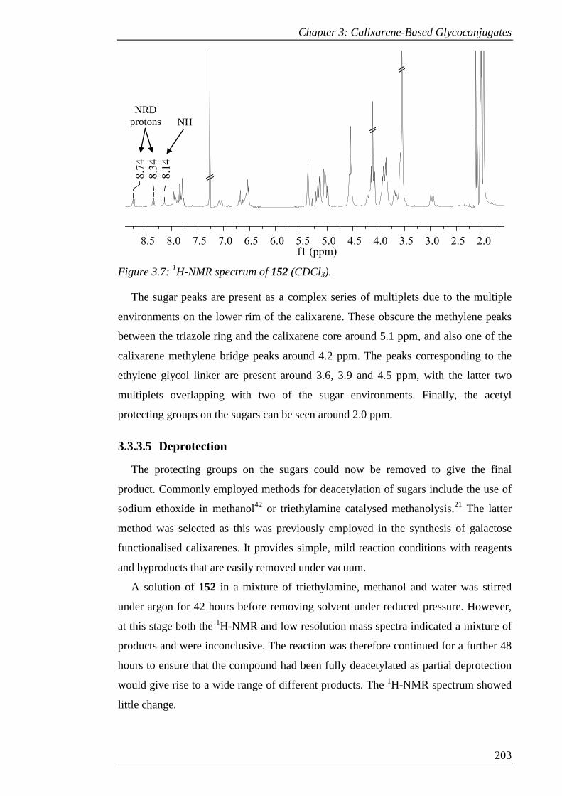
Chapter 3: Calixarene-Based Glycoconjugates
203
Figure 3.7: 1H-NMR spectrum of 152 (CDCl3).
The sugar peaks are present as a complex series of multiplets due to the multiple
environments on the lower rim of the calixarene. These obscure the methylene peaks
between the triazole ring and the calixarene core around 5.1 ppm, and also one of the
calixarene methylene bridge peaks around 4.2 ppm. The peaks corresponding to the
ethylene glycol linker are present around 3.6, 3.9 and 4.5 ppm, with the latter two
multiplets overlapping with two of the sugar environments. Finally, the acetyl
protecting groups on the sugars can be seen around 2.0 ppm.
3.3.3.5 Deprotection
The protecting groups on the sugars could now be removed to give the final
product. Commonly employed methods for deacetylation of sugars include the use of
sodium ethoxide in methanol42 or triethylamine catalysed methanolysis.21 The latter
method was selected as this was previously employed in the synthesis of galactose
functionalised calixarenes. It provides simple, mild reaction conditions with reagents
and byproducts that are easily removed under vacuum.
A solution of 152 in a mixture of triethylamine, methanol and water was stirred
under argon for 42 hours before removing solvent under reduced pressure. However,
at this stage both the 1H-NMR and low resolution mass spectra indicated a mixture of
products and were inconclusive. The reaction was therefore continued for a further 48
hours to ensure that the compound had been fully deacetylated as partial deprotection
would give rise to a wide range of different products. The 1H-NMR spectrum showed
little change.
2.02.53.03.54.04.55.05.56.06.57.07.58.08.5f1 (ppm)
8.1
4
8.3
4
8.7
4
NH NRD
protons
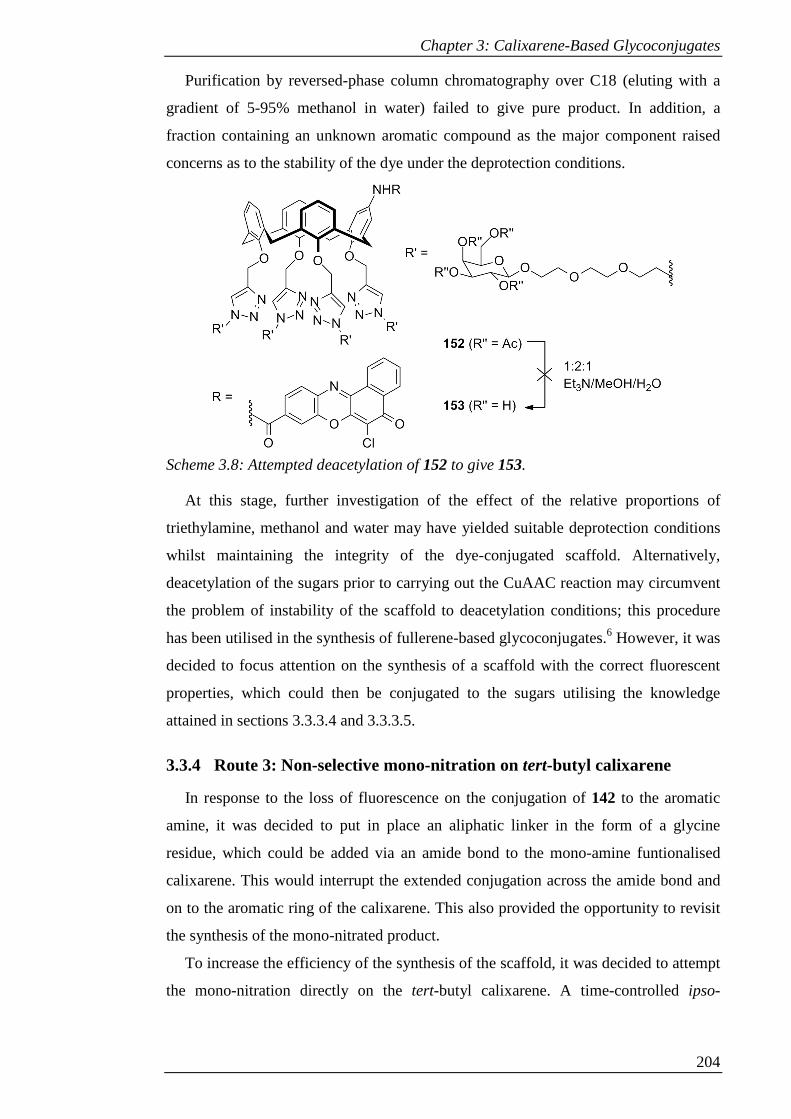
Chapter 3: Calixarene-Based Glycoconjugates
204
Purification by reversed-phase column chromatography over C18 (eluting with a
gradient of 5-95% methanol in water) failed to give pure product. In addition, a
fraction containing an unknown aromatic compound as the major component raised
concerns as to the stability of the dye under the deprotection conditions.
Scheme 3.8: Attempted deacetylation of 152 to give 153.
At this stage, further investigation of the effect of the relative proportions of
triethylamine, methanol and water may have yielded suitable deprotection conditions
whilst maintaining the integrity of the dye-conjugated scaffold. Alternatively,
deacetylation of the sugars prior to carrying out the CuAAC reaction may circumvent
the problem of instability of the scaffold to deacetylation conditions; this procedure
has been utilised in the synthesis of fullerene-based glycoconjugates.6 However, it was
decided to focus attention on the synthesis of a scaffold with the correct fluorescent
properties, which could then be conjugated to the sugars utilising the knowledge
attained in sections 3.3.3.4 and 3.3.3.5.
3.3.4 Route 3: Non-selective mono-nitration on tert-butyl calixarene
In response to the loss of fluorescence on the conjugation of 142 to the aromatic
amine, it was decided to put in place an aliphatic linker in the form of a glycine
residue, which could be added via an amide bond to the mono-amine funtionalised
calixarene. This would interrupt the extended conjugation across the amide bond and
on to the aromatic ring of the calixarene. This also provided the opportunity to revisit
the synthesis of the mono-nitrated product.
To increase the efficiency of the synthesis of the scaffold, it was decided to attempt
the mono-nitration directly on the tert-butyl calixarene. A time-controlled ipso-

Chapter 3: Calixarene-Based Glycoconjugates
205
nitration has been accomplished previously using acetic acid and 100% nitric acid in
much smaller amounts than used in the corresponding tetra-nitration.43
Compound 2 was first tetra-alkylated in the same way as in the synthesis of 147.
After stirring the reaction mixture for 48 hours the product was precipitated with
excess water, filtered and washed with water to give 154 as light-brown powder in
53% yield. This product was sufficiently pure to use in further reactions.
The nitration reaction was tested on 154 by stirring it in DCM with acetic acid and
100% nitric acid. After a 30, 60 or 90 minutes, the reaction was quenched by addition
of water. After aqueous washes, the product (now identical to 145, the synthesis of
which was previously attempted in section 3.3.2) was purified by column
chromatography over silica gel (eluting with 8-10% ethyl acetate in hexane) and the
proportions of starting material and products were assessed.
The results are shown in Table 3.1. A reaction time of 30 minutes gave the
optimum yield of 145 whilst returning maximal 154. Longer reaction times did not
improve the yield of product, whilst returning less starting material. At 90 minutes, a
significant amount of material was lost to a mixture of over-nitrated products.
Table 3.1: Change in proportions of product 145 and recovered 154 over time.
Reaction time / mins % recovery of 154 % yield of 145
30 40 26
60 13 23
90 2 22
In addition to removing one step from the synthesis, a noteworthy improvement in
this route was the ease of purification relative to that of 148. This made this synthetic
step a much more simple and efficient process.
Reduction was carried out as in section 3.3.3. Compound 145 was heated to reflux
with SnCl2 in ethanol for 18 hours, followed by removal of solvent and aqueous work
up with sodium hydroxide, giving 155 as yellow solid in 83% yield. However, in this
case, a small amount of unknown impurity was consistently found in the product.
When the reaction was allowed to continue for 48 hours, the purity of the product was
greatly diminished. Therefore 18 hours seems to be the optimum to allow the reaction
to run to completion without diminishing the yield due to impurities.
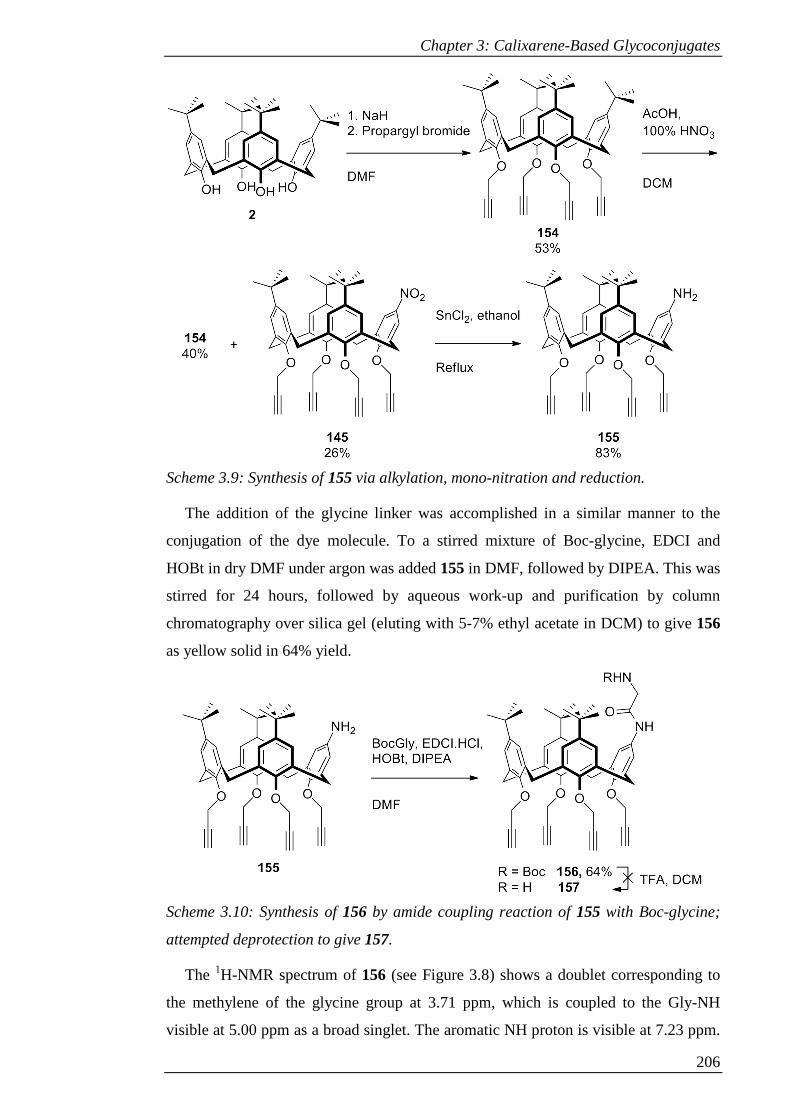
Chapter 3: Calixarene-Based Glycoconjugates
206
Scheme 3.9: Synthesis of 155 via alkylation, mono-nitration and reduction.
The addition of the glycine linker was accomplished in a similar manner to the
conjugation of the dye molecule. To a stirred mixture of Boc-glycine, EDCI and
HOBt in dry DMF under argon was added 155 in DMF, followed by DIPEA. This was
stirred for 24 hours, followed by aqueous work-up and purification by column
chromatography over silica gel (eluting with 5-7% ethyl acetate in DCM) to give 156
as yellow solid in 64% yield.
Scheme 3.10: Synthesis of 156 by amide coupling reaction of 155 with Boc-glycine;
attempted deprotection to give 157.
The 1H-NMR spectrum of 156 (see Figure 3.8) shows a doublet corresponding to
the methylene of the glycine group at 3.71 ppm, which is coupled to the Gly-NH
visible at 5.00 ppm as a broad singlet. The aromatic NH proton is visible at 7.23 ppm.

Chapter 3: Calixarene-Based Glycoconjugates
207
The methylenes of the alkynes adjacent to the functionalised ring once again give rise
to a pair of doublet of doublets around 4.9 ppm; however, in this case there is a
pronounced ‘rooftop’ effect due to the proximity of the two multiplets. The doublets
of the other two alkynes are overlapping with one of the methylene bridge peaks at
4.60 ppm.
Figure 3.8: 1H-NMR spectrum of 156 (CDCl3).
In order to perform the conjugation reaction with 142, the Boc protecting group
next needed to be removed to reveal the required amine. Based on the deprotection of
the amide-linked coumarin conjugate (132) carried out in Chapter 2, it was decided to
use the TFA deprotection method instead of using gaseous HCl as this appeared to be
a milder method and better suited to compounds featuring amide bonds. Compound
156 was stirred with TFA in DCM, resulting in a change in colour of the light orange-
yellow solution to orange then brown, finally gaining a pink hue. After 40 minutes the
reaction was quenched with water to give a grey emulsion. The acid was neutralised
with saturated aqueous NaHCO3; on shaking the emulsion resolved to give an orange
organic layer. However, the isolated orange-brown solid proved to be a complex
mixture of products by 1H-NMR.
It was unclear exactly what unexpected transformation was taking place in this
reaction. However, it was reasoned that such a complex mixture would likely arise
from side-reactions of the alkynes. The potential of propargyl ethers to give rise to
side-reactions was noted in Chapter 2. It was therefore decided to synthesise an
analogous structure with TBDMS-protected alkynes to determine if these groups were
resulting in the failure of this Boc deprotection reaction.
3.7
1
5.0
0
7.2
3
4.6
0
4.8
7
ArNH GlyNH
GlyCH2
OCH2 OCH2 and
ArCH2
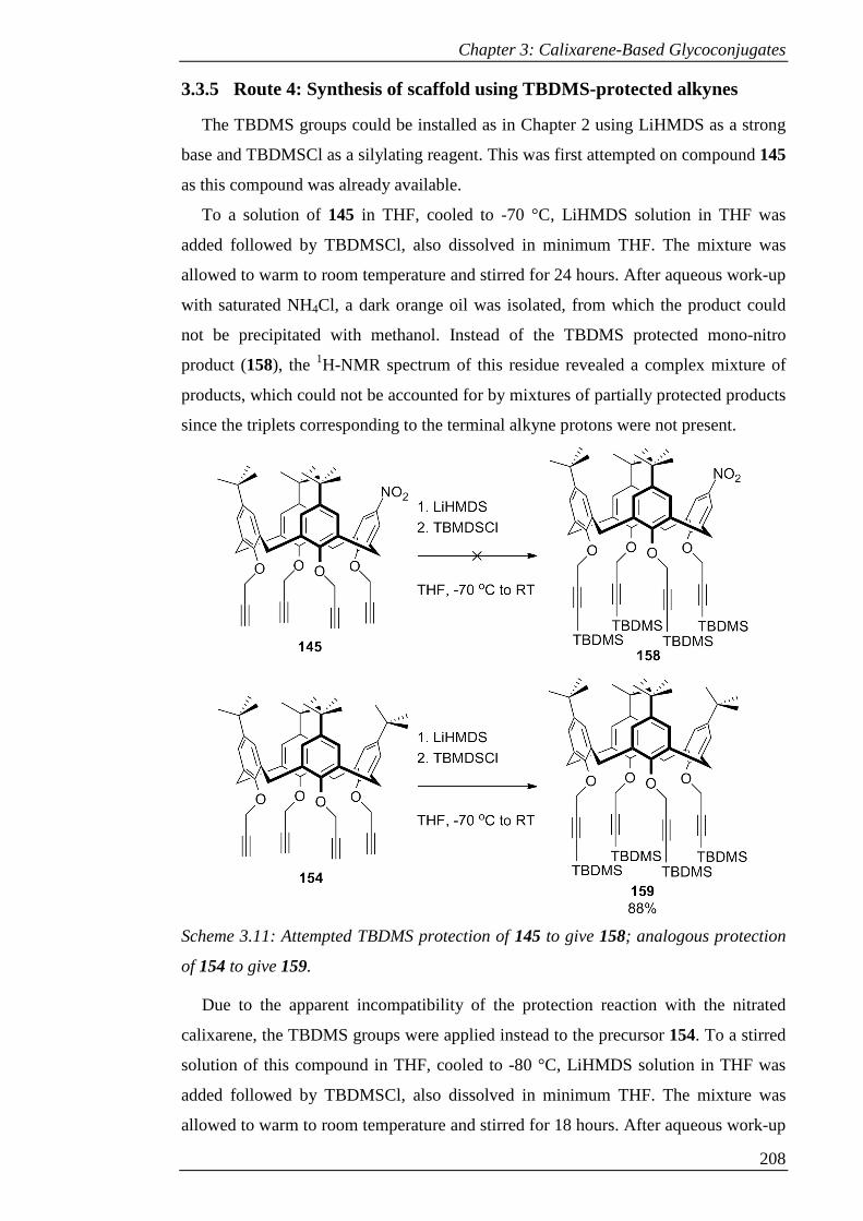
Chapter 3: Calixarene-Based Glycoconjugates
208
3.3.5 Route 4: Synthesis of scaffold using TBDMS-protected alkynes
The TBDMS groups could be installed as in Chapter 2 using LiHMDS as a strong
base and TBDMSCl as a silylating reagent. This was first attempted on compound 145
as this compound was already available.
To a solution of 145 in THF, cooled to -70 °C, LiHMDS solution in THF was
added followed by TBDMSCl, also dissolved in minimum THF. The mixture was
allowed to warm to room temperature and stirred for 24 hours. After aqueous work-up
with saturated NH4Cl, a dark orange oil was isolated, from which the product could
not be precipitated with methanol. Instead of the TBDMS protected mono-nitro
product (158), the 1H-NMR spectrum of this residue revealed a complex mixture of
products, which could not be accounted for by mixtures of partially protected products
since the triplets corresponding to the terminal alkyne protons were not present.
Scheme 3.11: Attempted TBDMS protection of 145 to give 158; analogous protection
of 154 to give 159.
Due to the apparent incompatibility of the protection reaction with the nitrated
calixarene, the TBDMS groups were applied instead to the precursor 154. To a stirred
solution of this compound in THF, cooled to -80 °C, LiHMDS solution in THF was
added followed by TBDMSCl, also dissolved in minimum THF. The mixture was
allowed to warm to room temperature and stirred for 18 hours. After aqueous work-up
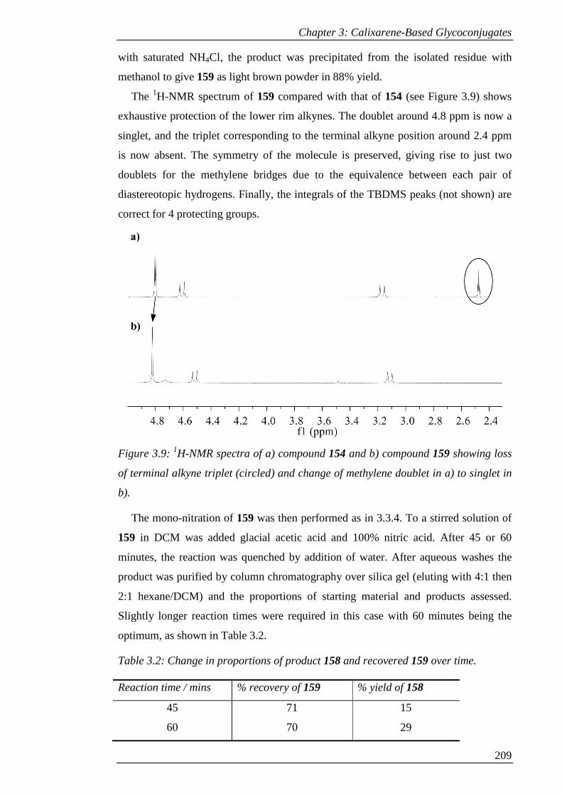
Chapter 3: Calixarene-Based Glycoconjugates
209
with saturated NH4Cl, the product was precipitated from the isolated residue with
methanol to give 159 as light brown powder in 88% yield.
The 1H-NMR spectrum of 159 compared with that of 154 (see Figure 3.9) shows
exhaustive protection of the lower rim alkynes. The doublet around 4.8 ppm is now a
singlet, and the triplet corresponding to the terminal alkyne position around 2.4 ppm
is now absent. The symmetry of the molecule is preserved, giving rise to just two
doublets for the methylene bridges due to the equivalence between each pair of
diastereotopic hydrogens. Finally, the integrals of the TBDMS peaks (not shown) are
correct for 4 protecting groups.
Figure 3.9: 1H-NMR spectra of a) compound 154 and b) compound 159 showing loss
of terminal alkyne triplet (circled) and change of methylene doublet in a) to singlet in
b).
The mono-nitration of 159 was then performed as in 3.3.4. To a stirred solution of
159 in DCM was added glacial acetic acid and 100% nitric acid. After 45 or 60
minutes, the reaction was quenched by addition of water. After aqueous washes the
product was purified by column chromatography over silica gel (eluting with 4:1 then
2:1 hexane/DCM) and the proportions of starting material and products assessed.
Slightly longer reaction times were required in this case with 60 minutes being the
optimum, as shown in Table 3.2.
Table 3.2: Change in proportions of product 158 and recovered 159 over time.
Reaction time / mins % recovery of 159 % yield of 158
45 71 15
60 70 29
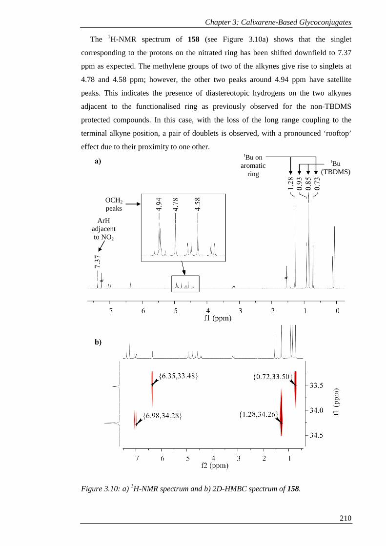
Chapter 3: Calixarene-Based Glycoconjugates
210
The 1H-NMR spectrum of 158 (see Figure 3.10a) shows that the singlet
corresponding to the protons on the nitrated ring has been shifted downfield to 7.37
ppm as expected. The methylene groups of two of the alkynes give rise to singlets at
4.78 and 4.58 ppm; however, the other two peaks around 4.94 ppm have satellite
peaks. This indicates the presence of diastereotopic hydrogens on the two alkynes
adjacent to the functionalised ring as previously observed for the non-TBDMS
protected compounds. In this case, with the loss of the long range coupling to the
terminal alkyne position, a pair of doublets is observed, with a pronounced ‘rooftop’
effect due to their proximity to one other.
Figure 3.10: a) 1H-NMR spectrum and b) 2D-HMBC spectrum of 158.
ArH adjacent to NO2
OCH2
peaks
tBu on aromatic
ring
tBu (TBDMS)
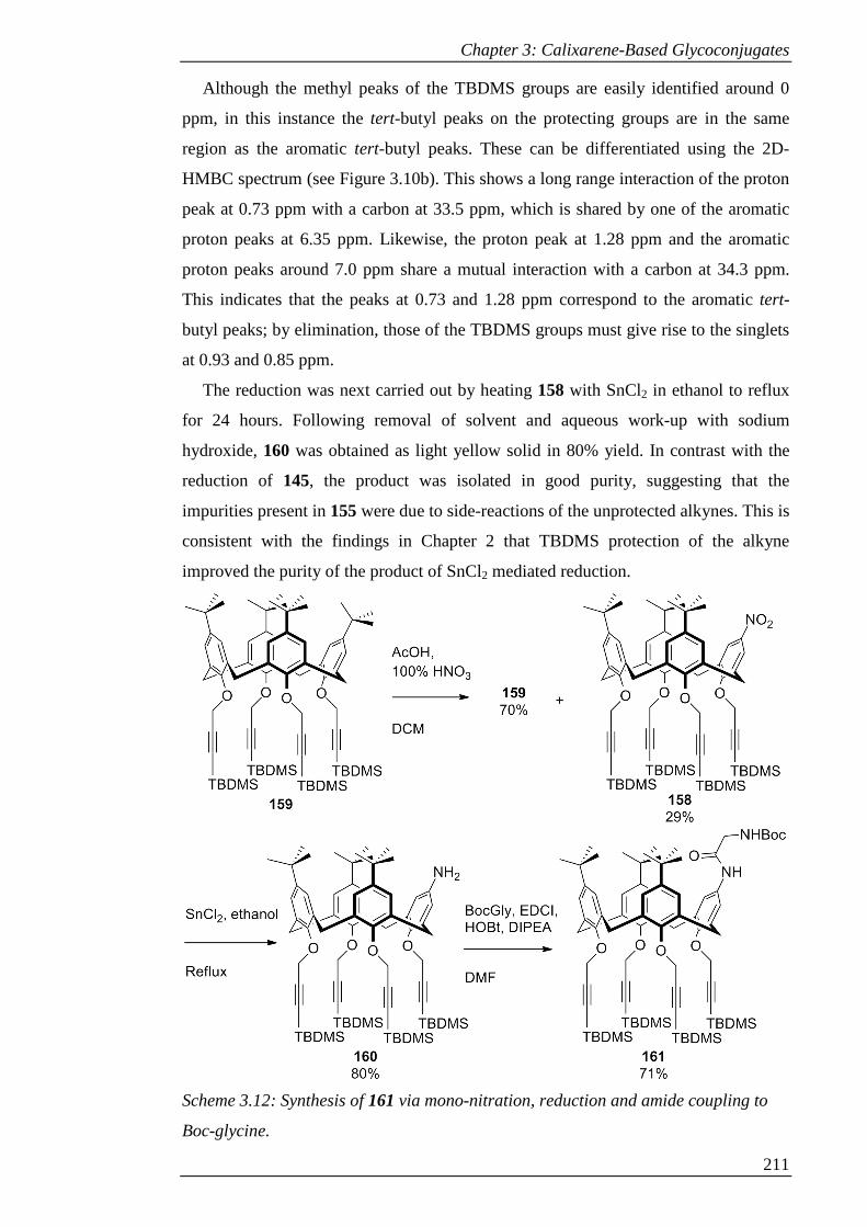
Chapter 3: Calixarene-Based Glycoconjugates
211
Although the methyl peaks of the TBDMS groups are easily identified around 0
ppm, in this instance the tert-butyl peaks on the protecting groups are in the same
region as the aromatic tert-butyl peaks. These can be differentiated using the 2D-
HMBC spectrum (see Figure 3.10b). This shows a long range interaction of the proton
peak at 0.73 ppm with a carbon at 33.5 ppm, which is shared by one of the aromatic
proton peaks at 6.35 ppm. Likewise, the proton peak at 1.28 ppm and the aromatic
proton peaks around 7.0 ppm share a mutual interaction with a carbon at 34.3 ppm.
This indicates that the peaks at 0.73 and 1.28 ppm correspond to the aromatic tert-
butyl peaks; by elimination, those of the TBDMS groups must give rise to the singlets
at 0.93 and 0.85 ppm.
The reduction was next carried out by heating 158 with SnCl2 in ethanol to reflux
for 24 hours. Following removal of solvent and aqueous work-up with sodium
hydroxide, 160 was obtained as light yellow solid in 80% yield. In contrast with the
reduction of 145, the product was isolated in good purity, suggesting that the
impurities present in 155 were due to side-reactions of the unprotected alkynes. This is
consistent with the findings in Chapter 2 that TBDMS protection of the alkyne
improved the purity of the product of SnCl2 mediated reduction.
Scheme 3.12: Synthesis of 161 via mono-nitration, reduction and amide coupling to
Boc-glycine.
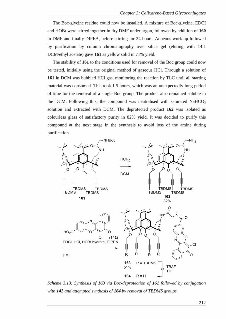
Chapter 3: Calixarene-Based Glycoconjugates
212
The Boc-glycine residue could now be installed. A mixture of Boc-glycine, EDCI
and HOBt were stirred together in dry DMF under argon, followed by addition of 160
in DMF and finally DIPEA, before stirring for 24 hours. Aqueous work-up followed
by purification by column chromatography over silica gel (eluting with 14:1
DCM/ethyl acetate) gave 161 as yellow solid in 71% yield.
The stability of 161 to the conditions used for removal of the Boc group could now
be tested, initially using the original method of gaseous HCl. Through a solution of
161 in DCM was bubbled HCl gas, monitoring the reaction by TLC until all starting
material was consumed. This took 1.5 hours, which was an unexpectedly long period
of time for the removal of a single Boc group. The product also remained soluble in
the DCM. Following this, the compound was neutralised with saturated NaHCO3
solution and extracted with DCM. The deprotected product 162 was isolated as
colourless glass of satisfactory purity in 82% yield. It was decided to purify this
compound at the next stage in the synthesis to avoid loss of the amine during
purification.
Scheme 3.13: Synthesis of 163 via Boc-deprotection of 161 followed by conjugation
with 142 and attempted synthesis of 164 by removal of TBDMS groups.
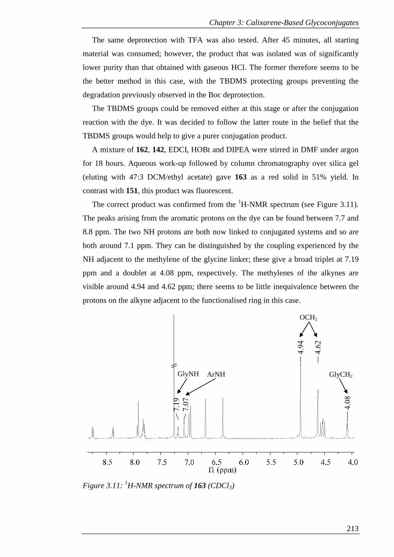
Chapter 3: Calixarene-Based Glycoconjugates
213
The same deprotection with TFA was also tested. After 45 minutes, all starting
material was consumed; however, the product that was isolated was of significantly
lower purity than that obtained with gaseous HCl. The former therefore seems to be
the better method in this case, with the TBDMS protecting groups preventing the
degradation previously observed in the Boc deprotection.
The TBDMS groups could be removed either at this stage or after the conjugation
reaction with the dye. It was decided to follow the latter route in the belief that the
TBDMS groups would help to give a purer conjugation product.
A mixture of 162, 142, EDCI, HOBt and DIPEA were stirred in DMF under argon
for 18 hours. Aqueous work-up followed by column chromatography over silica gel
(eluting with 47:3 DCM/ethyl acetate) gave 163 as a red solid in 51% yield. In
contrast with 151, this product was fluorescent.
The correct product was confirmed from the 1H-NMR spectrum (see Figure 3.11).
The peaks arising from the aromatic protons on the dye can be found between 7.7 and
8.8 ppm. The two NH protons are both now linked to conjugated systems and so are
both around 7.1 ppm. They can be distinguished by the coupling experienced by the
NH adjacent to the methylene of the glycine linker; these give a broad triplet at 7.19
ppm and a doublet at 4.08 ppm, respectively. The methylenes of the alkynes are
visible around 4.94 and 4.62 ppm; there seems to be little inequivalence between the
protons on the alkyne adjacent to the functionalised ring in this case.
Figure 3.11: 1H-NMR spectrum of 163 (CDCl3)
4.0
8
4.6
2
4.9
4
7.0
7
7.1
9
GlyNH ArNH
OCH2
GlyCH2
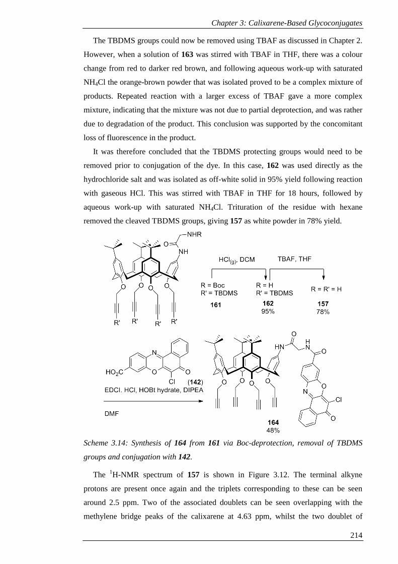
Chapter 3: Calixarene-Based Glycoconjugates
214
The TBDMS groups could now be removed using TBAF as discussed in Chapter 2.
However, when a solution of 163 was stirred with TBAF in THF, there was a colour
change from red to darker red brown, and following aqueous work-up with saturated
NH4Cl the orange-brown powder that was isolated proved to be a complex mixture of
products. Repeated reaction with a larger excess of TBAF gave a more complex
mixture, indicating that the mixture was not due to partial deprotection, and was rather
due to degradation of the product. This conclusion was supported by the concomitant
loss of fluorescence in the product.
It was therefore concluded that the TBDMS protecting groups would need to be
removed prior to conjugation of the dye. In this case, 162 was used directly as the
hydrochloride salt and was isolated as off-white solid in 95% yield following reaction
with gaseous HCl. This was stirred with TBAF in THF for 18 hours, followed by
aqueous work-up with saturated NH4Cl. Trituration of the residue with hexane
removed the cleaved TBDMS groups, giving 157 as white powder in 78% yield.
Scheme 3.14: Synthesis of 164 from 161 via Boc-deprotection, removal of TBDMS
groups and conjugation with 142.
The 1H-NMR spectrum of 157 is shown in Figure 3.12. The terminal alkyne
protons are present once again and the triplets corresponding to these can be seen
around 2.5 ppm. Two of the associated doublets can be seen overlapping with the
methylene bridge peaks of the calixarene at 4.63 ppm, whilst the two doublet of
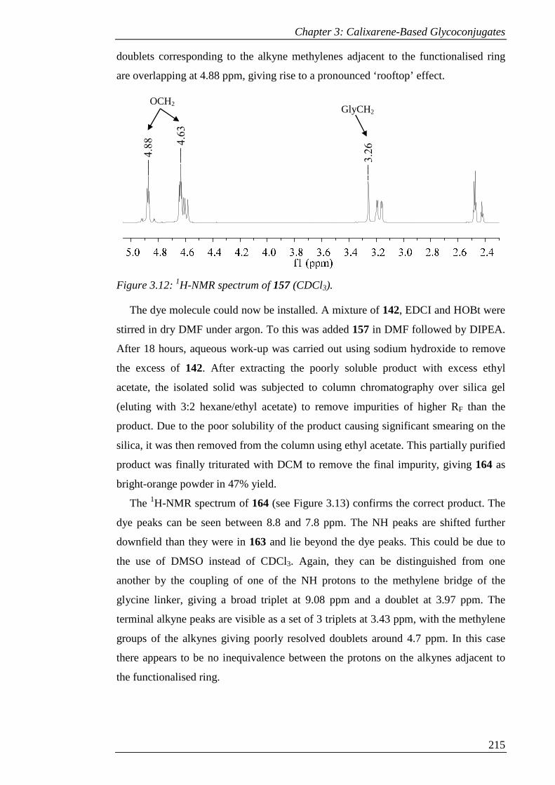
Chapter 3: Calixarene-Based Glycoconjugates
215
doublets corresponding to the alkyne methylenes adjacent to the functionalised ring
are overlapping at 4.88 ppm, giving rise to a pronounced ‘rooftop’ effect.
Figure 3.12: 1H-NMR spectrum of 157 (CDCl3).
The dye molecule could now be installed. A mixture of 142, EDCI and HOBt were
stirred in dry DMF under argon. To this was added 157 in DMF followed by DIPEA.
After 18 hours, aqueous work-up was carried out using sodium hydroxide to remove
the excess of 142. After extracting the poorly soluble product with excess ethyl
acetate, the isolated solid was subjected to column chromatography over silica gel
(eluting with 3:2 hexane/ethyl acetate) to remove impurities of higher RF than the
product. Due to the poor solubility of the product causing significant smearing on the
silica, it was then removed from the column using ethyl acetate. This partially purified
product was finally triturated with DCM to remove the final impurity, giving 164 as
bright-orange powder in 47% yield.
The 1H-NMR spectrum of 164 (see Figure 3.13) confirms the correct product. The
dye peaks can be seen between 8.8 and 7.8 ppm. The NH peaks are shifted further
downfield than they were in 163 and lie beyond the dye peaks. This could be due to
the use of DMSO instead of CDCl3. Again, they can be distinguished from one
another by the coupling of one of the NH protons to the methylene bridge of the
glycine linker, giving a broad triplet at 9.08 ppm and a doublet at 3.97 ppm. The
terminal alkyne peaks are visible as a set of 3 triplets at 3.43 ppm, with the methylene
groups of the alkynes giving poorly resolved doublets around 4.7 ppm. In this case
there appears to be no inequivalence between the protons on the alkynes adjacent to
the functionalised ring.
3.2
64.6
3
4.8
8
GlyCH2 OCH2
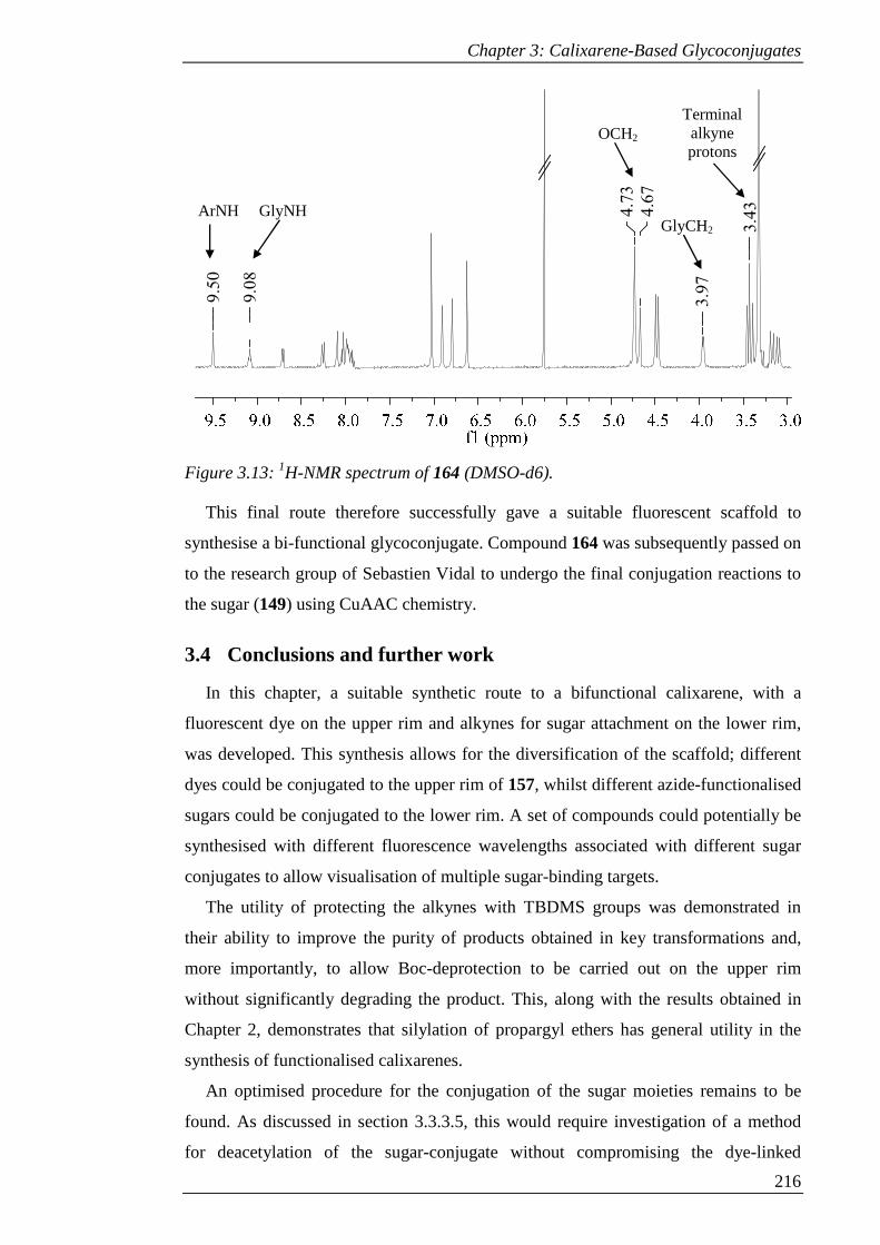
Chapter 3: Calixarene-Based Glycoconjugates
216
Figure 3.13: 1H-NMR spectrum of 164 (DMSO-d6).
This final route therefore successfully gave a suitable fluorescent scaffold to
synthesise a bi-functional glycoconjugate. Compound 164 was subsequently passed on
to the research group of Sebastien Vidal to undergo the final conjugation reactions to
the sugar (149) using CuAAC chemistry.
3.4 Conclusions and further work
In this chapter, a suitable synthetic route to a bifunctional calixarene, with a
fluorescent dye on the upper rim and alkynes for sugar attachment on the lower rim,
was developed. This synthesis allows for the diversification of the scaffold; different
dyes could be conjugated to the upper rim of 157, whilst different azide-functionalised
sugars could be conjugated to the lower rim. A set of compounds could potentially be
synthesised with different fluorescence wavelengths associated with different sugar
conjugates to allow visualisation of multiple sugar-binding targets.
The utility of protecting the alkynes with TBDMS groups was demonstrated in
their ability to improve the purity of products obtained in key transformations and,
more importantly, to allow Boc-deprotection to be carried out on the upper rim
without significantly degrading the product. This, along with the results obtained in
Chapter 2, demonstrates that silylation of propargyl ethers has general utility in the
synthesis of functionalised calixarenes.
An optimised procedure for the conjugation of the sugar moieties remains to be
found. As discussed in section 3.3.3.5, this would require investigation of a method
for deacetylation of the sugar-conjugate without compromising the dye-linked
3.4
3
3.9
7
4.6
7
4.7
3
9.0
8
9.5
0GlyCH2
Terminal alkyne protons
OCH2
ArNH GlyNH
Chapter 3: Calixarene-Based Glycoconjugates
217
scaffold, or alternatively the conjugation of deacetylated sugars. Whilst the latter
method would circumvent the problem of degradation of the molecule under
deacetylation conditions, the purification of the conjugate could prove to be
challenging. It can be expected that such a reaction would not be completely efficient
due to the aforementioned steric hindrance of the cone conformer; although 152 could
be successfully purified, an analogous purification by reversed-phase chromatography
would be more difficult to optimise.
Once the bifunctional scaffold has been successfully synthesised in its deacetylated
state, the biological tests will be carried out by co-workers of Sebastien Vidal in
Grenoble. This will allow confirmation of the binding to the conjugate to the free
lectins, followed by investigation of the binding to P. aeruginosa.
3.5 Experimental
5,11,17,23-Tetra-tert-butyl-25,26,27-tripropargyloxy-28-hydroxy-calix[4]arene
(143)44
p-tert-Butylcalix[4]arene 2 (20.10 g, 31.02 mmol) was dissolved in DMF (300 mL)
and heated to 30 °C. BaO (7.15 g, 46.78 mmol) and Ba(OH)2.8H2O (33.83 g, 107.39
mmol) were added and the mixture stirred for 30 mins. n-Propargyl bromide (94.29 g,
80% w/w in toluene, 660 mmol) was added and the mixture stirred for 18 hrs. Water
(200 mL) and dilute HCl (200 mL) were added and the product extracted with DCM
(3 x 100 mL), then washed with water (200 mL) and brine (200 mL). After drying
over MgSO4, the solvent was removed under reduced pressure. Purification by column
chromatography over silica gel (eluent: 2:1 DCM/Hexane) gave 143 as light yellow
crystals (6.7 g, 28%). Mp 155-157 °C; IR ν 1120, 1193, 1238, 1259, 1299, 1361,
1391, 1477, 1582, 1594, 2125, 2860, 2900, 2950, 3261, 3286, 3539 cm-1; 1H-NMR
(400 MHz, CDCl3): δ 7.10 (s, 2 H, ArH), 7.05 (s, 2 H, ArH), 6.56 (s, 2 H, ArH), 6.52
(s, 2 H, ArH), 5.60 (d, J = 2 Hz, 2 H, OCH2CCH), 6.62 (d, J = 2 Hz, 4 H, OCH2CCH),
4.60 (d, J = 13 Hz, 2 H, ArCH2Ar), 4.345 (d, J = 13 Hz, 2 H, ArCH2Ar), 3.275 (d, J =
13 Hz, 2 H, ArCH2Ar), 3.19 (d, J = 13 Hz, 2 H, ArCH2Ar), 2.48 (t, J = 2 Hz, 2 H,
OCH2CCH), 2.42 (t, J = 2 Hz, 4 H, OCH2CCH), 1.31 (s, 9 H, C(CH3)3), 1.30 (s, 9 H,
C(CH3)3), 0.82 (s, 18 H, C(CH3)3).
Chapter 3: Calixarene-Based Glycoconjugates
218
5,11,17-Tri-tert-butyl-23-nitro-25,26,27-tripropargyloxy-28-hydroxy-
calix[4]arene (144)
Compound 143 (1.00 g, 1.31 mmol) was dissolved in DCM (5 mL). Glacial acetic
acid (3 mL) was added, followed by 65% nitric acid (0.5 mL), dropwise over 2
minutes. After stirring for 4 minutes, the reaction was quenched with H2O (20 mL).
The product was extracted into DCM (20 mL), washed with water (2 x 20 mL) and
dried (MgSO4). Removal of solvent under reduced pressure followed by purification
by column chromatography over silica gel (eluent: 2:1 DCM/hexane) gave 144 as
yellow crystals (0.43 g, 44%). Mp 195-197 °C; IR ν 1100, 1118, 1192, 1247, 1288,
1327, 1361, 1391, 1435, 1474, 1510, 1592, 2861, 2900, 2954, 2960, 3301, 3315 cm-1; 1H NMR (400 MHz, CDCl3): δ 8.07 (s, 2 H, ArH), 7.14 (s, 2 H, ArH), 6.62 (d, J = 2
Hz, 2 H, ArH), 6.52 (d, J = 2 Hz, 2 H, ArH), 5.00 (d, J = 2 Hz, 2 H, OCH2CCH), 4.73
(dd, J = 16, 2 Hz, 2 H, OCH2CCH), 4.59 (dd, J = 16, 2 Hz, 2 H, OCH2CCH), 4.58 (d,
J = 13 Hz, 2 H, ArCH2Ar), 4.41 (d, J = 14 Hz, 2 H, ArCH2Ar), 3.43 (d, J = 14 Hz, 2
H, ArCH2Ar), 3.245 (d, J = 13 Hz, 2 H, ArCH2Ar), 2.56 (t, J = 2 Hz, 2 H,
OCH2CCH), 2.49 (t, J = 2 Hz, 4 H, OCH2CCH), 1.33 (s, 9 H, C(CH3)3), 0.84 (s, 18 H,
C(CH3)3); 13C NMR (100 MHz, CDCl3): δ 159.9, 152.2, 151.0 147.2, 147.0, 139.6,
136.1, 133.0, 130.6, 129.7, 126.2, 125.9, 124.8, 124.5, 81.7, 79.3, 76.2, 74.8, 63.1,
60.2, 53.7, 34.4, 34.0, 32.7, 31.9, 31.8, 31.2; HRMS (APCI) m/z: [M+H]+ Calcd for
C49H54NO6 752.3946; Found 752.3942.
5,11,17-Tri-tert-butyl-23-nitro-25,26,27,28-tetrapropargyloxy-calix[4]arene
(145)
Method A: To a stirred solution of propargyl alcohol (157 µL, 2.66 mmol) in
toluene (20 mL) was added TPP (0.70 g, 2.66 mmol). The solution was cooled in an
ice bath and DIAD (0.53 mL, 2.66 mmol) was added dropwise. After stirring for 10
minutes, 144 (1.00 g, 1.33 mmol) dissolved in minimum toluene was added. The
mixture was stirred for 16 hours at room temperature. The solvent was removed under
reduced pressure and the product triturated with MeOH/H2O, then filtered. 1H-NMR
showed no reaction.
Method B: To a stirred solution of propargyl alcohol (310 µl, 5.32 mmol) in
toluene (20 mL) was added TPP (1.4 g, 5.32 mmol), followed by DEAD (0.84 mL,
5.32 mmol), dropwise. After stirring for 10 minutes, 144 (1.00 g, 1.33 mmol)
dissolved in minimum toluene was added. The mixture was stirred for 72 hours at
room temperature and then the solvent removed under reduced pressure. After
Chapter 3: Calixarene-Based Glycoconjugates
219
attempted trituration with MeOH/H2O, the product was extracted into DCM and the
solvent removed under reduced pressure. 1H-NMR showed no reaction.
Calix[4]arene (146)39
To a mixture of tert-butyl calix[4]arene 2 (100 g, 154.08 mmol) and phenol (65
mL, 739 mmol) in 1 L of toluene was added AlCl3 (112.72 g, 133.34 mmol) in small
portions. After stirring for 4 hours the mixture was poured onto ice, mixed, then left to
separate. The organic layer was decanted from the surface, washed with water, and the
product extracted with DCM. Solvent was removed under reduced pressure and the
product precipitated from the residue with methanol to give 146 as off-white powder
(51.78 g, 79%). Mp 295-297 °C; IR ν 3150.5, 3093, 3058.5, 2931.5, 2867, 2769.5,
2706.5, 1608, 1594, 1466, 1463, 1448, 1446.5, 1410.5, 1376, 1299, 1267.5, 1259.5,
1245, 1236, 1213.5, 1202, 1194, 1145.5, 1094, 1077.5 cm-1; 1H NMR (400 MHz,
CDCl3); 10.18 (s, 4H, ArOH), 7.04 (d, 8H, J = 7.5 Hz, ArH), 6.72 (t, 4H, J = 7.5 Hz,
ArH), 4.25 (br s, 4H ArCH2Ar), 3.53 (br s, 4H, ArCH2Ar).
25,26,27,28- Tetra-propargyloxy-calix[4]arene (147)45
To a stirred solution of calix[4]arene (146) (10.00 g, 47.12 mmol) in DMF (400
mL) was added NaH (9.05 g, 376.96 mmol). The mixture stirred for 30 minutes before
addition of propargyl bromide (56.05 g, 80% w/w in toluene, 376.96 mmol). After
stirring for 72 hours, the solvent was removed under reduced pressure. Dilute HCl
(200 mL) was added and the product extracted with DCM (3 x 100 mL), then washed
with water (200 mL) and brine (200 mL). After drying over MgSO4, the solvent was
removed under reduced pressure. Purification by column chromatography over silica
(eluent: 2:1 hexane/toluene) gave 147 as fine white powder (10.00 g, 37%). Mp 156-
158 °C; IR ν 3298.5, 3278, 3058, 3010, 2982, 2972, 2950.5, 2911, 2847, 2117, 1585,
1455.5, 1428.5, 1364, 1295.5, 1278, 1268.5, 1248, 1241, 1198.5, 1184.5, 1178, 1156,
1088, 1076.5 cm-1; 1H NMR (300 MHz, CDCl3): δ 6.75-6.65 (m, 12H, ArH), 4.78 (d,
J = 2 Hz, 2H, OCH2CCH), 4.63 (d, J = 14 Hz, 4H, ArCH2Ar), 3.23 (d, J = 14 Hz, 4H,
ArCH2Ar), 2.47 (t, J = 2 Hz, 2H, OCH2CCH).
5-Nitro-25,26,27,28-tetra-propargyloxy-calix[4]arene (148)
To a stirred solution of 147 (1.5 g, 2.60 mmol) in DCM (150 mL) was added 100%
nitric acid (1.5 mL) and 95% sulphuric acid (6 drops). The solution was stirred for 1
hour before quenching with H2O (100 mL). The product was extracted with DCM (3 x
Chapter 3: Calixarene-Based Glycoconjugates
220
50 mL) then washed with water (3 x 100 mL) and brine (100 mL). After drying over
MgSO4 the solvent was removed under reduced pressure and the product purified by
column chromatography over silica gel (eluent: 1:1 DCM/hexane) to give 147 (0.93 g,
60%) and 148 (0.19 g, 12%). Impure fractions were recrystallized from
DCM/methanol to give further 148 as off-white crystals (0.15 g, 9%). Mp 157-159
°C; IR ν 1159, 1197, 1208, 1258, 1264, 1303, 1344, 1436, 1455, 1521, 1588, 2125,
2862, 2930, 2987, 3036, 3071, 3274, 3303 cm-1; 1H NMR (400 MHz, CDCl3): δ 7.38
(s, 2H, ArH), 6.94-6.82 (m, 6H, ArH), 6.50-6.43 (m, 3H, ArH), 4.85 (dd, J = 16, 2 Hz,
2H, OCH2CCH), 4.81 (d, J = 2 Hz, 2H, OCH2CCH), 4.75 (dd, J = 16, 2 Hz, 2H,
OCH2CCH), 4.71 (d, J = 14 Hz, 2H, ArCH2Ar), 4.65 (d, J = 2 Hz, 2H, OCH2CCH),
4.56 (d, J = 14 Hz, 2H, ArCH2Ar), 3.28 (d, J = 14 Hz, 2H, ArCH2Ar), 3.24 (d, J = 14
Hz, 2H, ArCH2Ar), 2.50-2.49 (m, 4H, OCH2CCH); 13C NMR (100 MHz, CDCl3) δ
159.99, 155.34, 154.78, 143.42, 137.03, 136.34, 134.97, 134.56, 129.44, 128.56,
128.19, 123.97, 123.46, 123.30, 80.42, 80.10, 79.01, 76.07, 75.12, 75.07, 61.69,
61.25, 32.08, 31.76; HRMS (APCI) m/z: [M+H]+ Calcd for C40H32NO6 622.2224;
Found 622.2225.
6-Chloro-5-oxo-5H-benzo[a]phenoxazine-9-carboxylic acid (142)29
A mixture of 2,3-dichloro-1,4-napthoquinone (2.27 g, 10 mmol), 4-amino-3-
hydroxybenzoic acid (1.53 g, 10 mmol) and potassium acetate (1.96 g, 20 mmol) were
suspended in methanol (20 mL) and heated to reflux, with stirring, for 24 hours. The
resulting orange-red suspension was cooled to room temperature, filtered and washed
with water. The crude product was washed with hot methanol to give 142 as red
powder (2.14 g, 66%). Mp >320 °C (decomp.); IR ν 3066, 1615.5, 1593, 1561, 1557,
1479, 1470.5, 1446.5, 1411, 1372.5, 1362, 1306.5, 1283, 1235, 1224.5, 1201.5, 1194,
1163, 1144, 1099, 1080, 1046, 1020.5 cm-1; 1H-NMR (400 MHz, 7:1 Tol-d8/DMSO-
d6) δ 8.53 – 8.51 (m, 1H, ArH), 8.30 – 8.28 (m, 1H, ArH), 8.10 (dd, J = 8.0, 1.5 Hz,
1H, ArH), 8.08 (d, J = 1.5 Hz, 1H, ArH), 7.65 (d, J = 8.0 Hz, 1H, ArH), 7.45 – 7.38
(m, 2H, ArH).
1-Azido-3,6-dioxaoct-8-yl-2’,3’,4’,6’-tetra-O-acetyl-β-D-galactopyranoside
(149)41
To a stirred solution of 2,3,4,6-tetra-O-acetyl-β-D-galactopyranoside (15 g, 38.43
mmol), AgCO2CF3 (12.72 g, 57.58 mmol) and 2-[2-(2-chloroethoxy)ethoxy]ethanol
(8.40 mL, 57.79 mmol) in dry DCM (300 mL) under argon was added SnCl4 (1 M in
Chapter 3: Calixarene-Based Glycoconjugates
221
DCM, 115.2 mL, 115.2 mmol) dropwise over 30 minutes. After 90 minutes, saturated
aq. NaHCO3 was added to adjust pH to >8, and the solution was stirred for 12 hours.
The biphasic solution was diluted with water (1000 mL) and product extracted with
DCM (3 × 450 mL). The combined organic layers were washed with saturated aq.
NaHCO3 (450 mL), water (3 × 450 mL) and brine (2 × 450 mL). After drying over
Na2SO4, the solvent was removed under reduced pressure. The yellow oil was
dissolved in DMF (450 mL) before adding sodium azide (12.48 g, 192 mmol) and
Bu4NI (14.19 g, 38.42 mmol). The mixture was stirred at 70°C under argon for 18 h
then cooled to RT, filtered, and the solid was washed with EtOAc (3 × 400 mL). The
filtrate was diluted with EtOAc (800 mL) then washed with saturated aq. NaHCO3 (3
× 900 mL), water (3 × 1000 mL), and brine (1000 mL). After drying over Na2SO4, the
solvent was removed under reduced pressure and the residue was purified by flash
column chromatography over silica gel (1:1 petroleum ether/ethyl acetate) to give 149
as viscous, light-yellow oil (10.01 g, 52% yield). IR ν 2937, 2871, 2102, 1743.5,
1436.5, 1367.5, 1214, 1174.5, 1132, 1122, 1073, 1055, 1043.5 cm-1; 1H-NMR (300
MHz, CDCl3) δ 5.38 (dd, J = 3.4, 1.0 Hz, 1H, CH), 5.21 (dd, J = 10.5, 7.9 Hz, 1H,
CH), 5.01 (dd, J = 10.5, 3.4 Hz, 1H, CH), 4.57 (d, J = 7.9 Hz, 1H, CH), 4.20 – 4.08
(m, 2H, CH2), 3.99 – 3.88 (m, 2H, CH, OCH2), 3.79 – 3.64 (m, 9H, OCH2), 3.39 (t, J
= 5.0 Hz, 2H, CH2N3), 2.14 (s, 3H, CH3), 2.06 (s, 3H, CH3), 2.04 (s, 3H, CH3), 1.98
(s, 3H, CH3); 13C-NMR (100 MHz, CDCl3) δ 169.80, 169.74, 169.52, 168.92, 100.77,
70.43, 70.20, 70.17, 70.14, 69.89, 69.54, 68.55, 68.40, 66.75, 60.92, 53.37, 50.18,
20.21, 20.11, 20.09, 20.02.
NRD appended tetra-propargyloxy-calix[4]arene (151)
A mixture of 148 (0.58 g, 0.93 mmol) and SnCl2·2H2O (1.37 g, 6.05 mmol) was
heated to reflux in ethanol (60 mL) with stirring for 48 hours. Solvent was removed
under reduced pressure and DCM (50 mL) added. After washing with 10% NaOH
(100 mL), the product was extracted with DCM (3 x 50 mL) and washed with water (2
x 100 mL) then brine (100 mL). After drying over MgSO4, the solvent was removed
under reduced pressure to give a brown glass, which was verified by 1H NMR as the
intermediate amine 150 (0.35 g, 63%). 1H-NMR (400 MHz, CDCl3) δ 6.82 – 6.67 (m,
9H, ArH), 6.12 (s, 2H, ArH), 4.84 (d, J = 2 Hz, 2H, OCH2CCH), 4.81 (d, J = 2 Hz,
2H, OCH2CCH), 4.81 (d, J = 2 Hz, 2H, OCH2CCH), 4.75 (d, J = 2 Hz, 2H,
OCH2CCH), 4.69 (d, J = 13 Hz, 2H, ArCH2Ar), 4.60 (d, J = 13 Hz, 2H, ArCH2Ar),
3.28 (d, J = 13 Hz, 2H, ArCH2Ar), 3.15 (d, J = 13 Hz, 2H, ArCH2Ar), 2.50 – 2.49 (m,

Chapter 3: Calixarene-Based Glycoconjugates
222
4H, OCH2CCH). The crude amine was stirred with 142 (0.19 g, 0.59 mmol),
EDCI·HCl (0.25 g, 1.30 mmol), HOBt hydrate (0.17 g, 1.30 mmol) and NMM (0.26
mL, 2.42 mmol) in DMF (6 mL) for 18 hours. Water was added and the precipitate
filtered. The solid was dissolved in ethyl acetate and washed with water (200 mL) and
brine (200 mL). After drying over MgSO4, the solvent was removed under reduced
pressure and the solid purified by column chromatography over silica gel (eluent:
DCM) to give 151 as a red solid (0.20 g, 37%). Mp 250 °C (decomp.); IR ν 3440,
3288.5, 3258, 3061, 3014, 2991, 2920.5, 2878.5, 2851, 2127, 2112, 1675.5, 1673,
1640.5, 1628.5, 1604, 1592.5, 1587, 1565, 1534, 1531.5, 1474, 1459.5, 1455, 1440,
1424, 1413, 1358.5, 1331.5, 1302.5, 1282.5, 1256.5, 1234.5, 1212.5, 1203.5, 1191,
1160, 1144.5, 1127.5, 1089, 1078, 1043.5, 1017.5, 1008.5 cm-1; 1H-NMR (400 MHz,
CDCl3) δ 8.75 – 8.73 (m, 1H, ArHNRD), 8.37 – 8.35 (m, 1H, ArHNRD), 7.93 – 7.76 (m,
5H, ArHNRD), 7.56 (br s, 1H, ArNH), 7.00 (br s, 2H, ArH), 6.84 – 6.79 (m, 4H, ArH),
6.74 – 6.71 (m, 4H, ArH), 6.67 – 6.62 (m, 1H, ArH), 4.82 (d, J = 2 Hz, 2H,
OCH2CCH), 4.82 (d, J = 2 Hz, 2H, OCH2CCH), 4.78 (d, J = 2 Hz, 2H, OCH2CCH),
4.77 (d, J = 2 Hz, 2H, OCH2CCH), 4.68 (d, J = 13 Hz, 2H, ArCH2Ar), 4.64 (d, J = 13
Hz, 2H, ArCH2Ar), 3.27 (d, J = 13 Hz, 2H, ArCH2Ar), 3.25 (d, J = 13 Hz, 2H,
ArCH2Ar), 2.50 – 2.48 (m, 4H, OCH2CCH). 13C-NMR (100 MHz, CDCl3) δ 177.35,
163.23, 155.08, 155.01, 152.35, 147.35, 146.40, 143.33, 137.96, 136.34, 135.48,
135.44, 135.02, 134.28, 132.73, 132.57, 131.15, 129.93, 129.76, 128.52, 128.38,
128.34, 126.71, 125.05, 124.43, 123.52, 123.22, 120.60, 115.24, 114.86, 80.44, 80.33,
75.10, 74.88, 74.84, 61.34, 61.27, 61.21, 32.10, 31.94; HRMS (APCI) m/z: [M+H]+
Calcd for C57H40ClN2O7 899.2519; Found 899.2533.
NRD appended 25,26,27,28-Tetra-{1’-[(2’’’,3’’’,4’’’ ,6’’’-tetra-O-acetyl- β-D-
galactopyranosyloxy)-3”,6”-dioxaoct-8”-yl]-1’,2’,3’ -triazol-4’-ylmethyleneoxy}-
calix[4]arene (152)
A mixture of 149 (169 mg, 333.96 µmol), 151 (50 mg, 55.66 µmol), CuSO4 (0.1 M
in water, 278 µL, 27.83 µmol) and sodium ascorbate (17 mg, 83.49 µmol) in DMF (3
mL) was stirred at room temperature under argon for 24 hours. Further 149 (28.17 mg,
55.66 µmol), CuSO4 (0.1 M in water, 278 µL, 27.83 µmol) and sodium ascorbate (17
mg, 83.49 µmol) was added and stirring continued for 24 hours. After removing
solvent under reduced pressure, the product was purified by column chromatography
over silica gel (eluent: 1:1 petroleum ether/ethyl acetate, ethyl acetate and 19:1 ethyl
acetate/methanol) to give 152 as dark-orange solid (0.16 mg, 44%). Mp 68-70 °C; IR
Chapter 3: Calixarene-Based Glycoconjugates
223
ν 2920.5, 2871.5, 1790, 1651, 1595.5, 1565.5, 1540.5, 1460, 1435, 1367, 1301.5,
1218.5, 1176.5, 1134, 1045, 1018.5 cm-1; 1H-NMR (300 MHz, CDCl3) δ 8.75 – 8.72
(m, 1H, ArHNRD), 8.37 – 8.34 (m, 1H, ArHNRD), 8.14 (s, 1H, NH), 7.96 – 7.75 (m, 9H,
ArHNRD and ArHTriazole), 7.08 (s, 1H, ArH), 7.04 (s, 1H, ArH), 6.70 – 6.52 (m, 9H,
ArH), 5.38 – 5.36 (m, 4H, CH), 5.21 – 5.14 (m, 8H, CH and OCH2C), 5.06 – 4.99 (m,
8H, CH and OCH2C), 4.57 – 4.52 (m, 12H, CH and CH2N), 4.22 – 4.07 (m, 12H,
ArCH2Ar and CH2OAc), 3.96 – 3.84 (m, 16H, CH and OCH2), 3.72 – 3.55 (m, 28H,
OCH2), 2.97 (d, J = 13 Hz, 4H, ArCH2Ar), 2.12 – 1.96 (m, 48H, CH3); 13C-NMR (75
MHz, CDCl3) δ 177.44, 170.46, 170.43, 170.29, 170.18, 169.64, 169.52, 163.13,
155.12, 154.75, 152.13, 147.52, 146.70, 143.97, 143.53, 138.05, 136.51, 135.86,
135.02, 134.62, 134.47, 132.80, 132.47, 131.37, 130.01, 128.42, 128.29, 128.15,
126.79, 125.16, 124.66, 122.88, 120.54, 115.25, 115.19, 101.38, 70.92, 70.67, 70.52,
70.20, 69.58, 69.15, 68.84, 67.09, 66.84, 61.28, 50.13, 31.95, 31.44, 29.72, 29.39,
22.72, 20.85, 20.74, 20.72, 20.64; ESI-MS m\z: [M+2H]2+ 1461.5, [M+2Na]2+
1483.5, [M]+ 2921.8, [M+Na]+ 2943.8.
NRD appended 25,26,27,28-Tetra-{1’-[(β-D-galactopyranosyloxy)-3”,6”-
dioxaoct-8”-yl]-1’,2’,3’-triazol-4’-ylmethyleneoxy} -calix[4]arene (153)
Compound 152 (62 mg, 21.22 µmol) was stirred in a mixture of 1:2:1
Et3N/methanol/water (2.2 mL) under argon for 42 hours, then the solvent removed
under reduced pressure. Attempted purification by reversed-phase column
chromatography over C18 failed to give the desired product.
5,11,17,23-Tetra-tert-butyl-25,26,27,28-tetra-propargyloxy-calix[4]arene
(154)21
To a stirred suspension of tert-butyl-calix[4]arene 2 (8 g, 12.33 mmol) in DMF
(160 mL) was added NaH (2.35 g, 97.92 mmol) and the mixture stirred for 1 hour.
Propargyl bromide (8.74 mL, 80% w/w in toluene, 81.12 mmol) was added and the
reaction stirred for 48 hours. Water was added and the precipitate filtered, washed
with water twice and air dried to give 154 as light brown powder (5.55 g, 53%). Mp
204-206 °C; IR ν 3309.5, 3283, 2965.5, 2951, 2921, 2906, 2861.5, 1602, 1583.5,
1478.5, 1474, 1438, 1434.5, 1414.5, 1392.5, 1363.5, 1299.5, 1273.5, 1261, 1237,
1192, 1121.5, 1107, 1019.5, 1009, 1003 cm-1; 1H-NMR (400 MHz, CDCl3) δ 6.79 (s,
8H, ArH), 4.80 (d, J = 2 Hz, 8H, OCH2CCH), 4.61 (d, J = 13 Hz, 4H, ArCH2Ar), 3.17
Chapter 3: Calixarene-Based Glycoconjugates
224
(d, J = 13 Hz, 4H, ArCH2Ar), 2.47 (t, J = 2 Hz, 4H, OCH2CCH), 1.08 (s, 36H,
C(CH3)3).
5,11,17-Tetra-tert-butyl-23-nitro-25,26,27,28-tetra-propargyloxy-calix[4]arene
(145)
To a stirred solution of 154 (1.00 g, 1.125 mmol) in DCM (72 mL) was added
glacial acetic acid (1.84 mL) and 100% HNO3 (1.04 mL). After stirring for 30
minutes, the reaction was quenched with water (50 mL). The product was extracted
with DCM (3 x 50 mL), washed with water (3 x 200 mL) and brine (200 mL). After
drying over MgSO4, the solvent was removed under reduced pressure and the residue
purified by column chromatography over silica gel (eluent: 8-10% ethyl acetate in
hexane) to give recovered 154 (0.4 g, 40%) as white powder and 145 (0.26 g, 26%) as
a light yellow solid. Mp 87-89 °C; IR ν 3296, 3156, 2961, 2934, 2874, 1731, 1669.5,
1609.5, 1583.5, 1518.5, 1478, 1463, 1437.5, 1392.5, 1361.5, 1341.5, 1308.5, 1261,
1231.5, 1194.5, 1162, 1117.5, 1089, 1043, 1002 cm-1; 1H-NMR (400 MHz, CDCl3) δ
7.37 (s, 2H, ArH), 7.12 (d, J = 2 Hz, 2H, ArH), 7.08 (d, J = 2 Hz, 2H, ArH), 6.31 (s,
2H, ArH), 4.98 (dd, J = 16, 2 Hz, 2H, OCH2CCH), 4.83 (dd, J = 16, 2 Hz, 2H,
OCH2CCH), 4.75 (d, J = 13 Hz, 2H, ArCH2Ar), 4.73 (d, J = 2 Hz, 2H, OCH2CCH),
4.56 (d, J = 2 Hz, 2H, OCH2CCH), 4.51 (d, J = 13 Hz, 2H, ArCH2Ar), 3.23 (d, J = 13
Hz, 2H, ArCH2Ar), 3.20 (d, J = 13 Hz, 2H, ArCH2Ar), 2.50 (t, J = 2 Hz, 2H,
OCH2CCH), 2.50 (t, J = 2 Hz, 1H, OCH2CCH), 2.47 (t, J = 2 Hz, 1H, OCH2CCH),
1.32 (s, 18H, C(CH3)3), 0.69 (s, 9H, C(CH3)3). 13C-NMR (100 MHz, CDCl3) δ
159.46, 153.05, 152.11, 146.81, 145.86, 143.39, 136.32, 136.21, 134.74, 132.63,
126.68, 125.20, 124.80, 123.17, 81.28, 80.24, 79.02, 75.94, 74.87, 74.72, 62.09,
61.95, 60.73, 34.32, 33.44, 32.47, 32.12, 31.67, 30.75. HRMS (APCI) m/z: [M+H]+
Clcd for C52H56NO6 790.4102; Found 790.4110.
5,11,17-Tetra-tert-butyl-23-BocGly-25,26,27,28-tetra-propargyloxy-
calix[4]arene (156)
A mixture of 145 (1.02 g, 1.29 mmol) and SnCl2·2H2O (2.91 g, 12.9 mmol) in
ethanol (70 mL) was heated to reflux for 18 hours. The solvent was removed under
reduced pressure and the residue stirred with 10% NaOH (100 mL) for 5 minutes. The
product was extracted with DCM (3 x 100 mL) then washed with water (200 mL) and
brine (200 mL). After drying over MgSO4, the solvent was removed under reduced
pressure to give the crude intermediate amine 155 as a yellow solid (crude yield: 0.81
Chapter 3: Calixarene-Based Glycoconjugates
225
g, 83%). 1H NMR (400 MHz, CDCl3) δ 7.02 (d, J = 2.5 Hz, 2H, ArH), 6.94 (d, J = 2.5
Hz, 2H, ArH), 6.42 (s, 2H, ArH), 5.83 (s, 2H, ArH), 4.88 (d, J = 2 Hz, 4H,
OCH2CCH), 4.63 (d, J = 2 Hz, 2H, OCH2CCH), 4.62 (d, J = 12.9 Hz, 2H), 4.58 (d, J
= 2 Hz, 2H, OCH2CCH), 4.54 (d, J = 13 Hz, 2H, ArCH2Ar), 3.18 (d, J = 13 Hz, 2H,
ArCH2Ar), 3.05 (d, J = 13 Hz, 2H, ArCH2Ar), 2.47 (t, J = 2 Hz, 1H, OCH2CCH), 2.46
(t, J = 2 Hz, 2H, OCH2CCH), 2.42 (t, J = 2 Hz, 1H, OCH2CCH), 1.26 (s, 18H,
C(CH3)3), 0.86 (s, 9H, C(CH3)3). A mixture of BocGly (0.70 g, 4.01 mmol),
EDCI·HCl (0.85 g, 4.42 mmol) and HOBt hydrate (0.60 g, 4.42 mmol) were stirred
under argon in dry DMF (20 mL). To this was added 155 in dry DMF (1 mL) and
DIPEA (1.44 mL, 8.24 mmol). The mixture was stirred for 24 hours and the solvent
removed under reduced pressure. The residue was redissolved in DCM (100 mL) and
washed with 10% NaOH (100 mL), water (100 mL) then brine (100 mL). After drying
over MgSO4, the solvent was removed under reduced pressure and the residue purified
by column chromatography over silica gel (eluent: 5-7% ethyl acetate in DCM) to
give 156 as a light yellow solid (1.18 g, 1.29 mmol). Mp 106-108 °C; IR ν 3405.5,
3290, 2954.5, 2931.5, 2906, 2866, 1685.5, 1603.5, 1539, 1505.5, 1476.5, 1437,
1417.5, 1392, 1363.5, 1285, 1274.5, 1251.5, 1241.5, 1193.5, 1166, 1132.5, 1116,
1054, 1014 cm-1; 1H-NMR (400 MHz, CDCl3) δ 7.23 (s, 1H, ArNH), 7.02 (s, 2H,
ArH), 6.97 (s, 2H, ArH), 6.65 (s, 2H, ArH), 6.39 (s, 2H, ArH), 4.99 (s, 1H, CH2NH),
4.90 (dd, J = 16, 2 Hz, 2H, OCH2CCH), 4.84 (dd, J = 16, 2 Hz, 2H, OCH2CCH), 4.63
– 4.57 (m, 8H, OCH2CCH and ArCH2Ar), 3.72 (d, J = 6 Hz, 2H, CH2NH), 3.18 (d, J
= 13 Hz, 2H, ArCH2Ar), 3.15 (d, J = 13 Hz, 2H, ArCH2Ar), 2.49 – 2.47 (m, 3H,
OCH2CCH), 2.43 (t, J = 2 Hz, 1H, OCH2CCH), 1.45 (s, 9H, CO2C(CH3)3), 1.26 (s,
18H, C(CH3)3), 0.81 (s, 9H, C(CH3)3); 13C-NMR (100 MHz, CDCl3) δ 166.70,
156.29, 153.10, 152.44, 151.38, 146.08, 145.31, 135.74, 135.26, 135.07, 133.37,
132.33, 125.90, 125.27, 124.85, 119.40, 81.45, 80.63, 80.51, 80.38, 74.90, 74.66,
74.47, 61.79, 61.54, 60.84, 45.41, 34.18, 33.68, 32.44, 32.25, 31.67, 31.02, 28.41;
HRMS (NSI) m/z: [M+NH4]+ Calcd for C59H72N3O7 934.5365; Found 934.5352.
5,11,17-Tetra-tert-butyl-23-Gly-25,26,27,28-tetra-propargyloxy-calix[4]arene
(157)
To a stirred solution of 156 (1.06 g, 1.16 mmol) in DCM (8 mL) was added TFA (4
mL). After stirring for 40 minutes, the solution was diluted with DCM (50 mL), then
water (50 mL) was added followed by sufficient saturated NaHCO3 to adjust to pH >
7. After mixing thoroughly the emulsion separated and the product was extracted with
Chapter 3: Calixarene-Based Glycoconjugates
226
DCM (3 x 50 mL) and washed with brine (100 mL). After drying over MgSO4, the
solvent was removed under reduced pressure to give an orange-brown solid that was
revealed by 1H NMR to be a complex mixture of products.
5,11,17-Tri-tert-butyl-23-nitro-25,26,27,28-tetra-[tert-butyl(dimethyl)silyl]-
propargyloxy-calix[4]arene (158)
A solution of 145 (2.88 g, 3.65 mmol) in THF (25 mL) was cooled to -80 °C. To
this was added 1 M LiHMDS in THF (21.9 mL, 21.9 mmol) followed by TBDMSCl
(3.30 g, 21.9 mmol) in minimum THF. The mixture was allowed to warm to room
temperature and stirred for 24 hours before quenching with saturated ammonium
chloride (100 mL). Water (200 mL) was added to dilute and the product extracted
with DCM (3 x 100 mL) then washed with brine (200 mL). After drying over MgSO4,
the solvent was removed under reduced pressure to give sticky dark-orange oil. 1H-
NMR showed loss of the terminal alkyne triplet but a complex mixture of products.
5,11,17,23-Tetra-tert-butyl-25,26,27,28-tetra-[tert-butyl(dimethyl)silyl]-
propargyloxy-calix[4]arene (159)
A solution of 154 (10.00 g, 12.48 mmol) in THF (87 mL) was cooled to -80 °C. To
this was added 1 M LiHMDS in THF (54.91 mL, 54.91 mmol) followed by
TBDMSCl (11.29 g, 74.88 mmol) in minimum THF. The mixture was allowed to
warm to room temperature and stirred for 18 hours before quenching with saturated
ammonium chloride (150 mL). The product was extracted with ethyl acetate (3 x 150
mL), washed with dilute HCl (200 mL) and brine (200 mL). After drying over
MgSO4, the solvent was removed under reduced pressure and the product precipitated
from the residue with methanol to give 159 as light brown powder (13.87 g, 88%).
Mp 219-221 °C; IR ν 2953, 2928.5, 2902, 2856.5, 2855, 2176, 1603, 1586, 1480.5,
1471, 1462.5, 1411.5, 1391, 1361.5, 1314.5, 1301.5, 1280, 1247.5, 1197, 1121, 1106,
1097, 1040.5 cm-1; 1H-NMR (400 MHz, CDCl3) δ 6.75 (s, 8H, ArH), 4.82 (s, 8H,
OCH2), 4.52 (d, J = 13 Hz, 4H, ArCH2Ar), 3.11 (d, J = 13 Hz, 4H, ArCH2Ar), 1.06 (s,
36H, C(CH3)3), 0.88 (s, 36H, SiC(CH3)3), 0.08 (s, 24H, SiCH3); 13C-NMR (100 MHz,
CDCl3) δ 152.09, 145.28, 134.79, 124.87, 103.58, 89.53, 61.39, 33.99, 32.75, 31.57,
26.30, 16.61, -4.43; MALDI-TOF m/z: [M+Na]+ 1281.40.

Chapter 3: Calixarene-Based Glycoconjugates
227
5,11,17-Tri-tert-butyl-23-nitro-25,26,27,28-tetra-[tert-butyl(dimethyl)silyl]-
propargyloxy-calix[4]arene (158)
To a stirred solution of 159 (1.57 g, 1.25 mmol) in DCM (72 mL) was added
glacial acetic acid (1.85 mL) and 100% nitric acid (1.04 mL). The solution was stirred
for 1 hour before quenching with water (100 mL). The product was extracted with
DCM (3 x 50 mL), washed with water (3 x 200 mL) and brine (200 mL). After drying
over MgSO4, the solvent was removed under reduced pressure and the product
purified by column chromatography over silica gel (eluent: 4:1 then 2:1 hexane/DCM)
to give recovered 159 (1.10 g, 70%) as white powder and 158 (0.45 g, 29%) as a light
yellow solid. Mp 200-202 °C; IR ν 2953, 2928, 2898.5, 2885, 2856.5, 2181, 1585,
1521.5, 1481, 1470.5, 1462, 1411.5, 1390.5, 1361.5, 1341, 1312.5, 1278.5, 1256,
1249, 1205, 1195, 1158, 1116.5, 1085.5, 1035 cm-1; 1H-NMR (400 MHz, CDCl3) δ
7.37 (s, 2H, ArH), 7.04 (d, J = 2 Hz, 2H, ArH), 6.98 (d, J = 2 Hz, 2H, ArH), 6.35 (s,
2H, ArH), 4.94 (s, 2H, OCH2), 4.92 (s, 2H, OCH2), 4.78 (s, 2H, OCH2), 4.66 (d, J =
13 Hz, 2H, ArCH2Ar), 4.58 (s, 2H, OCH2), 4.44 (d, J = 13 Hz, 2H, ArCH2Ar), 3.18
(d, J = 13 Hz, 2H, ArCH2Ar), 3.16 (d, J = 13.3 Hz, 2H, ArCH2Ar), 1.28 (s, 18H,
C(CH3)3), 0.92 (s, 9H, SiC(CH3)3), 0.86 (s, 9H, SiC(CH3)3), 0.85 (s, 18H, SiC(CH3)3),
0.73 (s, 9H, C(CH3)3), 0.12 (s, 6H, SiCH3), 0.07 (s, 6H, SiCH3), 0.06 (s, 12H, SiCH3); 13C-NMR (100 MHz, CDCl3) δ 159.61, 152.12, 152.02, 146.36, 145.71, 143.27,
136.68, 136.36, 135.02, 133.08, 126.30, 124.88, 124.73, 123.08, 103.17, 102.70,
101.14, 91.45, 90.07, 89.95, 62.62, 62.53, 60.67, 34.26, 33.52, 32.68, 32.33, 31.71,
30.87, 26.23, 26.21, 26.10, 16.58, 16.55, 16.49, -4.51, -4.61; MALDI-TOF m/z:
[M+Na]+ 1270.37, [M+K]+ 1286.33.
5,11,17-Tri-tert-butyl-23-amino-25,26,27,28-tetra-[tert-butyl(dimethyl)silyl]-
propargyloxy-calix[4]arene (160)
A mixture of 158 (7.81 g, 6.26 mmol) and SnCl2·2H2O (14.12 g, 62.60 mmol) in
ethanol (330 mL) were heated to reflux for 24 hours. The solvent was removed under
reduced pressure and the residue stirred with 10% NaOH for 5 minutes. The product
was extracted with DCM (3 x 100 mL) then washed with water (2 x 200 mL) and
brine (200 mL). After drying over MgSO4, the solvent was removed under reduced
pressure to give 160 as a light yellow solid (6.07 g, 80%). Mp 95-97 °C; IR ν 2952.5,
2927.5, 2899.5, 2885, 2855, 2176.5, 1741, 1615, 1605.5, 1589, 1479.5, 1471, 1463,
1411.5, 1390.5, 1361.5, 1317.5, 1303.5, 1281.5, 1248.5, 1194.5, 1137.5, 1116, 1028,
1005 cm-1; 1H-NMR (400 MHz, CDCl3) δ 6.96 (d, J = 2.5 Hz, 2H, ArH), 6.88 (d, J =
Chapter 3: Calixarene-Based Glycoconjugates
228
2.5 Hz, 2H, ArH), 6.41 (s, 2H, ArH), 5.82 (s, 2H; ArH), 4.93 (d, J = 1.5 Hz, 4H,
OCH2), 4.64 (s, 2H, OCH2), 4.55 (s, 2H, OCH2), 4.54 (d, J = 13 Hz, 2H, ArCH2Ar),
4.45 (d, J = 13 Hz, 2H, ArCH2Ar), 3.13 (d, J = 13 Hz, 2H, ArCH2Ar), 3.00 (d, J = 13
Hz, 2H, ArCH2Ar), 2.87 (s, 2H, NH2), 1.24 (s, 18H, C(CH3)3), 0.93 (s, 9H,
SiC(CH3)3), 0.92 (s, 9H, SiC(CH3)3), 0.85 (s, 9H, C(CH3)3), 0.85 (s, 18H, SiC(CH3)3),
0.11 (s, 6H, SiCH3), 0.11 (s, 6H, SiCH3), 0.04 (s, 12H, SiCH3); 13C-NMR (100 MHz,
CDCl3) δ 152.27, 152.25, 147.90, 145.36, 144.92, 140.94, 136.44, 135.90, 135.05,
133.61, 125.34, 124.85, 124.72, 114.86, 103.66, 103.41, 103.14, 89.56, 89.52, 89.20,
62.47, 62.33, 60.51, 34.14, 33.75, 32.57, 32.54, 31.77, 31.71, 31.14, 26.27, 26.26,
26.24, 16.59, 16.58, 16.55, -4.45, -4.48, -4.50; MALDI-TOF m/z: [M+Na]+ 1239.97,
[M+K] + 1255.95.
5,11,17-Tri-tert-butyl-23-BocGly-25,26,27,28-tetra-[tert-butyl(dimethyl)silyl]-
propargyloxy-calix[4]arene (161)
A mixture of Boc-glycine (1.71 g, 9.78 mmol), EDCI (1.87 g, 9.78 mmol) and
HOBt (1.32 g, 9.78 mmol) was stirred in DMF (10 mL) under argon for 5 minutes. To
this was added a solution of 160 (5.95 g, 4.89 mmol) in DMF (40 mL) and DIPEA
(3.41 mL, 19.56 mmol). The mixture was stirred for 24 hours, the solvent removed
under reduced pressure and water (200 mL) added. The product was extracted with
DCM (3 x 100 mL), washed with water (2 x 200 mL) and brine (200 mL). After
drying over MgSO4, the solvent was removed under reduced pressure and the product
purified by column chromatography over silica gel (eluent: 19:1 DCM/ethyl acetate)
to give 161 as a yellow solid. Mp 114-116 °C; IR ν 2952.5, 2927, 2898.5, 2855.5,
2173, 2160.5, 1731, 1701, 1698, 1685.5, 1680, 1603.5, 1558, 1539, 1504, 1478, 1470,
1462.5, 1420, 1417.5, 1390.5, 1361.5, 1314.5, 1281, 1248.5, 1210, 1194, 1169,
1132.5, 1115.5, 1028 cm-1; 1H NMR (400 MHz, CDCl3) δ 7.17 (s, 1H, ArNH), 6.95
(s, 2H, ArH), 6.91 (s, 2H, ArH), 6.63 (s, 2H, ArH), 6.39 (s, 2H, ArH), 5.01 (s, 1H,
CH2NH), 4.91 (s, 4H, OCH2), 4.65 (s, 2H, OCH2), 4.62 (s, 2H, OCH2), 4.52 (d, J = 13
Hz, 2H, ArCH2Ar), 4.51 (d, J = 13 Hz, 2H, ArCH2Ar), 3.73 (d, J = 6 Hz, 2H,
CH2NH), 3.13 (d, J = 13 Hz, 2H, ArCH2Ar), 3.10 (d, J = 13 Hz, 2H, ArCH2Ar), 1.45
(s, 9H, NHCO2C(CH3)3), 1.23 (s, 18H, C(CH3)3), 0.91 (s, 18H, SiC(CH3)3), 0.85 (s,
18H, SiC(CH3)3), 0.82 (s, 9H, C(CH3)3), 0.11 (s, 6H, SiCH3), 0.10 (s, 6H, SiCH3),
0.05 (s, 12H, SiCH3); 13C NMR (100 MHz, CDCl3) δ 166.45, 156.08, 152.09, 152.07,
151.43, 145.51, 144.84, 136.10, 135.32, 133.56, 131.88, 125.42, 124.78, 124.55,
119.38, 103.32, 102.90, 102.67, 89.70, 89.56, 80.41, 62.16, 60.53, 60.40, 45.25,
Chapter 3: Calixarene-Based Glycoconjugates
229
34.01, 33.55, 32.44, 32.33, 31.60, 30.95, 28.30, 26.10, 26.09, 16.45, 16.41, -4.64;
MALDI-TOF m/z: [M+Na]+ 1397.63, [M+K]+ 1413.49.
NRD appended tetra-[tert-butyl(dimethyl)silyl]propargyloxy-calix[4]arene
(163)
Through a stirred solution of 161 (0.15 g, 0.11 mmol) in DCM (10 mL) was
bubbled HCl(g) for 1.5 hours, at which point no more starting material could be seen
by TLC (19:1 DCM/ethyl acetate). Water (10 mL) was added then the pH adjusted to
above 7 with saturated NaHCO3. The product was extracted with DCM (3 x 50 mL)
and washed with brine (100 mL). After drying over MgSO4, the solvent was removed
under reduced pressure to give the intermediate amine 162 as a colourless glass (crude
yield 0.11 g, 82%). To a solution of this in dry DMF (1 mL) was added 142 (0.085 g,
0.25 mmol), EDCI·HCl (0.10 g, 0.52 mmol), HOBt hydrate (0.11 g, 0.78 mmol) and
DIPEA (0.059 mL, 0.34 mmol). The mixture was stirred under argon for 18 hours.
Water (50 mL) was added and the product extracted with DCM (3 x 50 mL), washed
with 10% NaOH (50 mL), dilute HCl (50 mL) and brine (100 mL). After drying over
MgSO4, the solvent was removed under reduced pressure and the product purified by
column chromatography over silica gel (eluent: 95:6 DCM/ethyl acetate) to give 163
as a red solid (0.07 g, 51%). Mp 275-277 °C; IR ν 3347, 2953, 2927.5, 2898.5,
2855.5, 2176.5, 1694, 1658, 1650, 1633.5, 1596.5, 1565.5, 1537.5, 1510.5, 1478,
1470.5, 1462, 1435, 1418, 1390, 1361.5, 1301, 1279.5, 1248.5, 1222.5, 1206.5,
1193.5, 1159.5, 1135.5, 1128, 1115.5, 1028, 1020, 1004.5 cm-1; 1H-NMR (400 MHz,
CDCl3) δ 8.76 – 8.74 (m, 1H, ArHNRD), 8.38 – 8.36 (m, 1H, ArHNRD), 7.93 – 7.91 (m,
2H, ArHNRD), 7.85 – 7.78 (m, 3H, ArHNRD), 7.19 (t, J = 4.5 Hz, 1H, CH2NH), 7.07 (s,
1H, ArNH), 6.99 (d, J = 2 Hz, 2H, ArH), 6.95 (d, J = 2 Hz, 2H, ArH), 6.68 (s, 2H,
ArH), 6.36 (s, 2H, ArH), 4.94 (s, 4H, OCH2), 4.63 (s, 2H, OCH2), 4.62 (s, 2H, OCH2),
4.55 (d, J = 13 Hz, 2H, ArCH2Ar), 4.51 (d, J = 13 Hz, 2H, ArCH2Ar), 4.09 (d, J = 4.5
Hz, 2H, CH2NH), 3.14 (d, J = 13 Hz, 2H, ArCH2Ar), 3.12 (d, J = 13 Hz, 2H,
ArCH2Ar), 1.27 (s, 18H, C(CH3)3), 0.92 (s, 18H, SiC(CH3)3), 0.85 (s, 18H,
SiC(CH3)3), 0.78 (s, 9H, C(CH3)3), 0.11 (s, 6H, SiCH3), 0.11 (s, 6H, SiCH3), 0.05 (s,
12H, SiCH3); 13C-NMR (100 MHz, CDCl3) δ 177.38, 165.87, 165.66, 152.22, 152.18,
151.69, 147.83, 146.62, 145.70, 144.84, 143.65, 136.42, 136.11, 135.52, 134.81,
133.58, 132.85, 132.17, 131.43, 130.09, 130.02, 126.89, 125.63, 125.31, 124.91,
124.66, 124.32, 119.21, 115.67, 115.51, 103.40, 102.95, 102.66, 89.92, 89.78, 89.74,

Chapter 3: Calixarene-Based Glycoconjugates
230
62.49, 62.38, 60.57, 44.80, 34.16, 33.63, 32.62, 32.45, 31.72, 31.06, 26.25, 26.21,
16.56, 16.54, -4.48, -4.51; MALDI-TOF m/z: [M+Na]+ 1605.92, [M+K]+ 1621.92.
NRD appended tetra-propargyloxy-calix[4]arene (164)
To a stirred solution of 163 (0.04 g, 0.025 mmol) in THF (0.25 mL) was added 1 M
TBAF in THF (1 mL, 1 mmol). The solution was stirred for 18 hours then quenched
with saturated ammonium chloride (10 mL). The product was extracted with DCM (3
x 20 mL), then washed with water (50 mL) and brine (50 mL). After drying over
MgSO4, the solvent was removed under reduced pressure and the residue triturated
with hexane to precipitate the product. This was filtered, washed with hexane and air
dried to give a dark red-brown powder. 1H-NMR showed a complex mixture of
products.
5,11,17-Tri-tert-butyl-23-Gly-25,26,27,28-tetra-propargyloxy-calix[4]arene
(157)
Through a stirred solution of 162 (1.00 g, 0.73 mmol) in DCM (20 mL) was
bubbled HCl(g) for 3 hours, at which point no more starting material could be seen by
TLC (19:1 DCM/ethyl acetate). Solvent was removed under reduced pressure to give
the hydrochloride salt of the intermediate amine 162 as an off-white solid (crude
yield: 0.91 g, 95%). 1H-NMR (400 MHz, CDCl3) δ 6.92 (s, 2H, ArH), 6.89 (s, 2H,
ArH), 6.87 (s, 2H, ArH), 6.49 (s, 2H, ArH), 4.91 – 4.82 (m, 4H, OCH2), 4.72 (s, 2H,
OCH2), 4.65 (s, 2H, OCH2), 4.52 (d, J = 13 Hz, 2H, ArCH2Ar), 4.50 (d, J = 13 Hz,
2H, ArCH2Ar), 3.82 (s, 2H, CH2NH), 3.13 (d, J = 13 Hz, 2H, ArCH2Ar), 3.10 (d, J =
13 Hz, 2H, ArCH2Ar), 1.18 (s, 18H, C(CH3)3), 0.92 (s, 9H, C(CH3)3), 0.90 (s, 9H,
C(CH3)3), 0.86 (s, 27H, C(CH3)3), 0.10 (s, 6H, SiCH3), 0.09 (s, 6H, SiCH3), 0.06 (s,
12H, SiCH3). To this was added 1 M TBAF in THF (27.6 mL, 27. 6 mmol) and the
solution stirred for 18 hours. The reaction was quenched with saturated ammonium
chloride solution (50 mL), diluted with water (100 mL) and the product extracted with
DCM (3 x 50 mL) then washed with water (100 mL) and brine (100 mL). After drying
over MgSO4, the solvent was removed under reduced pressure and the residue
triturated with hexane to precipitate the product. This was filtered and washed with
hexane to give 157 as white powder (0.44 g, 78%). Mp 115-117 °C; IR ν 3293, 2953,
2926, 2906, 2862.5, 2113, 1682, 1604, 1526, 1477, 1435, 1415, 1392.5, 1361.5,
1300.5, 1273, 1260.5, 1238, 1194, 1135.5, 1116.5, 1075, 1014 cm-1; 1H-NMR (400
MHz, CDCl3) δ 8.70 (s, 1H, ArNH), 7.01 (d, J = 2 Hz, 2H, ArH), 6.99 (d, J = 2 Hz,

Chapter 3: Calixarene-Based Glycoconjugates
231
2H, ArH), 6.75 (s, 2H, ArH), 6.41 (s, 2H, ArH), 4.90 (dd, J = 16.5, 2.5 Hz, 2H,
OCH2CCH), 4.85 (dd, J = 16.5, 2.5 Hz, 2H, OCH2CCH), 4.64 (d, J = 2.5 Hz, 2H,
OCH2CCH), 4.63 (d, J = 2.5 Hz, 2H, OCH2CCH), 4.62 (d, J = 13 Hz, 2H, ArCH2Ar),
4.60 (d, J = 13 Hz, 2H, ArCH2Ar), 3.26 (s, 2H, CH2NH2), 3.18 (d, J = 13 Hz, 2H,
ArCH2Ar), 3.17 (d, J = 13 Hz, 2H, ArCH2Ar), 2.48 (t, J = 2.5 Hz, 1H, OCH2CCH),
2.47 (t, J = 2.5 Hz, 2H, OCH2CCH), 2.42 (t, J = 2.5 Hz, 1H, OCH2CCH), 1.26 (s,
18H, C(CH3)3), 0.82 (s, 9H, C(CH3)3); 13C-NMR (100 MHz, CDCl3) δ 169.60,
153.04, 152.40, 150.99, 146.01, 145.18, 135.66, 135.15, 135.00, 133.32, 132.59,
125.77, 125.34, 124.81, 118.90, 81.44, 80.59, 80.41, 74.80, 74.65, 74.44, 61.75,
61.51, 60.75, 44.93, 34.15, 33.62, 32.37, 32.23, 31.64, 31.03; HRMS (NSI) m/z:
[M+H] + Calcd for C54H61N2O5 817.4575; Found 817.4565.
NRD appended tetra-propargyloxy-calix[4]arene (164)
A mixture of 142 (0.28 g, 0.86 mmol), EDCI·HCl (0.16 g, 0.86 mmol) and HOBT
(0.12 g, 0.86 mmol) was stirred in DMF (1 mL) for 10 minutes before adding 157
(0.39 g, 0.43 mmol) in DMF (3.5 mL). DIPEA (0.30 mL, 1.72 mmol) was added and
the mixture stirred for 18 hours. The suspension was mixed with 10% NaOH (200
mL) and the product extracted with ethyl acetate (3 x 200 mL), washed with water
(300 mL) and brine (300 mL). After drying over MgSO4, the solvent was removed
under reduced pressure and the crude product subjected to column chromatography
over silica gel (eluent: 3:2 hexane/ethyl acetate then ethyl acetate). The isolated
product was triturated with DCM, filtered and air dried to give 164 as bright orange
powder (0.23 g, 47%). Mp 223-225 °C; IR ν 3338, 3321, 3311, 3282.5, 3266, 3260.5,
2966, 2951.5, 2920.5, 2909, 2863, 2113, 1686.5, 1651.5, 1628.5, 1607.5, 1593, 1563,
1556, 1537.5, 1519.5, 1474.5, 1435.5, 1418, 1393, 1363.5, 1300, 1278, 1253.5,
1237.5, 1213, 1193, 1157.5, 1141.5, 1131.5, 1116.5, 1078, 1043.5, 1018.5 cm-1; 1H-
NMR (400 MHz, DMSO-d6) δ 9.50 (s, 1H, ArNH), 9.08 (t, J = 6 Hz, 1H, CH2NH),
8.71 (d, J = 7 Hz, 1H, ArHNRD), 8.25 (dd, J = 8, 1 Hz, 1H, ArHNRD), 8.09 (d, J = 1 Hz,
1H, ArHNRD), 8.04 – 7.90 (m, 4H, ArHNRD), 7.03 (s, 2H, ArH), 6.91 (d, J = 2 Hz, 2H,
ArH), 6.80 (d, J = 2 Hz, 2H, ArH), 6.63 (s, 2H, ArH), 4.74 – 4.72 (m, 6H,
OCH2CCH), 4.67 (d, J = 2 Hz, 2H, OCH2CCH), 4.48 (d, J = 13 Hz, 4H, ArCH2Ar),
3.96 (d, J = 6 Hz, 2H, CH2NH), 3.46 (t, J = 2 Hz, 1H, OCH2CCH), 3.43 (t, J = 2 Hz,
2H, OCH2CCH), 3.39 (t, J = 2 Hz, 1H, OCH2CCH), 3.18 (d, J = 13 Hz, 2H,
ArCH2Ar), 3.10 (d, J = 13 Hz, 2H, ArCH2Ar), 1.11 (s, 18H, C(CH3)3), 0.93 (s, 9H,
C(CH3)3); 13C-NMR (100 MHz, DMSO) δ 176.54, 166.39, 164.61, 152.32, 152.18,
Chapter 3: Calixarene-Based Glycoconjugates
232
149.98, 147.36, 147.10, 144.86, 144.76, 143.03, 136.52, 134.79, 134.54, 133.99,
133.87, 133.63, 132.90, 132.63, 130.77, 129.84, 129.36, 125.91, 125.25, 124.92,
124.78, 124.64, 124.51, 118.51, 114.83, 112.93, 80.93, 80.71, 80.53, 77.54, 77.45,
77.30, 61.02, 60.55, 60.49, 54.89, 43.40, 33.62, 33.39, 32.04, 31.67, 31.22, 30.86;
HRMS (NSI) m/z: [M+Na]+ Calcd for C71H66ClN3O8Na 1146.4431; Found
1146.4403.
3.6 References
(1) Collins, B. E.; Paulson, J. C. Curr. Opin. Chem. Biol. 2004, 8, 617–25.
(2) Lee, Y. C.; Lee, R. T. Acc. Chem. Res. 1995, 28, 321–327.
(3) Rendle, P. M.; Seger, A.; Rodrigues, J.; Oldham, N. J.; Bott, R. R.; Jones, J. B.; Cowan, M. M.; Davis, B. G. J. Am. Chem. Soc. 2004, 126, 4750–1.
(4) Ponader, D.; Wojcik, F.; Beceren-Braun, F.; Dernedde, J.; Hartmann, L. Biomacromolecules 2012, 13, 1845–52.
(5) André, S.; Pieters, R. J.; Vrasidas, I.; Kaltner, H.; Kuwabara, I.; Liu, F. T.; Liskamp, R. M.; Gabius, H. J. Chembiochem 2001, 2, 822–30.
(6) Cecioni, S.; Oerthel, V.; Iehl, J.; Holler, M.; Goyard, D.; Praly, J.-P.; Imberty, A.; Nierengarten, J.-F.; Vidal, S. Chem.--Eur. J. 2011, 17, 3252–61.
(7) Krenek, K.; Kuldova, M.; Hulikova, K.; Stibor, I.; Lhotak, P.; Dudic, M.; Budka, J.; Pelantova, H.; Bezouska, K.; Fiserova, A.; Kren, V.; Křenek, K.; Kuldová, M.; Hulíková, K.; Lhoták, P.; Dudič, M.; Pelantová, H.; Bezouška, K.; Fišerová, A.; Křen, V. Carbohydr. Res. 2007, 342, 1781–1792.
(8) Geraci, C.; Consoli, G. M. L.; Galante, E.; Bousquet, E.; Pappalardo, M.; Spadaro, A. Bioconjug. Chem. 2008, 19, 751–758.
(9) Marra, A.; Moni, L.; Pazzi, D.; Corallini, A.; Bridi, D.; Dondoni, A. Org. Biomol. Chem. 2008, 6, 1396–1409.
(10) Arosio, D.; Fontanella, M.; Baldini, L.; Mauri, L.; Bernardi, A.; Casnati, A.; Sansone, F.; Ungaro, R. J. Am. Chem. Soc. 2005, 127, 3660–3661.
(11) Viola, S.; Consoli, G. M. L.; Merlo, S.; Drago, F.; Sortino, M. A.; Geraci, C. J. Neurochem. 2008, 107, 1047–55.
(12) Lis, H.; Sharon, N. Chem. Rev. 1998, 98, 637–674.
(13) Mesaros, N.; Nordmann, P.; Plésiat, P.; Roussel-Delvallez, M.; Van Eldere, J.; Glupczynski, Y.; Van Laethem, Y.; Jacobs, F.; Lebecque, P.; Malfroot, A.; Tulkens, P. M.; Van Bambeke, F. Clin. Microbiol. Infect. 2007, 13, 560–78.
(14) Morrison, A.; Wenzel, R. Rev. Infect. Dis. 1984, 6, S627–S642.

Chapter 3: Calixarene-Based Glycoconjugates
233
(15) Ratjen, F. Paediatr. Respir. Rev. 2006, 7, S151–S153.
(16) Tielker, D.; Hacker, S.; Loris, R.; Strathmann, M.; Wingender, J.; Wilhelm, S.; Rosenau, F.; Jaeger, K.-E. Microbiology 2005, 151, 1313–23.
(17) Imberty, A.; Wimmerová, M.; Mitchell, E. P.; Gilboa-Garber, N. Microbes Infect. 2004, 6, 221–228.
(18) Chemani, C.; Imberty, A.; de Bentzmann, S.; Pierre, M.; Wimmerová, M.; Guery, B. P.; Faure, K. Infect. Immun. 2009, 77, 2065–75.
(19) Hauber, H.-P.; Schulz, M.; Pforte, A.; Mack, D.; Zabel, P.; Schumacher, U. Int. J. Med. Sci. 2008, 5, 371–6.
(20) Moni, L.; Pourceau, G.; Zhang, J.; Meyer, A.; Vidal, S.; Souteyrand, E.; Dondoni, A.; Morvan, F.; Chevolot, Y.; Vasseur, J.-J.; Marra, A. Chembiochem 2009, 10, 1369–1378.
(21) Cecioni, S.; Lalor, R.; Blanchard, B.; Praly, J.-P.; Imberty, A.; Matthews, S. E.; Vidal, S. Chem.--Eur. J. 2009, 15, 13232–13240.
(22) Consoli, G. M. L.; Granata, G.; Cafiso, V.; Stefani, S.; Geraci, C. Tetrahedron Lett. 2011, 52, 5831–5834.
(23) Cecioni, S.; Praly, J.-P.; Matthews, S. E.; Wimmerová, M.; Imberty, A.; Vidal, S. Chem.--Eur. J. 2012, 18, 6250–63.
(24) Cecioni, S.; Faure, S.; Darbost, U.; Bonnamour, I.; Parrot-Lopez, H.; Roy, O.; Taillefumier, C.; Wimmerová, M.; Praly, J.-P.; Imberty, A.; Vidal, S. Chem.--Eur. J. 2011, 17, 2146–2159.
(25) Sicard, D.; Cecioni, S.; Iazykov, M.; Chevolot, Y.; Matthews, S. E.; Praly, J.-P.; Souteyrand, E.; Imberty, A.; Vidal, S.; Phaner-Goutorbe, M. Chem. Commun. 2011, 47, 9483–9485.
(26) Imberty, A.; Vidal, S.; Matthews, S.; Faure, K.; Guery, B.; Cecioni, S. Glycomimetic compounds as anti-infectious against pathogens lectins. WO 2012/076934A1 2012.
(27) Gerland, B.; Goudot, A.; Pourceau, G.; Meyer, A.; Vidal, S.; Souteyrand, E.; Vasseur, J.-J.; Chevolot, Y.; Morvan, F. J. Org. Chem. 2012, 77, 7620–6.
(28) Deguise, I.; Lagnoux, D.; Roy, R. New J. Chem. 2007, 31, 1321.
(29) Waller, Z. A. E. Targeting G-Quadruplex DNA with Triarylpyridines and Nile Red Derivatives. PhD Thesis, University of Cambridge, 2009.
(30) Lei, L.; Ping, L.; Chunying, X.; Limin, Z. Indian J. Chem. 2006, 45B, 2118–2122.
(31) Van Loon, J.-D.; Arduini, A.; Coppi, L.; Verboom, W.; Pochini, A.; Ungaro, R.; Harkema, S.; Reinhoudt, D. N. J. Org. Chem. 1990, 55, 5639–5646.
Chapter 3: Calixarene-Based Glycoconjugates
234
(32) Rashidi-Ranjbar, P.; Taghvaei-Ganjali, S.; Shaabani, B.; Akbari, K. Molecules 2000, 5, 941–944.
(33) Rudzevich, Y.; Rudzevich, V.; Bolte, M.; Böhmer, V. Synthesis 2008, 5, 754–762.
(34) Miao, R.; Zheng, Q.-Y.; Chen, C.-F.; Huang, Z.-T. J. Org. Chem. 2005, 70, 7662–2671.
(35) Dulcere, J. P.; Crandall, J.; Faure, R.; Santelli, M.; Agati, V.; Mihoubi, M. N. J. Org. Chem. 1993, 58, 5702–5708.
(36) Wang, J.; Gutsche, C. D. Struct. Chem. 2001, 12, 267–274.
(37) Zhang, D.; Cao, X.; Purkiss, D. W.; Bartsch, R. A. Org. Biomol. Chem. 2007, 5, 1251–1259.
(38) Li, Z.; Ma, J.; Chen, J.; Pan, Y.; Jiang, J.; Wang, L. Chinese J. Chem. 2009, 27, 2031–2036.
(39) Gutsche, C. D.; Lin, L.-G. Tetrahedron 1986, 42, 1633–1640.
(40) Schroeder, L. R.; Green, J. W. J. Chem. Soc. C 1966, 530–531.
(41) Cecioni, S.; Almant, M.; Praly, J.-P.; Vidal, S. In Carbohydrate Chemistry: Proven Synthetic Methods; 2012; pp. 175–180.
(42) Wang, Z. In Comprehensive Organic Name Reactions and Reagents; John Wiley & Sons, Inc.: Hoboken, NJ, USA, 2010; pp. 3123–3128.
(43) Mogck, O.; Parzuchowski, P.; Nissinen, M.; Böhmer, V.; Rokicki, G.; Rissanen, K. Tetrahedron 1998, 54, 10053–10068.
(44) Lim, C.; Sandman, D.; Sukwattanasinitt, M. Macromolecules 2008, 41, 675–681.
(45) Xu, W.; Vittal, J. J.; Puddephatt, R. J. Can. J. Chem. 1996, 74, 766–774.