Embed Size (px)
Citation preview
JOURNAL OF VIROLOGY,0022-538X/00/$04.0010
Jan. 2000, p. 436–446 Vol. 74, No. 1
Copyright © 2000, American Society for Microbiology. All Rights Reserved.
Identification of the Novel K15 Gene at the Rightmost End ofthe Kaposi’s Sarcoma-Associated Herpesvirus GenomeJOONG-KOOK CHOI, BOK-SOO LEE, SUNG N. SHIM, MENGTAO LI, AND JAE U. JUNG*
Department of Microbiology and Molecular Genetics, New England Regional Primate Research Center,Harvard Medical School, Southborough, Massachusetts 01772
Received 8 July 1999/Accepted 10 September 1999
Kaposi’s sarcoma-associated herpesvirus (KSHV) encodes a distinct open reading frame called K15 at aposition equivalent to the gene encoding LMP2A of Epstein-Barr virus (EBV). K15 isolates from bodycavity-based lymphoma (BCBL) cells exhibited a dramatic sequence variation and a complex splicing pattern.However, all K15 alleles are organized similarly with the potential SH2 and SH3 binding motifs in theircytoplasmic regions. Northern blot analysis showed that K15 was weakly expressed in latently infected BCBL-1cells, and the level of its expression was significantly induced by tetradecanoyl phorbol acetate stimulation. K15encoded 40- to 55-kDa proteins, as determined by sodium dodecyl sulfate-polyacrylamide gel electrophoresis,and was localized at the cytoplasm and plasma membrane. To demonstrate the signal-transducing activity ofthe K15 protein, we constructed a chimeric protein in which the cytoplasmic tail of the human CD8apolypeptide was replaced with that of KSHV K15. While the CD8-K15 chimera was not capable of elicitingcellular signal transduction upon stimulation with an anti-CD8 antibody, it significantly inhibited B-cellreceptor signaling, as evidenced by a suppression of tyrosine phosphorylation and intracellular calciummobilization. This inhibition required the putative SH2 or SH3 binding motif in the cytoplasmic region of K15.Biochemical study of CD8-K15 chimeras showed that the cytoplasmic region of K15 was constitutively tyrosinephosphorylated and that the tyrosine residue within the putative SH2 binding motif of K15 was a primary siteof phosphorylation. These results demonstrate that KSHV K15 resembles LMP2A in genomic location, splicingpattern, and protein structure and by the presence of functional signal-transducing motifs in the cytoplasmicregion. Thus, KSHV K15 is likely a distant evolutionary relative of EBV LMP2A.
DNA sequences of a novel member of the herpesvirusgroup, called Kaposi’s sarcoma-associated herpesvirus(KSHV) or human herpesvirus 8, have been widely identifiedin Kaposi’s sarcoma (KS) tumors from human immunodefi-ciency virus-positive and -negative patients (4, 5, 21, 32, 35).KSHV has also been identified in body cavity-based lymphoma(BCBL) and some forms of Castleman’s disease (4, 5, 28).These are principally or exclusively of B-cell origin. Cell lineshave been derived from some of the BCBL, and while someharbor both Epstein-Barr virus (EBV) and KSHV, others har-bor KSHV only. The genomic sequence indicates that KSHV isa gammaherpesvirus that is closely related to herpesvirussaimiri (HVS) (25, 29) and the recently isolated rhesus monkeyrhadinovirus (7, 33).
In primary lymphocytes, cross-linking the B-cell receptor(BCR) or T-cell receptor (TCR) leads to an intricate signalcascade including the recruitment and activation of the srcfamily tyrosine kinases; the subsequent activation and recruit-ment of other kinases, phosphatases, or adapter proteins; thehydrolysis of phospholipids; the mobilization of intracellularcalcium; the activation of protein kinase C; the activation ofnuclear transcription factors; and the transcription of BCR orTCR signal-specific genes (3, 37). In contrast, these signaltransduction cascades are blocked in EBV-transformed B cellsand HVS-transformed T cells (13, 22, 24). Latent membraneprotein 2A (LMP2A) of EBV and tyrosine kinase-interactingprotein (tip) of HVS have been proposed to be responsible forthis phenotype (12, 13, 20, 24).
LMP2A is one of nine viral proteins expressed in B cellslatently infected with EBV in vitro (14, 18, 19). LMP2A con-tains 12 transmembrane domains and short stretches of aminoand carboxyl termini and is expressed in aggregates at theplasma membranes of latently infected B cells. The aminoterminus of LMP2A contains a functional immunoreceptortyrosine-based activation motif (ITAM) (19). This motif istyrosine phosphorylated and is necessary for association ofLMP2A with the SH2 domain of the src family kinases and sykkinase. In addition, by using CD8 chimeras, this motif has beenshown to be capable of triggering cellular signal transductionleading to intracellular calcium mobilization and cytokine pro-duction (1). Furthermore, a recent study with transgenic micehas demonstrated that LMP2A provides a constitutive survivalsignaling activity in primary B cells of transgenic mice (2).While LMP2A is not required for EBV-induced B-lymphocytetransformation, studies with EBV-transformed lymphoblastoidcell lines suggest that ITAM-mediated signaling of LMP2A isnecessary for establishing or maintaining viral latency in vivo(19, 22–24).
In this report, we demonstrate that KSHV contains a distinctopen reading frame called K15 at a position equivalent to thegene encoding LMP2A of EBV. Although K15 does not ex-hibit homology to LMP2A, both proteins contain a similarstructural organization, including the amino-terminal multipletransmembrane domain and the carboxyl signal-transducingdomain. Biochemical studies of CD8-K15 chimeras demon-strate that unlike LMP2A, K15 is not capable of eliciting cel-lular signal transduction. In the other hand, like LMP2A, K15is capable of blocking BCR signal transduction. Thus, theseresults suggest that KSHV K15 modulates lymphocyte signal-ing in a manner similar to but distinct from EBV LMP2A.
* Corresponding author. Mailing address: New England RegionalPrimate Research Center, Harvard Medical School, 1 Pine Hill Dr.,Southborough, MA 01772. Phone: (508) 624-8083. Fax: (508) 786-1416. E-mail: [email protected].
436
on Decem
ber 3, 2014 by guesthttp://jvi.asm
.org/D
ownloaded from
MATERIALS AND METHODS
Cell culture and transfection. BJAB, BC-1, and BCBL-1 cells were grown inRPMI medium supplemented with 10% fetal calf serum. COS-1 cells were grownin Dulbecco modified Eagle medium supplemented with 10% fetal calf serum. Afusin lipofection (Boehringer Mannheim) transfection procedure was used for
transient expression in COS-1 cells. The pcDNA3-CD8-K15 chimeric constructs(20 mg) were introduced into BJAB cells by electroporation at 250 V and 960 mFin serum-free Dulbecco modified Eagle medium. After a 48-h incubation, thecells were cultured with selection medium containing 2 mg of neomycin per mlfor 5 weeks.
Plasmid constructions. KSHV K15 cDNA was cloned by reverse transcriptasePCR (RT-PCR). mRNA from BCBL-1 cells or BC-1 cells was isolated by usingan mRNA isolation kit from Qiagen (Santa Clarita, Calif.). Approximately 0.3 mgof mRNA was reverse transcribed by murine leukemia virus RT in a 20-mlreaction mixture with poly(dT) primer for 20 min at 42°C. As a control, cDNAsynthesis was performed without RT. One microliter of the same cDNA prepa-ration was used for PCR amplification in a 50-ml-volume reaction mixture with100 pmol of oligo(dT) per liter as the 39 primer and 3652ATGAAGACACTCATATTCT3634 or 3623ATGGCTTTGGGCCCTACTGG3604 as the 59 primer,which overlapped with the potential translational initiation site at the 59 end ofthe gene (26). The primers used for PCR contain an EcoRI site at the 59 end anda XhoI site at the 39 end for subsequent cloning. Amplified DNA was directlycloned into the Topo-II vector (Invitrogen). Twenty independent clones weresubsequently sequenced on both strands by using an ABI PRISM 377 automaticDNA sequencer. The cytoplasmic region of K15 was amplified by PCR and wasfused in frame to the human CD8a containing a deletion of its carboxyl terminus(CD8D) in the pSP72 vector (Promega Biotech). For stable expression, the KpnIand BglII DNA fragment containing CD8-K15 chimera sequence was cloned intothe KpnI and BamHI sites of pcDNA3-Neo (Invitrogen). All mutations in theK15 gene were generated by PCR using oligonucleotide-directed mutagenesis(8). The amplified DNA fragments containing mutations in K15 were purifiedand cloned into the pSP72 vector. Each K15 mutant was completely sequencedto verify the presence of the mutation and the absence of any other changes.After confirmation of the DNA sequence, DNA containing the desired K15mutation was recloned into pcDNA3-Neo vector containing CD8D.
Recombinant K15 protein and antibodies. For purification of recombinantK15 protein from Escherichia coli, the K15 DNA fragment corresponding to thecytoplasmic region of K15 from BCBL-1 cells was amplified by PCR with primerscontaining BamHI and SalI recognition sequences at the ends and subcloned intoBamHI and SalI cloning sites of the pQE-40 expression vector (Qiagen, SanDiego, Calif.) with the potential of incorporating six histidines at the aminoterminus. Clones were sequenced to ensure the presence of the exact desiredsequence. When E. coli XL-1 Blue containing plasmid pQE40-K15 reached anoptical density at 600 nm of approximately 0.6, 1 mM IPTG (isopropyl-b-D-thiogalactopyranoside) was added, and the cells were harvested 3 h after induc-tion. Cells were solubilized with 6 M guanidine hydrochloride. Due to thepresence of the affinity tail, six-histidine-tagged K15 protein was purified tovirtual homogeneity in one step by Ni21-chelate affinity chromatography. Thepurified recombinant His6-K15 proteins were used to generate polyclonal anti-body in New Zealand White rabbits. A Ni21-chelate affinity column containingK15 protein was used to purify the antigen-specific antibodies. Antibody specificfor K15 was eluted with a high-pH solution (0.1 M triethylamine, pH 11.5).
Immunoprecipitation and immunoblotting. Cells were harvested and lysedwith lysis buffer (0.15 M NaCl, 1% Nonidet P-40, and 50 mM HEPES buffer [pH8.0]) containing 1 mM Na2VO3, 1 mM NaF, and protease inhibitors (leupeptin,aprotinin, phenylmethylsulfonyl fluoride [PMSF], and bestatin). Immunoprecipi-
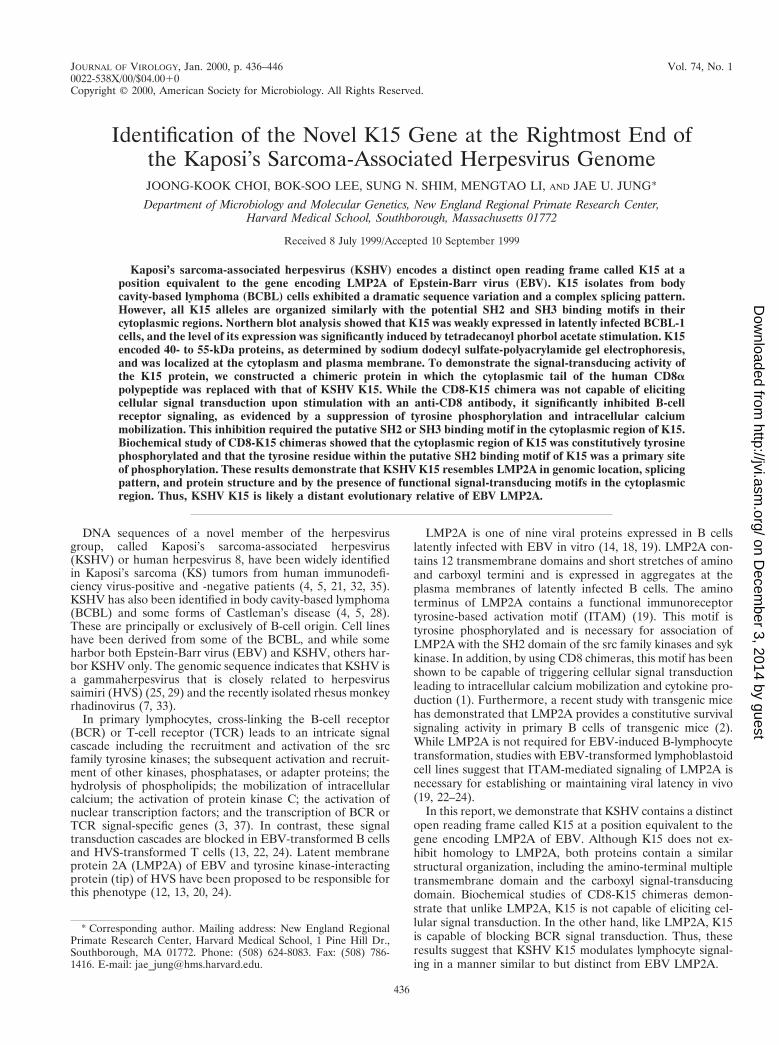
FIG. 1. Overall splicing pattern of KSHV K15 gene. Sequence analysis with 20 independent cDNA clones from BCBL-1 cells identified four differently spliced formsof K15. The boxes indicate each coding exon, and the numbers above the boxes, which are based on the sequence data from Nicholas et al. (26), indicate the potentialsplicing junction sites.
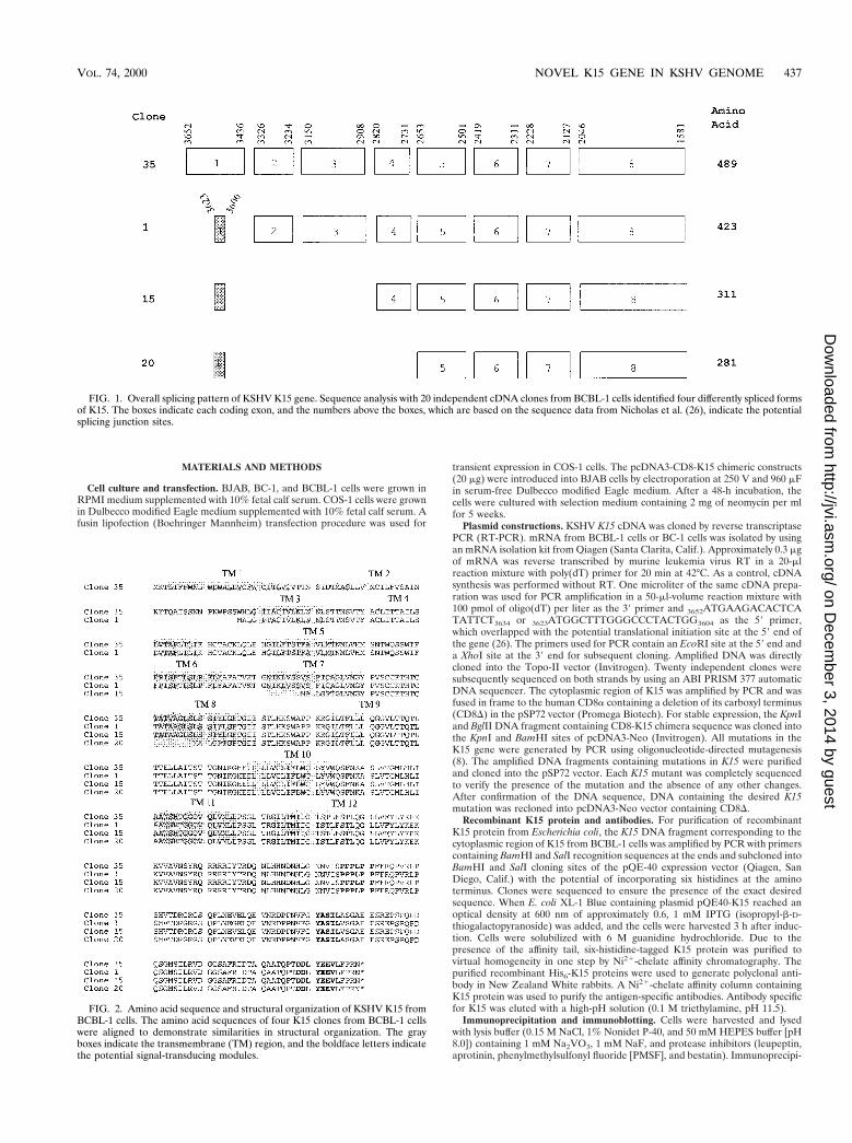
FIG. 2. Amino acid sequence and structural organization of KSHV K15 fromBCBL-1 cells. The amino acid sequences of four K15 clones from BCBL-1 cellswere aligned to demonstrate similarities in structural organization. The grayboxes indicate the transmembrane (TM) region, and the boldface letters indicatethe potential signal-transducing modules.
VOL. 74, 2000 NOVEL K15 GENE IN KSHV GENOME 437
on Decem
ber 3, 2014 by guesthttp://jvi.asm
.org/D
ownloaded from
tation was performed with 1:500-diluted antibody together with 30 ml of proteinA/G-agarose beads. For protein immunoblots, polypeptides in cell lysates cor-responding to 105 cells were resolved by sodium dodecyl sulfate-polyacrylamidegel electrophoresis (SDS-PAGE) and transferred to a nitrocellulose membrane
filter. Protein detection was performed with a 1:1,000 or 1:3,000 dilution ofprimary antibody by using an enhanced chemiluminescence system (Amersham,Chicago, Ill.).
Northern blot analysis. Northern blot analysis was performed under standardconditions with randomly labeled probes representing either full-length K15 orthe 59 half of K15. Total RNA was purified from BJAB, unstimulated BCBL-1,and tetradecanoyl phorbol myristate (TPA)-stimulated BCBL-1 cells accordingto the manufacturer’s instructions (Qiagen), and 10 mg of total RNA was loadedin each lane. The filters were baked at 80°C for 2 h and then hybridized withradioactive probes.
Immunofluorescence. Cells were fixed with 4% paraformaldehyde for 15 min,permeablized with 70% ethanol for 15 min, blocked with 10% goat serum inphosphate-buffered saline (PBS) for 30 min, and reacted with 1:100-dilutedprimary antibody in PBS for 30 min at room temperature. After incubation, cellswere washed extensively with PBS, incubated with 1:100-diluted secondary an-tibody (Vector) in PBS for 30 min at room temperature, and washed three timeswith PBS. Protein staining was performed with 1:500-diluted Sypro (MolecularProbes) for 1 min. Immunofluorescence was detected with a Leica immunoflu-orescence confocal microscope.
Flow cytometry analysis. A total of 5 3 105 cells were washed with RPMImedium containing 10% fetal calf serum and incubated with fluorescein isothio-cyanate (FITC)-conjugated or phycoerythrin-conjugated monoclonal antibodiesfor 30 min at 4°C. After being washed, each sample was fixed with 1% formalinsolution, and flow cytometry analysis was performed with a FACS Scan (BectonDickinson Co., Mountainview, Calif.). For cell sorting, 2 3 107 cells were stainedwith FITC-conjugated CD8 (UCHT2; PharMingen) antibody for 30 min at 4°C.Stained cells were sorted based on CD8 surface expression, using a FACSVantage (Becton Dickinson). After being sorted, cells were washed twice withPBS and cultured with RPMI–10% fetal calf serum medium. UCHT2 CD8 andG20-127 antibodies used for fluorescence-activated cell sorter (FACS) analysiswere obtained from PharMingen, and OKT8 antibody used for stimulation wasobtained from the American Type Culture Collection.
Antibody stimulation. Next, 107 cells were incubated with 10 mg of anti-CD8(OKT8) or anti-immunoglobulin M (IgM) antibody at 37°C for various times.After stimulation, cells were immediately frozen and lysed with cold lysis buffercontaining 1 mM Na2VO3, 1 mM NaF, and protease inhibitors (leupeptin,aprotinin, PMSF, and bestatin). Precleared cell lysates were used for immuno-blotting or for immunoprecipitation.
Calcium mobilization analysis. A total of 2 3 106 cells were loaded with 1 mMIndo-1 in 2 ml of RPMI complete medium for 20 min at 37°C. A detailedprotocol for this process has been described previously (24). Baseline calcium
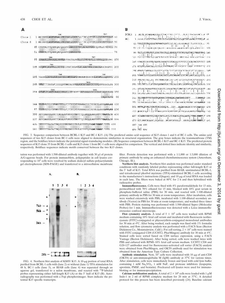
FIG. 3. Sequence comparison between BCBL-1 K15 and BC-1 K15. (A) The predicted amino acid sequence of K15 clones 1 and 6 of BC-1 cells. The amino acidsequences of two K15 clones from BC-1 cells were aligned to demonstrate similarities in structural organization. The gray boxes indicate the transmembrane (TM)region, and the boldface letters indicate the potential signal-transducing modules. (B) Sequence comparison between BCBL-1 K15 and BC-1 K15. The predicted proteinsequences of K15 clone 35 from BCBL-1 cells and K15 clone 1 from BC-1 cells were aligned for comparison. The vertical and dotted lines indicate identity and similarity,respectively. Boldface sequences indicate motifs conserved between the two K15 clones.
FIG. 4. Northern blot analysis of KSHV K15. A 10-mg portion of total RNApurified from BCBL-1 cells with (lane 2) or without (lane 1) TPA stimulation for48 h, BC-1 cells (lane 3), or BJAB cells (lane 4) was separated through anagarose gel, transferred to a nylon membrane, and reacted with 32P-labeledprobes representing either full-length K15 (A) or the 59 half of K15 (B). Auto-radiography was performed with a Fuji phosphorimager. Stars indicate the po-tential K15 specific transcripts.
438 CHOI ET AL. J. VIROL.
on Decem
ber 3, 2014 by guesthttp://jvi.asm
.org/D
ownloaded from
levels were established for 1 min prior to the addition of the antibody. Cells werestimulated with 10 mg of mouse anti-CD8 (OKT8) antibody followed by 10 mg ofgoat anti-mouse antibody, or 10 mg of mouse anti-IgM antibody alone, and datawere then collected for 4 min. Baseline absolute intracellular calcium levels weredetermined by using an ionophore and EGTA. Data were collected and analyzedon a FACS Vantage (Becton Dickinson).
RESULTS
Cloning and sequence analysis of K15 from BCBL. At aposition and transcriptional direction equivalent to theLMP2A gene of EBV, KSHV contains a distinct reading framecalled K15. Initial DNA sequence analysis of the KSHV ge-nome did not reveal this open reading frame because of itsmultiple splicing (29). KSHV K15 cDNA was cloned frommRNA of unstimulated BCBL-1 cells by RT-PCR as describedin Materials and Methods, using an oligo(dT) primer at the 39end and a gene-specific primer which overlapped with thepotential translational initiation site at the 59 end of the gene(26, 29). Sequence analysis of 20 independent cDNA clonesidentified at least four differently spliced forms of K15 inBCBL-1 cells (Fig. 1). Clones 35 and 1 contained eight exons,clone 15 contained six exons, and clone 20 contained five exons(Fig. 1). While all clones shared exons 5 to 8, clone 35 con-tained a first exon which was entirely different from that ofclones 1, 15, and 20. The first exon of clone 35 encoded 72amino acids, and the first exon of clones 1, 15, and 20 encodedonly 6 amino acids. These spliced forms were predicted to encodeproducts of from 281 to 489 amino acids (Fig. 1).
A comparison of the primary amino acid sequences of thefour K15 isolates revealed extensive size variation in the ami-no-terminal hydrophobic region because of complex splicing(Fig. 2). All cDNA clone products are predicted to be largeintegral membrane proteins consisting of 4 to 12 hydrophobicmembrane-spanning domains (Fig. 2). Regardless of the num-ber of transmembrane domains, all cDNA clone products havethe same carboxyl-terminal region, which is predicted to con-tain 142 amino acids. The presence of potential motifs in thecytoplasmic region was assessed based on the primary aminoacid sequence. All spliced forms of K15 contained a conserved
YEEV sequence at amino acids 480 to 483 (based on theprimary amino acid sequence of clone 35), which is precededby two negatively charged glutamic acid residues (Fig. 2). Thismotif matches very well with the consensus sequence for SH2binding (EExxYEEV/I) to src family kinases (34). This is des-ignated as an SH2-binding (SH2-B) motif. In addition, a pro-line-rich region from amino acid residues 385 to 390 of theclone 35 product shows homology with the consensus se-quences for binding to the SH3 domains of signal-transducingproteins (6, 27, 38). This is designated as an SH3-binding (SH3-B)motif. In addition, while the YASIL sequence at amino acids 431to 434 is consistent with an SH2-binding motif, it is not precededby negatively charged amino acids, suggesting that it may beinvolved in activities other than SH2 binding. Thus, besides aconsiderable resemblance to LMP2A of EBV in genomic lo-cation, overall protein structure, and complex splicing pattern,K15 also contains potential SH2-B and SH3-B motifs in thecytoplasmic region as has been shown in LMP2A (19).
Genetic variation of K15 alleles. Primary amino acid se-quences of K15 from BC-1 cells were also determined by RT-PCR and DNA sequence analysis (Fig. 3). Sequence analysis of20 independent cDNA clones detected two major splicedforms of K15 in BC-1 cells (Fig. 3A). K15 clones 1 and 6 fromBC-1 cells are predicted to encode 498 and 235 amino acids,respectively (Fig. 3A). To our surprise, a remarkable sequencevariation was detected between K15 genes derived fromBCBL-1 and BC-1 cells (Fig. 3B). K15 genes from BC-1 andBCBL-1 cell lines exhibited only 30% identity and 40% ho-mology at the amino acid level. Despite this dramatic sequencevariation, both K15 proteins are predicted to have a similarstructure: an amino-terminal multiple transmembrane regionand a carboxyl-terminal cytoplasmic region. In addition, thethree potential motifs, SH2-B, SH3-B, and YASIL, were com-pletely conserved in the cytoplasmic region of both K15 pro-teins (Fig. 3B). This suggests that the conserved SH2-B,SH3-B, and YASIL motifs of the cytoplasmic region are likelyimportant for K15 function.
Latent and lytic expression of K15. To determine K15 ex-pression, Northern blot analysis was performed with totalRNA from BCBL-1 cells with or without TPA stimulation for48 h. When sequences representing the full-length K15 genewere used as a probe, multiple transcripts of 3, 4, 5.5, 7, and 10kb were detected in BCBL-1 cells. These transcripts wereweakly detected in unstimulated BCBL-1 cells, and their levelof expression was significantly increased after TPA stimulation(Fig. 4A, lanes 1 and 2). No specific transcripts were detectedin control BC-1 and BJAB cells (Fig. 4A, lanes 3 and 4). Tofurther determine specificity, sequences representing the 59half of the K15 gene were used as a probe in Northern blotanalysis. This probe specifically detected only the 7- and 10-kbtranscripts among the five transcripts which were identified bythe full-length K15 probe (Fig. 4B, lanes 1 and 2). These resultssuggest that the 7- and 10-kb transcripts likely encode the K15gene and the 3-, 4-, and 5.5-kb transcripts may encode otherKSHV genes or unidentified alternatively spliced forms of K15.These results indicate that K15 is weakly expressed duringlatency but that its expression is significantly induced duringlytic viral replication.
Identification of K15 protein. To analyze the K15 gene prod-uct of BCBL-1 cells, we generated a rabbit polyclonal antibodyagainst a purified bacterial six-histidine-tagged K15 fusion pro-tein which contained the putative cytoplasmic portion ofBCBL-1 K15 as described in Materials and Methods. Expres-sion of K15 clones 35 and 20 derived from BCBL-1 cells wasthen demonstrated. At 48 h posttransfection, heat-treatedCOS-1 cell lysates were used for an immunoblot assay with
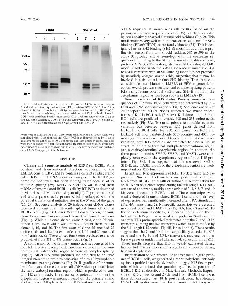
FIG. 5. Identification of the KSHV K15 protein. COS-1 cells were trans-fected with transient expression vector pFJ containing BCBL-1 K15 clone 35 orclone 20. Boiled or nonboiled cell lysates were fractionated by SDS-PAGE,transferred to nitrocellulose, and reacted with an anti-K15 antibody. Lane 1,COS-1 cells transfected with vector; lane 2, COS-1 cells transfected with 10 mg ofpFJ-K15 clone 20; lane 3, COS-1 cells transfected with 5 mg of pFJ-K15 clone 20;lane 4, COS-1 cells transfected with 5 mg of pFJ-K15 clone 35.
VOL. 74, 2000 NOVEL K15 GENE IN KSHV GENOME 439
on Decem
ber 3, 2014 by guesthttp://jvi.asm
.org/D
ownloaded from
antibody specific for BCBL-1 K15. This antibody reacted spe-cifically with the 50- and 55-kDa protein species of BCBL-1K15 clone 35 and with 40- and 45-kDa protein species ofBCBL-1 K15 clone 20 (Fig. 5). In contrast, these proteins werenot detected in control COS-1 cells not expressing the K15gene (Fig. 5).
LMP2A, which contains 12 transmembrane domains, ispresent as aggregates in plasma membranes (19). DNA se-quence analysis predicts that K15 clones 20 and 35 have 4 and12 transmembrane domains, respectively. To determine thepotential aggregation of K15, cell lysates containing K15 pro-tein were subjected to SDS-PAGE without heat treatment and
reacted with an anti-K15 antibody in an immunoblot analysis.This analysis showed that the migration of 50- and 55-kDaprotein species of K15 clone 35 was greatly retarded to ca. 200kDa in SDS-PAGE (Fig. 5). In striking contrast, the migrationof 40- and 45-kDa species of the K15 clone 20 was not alteredin SDS-PAGE under the same conditions (Fig. 5). These datasuggest that K15 clone 35, which contains 12 transmembranedomains, is primarily present as aggregates, whereas K15 clone20, which contains 3 transmembrane domains, is far less abun-dant as aggregates.
Localization of KSHV K15. To determine the subcellularlocalization of K15 by indirect immunofluorescence tests,
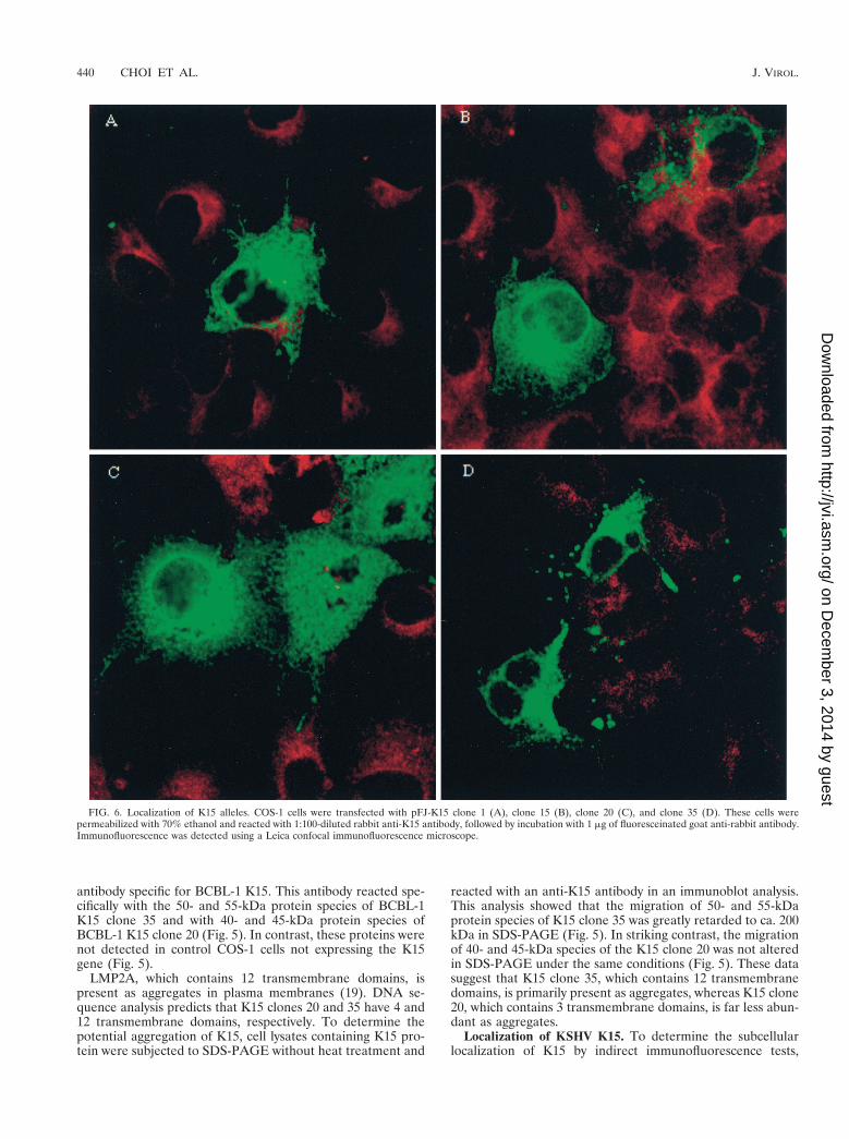
FIG. 6. Localization of K15 alleles. COS-1 cells were transfected with pFJ-K15 clone 1 (A), clone 15 (B), clone 20 (C), and clone 35 (D). These cells werepermeabilized with 70% ethanol and reacted with 1:100-diluted rabbit anti-K15 antibody, followed by incubation with 1 mg of fluoresceinated goat anti-rabbit antibody.Immunofluorescence was detected using a Leica confocal immunofluorescence microscope.
440 CHOI ET AL. J. VIROL.
on Decem
ber 3, 2014 by guesthttp://jvi.asm
.org/D
ownloaded from
COS-1 cells transfected with K15 clones 1, 15, 20, and 35 fromBCBL-1 cells were reacted with an anti-K15 antibody. Thestaining pattern in COS-1 cells suggested that all K15 cloneslocalized primarily in the cytoplasm and at the plasma mem-brane (Fig. 6). In addition, a strong fluorescence was detectedin the perinuclear region. This staining pattern is similar to that
obtained when the Golgi is visualized. Thus, immunofluores-cence tests demonstrated that, independent of the number oftransmembrane domains, K15 alleles were located principallyin the cytoplasm, Golgi, and plasma membrane.
Construction of a CD8a chimera with the cytoplasmic re-gion of K15. To determine if the cytoplasmic region of K15 iscapable of inducing signals or altering cellular signal transduc-tion, we analyzed the signaling capacity of the cytoplasmic tailindependent of the transmembrane domains of K15. Antibodycross-linking of chimeric molecules composed of the extracel-lular and the transmembrane domains of the CD8a moleculeand the cytoplasmic region of KSHV K1 or EBV LMP2A hasbeen shown to be sufficient to elicit early and late signal-transducing events (1, 16). We constructed a chimeric proteinin which 27 amino acids of the cytoplasmic tail of human CD8aprotein were replaced with 142 amino acids of the cytoplasmictail of BCBL-1 K15 (CD8-K15). CD8D, from which the cyto-plasmic region has been deleted, was used as a control. Sincemutations in the signal-transducing modules of cellular pro-teins abrogate their capacity to induce signaling (3, 9), we also
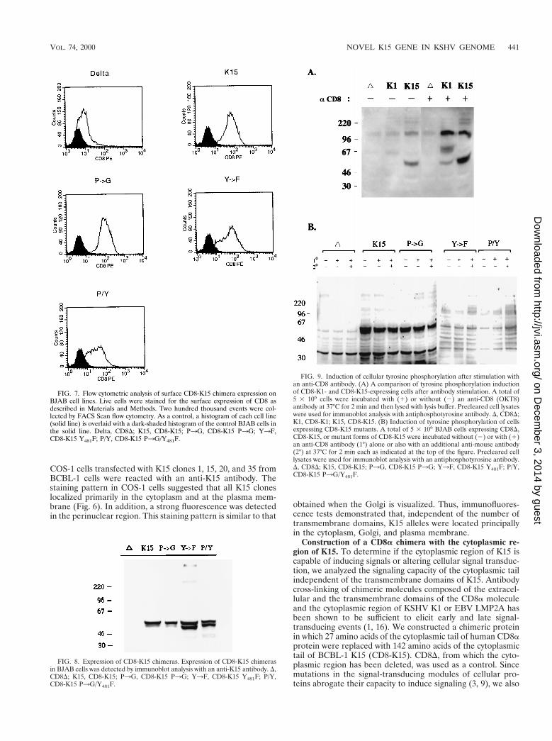
FIG. 7. Flow cytometric analysis of surface CD8-K15 chimera expression onBJAB cell lines. Live cells were stained for the surface expression of CD8 asdescribed in Materials and Methods. Two hundred thousand events were col-lected by FACS Scan flow cytometry. As a control, a histogram of each cell line(solid line) is overlaid with a dark-shaded histogram of the control BJAB cells inthe solid line. Delta, CD8D; K15, CD8-K15; P3G, CD8-K15 P3G; Y3F,CD8-K15 Y481F; P/Y, CD8-K15 P3G/Y481F.
FIG. 8. Expression of CD8-K15 chimeras. Expression of CD8-K15 chimerasin BJAB cells was detected by immunoblot analysis with an anti-K15 antibody. D,CD8D; K15, CD8-K15; P3G, CD8-K15 P3G; Y3F, CD8-K15 Y481F; P/Y,CD8-K15 P3G/Y481F.
FIG. 9. Induction of cellular tyrosine phosphorylation after stimulation withan anti-CD8 antibody. (A) A comparison of tyrosine phosphorylation inductionof CD8-K1- and CD8-K15-expressing cells after antibody stimulation. A total of5 3 106 cells were incubated with (1) or without (2) an anti-CD8 (OKT8)antibody at 37°C for 2 min and then lysed with lysis buffer. Precleared cell lysateswere used for immunoblot analysis with antiphosphotyrosine antibody. D, CD8D;K1, CD8-K1; K15, CD8-K15. (B) Induction of tyrosine phosphorylation of cellsexpressing CD8-K15 mutants. A total of 5 3 106 BJAB cells expressing CD8D,CD8-K15, or mutant forms of CD8-K15 were incubated without (2) or with (1)an anti-CD8 antibody (1°) alone or also with an additional anti-mouse antibody(2°) at 37°C for 2 min each as indicated at the top of the figure. Precleared celllysates were used for immunoblot analysis with an antiphosphotyrosine antibody.D, CD8D; K15, CD8-K15; P3G, CD8-K15 P3G; Y3F, CD8-K15 Y481F; P/Y,CD8-K15 P3G/Y481F.
VOL. 74, 2000 NOVEL K15 GENE IN KSHV GENOME 441
on Decem
ber 3, 2014 by guesthttp://jvi.asm
.org/D
ownloaded from
introduced mutations at the SH2-B and SH3-B motifs as fol-lows: CD8-K15 P3G contained mutations at positions 386,387, 388, 390, and 341 of prolines to glycines; CD8-K15 Y481Fcontained a mutation at position 481 of tyrosine to phenylal-anine; and CD8-K15 P3G/Y481F contained both mutations inthe SH2-B and SH3-B motifs.
Construction of BJAB cell lines expressing CD8-K15 chime-ras. To assess the signal-transducing activity of CD8-K15 chi-meras, BJAB cells (KSHV and EBV negative) were used to
establish stable lines expressing the CD8-K15 chimeric genes.The CD8D, CD8-K15, CD8-K15 P3G, CD8-Y481F, and CD8-K15 P3G/Y481F chimeric genes were cloned into the expres-sion vector pcDNA3-Neo. After electroporation of the expres-sion vector into BJAB cells, cell lines were selected by growthin medium containing 2 mg of neomycin per ml for 5 weeks.Since CD8 is not expressed on the surface of BJAB cells,neomycin-resistant cells were sorted by FACS analysis basedon the surface expression of the CD8. Comparable levels of
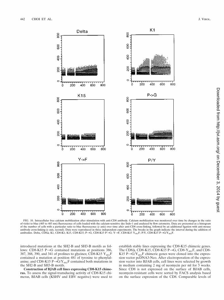
FIG. 10. Intracellular free calcium mobilization after stimulation with anti-CD8 antibody. Calcium mobilization was monitored over time by changes in the ratioof violet to blue (405 to 485 nm) fluorescence of cells loaded with the calcium-sensitive dye Indo-1 and analyzed by flow cytometry. Data are presented as a histogramof the number of cells with a particular ratio to blue fluorescence (y axis) over time after anti-CD8 cross-linking, followed by an additional ligation with anti-mouseantibody cross-linking (x axis, second). Data were reproduced in three independent experiments. The breaks in the graph indicate the interval during the addition ofantibodies. Delta, CD8D; K1, CD8-K1; K15, CD8-K15; P3G, CD8-K15 P3G; Y3F, CD8-K15 Y481F; P/Y, CD8-K15 P3G/Y481F.
442 CHOI ET AL. J. VIROL.
on Decem
ber 3, 2014 by guesthttp://jvi.asm
.org/D
ownloaded from
CD8 surface expression of FACS-sorted cells were detected inmost of the cells expressing the CD8-K15 chimeras, with theexception of CD8D cells (Fig. 7). The reduced level of CD8surface expression in CD8D cells was likely caused by theabsence of its cytoplasmic region. To demonstrate the expres-sion of these chimeras, BJAB cells expressing the CD8-K15chimeras were used for immunoblot analysis with an anti-K15antibody. This assay revealed that CD8-K15 chimeras wereexpressed at somewhat variable but still comparable levels inBJAB cells (Fig. 8).
Lack of cellular signal-transducing ability of CD8-K15 chi-meras upon antibody stimulation. The early biochemical eventsubsequent to TCR or BCR stimulation is the induction oftyrosine phosphorylation of a number of cellular proteins (3,36). We examined the effects of CD8-K15 chimera expressionon cellular tyrosine phosphorylation upon stimulation with ananti-CD8 antibody. BJAB cells expressing CD8D or CD8-K15chimera were stimulated with an anti-CD8 antibody and thelevel of tyrosine phosphorylation induction was observed byimmunoblot assay with an antiphosphotyrosine antibody (Fig.9). CD8-K1, which has been shown to induce cellular tyrosinephosphorylation upon stimulation with an anti-CD8 antibody(16), was included as a positive control. The stimulation ofBJAB cells expressing the CD8-K15 chimera with a mouseanti-CD8 antibody did not induce cellular tyrosine phosphor-ylation (Fig. 9A). In contrast, tyrosine phosphorylation of a60-kDa protein was strongly detected in BJAB cells expressingCD8-K15 independent of antibody stimulation (Fig. 9A). Tofurther potentiate stimulating activity, an additional ligationwith a goat anti-mouse antibody was included in the stimula-tion conditions. This experiment also revealed that expressionof the CD8-K15 chimera did not induce tyrosine phosphory-lation of cellular proteins upon stimulation except for a 60-kDaprotein (Fig. 9B). Under the same conditions, an enhancedlevel of tyrosine phosphorylation induction was detected inCD8-K1-expressing cells (Fig. 9A). Mutant forms of the CD8-K15 chimera were also examined for their ability to inducecellular tyrosine phosphorylation under the same conditions.Like CD8-K15, none of the CD8-K15 mutants induced cellulartyrosine phosphorylation upon stimulation (Fig. 9B). Theseresults showed that the cytoplasmic region of K15 was notcapable of inducing tyrosine phosphorylation upon antibodystimulation. However, tyrosine phosphorylation of a 60-kDa
protein was strongly detected in BJAB cells expressing CD8-K15 or CD8-K15 P3G independent of antibody stimulation.
To further determine the potential ability of the cytoplasmicregion of K15 to induce late signaling events, the cytoplasmicfree calcium concentration was examined for increases afterantibody stimulation. BJAB cells expressing CD8-K15 chime-ras were treated with mouse anti-CD8 antibody, followed by agoat anti-mouse antibody, and intracellular free calcium levelswere monitored by flow cytometry in three independent exper-iments. CD8-K1, which has been shown to induce intracellularcalcium mobilization upon stimulation with an anti-CD8 anti-body (16), was used as a positive control. Control CD8-K1 cellsexhibited a rapid increase in intracellular calcium concentra-tion immediately after anti-CD8 treatment, whereas none ofCD8-K15 chimeras induced an increase of intracellular freecalcium concentration upon stimulation with anti-CD8 aloneor together with a goat anti-mouse antibody (Fig. 10). Thus,unlike those of KSHV K1 (16) and EBV LMP2A (1), thecytoplasmic region of K15 is not capable of transducing a signalto induce tyrosine phosphorylation or intracellular calciummobilization.
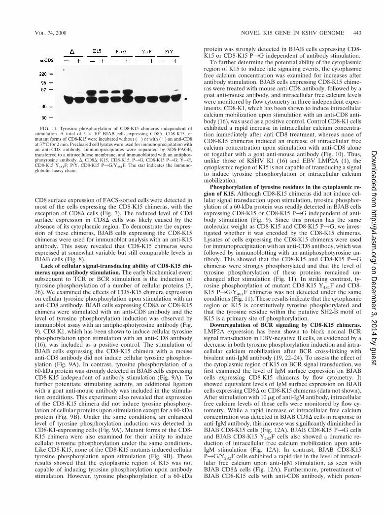
Phosphorylation of tyrosine residues in the cytoplasmic re-gion of K15. Although CD8-K15 chimeras did not induce cel-lular signal transduction upon stimulation, tyrosine phosphor-ylation of a 60-kDa protein was readily detected in BJAB cellsexpressing CD8-K15 or CD8-K15 P3G independent of anti-body stimulation (Fig. 9). Since this protein has the samemolecular weight as CD8-K15 and CD8-K15 P3G, we inves-tigated whether it was encoded by the CD8-K15 chimeras.Lysates of cells expressing the CD8-K15 chimeras were usedfor immunoprecipitation with an anti-CD8 antibody, which wasfollowed by immunoblotting with an antiphosphotyrosine an-tibody. This showed that the CD8-K15 and CD8-K15 P3Gchimeras were strongly phosphorylated and that the level oftyrosine phosphorylation of these proteins remained un-changed after stimulation (Fig. 11). In striking contrast, ty-rosine phosphorylation of mutant CD8-K15 Y481F and CD8-K15 P3G/Y481F chimeras was not detected under the sameconditions (Fig. 11). These results indicate that the cytoplasmicregion of K15 is constitutively tyrosine phosphorylated andthat the tyrosine residue within the putative SH2-B motif ofK15 is a primary site of phosphorylation.
Downregulation of BCR signaling by CD8-K15 chimeras.LMP2A expression has been shown to block normal BCRsignal transduction in EBV-negative B cells, as evidenced by adecrease in both tyrosine phosphorylation induction and intra-cellular calcium mobilization after BCR cross-linking withbivalent anti-IgM antibody (19, 22–24). To assess the effect ofthe cytoplasmic region of K15 on BCR signal transduction, wefirst examined the level of IgM surface expression on BJABcells expressing CD8-K15 chimeras by flow cytometry. Itshowed equivalent levels of IgM surface expression on BJABcells expressing CD8D or CD8-K15 chimeras (data not shown).After stimulation with 10 mg of anti-IgM antibody, intracellularfree calcium levels of these cells were monitored by flow cy-tometry. While a rapid increase of intracellular free calciumconcentration was detected in BJAB CD8D cells in response toanti-IgM antibody, this increase was significantly diminished inBJAB CD8-K15 cells (Fig. 12A). BJAB CD8-K15 P3G cellsand BJAB CD8-K15 Y282F cells also showed a dramatic re-duction of intracellular free calcium mobilization upon anti-IgM stimulation (Fig. 12A). In contrast, BJAB CD8-K15P3G/Y282F cells exhibited a rapid rise in the level of intracel-lular free calcium upon anti-IgM stimulation, as seen withBJAB CD8D cells (Fig. 12A). Furthermore, pretreatment ofBJAB CD8-K15 cells with anti-CD8 antibody, which poten-
FIG. 11. Tyrosine phosphorylation of CD8-K15 chimeras independent ofstimulation. A total of 5 3 106 BJAB cells expressing CD8D, CD8-K15, ormutant forms of CD8-K15 were incubated without (2) or with (1) an anti-CD8at 37°C for 2 min. Precleared cell lysates were used for immunoprecipitation withan anti-CD8 antibody. Immunoprecipitates were separated by SDS-PAGE,transferred to a nitrocellulose membrane, and immunoblotted with an antiphos-photyrosine antibody. D, CD8D; K15, CD8-K15; P3G, CD8-K15 P3G; Y3F,CD8-K15 Y481F; P/Y, CD8-K15 P3G/Y481F. The star indicates the immuno-globulin heavy chain.
VOL. 74, 2000 NOVEL K15 GENE IN KSHV GENOME 443
on Decem
ber 3, 2014 by guesthttp://jvi.asm
.org/D
ownloaded from
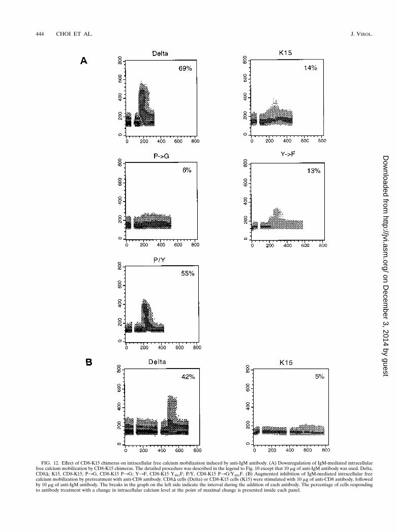
FIG. 12. Effect of CD8-K15 chimeras on intracellular free calcium mobilization induced by anti-IgM antibody. (A) Downregulation of IgM-mediated intracellularfree calcium mobilization by CD8-K15 chimeras. The detailed procedure was described in the legend to Fig. 10 except that 10 mg of anti-IgM antibody was used. Delta,CD8D; K15, CD8-K15; P3G, CD8-K15 P3G; Y3F, CD8-K15 Y481F; P/Y, CD8-K15 P3G/Y481F. (B) Augmented inhibition of IgM-mediated intracellular freecalcium mobilization by pretreatment with anti-CD8 antibody. CD8D cells (Delta) or CD8-K15 cells (K15) were stimulated with 10 mg of anti-CD8 antibody, followedby 10 mg of anti-IgM antibody. The breaks in the graph on the left side indicate the interval during the addition of each antibody. The percentage of cells respondingto antibody treatment with a change in intracellular calcium level at the point of maximal change is presented inside each panel.
444 CHOI ET AL. J. VIROL.
on Decem
ber 3, 2014 by guesthttp://jvi.asm
.org/D
ownloaded from
tially oligomerized the CD8-K15 chimera on the cell surface,blocked anti-IgM-mediated intracellular free calcium mobili-zation to a greater degree (Fig. 12B).
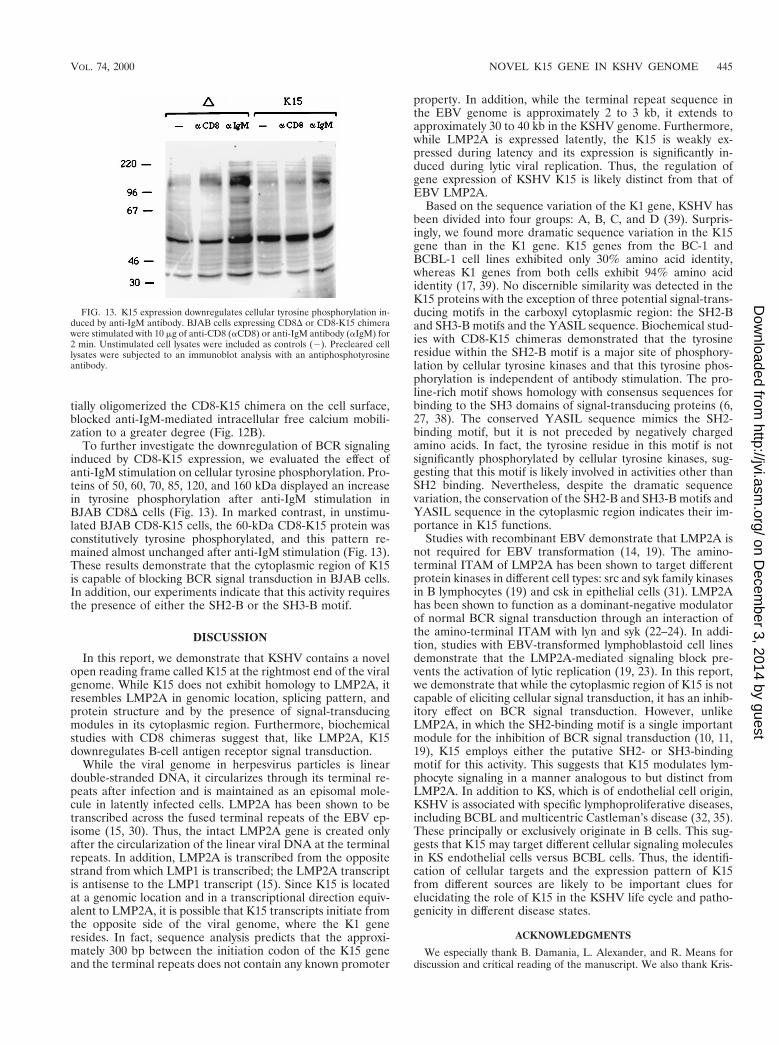
To further investigate the downregulation of BCR signalinginduced by CD8-K15 expression, we evaluated the effect ofanti-IgM stimulation on cellular tyrosine phosphorylation. Pro-teins of 50, 60, 70, 85, 120, and 160 kDa displayed an increasein tyrosine phosphorylation after anti-IgM stimulation inBJAB CD8D cells (Fig. 13). In marked contrast, in unstimu-lated BJAB CD8-K15 cells, the 60-kDa CD8-K15 protein wasconstitutively tyrosine phosphorylated, and this pattern re-mained almost unchanged after anti-IgM stimulation (Fig. 13).These results demonstrate that the cytoplasmic region of K15is capable of blocking BCR signal transduction in BJAB cells.In addition, our experiments indicate that this activity requiresthe presence of either the SH2-B or the SH3-B motif.
DISCUSSION
In this report, we demonstrate that KSHV contains a novelopen reading frame called K15 at the rightmost end of the viralgenome. While K15 does not exhibit homology to LMP2A, itresembles LMP2A in genomic location, splicing pattern, andprotein structure and by the presence of signal-transducingmodules in its cytoplasmic region. Furthermore, biochemicalstudies with CD8 chimeras suggest that, like LMP2A, K15downregulates B-cell antigen receptor signal transduction.
While the viral genome in herpesvirus particles is lineardouble-stranded DNA, it circularizes through its terminal re-peats after infection and is maintained as an episomal mole-cule in latently infected cells. LMP2A has been shown to betranscribed across the fused terminal repeats of the EBV ep-isome (15, 30). Thus, the intact LMP2A gene is created onlyafter the circularization of the linear viral DNA at the terminalrepeats. In addition, LMP2A is transcribed from the oppositestrand from which LMP1 is transcribed; the LMP2A transcriptis antisense to the LMP1 transcript (15). Since K15 is locatedat a genomic location and in a transcriptional direction equiv-alent to LMP2A, it is possible that K15 transcripts initiate fromthe opposite side of the viral genome, where the K1 generesides. In fact, sequence analysis predicts that the approxi-mately 300 bp between the initiation codon of the K15 geneand the terminal repeats does not contain any known promoter
property. In addition, while the terminal repeat sequence inthe EBV genome is approximately 2 to 3 kb, it extends toapproximately 30 to 40 kb in the KSHV genome. Furthermore,while LMP2A is expressed latently, the K15 is weakly ex-pressed during latency and its expression is significantly in-duced during lytic viral replication. Thus, the regulation ofgene expression of KSHV K15 is likely distinct from that ofEBV LMP2A.
Based on the sequence variation of the K1 gene, KSHV hasbeen divided into four groups: A, B, C, and D (39). Surpris-ingly, we found more dramatic sequence variation in the K15gene than in the K1 gene. K15 genes from the BC-1 andBCBL-1 cell lines exhibited only 30% amino acid identity,whereas K1 genes from both cells exhibit 94% amino acididentity (17, 39). No discernible similarity was detected in theK15 proteins with the exception of three potential signal-trans-ducing motifs in the carboxyl cytoplasmic region: the SH2-Band SH3-B motifs and the YASIL sequence. Biochemical stud-ies with CD8-K15 chimeras demonstrated that the tyrosineresidue within the SH2-B motif is a major site of phosphory-lation by cellular tyrosine kinases and that this tyrosine phos-phorylation is independent of antibody stimulation. The pro-line-rich motif shows homology with consensus sequences forbinding to the SH3 domains of signal-transducing proteins (6,27, 38). The conserved YASIL sequence mimics the SH2-binding motif, but it is not preceded by negatively chargedamino acids. In fact, the tyrosine residue in this motif is notsignificantly phosphorylated by cellular tyrosine kinases, sug-gesting that this motif is likely involved in activities other thanSH2 binding. Nevertheless, despite the dramatic sequencevariation, the conservation of the SH2-B and SH3-B motifs andYASIL sequence in the cytoplasmic region indicates their im-portance in K15 functions.
Studies with recombinant EBV demonstrate that LMP2A isnot required for EBV transformation (14, 19). The amino-terminal ITAM of LMP2A has been shown to target differentprotein kinases in different cell types: src and syk family kinasesin B lymphocytes (19) and csk in epithelial cells (31). LMP2Ahas been shown to function as a dominant-negative modulatorof normal BCR signal transduction through an interaction ofthe amino-terminal ITAM with lyn and syk (22–24). In addi-tion, studies with EBV-transformed lymphoblastoid cell linesdemonstrate that the LMP2A-mediated signaling block pre-vents the activation of lytic replication (19, 23). In this report,we demonstrate that while the cytoplasmic region of K15 is notcapable of eliciting cellular signal transduction, it has an inhib-itory effect on BCR signal transduction. However, unlikeLMP2A, in which the SH2-binding motif is a single importantmodule for the inhibition of BCR signal transduction (10, 11,19), K15 employs either the putative SH2- or SH3-bindingmotif for this activity. This suggests that K15 modulates lym-phocyte signaling in a manner analogous to but distinct fromLMP2A. In addition to KS, which is of endothelial cell origin,KSHV is associated with specific lymphoproliferative diseases,including BCBL and multicentric Castleman’s disease (32, 35).These principally or exclusively originate in B cells. This sug-gests that K15 may target different cellular signaling moleculesin KS endothelial cells versus BCBL cells. Thus, the identifi-cation of cellular targets and the expression pattern of K15from different sources are likely to be important clues forelucidating the role of K15 in the KSHV life cycle and patho-genicity in different disease states.
ACKNOWLEDGMENTS
We especially thank B. Damania, L. Alexander, and R. Means fordiscussion and critical reading of the manuscript. We also thank Kris-
FIG. 13. K15 expression downregulates cellular tyrosine phosphorylation in-duced by anti-IgM antibody. BJAB cells expressing CD8D or CD8-K15 chimerawere stimulated with 10 mg of anti-CD8 (aCD8) or anti-IgM antibody (aIgM) for2 min. Unstimulated cell lysates were included as controls (2). Precleared celllysates were subjected to an immunoblot analysis with an antiphosphotyrosineantibody.
VOL. 74, 2000 NOVEL K15 GENE IN KSHV GENOME 445
on Decem
ber 3, 2014 by guesthttp://jvi.asm
.org/D
ownloaded from
ten Toohey for photography support and Maryann DeMaria for flowcytometry analysis.
This work was supported by U.S. Public Health Service grantsCA31363, CA82057, and RR00168.
ADDENDUM
After the manuscript was submitted, Poole et al. (26a) andGlenn et al. (11a) published similar results.
REFERENCES
1. Beaufils, P., D. Choquet, R. Z. Mamoun, and B. Malissen. 1993. The(YXXL/I)2 signalling motif found in the cytoplasmic segments of the bovineleukaemia virus envelope protein and Epstein-Barr virus latent membraneprotein 2A can elicit early and late lymphocyte activation events. EMBO J.12:5105–5112.
2. Caldwell, R. G., J. B. Wilson, S. J. Anderson, and R. Longnecker. 1998.Epstein-Barr virus LMP2A drives B cell development and survival in theabsence of normal B cell receptor signals. Immunity 9:405–411.
3. Cambier, J. C. 1995. Antigen and Fc receptor signaling. The awesome powerof the immunoreceptor tyrosine-based activation motif (ITAM). J. Immunol.155:3281–3285.
4. Cesarman, E., Y. Chang, P. S. Moore, J. W. Said, and D. M. Knowles. 1995.Kaposi’s sarcoma-associated herpesvirus-like DNA sequences in AIDS-re-lated body-cavity-based lymphomas. N. Engl. J. Med. 332:1186–1191.
5. Chang, Y., E. Cesarman, M. S. Pessin, F. Lee, J. Culpepper, D. M. Knowles,and P. S. Moore. 1994. Identification of herpesvirus-like DNA sequences inAIDS-associated Kaposi’s sarcoma. Science 266:1865–1869.
6. Chang, Y., E. Cesarman, M. S. Pessin, F. Lee, J. Culpepper, D. M. Knowles,and P. S. Moore. 1994. Identification of herpesvirus-like DNA sequences inAIDS-associated Kaposi’s sarcoma. Science 266:1865–1869.
7. Desrosiers, R. C., V. G. Sasseville, S. C. Czajak, X. Zhang, K. G. Mansfield,A. Kaur, R. P. Johnson, A. A. Lackner, and J. U. Jung. 1997. A herpesvirusof rhesus monkeys related to human Kaposi’s sarcoma-associated herpesvi-rus. J. Virol. 71:9764–9769.
8. Du, Z., D. A. Regier, and R. C. Desrosiers. 1995. Improved recombinant PCRmutagenesis procedure that uses alkaline-denatured plasmid template. Bio-Techniques 18:376–378.
9. Flaswinkel, H., and M. Reth. 1994. Dual role of the tyrosine activation motifof the Ig-a protein during signal transduction via the B cell antigen receptor.EMBO J. 13:83–89.
10. Fruehling, S., S. K. Lee, R. Herrold, B. Frech, G. Laux, E. Kremmer, F. A.Grasser, and R. Longnecker. 1996. Identification of latent membrane pro-tein 2A (LMP2A) domains essential for the LMP2A dominant-negativeeffect on B-lymphocyte surface immunoglobulin signal transduction. J. Virol.70:6216–6226.
11. Fruehling, S., and R. Longnecker. 1997. The immunoreceptor tyrosine-basedactivation motif of Epstein-Barr virus LMP2A is essential for blocking BCR-mediated signal transduction. Virology 235:241–251.
11a.Glenn, M., L. Rainbow, F. Aurad, A. Davison, and T. F. Schulz. 1999.Identification of a spliced gene from Kaposi’s sarcoma-associated herpesvi-rus encoding a protein with similarities to latent membrane proteins 1 and2A of Epstein-Barr virus. J. Virol. 73:6953–6963.
12. Jung, J. U., S. M. Lang, U. Friedrich, T. Jun, T. M. Roberts, R. C. Desro-siers, and B. Biesinger. 1995. Identification of lck-binding elements in Tip ofherpesvirus saimiri. J. Biol. Chem. 270:20660–20667.
13. Jung, J. U., S. M. Lang, T. Jun, T. M. Roberts, A. Veillette, and R. C.Desrosiers. 1995. Downregulation of Lck-mediated signal transduction by tipof herpesvirus saimiri. J. Virol. 69:7814–7822.
14. Kieff, E. 1996. Epstein-Barr virus and its replication, p. 2343–2396. In B. N.Fields, D. M. Knipe, P. M. Howley, et al. (ed.), Fields virology, vol. 74.Lippincott-Raven, Philadelphia, Pa.
15. Laux, G., M. Perricaudet, and P. Farrell. 1988. A spliced Epstein-Barr virusgene expressed in immortalized lymphocytes is created by circularization ofthe linear viral genome. EMBO J. 7:769–774.
16. Lee, H., J. Guo, M. Li, J.-K. Choi, M. DeMaria, M. Rosenzweig, and J. U.Jung. 1998. Identification of an immunoreceptor tyrosine-based activationmotif of K1 transforming protein of Kaposi’s sarcoma-associated herpesvi-rus. Mol. Cell. Biol. 18:5219–5228.
17. Lee, H., R. Veazey, K. Williams, M. Li, J. Guo, F. Neipel, B. Fleckenstein,A. A. Lackner, R. C. Desrosiers, and J. U. Jung. 1998. Deregulation of cellgrowth by the Kaposi’s sarcoma-associated herpesvirus K1 gene. Nat. Med.4:435–440.
18. Longnecker, R., and E. Kieff. 1990. A second Epstein-Barr virus membraneprotein (LMP2) is expressed in latent infection and colocalizes with LMP1.J. Virol. 64:2319–2326.
19. Longnecker, R., and C. L. Miller. 1996. Regulation of Epstein-Barr viruslatency by latent membrane protein 2. Trends Microbiol. 4:38–42.
20. Longnecker, R., C. L. Miller, X.-Q. Miao, B. Tomkinson, and E. Kieff. 1993.The last seven transmembrane and carboxy-terminal cytoplasmic domains ofEpstein-Barr virus latent membrane protein 2 (LMP2) are dispensable forlymphocyte infection and growth transformation in vitro. J. Virol. 67:2006–2013.
21. Mesri, E. A., E. Cesarman, L. Arvanitakis, S. Rafii, M. A. S. Moore, D. N.Posnett, D. M. Knowles, and A. S. Asch. 1996. Human herpesvirus-8/Kapo-si’s sarcoma-associated herpesvirus is a new transmissible virus that infects Bcells. J. Exp. Med. 183:2385–2389.
22. Miller, C. L., A. L. Burkhard, J. H. Lee, B. Stealey, R. Longnecker, J. B.Bolen, and E. Kieff. 1995. Integral membrane protein 2 of Epstein-Barr virusregulates reactivation from latency through dominant negative effects onprotein-tyrosine kinases. Immunity 2:155–166.
23. Miller, C. L., J. H. Lee, E. Kieff, and R. Longnecker. 1994. An integralmembrane protein (LMP2) blocks reactivation of Epstein-Barr virus fromlatency following surface immunoglobulin crosslinking. Proc. Natl. Acad. Sci.USA 91:772–776.
24. Miller, C. L., R. Longnecker, and E. Kieff. 1993. Epstein-Barr virus latentmembrane protein 2A blocks calcium mobilization in B lymphocytes. J. Vi-rol. 67:3087–3094.
25. Neipel, F., J.-C. Albrecht, and B. Fleckenstein. 1997. Cell-homologous genesin the Kaposi’s sarcoma-associated rhadinovirus human herpesvirus 8: de-terminants of its pathogenicity? J. Virol. 71:4187–4192.
26. Nicholas, J., J.-C. Zong, D. J. Alcendor, D. M. Ciufo, L. J. Poole, R. T.Sarisky, C.-J. Chiou, X. Zhang, X. Wan, H.-G. Guo, M. S. Reitz, and G. S.Hayward. 1998. Novel organizational features, captured cellular genes, andstrain variability within the genome of KSHV/HHV8. J. Natl. Cancer Inst.Monogr. 23:79–88.
26a.Poole, L. J., J. C. Zong, D. Ciufo, D. Alcendor, J. S. Cannon, R. Ambinder,J. M. Orenstein, M. S. Reitz, and G. S. Hayward. 1999. Comparison ofgenetic variability at multiple loci across the genomes of the major subtypesof Kaposi’s sarcoma-associated herpesvirus reveals evidence for recombina-tion and for two distinct types of open reading frame K15 alleles at theright-hand end. J. Virol. 73:6646–6660.
27. Ren, R., B. J. Mayer, P. Cicchetti, and D. Baltimore. 1993. Identification ofa ten-amino acid proline-rich SH3 binding site. Science 259:1159–1161.
28. Renne, R., W. Zhong, B. Herndier, M. McGrath, N. Abbey, and D. Ganem.1996. Lytic growth of Kaposi’s sarcoma-associate herpesvirus (human her-pesvirus 8) in culture. Nat. Med. 2:342–346.
29. Russo, J. J., R. A. Bohenzxy, M.-C. Chien, J. Chen, M. Yan, D. Maddalena,J. P. Parry, D. Peruzzi, I. S. Edelman, Y. Chang, and P. S. Moore. 1996.Nucleotide sequence of the Kaposi’s sarcoma-associated herpesvirus(HHV8). Proc. Natl. Acad. Sci. USA 93:14862–14867.
30. Sample, J., D. Liebowitz, and E. Kieff. 1989. Two related Epstein-Barr virusmembrane proteins are encoded by separate genes. J. Virol. 63:933–937.
31. Scholle, F., R. Longnecker, and N. Raab-Traub. 1999. Epithelial cell adhe-sion to extracellular matrix proteins induces tyrosine phosphorylation of theEpstein-Barr virus latent membrane protein 2: a role for C-terminal Srckinase. J. Virol. 73:4767–4775.
32. Schulz, T. F., Y. Chang, and P. S. Moore. 1998. Kaposi’s sarcoma-associatedherpesvirus (human herpesvirus 8). American Society for Microbiology,Washington, D.C.
33. Searles, R. P., E. P. Bergquam, M. K. Axthelm, and S. W. Wong. 1999.Sequence and genomic analysis of a rhesus macaque rhadinovirus with sim-ilarity to Kaposi’s sarcoma-associated herpesvirus/human herpesvirus 8.J. Virol. 73:3040–3053.
34. Songyang, Z., S. E. Shoelson, M. Chaudhuri, G. Gish, T. Pawson, W. G.Haser, F. King, T. Roberts, S. Ratnofsky, R. J. Lechleider, B. G. Neel, R. B.Birge, J. E. Fajardo, M. M. Chou, H. Hanafusa, B. Schaffhausen, and L. C.Cantley. 1993. SH2 domains recognize specific phosphopeptide sequences.Cell 72:767–778.
35. Soulier, J., L. Grollet, E. Oksenhendler, P. Cacoub, D. Cazals-Hatem, P.Babinet, M. F. d’Agay, J. P. Clauvel, M. Raphael, L. Degos, and F. Sigaux.1997. Kaposi’s sarcoma-associated herpesvirus-like DNA sequences in mul-ticentric Castleman’s disease. Blood 86:1276–1280.
36. Wange, R. L., and L. E. Samelson. 1996. Complex complexes: signaling at theTCR. Immunity 5:197–205.
37. Weiss, A., and D. R. Littman. 1994. Signal transduction by lymphocyteantigen receptors. Cell 76:263–274.
38. Yu, H., J. K. Chen, S. Feng, D. C. Dalgarno, A. W. Brauer, and S. L.Schreiber. 1994. Structural basis for the binding of proline-rich peptides toSH3 domains. Cell 76:933–945.
39. Zong, J. C., D. M. Ciufo, D. J. Alcendor, X. Wan, J. Nicholas, P. J. Browning,P. L. Rady, S. K. Tyring, J. M. Orenstein, C. S. Rabkin, I. J. Su, K. F. Powell,M. Croxson, K. E. Foreman, B. J. Nickoloff, S. Alkan, and G. S. Hayward.1999. High-level variability in the ORF-K1 membrane protein gene at theleft end of the Kaposi’s sarcoma-associated herpesvirus genome defines fourmajor virus subtypes and multiple variants or clades in different humanpopulations. J. Virol. 73:4156–4170.
446 CHOI ET AL. J. VIROL.
on Decem
ber 3, 2014 by guesthttp://jvi.asm
.org/D
ownloaded from