Embed Size (px)
Citation preview
S
GcG
PEMa
Gb
ac
2
a
ARRA
KBMMN
eST
a
0
Veterinary Parasitology 204 (2014) 381–387
Contents lists available at ScienceDirect
Veterinary Parasitology
jo u r nal homep age: www.elsev ier .com/ locate /vetpar
hort communication
enetic characterisation of Neospora caninum strains fromlinical samples of zebuine foetuses obtained in abattoirs inoiás, Brazil
aula Rogério Fernandes Broma, Javier Regidor-Cerrilloc,sther Collantes-Fernándezc, Luis Miguel Ortega-Morac,arcelo Sales Guimarãesa,∗, Andréa Caetano da Silvab,∗
Veterinary Parasitology Center/Veterinary and Zootechnics School, Federal University of Goiás, Caixa postal 131, CEP: 74001-970,oiânia, Goiás, BrazilVeterinary Parasitology Laboratory/Department of Microbiology, Immunology, Parasitology and Pathology, Institute of Public Healthnd Tropical Pathology, Federal University of Goiás, Rua 235, s/n, Setor Universitário, CEP 74605-050, Goiânia, Goiás, BrazilSALUVET, Animal Health Department, Faculty of Veterinary Sciences, Complutense University of Madrid, Ciudad Universitaria s/n,8040 Madrid, Spain
r t i c l e i n f o
rticle history:eceived 15 September 2013eceived in revised form 3 May 2014ccepted 6 May 2014
eywords:os indicusicrosatellitesultilocus genotypingeosporosis
a b s t r a c t
The Neospora caninum microsatellite markers were applied to clinical samples. Genotypingtechnology involving fluorescently labelled DNA fragment analysis was used in combina-tion with DNA sequencing for markers with complex repetitive sequences. Nineteen DNAsamples from 15 brains and four hearts of naturally infected non-aborted zebuine foetusesfrom abattoirs in Goiás, Brazil. N. caninum had been detected in these foetuses by nested-PCR of the internal transcribed spacer-1 rRNA region, and the samples were analysed usingthese microsatellites. Seven complete or nearly complete allele profiles were obtained fromsix foetuses. Three distinct profiles of N. caninum were identified in a unique microregion(Meia Ponte) of Goiás. Two alleles for the same marker were detected in a unique foetusthat was probably infected with two different strains. A new allele for one of the microsatel-lites is described. The multilocus analysis performed here revealed a preliminary means ofdiscriminating between individual strains according to their geographical origins. These
are the first results that have been obtained regarding the molecular characterisation ofstrains of N. caninum from infected zebuine foetuses in South America and reveal for thefirst time that there are genotypic differences in the strains that are responsible for foetaltransmission in zebuine foetuses.© 2014 Elsevier B.V. All rights reserved.
∗ Corresponding authors at: Center for Veterinary Parasitology, Vet-rinary and Zootechnics School, Federal University of Goiás, Campusamambaia, Campus II, PO Box 131, 74001-970 Goiânia, GO, Brazil.el.: +55 62 3521 1574; fax: +55 62 3521 1839.
E-mail addresses: [email protected] (M.S. Guimarães),[email protected] (A.C.d. Silva).
http://dx.doi.org/10.1016/j.vetpar.2014.05.011304-4017/© 2014 Elsevier B.V. All rights reserved.
1. Introduction
Neospora caninum is a protozoan of the phylum Apicom-plexa. It is considered to be the major cause of abortionin cattle (Dubey et al., 2007). The global distribution and
broad host range of these parasites, in addition to theircapacity for sexual reproduction, suggest that significantbiological and genetic diversity exist in N. caninum. Indeed,biological diversity has been reported among some isolates
y Parasi
382 P.R.F. Brom et al. / Veterinarregarding their capacities to produce pathology in experi-mental murine (Collantes-Fernández et al., 2006; PereiraGarcía-Melo et al., 2010) and bovine infections (Rojo-Montejo et al., 2009a) and in in vitro studies (Pérez-Zaballoset al., 2005; Rojo-Montejo et al., 2009b).
Genetic diversity in N. caninum has also been shownwith a variety of molecular techniques and genetic markers(Gondim et al., 2004; Regidor-Cerrillo et al., 2006; Pedraza-Díaz et al., 2009). Microsatellite sequences, also known assimple sequence repeats (SSRs), have been shown to bethe most suitable polymorphic markers for the typing ofN. caninum isolates and have been applied to geneticallycharacterise sets of isolates that have been obtained fromcattle and canines worldwide. Microsatellite sequenceshave allowed for the detection of extensive intra-speciesdiversity within healthy and clinically infected animals(Regidor-Cerrillo et al., 2006, 2008; Al-Qassab et al., 2009;Basso et al., 2009; García-Melo et al., 2009; Pedraza-Díazet al., 2009; Rojo-Montejo et al., 2009b). Microsatelliteanalysis has improved in recent years, and 12 polymor-phic microsatellite loci for N. caninum that exhibit three tonine separate alleles have been described in isolates grownin vitro (Regidor-Cerrillo et al., 2006).
Nevertheless, detailed information about the geneticdiversity of isolates of N. caninum from different geograph-ical locations is scarce (Al-Qassab et al., 2009). The actualgenetic diversity of N. caninum might be determined bythe analyses of N. caninum isolates from infected but non-aborted foetuses as has been proposed for aborted foetusesby Pedraza-Díaz et al. (2009). If so, microsatellite typingcould be used to identify infection sources in molecularepidemiological studies and to determine the sources of theisolates circulating in a delimited region (Basso et al., 2010).If applied to N. caninum isolates, this technique may aid thegenetic characterisation and discrimination of different N.caninum isolates, which in turn, will be central to preven-tion, surveillance, and the application of suitable controlmeasures for bovine neosporosis. Here, we report on thegenetic analyses of N. caninum strains from clinical sam-ples of zebuine (Bos indicus) foetuses that were naturallyinfected by vertical transmission.
2. Materials and methods
2.1. Sample collection, DNA extraction, and nested-PCRITS-1 amplification
Tissue samples were obtained randomly from deadzebuine (B. indicus) foetuses at a local commercial slaugh-terhouse in Goiás. Data about the locations at which thefoetuses were collected were recorded. A total of 195 foe-tuses were obtained during the slaughtering and used tocollect 585 different fresh sections of brain, heart, and liver,which were stored at −80 ◦C until DNA extraction. DNAwas prepared from 20 mg of tissue using a commerciallyavailable kit (Real Pure, Durviz, Patema, Spain) accordingto the manufacturer’s instructions. The concentrations of
DNA were determined by spectrophotometric analyses atA260/280, and all DNA samples were adjusted to a finalconcentration of 60 ng/mL. The DNA samples were storedat −20 ◦C until PCR analysis. All foetal DNA samples weretology 204 (2014) 381–387
subjected to an N. caninum-specific nested-PCR of the inter-nal transcribed spacer 1 (ITS-1) sequence with the primerspairs NN1/NN2 and NP1/NP2 (Buxton et al., 1998). To avoidfalse positive reactions, DNA extraction, PCR sample prepa-ration, and electrophoresis were performed in separaterooms using different sets of equipment and aerosol bar-rier tips. DNA from the cultured N. caninum tachyzoites wasused as a positive control, and pure water was used as anegative control. The controls were included at all stagesand for all batches.
2.2. Evaluation of parasite burden
Parasite loads were determined using real-time PCR onthe brain, heart and the tissues that had previously beenfound to be positive by nested-PCR ITS-1. We used primerpairs from the N. caninum Nc-5 sequence to quantify theparasites and primers from the 28S rRNA gene to quantifythe host DNA. Reactions for Neospora Nc5 and 28S rRNAwere performed as described by Collantes-Fernández et al.(2002). The data were analysed with Sequence DetectionSystem Software v.1.6 (PE Applied Biosystems, FosterCity, CA), and the results were exported to MicrosoftExcel for statistical analyses. The parasite numbers in thetissue samples (i.e., the parasite loads) are expressed asparasite number/�g host DNA. After amplification of theN. caninum Nc5 sequence, PCR product melting curveswere acquired by a stepwise temperature increase from55 to 95 ◦C over 20 min. The data were analysed using theDissociation Curves 1.0f software (PE Applied Biosystems,Foster City, CA).
2.3. PCR amplification of microsatellites
The specimens that tested positive in the nested-PCR ITS-1 assays were further analysed with multilocusmicrosatellite typing for nine markers (MS4, MS5, MS6A,MS6B, MS7, MS8, MS10, MS12, and MS21) of N. caninumas described by Regidor-Cerrillo et al. (2006) and Pedraza-Díaz et al. (2009) to determine the genetic profiles of the N.caninum strains that infected the population analysed here.The primers were specific for the parasite and amplified afragment of approximately 300 bp.
2.4. Automated allele sizing
Allele determination was performed for all microsatel-lite markers according to the sizes obtained by capillaryelectrophoresis. The allele sizes amplified in each samplewere determined by fragment analysis using reverseprimers that were fluorescent end-labelled with 6-FAMin the secondary PCR. The dilutions of PCR products wereprepared in sterile distilled water to a concentration ofapproximately 1/10–1/20 ng/�L. Next, 13.75 �L of HiDiformamide and 0.25 �L of Gene Scan-500 (LIZ) Size Stan-dards (Applied Biosystems, Foster City, CA) were added
to 1 �L of each of the diluted PCR products. The sizesof the fluorescent PCR products were determined usinga 48-capillary 3730 DNA analyser (Applied Biosystems)and the GeneScanTM 500 LIZ® Size Standard (Applied
y Parasitology 204 (2014) 381–387 383
Bt
2
ssmTU
2
gaatb
3
3
fpltdts3et
3
ps
3m
taoiooboet
omw
Table 1Allele sizes and number allocations for each of the nine markers for themicrosatellite analyses of Neospora caninum.
Marker Repeat Allelesize (bp.)
Allele no.
MS4
(AT)16 296 2(AT)17 298 3(AT)18 300 4(AT)19 302 5(AT)20 304 6
MS5
(TA)11 304 1(TA)12 306 2(TA)19 320 7(TA)17 316 8(TA)18 318 10
MS6A(TA)14 302 3(TA)15 304 4(TA)17 308 8
MS6B (AT)12 289 2
MS7(TA)10 279 1(TA)11 281 5
MS8(AT)13 289 1(AT)12 287 7
MS10
(ACT)x–(AGA)y–(TGA)z 298 AA
(ACT)x–(AGA)y–(TGA)z 301 BA
(ACT)x–(AGA)y–(TGA)z 307 CA
(ACT)6–(AGA)14–(TGA)10 304 28
MS12(GT)16 308 2(GT)14 304 4B
MS21 (TACA)10 303 1
P.R.F. Brom et al. / Veterinar
iosystems) as the size standard and were analysed withhe GeneMapper® V 3.5 Software.
.5. Sequencing of microsatellites
The microsatellite markers MS7 and MS10 wereequenced as reported by Regidor-Cerrillo et al. (2006). Theequences were analysed using BioEdit Sequence Align-ent Editor v.7.0.1 (Hall, 1999) (Copyright© 1997–2004
om Hall, Ibis Therapeutics, Carlsbad, California 92008,SA).
.6. Data analysis
A multiloci genotype for each genomic DNA sample wasenerated by combining the data for each across the ninenalysed loci. A Roman numeral was designated for eachllele according to the length of the repeat (allele size), andhe differences found in the sequences have been reportedy Regidor-Cerrillo et al. (2006).
. Results
.1. Parasite detection by nested-PCR ITS-1
N. caninum DNA was detected in 40 (20.51%) of the 195oetuses by nested-PCR ITS-1. Thirty-one foetuses wereositive only in brain, three in the heart, and two in the
iver. Four foetuses were positive in the samples from bothhe brain and heart. No parasites were simultaneouslyetected in the hearts and livers or in the three organs ofhe same animal. A total of 44 (7.52%) of the 585 sample tis-ues were PCR positive, and the positive samples included5 (5.98%) brains, seven (1.2%) hearts, and two (0.34%) liv-rs. It was not possible to determine the parasite burden inhese brain, heart, and liver samples.
.2. Evaluation of parasite burden
The quantification control curve worked well, but thearasite burdens of the brain, heart, and liver PCR-positiveamples were not determined by real-time PCR.
.3. Microsatellite amplification, allelic variation, andultilocus analysis
In 21 foetuses (17 foetal brain tissues, three foetal heartissues, and two foetal liver tissues), nested-PCR failed tomplify any microsatellite. Of the remaining 19 foetuses,nly one or two microsatellite markers were amplified anddentified from 13 tissue samples (12 foetal brain tissues,ne foetal heart tissue), and more than three markers werebtained exclusively from six tissue samples (three foetalrain tissues and three foetal heart tissues). The lengthsf the repetitive motifs, the sizes of the allele amplified forach marker, and the complete list of the alleles signed withheir corresponding numbers are given in Table 1.
There was no apparent correlation between the numberf repeats and the degree of polymorphism. The most poly-orphic sequences were the microsatellites MS4 and MS5,hich exhibited five alleles, while the MS6B and MS21
A Alleles A, B, C from the MS10 marker were assigned to capital lettersaccording to their size because we were unable to sequence these alleles.
B New allele.
markers were found to be monomorphic across this set ofsamples. The MS7, MS8, and MS12 markers each exhibitedtwo different alleles. The remaining microsatellite mark-ers of MS6A and MS10 displayed three and four differentalleles, respectively.
Multiloci profiles with three or more microsatellitemarkers were obtained for only six foetuses, which werenumbered 102, 155, 156, 180, 190, and 200 (Table 2) andoriginated from the geographic regions represented on themap (Fig. 1). For one of these foetuses (102), no data aboutlocation were obtained, two (180 and 190) were from thesame microregion (Meia Ponte), and one of the latter foe-tuses (190) simultaneously exhibited two allele profiles forMS8 (Fig. 2).
4. Discussion
The brain is considered by several authors to be the ref-erence organ for the amplification of the ITS-1 fragment(Pereira-Bueno et al., 2003; Collantes-Fernández et al.,2006; Cabral et al., 2009). Similarly, in the present work,the brain exhibited the largest amplification ratio for themicrosatellites, which varied from 50 to 100%. Most of themarkers used in the present study have been successfullyapplied to the molecular characterisation of N. caninum iso-lates from calves and foetuses from Spain (Regidor-Cerrilloet al., 2008; Pedraza-Díaz et al., 2009; Rojo-Montejo et al.,
2009a) and from dog isolates from Germany (Basso et al.,2009).Parasitic levels are known to be highly variable in nat-ural infections; these levels are frequently low, and the
384 P.R.F. Brom et al. / Veterinary Parasitology 204 (2014) 381–387
Table 2Summary of the microsatellite analyses of Neospora caninum.
N. of foetus Foetal age (m) Geographical origin (microregion) Microsatellites loci and alleles
MS4 MS5 MS6A MS6B MS7 MS8 MS10 MS12 MS21
14 (brain) 9 ND – 1 – – 5 – – – –15 (brain) 8 ND – – – – 5 – – – –17 (brain) 4 ND – – – – 1 – – – –18 (brain) 6 ND – – – – – – – 2 –20 (brain) 6 ND – 2 – – 1 – – – –34 (brain) 8 ND 6 – – – – – – – –39 (brain) 8 ND – – – – – – – 2 –49 (brain) 7 ND – – – – 1 – – – –102 (heart)a 6 ND 3 – – – 1b – Cc – –152 (brain) 7 Vale do Rio dos Bois – – – – – – Ac – –154 (brain) 7 Vale do Rio dos Bois – – – – – – – –155 (heart)a 7 Vale do Rio dos Bois 3 7 – – – – Bc 2 –156 (heart)a 5 Rio Vermelho 3 10 4 2 – – Ac 2 –177 (brain) 7 Meia Ponte 2 – – – – – – 4 –180 (brain)a 6 Meia Ponte 4 8 3 2 1b 1 28b 2 1190 (brain)a 7 Meia Ponte 3 10 8 2 1b 1d7d 28b – 1198 (brain) Porangatu 3 – – – – – – – –200 (brain)a 7 Porangatu 5 8 4 2 1 – – 2 1204 (heart) 5 Porangatu – – – – – – – 2 –
ND—no data.a
markerples.
ot sequ
Multilocus profiles with amplification of three or more microsatelliteb MS7 was sequenced for all samples, and MS10 sequenced for two samc The MS10 markers from the foetuses 102, 152, 155 Y, and 156 were nd Two alleles for the MS8 marker were detected in foetus no. 190.
amount of parasitic DNA is lower than that of the host DNA.Thus, authors such as Basso et al. (2009) and Pedraza-Díazet al. (2009) have reported on the use of nested-PCR pro-tocols to increase the sensitivity of microsatellite analysesof clinical samples. The low parasite load in the analysedsamples can influence the sensitivities of real-time PCRand multilocus microsatellite typing, which are both areless sensitive than nested-PCR ITS-1. This difference insensitivity may explain why we could not determine theparasite burden by real-time PCR of the brain, heart, orliver PCR-positive samples and why the amplification ofthe microsatellites could not be achieved for all of the sam-ples that were analysed here. Indeed, the amplification ofthe microsatellites was more successful for those samplesin which the amplification of the ITS-1 fragment was moreintense. These difficulties may have prevented the amplifi-cation of any microsatellite markers in 21 of the 40 analysedPCR-positive zebuine foetuses.
The numbers of alleles for each microsatellite amplifiedin the foetal samples ranged from 1 to 5. All alleles iden-tified in the present work have previously been observedin other studies (Regidor-Cerrillo et al., 2006, 2008; Rojo-Montejo et al., 2009a) with the exception of allele 4 forMS12 (Table 1).
When considering the number of alleles and the numberof samples for which the marker was amplified, the mostpolymorphic microsatellite was MS8, followed by MS6A,MS5, and MS10 (Table 2). In the study of Al-Qassab et al.(2009), the most polymorphic marker was found to beMS10. Similar to the results reported by Regidor-Cerrilloet al. (2006, 2008) and Pedraza-Díaz et al. (2009), our
results revealed high levels of polymorphism for MS10 andMS5 (>65%). The sequencing of three alleles at this MS10locus (Table 1) failed, likely because of low parasite loadsthat were not sufficient to obtain sequences. However, eachs.
enced.
of these alleles was designated with a capital letter in theorder of size to distinguish it from the remaining alleleswithin this locus. The sequences observed for this markerhave not previously been described for any Neospora-likeprotozoan, and these findings demonstrate the specificityand sensitivity of the nested-PCR protocols for microsatel-lites used here.
In attempts to determine the level of polymorphism ofa given marker, Tables 1 and 2 should be analysed care-fully because relatively few of the amplified microsatellitemarkers have been observed. Table 2 illustrates the anal-yses of the multilocus genotypes for each genomic DNAsample, and from this table, it can be seen that we, forthe first time, observed six zebuine foetuses that presentedwith seven different genetic profiles. These profiles wereobtained from the analyses of the DNA samples from whichmore than three microsatellites were amplified; we con-sider this to be enough for the genetic characterisation of N.caninum, which agrees with the consideration of Pedraza-Díaz et al. (2009) that was based on natural clinical samplesof aborted Bos taurus foetuses. None of the seven geneticprofiles listing in Table 2 exhibited identical genetic pat-terns; all profiles produced unique multilocus genotypesthat were distinct from each other and from the worldwideisolates that have previously been identified and publishedin previous studies (Regidor-Cerrillo et al., 2006, 2008;Basso et al., 2009; Pedraza-Díaz et al., 2009; Rojo-Montejoet al., 2009a). These genotypes may represent new, genet-ically different strains of the N. caninum species.
As the parasite is considered to be haploid (Mallon et al.,2003), only a single allele would be detected in each strain
unless the host has been infected with parasites of morethan one genotype. Such multiplicity of infection has beenreported by Pedraza-Díaz et al. (2009). Therefore, the appli-cability of MS8, the most polymorphic marker, was also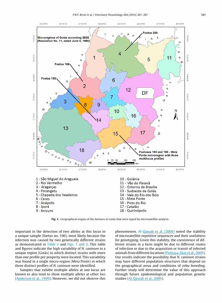
P.R.F. Brom et al. / Veterinary Parasitology 204 (2014) 381–387 385
in Goiás
iaiaautwt
k(
Fig. 1. Geographical origins of the foetuses
mportant in the detection of two alleles at this locus in unique sample (foetus no. 190), most likely because thenfection was caused by two genetically different strainss demonstrated in Table 2 and Figs. 1 and 2. This tablend figures indicate the high variability of N. caninum in anique region (Goiás) in which distinct strains with morehan one profile per property were located. This variabilityas found in a single micro-region (Meia Ponte) in which
hree distinct profiles of N. caninum were identified.Samples that exhibit multiple alleles at one locus are
nown to also tend to show multiple alleles at other lociAnderson et al., 1999). However, we did not observe this
that were typed by microsatellite analysis.
phenomenon. Al-Qassab et al. (2009) noted the stabilityof microsatellite repetitive sequences and their usefulnessfor genotyping. Given this stability, the coexistence of dif-ferent strains in a farm might be due to different routesof infection or due to the acquisition or transit of infectedanimals from different locations (Pedraza-Díaz et al., 2009).Our results indicate the possibility that N. caninum strainsmay have different population structures that depend on
the geographical areas and conditions of zebu breeding.Further study will determine the value of this approachthrough future epidemiological and population geneticstudies (Al-Qassab et al., 2009).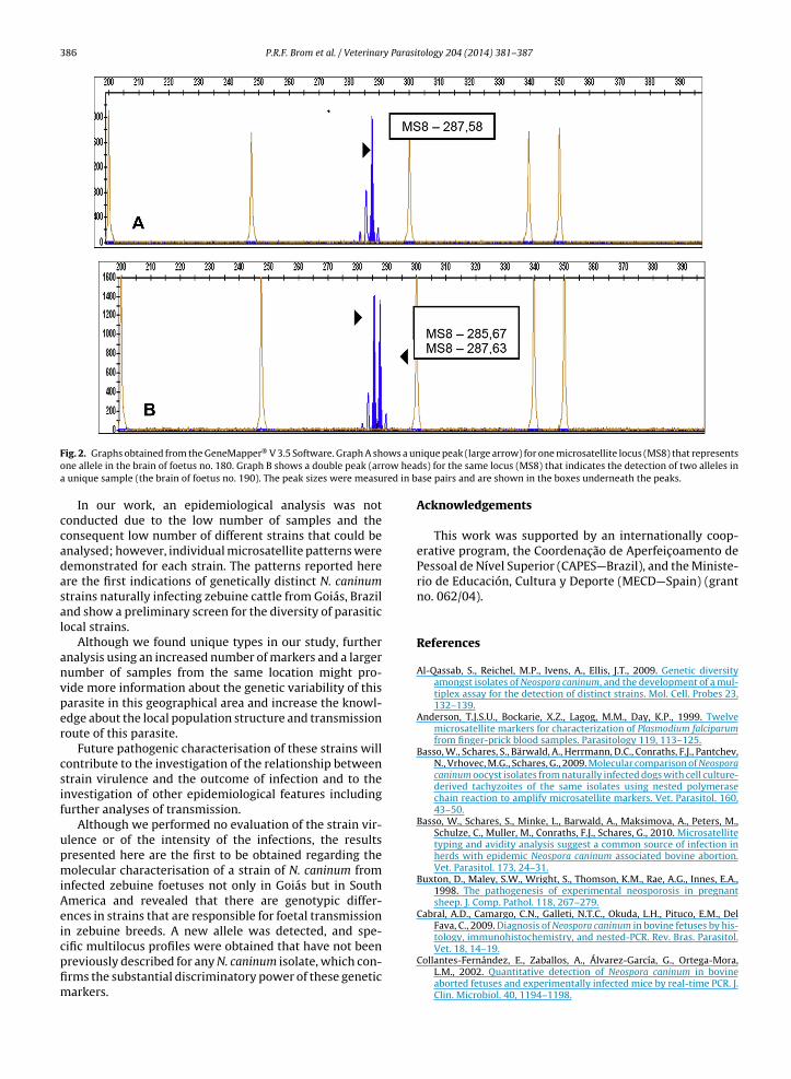
386 P.R.F. Brom et al. / Veterinary Parasitology 204 (2014) 381–387
ows a unow heared in b
Fig. 2. Graphs obtained from the GeneMapper® V 3.5 Software. Graph A shone allele in the brain of foetus no. 180. Graph B shows a double peak (arra unique sample (the brain of foetus no. 190). The peak sizes were measu
In our work, an epidemiological analysis was notconducted due to the low number of samples and theconsequent low number of different strains that could beanalysed; however, individual microsatellite patterns weredemonstrated for each strain. The patterns reported hereare the first indications of genetically distinct N. caninumstrains naturally infecting zebuine cattle from Goiás, Braziland show a preliminary screen for the diversity of parasiticlocal strains.
Although we found unique types in our study, furtheranalysis using an increased number of markers and a largernumber of samples from the same location might pro-vide more information about the genetic variability of thisparasite in this geographical area and increase the knowl-edge about the local population structure and transmissionroute of this parasite.
Future pathogenic characterisation of these strains willcontribute to the investigation of the relationship betweenstrain virulence and the outcome of infection and to theinvestigation of other epidemiological features includingfurther analyses of transmission.
Although we performed no evaluation of the strain vir-ulence or of the intensity of the infections, the resultspresented here are the first to be obtained regarding themolecular characterisation of a strain of N. caninum frominfected zebuine foetuses not only in Goiás but in SouthAmerica and revealed that there are genotypic differ-ences in strains that are responsible for foetal transmissionin zebuine breeds. A new allele was detected, and spe-
cific multilocus profiles were obtained that have not beenpreviously described for any N. caninum isolate, which con-firms the substantial discriminatory power of these geneticmarkers.ique peak (large arrow) for one microsatellite locus (MS8) that representsds) for the same locus (MS8) that indicates the detection of two alleles inase pairs and are shown in the boxes underneath the peaks.
Acknowledgements
This work was supported by an internationally coop-erative program, the Coordenac ão de Aperfeic oamento dePessoal de Nível Superior (CAPES—Brazil), and the Ministe-rio de Educación, Cultura y Deporte (MECD—Spain) (grantno. 062/04).
References
Al-Qassab, S., Reichel, M.P., Ivens, A., Ellis, J.T., 2009. Genetic diversityamongst isolates of Neospora caninum, and the development of a mul-tiplex assay for the detection of distinct strains. Mol. Cell. Probes 23,132–139.
Anderson, T.J.S.U., Bockarie, X.Z., Lagog, M.M., Day, K.P., 1999. Twelvemicrosatellite markers for characterization of Plasmodium falciparumfrom finger-prick blood samples. Parasitology 119, 113–125.
Basso, W., Schares, S., Bärwald, A., Herrmann, D.C., Conraths, F.J., Pantchev,N., Vrhovec, M.G., Schares, G., 2009. Molecular comparison of Neosporacaninum oocyst isolates from naturally infected dogs with cell culture-derived tachyzoites of the same isolates using nested polymerasechain reaction to amplify microsatellite markers. Vet. Parasitol. 160,43–50.
Basso, W., Schares, S., Minke, L., Barwald, A., Maksimova, A., Peters, M.,Schulze, C., Muller, M., Conraths, F.J., Schares, G., 2010. Microsatellitetyping and avidity analysis suggest a common source of infection inherds with epidemic Neospora caninum associated bovine abortion.Vet. Parasitol. 173, 24–31.
Buxton, D., Maley, S.W., Wright, S., Thomson, K.M., Rae, A.G., Innes, E.A.,1998. The pathogenesis of experimental neosporosis in pregnantsheep. J. Comp. Pathol. 118, 267–279.
Cabral, A.D., Camargo, C.N., Galleti, N.T.C., Okuda, L.H., Pituco, E.M., DelFava, C., 2009. Diagnosis of Neospora caninum in bovine fetuses by his-tology, immunohistochemistry, and nested-PCR. Rev. Bras. Parasitol.
Vet. 18, 14–19.Collantes-Fernández, E., Zaballos, A., Álvarez-García, G., Ortega-Mora,L.M., 2002. Quantitative detection of Neospora caninum in bovineaborted fetuses and experimentally infected mice by real-time PCR. J.Clin. Microbiol. 40, 1194–1198.
y Parasi
C
D
G
G
H
M
P
P
P.R.F. Brom et al. / Veterinar
ollantes-Fernández, E., Rodríguez-Bertos, A., Arnáiz-Seco, I., Moreno-Burgos, B., Aduriz, G., Ortega-Mora, L.M., 2006. Influence of the stageof pregnancy on Neospora caninum distribution, parasite loads andlesions in aborted bovine foetuses. Theriogenology 65, 629–641.
ubey, J.P., Schares, G., Ortega-Mora, L.M., 2007. Epidemiology and con-trol of neosporosis and Neospora caninum. Clin. Microbiol. Rev. 20,323–367.
arcía-Melo, D.P., Regidor-Cerrillo, J., Ortega-Mora, L.M., Collantes-Fernández, E., de Oliveira, V.S.F., Oliveira, M.A.P., Silva, A.C., 2009.Isolation and biological characterisation of a new isolate of Neosporacaninum from an asymptomatic calf in Brazil. Acta Parasitol. 54,180–185.
ondim, L.F., Laski, P., Gao, L., Mcallister, M.M., 2004. Variation of the inter-nal transcribed spacer 1 sequence within individual strains and amongdifferent strains of Neospora caninum. J. Parasitol. 90, 119–122.
all, T.A., 1999. BioEdit: a user-friendly biological sequence alignmenteditor and analysis program for Windows 95/98/NT. Nucleic AcidsSymp. Ser. 41, 95–98.
allon, M.E., MacLeod, A., Wastling, J.M., Smith, H., Tait, A., 2003.Multilocus genotyping of Cryptosporidium parvum Type 2: pop-ulation genetics and sub-structuring. Infect. Genet. Evol. 3,207–218.
edraza-Díaz, S., Marugán-Hernández, V., Collantes-Fernández, E.,Regidor-Cerrillo, J., Rojo-Montejo, S., Gómez-Bautista, M., Ortega-Mora, L.M., 2009. Microsatellite markers for the molecular charac-
terization of Neospora caninum: application to clinical samples. Vet.Parasitol. 166, 38–46.ereira-Bueno, J., Quintanilla-Gozalo, A., Pérez-Pérez, V., Espi-Felgueroso,A., Álvarez-García, G., Collantes-Fernández, E., Ortega-Mora, L.M.,2003. Evaluation by different diagnostic techniques of bovine
tology 204 (2014) 381–387 387
abortion associated with Neospora caninum in Spain. Vet. Parasitol.111, 143–152.
Pereira García-Melo, D., Regidor-Cerrillo, J., Collantes-Fernández, E.,Aguado-Martínez, A., Del Pozo, I., Minguijón, E., Gómez-Bautista,M., Aduriz, G., Ortega-Mora, L.M., 2010. Pathogenic characterizationin mice of Neospora caninum isolates obtained from asymptomaticcalves. Parasitology 137, 1057–1068.
Pérez-Zaballos, F.J., Ortega-Mora, L.M., Álvarez-García, G., Collantes-Fernández, E., Navarro-Lozano, V., García-Villada, L., Costas, E., 2005.Adaptation of Neospora caninum isolates to cell-culture changes: anargument in favor of its clonal population structure. J. Parasitol. 91,507–510.
Regidor-Cerrillo, J., Pedraza-Díaz, S., Gómez-Bautista, M., Ortega-Mora,L.M., 2006. Multilocus microsatellite analysis reveals extensivegenetic diversity in Neospora caninum. J. Parasitol. 92, 517–524.
Regidor-Cerrillo, J., Gómez-Bautista, M., Pereira-Bueno, J., Aduriz,G., Navarro-Lozano, V., Risco-Castillo, V., Fernandez-García, A.,Pedraza-Díaz, S., Ortega-Mora, L.M., 2008. Isolation and genetic char-acterization of Neospora caninum from asymptomatic calves in Spain.Parasitology 135, 1651–1659.
Rojo-Montejo, S., Collantes-Fernández, E., Regidor-Cerrillo, J., Álvarez-García, G., Marugán-Hernández, V., Pedraza-Díaz, S., Blanco-Murcia,J., Prenafeta, A., Ortega-Mora, L.M., 2009a. Isolation and characteriza-tion of a bovine isolate of Neospora caninum with low virulence. Vet.Parasitol. 159, 7–16.
Rojo-Montejo, S., Collantes-Fernández, E., Blanco-Murcia, J., Rodríguez-Bertos, A., Risco-Castillo, V., Ortega-Mora, L.M., 2009b. Experimentalinfection with a low virulence isolate of Neospora caninum at 70days gestation in cattle did not result in foetopathy. Vet. Res.40, 49.


