Embed Size (px)
Citation preview
Journal of Cell and Molecular Research (2013) 5 (1), 03-12
Cloning, nucleotide sequencing and bioinformatics study of NcGRA7, an
immunogen from Neospora caninum
Mahdi Soltani1, Mohammadreza Nassiri1,2*, Alireza Sadrebazzaz3, Mojtaba Tahmoorespoor1,2
1 - Department of Animal Sciences, College of Agriculture, Ferdowsi University of Mashhad, Mashhad, Iran.
2 - Agricultural Biotechnology Research Group, Institute of Biotechnology, Ferdowsi University of Mashhad, Mashhad, Iran.
3 - Razi Serum and Vaccine Research Institute, Mashhad, Iran
Received 20 July 2013 Accepted 01 September 2013
Abstract
Neospora caninum is an obligate intracellular parasitic protozoa and considered as causal agent of Neosporosis
which infect wide variety of hosts. NcGRA7 is an immunodominant antigen recognized by sera from bovines, naturally
infected by N. caninum, which is used as a powerful target for recombinant or DNA vaccine preparation against
neosporosis. There is no study about identifying the molecular structure of Neospora caninum in Iran, so as first step,
current study tried to identify NcGRA7 gene in this parasite in Iran. After extraction of total RNA from N. caninum
tachyzoites, cDNA was synthesized and NcGRA7 gene was amplified using cDNA as template. Then the PCR product
was cloned into pTZ57R/T vector and transformed into Escherichia coli (DH5α strain), and the resulted recombinant
plasmid was submitted for sequencing, followed by bioinformatics analysis. The data obtained from sequencing of
native NcGRA7 was recorded in GenBank. The deduced amino acid sequence of NcGRA7 in current study was
compared with other N. caninum NcGRA7 sequences and showed some identities and differences. NcGRA7 gene of N.
caninum was successfully cloned into the pTZ57R/T vector and recombination was confirmed by sequencing, colony
PCR, and enzymatic digestion, making it ready expression of recombinant protein for further studies.
Keywords: Neospora caninum, NcGRA7, Cloning, Sequencing
Introduction
Bovine neosporosis is the most frequently
diagnosed cause of bovine abortion worldwide
(Monney et al., 2011). Neospora caninum, a
persistent protozoan parasite capable of infecting
almost any warm-blooded vertebrate, is a member
of phylum apicomplexa and has a complex lifestyle
involving two phases of growth: an intestinal phase
in canine hosts, and an extra-intestinal phase in
other mammals (Dubey and Schares, 2011). It was
originally identified in tissues of paralyzed dogs
(Bjerkas and Presthus, 1988; Dubey et al., 1988).
As revealed by molecular analyses, N. caninum is
closely related to other coccidian parasite,
Toxoplasma gondii, and therefore many of
previously described T. gondii biological
characteristics can be attributed to N. caninum so
they would employ similar mechanisms for
adhesion and invasion processes (Monney et al.,
2011). Results of studies on Neospora caninum
infection in Iran showed that this parasite could be
considered as a cause of economic loss in dairy
cattle (Salehi et al., 2009). From several areas in
Iran, Neospora infection has been reported in cattle
Corresponding author E-mail:
(Nematollahi et al., 2011; Nourollahi Fard et al.,
2008; Razmi et al., 2006; Sadrebazzaz et al., 2007;
Sadrebazzaz et al., 2004), dogs (Haddadzadeh et
al., 2007; Hosseininejad et al., 2010; Malmasi et al.,
2007; Yagoob, 2011) and camels (Hosseininejad et
al., 2009; Sadrebazzaz et al., 2006).
Current studies on N. caninum are mainly
focused on the mechanisms and antigens involved
in the tachyzoite adhesion, invasion and its
proliferation and persistence in the host cell and
using these antigens for immunological purposes
(Dubey and Schares, 2011). NcSRS2 was one of
the most worked targets for developing
recombinant vaccines and diagnostic kits against
neosporosis (Soltani et al., 2013).
N. caninum exploits different secretory and
antigenic proteins to invade a host cell and gain
access to its intracellular environment. These
proteins originate from distinct organelles termed
micronemes, rhoptries, and dense granules. They
are released at specific times during invasion to
ensure the proteins are allocated to their correct
target destinations(Howe and Sibley, 1999). Dense
granule antigens (GRAs) are secreted by dense
granules to the parasitophorous vacuole during
parasite intracellular development (Cesbron-
delauw, 1994). Dense-granule secretion shares
several features with the regulated secretory
Cloning, nucleotide sequencing and bioinformatics study of NcGRA7…
4
pathway: (1) packaging in electron-dense vesicles;
(2) fusion of these vesicles with the plasma
membrane; and (3) calcium-regulated exocytosis. It
has been suggested that dense granule antigens
stimulate humoral immunity in the host. GRA7 is a
highly immunogenic, dense granule protein in both
T. gondii and N. caninum (Lally et al., 1997;
Vonlaufen et al., 2004). Moreover, although GRA
proteins appear to be related to intracellular parasite
development, previous studies revealed that
NcGRA7 might be involved in the initial host cell
invasion process of N. caninum (Augustine et al.,
1999; Cho et al., 2005). It has been showed that this
immunogenic protein provides some protection
against experimental N. caninum infection (Jenkins
et al., 2004; Liddell et al., 2003; Nishikawa et al.,
2009). Thus, the NcGRA7 protein could be
considered as a vaccine candidate against
neosporosis. Moreover, the immunogenicity of
NcGRA7 has led to investigation of this antigen as
a diagnostic reagent (Huang et al., 2007).
In the framework of the investigations on
designing recombinant vaccines against
neosporosis, this work focuses on the cloning and
sequencing of NcGRA7 from Iranian isolate of N.
caninum for the first time and bioinformatics based
characterization of the important properties of its
deduced protein. This work is first step in an
attempt to design vaccine studies against
neosporosis using NcGRA7 antigen that will be
studied in the future.
Materials and Methods
Production of N. caninum tachyzoites
All cell culture reagents were purchased from
Gibco-BRL (Zurich, Switzerland) and chemicals
were from Sigma (St. Louis, MO, USA). Vero cells
were routinely cultured in 25 cm2 tissue culture
flasks in 5 ml of RPMI 1640 medium supplemented
with 10% heat-inactivated FCS, 2 mM glutamine,
50 U of penicillin ⁄ mL and 50 ug of streptomycin ⁄
ml and incubated at 37ºC with 5% CO2. A strain of
Neospora caninum was kindly provided by Dr.
Sadrebazzaz (Razi vaccine and serum research
institute, Mashhad branch). N. caninum cells were
maintained in BALB/c mice by serial
intraperitoneal inoculation of parasites was used for
the experiment. N. caninum tachyzoites was
maintained by serial passages in Vero cells.
Cultures were passaged at least once per week.
When 80% of the Vero cells that had been infected
with N. caninum tachyzoites shows cytopathic
effect (typically 3-4 days p.i.), the cell monolayers
were removed by scraping, twice washed with
phosphate buffered saline (PBS) solution, and then
centrifuged at 1000 g for 10 min. Purified
tachyzoites were checked for viability using trypan
blue staining. Infected cells were trypsinized,
washed twice in cold RPMI 1640 medium and the
resulting pellet resuspended in 2 ml cold RPMI
1640 medium. Cells were repeatedly passed
through a 25G needle.
RNA isolation and first strand cDNA synthesis
Total RNA was isolated from 2 × 10⁶ purified N.
caninum tachyzoites using NucleoSpin® RNA II
kit (Machery-Nagel, Germany) according to the
manufacturer’s instructions using gene specific
primers. RNA concentration was measured with the
NanoDrop ND1000 (Thermo Scientific, Delaware,
US) system.
Single-stranded cDNA was synthesized from
isolated total RNA using a cDNA synthesis kit
(RevertAid™ First Strand cDNA Synthesis Kit,
Fermentas, Germany) according to the standard
protocol for first strand cDNA synthesis. Briefly,
first strand cDNA synthesis reaction was performed
in a 20 µl reaction mixture containing 100 ng of
total RNA, 4 µl 5X reaction buffer, 2 µl 10 mM
dNTP Mix, 12 µl nuclease-free water, 1 µl
RiboLock™ RNase Inhibitor (20 u/μl), 1 µl
RevertAid™ M-MuLV Reverse Transcriptase (200
u/μl) and 15 pmol of each gene specific primers.
Reaction mixtures were incubated for 5 minutes at
25 °C followed by 60 minutes at 42 °C and the
reactions were terminated by heating at 70 °C for 5
minutes.
PCR amplification
A pair of gene-specific primers were designed
using Primer Premiere software (Biosoft) based on
published NcGRA7 gene sequence in the GenBank
to amplify NcGRA7 gene. Primers were
synthesized by as follows: NG71-F (5´-
CGAGAATTCAAAATGGCCCGACAAGC-3´)
and NG71-R (5´-
CGCAGGATCCTAACTATTCGGTGTCTAC-3´)
(Bioneer, South Korea). PCR reactions were
performed using total cDNA as template. Reaction
was carried out in 25 µl volume containing
approximately 100 ng of cDNA template, 50 mM
Tris buffer (pH: 8.3), 1.5 mM MgCl2, 200 mM of
each ddNTPs, 0.5 U of Pfu DNA polymerase and
100 pM of each primers. Amplification reaction
was performed using the following thermal profile:
95°C for 5 min, 35 amplification cycles (94°C for
40 sec, 62.5°C for 40 sec, and 72°C for 40 sec.),
followed by a 72°C final extension for 10 min.
Furthermore, false-negative results, caused by
inhibitory compounds in the PCR reactions, were
Journal of Cell and Molecular Research
excluded by performing a simultaneous positive
control reaction using the DNA extracted from
tachyzoites of the NC-1 strain. The negative control
consisted of dH2O without DNA. A positive and
negative control was included in each reaction.
Amplified PCR products were analyzed by
electrophoresis of 5 µl of each sample on 1%
(W/V) agarose gel at a constant voltage of 100 v for
40 minutes, stained with SYBR® Safe DNA Gel
Stain (Invitrogen, Paisley, UK). GeneRulerTM 100
bp Plus DNA Ladder (Fermentas) was used to
compare the DNA fragment sizes. Agarose gel
illuminated under UV, and photographed with an
UVidoc Gel Documentation System (UVitec, UK).
Gel extraction of PCR products
The specific amplimers containing desired gene
sequence were purified from the agarose gel by
QIAquick Gel Extraction Kit (Qiagen, Germany)
based on manufacturer’s recommendations. This kit
follows a simple bind-wash-elute procedure. Gel
slices were dissolved in a buffer containing a pH
indicator, allowing easy determination of the
optimal pH for DNA binding, then mixtures were
applied to the QIAquick spin column. Nucleic acids
adsorbed to the silica membrane in the conditions
provided by the buffer. Impurities were washed
away and pure DNA was eluted with a small
volume of low-salt buffer provided.
A tailing of PCR products
As exonuclease activity of the proofreading
polymerases removes the 3´-A overhangs necessary
for TA cloning, 3´-A overhangs must be added to
fragments taking advantages of non-template
activity of Taq DNA polymerase after PCR
amplification since Taq polymerase preferentially
adds an A to the 3'-ends in the presence of all four
dNTPs. Briefly, a reaction was set up containing 25
μl purified PCR product, 5 μl 10X Taq reaction
buffer, 5 μl MgCl2, 5 μl dNTP (10 mM stock), 1 μl
Taq polymerase, 9 μl H2O. Then the mixture was
incubated at 70 ºC for 30 min. Finally, 3 µl of
reaction mixture was run on a gel to quantify. This
reaction product can directly be used in ligation
reaction without any need to clean up reaction.
Ligation into pTZ57R/T vector
Tailed PCR products were ligated into
pTZ57R/T Vector (Fermentas, Germany) based on
TA cloning scheme according to the manufacturer’s
instructions. A 1:3 (vector to insert) molar ratio was
used. Ligation reaction sat up in 30 µl volume
containing 3 µl pTZ57R/T plasmid, 10 µl of A
tailed PCR product, 1 µl T4 DNA ligase enzyme, 6
µl 5X buffer and 10 µl nuclease free distilled water.
After gentle mix and a brief centrifuge, the ligation
reaction mixture was incubated overnight at 10ºC.
The resulting plasmid was designated as pTZ-
NcGRA7. Recombinant vector were stored at -20°C
until transformation.
Transformation, Screening and Colony PCR
Preparation of competent cells from Escherichia
coli strain DH5-α was performed by calcium
chloride method (Sambrook et al., 1989).
Advantages of chemical preparation of competent
cells include simple procedure; no special
equipment required and gives good transformation
efficiencies. In general, it is the best method to use
when the transformation efficiencies is not the
problem. For transformation, 10 µl of ligation
reaction product was added to 150 µl of competent
cells and placed on ice for 40 minutes after vortex
and spin. Then the mixture was incubated at 42 ºC
for 90 s and immediately was placed on ice for 5
minutes. Then 1 ml of LB antibiotic free medium
was added to the transformed cells and allowed to
recover by incubation at 37 ºC for 2 hours with
shaking. Cells harboring pTZ-NcGRA7 plasmid
was plated and grown overnight at 37 ºC on a LB
agar plate (10 g NaCl, 5 g yeast extract, 10 g bacto
tryptone) with ampicillin (100 µg/ml), X-Gal
(Fermentas) and IPTG (Fermentas) for blue-white
screening. After overnight incubation, plate was
placed at 4 ºC for 2 hours and cells from white
colonies were harvested and cultured on antibiotic
containing LB agar plates. After 16 hours
incubation at 37 ºC, cells harboring the recombinant
plasmid grew up. Recombination confirmed by
colony PCR with NcGRA7 gene specific primers.
This technique was used to determine insert size in
the vector. Briefly, a colony was picked with
toothpick and swirl into 50 μl of ddH2O in 1.5 ml
microfuge tube. Then the tubes were heated at 95
ºC for 10 minutes. Tubes were centrifuged for 5
minutes at top speed in microfuge and 40 μl of
supernatant was transferred to 0.5 ml microfuge
tubes and 2 μl of it was used as template in PCR
reaction. All other PCR reactions conditions were
as explained before.
Plasmid Purification
Cells harboring the recombinant plasmid were
cultured in antibiotic containing LB medium for 16
hours at 37 ºC in a shaker incubator. GeneJET
Plasmid Miniprep Kit (Fermentas) was used to
purify plasmids from E. coli DH5α following the
manufacturer’s instructions. Briefly, 4 ml bacterial
culture was harvested and lysed. The lysate was
then cleared by centrifugation and applied on the
silica column to selectively bind DNA molecules.
Cloning, nucleotide sequencing and bioinformatics study of NcGRA7…
6
The adsorbed DNA was washed to remove
contaminants, and the pure plasmid DNA was
eluted in a small volume of elution buffer. Plasmid
DNA concentrations were determined by
absorbance at 260 nm using NanoDrop ND1000
(Thermo Scientific, Delaware, US) system. The
integrity of the DNA plasmids was checked by
agarose gel electrophoresis. Also resultant
recombinant plasmid (pTZ-NcGRA7) was
compared with native plasmid (pTZ57R/T) by
electrophoresis of 3 µl of extracted plasmid on a
1% agarose gel.
Enzymatic Digestion of pTZ-NcGRA7
With regard to presence of BamHI and EcoRI
restriction sites on recombinant plasmid extracted
from white colonies, the recombinant plasmid was
characterized for the presence and size of inserts by
double digestion with EcoRI and BamHI. Each 20
µl digestion reaction contained 10 µl of plasmid, 1
µl of each restriction enzyme, 2 µl of 10X buffer
(buffer R, based on Fermentas recommendations)
and 6 µl of dH2O. Digestion was performed by
incubation at 37 ºC for 2 hours. Digestion products
were analyzed by electrophoresis on 1% agarose
gel containing SYBR® Safe DNA Gel Stain
(Invitrogen, Paisley, UK).
Sequencing of NcGRA7 gene
The nucleotide sequence of the inserts
(NcGRA7) in the recombinant plasmid pTZ-
NcGRA7 was verified by sequencing in the forward
and reverse directions using primer walking
approach (Eurofins MWG Operon, Germany). M13
uni (-21) forward primer (5´-
TGTAAAACGACGGCCAGT-3´) and M13 rev (-
29) reverse primer (5´-
CAGGAAACAGCTATGACC-3´) were used for
sequencing. DNA Baser v3 (Heracle BioSoft,
Romania) was used for sequencing data assembly
to produce a consensus sequence for each DNA
sample used.
Blast search and bioinformatics study
The nucleotide sequence of NcGRA7 was
submitted to the BLAST search (megablast
algorithm) at NCBI server
(http://www.ncbi.nlm.nih.gov/blast/) to compare
with sequences presented in the GenBank. For
detailed analysis, all closely related sequences and
deduced amino acid sequences between published
sequences were aligned by ClustalW2 multiple
sequence alignment program
(http://www.ebi.ac.uk/Tools/clustalw2/) (Larkin et
al., 2007).
The sequences were analyzed for signal peptides
using SignalP 4.0
(http://www.cbs.dtu.dk/services/SignalP/) (Petersen
et al., 2011), protein domains using Prosite
(http://prosite.expasy.org/) (Sigrist et al., 2010) and
potential transmembrane regions were checked with
the ProtScale tool on the Expasy server
(http://expasy.org/tools/protscale.html).
Hydrophobicity plot of NcGRA7 protein was
also drawn which characterizes its hydrophobic and
hydrophilic characteristics that may be useful in
predicting membrane-spanning domains, potential
antigenic sites and regions that are likely exposed
on the protein surface (Hopp and Woods, 1981;
Kyte and Doolittle, 1982).
Phylogenetic and molecular evolutionary
analyses were conducted using CLC main
workbench software (CLC bio) by bootstrap test
with 1000 replications was applied to estimate the
confidence of branching patterns of the UPGMA
tree. Also, pairwise comparisons were done to
clarify the pairwise distances and percent identities.
Results
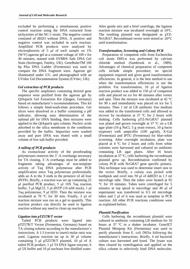
Production of N. caninum tachyzoites
Vero cells became confluent on day 3 and then
were infected with Neospora caninum tachyzoites.
Tachyzoites grew well in Vero monolayers (Fig. 1).
N. caninum tachyzoites were maintained in and
purified from, Vero cell monolayers and were
immediately used for RNA extraction.
Figure 1. (a) Confluent Vero cells on day 3. (b) N.
caninum tachyzoite infected Vero cells.
RNA isolation and first strand cDNA synthesis
Extracted RNA samples had very good quality
and integrity based on Nanodrop analysis results.
The OD 260/280 ratio for purified RNA was
between 1.80–1.95, indicating that preparations
were free of any major protein contamination.
NanoDrop results showed that first strand cDNA
synthesis reaction was successful.
PCR amplification
As PCR results showed, synthesized cDNA was
successfully amplified by PCR reaction. The
Journal of Cell and Molecular Research
presence of amplicons is characteristic for the
presence of the N. caninum DNA. Length of
NcGRA7 specific product was about 679 bp. The
intensity and size of bands was identical with N.
caninum (NC-1) positive controls that confirmed
the accuracy of performed reactions. Furthermore,
no visible bands can be seen in negative control
lanes. PCR products were used for ligation into
pTZ57R/T vector after A-tailing process.
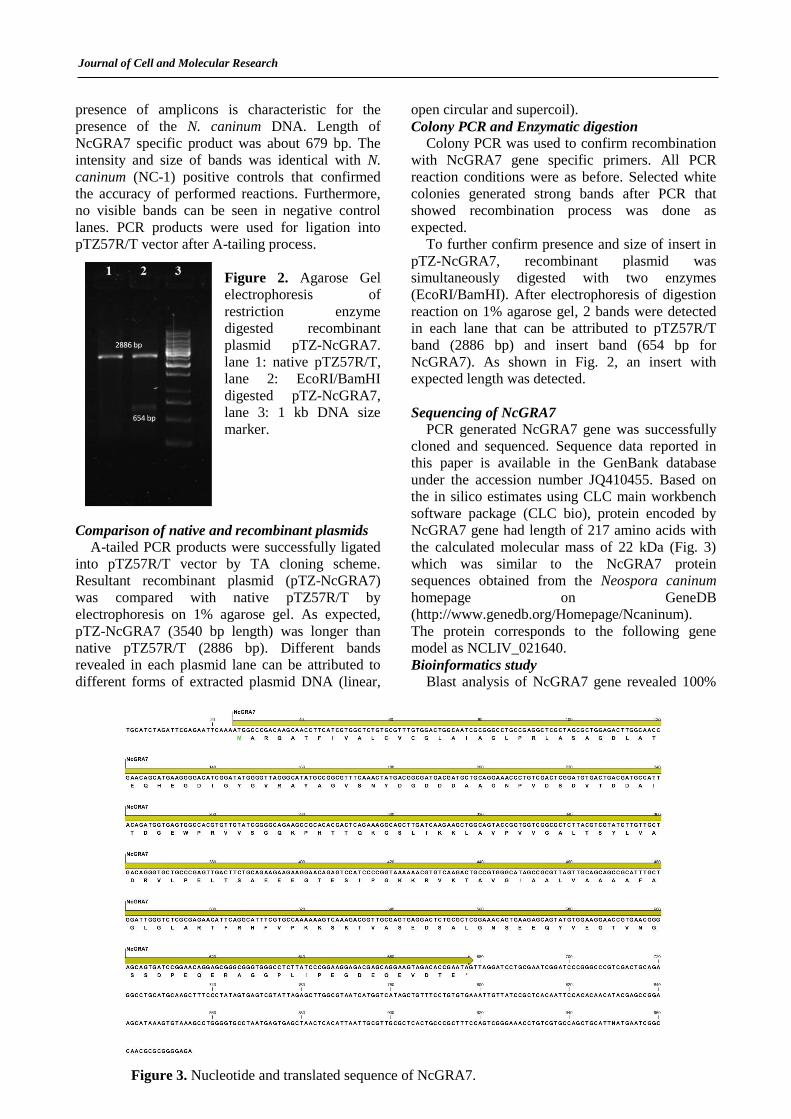
Figure 2. Agarose Gel
electrophoresis of
restriction enzyme
digested recombinant
plasmid pTZ-NcGRA7.
lane 1: native pTZ57R/T,
lane 2: EcoRI/BamHI
digested pTZ-NcGRA7,
lane 3: 1 kb DNA size
marker.
Comparison of native and recombinant plasmids
A-tailed PCR products were successfully ligated
into pTZ57R/T vector by TA cloning scheme.
Resultant recombinant plasmid (pTZ-NcGRA7)
was compared with native pTZ57R/T by
electrophoresis on 1% agarose gel. As expected,
pTZ-NcGRA7 (3540 bp length) was longer than
native pTZ57R/T (2886 bp). Different bands
revealed in each plasmid lane can be attributed to
different forms of extracted plasmid DNA (linear,
open circular and supercoil).
Colony PCR and Enzymatic digestion
Colony PCR was used to confirm recombination
with NcGRA7 gene specific primers. All PCR
reaction conditions were as before. Selected white
colonies generated strong bands after PCR that
showed recombination process was done as
expected.
To further confirm presence and size of insert in
pTZ-NcGRA7, recombinant plasmid was
simultaneously digested with two enzymes
(EcoRI/BamHI). After electrophoresis of digestion
reaction on 1% agarose gel, 2 bands were detected
in each lane that can be attributed to pTZ57R/T
band (2886 bp) and insert band (654 bp for
NcGRA7). As shown in Fig. 2, an insert with
expected length was detected.
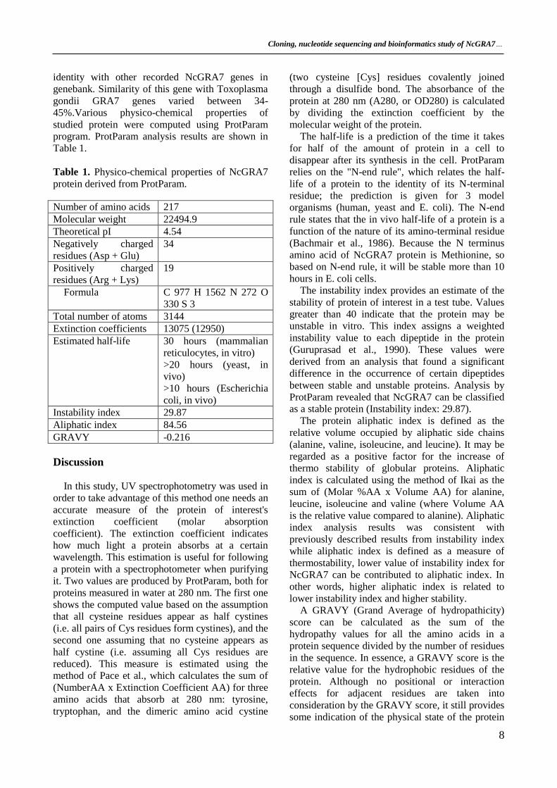
Sequencing of NcGRA7
PCR generated NcGRA7 gene was successfully
cloned and sequenced. Sequence data reported in
this paper is available in the GenBank database
under the accession number JQ410455. Based on
the in silico estimates using CLC main workbench
software package (CLC bio), protein encoded by
NcGRA7 gene had length of 217 amino acids with
the calculated molecular mass of 22 kDa (Fig. 3)
which was similar to the NcGRA7 protein
sequences obtained from the Neospora caninum
homepage on GeneDB
(http://www.genedb.org/Homepage/Ncaninum).
The protein corresponds to the following gene
model as NCLIV_021640.
Bioinformatics study
Blast analysis of NcGRA7 gene revealed 100%
Figure 3. Nucleotide and translated sequence of NcGRA7.
Cloning, nucleotide sequencing and bioinformatics study of NcGRA7…
8
identity with other recorded NcGRA7 genes in
genebank. Similarity of this gene with Toxoplasma
gondii GRA7 genes varied between 34-
45%.Various physico-chemical properties of
studied protein were computed using ProtParam
program. ProtParam analysis results are shown in
Table 1.
Table 1. Physico-chemical properties of NcGRA7
protein derived from ProtParam.
Discussion
In this study, UV spectrophotometry was used in
order to take advantage of this method one needs an
accurate measure of the protein of interest's
extinction coefficient (molar absorption
coefficient). The extinction coefficient indicates
how much light a protein absorbs at a certain
wavelength. This estimation is useful for following
a protein with a spectrophotometer when purifying
it. Two values are produced by ProtParam, both for
proteins measured in water at 280 nm. The first one
shows the computed value based on the assumption
that all cysteine residues appear as half cystines
(i.e. all pairs of Cys residues form cystines), and the
second one assuming that no cysteine appears as
half cystine (i.e. assuming all Cys residues are
reduced). This measure is estimated using the
method of Pace et al., which calculates the sum of
(NumberAA x Extinction Coefficient AA) for three
amino acids that absorb at 280 nm: tyrosine,
tryptophan, and the dimeric amino acid cystine
(two cysteine [Cys] residues covalently joined
through a disulfide bond. The absorbance of the
protein at 280 nm (A280, or OD280) is calculated
by dividing the extinction coefficient by the
molecular weight of the protein.
The half-life is a prediction of the time it takes
for half of the amount of protein in a cell to
disappear after its synthesis in the cell. ProtParam
relies on the "N-end rule", which relates the half-
life of a protein to the identity of its N-terminal
residue; the prediction is given for 3 model
organisms (human, yeast and E. coli). The N-end
rule states that the in vivo half-life of a protein is a
function of the nature of its amino-terminal residue
(Bachmair et al., 1986). Because the N terminus
amino acid of NcGRA7 protein is Methionine, so
based on N-end rule, it will be stable more than 10
hours in E. coli cells.
The instability index provides an estimate of the
stability of protein of interest in a test tube. Values
greater than 40 indicate that the protein may be
unstable in vitro. This index assigns a weighted
instability value to each dipeptide in the protein
(Guruprasad et al., 1990). These values were
derived from an analysis that found a significant
difference in the occurrence of certain dipeptides
between stable and unstable proteins. Analysis by
ProtParam revealed that NcGRA7 can be classified
as a stable protein (Instability index: 29.87).
The protein aliphatic index is defined as the
relative volume occupied by aliphatic side chains
(alanine, valine, isoleucine, and leucine). It may be
regarded as a positive factor for the increase of
thermo stability of globular proteins. Aliphatic
index is calculated using the method of Ikai as the
sum of (Molar %AA x Volume AA) for alanine,
leucine, isoleucine and valine (where Volume AA
is the relative value compared to alanine). Aliphatic
index analysis results was consistent with
previously described results from instability index
while aliphatic index is defined as a measure of
thermostability, lower value of instability index for
NcGRA7 can be contributed to aliphatic index. In
other words, higher aliphatic index is related to
lower instability index and higher stability.
A GRAVY (Grand Average of hydropathicity)
score can be calculated as the sum of the
hydropathy values for all the amino acids in a
protein sequence divided by the number of residues
in the sequence. In essence, a GRAVY score is the
relative value for the hydrophobic residues of the
protein. Although no positional or interaction
effects for adjacent residues are taken into
consideration by the GRAVY score, it still provides
some indication of the physical state of the protein
Number of amino acids 217
Molecular weight 22494.9
Theoretical pI 4.54
Negatively charged
residues (Asp + Glu)
34
Positively charged
residues (Arg + Lys)
19
Formula C 977 H 1562 N 272 O
330 S 3
Total number of atoms 3144
Extinction coefficients 13075 (12950)
Estimated half-life 30 hours (mammalian
reticulocytes, in vitro)
>20 hours (yeast, in
vivo)
>10 hours (Escherichia
coli, in vivo)
Instability index 29.87
Aliphatic index 84.56
GRAVY -0.216
Journal of Cell and Molecular Research
(Kyte and Doolittle, 1982).
This index indicates the solubility of the proteins:
positive GRAVY protein is hydrophobic while
negative GRAVY protein is hydrophilic (Kyte and
Doolittle, 1982). As derived from ProtParam
analysis, NcGRA7 gained a negative GRAVY
score so it can be inferred that NcGRA7 is a
hydrophilic protein. According to Kyte and
Doolittle (1982), integral membrane proteins
typically have higher GRAVY scores than do
globular proteins. Though this score is another
helpful piece of information, it cannot reliably
predict the structure without the help of hydropathy
plots.
There are some methods for evaluation of the
degree of interaction of polar solvents such as water
with specific amino acids. In these methods a
hydrophobicity plot is created that is a quantitative
analysis of the degree of hydrophobicity or
hydrophilicity of amino acids in a protein (Kyte-
Doolittle scale indicates hydrophobic amino acids,
while the Hopp-Woods scale measures hydrophilic
residues). This measure is implicated to identify
possible structure or domains of a protein. Plot
shape analysis prepares information about partial
structure of the protein of interest. For example,
extension of about 20 amino acids with positive
shows that these amino acids may be part of alpha-
helix spanning across a lipid bilayer, which is
composed of hydrophobic fatty acids. On the other
hand, stretch of amino acids with negative
hydrophobicity indicates that these residues are in
contact with solvent or water, and that they are
probably resided on the outer surface of the protein.
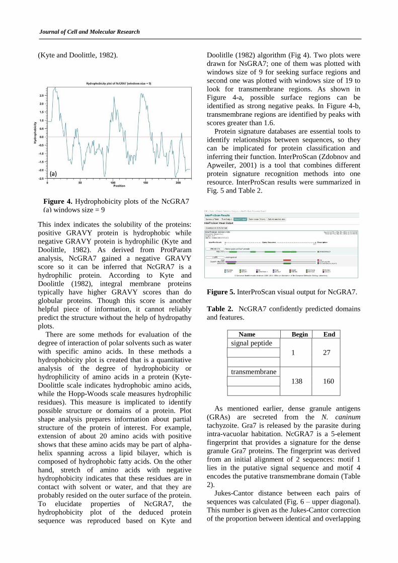
To elucidate properties of NcGRA7, the
hydrophobicity plot of the deduced protein
sequence was reproduced based on Kyte and
Doolitlle (1982) algorithm (Fig 4). Two plots were
drawn for NsGRA7; one of them was plotted with
windows size of 9 for seeking surface regions and
second one was plotted with windows size of 19 to
look for transmembrane regions. As shown in
Figure 4-a, possible surface regions can be
identified as strong negative peaks. In Figure 4-b,
transmembrane regions are identified by peaks with
scores greater than 1.6.
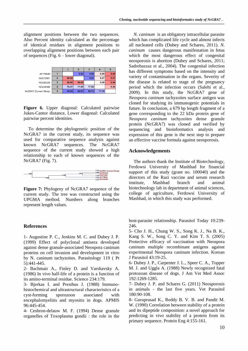
Protein signature databases are essential tools to
identify relationships between sequences, so they
can be implicated for protein classification and
inferring their function. InterProScan (Zdobnov and
Apweiler, 2001) is a tool that combines different
protein signature recognition methods into one
resource. InterProScan results were summarized in
Fig. 5 and Table 2.
Figure 5. InterProScan visual output for NcGRA7.
Table 2. NcGRA7 confidently predicted domains
and features.
Name Begin End
signal peptide
1 27
transmembrane
138 160
As mentioned earlier, dense granule antigens
(GRAs) are secreted from the N. caninum
tachyzoite. Gra7 is released by the parasite during
intra-vacuolar habitation. NcGRA7 is a 5-element
fingerprint that provides a signature for the dense
granule Gra7 proteins. The fingerprint was derived
from an initial alignment of 2 sequences: motif 1
lies in the putative signal sequence and motif 4
encodes the putative transmembrane domain (Table
2).
Jukes-Cantor distance between each pairs of
sequences was calculated (Fig. 6 – upper diagonal).
This number is given as the Jukes-Cantor correction
of the proportion between identical and overlapping
Figure 4. Hydrophobicity plots of the NcGRA7
(a) windows size = 9
Cloning, nucleotide sequencing and bioinformatics study of NcGRA7…
10
alignment positions between the two sequences.
Also Percent identity calculated as the percentage
of identical residues in alignment positions to
overlapping alignment positions between each pair
of sequences (Fig. 6 – lower diagonal).
Figure 6. Upper diagonal: Calculated pairwise
Jukes-Cantor distance, Lower diagonal: Calculated
pairwise percent identities.
To determine the phylogenetic position of the
NcGRA7 in the current study, its sequence was
used for comparative sequence analysis against
known NcGRA7 sequences. The NcGRA7
sequence of the current study showed a high
relationship to each of known sequences of the
NcGRA7 (Fig. 7).
Figure 7: Phylogeny of NcGRA7 sequence of the
current study. The tree was constructed using the
UPGMA method. Numbers along branches
represent length values.
N. caninum is an obligatory intracellular parasite
which has complicated life cycle and almost infects
all nucleated cells (Dubey and Schares, 2011). N.
caninum causes dangerous manifestation in fetus
which the most dangerous effect of congenital
neosporosis is abortion (Dubey and Schares, 2011;
Sadrebazzaz et al., 2004). The congenital infection
has different symptoms based on the intensity and
variety of contamination in the organs. Severity of
the disease is related to stage of the pregnancy
period which the infection occurs (Salehi et al.,
2009). In this study, the NcGRA7 gene of
Neospora caninum tachyzoites surface antigen was
cloned for studying its immunogenic potentials in
future. In conclusion, a 679 bp length fragment of a
gene corresponding to the 22 kDa protein gene of
Neospora caninum tachyzoites dense granule
protein (NcGRA7) was cloned and verified by
sequencing and bioinformatics analysis and
expression of this gene is the next step to prepare
an effective vaccine formula against neosporosis.
Acknowledgements
The authors thank the Institute of Biotechnology,
Ferdowsi University of Mashhad for financial
support of this study (grant no. 100040) and the
directors of the Razi vaccine and serum research
institute, Mashhad branch and animal
biotechnology lab in department of animal sciences,
college of agriculture, Ferdowsi University of
Mashhad, in which this study was performed.
References
1- Augustine P. C., Jenkins M. C. and Dubey J. P.
(1999) Effect of polyclonal antisera developed
against dense granule-associated Neospora caninum
proteins on cell invasion and development in vitro
by N. caninum tachyzoites. Parasitology 119 ( Pt
5):441-445.
2- Bachmair A., Finley D. and Varshavsky A.
(1986) In vivo half-life of a protein is a function of
its amino-terminal residue. Science 234:179.
3- Bjerkas I. and Presthus J. (1988) Immuno-
histochemical and ultrastructural characteristics of a
cyst-forming sporozoon associated with
encephalomyelitis and myositis in dogs. APMIS
96:445-454.
4- Cesbron-delauw M. F. (1994) Dense granule
organelles of Toxoplasma gondii : the role in the
host-parasite relationship. Parasitol Today 10:239-
246.
5- Cho J. H., Chung W. S., Song K. J., Na B. K.,
Kang S. W., Song C. Y. and Kim T. S. (2005)
Protective efficacy of vaccination with Neospora
caninum multiple recombinant antigens against
experimental Neospora caninum infection. Korean
J Parasitol 43:19-25.
6- Dubey J. P., Carpenter J. L., Speer C. A., Topper
M. J. and Uggla A. (1988) Newly recognized fatal
protozoan disease of dogs. J Am Vet Med Assoc
192:1269-1285.
7- Dubey J. P. and Schares G. (2011) Neosporosis
in animals - the last five years. Vet Parasitol
180:90-108.
8- Guruprasad K., Reddy B. V. B. and Pandit M.
W. (1990) Correlation between stability of a protein
and its dipeptide composition: a novel approach for
predicting in vivo stability of a protein from its
primary sequence. Protein Eng 4:155-161.
Journal of Cell and Molecular Research
9- Haddadzadeh H. R., Sadrebazzaz A., Malmasi
A., Talei Ardakani H., Khazraii Nia P. and
Sadreshirazi N. (2007) Seroprevalence of Neospora
caninum infection in dogs from rural and urban
environments in Tehran, Iran. Parasitol Res
101:1563-1565.
10- Hopp T. P. and Woods K. R. (1981) Prediction
of protein antigenic determinants from amino acid
sequences. Proceedings of the National Academy of
Sciences 78:3824.
11- Hosseininejad M., Hosseini F., Mahzounieh M.,
Raisi Nafchi A. and Mosharraf M. (2010)
Seroprevalence of Neospora caninum infection in
dogs in Chaharmahal-va-Bakhtiari Province, Iran.
Comp Clin Pathol 19:269-270.
12- Hosseininejad M., Pirali-Kheirabadi K. and
Hosseini F. (2009) Seroprevalence of Neospora
caninum Infection in Camels (Camelus
dromedarius) in Isfahan Province, Center of Iran.
Iranian J Parasitol 4:61-64.
13- Howe D. K. and Sibley L. D. (1999)
Comparison of the major antigens of Neospora
caninum and Toxoplasma gondii. Int J Parasitol
29:1489-1496.
14- Huang P., Liao M., Zhang H., Lee E. G.,
Nishikawa Y. and Xuan X. (2007) Dense-granule
protein NcGRA7, a new marker for the
serodiagnosis of Neospora caninum infection in
aborting cows. Clin Vaccine Immunol 14:1640-
1643.
15- Jenkins M., Parker C., Tuo W., Vinyard B. and
Dubey J. P. (2004) Inclusion of CpG adjuvant with
plasmid DNA coding for NcGRA7 improves
protection against congenital neosporosis. Infect.
Immun. 72:1817-1819.
16- Kyte J. and Doolittle R. F. (1982) A simple
method for displaying the hydropathic character of
a protein. J Mol Biol 157:105-132.
17- Lally N., Jenkins M., Liddell S. and Dubey J. P.
(1997) A dense granule protein (NCDG1) gene
from Neospora caninum. Mol Biochem Parasitol
87:239-243.
18- Larkin M., Blackshields G., Brown N., Chenna
R., McGettigan P., McWilliam H., Valentin F.,
Wallace I., Wilm A. and Lopez R. (2007) Clustal
W and Clustal X version 2.0. Bioinformatics
23:2947-2948.
19- Liddell S., Parker C., Vinyard B., Jenkins M.
and Dubey J. P. (2003) Immunization of mice with
plasmid DNA coding for NcGRA7 or NcsHSP33
confers partial protection against vertical
transmission of Neospora caninum. J Parasitol
89:496-500.
20- Malmasi A., Hosseininejad M., Haddadzadeh
H., Badii A. and Bahonar A. (2007) Serologic study
of anti-Neospora caninum antibodies in household
dogs and dogs living in dairy and beef cattle farms
in Tehran, Iran. Parasitol Res 100:1143-1145.
21- Monney T., Debache K. and Hemphill A.
(2011) Vaccines against a Major Cause of Abortion
in Cattle, Neospora caninum Infection. Animals
1:306-325.
22- Nematollahi A., Jaafari R. and Moghaddam G.
(2011) Seroprevalence of Neospora caninum
Infection in Dairy Cattle in Tabriz, Northwest Iran.
Iran J Parasitol 6:95-98.
23- Nishikawa Y., Zhang H., Ikehara Y., Kojima
N., Xuan X. and Yokoyama N. (2009)
Immunization with oligomannose-coated liposome-
entrapped dense granule protein 7 protects dams
and offspring from Neospora caninum infection in
mice. Clin Vaccine Immunol 16:792-797.
24- Nourollahi Fard S. R., Khalili M. and
Aminzadeh A. (2008) Prevalence of antibodies to
Neospora caninum in cattle in Kerman province,
South East Iran. Vet Arh 78:253.
25- Petersen T. N., Brunak S., von Heijne G. and
Nielsen H. (2011) SignalP 4.0: discriminating
signal peptides from transmembrane regions. Nat.
Methods 8:785-786.
26- Razmi G. R., Mohammadi G. R., Garrosi T.,
Farzaneh N., Fallah A. H. and Maleki M. (2006)
Seroepidemiology of Neospora caninum infection
in dairy cattle herds in Mashhad area, Iran. Vet
Parasitol 135:187-189.
27- Sadrebazzaz A., Habibi G., Haddadzadeh H.
and Ashrafi J. (2007) Evaluation of bovine abortion
associated with Neospora caninum by different
diagnostic techniques in Mashhad, Iran. Parasitol
Res 100:1257-1260.
28- Sadrebazzaz A., Haddadzadeh H., Esmailnia
K., Habibi G., Vojgani M. and Hashemifesharaki R.
(2004) Serological prevalence of Neospora
caninum in healthy and aborted dairy cattle in
Mashhad, Iran. Vet Parasitol 124:201-204.
29- Sadrebazzaz A., Haddadzadeh H. and Shayan
P. (2006) Seroprevalence of Neospora caninum and
Toxoplasma gondii in camels (Camelus
dromedarius) in Mashhad, Iran. Parasitol Res
98:600-601.
30- Salehi N., Haddadzadeh H., Ashrafihelan J.,
Shayan P. and Sadrebazzaz A. (2009) Molecular
and Pathological Study of Bovine Aborted Fetuses
and Placenta from Neospora caninum Infected
Dairy Cattle. Iran J Parasitol 4:40-51.
31- Sambrook J., Fritsch E. F. and Maniatis T.
1989. Molecular cloning: a laboratory manual. New
York: Cold Spring Harbor Laboratory Press.
32- Sigrist C. J. A., Cerutti L., De Castro E.,
Langendijk-Genevaux P. S., Bulliard V., Bairoch
A. and Hulo N. (2010) PROSITE, a protein domain
Cloning, nucleotide sequencing and bioinformatics study of NcGRA7…
12
database for functional characterization and
annotation. Nucleic Acids Res 38:D161-D166.
33- Soltani M., Sadrebazzaz A., Nassiri M. and
Tahmoorespoor M. (2013) Cloning, Nucleotide
Sequencing and Bioinformatics Study of NcSRS2
Gene, an Immunogen from Iranian Isolate of
Neospora caninum. Iranian journal of parasitology
8:114.
34- Vonlaufen N., Guetg N., Naguleswaran A.,
Muller N., Bjorkman C., Schares G., von
Blumroeder D., Ellis J. and Hemphill A. (2004) In
vitro induction of Neospora caninum bradyzoites in
vero cells reveals differential antigen expression,
localization, and host-cell recognition of
tachyzoites and bradyzoites. Infect. Immun.
72:576-583.
35- Yagoob G. (2011) Seroprevalence of Neospora
Caninum in Stray Dogs. Am J Anim Vet Sci 6:100-
104.
36- Zdobnov E. M. and Apweiler R. (2001)
InterProScan–an integration platform for the
signature-recognition methods in InterPro.
Bioinformatics 17:847-848.


