Embed Size (px)
Citation preview

Metabolic footprinting of extracellular metabolitesof brain endothelium infected with Neosporacaninum in vitroElsheikha et al.
Elsheikha et al. BMC Research Notes 2014, 7:406http://www.biomedcentral.com/1756-0500/7/406
Elsheikha et al. BMC Research Notes 2014, 7:406http://www.biomedcentral.com/1756-0500/7/406
RESEARCH ARTICLE Open Access
Metabolic footprinting of extracellular metabolitesof brain endothelium infected with Neosporacaninum in vitroHany M Elsheikha1*, Mamdowh Alkurashi1,2, Kenny Kong3 and Xing-Quan Zhu4
Abstract
Background: The survival of the intracellular protozoan parasite Neospora caninum depends on its ability to adaptto changing metabolic conditions of the host cell. Thus, defining cellular and metabolic changes in affected targettissues may aid in delineating pathogenetic mechanism. We undertook this study to assess the metabolic responseof human brain microvascular endothelial cells (HBMECs) to N. caninum infection in vitro.
Methods: HBMECs were exposed to N. caninum infection and the cytotoxic effects of infection were analyzed bythe 3-(4,5-dimethyl-2-thiazolyl)-2,5-diphenyl-2H-tetrazoliumbromidin (MTT) assay and lactate dehydrogenase (LDH)release assay. Metabolic footprinting of the extracellular metabolites of parasite-infected and non-infected culturesupernatant was determined by using targeted (Randox RX Imola clinical chemistry analyser) and unbiased RS (Ramanmicrospectroscopy) approaches.
Results: The MTT assay did not reveal any cytotoxic effect of N. caninum challenge on host cell viability. Measurementof LDH activity showed that N. caninum significantly induced loss of cell membrane integrity in a time-dependent anddose-dependent manner compared to control cells. Targeted biochemical analysis revealed that beta hydroxybutyrate,pyruvate, ATP, total protein, non-esterified fatty acids, and triglycerides are significantly different in infected cellscompared to controls. RS-based footprinting with principal component analysis (PCA) were able to correctly distinguishextracellular metabolites obtained from infected and control cultures, and revealed infection-related spectral signatures at865 cm−1, 984 cm−1, 1046 cm−1, and 1420 cm−1, which are attributed to variations in the content of lipids and nucleic acidsin infected cultures.
Conclusions: The changing pattern of extracellular metabolites suggests that HBMECs are target of metabolic alterations inN. caninum infection, which seem to reflect the changing metabolic state of infected cells and constitute a level ofinformation exchange that host and parasite use to coordinate activities.
Keywords: Adaptation, Blood brain barrier, Host-pathogen interaction, Metabolic footprinting, Metabolomics, Neosporacaninum
BackgroundThe complex interaction between intracellular (IC) patho-gens and their eukaryotic host cells embodies the funda-mental evolutionary struggle of eukaryotic cells to surviveunder a continuous challenge caused by the infectingpathogen. Despite significant progress in the past decades,how an obligatory IC apicomplexan protozoan parasite,
* Correspondence: [email protected] of Veterinary Medicine and Science, Faculty of Medicine and HealthSciences, University of Nottingham, Sutton Bonington Campus, LeicestershireLE12 5RD, UKFull list of author information is available at the end of the article
© 2014 Elsheikha et al.; licensee BioMed CentrCommons Attribution License (http://creativecreproduction in any medium, provided the orDedication waiver (http://creativecommons.orunless otherwise stated.
such as Neospora caninum adapts to host cell microenvir-onment, and the implication of this on the viability of thehost cell and the fitness of the parasite remains largely un-known. This parasite infects a large number of vertebrateanimals and is responsible for abortion and infertilityproblems in cattle and neuromuscular disease in dogs[1,2]. However, N. caninum infection is generally latentand asymptomatic, and results in the formation of dor-mant cysts that remain in the brain and other tissues forlife [1]. As an obligate IC pathogen, N. caninum survival isdependent upon entry, growth and development within
al Ltd. This is an Open Access article distributed under the terms of the Creativeommons.org/licenses/by/4.0), which permits unrestricted use, distribution, andiginal work is properly credited. The Creative Commons Public Domaing/publicdomain/zero/1.0/) applies to the data made available in this article,

Elsheikha et al. BMC Research Notes 2014, 7:406 Page 2 of 10http://www.biomedcentral.com/1756-0500/7/406
the eukaryotic host cell and then exiting to initiate a newinfection cycle. The IC infection cycle ends up with lysisof the host cell and release of the parasite progeny. Des-pite significant research efforts understanding of the cellu-lar processes by which N. caninum initiates infection andcause disease remains incomplete, partly, due to the com-plexity of N. caninum-host interaction, which determinesthe host response and outcome of infection.Critical aspects of N. caninum infection occur in CNS
tissues, particularly at the blood brain barrier (BBB) inter-face. N. caninum is a neuro-pathogen with a remarkablecapacity to cross the BBB and infect neurons and otherbrain cells, with adverse consequences [3,1]. However,many aspects of the molecular basis of neuropatho-genicity of N. caninum have not been fully elucidated.For example, our knowledge about the substrates used byN. caninum during infection, and the effect of N. caninuminfection on the metabolism of the host cell is unknown.Giving the significant animal health implication and eco-nomic losses associated with N. caninum infection betterunderstanding of the biochemical and metabolic changesin BBB cells induced by N. caninum and the metabolicrequirement of N. caninum during infection is essen-tial in order to understand the parasite neuro-pathogenesis.Recently, we investigated changes in BBB endothelial cellbioenergetics in response to N. caninum infection [4]. It isimportant to examine what makes the BBB endothelial cellsa tolerant environment for the growth of N. caninum.N. caninum is largely dependent on the host cell to
obtain the nutrients that are necessary for its replication.N. caninum-infected cells are expected to have differentmetabolic requirements from their normal (uninfected)counterparts because replication of IC parasites requiresenergy for synthesis of macromolecules, such as pro-teins, DNA, lipids, which are essential for the assemblyof the growing parasites. Hence, N. caninum-infectedcell faces two major metabolic challenges: (1) how tomeet the bioenergetic and biosynthetic demands of thegrowing IC parasites and (2) how to adapt to the fluctu-ations in the extracellular nutrient availability. Under-standing the consequences of this differential metabolicstress requires a detailed understanding of host cell metab-olism and viability in infected cells compared to healthycontrol cells. Metabolomic technologies can provide a glo-bal analysis of cellular phenotype in response to infection.However, methods used for sampling intracellular metabo-lites without changing their relative concentrations orintroducing contamination from supernatant metabolites isnot optimal [5,6]. In contrast, exometabolome or metabolicfootprinting is simple, and extracellular metabolites canexhibit very large changes in pool size. Exometabo-lome analysis has been used for phenotyping of micro-bial populations [7-10] or studying cellular response todrugs [11].
In the present study we investigated the hypothesisthat pathophysiological effect of N. caninum infectionon host tissue can be attributed to changes in the meta-bolic status of host cells during the course of infection.Our goals in this study were to: (i) seek direct evidencefor the alteration in the metabolic response of brainmicrovascular endothelial cells, a fundamental compo-nent of the BBB, to N. caninum infection and (ii) deter-mine the level and kinetic of extracellular metabolites inculture medium from N. caninum-infected versus con-trol cells.
MethodsEthical considerationThis study was reviewed by the University of Nottingham(UK) School of Veterinary Medicine and Science (SVMS)Ethical Review Committee. The Committee reviews all re-search studies involving School personnel and is chairedby Professor David Haig. The committee passed thisin vitro study as good to proceed, not requiring any fur-ther ethical review as it doesn’t involve vertebrate or inver-tebrate animals.
Cell lineHuman brain microvascular endothelial cells (HBMECs)were maintained as described previously [4], in completeRPMI-1640 (cRPMI) medium supplemented with 20%heat inactivated fetal calf serum (FBS), 2 mM L-glutamine,1 mM Sodium Pyruvate, 1 mM MEM non-essential aminoacids, 1% MEM vitamins and 100 units/mL penicillin/streptomycin at 37°C under humidified 5% CO2 conditions.When cells were confluent they were harvested withtrypsin-EDTA and passaged at a sub-cultivation ratioof 1:3 into new culture flasks with fresh medium. Cellswere considered confluent when their expansion hadreached a point where cells touched each other on allsides, leaving no intercellular spaces. To exclude if cellviability could be regarded as a factor affecting responseof the host cell to parasite infection and hence any subse-quent metabolic analysis, the number of viable cells wasdetermined on a minimum of 100 cells by hemocytometerunder a light microscope after staining with 0.15% trypanblue solution. Cells used in the experiments had a viabilitynot less than 99% at all times.
Parasites cultureNeospora caninum (Nc-Liverpool) strain was propagatedin Vero cells as described [12]. Infected host cell mono-layers were scraped, parasites were isolated from hostcells by passage through 25- and 27-gauge needles andpurified by using PD-10 Desalting Columns prepackedwith Sephadex G-25 medium as described previously [13].Purified parasites were centrifuged at 800 × g, washedtwice with fresh cRPMI, re-suspended in fresh medium

Elsheikha et al. BMC Research Notes 2014, 7:406 Page 3 of 10http://www.biomedcentral.com/1756-0500/7/406
and quantified using a hemacytometer. The final volumeof suspension was adjusted with cRPMI medium to achievea ratio of 2:1 parasite/host cell for subsequent infectionexperiments. Parasite viability was checked by usingtrypan blue staining assay and parasite with more than97% viability were used.
In vitro infection protocolCells (3 × 10⁵ cells/mL) were seeded at the bottom of 6-wellculture plates with a volume of 2 mL cRPMI medium/well. Cells were allowed to grow overnight by incubationat 37°C in a humidified atmosphere with 5% CO2 in air.Before infection, cell growth medium was removed andcells were washed three times with sterile PBS (8 g/LNaCl, 0.2 g/L KCl, 0.2 g/L KH2PO4, 1.15 g/L Na2HPO4).Then, in each 6-well plate, three wells were infected withparasites at a MOI of 2 in 2-ml fresh medium, and theremaining three wells received only 2-ml fresh medium(mock-infected) and considered controls. Culture plateswere then incubated to allow infection to progress withincells. Culture media were sampled at different time pointpost infection (PI) starting from 0 h, and then, at 1, 2, 3, 6,12, 18, 24, 48 h PI. At each sampling time six wells (threeinfected and three controls) were collected and centri-fuged at 1000 × g for 3 min, and the supernatants col-lected and kept at −80°C until analysis of extracellularmetabolites.
MTT assayThe nonradioactive metabolic assay MTT (3-(4,5-dimethyl-2-thiazolyl)-2,5-diphenyl-2H-tetrazoliumbromidin ) wasused to assess the effect of N. caninum infection onthe viability of host cells. HBME cells were trypsinizedfrom T-75 culture flasks, seeded into 96-well tissue cul-ture microtiter plates (Nunc) at 1 × 10⁴ cells per well in100 μl of culture medium, and incubated for 18 h in ahumidified incubator (37°C, 5% CO2) until becomeconfluent. N. caninum tachyzoites were added to thecells at 2 MOI for 2 h, followed by removal of themedium and 2x washing with fresh medium to removeunbound parasites and cellular debris. Each well wasthen filled with 100 μl of fresh culture medium and plateswere incubated at the above culture conditions. As a posi-tive control, cells were treated with 1 μM staurosporine,an apoptotic agent. Cell viability was measured at 3, 6, 12,and 24 h PI by the reduction of MTT in a colorimetricassay. Briefly, MTT (Sigma Chemical, St. Louis, MO,USA) was added to each well (to a final concentrationof 0.5 mg/ml), and incubation was continued for 4 h inthe dark at 37°C. The cells were then incubated for 1 hin solubilization solution (50% sodium dodecyl sulfatein 0.1 mM/L HCl). The spectrophotometric absorbanceof the samples was subsequently measured with a microti-ter enzyme-linked immunosorbent assay (ELISA) plate
reader using a 570-nm filter. The level of MTT reductionwas expressed as a percentage of that of the non-infectedcontrol cells. The assay was performed in triplicate.
Lactate dehydrogenase assayLactate dehydrogenase (LDH) activity released into theculture medium (a measure of cell membrane lysis due tonecrotic cell death) was assayed using a CytoTox 96 Kit(Promega, Madison, Wis.) according to the manufacturer’sinstructions. Briefly, 1 × 10⁴ HBMECs were seeded ontosterile 96-well plates and grown until 90% confluence andsubsequently infected with N. caninum tachyzoites usingdifferent multiplicities of infection (MOIs) ranging from0.5 to 4. After 3, 6, 9, 12, 18, and 24 h of incubation, thesupernatants were collected, centrifuged to obtain cell-free supernatants. Of each sample, 50 μl per well wastransferred to new 96-well plates. LDH activity wasdetected by the addition of freshly prepared reagentsfollowed by incubation for 30 min in the dark at ambienttemperature. LDH activity was measured by a redox reac-tion that couples the oxidation of lactate iodotetrazoliumchloride to a colored formazan salt, using NADH as theelectron transfer agent and NADH diaphorase as the cata-lyst. The absorbance at 490 nm was read using a Bio-TekInstruments EL311SX plate reader. The cytotoxicity wasexpressed as a percentage of maximum LDH release,i.e., 100 × (optical density at 490 nm [OD490] of infectedcells − OD490 of uninfected cells)/(OD490 of 2% TritonX-100-lysed uninfected cells − OD490 of uninfected cells).This assay was performed in triplicate wells, and the datarepresent the mean ± standard error of the mean (SEM)from at least three separate experiments. Statistical ana-lysis was calculated by Student’s t-test using the GraphPad Prism 3.0 statistical program (GraphPad SoftwareInc., San Diego, CA). P < 0.05 was taken to indicatestatistical significance.
Biochemical analysis of extracellular metabolitesThe level of the 20 metabolites was determined colorimet-rically in culture medium obtained from infected and con-trol wells at different time points PI using commerciallyavailable kits and a Randox RX Imola clinical chemistryanalyzer (Randox Laboratories Ltd., Belfast, UK) accordingto the manufacturer’s specifications. Biochemical parame-ters measured included albumin (AB3800), glucose hexoki-nase (GL3816), calcium (CA3871), magnesium (MG3880),phosphorus (PH3820), NEFA (FA115), BHB (RB1007), chol-esterol (CH3810), TGA (TR3823), total protein (TP3869),urea (UR3825), lactate (LC3980), chloride (CL1645), so-dium (NA3851), potassium (PT3852), iron (SI3821),HDL (CH3811), and LDL (CH3841). Additionally, pyru-vate and ATP were measured. All reagents used in the ex-periment were of analytical grade, or better. All of the 20metabolites were quantified at each sampling time in
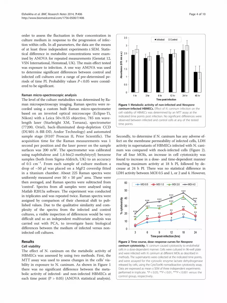
Figure 1 Metabolic activity of non-infected and Neosporacaninum-infected HBMECs. Effect of N. caninum infection on thecell viability of HBMECs was determined by an MTT assay at theindicated time points post infection. No significant differences wereobserved between infected and control cells at any of the testedtime points.
Figure 2 Time course, dose–response curves for Neosporacaninum cytotoxicity. N. caninum caused cytotoxicity to endothelialcells in a dose-dependent manner. Cells were cultured in 96-well plateand were infected with N. caninum at different MOIs as described inmethods. The supernatants were collected at the indicated time points,and were assayed for the cytosolic enzyme lactate dehydrogenasereleased by cells, using the CytoTox96 nonradioactive cytotoxicity assay.Data are expressed as mean ± SEM of three independent experimentsperformed in triplicate. *P < 0.05; **P < 0.01; ***P < 0.001 versus thecontrol group, respectively.
Elsheikha et al. BMC Research Notes 2014, 7:406 Page 4 of 10http://www.biomedcentral.com/1756-0500/7/406
order to assess the fluctuation in their concentration inculture medium in response to the progression of infec-tion within cells. In all parameters, the data are the meansof at least three independent experiments ± SEM. Statis-tical difference in metabolite concentrations were exam-ined by ANOVA for repeated measurements (Genstat 12,VSN International, Hemstead, UK). The main effect testedwas exposure to infection. A one way ANOVA was usedto determine significant differences between control andinfected cell cultures over a range of pre-determined pe-riods of time PI. Probability values P < 0.05 were consid-ered to be significant.
Raman micro-spectroscopic analysisThe level of the culture metabolites was determined by Ra-man microspectroscopy imaging. Raman spectra were re-corded using a custom built Raman micro-spectrometerbased on an inverted optical microscope (Eclipse-Ti,Nikon) with a Leica 50×/0.55 objective, 785 nm wave-length laser (Starbright XM, Torsana), spectrometer(77200, Oriel), back-illuminated deep-depletion CCD(DU401-A-BR-DD, Andor Technology) and automatedsample stage (H107 Proscan II, Prior Scientific). Theacquisition time for the Raman measurements was 1second per position and the laser power on the samplesurfaces was 200 mW. The spectrometer was calibratedusing naphthalene and 1,4-bis(2-methylstyryl) benzenesamples (both from Sigma-Aldrich, UK) to an accuracyof 0.5 cm−1. From each sample of culture medium adrop of ~50 μl was placed on a MgF2 coverslip fittedin a titanium chamber. About 225 Raman spectra wereuniformly measured over 50 × 50 μm2 area. These werethen averaged, and Raman spectra were subtracted from‘control’. Spectra from all samples were analysed usingMatlab R2013a software. The experiment was conductedin triplicates and was repeated twice. Raman spectra wereassigned by comparison of their chemical shift to pub-lished values. Due to the qualitative similarity and com-plexity of the spectra from the infected and controlcultures, a visible inspection of differences would be verydifficult and so an independent multivariate analysis wascarried out with PCA, to investigate basic biologicaldifferences between the medium of infected versus non-infected cell cultures.
ResultsCell viabilityThe effect of N. caninum on the metabolic activity ofHBMECs was assessed by using two methods. First, theMTT assay was used to assess changes in the cells’ via-bility in exposure to N. caninum. As shown in Figure 1there was no significant difference between the meta-bolic activity of infected- and non-infected HBMECs ateach time point (P > 0.05) (ANOVA statistical analysis).
Secondly, to determine if N. caninum has any adverse ef-fect on the membrane permeability of infected cells, LDHactivity in supernatants of HBMECs infected with N. cani-num was compared with mock-infected cells (Figure 2).For all four MOIs, an increase in cell cytotoxicity wasfound to increase in a dose- and time-dependent mannerreaching maximum activity at 18 h PI, followed by de-crease at 24 h PI. There was no statistical difference inLDH activity between MOI 0.5 and 1, or 2 and 4. However,

Elsheikha et al. BMC Research Notes 2014, 7:406 Page 5 of 10http://www.biomedcentral.com/1756-0500/7/406
significant statistical difference was observed between thecontrol and all different MOIs during the whole experi-mental period.
N. caninum triggers metabolic changes in HBMECsDynamic changes in the concentration of 20 biochemicalparameters were determined in the culture media of in-fected and control cultures. The following 14 metabolitesdid not show statistically significant difference between in-fected and control cultures: phosphorus, magnesium, cal-cium, chloride, sodium, potassium, iron, lactate, HDL,LDL, glucose hexokinase, urea, albumin, and cholesterol.On the other hand, the levels of pyruvate, ATP, BHB, totalprotein, NEFA, and TGA were statistically different be-tween infected and control cell cultures at certain timepoints PI (Figure 3). Pyruvate level was significantly higherin infected culture compared to control at 48 h PI. ATPlevel in the culture supernatant changed during the para-site growth (Figure 3). During the first 24 h PI therewas no difference in the level of ATP between infected
Figure 3 Time-specific metabolic changes in medium of Neospora caninuendothelial cells. Temporal changes in the concentrations of pyruvate (a), AT(NEFA; e), and triglycerides (TGA, f) in culture medium of infected (red line) verpoint separately. *P< 0.05; **P< 0.01; ***P< 0.001 versus the control group, res
and control culture, followed by increased consump-tion (i.e. reduced extracellular concentration) of ATP inN. caninum-infected cells till the end of the experiment(72 h PI), corresponding with the increase in the parasitegrowth within host cells. BHB levels were elevated in in-fected culture from 3 to 48 h PI, and this elevation was sig-nificantly higher (p < 0.05) in infected culture compared tocontrol at 6 h PI and between 12 and 48 h PI (Figure 3). Areduction in the levels of total protein, NEFA, and TGAwere observed in control culture media from 24 to 72 h PI.
Raman spectra of culture mediaRaman spectral patterns of medium from non-infected andinfected cultures were obtained at different time points PI.Label-free imaging analysis was able to detect variations inthe biochemistry of culture media between infected andcontrol cell cultures and to reveal spectral signatures ofeven low-concentration metabolites. As shown in Figure 4comparative Raman spectral analysis revealed manysimilarities and few differences, with prominent peaks
m-infected and mock-infected culture of human brain microvascularP (b), β-hydroxybutyrate (BHB; c), total protein (d), non-esterified fatty acidssus control (blue line). The statistical analysis was performed for every timepectively. Error bars indicate the standard errors of the means (n= 6).
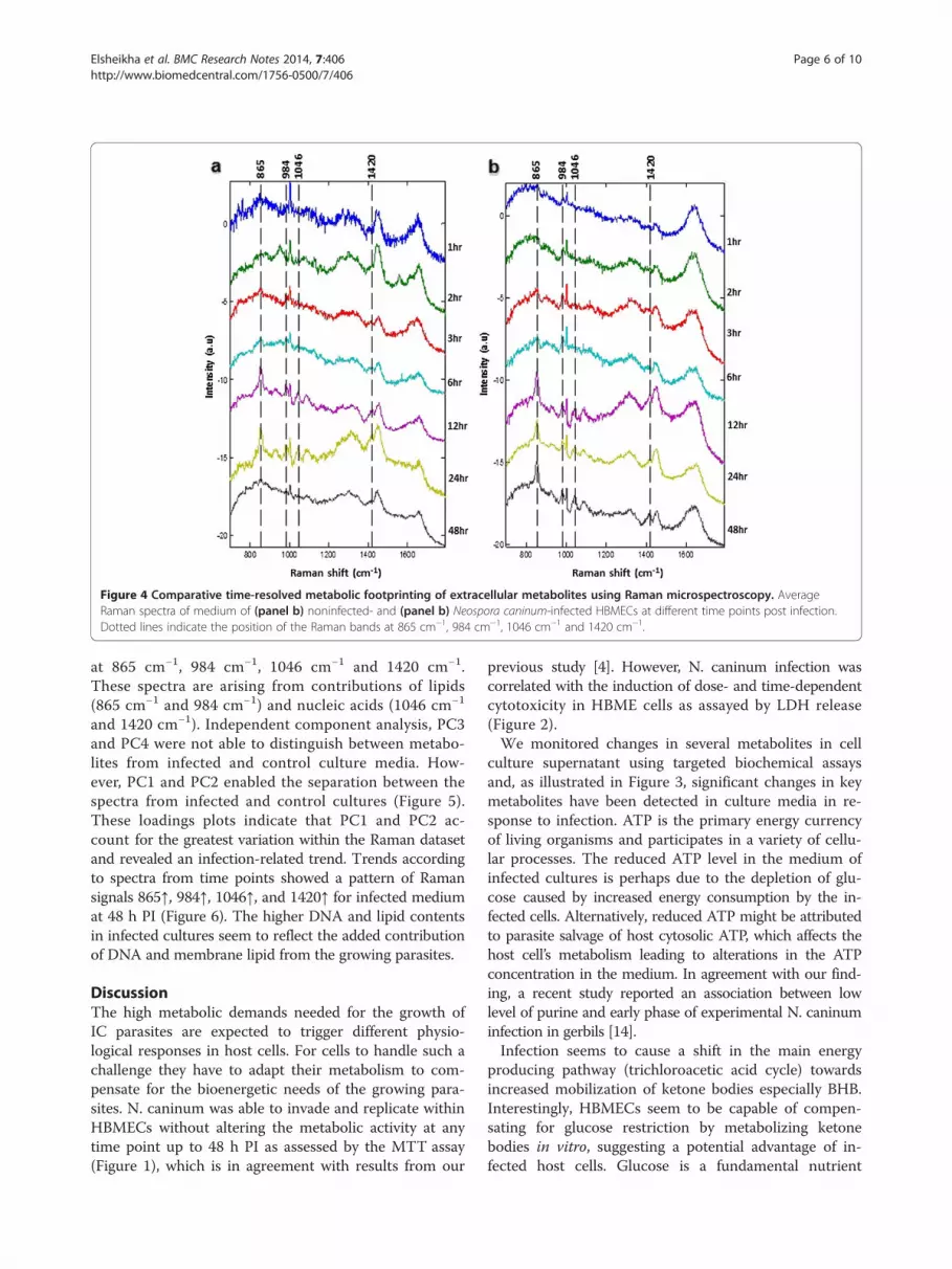
Figure 4 Comparative time-resolved metabolic footprinting of extracellular metabolites using Raman microspectroscopy. AverageRaman spectra of medium of (panel b) noninfected- and (panel b) Neospora caninum-infected HBMECs at different time points post infection.Dotted lines indicate the position of the Raman bands at 865 cm−1, 984 cm−1, 1046 cm−1 and 1420 cm−1.
Elsheikha et al. BMC Research Notes 2014, 7:406 Page 6 of 10http://www.biomedcentral.com/1756-0500/7/406
at 865 cm−1, 984 cm−1, 1046 cm−1 and 1420 cm−1.These spectra are arising from contributions of lipids(865 cm−1 and 984 cm−1) and nucleic acids (1046 cm−1
and 1420 cm−1). Independent component analysis, PC3and PC4 were not able to distinguish between metabo-lites from infected and control culture media. How-ever, PC1 and PC2 enabled the separation between thespectra from infected and control cultures (Figure 5).These loadings plots indicate that PC1 and PC2 ac-count for the greatest variation within the Raman datasetand revealed an infection-related trend. Trends accordingto spectra from time points showed a pattern of Ramansignals 865↑, 984↑, 1046↑, and 1420↑ for infected mediumat 48 h PI (Figure 6). The higher DNA and lipid contentsin infected cultures seem to reflect the added contributionof DNA and membrane lipid from the growing parasites.
DiscussionThe high metabolic demands needed for the growth ofIC parasites are expected to trigger different physio-logical responses in host cells. For cells to handle such achallenge they have to adapt their metabolism to com-pensate for the bioenergetic needs of the growing para-sites. N. caninum was able to invade and replicate withinHBMECs without altering the metabolic activity at anytime point up to 48 h PI as assessed by the MTT assay(Figure 1), which is in agreement with results from our
previous study [4]. However, N. caninum infection wascorrelated with the induction of dose- and time-dependentcytotoxicity in HBME cells as assayed by LDH release(Figure 2).We monitored changes in several metabolites in cell
culture supernatant using targeted biochemical assaysand, as illustrated in Figure 3, significant changes in keymetabolites have been detected in culture media in re-sponse to infection. ATP is the primary energy currencyof living organisms and participates in a variety of cellu-lar processes. The reduced ATP level in the medium ofinfected cultures is perhaps due to the depletion of glu-cose caused by increased energy consumption by the in-fected cells. Alternatively, reduced ATP might be attributedto parasite salvage of host cytosolic ATP, which affects thehost cell’s metabolism leading to alterations in the ATPconcentration in the medium. In agreement with our find-ing, a recent study reported an association between lowlevel of purine and early phase of experimental N. caninuminfection in gerbils [14].Infection seems to cause a shift in the main energy
producing pathway (trichloroacetic acid cycle) towardsincreased mobilization of ketone bodies especially BHB.Interestingly, HBMECs seem to be capable of compen-sating for glucose restriction by metabolizing ketonebodies in vitro, suggesting a potential advantage of in-fected host cells. Glucose is a fundamental nutrient
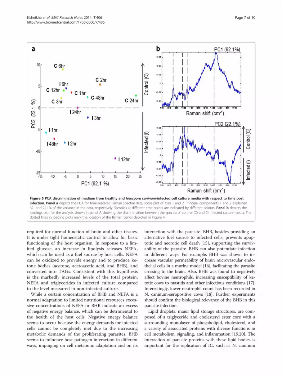
Figure 5 PCA discrimination of medium from healthy and Neospora caninum-infected cell culture media with respect to time postinfection. Panel a depicts the PCA for time-resolved Raman spectral data, score plot of axes 1 and 2. Principal components 1 and 2 explained62.1and 22.1% of the variance in the data, respectively. Samples at different time points are indicated by different colours. Panel b depicts theloadings plot for the analysis shown in panel A showing the discrimination between the spectra of control (C) and (I) infected culture media. Thedotted lines in loading plots mark the location of the Raman bands depicted in Figure 4.
Elsheikha et al. BMC Research Notes 2014, 7:406 Page 7 of 10http://www.biomedcentral.com/1756-0500/7/406
required for normal function of brain and other tissues.It is under tight homeostatic control to allow for basicfunctioning of the host organism. In response to a lim-ited glucose, an increase in lipolysis releases NEFA,which can be used as a fuel source by host cells. NEFAcan be oxidized to provide energy and to produce ke-tone bodies (acetone, acetoacetic acid, and BHB), andconverted into TAGs. Consistent with this hypothesisis the markedly increased levels of the total protein,NEFA and triglycerides in infected culture comparedto the level measured in non-infected culture.While a certain concentration of BHB and NEFA is a
normal adaptation to limited nutritional resources exces-sive concentrations of NEFA or BHB indicate an excessof negative energy balance, which can be detrimental tothe health of the host cells. Negative energy balanceseems to occur because the energy demands for infectedcells cannot be completely met due to the increasingmetabolic demands of the proliferating parasites. BHBseems to influence host-pathogen interaction in differentways, impinging on cell metabolic adaptation and on its
interaction with the parasite. BHB, besides providing analternative fuel source to infected cells, prevents apop-totic and necrotic cell death [15], supporting the surviv-ability of the parasite. BHB can also potentiate infectionin different ways. For example, BHB was shown to in-crease vascular permeability of brain microvascular endo-thelial cells in a murine model [16], facilitating the parasitecrossing to the brain. Also, BHB was found to negativelyaffect bovine neutrophils, increasing susceptibility of ke-totic cows to mastitis and other infectious conditions [17].Interestingly, lower neutrophil count has been recorded inN. caninum-seropositive cows [18]. Further experimentsshould confirm the biological relevance of the BHB in thisparasite infection.Lipid droplets, major lipid storage structures, are com-
posed of a triglyceride and cholesteryl ester core with asurrounding monolayer of phospholipid, cholesterol, anda variety of associated proteins with diverse functions incell metabolism, signaling, and inflammation [19,20]. Theinteraction of parasite proteins with these lipid bodies isimportant for the replication of IC, such as N. caninum
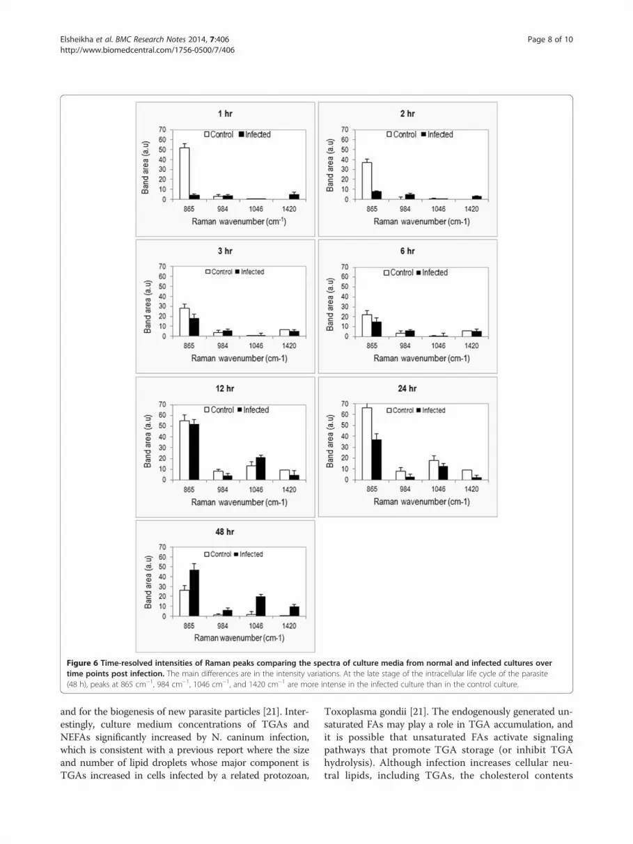
Figure 6 Time-resolved intensities of Raman peaks comparing the spectra of culture media from normal and infected cultures overtime points post infection. The main differences are in the intensity variations. At the late stage of the intracellular life cycle of the parasite(48 h), peaks at 865 cm−1, 984 cm−1, 1046 cm−1, and 1420 cm−1 are more intense in the infected culture than in the control culture.
Elsheikha et al. BMC Research Notes 2014, 7:406 Page 8 of 10http://www.biomedcentral.com/1756-0500/7/406
and for the biogenesis of new parasite particles [21]. Inter-estingly, culture medium concentrations of TGAs andNEFAs significantly increased by N. caninum infection,which is consistent with a previous report where the sizeand number of lipid droplets whose major component isTGAs increased in cells infected by a related protozoan,
Toxoplasma gondii [21]. The endogenously generated un-saturated FAs may play a role in TGA accumulation, andit is possible that unsaturated FAs activate signalingpathways that promote TGA storage (or inhibit TGAhydrolysis). Although infection increases cellular neu-tral lipids, including TGAs, the cholesterol contents

Elsheikha et al. BMC Research Notes 2014, 7:406 Page 9 of 10http://www.biomedcentral.com/1756-0500/7/406
did not seem to be significantly affected by N. caninuminfection.The marked increase in the concentrations of TGAs
and NEFA in infected culture medium compared to con-trol and the significant changes in expression of genesinvolved in lipid biogenesis (unpublished data) demon-strate that lipids play a very important role in the IC lifecycle of N. caninum. Increased lipid droplets’ formationand association with the parasitophorous vacuole hasbeen demonstrated in infections by other parasites in-cluding T. gondii [22,23], Trypanosoma cruzi [21,24,25],Leishmania amazonensis [26], Plasmodium falciparum[27], and P. berghei [28]. However, to date the mecha-nisms that govern lipid droplets’ biogenesis and its roleto N. caninum pathogenesis are not known.Further, we determined the temporal changes in exo-
metabolome composition by obtaining Raman spectrafrom medium of infected and control cultures. PCAscores plots and loadings plots showed a clear separationbetween samples taken from infected and control cul-tures (Figure 5), supporting the feasibility of this methodfor the investigation of the biochemical differences be-tween control and infected cultures. These data also in-dicate that footrpinting metabolic analysis using label-freeRaman spectroscopic imaging combined with multivariatechemometric analysis has enough resolution to monitorinfection-related metabolic changes over the course oftime spanning the IC life cycle of the parasite. This time-resolved-based analysis is essential since metabolic differ-ences can be highly dependent on growth phase of thecell, and cellular biochemistry changes during growth ofboth the cell and the parasite.The detection of signals for nucleic acids (1046 cm−1
and 1420 cm−1) in culture medium of control and in-fected cells was interesting (Figure 6). The source of thenucleic acids’ traces, host or parasite origin, is unknown.However, the concentration of nucleic acids was foundto increase in the medium of infected cultures in pro-portion to the proliferation of parasite, suggesting thesource of the nucleic acids to be of parasite origin.Apoptosis and/or necrosis are the two main mechanismsinvolved in release of DNA from normal or diseased liv-ing cells. However, parasite-infected cells are known toresist apoptosis [12,29], countering the notion of apop-tosis as the main mechanism for generating free DNA.The increase in nucleic acid signals in infected culture at18 h PI followed by a decrease at 24 h PI (Figure 6) cor-relates with the measurement of the intracellular LDHenzyme release, where N. caninum was found to com-promise the membrane integrity of infected cells in thefirst 18 h PI (Figure 2), increasing the nucleic acids’ per-meability. Interestingly, the nucleic acids Raman signalspeak again at 48 hr PI around the time the parasite isabout to exit the cells. These findings indicate that
although necrosis (and perhaps apoptosis) may con-tribute to the supernatant DNA, both mechanisms arethe not the only source of extracellular DNA. More re-search is needed in order to determine the mechanism(s)of release and the significance of circulating DNA in thesupernatant of both control and infected cultures.
ConclusionThe main novelty of our study is that we characterizedthe metabolic response and viability of BBB endothelialcells to protozoal infection using a multidisciplinary ap-proach. Analysis of the alterations in the biochemicalcomposition of culture media obtained by using Ramanmicrospectroscopy footprinting and chemometric analysiscomplemented data provided by standard biochemical as-says. This integrated approach allowed the determinationof the extracellular metabolites that are secreted and/orexcreted from infected and non-infected cells into growthmedia. PCA scores plots showed a clear separation be-tween metabolites from infected and control cultures.N. caninum challenge induced changes in energy statusof infected cells and lipid composition of culturemedia. Levels of precursors needed for lipid biosyn-thesis increased in infected HBMECs, confirming thecrucial role of lipid metabolism in the membrane bio-genesis of new parasite particles. Differences detectedby Raman imaging were attributed to variations in contentof lipids and nucleic acids in infected cultures. At this mo-ment, we do not know which biosynthetic step is criticalfor producing these changes in infected cells. We ex-pect this and other questions to be answered in futureexperiments.
AbbreviationsATP: Adenosine 5′-triphosphate; BBB: Blood brain barrier; BHB: β-hydroxybutyrate;CNS: Central nervous system; ELISA: Enzyme-linked immunosorbent assay;HBMECs: Human brain microvascular endothelial cells; HDL: High-densitylipoprotein; IC: Intracellular; LDH: Lactate dehydrogenase; LDL: Low-densitylipoprotein; MOI: Multiplicity of infection; NEFA: Non-esterified (unsaturated) fattyacids; PBS: Phosphate-buffered saline; PCA: Principal component analysis;RPMI: Roswell Park Memorial Institute medium; PI: Post infection;TGA: Triglycerides.
Competing interestsThe authors declare that they have no competing interests associated withthe publication of this manuscript.
Authors’ contributionsHME conceived and designed the experiments. HME and MMA performedthe experiments and wrote the initial draft of the manuscript. KK assistedwith Raman Spectroscopy analysis. ZXQ provided intellectual advice duringthe planning and execution of the experiments. All authors read, revised andapproved the final manuscript.
AcknowledgementsThe authors wish to thank Professor Lord Sandy Trees for providing N.caninum (Nc-Liverpool) strain, Professor David Haig for inspiring discussionson the project and Professor Naveed Khan for providing the HBME cells.MMA was supported by scholarship from Saudi Ministry of Higher Education.

Elsheikha et al. BMC Research Notes 2014, 7:406 Page 10 of 10http://www.biomedcentral.com/1756-0500/7/406
Author details1School of Veterinary Medicine and Science, Faculty of Medicine and HealthSciences, University of Nottingham, Sutton Bonington Campus, LeicestershireLE12 5RD, UK. 2Animal Production Department, College of Food andAgricultural Sciences, King Saud University, Riyadh 11451, Saudi Arabia.3School of Physics and Astronomy, University of Nottingham,Nottinghamshire NG7 2RD, UK. 4State Key Laboratory of Veterinary EtiologicalBiology, Key Laboratory of Veterinary Parasitology of Gansu Province,Lanzhou Veterinary Research Institute, Chinese Academy of AgriculturalSciences, Lanzhou, Gansu Province PR 730046, China.
Received: 13 March 2014 Accepted: 13 June 2014Published: 28 June 2014
References1. Dubey JP, Schares G, Ortega-Mora LM: Epidemiology and control of neosporosis
and Neospora caninum. Clin Microbiol Rev 2007, 20:323–367.2. Innes EA: The host-parasite relationship in pregnant cattle infected with
Neospora caninum. Parasitology 2007, 134:1903–1910.3. Vonlaufen N, Gianinazzi C, Müller N, Simon F, Björkman C, Jungi TW, Leib SL,
Hemphill A: Infection of organotypic slice cultures from rat centralnervous tissue with Neospora caninum: an alternative approach to studyhost-parasite interactions. Int J Parasitol 2002, 32:533–542.
4. Elsheikha HM, McKinlay CL, Elsaied NA, Smith PA: Elsheikha Effects ofNeospora caninum infection on brain microvascular endothelial cellsbioenergetics. Parasit Vectors 2013, 6:24.
5. Villas-Boas SG, Bruheim P: Cold glycerol-saline: the promising quenchingsolution for accurate intracellular metabolite analysis of microbial cells.Anal Biochem 2007, 370:87–97.
6. Winder CL, Dunn WB, Schuler S, Broadhurst D, Jarvis R, Stephens GM,Goodacre R: Global metabolic profiling of Escherichia coli cultures: anevaluation of methods for quenching and extraction of intracellularmetabolites. Anal Chem 2008, 80:2939–2948.
7. Allen J, Davey HM, Broadhurst D, Heald JK, Rowland JJ, Oliver SG, Kell DB:High-throughput classification of yeast mutants for functional genomicsusing metabolic footprinting. Nat Biotechnol 2003, 21:692–696.
8. Kell DB, Brown M, Davey HM, Dunn WB, Spasic I, Oliver SG: Metabolicfootprinting and systems biology: the medium is the message. Nat RevMicrobiol 2005, 3:557–565.
9. Mapelli V, Olsson L, Nielsen J: Metabolic footprinting in microbiology:methods and applications in functional genomics and biotechnology.Trends Biotechnol 2008, 26:490–497.
10. Behrends V, Ebbels TM, Williams HD, Bundy JG: Time-resolved metabolicfootprinting for nonlinear modeling of bacterial substrate utilization.Appl Environ Microbiol 2009, 75:2453–2463.
11. Kim DH, Jarvis RM, Xu Y, Oliver AW, Allwood JW, Hampson L, Hampson IN,Goodacre R: Combining metabolic fingerprinting and footprinting tounderstand the phenotypic response of HPV16 E6 expressing cervicalcarcinoma cells exposed to the HIV anti-viral drug lopinavir. Analyst 2010,135(6):1235–1244.
12. Alkurashi M, Eastick FA, Kuchipudi SV, Rauch C, Madouasse A, Zhu XQ,Elsheikha HM: Influence of culture medium pH on internalization, growthand phenotypic plasticity of Neospora caninum. Vet Parasitol 2011,177:267–274.
13. Elsheikha HM, Rosenthal BM, Murphy AJ, Dunams DB, Neelis DA, Mansfield LS:Generally applicable methods to purify intracellular coccidia from cellcultures and to quantify purification efficacy using quantitative PCR. VetParasitol 2006, 135:223–234.
14. Tonin AA, Da Silva AS, Thomé GR, Schirmbeck GH, Cardoso VV, Casali EA,Toscan G, Vogel FF, Flores MM, Fighera R, Lopes ST: Changes in purinelevels associated with cellular brain injury in gerbils experimentallyinfected with Neospora caninum. Res Vet Sci 2014, 96(3):507–511.
15. Xiao XQ, Zhao Y, Chen GQ: The effect of 3-hydroxybutyrate and itsderivatives on the growth of glial cells. Biomaterials 2007, 28:3608–3616.
16. Isales CM, Min L, Hoffman WH: Acetoacetate and beta-hydroxybutyratedifferentially regulate endothelin-1 and vascular endothelial growthfactor in mouse brain microvascular endothelial cells. J DiabetesComplications 1999, 13(2):91–97.
17. Grinberg N, Elazar S, Rosenshine I, Shpigel NY: Beta-hydroxybutyrateabrogates formation of bovine neutrophil extracellular traps and
bactericidal activity against mammary pathogenic Escherichia coli. InfectImmun 2008, 76(6):2802–2807.
18. Serrano B, Almería S, García-Ispierto I, Yániz JL, Abdelfattah-Hassan A,López-Gatius F: Peripheral white blood cell counts throughout pregnancy innon-aborting Neospora caninum-seronegative and seropositive high-producingdairy cows in a Holstein Friesian herd. Res Vet Sci 2011, 90(3):457–462.
19. Bozza PT, D’Avila H, Almeida PE, Magalhães KG, Molinaro R, Almeida CJ,Maya-Monteiro CM: Lipid droplets in host-pathogen interactions. ClinLipidol 2009, 4:791–807.
20. Farese RV Jr, Walther TC: Lipid droplets finally get a little R-E-S-P-E-C-T.Cell 2009, 139:855–860.
21. Melo RCN, Fabrino DL, Dias FF, Parreira GG: Lipid bodies: structuralmarkers of inflammatory macrophages in innate immunity. Inflamm Res2006, 55:342–348.
22. Charron AJ, Sibley LD: Host cells: mobilizable lipid resources for theintracellular parasite Toxoplasma gondii. J Cell Sci 2002, 115:3049–3059.
23. Nishikawa Y, Quittnat F, Stedman TT, Voelker DR, Choi JY, Zahn M, Yang M,Pypaert M, Joiner KA, Coppens I: Host cell lipids control cholesteryl estersynthesis and storage in intracellular Toxoplasma. Cell Microbiol 2005,7:849–867.
24. Melo RCN, D’Avila H, Fabrino DL, Almeida PE, Bozza PT: Macrophage lipidbody induction by Chagas disease in vivo: putative intracellular domainsfor eicosanoid formation during infection. Tissue Cell 2003, 35:59–67.
25. D’Avila H, Melo RCN, Parreira GG, Werneck-Barroso E, Castro-Faria-Neto HC,Bozza PT: Mycobacterium bovis bacillus Calmette-Guerin inducesTLR2-mediated formation of lipid bodies: intracellular domains for eicosanoidsynthesis in vivo. J Immunol 2006, 176:3087–3097.
26. Pinheiro RO, Nunes MP, Pinheiro CS, D’Avila H, Bozza PT, Takiya CM, Côrte-Real S,Freire-de-Lima CG, DosReis GA: Induction of autophagy correlates withincreased parasite load of Leishmania amazonensis in BALB/c but notC57BL/6 macrophages. Microbes Infect 2009, 11:181–190.
27. Jackson KE, Klonis N, Ferguson DJ, Adisa A, Dogovski C, Tilley L: Foodvacuole-associated lipid bodies and heterogeneous lipid environmentsin the malaria parasite, Plasmodium falciparum. Mol Microbiol 2004,54:109–122.
28. Rodríguez-Acosta A, Finol HJ, Pulido-Méndez M, Márquez A, Andrade G,González N, Aguilar I, Girón ME, Pinto A: Liver ultrastructural pathology inmice infected with Plasmodium berghei. J Submicrosc Cytol Pathol 1998,30:299–307.
29. Herman RK, Molestina RE, Sinai AP, Howe DK: The apicomplexan pathogenNeospora caninum inhibits host cell apoptosis in the absence ofdiscernible NF-kappa B activation. Infect Immun 2007, 75(9):4255–4262.
doi:10.1186/1756-0500-7-406Cite this article as: Elsheikha et al.: Metabolic footprinting of extracellularmetabolites of brain endothelium infected with Neospora caninumin vitro. BMC Research Notes 2014 7:406.
Submit your next manuscript to BioMed Centraland take full advantage of:
• Convenient online submission
• Thorough peer review
• No space constraints or color figure charges
• Immediate publication on acceptance
• Inclusion in PubMed, CAS, Scopus and Google Scholar
• Research which is freely available for redistribution
Submit your manuscript at www.biomedcentral.com/submit


