Embed Size (px)
Citation preview
Expression and characterization of SARS-CoV-2spike proteinsJeffrey M. Schaub1,5, Chia-Wei Chou 1,5, Hung-Che Kuo1,5, Kamyab Javanmardi1,5,Ching-Lin Hsieh 1, Jory Goldsmith1, Andrea M. DiVenere2, Kevin C. Le 2, Daniel Wrapp1,Patrick O. Byrne 1, Christy K. Hjorth 1, Nicole V. Johnson 1, John Ludes-Meyers1,Annalee W. Nguyen 2, Nianshuang Wang1, Jason J. Lavinder1,2, Gregory C. Ippolito 1,3,Jennifer A. Maynard 2, Jason S. McLellan 1 and Ilya J. Finkelstein 1,4✉
The severe acute respiratory syndrome coronavirus 2 spike protein is a critical component of coronavirus disease 2019vaccines and diagnostics and is also a therapeutic target. However, the spike protein is difficult to produce recombinantlybecause it is a large trimeric class I fusion membrane protein that is metastable and heavily glycosylated. We recentlydeveloped a prefusion-stabilized spike variant, termed HexaPro for six stabilizing proline substitutions, that can beexpressed with a yield of >30 mg/L in ExpiCHO cells. This protocol describes an optimized workflow for expressing andbiophysically characterizing rationally engineered spike proteins in Freestyle 293 and ExpiCHO cell lines. Although wefocus on HexaPro, this protocol has been used to purify over a hundred different spike variants in our laboratories. Wealso provide guidance on expression quality control, long-term storage, and uses in enzyme-linked immunosorbent assays.The entire protocol, from transfection to biophysical characterization, can be completed in 7 d by researchers with basictissue cell culture and protein purification expertise.
Introduction
The coronavirus disease 2019 (COVID-19) pandemic is a global health emergency that has resulted inover four million deaths as of summer 2021. The causative agent of COVID-19 is severe acuterespiratory syndrome coronavirus 2 (SARS-CoV-2), a coronavirus-family RNA virus. SARS-CoV-2encodes at least 12 canonical open reading frames in its ~29.9 kilobase RNA genome1,2. Viral RNA isinitially translated into two polyproteins with subsequent expression of multiple subgenomic mRNAsthat are further processed into smaller proteins by virally encoded proteases3. Mature virions includespike (S), the nucleocapsid protein (N), an ion channel (E) and an integral membrane protein (M).Spike is the most immunogenic of these proteins in other coronaviruses and is thus a major focus forvaccine, therapeutic and diagnostic development4–6.
Spike is a transmembrane homotrimeric class I fusion protein. Each virion is decorated with anaverage of 25–50 spike trimers7–10 per virion, although other coronaviruses possess ~90 trimers11.Spike mediates host cell entry by binding the cell angiotensin-converting enzyme 2 (ACE2) receptorand by subsequent virion–host membrane fusion. ACE2 is recognized by the spike receptor-bindingdomain (RBD) in the up conformation, located within the S1 subunit of the spike protein. Membranefusion is enhanced by spike cleavage at the furin site separating the S1 and S2 domains and furtherincreasing RBD-open states12–14. Furthermore, cleavage at the furin site increases SARS-CoV-2infectivity12,15,16. A second cleavage within the S2 subdomain may further enhance infectivity viaexposure of the fusion peptide (FP) necessary for viral entry, similar to SARS-CoV-117,18.
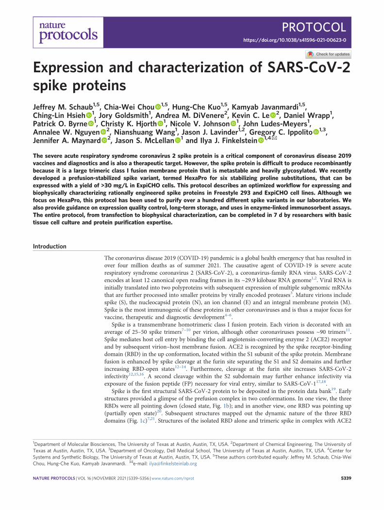
Spike is the first structural SARS-CoV-2 protein to be deposited in the protein data bank19. Earlystructures provided a glimpse of the prefusion complex in two conformations. In one view, the threeRBDs were all pointing down (closed state, Fig. 1b); and in another view, one RBD was pointing up(partially open state)20. Subsequent structures mapped out the dynamic nature of the three RBDdomains (Fig. 1c)7,21. Structures of the isolated RBD alone and trimeric spike in complex with ACE2
1Department of Molecular Biosciences, The University of Texas at Austin, Austin, TX, USA. 2Department of Chemical Engineering, The University ofTexas at Austin, Austin, TX, USA. 3Department of Oncology, Dell Medical School, The University of Texas at Austin, Austin, TX, USA. 4Center forSystems and Synthetic Biology, The University of Texas at Austin, Austin, TX, USA. 5These authors contributed equally: Jeffrey M. Schaub, Chia-WeiChou, Hung-Che Kuo, Kamyab Javanmardi. ✉e-mail: [email protected]
NATURE PROTOCOLS | VOL 16 |NOVEMBER 2021 | 5339–5356 |www.nature.com/nprot 5339
PROTOCOLhttps://doi.org/10.1038/s41596-021-00623-0
1234
5678
90():,;
1234567890():,;
were solved shortly thereafter16,22–25. These structures revealed that ACE2 interacts with the receptor-binding motif, approximately composed of residues 438–508 within the RBD26. SARS-CoV-2receptor-binding motif has a more pronounced receptor-interacting ridge and stabilizing residuesthat increase the affinity for ACE2 compared with SARS-CoV-127. The pace of spike structuresdeposited in the protein data bank continues to accelerate, with multiple high-resolution structures ofspike-binding antibodies and a structure of the post-fusion conformation18. We direct the reader toseveral reviews summarizing these structural findings28–31.
Development of the protocolPrefusion spike is the major target for vaccine and therapeutic antibody development. However,prefusion-stabilized spike is difficult to produce recombinantly because it is a large (~141 kDa permonomer), metastable and heavily glycosylated transmembrane protein. Nearly all structural studiesof spike have thus focused on the soluble ectodomain (Fig. 1a)20,28,29,32–35. The first prefusion-stabilized SARS-CoV-2 spike was informed by our structure-based engineering of the Middle Eastrespiratory syndrome (MERS) coronavirus S protein32. We showed that inserting two prolines in aloop between the first heptad repeat (HR1) and the central helix increased the homogeneity andrecombinant expression yields of prefusion MERS-CoV S ectodomains32. We and others followed a
d
4A8(PDB: 7C2L)
CR3022(PDB: 6W41)
c
Up
DownUp
90°817P892P
899P
942P
986P/987P
b
Down
DownDown
90°817P892P
899P
942P
986P/987P
a
NTD RBD
HR
1
CH
CD
FP
S1 S2
Glycans:
1 1,273
817P
892P89
9P94
2P98
6P98
7P
Fig. 1 | Overview of SARS-CoV-2 spike structure and antigenicity. a, Schematic of the SARS-CoV-2 spike domains subdivided by color. NTD,N-terminal domain; RBD, receptor-binding domain; FP, fusion peptide; HR1, heptad repeat 1; CH, central helix; CD, connector domain. White region isunresolved in cryo-EM structures of the ectodomain. Glycan positions noted on top of the primary structure as previously reported63. Scissors denotethe S1/S2 and S2′ proteolytic cleavage sites. Location of stabilizing HexaPro prolines is denoted. b,c, Spike structures in the three RBD down(PDB:6VXX) (b) and two RBD up prefusion states (PDB:6X2B) (c). Left: front view, Right: top view. HexaPro prolines shown in black. d, Compositestructure of a spike monomer bound to neutralizing antibodies CR3022 (PDB: 6W41) and 4A8 (PDB:7C2L).
PROTOCOL NATURE PROTOCOLS
5340 NATURE PROTOCOLS | VOL 16 |NOVEMBER 2021 | 5339–5356 |www.nature.com/nprot
similar strategy to insert two prolines into the SARS-CoV-2 ectodomain, termed S-2P (Table 1)19,20.However, S-2P yield continued to be relatively poor19. Thus, we developed a second-generationprefusion-stabilized SARS-CoV-2 ectodomain by introducing four additional prolines, termedHexaPro (Addgene, 154754), that further stabilized the trimeric spike structure in the prefusionconformation and increased recombinant protein yield by an additional tenfold above spike-2P36.Despite the increased stability of HexaPro, the RBDs continue to sample multiple positions36,37.
Here, we describe the expression, purification and biophysical characterization of HexaPro,a second-generation prefusion-stabilized SARS-CoV-2 spike glycoprotein from mammalian cells36.In addition to developing HexaPro, we have used this protocol to rapidly screen circulatingspike mutants, to develop new spike antigens, and for structure–function studies of spike con-formations37–40. We also describe three widely available assays to characterize recombinant spikes:(1) spike thermostability; (2) affinity for ACE2; and (3) binding by patient sera or monoclonalantibodies. Although beyond the scope of this paper, we also recommend visualizing recombinantspikes via negative-stain electron microscopy to check for monodispersity and the overall distributionof conformations. We anticipate that this protocol will be useful for research groups that needto express and purify spike variants and broadly applicable to studies of other trimeric viralfusion proteins.
Applications of the methodStructure–function studies of recombinant SARS-CoV-2 spike protein alone and in complex withACE2 are critical for understanding how the virus infects human cells2,3. Prefusion spike is in ametastable state that undergoes conformational ‘breathing’ of the RBDs19,37. This allows the threeRBDs to adopt an open conformation that can interact with the ACE2 cell surface receptors to triggerthe post-fusion conformation and viral entry. Binding affinity studies with recombinant SARS-CoV-2spike showed a higher affinity than SARS-CoV-1 for human ACE219,34. This interaction is unaffectedby the glycan shield near the RBD41.
Recombinant SARS-CoV-2 spike is essential for probing the immune response of convalescentpatients and developing monoclonal antibody (mAb) therapeutics. For example, enzyme-linkedimmunosorbent assays (ELISAs) can detect seroconverted individuals42. While the soluble RBD canbe used for some assays43,44, full-length spike is essential for characterizing antibodies that bindoutside the RBD, including the N-terminal domain (NTD45,46) and the S2 stalk47,48. Targeting theseregions of the protein is especially important because they provide alternative neutralizing epitopes
Table 1 | Prefusion-stabilized spike trimer variants and characteristics
Variant Stabilizing mutations Rationale Yield Cell line Ref
S-2P K986P, V987P, furina Proline 0.5 mg/L FreeStyle 293-F Wrapp et al.19
HexaPro K986P, V987P, F817P, A892P,A899P, A942P, furina
Proline 10.5 mg/L(FreeStyle 293-F)32.5 mg/L (ExpiCHO)
FreeStyle 293-FExpiCHO
Hsieh et al.36
rS2d K986P, V987P, S383C, D985C, furina Disulfide Not reported FreeStyle 293-F Henderson et al.21
u1S2q A570L T572I F855Y N856I (u1S2q)K986P, V987P, A570L, T572I, F855Y,N856I, furina
Cavity filling Not reported FreeStyle 293-F Henderson et al. 21
S-R/PP/x1 K986P, V987P, S383C, D985C, furin b Disulfide ~0.4 mg/L Expi293F Xiong et al.57
S-R/x2 K986P, G413C, V987C, furinb Disulfide ~0.4 mg/L Expi293F Xiong et al.57
S-closed +Fd K986P, V987P, D614N, A892P,A942P, V987P, furina
Cavity filling/proline
Sixfold increasedrelative to S-2P
Expi293F Juraszek et al.53
2P DS S K986P, V987P, S383C/D985C, furinc Disulfide Tenfold reducedrelative to S-2P
FreeStyle 293-F McCallum et al.56
N234A K986P, V987P, N234A Glycan deletion 2.0 mg/L FreeStyle 293-F Henderson et al.58
VFLIP F817P, A892P, A899P, A942P,K986P, Y707C, T883C, K986, furind
Proline,disulfide, linker
7.1 mg/L (HEK293F)157 mg/L (ExpiCHO)
HEK293FExpiCHO
Olmedillas et al.73
aS1/S2 furin cleavage site (682RRAR685) mutated to 682GSAS685bS1/S2 furin cleavage site mutated to 681PRRAR685.
cS1/S2 furin cleavage site mutated to 682SGAG685.dS1/S2 furin cleavage site
mutated to 675GGSGGGS691.
NATURE PROTOCOLS PROTOCOL
NATURE PROTOCOLS | VOL 16 |NOVEMBER 2021 | 5339–5356 |www.nature.com/nprot 5341
for antibody therapeutics40,49,50. In addition, serological assays with recombinant spike can measurepatient immune response and neutralizing convalescent plasma.
Prefusion spike is a critical antigenic target for eliciting neutralizing antibodies in leadingSARS-CoV-2 vaccine candidates51,52. Recombinant production of wild-type MERS and SARS-CoV-1spikes included a heterogeneous mixture of both pre- and post-fusion conformations32. Class I fusionproteins are inherently unstable, and the SARS-CoV-2 spike dissociates into the S1 andS2 subdomains. Immunization with constructs that adopt post-fusion conformations reduces specificS1 neutralizing or ACE2 blocking antibodies53. Therefore, leading SARS-CoV-2 subunit vaccines,including those from Pfizer, Moderna and NovaVax, are using prefusion-stabilized spikes51,52,54,55.These stabilized constructs display the spike proteins in a trimeric, prefusion conformation with thesame glycosylated residues as in the wild-type virus (but possibly different glycan linkages)20,55.Additionally, the stabilized constructs may increase the functional protein yield and further increasevaccine response. Thus, multiple vaccine candidates use stabilized, full-length spike glycoprotein toelicit an immune response.
Comparison with other methodsAlthough HexaPro is highly stable with increased yield, the following complementary approacheshave been reported to stabilize prefusion coronavirus spike proteins: (1) stabilizing the hinge loopundergoing conformational change to extended helix32; (2) adding disulfide bonds to prevent con-formational changes during the pre- to post-fusion states21; (3) introducing salt bridges to neutralizecharge imbalances36; (4) point-mutating to hydrophobic residues to fill internal cavities21,53; and(5) changing the surface glycosylation pattern21. Table 1 summarizes these approaches applied to theSARS-CoV-2 spike and the reported yields per liter of cell culture. One approach locks the RBD ina closed state via an engineered intermolecular disulfide bond between an RBD residue (S383C)and the hairpin preceding the S2 central helix (D985C) from a neighboring protomer21,56,57. Thecryo-electron microscopy (cryo-EM) structure of this variant (rS2d and S-R/PP/x1, Table 1) con-firmed that spike is covalently trapped in an all-down conformation21. Conformations favoring thethree RBD-down state may limit exposure of cryptic epitopes for antibody neutralization. A singlemutation of an N-glycan site (N234A) reduces the population of up-RBD spikes58. This change isattributed to additional occupancy of the cleft formed by the NTD and RBD. Another spike variantincreased overall yield five- to sixfold by adding one cavity-filling mutation and five proline sub-stitutions53. Finally, engineering spikes also abolish the furin cleavage site. Furin cleavage between S1and S2 is pivotal for SARS-CoV-2 pathogenesis12. Mutating the furin cleavage site prevents separationof the S1 and S2 domains and stabilizes the full-length prefusion ectodomain15.
LimitationsThe differences between HexaPro and other stabilized spike constructs may be especially notable inthe context of eliciting neutralizing antibodies, as the precise contribution of conformations tostimulating our immune response is unclear. HexaPro, additionally, removes the furin cleavage at theS1/S2 interface. This is crucial for the downstream utilization of stabilized spikes, such as viral uptakeassays. The presence of the furin cleavage site is additionally critical for understanding the viraltransmissibility in clinical variants such as D614G, where increased furin site accessibility andcleavage may be responsible for increase infectivity59. ELISAs utilizing the HexaPro stabilized trimershowed increased sensitivity for seroconversion compared with individual spike subunits60. Addi-tionally, HexaPro does not transition into the post-fusion conformation, eliminating any newlyexposed post-fusion antigenic sites18. Nonetheless, we anticipate that some epitopes may be obscuredby prefusion-stabilized HexaPro.
SARS-CoV-2 spike is highly glycosylated with 22 predicted N-linked glycosylated sites and fivepredicted O-linked glycosylated sites20,61,62. Several cryo-EM structures and mass spectrometryanalyses of recombinant spike purified from FreeStyle 293-F cells (HEK293 derivative) confirmed 22N-linked glycosylated sites, strong occupation of O-linked glycosylation at Thr323, and trace levels ofO-linked glycosylation at Thr32520,62,63. Recombinant spike has 89% complex-type and 11% oligo-mannose/hybrid N-glycans, whereas virus-derived spike has a slight difference, with 79% complex-type and 21% oligomannose/hybrid N-glycans64. Despite minimal large-scale N-glycan change,specific N-glycan sites (for example, Asn61, Asn234 and Asn603) are predominantly underprocessedoligomannose in virus-derived spike. In contrast, Asn234 was mixed complex and oligomannose/hybrid N-glycans, while Asn61 and Asn603 was mostly complex N-glycans in recombinant spike.
PROTOCOL NATURE PROTOCOLS
5342 NATURE PROTOCOLS | VOL 16 |NOVEMBER 2021 | 5339–5356 |www.nature.com/nprot
Moreover, Thr678 modifications identified on the virus-derived spike are absent on recombinantspike. Interestingly, multiple N-linked glycosylations and Thr323/Thr325 O-linked glycosylationsare located on the RBD64. The glycosylation patterns of recombinant spike purified from CHOcells are unexplored65,66. The functional differences in glycosylation patterns between HEK293- orCHO-purified spike are unknown62,63 but should be considered when interpreting differencesbetween binding affinities for ACE2 or mAbs across different constructs.
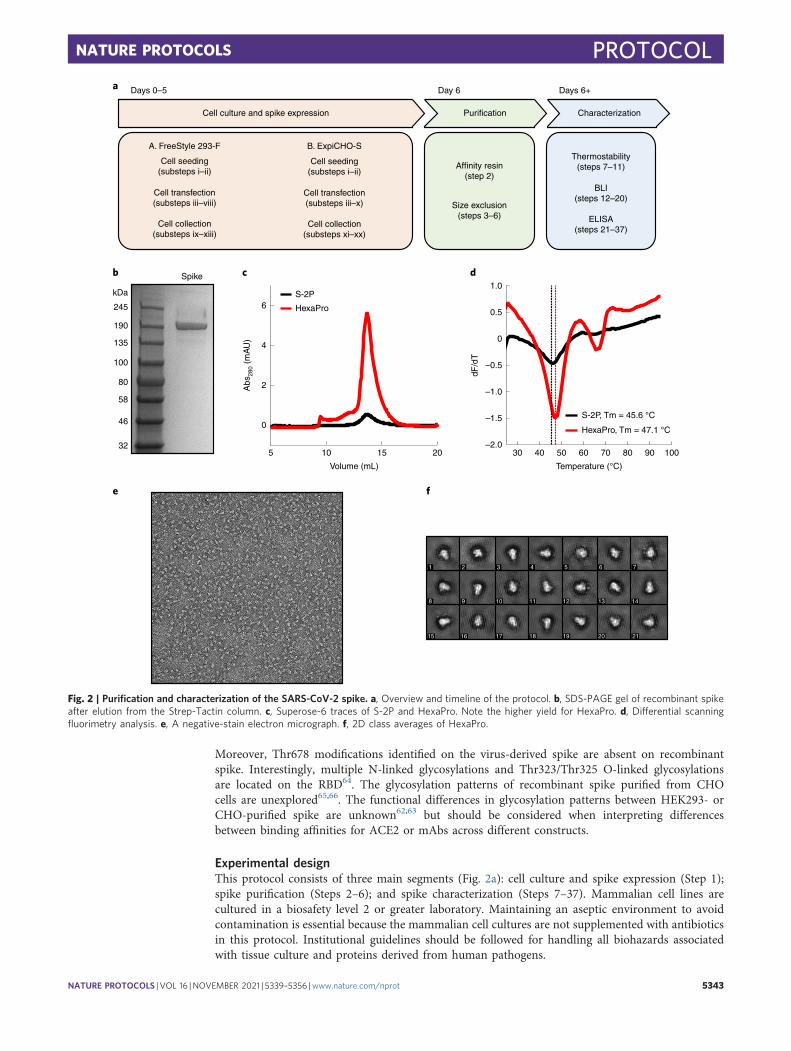
Experimental designThis protocol consists of three main segments (Fig. 2a): cell culture and spike expression (Step 1);spike purification (Steps 2–6); and spike characterization (Steps 7–37). Mammalian cell lines arecultured in a biosafety level 2 or greater laboratory. Maintaining an aseptic environment to avoidcontamination is essential because the mammalian cell cultures are not supplemented with antibioticsin this protocol. Institutional guidelines should be followed for handling all biohazards associatedwith tissue culture and proteins derived from human pathogens.
fe
d
Temperature (°C)
30 9040 10050 60 70 80
S-2P, Tm = 45.6 °C
HexaPro, Tm = 47.1 °C
c
Abs
280
(mA
U)
6
4
2
0
Volume (mL)
5 1510 20
S-2P
HexaPro
dF/d
T
1.0
–1.0
–1.5
–2.0
0.5
0
–0.5
b
245
190
135
100
80
58
46
32
kDa
Spike
a
Cell culture and spike expression Purification Characterization
Cell seeding(substeps i–ii)
Cell transfection(substeps iii–viii)
Cell collection(substeps ix–xiii)
Affinity resin(step 2)
Size exclusion(steps 3–6)
Thermostability(steps 7–11)
BLI(steps 12–20)
ELISA(steps 21–37)
A. FreeStyle 293-F B. ExpiCHO-S
Cell seeding(substeps i–ii)
Cell transfection(substeps iii–x)
Cell collection(substeps xi–xx)
Days 0–5 Day 6 Days 6+
1 2 3 4 5 6 7
8 9 10 11 12 13 14
15 16 17 18 19 20 21
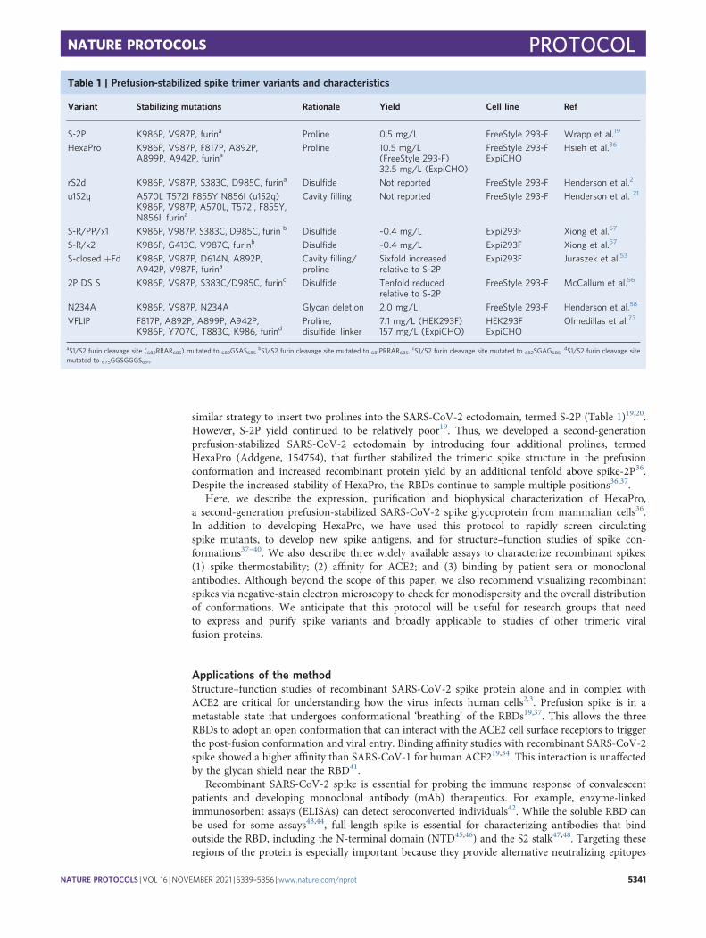
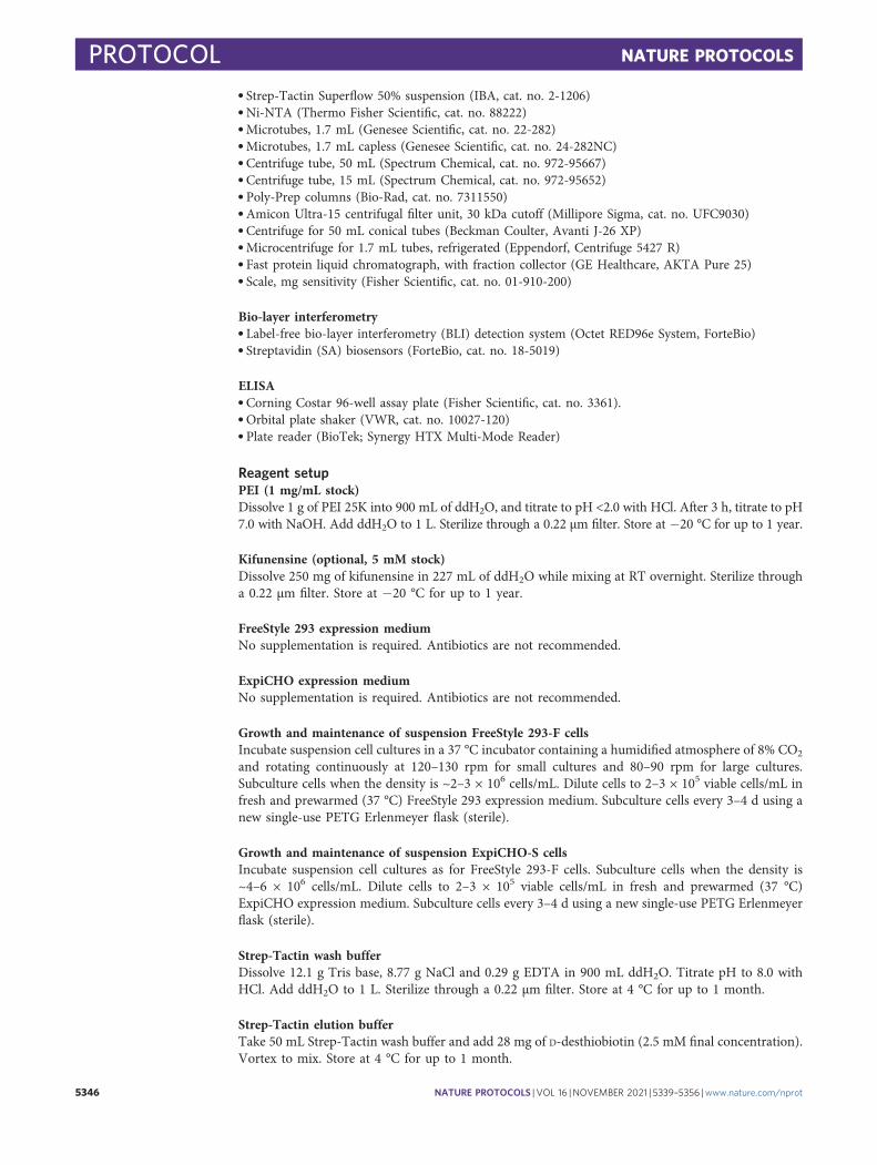
Fig. 2 | Purification and characterization of the SARS-CoV-2 spike. a, Overview and timeline of the protocol. b, SDS-PAGE gel of recombinant spikeafter elution from the Strep-Tactin column. c, Superose-6 traces of S-2P and HexaPro. Note the higher yield for HexaPro. d, Differential scanningfluorimetry analysis. e, A negative-stain electron micrograph. f, 2D class averages of HexaPro.
NATURE PROTOCOLS PROTOCOL
NATURE PROTOCOLS | VOL 16 |NOVEMBER 2021 | 5339–5356 |www.nature.com/nprot 5343
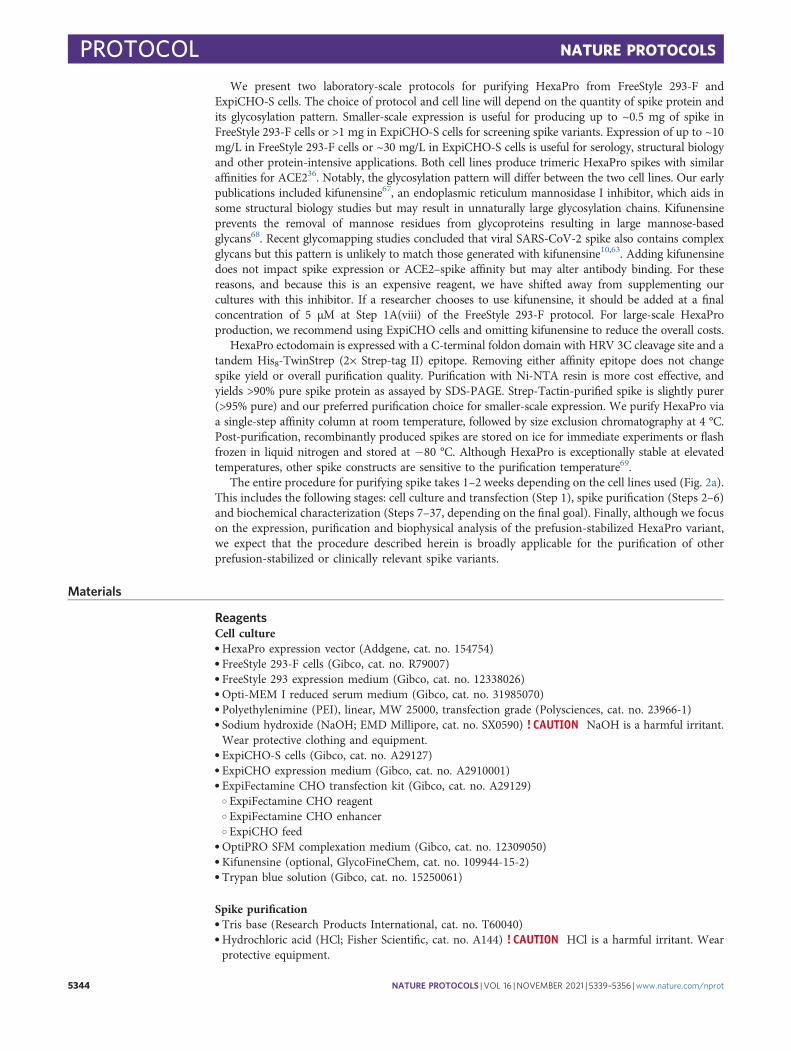
We present two laboratory-scale protocols for purifying HexaPro from FreeStyle 293-F andExpiCHO-S cells. The choice of protocol and cell line will depend on the quantity of spike protein andits glycosylation pattern. Smaller-scale expression is useful for producing up to ~0.5 mg of spike inFreeStyle 293-F cells or >1 mg in ExpiCHO-S cells for screening spike variants. Expression of up to ~10mg/L in FreeStyle 293-F cells or ~30 mg/L in ExpiCHO-S cells is useful for serology, structural biologyand other protein-intensive applications. Both cell lines produce trimeric HexaPro spikes with similaraffinities for ACE236. Notably, the glycosylation pattern will differ between the two cell lines. Our earlypublications included kifunensine67, an endoplasmic reticulum mannosidase I inhibitor, which aids insome structural biology studies but may result in unnaturally large glycosylation chains. Kifunensineprevents the removal of mannose residues from glycoproteins resulting in large mannose-basedglycans68. Recent glycomapping studies concluded that viral SARS-CoV-2 spike also contains complexglycans but this pattern is unlikely to match those generated with kifunensine10,63. Adding kifunensinedoes not impact spike expression or ACE2–spike affinity but may alter antibody binding. For thesereasons, and because this is an expensive reagent, we have shifted away from supplementing ourcultures with this inhibitor. If a researcher chooses to use kifunensine, it should be added at a finalconcentration of 5 µM at Step 1A(viii) of the FreeStyle 293-F protocol. For large-scale HexaProproduction, we recommend using ExpiCHO cells and omitting kifunensine to reduce the overall costs.
HexaPro ectodomain is expressed with a C-terminal foldon domain with HRV 3C cleavage site and atandem His8-TwinStrep (2× Strep-tag II) epitope. Removing either affinity epitope does not changespike yield or overall purification quality. Purification with Ni-NTA resin is more cost effective, andyields >90% pure spike protein as assayed by SDS-PAGE. Strep-Tactin-purified spike is slightly purer(>95% pure) and our preferred purification choice for smaller-scale expression. We purify HexaPro viaa single-step affinity column at room temperature, followed by size exclusion chromatography at 4 °C.Post-purification, recombinantly produced spikes are stored on ice for immediate experiments or flashfrozen in liquid nitrogen and stored at −80 °C. Although HexaPro is exceptionally stable at elevatedtemperatures, other spike constructs are sensitive to the purification temperature69.
The entire procedure for purifying spike takes 1–2 weeks depending on the cell lines used (Fig. 2a).This includes the following stages: cell culture and transfection (Step 1), spike purification (Steps 2–6)and biochemical characterization (Steps 7–37, depending on the final goal). Finally, although we focuson the expression, purification and biophysical analysis of the prefusion-stabilized HexaPro variant,we expect that the procedure described herein is broadly applicable for the purification of otherprefusion-stabilized or clinically relevant spike variants.
Materials
ReagentsCell culture● HexaPro expression vector (Addgene, cat. no. 154754)● FreeStyle 293-F cells (Gibco, cat. no. R79007)● FreeStyle 293 expression medium (Gibco, cat. no. 12338026)● Opti-MEM I reduced serum medium (Gibco, cat. no. 31985070)● Polyethylenimine (PEI), linear, MW 25000, transfection grade (Polysciences, cat. no. 23966-1)● Sodium hydroxide (NaOH; EMD Millipore, cat. no. SX0590) ! CAUTION NaOH is a harmful irritant.Wear protective clothing and equipment.
● ExpiCHO-S cells (Gibco, cat. no. A29127)● ExpiCHO expression medium (Gibco, cat. no. A2910001)● ExpiFectamine CHO transfection kit (Gibco, cat. no. A29129)
○ ExpiFectamine CHO reagent○ ExpiFectamine CHO enhancer○ ExpiCHO feed
● OptiPRO SFM complexation medium (Gibco, cat. no. 12309050)● Kifunensine (optional, GlycoFineChem, cat. no. 109944-15-2)● Trypan blue solution (Gibco, cat. no. 15250061)
Spike purification● Tris base (Research Products International, cat. no. T60040)● Hydrochloric acid (HCl; Fisher Scientific, cat. no. A144) ! CAUTION HCl is a harmful irritant. Wearprotective equipment.
PROTOCOL NATURE PROTOCOLS
5344 NATURE PROTOCOLS | VOL 16 |NOVEMBER 2021 | 5339–5356 |www.nature.com/nprot
● Sodium chloride (VWR, cat. no. BDH9286)● Ethylenediaminetetraacetic acid (EDTA; Fisher Scientific, cat. no. S312) ! CAUTION EDTA is aharmful irritant. Wear protective equipment.
● Sodium azide (Fisher Scientific, cat. no. S2271) ! CAUTION Sodium azide is toxic. Wear protective equipment.● Imidazole (Sigma-Aldrich, cat. no. 792527)● D-desthiobiotin (Sigma-Aldrich, cat. no. D1411)● 2× Laemmli sample buffer (Biorad, cat. no. 1610737)● 2-mercaptoethanol (Fisher Scientific, cat. no. BP176) ! CAUTION 2-mercaptoethanol is a harmfulirritant. Wear protective equipment.
Thermostability assay● SYPRO orange protein gel stain (Thermo Fisher Scientific, cat. no. S6650)● MicroAmp Fast Optical 96-Well Reaction Plate, 0.1 mL (Thermo Fisher Scientific, cat. no. 4346970)
Bio-layer interferometry● HBS-EP+ Buffer 10× (Cytiva, cat. no. BR100669)● Bovine serum albumin (New England Biolabs, cat. no. B9000S)
Enzyme-linked immunosorbent assay● Phosphate-buffered saline (PBS; Thermo Fisher Scientific, cat. no. 10010023)● Tween 20 (Sigma-Aldrich, cat. no. P9416)● Nonfat dry milk (Sigma-Aldrich, cat. no. M7409)● Anti-spike antibodies, such as mAb CR3022 (Abcam, cat. no. ab273073)● Anti-human IgG Fab horseradish peroxidase (HRP) (Sigma-Aldrich, cat. no. A0293)● 1-Step Ultra TMB ELISA substrate (Thermo Fisher Scientific, cat. no. 34028)● Sulfuric acid (H2SO4; Fisher Scientific, cat. no. 7664-93-9) ! CAUTION H2SO4 is a harmful irritant.Wear protective equipment.
EquipmentCell culture● Celltron benchtop shaker for CO2 incubators, 25 mm (Infors-HT, cat. no. I69222)● LUNA-II automated cell counter (Logos Biosystems, cat. no. L40002)● LUNA cell counting slides (Logos Biosystems, cat. no. L12001)● CellXpert C170 CO2 incubator (Eppendorf, cat. no. 6734010015)● CO2 gas● Laminar flow tissue culture hood● Water bath, 37 °C (Thermo Fisher Scientific, cat. no. TSGP05)● Nalgene; single-use polyethylene terephthalate (PETG) Erlenmeyer flasks with plain bottom: sterile(Thermo Fisher Scientific, cat. no. 4115-0125)
● Nalgene; single-use PETG Erlenmeyer flasks with plain bottom: sterile (Thermo Fisher Scientific,cat. no. 4115-0500)
● Nalgene; single-use PETG Erlenmeyer flasks with plain bottom: sterile (Thermo Fisher Scientific,cat. no. 4115-2800)
● 0.22 μm syringe tip filter (Genesee Scientific, cat. no. 25-244)● Plastic syringes 10 mL and 60 mL (BD, cat. nos. 302995 and 309653)● Centrifuge tube, 250 mL (Corning, cat. no. 430776)
Protein purification● pH probe (Fisher Scientific, cat. no. 01-913-996)● Water filtration source (Millipore Sigma)● Superose 6 Increase 10/300 GL (GE Healthcare, cat. no. 29091596)● SDS-PAGE gels, 4–15% (Biorad, cat. no. 4561084)● Electrophoresis equipment, MiniPROTEAN Tetra Cell (Biorad, cat. no. 1658001EDU)● Electrophoresis power supply (Biorad, cat. no. 1645050EDU)● Color pre-stained protein standard, broad range (NEB, cat. no. P7712S)● Electrophoresis gel stain (VWR, cat. no. 82021)● 10× Buffer R; Strep-Tactin regeneration buffer with HABA (IBA, cat. no. 2-1002)
NATURE PROTOCOLS PROTOCOL
NATURE PROTOCOLS | VOL 16 |NOVEMBER 2021 | 5339–5356 |www.nature.com/nprot 5345
● Strep-Tactin Superflow 50% suspension (IBA, cat. no. 2-1206)● Ni-NTA (Thermo Fisher Scientific, cat. no. 88222)● Microtubes, 1.7 mL (Genesee Scientific, cat. no. 22-282)● Microtubes, 1.7 mL capless (Genesee Scientific, cat. no. 24-282NC)● Centrifuge tube, 50 mL (Spectrum Chemical, cat. no. 972-95667)● Centrifuge tube, 15 mL (Spectrum Chemical, cat. no. 972-95652)● Poly-Prep columns (Bio-Rad, cat. no. 7311550)● Amicon Ultra-15 centrifugal filter unit, 30 kDa cutoff (Millipore Sigma, cat. no. UFC9030)● Centrifuge for 50 mL conical tubes (Beckman Coulter, Avanti J-26 XP)● Microcentrifuge for 1.7 mL tubes, refrigerated (Eppendorf, Centrifuge 5427 R)● Fast protein liquid chromatograph, with fraction collector (GE Healthcare, AKTA Pure 25)● Scale, mg sensitivity (Fisher Scientific, cat. no. 01-910-200)
Bio-layer interferometry● Label-free bio-layer interferometry (BLI) detection system (Octet RED96e System, ForteBio)● Streptavidin (SA) biosensors (ForteBio, cat. no. 18-5019)
ELISA● Corning Costar 96-well assay plate (Fisher Scientific, cat. no. 3361).● Orbital plate shaker (VWR, cat. no. 10027-120)● Plate reader (BioTek; Synergy HTX Multi-Mode Reader)
Reagent setupPEI (1 mg/mL stock)Dissolve 1 g of PEI 25K into 900 mL of ddH2O, and titrate to pH <2.0 with HCl. After 3 h, titrate to pH7.0 with NaOH. Add ddH2O to 1 L. Sterilize through a 0.22 μm filter. Store at −20 °C for up to 1 year.
Kifunensine (optional, 5 mM stock)Dissolve 250 mg of kifunensine in 227 mL of ddH2O while mixing at RT overnight. Sterilize througha 0.22 μm filter. Store at −20 °C for up to 1 year.
FreeStyle 293 expression mediumNo supplementation is required. Antibiotics are not recommended.
ExpiCHO expression mediumNo supplementation is required. Antibiotics are not recommended.
Growth and maintenance of suspension FreeStyle 293-F cellsIncubate suspension cell cultures in a 37 °C incubator containing a humidified atmosphere of 8% CO2
and rotating continuously at 120–130 rpm for small cultures and 80–90 rpm for large cultures.Subculture cells when the density is ~2–3 × 10⁶ cells/mL. Dilute cells to 2–3 × 10⁵ viable cells/mL infresh and prewarmed (37 °C) FreeStyle 293 expression medium. Subculture cells every 3–4 d using anew single-use PETG Erlenmeyer flask (sterile).
Growth and maintenance of suspension ExpiCHO-S cellsIncubate suspension cell cultures as for FreeStyle 293-F cells. Subculture cells when the density is~4–6 × 10⁶ cells/mL. Dilute cells to 2–3 × 10⁵ viable cells/mL in fresh and prewarmed (37 °C)ExpiCHO expression medium. Subculture cells every 3–4 d using a new single-use PETG Erlenmeyerflask (sterile).
Strep-Tactin wash bufferDissolve 12.1 g Tris base, 8.77 g NaCl and 0.29 g EDTA in 900 mL ddH2O. Titrate pH to 8.0 withHCl. Add ddH2O to 1 L. Sterilize through a 0.22 µm filter. Store at 4 °C for up to 1 month.
Strep-Tactin elution bufferTake 50 mL Strep-Tactin wash buffer and add 28 mg of D-desthiobiotin (2.5 mM final concentration).Vortex to mix. Store at 4 °C for up to 1 month.
PROTOCOL NATURE PROTOCOLS
5346 NATURE PROTOCOLS | VOL 16 |NOVEMBER 2021 | 5339–5356 |www.nature.com/nprot
Ni-NTA wash bufferDissolve 12.1 g Tris base, 8.77 g NaCl and 1.7 g imidazole (25 mM final) in 900 mL ddH2O. TitratepH to 8.0 with HCl. Add ddH2O to 1 L. Sterilize through a 0.22 µm filter. Store at 4 °C for up to1 month. Protect imidazole from excess light.
Ni-NTA elution bufferDissolve 6.06 g Tris base, 4.38 g NaCl and 8.51 g imidazole (250 mM final) in 500 mL ddH2O. TitratepH to 8.0 with HCl. Add ddH2O to 500 mL. Sterilize through a 0.22 µm filter. Store at 4 °C for up to1 month. Protect imidazole from excess light.
Superose 6 SEC bufferDissolve 0.24 g Tris base, 11.69 g NaCl and 0.2 g sodium azide in 900 mL ddH2O. Titrate pH to 8.0with HCl. Add ddH2O to 1 L. Sterilize through a 0.22 µm filter. Store at 4 °C for up to 1 month.
2× Laemmli sample buffer preparationMix 950 µL 2× Laemmli sample buffer and 50 µL 2-mercaptoethanol. Vortex to mix. Store at RTindefinitely.
PBST (0.1%)Mix 100 mL of PBS pH 7.5 with 100 μL of Tween 20. Sterilize through a 0.22 µm filter. Store at 4 °Cfor up to 1 month.
PBSTM (1%)Mix 100 mL of PBS pH 7.5 with 50 μL of Tween 20 and 1 g of nonfat dried milk. Sterilize through a0.22 µm filter. Store at 4 °C for up to 1 week.
BLI bufferDilute 5 mL of 10× HBS-EP+ buffer to 50 mL. Vortex to mix. Store at RT for up to 1 week.
Procedure
Cell culture and spike expression ● Timing 6–14 d1 Express SARS-CoV-2 spike in either FreeStyle 293-F (option A) or ExpiCHO-S (option B) cells.
! CAUTION Cell cultures are a potential biohazard. Ensure that mammalian cell work is performedin an approved laminar flow hood using aseptic technique. Follow institutional and governmentalguidelines for recommended personal protective equipment and waste disposal while working withcell cultures.? TROUBLESHOOTING(A) Expression in FreeStyle 293-F cells ● Timing 6 d
(i) Cell seeding. For small-scale expression, dilute cells to ~5 × 10⁵ cells/mL in a 125 mL single-use PETG Erlenmeyer flask to a total volume of 36 mL (~1.8 × 10⁷ cells) with fresh,prewarmed FreeStyle 293 expression medium. Alternatively, for large-scale expression, dilutecells to ~5 × 10⁵ cell/mL in a 2,800 mL single-use PETG Erlenmeyer flask to a total volume of900 mL (~4.5 × 10⁸ cells) with fresh, prewarmed FreeStyle 293 expression medium.
c CRITICAL STEP All cell culture solutions and equipment must be sterile. Culturesuspension should not exceed 30–40% of the total flask volume. Ensure that cell viability isalways >95%.
(ii) Incubate cultures at 37 °C with 8% CO2, shaking (120–130 rpm for small scale or80–90 rpm for large scale) for 16–20 h.
(iii) Cell transfection. Mix 20 µL of seeded culture with 20 µL of trypan blue solution (1:1 ratio),and assess viability and density on a cell counting slide. Ensure that cell density is ~1.0–1.2 ×10⁶ cells/mL and cell viability is >95% for optimal transfection efficiency.? TROUBLESHOOTING
(iv) For small-scale experiments, add 2 mL of room temperature (RT, 25 °C) OptiMEM toa labeled 15 mL conical tube for each transfection, add 25 µg of plasmid DNA to each tubeand mix by vortexing. For large-scale experiments, add 50 mL of RT OptiMEM to a labeled50 mL conical tube for each transfection, add 1 mg of plasmid DNA to each tube and mix byvortexing.
NATURE PROTOCOLS PROTOCOL
NATURE PROTOCOLS | VOL 16 |NOVEMBER 2021 | 5339–5356 |www.nature.com/nprot 5347
(v) Create a transfection mix by adding 3 µL PEI per 1 µg plasmid DNA and OptiMEM (2 mL ina 15 mL conical tube per small-scale transfection or 50 mL in a 250 mL conical tube perlarge-scale transfection) for each transfection. Mix by vortexing.
(vi) Filter the OptiMEM/plasmid DNA mix (2 mL for small scale or 50 mL for large scale)through a 0.22 µm syringe filter into the conical tube containing an equal volume oftransfection mix. Mix by inversion.
(vii) Wait 20 min and slowly add each final transfection mix (4 mL for small scale or 100 mL forlarge scale) to the cell cultures.
(viii) Incubate cultures at 37 °C with 8% CO2, shaking until time for collection.
c CRITICAL STEP Ensure that the plasmid DNA is endotoxin free and concentrated to>500 ng/μL. Do not syringe filter the transfection master mix or the final transfection mixafter the DNA and PEI have been added. This will reduce the transfection efficiency.
(ix) Cell collection. Pour cultures into 50 mL (small scale) or 250 mL (large scale) conical tubes.(x) Centrifuge at 500g for 10 min at 4 °C to separate cells from the supernatant.(xi) Transfer the supernatant to fresh tubes without disturbing the cell pellet.(xii) Centrifuge supernatants at 10,000g for 20 min at 4 °C to separate debris from the super-
natant. Transfer purified supernatant to fresh conical tubes without disturbing the pellet.(xiii) Keep supernatant at 4 °C for same-day protein purification.
j PAUSE POINT Store supernatants at −80 °C for later purification. We did not observe anychanges in purified spike quality between supernatants that were frozen at −80 °C and thosethat were used immediately.
(B) Expression in ExpiCHO-S Cells ● Timing 14 d(i) Cell seeding. For small-scale expression, dilute cells to ~4 × 10⁶ cells/mL in a 125 mL
single-use PETG Erlenmeyer flask to a total volume of 25 mL (~1 × 10⁸ cells) with fresh,prewarmed ExpiCHO expression medium. For large-scale expression, dilute cells to~4 × 10⁶ cells/mL in a 2,800 mL single-use PETG Erlenmeyer flask to a total volume of750 mL (~3 × 10⁹ cells) with fresh, prewarmed ExpiCHO expression medium.
c CRITICAL STEP Ensure that cell viability is always >95% prior to transfections. Avoidvortexing cells.? TROUBLESHOOTING
(ii) Incubate cultures at 37 °C with 8% CO2, shaking (120–130 rpm for small scale or80–90 rpm for large scale) for 16–20 h.
(iii) Cell transfection. Mix 20 µL of seeded culture with 20 µL of trypan blue solution (1:1 ratio),and assess viability and density on a cell counting slide. Ensure that cell density is ~7–10 ×106 cells/mL and cell viability is >95% for optimal transfection efficiency. Dilute cultures to6 × 106 cells/mL by discarding excess cells and diluting with fresh, prewarmed ExpiCHOexpression medium back to the original culture volume.
(iv) Chill the ExpiFectamine CHO reagent and plasmid DNA to 4 °C.(v) For small-scale experiments, add 1 mL of OptiPRO medium and 20 µg of plasmid DNA to
a 1.5 mL tube for each transfection and mix by inversion. For large-scale experiments, add30 mL of OptiPRO medium and 600 µg of plasmid DNA to a 50 mL conical tube for eachtransfection and mix by inversion.
(vi) Invert the ExpiFectamine CHO reagent several times prior to use.(vii) For small-scale experiments, add 80 µL of ExpiFectamine CHO reagent to 920 µL of
OptiPRO medium in a 1.5 mL tube and mix by inversion. For large-scale experiments, add2.4 mL of ExpiFectamine CHO reagent to 28 mL of OptiPRO medium in a fresh 250 mLconical tube and mix by inversion.
(viii) Add the diluted plasmid DNA from step (v) to the ExpiFectamine CHO solution from step(vii), and mix by inversion.
(ix) Incubate ExpiFectamine CHO/plasmid DNA complex at RT for 5 min. Then, slowly addthe solution to the diluted cell culture from step (iii), swirling the flask gently during theaddition.
(x) Incubate the flask in a humidified atmosphere of 8% CO2 for 18–22 h, shaking at 37 °C.
c CRITICAL STEP Ensure that stock plasmid DNA concentrations are >500 ng/μL andendotoxin free. Do not add ExpiFectamine CHO/plasmid DNA solution to cells quickly.This will reduce titers.
(xi) Adding enhancers. For small-scale experiments, add 150 µL of ExpiFectamine CHOEnhancer and 6 mL of ExpiCHO Feed to each flask, swirling the flask gently during the
PROTOCOL NATURE PROTOCOLS
5348 NATURE PROTOCOLS | VOL 16 |NOVEMBER 2021 | 5339–5356 |www.nature.com/nprot
addition. For large-scale experiments, add 4.5 mL of ExpiFectamine CHO Enhancer and180 mL of ExpiCHO Feed to each flask, swirling the flask gently during the addition.
(xii) Incubate the flask in a humidified atmosphere of 5% CO2, shaking at 32 °C.(xiii) First supernatant collection. Pour cultures into labeled conical tubes.(xiv) Centrifuge at 500g for 10 min at 4 °C to separate cells from the supernatant.(xv) Transfer the protein containing supernatant to fresh, labeled conical tubes without
disturbing the cell pellet.(xvi) Resuspend the cell pellet with fresh, prewarmed ExpiCHO expression medium (29 mL
for small-scale experiments and 750 mL for large-scale experiments), and transferback to the original flask. Add ExpiCHO Feed to the flask (6 mL for small-scaleexperiments or 180 mL for large-scale experiments), swirling the flask gently during theaddition.
(xvii) Incubate the flask in a humidified atmosphere of 5% CO2, shaking at 32 °C.j PAUSE POINT The protein containing supernatant from step (xv) can either be stored at
−80 °C to be combined with the supernatant from the day 12 collection (see below) orpurified immediately.
c CRITICAL STEP Avoid contamination of cell cultures during supernatant removal byusing aseptic technique.
(xviii) Second supernatant collection. Pour cultures into labeled conical tubes.(xix) Centrifuge at 500g for 10 min at 4 °C to separate cells from the supernatant.(xx) Transfer the protein containing supernatant to fresh, labeled conical tubes without
disturbing the cell pellet.
j PAUSE POINT The protein containing supernatants from Step 1B(xx) can be combinedand purified or can be stored long term at −80 °C.
Spike purification ● Timing 6 h2 Purify the SARS-CoV-2 spike using either a Strep-Tactin (option A) or a Ni-NTA (option B) resin.
(A) Purification with Strep-Tactin resin ● Timing 4 h(i) Add 2 mL of Strep-Tactin resin 50% slurry to Poly-Prep columns (Biorad). Equilibrate the
resin with 5 mL Strep-Tactin wash buffer (five column volumes).(ii) Add cleared supernatant to the column. To improve spike retention, reduce the flow rate to
<3 mL min−1.(iii) Wash the column with 5 mL Strep-Tactin wash buffer (five column volumes).(iv) Add 4 mL Strep-Tactin elution buffer for protein elution, and collect eluate in a single tube.(v) Concentrate the eluate in a 30 kDa cutoff spin concentrator (Amicon Ultra-15) at 4,000g
for 5 min (4 °C) or until concentrated to ~5 mg mL−1 (maximum 10 mg mL−1) or below500 μL if the protein will be further separated via size exclusion chromatography. Aliquotand store at −80 °C in Superose 6 SEC Buffer, or continue to the size exclusionchromatography step.
(B) Purification with Ni-NTA resin ● Timing 4 h(i) Add 2 mL of Ni-NTA resin slurry (50% beads in 30% ethanol) to Poly-Prep Columns
(Biorad). Equilibrate the resin with 5 mL Ni-NTA wash buffer (five column volumes).(ii) Supplement cleared supernatant with 10 mM imidazole, and add to the column for protein
binding. Reduce the flow rate to <3 mL/min to improve spike retention.(iii) Wash the column with 5 mL Ni-NTA wash buffer (five column volumes).(iv) Add 4 mL Ni-NTA elution buffer for protein elution, and collect eluate.(v) Concentrate the eluate in a 30 kDa cutoff spin concentrator (Amicon Ultra-15) at 4,000g
for 5 min (4 °C) or until concentrated to ~5 mg mL−1 (maximum 10 mg mL−1) or below500 μL if the protein will be further separated via size exclusion chromatography. Aliquotand store at −80 °C in Superose 6 SEC Buffer, or continue to the size exclusionchromatography step.? TROUBLESHOOTING
3 Equilibrate Superose 6 Column (Superose 6 Increase 10/300 GL, GE) on a fast protein liquidchromatograph with >1 column volume of Superose 6 SEC buffer.
4 Load purified spike protein into a 500 μL loop. Inject the sample at 0.5 mL/min. Collect 0.5 mLfractions.
5 Pool peak fractions based on the chromatogram. Peak fraction should elute between 13 and 14 mL.
NATURE PROTOCOLS PROTOCOL
NATURE PROTOCOLS | VOL 16 |NOVEMBER 2021 | 5339–5356 |www.nature.com/nprot 5349
6 Concentrate the eluate in a 30 kDa cutoff spin concentrator (Amicon Ultra-15) at 4,000g for 5 min(4 °C) or until concentrated to ~5 mg mL−1 (maximum 10 mg mL−1). Aliquot and store at −80 °C,or continue to thermostability assay.
j PAUSE POINT Concentrated protein can be flash frozen in liquid nitrogen. Plan to minimizefreeze–thaw cycles. We recommend only a single freeze–thaw cycle and to store concentrationprotein for downstream assays or size exclusion chromatography.
(Optional) Thermostability assay ● Timing 2 h7 Dilute spike protein to a final concentration of 0.25 mg/mL, and deposit 15 μL per well into a
MicroAmp Fast 96-Well Reaction Plate (0.1 mL). Plan to have enough protein for at least threereplicates per sample.
8 Create a SYPRO Orange solution by diluting the 5,000× stock to 20× (0.5 μL in 125 μL buffer).Make sure that your SYPRO Orange dilution buffer matches your protein buffer.
9 Inject 5 μL of 20× SYPRO Orange into each protein-containing well for a final concentration of1× spike, 5× SYPRO Orange and a volume of 20 μL.
10 Cover the plate using an optical adhesive cover, and gently vortex for ~15 s. Once the plate has beenadequately mixed, centrifuge to eliminate any air bubbles (600 rcf for 3 min).
11 Perform continuous fluorescence measurements (λex = 465 nm, λem = 580 nm) on differentialscanning fluorimetry, using a ramp rate of 4 °C/min from 25 °C to 95 °C.
(Optional) Bio-layer interferometry ● Timing 3 h12 Set instrument parameters with a shaking rate of 1,000 rpm at 25 °C, although higher temperatures
can also be used as necessary.13 Dilute spike variants to 10 nM. Serial dilute ACE2 to 200 nM, 100 nM, 50 nM and 25 nM in buffer.
Include a well without ACE2 as a reference.14 Immerse SA sensor tips in BLI buffer for at least 600 s.
? TROUBLESHOOTING15 Immobilize spike variants on the tips until 0.6 response (nm) is achieved, and move to the next step.16 Equilibrate spike-binding tips in BLI buffer for 60 s.17 Move the sensor tips to the wells with indicated concentrations of ACE2, and immerse for 600 s for
the association step. Include a well with no ACE2 as reference.? TROUBLESHOOTING
18 Move the sensor tips to other wells containing BLI buffer and immerse for 600 s as the dissociation step.19 Subtract all the data with reference sensor, and align curves to the baseline.20 Fit the curves globally with a 1:1 binding model. Care should be taken to use an appropriate model
when fitting antibody binding curves because these also include additional avidity effects. To aidfurther experiment design, we direct the reader to several excellent BLI reviews and methods70–72.
(Optional) Enzyme-linked immunosorbent assays ● Timing 2 d! CAUTION Handle patient serum (or plasma) using universal precautions for the prevention ofbloodborne pathogens. Work should be performed in a biosafety level 2 rated laboratory while wearingpersonal protective equipment such as gloves and safety glasses.21 Prepare coating antigens (SARS-CoV-2 spike ectodomain and SARS-CoV-2 RBD) at 2 μg/mL in
PBS (pH 7.5), and add 50 μL/well to a 96-well assay plate (high-binding polystyrene, Corning,cat. no. 3361).
22 Incubate at 4 °C, shaking at 100 rpm (2 mm orbital shaker) overnight.23 Discard coating antigen, and blot plate dry on paper towels. Dispense 200 μL/well of 2% milk in
PBS, and block plate at RT for 2 h.24 Wash plate three times with 300 μL/well of PBST (PBS with 0.1% Tween20).25 Add 50 μL/well PBSMT (1% milk in PBS, 0.05% Tween 20) to blocked plate.26 Prepare samples, along with a negative serum control (for our study, we used donor GNEG from
July 2019) and positive control mAb or serum, in a separate PCR plate by adding 6 μL of serum to94 μL of PBSMT (1:16.7). Prepare positive control mAb similarly by diluting in PBSMT to desiredconcentration (for our study, we used mAb CR3022 at 9 μg/mL).
27 Mix samples by pipetting up and down, and spin down plate at RT for 2 min at 4,000g.28 Serially dilute samples using a multichannel pipette to transfer 25 μL of each sample from
PCR plate into the initial dilution series on the ELISA plate (e.g., row A—first dilution at 1:50 serum
PROTOCOL NATURE PROTOCOLS
5350 NATURE PROTOCOLS | VOL 16 |NOVEMBER 2021 | 5339–5356 |www.nature.com/nprot
in PBSMT). Pipette up and down ten times to mix, and transfer 25 μL into the next row, repeatingprocess to serially dilute samples threefold per series across the ELISA plate.
c CRITICAL STEP Pipette serial dilutions with care to avoid propagating error. Allow HRPsubstrate to reach RT prior to use. Do not use cold HRP substrate.
29 Incubate plate at RT for 1 h for binding.30 After binding, wash plate three times with 300 μL/well of PBST.31 Prepare anti-human IgG Fab HRP (Sigma A0293) at 1:5,000 dilution in PBSTM. Add this
secondary antibody working solution to plate at 50 μL/well.
ckon : 4.0 × 10⁵ ± 6 × 10³ (M−1s−1)
koff : 6.3 × 10⁻³ ± 3 × 10⁻⁵ (s−1)
kD : 15.7 ± 0.2 nM
Time (s)
0.0
0.02
0.04
0.06
0 200
Res
pons
e (n
m)
400 600 800 1,000 1,200
ACE2 concentration (nM)200100502512.5
Time (s)
Res
pons
e (n
m)
Spike only
ACE2 only
0.0
0.2
0.4
0.6
0 100 200 500 1,000 1,500
b
Spike + ACE2
Association DissociationBaseline
Bas
elin
e
Load
inga
Spike
ACE2
Streptavidin
⥮
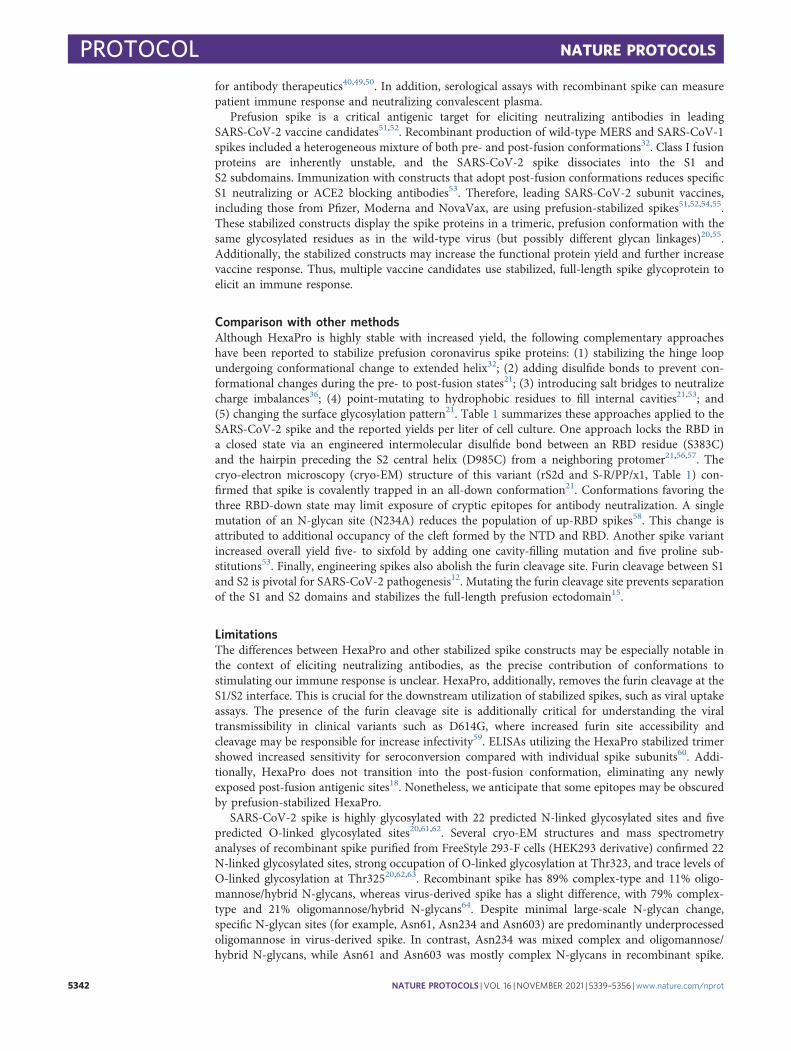
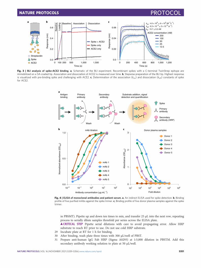
Fig. 3 | BLI analysis of spike-ACE2 binding. a, Schematic of the BLI experiment. Recombinant spikes with a C-terminal TwinStrep epitope areimmobilized on a SA-coated tip. Association and dissociation of ACE2 is measured over time. b, Stepwise preparation of the BLI tip. Highest responseis visualized with pre-binding spike and challenging with ACE2. c, Determination of the association (kon) and dissociation (koff) constants of spikefor ACE2.
c Donor plasma samples
Abs
orba
nce
(OD
450)
3
2
1
0
Fold dilution
101 102 103 104 106105
Donor 1
Donor 2
Donor 3
Donor 4
Donor 5
b mAb titration
Antibody concentration (μg mL–1)
Nor
mal
ized
abs
orba
nce
(OD
450)
1.0
0.5
0.0
10–2 10–1 100 101 102
mAb 1
mAb 2
mAb 3
mAb 4
mAb 5
aAntigenbinding
Primaryantibody
Secondaryantibody
Substrate addition, signaldetection and quantification
Wash Wash
Spike
Primaryantibody
Secondaryantibody (HRP)
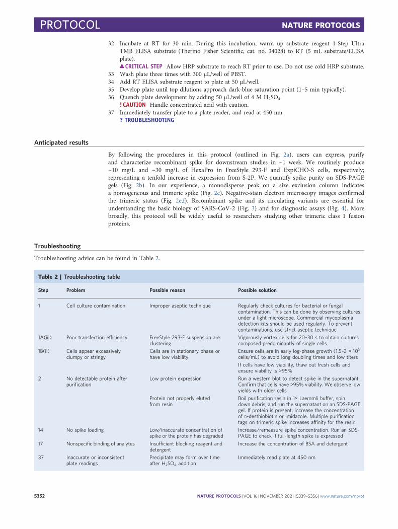
Fig. 4 | ELISA of monoclonal antibodies and patient serum. a, An indirect ELISA used for spike detection. b, Bindingprofile of five purified mAbs against the spike trimer. c, Binding profile of five donor plasma samples against the spiketrimer.
NATURE PROTOCOLS PROTOCOL
NATURE PROTOCOLS | VOL 16 |NOVEMBER 2021 | 5339–5356 |www.nature.com/nprot 5351
32 Incubate at RT for 30 min. During this incubation, warm up substrate reagent 1-Step UltraTMB ELISA substrate (Thermo Fisher Scientific, cat. no. 34028) to RT (5 mL substrate/ELISAplate).
c CRITICAL STEP Allow HRP substrate to reach RT prior to use. Do not use cold HRP substrate.33 Wash plate three times with 300 μL/well of PBST.34 Add RT ELISA substrate reagent to plate at 50 μL/well.35 Develop plate until top dilutions approach dark-blue saturation point (1–5 min typically).36 Quench plate development by adding 50 μL/well of 4 M H2SO4.
! CAUTION Handle concentrated acid with caution.37 Immediately transfer plate to a plate reader, and read at 450 nm.
? TROUBLESHOOTING
Anticipated results
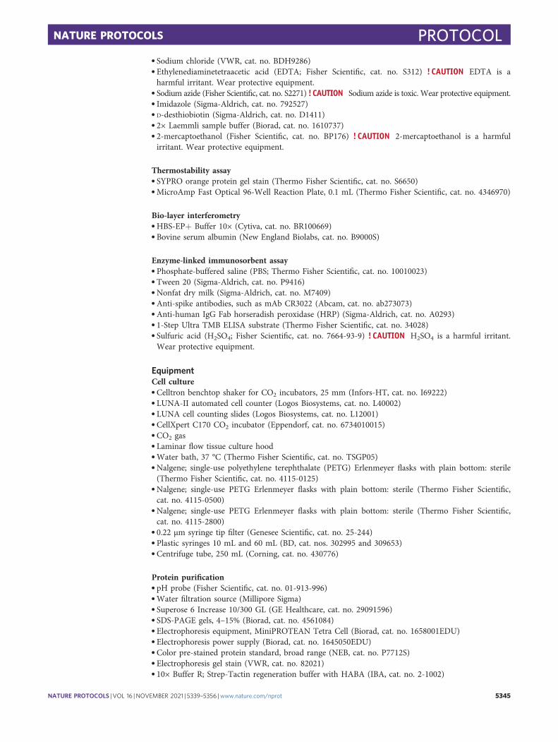
By following the procedures in this protocol (outlined in Fig. 2a), users can express, purifyand characterize recombinant spike for downstream studies in ~1 week. We routinely produce~10 mg/L and ~30 mg/L of HexaPro in FreeStyle 293-F and ExpiCHO-S cells, respectively;representing a tenfold increase in expression from S-2P. We quantify spike purity on SDS-PAGEgels (Fig. 2b). In our experience, a monodisperse peak on a size exclusion column indicatesa homogeneous and trimeric spike (Fig. 2c). Negative-stain electron microscopy images confirmedthe trimeric status (Fig. 2e,f). Recombinant spike and its circulating variants are essential forunderstanding the basic biology of SARS-CoV-2 (Fig. 3) and for diagnostic assays (Fig. 4). Morebroadly, this protocol will be widely useful to researchers studying other trimeric class 1 fusionproteins.
Troubleshooting
Troubleshooting advice can be found in Table 2.
Table 2 | Troubleshooting table
Step Problem Possible reason Possible solution
1 Cell culture contamination Improper aseptic technique Regularly check cultures for bacterial or fungalcontamination. This can be done by observing culturesunder a light microscope. Commercial mycoplasmadetection kits should be used regularly. To preventcontaminations, use strict aseptic technique
1A(iii) Poor transfection efficiency FreeStyle 293-F suspension areclustering
Vigorously vortex cells for 20–30 s to obtain culturescomposed predominantly of single cells
1B(ii) Cells appear excessivelyclumpy or stringy
Cells are in stationary phase orhave low viability
Ensure cells are in early log-phase growth (1.5–3 × 10⁵
cells/mL) to avoid long doubling times and low titers
If cells have low viability, thaw out fresh cells andensure viability is >95%
2 No detectable protein afterpurification
Low protein expression Run a western blot to detect spike in the supernatant.Confirm that cells have >95% viability. We observe lowyields with older cells
Protein not properly elutedfrom resin
Boil purification resin in 1× Laemmli buffer, spindown debris, and run the supernatant on an SDS-PAGEgel. If protein is present, increase the concentrationof D-desthiobiotin or imidazole. Multiple purificationtags on trimeric spike increases affinity for the resin
14 No spike loading Low/inaccurate concentration ofspike or the protein has degraded
Increase/remeasure spike concentration. Run an SDS-PAGE to check if full-length spike is expressed
17 Nonspecific binding of analytes Insufficient blocking reagent anddetergent
Increase the concentration of BSA and detergent
37 Inaccurate or inconsistentplate readings
Precipitate may form over timeafter H2SO4 addition
Immediately read plate at 450 nm
PROTOCOL NATURE PROTOCOLS
5352 NATURE PROTOCOLS | VOL 16 |NOVEMBER 2021 | 5339–5356 |www.nature.com/nprot
Timing
Step 1, cell culture and spike expression: 6–14 dA, expression in FreeStyle 293-F cells: 6 d
i–ii, cell seeding: 20 miniii–viii, cell transfection: 4 hix–xiii, cell collection: 1 h
B, expression in ExpiCHO-S cells: 14 di–ii, cell seeding: 20 miniii–x, cell transfection: 4 hxi–xii, adding enhancers: 20 minxiii–xvii, first supernatant collection: 30 minxviii–xx, second supernatant collection: 30 min
Steps 2–6, spike purification, 6 hA, purification with Strep-Tactin resin: 4 hB, purification with Ni-NTA resin: 4 h
Steps 7–11, thermostability assay, 2 hSteps 12–20, BLI, 3 hSteps 21–37, ELISA, 2 d
Reporting SummaryFurther information on research design is available in the Nature Research Reporting Summarylinked to this article.
Data availabilityThe data presented in these figures were generated as part of ref. 36. Raw files associated with Fig. 3are available from the corresponding author upon request.
References
1. Kim, D. et al. The architecture of SARS-CoV-2 transcriptome. Cell 181, 914–921.e10 (2020).2. Finkel, Y. et al. The coding capacity of SARS-CoV-2. Nature https://doi.org/10.1038/s41586-020-2739-1
(2020).3. Ullrich, S. & Nitsche, C. The SARS-CoV-2 main protease as drug target. Bioorg. Med. Chem. Lett. 30, 127377
(2020).4. Buchholz, U. J. et al. Contributions of the structural proteins of severe acute respiratory syndrome
coronavirus to protective immunity. Proc. Natl Acad. Sci. USA 101, 9804–9809 (2004).5. Gavor, E., Choong, Y. K., Er, S. Y., Sivaraman, H. & Sivaraman, J. Structural basis of SARS-CoV-2 and
SARS-CoV antibody interactions. Trends Immunol. 41, 1006–1022 (2020).6. Jeyanathan, M. et al. Immunological considerations for COVID-19 vaccine strategies. Nat. Rev. Immunol. 20,
615–632 (2020).7. Ke, Z. et al. Structures and distributions of SARS-CoV-2 spike proteins on intact virions. Nature
https://doi.org/10.1038/s41586-020-2665-2 (2020).8. Turoňová, B. et al. In situ structural analysis of SARS-CoV-2 spike reveals flexibility mediated by three
hinges. Science https://doi.org/10.1126/science.abd5223 (2020).9. Klein, S. et al. SARS-CoV-2 structure and replication characterized by in situ cryo-electron tomography.
Nat. Commun. 11, 5885 (2020).10. Yao, H. et al. Molecular architecture of the SARS-CoV-2 virus. Cell 183, 730–738.e13 (2020).11. Neuman, B. W. et al. A structural analysis of M protein in coronavirus assembly and morphology. J. Struct.
Biol. 174, 11–22 (2011).12. Hoffmann, M., Kleine-Weber, H. & Pöhlmann, S. A multibasic cleavage site in the spike protein of
SARS-CoV-2 is essential for infection of human lung cells. Mol. Cell 78, 779–784.e5 (2020).13. Coutard, B. et al. The spike glycoprotein of the new coronavirus 2019-nCoV contains a furin-like cleavage
site absent in CoV of the same clade. Antivir. Res. 176, 104742 (2020).14. Wrobel, A. G. et al. SARS-CoV-2 and bat RaTG13 spike glycoprotein structures inform on virus evolution
and furin-cleavage effects. Nat. Struct. Mol. Biol. 27, 763–767 (2020).15. Papa, G. et al. Furin cleavage of SARS-CoV-2 spike promotes but is not essential for infection and cell-cell
fusion. PloS Pathog. 17, e1009246 (2021).16. Shang, J. et al. Cell entry mechanisms of SARS-CoV-2. Proc. Natl Acad. Sci. USA 117, 11727–11734 (2020).17. Belouzard, S., Chu, V. C. & Whittaker, G. R. Activation of the SARS coronavirus spike protein via sequential
proteolytic cleavage at two distinct sites. Proc. Natl Acad. Sci. USA 106, 5871–5876 (2009).18. Cai, Y. et al. Distinct conformational states of SARS-CoV-2 spike protein. Science 369, 1586–1592 (2020).
NATURE PROTOCOLS PROTOCOL
NATURE PROTOCOLS | VOL 16 |NOVEMBER 2021 | 5339–5356 |www.nature.com/nprot 5353
19. Wrapp, D. et al. Cryo-EM structure of the 2019-nCoV spike in the prefusion conformation. Science 367,1260–1263 (2020).
20. Walls, A. C. et al. Structure, function, and antigenicity of the SARS-CoV-2 spike glycoprotein. Cell 181,281–292.e6 (2020).
21. Henderson, R. et al. Controlling the SARS-CoV-2 spike glycoprotein conformation. Nat. Struct. Mol. Biol. 27,925–933 (2020).
22. Lan, J. et al. Structure of the SARS-CoV-2 spike receptor-binding domain bound to the ACE2 receptor.Nature 581, 215–220 (2020).
23. Yan, R. et al. Structural basis for the recognition of SARS-CoV-2 by full-length human ACE2. Science 367,1444–1448 (2020).
24. Benton, D. J. et al. Receptor binding and priming of the spike protein of SARS-CoV-2 for membrane fusion.Nature 588, 327–330 (2020).
25. Zhou, T. et al. Cryo-EM structures of SARS-CoV-2 spike without and with ACE2 reveal a pH-dependentswitch to mediate endosomal positioning of receptor-binding domains. Cell Host Microbe 28, 867–879.e5(2020).
26. Yi, C. et al. Key residues of the receptor binding motif in the spike protein of SARS-CoV-2 that interact withACE2 and neutralizing antibodies. Cell Mol. Immunol. 17, 621–630 (2020).
27. Shang, J. et al. Structural basis of receptor recognition by SARS-CoV-2. Nature 581, 221–224 (2020).28. Huang, Y., Yang, C., Xu, X.-F., Xu, W. & Liu, S.-W. Structural and functional properties of SARS-CoV-2
spike protein: potential antivirus drug development for COVID-19. Acta Pharmacol. Sin. 41, 1141–1149(2020).
29. Sternberg, A. & Naujokat, C. Structural features of coronavirus SARS-CoV-2 spike protein: targets forvaccination. Life Sci. 257, 118056 (2020).
30. Chen, J., Gao, K., Wang, R., Nguyen, D. D. & Wei, G.-W. Review of COVID-19 antibody therapies.Annu. Rev. Biophys. https://doi.org/10.1146/annurev-biophys-062920-063711 (2020).
31. Tay, M. Z., Poh, C. M., Rénia, L., MacAry, P. A. & Ng, L. F. P. The trinity of COVID-19: immunity,inflammation and intervention. Nat. Rev. Immunol. 20, 363–374 (2020).
32. Pallesen, J. et al. Immunogenicity and structures of a rationally designed prefusion MERS-CoV spike antigen.Proc. Natl Acad. Sci. USA 114, E7348–E7357 (2017).
33. Kirchdoerfer, R. N. et al. Pre-fusion structure of a human coronavirus spike protein. Nature 531, 118–121(2016).
34. Walls, A. C. et al. Cryo-electron microscopy structure of a coronavirus spike glycoprotein trimer. Nature 531,114–117 (2016).
35. Walls, A. C. et al. Glycan shield and epitope masking of a coronavirus spike protein observed bycryo-electron microscopy. Nat. Struct. Mol. Biol. 23, 899–905 (2016).
36. Hsieh, C.-L. et al. Structure-based design of prefusion-stabilized SARS-CoV-2 spikes. Sciencehttps://doi.org/10.1126/science.abd0826 (2020).
37. Costello, S. M. et al. The SARS-CoV-2 spike reversibly samples an open-trimer conformation exposing novelepitopes. Preprint at bioRxiv https://doi.org/10.1101/2021.07.11.451855 (2021).
38. Huang, Y. et al. Identification of a conserved neutralizing epitope present on spike proteins from all highlypathogenic coronaviruses. Preprint at bioRxiv https://doi.org/10.1101/2021.01.31.428824 (2021).
39. Javanmardi, K. et al. Rapid characterization of spike variants via mammalian cell surface display. Preprint atbioRxiv https://doi.org/10.1101/2021.03.30.437622 (2021).
40. Long, S. W. et al. Molecular architecture of early dissemination and massive second wave of the SARS-CoV-2virus in a major metropolitan area. mBio 11, e02707-20 (2020).
41. Sun, Z. et al. Mass spectrometry analysis of newly emerging coronavirus HCoV-19 spike protein and humanACE2 reveals camouflaging glycans and unique post-translational modifications. Engineering (Beijing)https://doi.org/10.1016/j.eng.2020.07.014 (2020).
42. Amanat, F. et al. A serological assay to detect SARS-CoV-2 seroconversion in humans. Nat. Med. 26,1033–1036 (2020).
43. Crawford, K. H. D. et al. Dynamics of neutralizing antibody titers in the months after SARS-CoV-2 infection.J. Infect. Dis. https://doi.org/10.1093/infdis/jiaa618 (2020).
44. Rogers, T. F. et al. Isolation of potent SARS-CoV-2 neutralizing antibodies and protection from disease in asmall animal model. Science 369, 956–963 (2020).
45. Chi, X. et al. A neutralizing human antibody binds to the N-terminal domain of the Spike protein ofSARS-CoV-2. Science 369, 650–655 (2020).
46. Liu, L. et al. Potent neutralizing antibodies against multiple epitopes on SARS-CoV-2 spike. Nature 584,450–456 (2020).
47. Wec, A. Z. et al. Broad neutralization of SARS-related viruses by human monoclonal antibodies. Science 369,731–736 (2020).
48. Zheng, Z. et al. Monoclonal antibodies for the S2 subunit of spike of SARS-CoV-1 cross-react with the newly-emerged SARS-CoV-2. Euro Surveill. 25, 2000291 (2020).
49. Starr, T. N. et al. Deep mutational scanning of SARS-CoV-2 receptor binding domain reveals constraints onfolding and ACE2 binding. Cell 182, 1295–1310.e20 (2020).
50. Greaney, A. J. et al. Complete mapping of mutations to the SARS-CoV-2 spike receptor-binding domain thatescape antibody recognition. Cell Host Microbe 29, 44–57.e9 (2021).
PROTOCOL NATURE PROTOCOLS
5354 NATURE PROTOCOLS | VOL 16 |NOVEMBER 2021 | 5339–5356 |www.nature.com/nprot
51. Jackson, L. A. et al. An mRNA vaccine against SARS-CoV-2—preliminary report. N. Engl. J. Med.https://doi.org/10.1056/NEJMoa2022483 (2020).
52. Keech, C. et al. Phase 1-2 trial of a SARS-CoV-2 recombinant spike protein nanoparticle vaccine. N. Engl. J.Med. https://doi.org/10.1056/NEJMoa2026920 (2020).
53. Juraszek, J. et al. Stabilizing the closed SARS-CoV-2 spike trimer. Nat. Commun. 12, 244 (2021).54. Walsh, E. E. et al. Safety and immunogenicity of two RNA-based Covid-19 vaccine candidates. N. Engl. J.
Med. https://doi.org/10.1056/NEJMoa2027906 (2020).55. Bangaru, S. et al. Structural analysis of full-length SARS-CoV-2 spike protein from an advanced vaccine
candidate. Science https://doi.org/10.1126/science.abe1502 (2020).56. McCallum, M., Walls, A. C., Bowen, J. E., Corti, D. & Veesler, D. Structure-guided covalent stabilization of
coronavirus spike glycoprotein trimers in the closed conformation. Nat. Struct. Mol. Biol. 27, 942–949 (2020).57. Xiong, X. et al. A thermostable, closed SARS-CoV-2 spike protein trimer. Nat. Struct. Mol. Biol. 27, 934–941
(2020).58. Henderson, R. et al. Glycans on the SARS-CoV-2 spike control the receptor binding domain conformation.
Preprint at bioRxiv https://doi.org/10.1101/2020.06.26.173765 (2020).59. Gobeil, S. M.-C. et al. D614G mutation alters SARS-CoV-2 spike conformation and enhances protease
cleavage at the S1/S2 junction. Cell Rep. 34, 108630 (2021).60. Jagtap, S. et al. Evaluation of spike protein antigens for SARS-CoV-2 serology. Preprint at medRxiv
https://doi.org/10.1101/2021.01.27.21250382 (2021).61. Andersen, K. G., Rambaut, A., Lipkin, W. I., Holmes, E. C. & Garry, R. F. The proximal origin of
SARS-CoV-2. Nat. Med. 26, 450–452 (2020).62. Shajahan, A., Supekar, N. T., Gleinich, A. S. & Azadi, P. Deducing the N- and O-glycosylation profile of the
spike protein of novel coronavirus SARS-CoV-2. Glycobiology 30, 981–988 (2020).63. Watanabe, Y., Allen, J. D., Wrapp, D., McLellan, J. S. & Crispin, M. Site-specific glycan analysis of the
SARS-CoV-2 spike. Science https://doi.org/10.1126/science.abb9983 (2020).64. Brun, J. et al. Analysis of SARS-CoV-2 spike glycosylation reveals shedding of a vaccine candidate. Preprint at
bioRxiv https://doi.org/10.1101/2020.11.16.384594 (2020).65. Johari, Y. B. et al. Production of trimeric SARS-CoV-2 spike protein by CHO cells for serological COVID-19
testing. Biotechnol. Bioeng. 118, 1013–1021 (2021).66. Esposito, D. et al. Optimizing high-yield production of SARS-CoV-2 soluble spike trimers for serology assays.
Protein Expr. Purif. 174, 105686 (2020).67. Asano, N. Glycosidase inhibitors: update and perspectives on practical use. Glycobiology 13, 93R–104R
(2003).68. Elbein, A. D., Tropea, J. E., Mitchell, M. & Kaushal, G. P. Kifunensine, a potent inhibitor of the glycoprotein
processing mannosidase I. J. Biol. Chem. 265, 15599–15605 (1990).69. Edwards, R. J. et al. Cold sensitivity of the SARS-CoV-2 spike ectodomain. Nat. Struct. Mol. Biol.
https://doi.org/10.1038/s41594-020-00547-5 (2021).70. Dzimianski, J. V. et al. Rapid and sensitive detection of SARS-CoV-2 antibodies by biolayer interferometry.
Sci. Rep. 10, 21738 (2020).71. Shah, N. B. & Duncan, T. M. Bio-layer interferometry for measuring kinetics of protein-protein interactions
and allosteric ligand effects. J. Vis. Exp. https://doi.org/10.3791/51383 (2014).72. Kumaraswamy, S. & Tobias, R. Label-free kinetic analysis of an antibody-antigen interaction using biolayer
interferometry. Methods Mol. Biol. 1278, 165–182 (2015).73. Olmedillas, E. et al. Structure-based design of a highly stable, covalently-linked SARS-CoV-2 spike trimer
with improved structural properties and immunogenicity. Preprint at bioRxiv https://doi.org/10.1101/2021.05.06.441046 (2021).
AcknowledgementsWe thank members of the Maynard, Finkelstein and McLellan Laboratories for providing helpful comments on the manuscript. Thiswork was supported by NIH grant R01-AI127521 (J.S.M.), GM120554 and GM124141 (I.J.F.); the Bill & Melinda Gates Foundation INV-017592 (J.A.M., I.J.F. and J.S.M.); Welch Foundation grants F-1767 (J.A.M.) and F-1808 (I.J.F.); and the NSF 1453358 to (I.J.F.). I.J.F. is aCPRIT Scholar in Cancer Research. This research has been funded in part with federal funds under a contract from the National Instituteof Allergy and Infectious Diseases, National Institutes of Health, contract number 75N93019C00050 (G.C.I.). This research was, in part,supported by the National Cancer Institute’s National Cryo-EM Facility at the Frederick National Laboratory for Cancer Research undercontract HSSN261200800001E. The Sauer Structural Biology Laboratory is supported by the University of Texas College of NaturalSciences and by award RR160023 from the Cancer Prevention and Research Institute of Texas (CPRIT).
Author contributionsAll coauthors contributed to HexaPro design, purification and characterization. J.M.S., C.-W.C., H.-C.K., K.J. and I.J.F. wrote the protocolwith input from all coauthors.
Competing interestsN.W. and J.S.M. are inventors on US patent application no. 62/412,703 (‘Prefusion Coronavirus Spike Proteins and Their Use’). D.W.,N.W. and J.S.M. are inventors on US patent application no. 62/972,886 (‘2019-nCoV Vaccine’). C.-L.H., J.A.G., J.M.S., C.-W.C., A.M.D.,K.J., H.-C.K., D.W., P.O.B., C.K.H., N.V.J., N.W., J.A.M., I.J.F. and J.S.M. are inventors on US patent application no. 63/032,502(‘Engineered Coronavirus Spike (S) Protein and Methods of Use Thereof’).
NATURE PROTOCOLS PROTOCOL
NATURE PROTOCOLS | VOL 16 |NOVEMBER 2021 | 5339–5356 |www.nature.com/nprot 5355
Additional informationSupplementary information The online version contains supplementary material available at https://doi.org/10.1038/s41596-021-00623-0.
Correspondence and requests for materials should be addressed to Ilya J. Finkelstein.
Peer review information Nature Protocols thanks the anonymous reviewers for their contribution to the peer review of this work.
Reprints and permissions information is available at www.nature.com/reprints.
Publisher’s note Springer Nature remains neutral with regard to jurisdictional claims in published maps and institutional affiliations.
Received: 21 December 2020; Accepted: 6 September 2021;Published online: 5 October 2021
Related linksKey references using this protocolHsieh, C. L. et al. Science 369, 1501–1505 (2020): https://doi.org/10.1126/science.abd0826Javanmardi, K. et al. Preprint at bioRxiv (2021): https://doi.org/10.1101/2021.03.30.437622Huang, Y. et al. Preprint at bioRxiv (2021): https://doi.org/10.1101/2021.01.31.428824
PROTOCOL NATURE PROTOCOLS
5356 NATURE PROTOCOLS | VOL 16 |NOVEMBER 2021 | 5339–5356 |www.nature.com/nprot

1
nature research | reporting summ
aryApril 2020
Corresponding author(s): Ilya Finkelstein
Last updated by author(s): August 20, 2021
Reporting SummaryNature Research wishes to improve the reproducibility of the work that we publish. This form provides structure for consistency and transparency in reporting. For further information on Nature Research policies, see our Editorial Policies and the Editorial Policy Checklist.
StatisticsFor all statistical analyses, confirm that the following items are present in the figure legend, table legend, main text, or Methods section.
n/a Confirmed
The exact sample size (n) for each experimental group/condition, given as a discrete number and unit of measurement
A statement on whether measurements were taken from distinct samples or whether the same sample was measured repeatedly
The statistical test(s) used AND whether they are one- or two-sided Only common tests should be described solely by name; describe more complex techniques in the Methods section.
A description of all covariates tested
A description of any assumptions or corrections, such as tests of normality and adjustment for multiple comparisons
A full description of the statistical parameters including central tendency (e.g. means) or other basic estimates (e.g. regression coefficient) AND variation (e.g. standard deviation) or associated estimates of uncertainty (e.g. confidence intervals)
For null hypothesis testing, the test statistic (e.g. F, t, r) with confidence intervals, effect sizes, degrees of freedom and P value noted Give P values as exact values whenever suitable.
For Bayesian analysis, information on the choice of priors and Markov chain Monte Carlo settings
For hierarchical and complex designs, identification of the appropriate level for tests and full reporting of outcomes
Estimates of effect sizes (e.g. Cohen's d, Pearson's r), indicating how they were calculated
Our web collection on statistics for biologists contains articles on many of the points above.
Software and codePolicy information about availability of computer code
Data collection N/A
Data analysis N/A
For manuscripts utilizing custom algorithms or software that are central to the research but not yet described in published literature, software must be made available to editors and reviewers. We strongly encourage code deposition in a community repository (e.g. GitHub). See the Nature Research guidelines for submitting code & software for further information.
DataPolicy information about availability of data
All manuscripts must include a data availability statement. This statement should provide the following information, where applicable: - Accession codes, unique identifiers, or web links for publicly available datasets - A list of figures that have associated raw data - A description of any restrictions on data availability
The data presented in these figures were generated as part of ref. 36. Raw files associated with figure 3 are available from the corresponding author upon request.

2
nature research | reporting summ
aryApril 2020
Field-specific reportingPlease select the one below that is the best fit for your research. If you are not sure, read the appropriate sections before making your selection.
Life sciences Behavioural & social sciences Ecological, evolutionary & environmental sciences
For a reference copy of the document with all sections, see nature.com/documents/nr-reporting-summary-flat.pdf
Life sciences study designAll studies must disclose on these points even when the disclosure is negative.
Sample size N/A
Data exclusions N/A
Replication N/A
Randomization N/A
Blinding N/A
Reporting for specific materials, systems and methodsWe require information from authors about some types of materials, experimental systems and methods used in many studies. Here, indicate whether each material, system or method listed is relevant to your study. If you are not sure if a list item applies to your research, read the appropriate section before selecting a response.
Materials & experimental systemsn/a Involved in the study
Antibodies
Eukaryotic cell lines
Palaeontology and archaeology
Animals and other organisms
Human research participants
Clinical data
Dual use research of concern
Methodsn/a Involved in the study
ChIP-seq
Flow cytometry
MRI-based neuroimaging
AntibodiesAntibodies used CR3022 (Abcam, ab273073), anti-human IgG Fab HRP (Sigma-Aldrich, A0293)
Validation Abcam notes CR3022 is an anti-spike S1 antibody suitable for ELISAs. Sigma-Aldrich notes anti-human IgG Fab HRP is suitable for ELISAs.
Eukaryotic cell linesPolicy information about cell lines
Cell line source(s) FreeStyle 293-F (Gibco, R79007) and ExpiCHO-S (Gibco, A29127)
Authentication Cell lines were purchased commercially and were not further validated.
Mycoplasma contamination FreeStyle 293-F (Gibco, R79007) and ExpiCHO-S (Gibco, A29127) have tested negative for mycoplasma contamination.
Commonly misidentified lines(See ICLAC register)
N/A