Embed Size (px)
Citation preview
�����������������
Citation: Nijakowski, K.; Wyzga, S.;
Singh, N.; Podgórski, F.; Surdacka, A.
Oral Manifestations in SARS-CoV-2
Positive Patients: A Systematic
Review. J. Clin. Med. 2022, 11, 2202.
https://doi.org/10.3390/jcm11082202
Academic Editor: Pierpaolo Di Micco
Received: 31 March 2022
Accepted: 13 April 2022
Published: 14 April 2022
Publisher’s Note: MDPI stays neutral
with regard to jurisdictional claims in
published maps and institutional affil-
iations.
Copyright: © 2022 by the authors.
Licensee MDPI, Basel, Switzerland.
This article is an open access article
distributed under the terms and
conditions of the Creative Commons
Attribution (CC BY) license (https://
creativecommons.org/licenses/by/
4.0/).
Journal of
Clinical Medicine
Systematic Review
Oral Manifestations in SARS-CoV-2 Positive Patients: ASystematic ReviewKacper Nijakowski 1,* , Sylvia Wyzga 2,† , Nisha Singh 2,†, Filip Podgórski 2,† and Anna Surdacka 1
1 Department of Conservative Dentistry and Endodontics, Poznan University of Medical Sciences,60-812 Poznan, Poland; [email protected]
2 Student’s Scientific Group in Department of Conservative Dentistry and Endodontics,Poznan University of Medical Sciences, 60-812 Poznan, Poland; [email protected] (S.W.);[email protected] (N.S.); [email protected] (F.P.)
* Correspondence: [email protected]† These authors contributed equally to this work.
Abstract: The COVID-19 pandemic has severely affected the human population by revealing manyhealth problems, including within the oral cavity. This systematic review was designed to an-swer the question “Is there a relationship between oral manifestations and SARS-CoV-2 infection?”.Following the inclusion and exclusion criteria, twenty-seven studies were included (according toPRISMA statement guidelines). Based on the meta-analysis, nearly two-thirds of the SARS-CoV-2positive patients reported oral symptoms, in particular taste alterations, xerostomia and ulcerations(54.73% [95% CI: 46.28–63.04%], 37.58% [95% CI: 26.35–49.53%], and 21.43% [95% CI: 13.17–31.06%],respectively). In conclusion, despite the conducted systematic review, the increased prevalence of oralmanifestations in SARS-CoV-2 infection cannot be clearly established due to the possible associationof other factors, e.g., individual or environmental factors.
Keywords: COVID-19; SARS-CoV-2; oral manifestation; oral lesion; oral health
1. Introduction
Multiple cases of pneumonia of unknown etiology were reported in medical facilitiesin Wuhan city at the end of 2019. Researchers confirmed that the acute respiratory infectionwas caused by a novel coronavirus. On 7 January 2020 the Chinese Centre for DiseaseControl and Prevention (CCDC) identified the causative agent and named its Severe AcuteRespiratory Syndrome Coronavirus 2 (SARS-CoV-2) [1]. The disease was named Coron-avirus disease 2019 (COVID-19) by Director-General of The World Health Organization(WHO). The disease quickly spread among many parts of the world [2]. WHO declared theoutbreak of a global pandemic of COVID-19 on 11 March 2020 [3].
SARS-CoV-2 is the seventh known coronavirus able to infect humans and is capableof causing severe infection alongside SARS-CoV and MERS-CoV (Middle East Respira-tory Syndrome Coronavirus) [4]. Transmission of the virus is based on direct contact ofrespiratory droplets with mucous membrane [5]. Studies show that aerosol and airbornemethods of transmission of SARS-CoV-2 are a crucial pathway of disease spreading [6,7].SARS-CoV-2 RNA (ribonucleic acid) was present in air samples from hospitals in Wuhancity [8]. It is also possible to become infected through contact with contaminated objects [9].
The asymptomatic incubation period of SARS-CoV-2 was estimated to be between2.2 and 12.5 days [10]. The most common systemic symptoms of COVID-19 are fever,cough, diarrhea and fatigue [11,12]. However, some of the patients with a severe course ofdisease have developed fatal complications such as organ failure, septic shock, pulmonaryedema, severe pneumonia, and Acute Respiratory Distress Syndrome (ARDS) [13]. Antivi-ral agents such as lopinavir or remdesivir are helpful in suppressing the progression ofCOVID-19. Furthermore, an improvement in survivability was found in patients treated
J. Clin. Med. 2022, 11, 2202. https://doi.org/10.3390/jcm11082202 https://www.mdpi.com/journal/jcm
J. Clin. Med. 2022, 11, 2202 2 of 23
with plasma and hyperimmune immunoglobulins [14]. Social isolation, washing and dis-infecting hands, and wearing a mask in public proved effective in avoiding disease andreducing transmission of the virus [9].
The outbreak of the pandemic also made a huge impact on human mental health. Eachindividual dealt with various stressors such as fear of death or health complications, worryabout one’s financial status related to the massive loss of workplaces due to restrictions,and social isolation [15,16]. People also started to experience more negative emotions suchas loneliness, unhappiness, depression, anger, emotional exhaustion and more [17–19].Despite all of these changes people adapted well to the situation and seemed to find a wayof fulfilling their social and emotional needs [20].
The COVID-19 pandemic had its challenging outcome on dental care. The shutdownof academic institutions had its impact on the quality of education of dental students andsubsequently led to a growth in the number of patients in need of urgent dental care [21–23].The transmission of SARS-CoV-2 through aerosol is a main problem during dental treat-ment, associating it with a high risk of infection. In order to prevent the transmission of thevirus, guidelines recommend wearing an N95 mask, protective clothing, eye protector, latexgloves, surgical cap and shoe covers [24]. The increasing number of COVID-19 survivorsforced dentists to come out with solutions and find ways to provide patients in need ofdental care with a multi-professional approach [25]. The factors discussed above regardingpsychological isolation and limited access to dental care may have influenced the occurrenceand development of changes in the oral mucosa in the course of SARS-CoV-2 infection.
Our systematic review was designed in order to answer the question: “Is there a rela-tionship between oral manifestations and SARS-CoV-2 infection?”; formulated accordingto PICO (“Population”, “Intervention”, “Comparison” and “Outcome”).
2. Materials and Methods2.1. Search Strategy and Data Extraction
A systematic review was conducted up to 5th March 2022, according to the Pre-ferred Reporting Items for Systematic Reviews and Meta-Analyses (PRISMA) statementguidelines [26], using the databases PubMed, Scopus and Web of Science. The searchformulas included:
- For PubMed: COVID AND (oral AND ((manifestation*) OR (lesion*)))- For Scopus: TITLE-ABS-KEY(COVID* AND (oral AND (manifestation* OR lesion*)))- For Web of Science: TS = (COVID* AND (oral AND (manifestation* OR lesion*))).
Records were screened by the title, abstract and full text by two independent inves-tigators. Studies included in this review matched all the predefined criteria according toPICOS (“Population”, “Intervention”, “Comparison”, “Outcomes”, and “Study design”),as shown in Table 1. A detailed search flowchart is presented in the Results section.
Table 1. Inclusion and exclusion criteria according to the PICOS.
Parameter Inclusion Criteria Exclusion Criteria
Population SARS-CoV-2 positive patients—agedfrom 0 to 99 years, both genders patients with other infectious diseases
Intervention not applicable
Comparison not applicable
Outcomes determined the presence of oralmanifestations, including oral lesions
determined only the presence of gastrointestinalmanifestations, such as dysphagia
Study designcase-control, cohort and cross-sectional
studies, case series with min. 10 patientsliterature reviews, case reports, expert opinion,
conference reports
published between 2020 and 2022 not published in English
J. Clin. Med. 2022, 11, 2202 3 of 23
The study protocol was registered in the International prospective register of system-atic reviews PROSPERO (CRD42022315223). The results of the meta-analysis are presentedin forest plots using MedCalc Statistical Software version 19.5.3 (MedCalc Software Ltd.,Ostend, Belgium).
2.2. Quality Assessment and Critical Appraisal for the Systematic Review of Included Studies
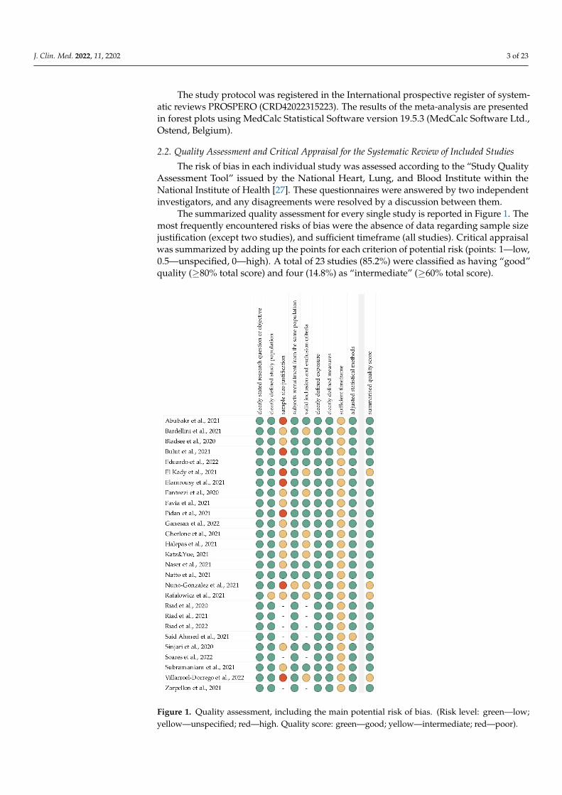
The risk of bias in each individual study was assessed according to the “Study QualityAssessment Tool” issued by the National Heart, Lung, and Blood Institute within theNational Institute of Health [27]. These questionnaires were answered by two independentinvestigators, and any disagreements were resolved by a discussion between them.
The summarized quality assessment for every single study is reported in Figure 1. Themost frequently encountered risks of bias were the absence of data regarding sample sizejustification (except two studies), and sufficient timeframe (all studies). Critical appraisalwas summarized by adding up the points for each criterion of potential risk (points: 1—low,0.5—unspecified, 0—high). A total of 23 studies (85.2%) were classified as having “good”quality (≥80% total score) and four (14.8%) as “intermediate” (≥60% total score).
J. Clin. Med. 2022, 11, x FOR PEER REVIEW 4 of 24
Figure 1. Quality assessment, including the main potential risk of bias. (Risk level: green—low;
yellow—unspecified; red—high. Quality score: green—good; yellow—intermediate; red—poor).
3. Results
Following the search criteria, our systematic review included twenty‐seven studies,
demonstrating data collected in twelve different countries from a total of 6722 partici‐
pants with diagnosed SARS‐CoV‐2 infection (including 2476 females and 2990 males, and
1256 patients without reported gender). Figure 2 shows the detailed selection strategy of
the articles. The inclusion and exclusion criteria are presented in Table 1 (in the Section 2).
Figure 1. Quality assessment, including the main potential risk of bias. (Risk level: green—low;yellow—unspecified; red—high. Quality score: green—good; yellow—intermediate; red—poor).
J. Clin. Med. 2022, 11, 2202 4 of 23
The level of evidence was assessed using the classification of the Oxford Centre forEvidence-Based Medicine levels for diagnosis [28]. All of the included studies have thethird or fourth level of evidence (in this 5-graded scale).
3. Results
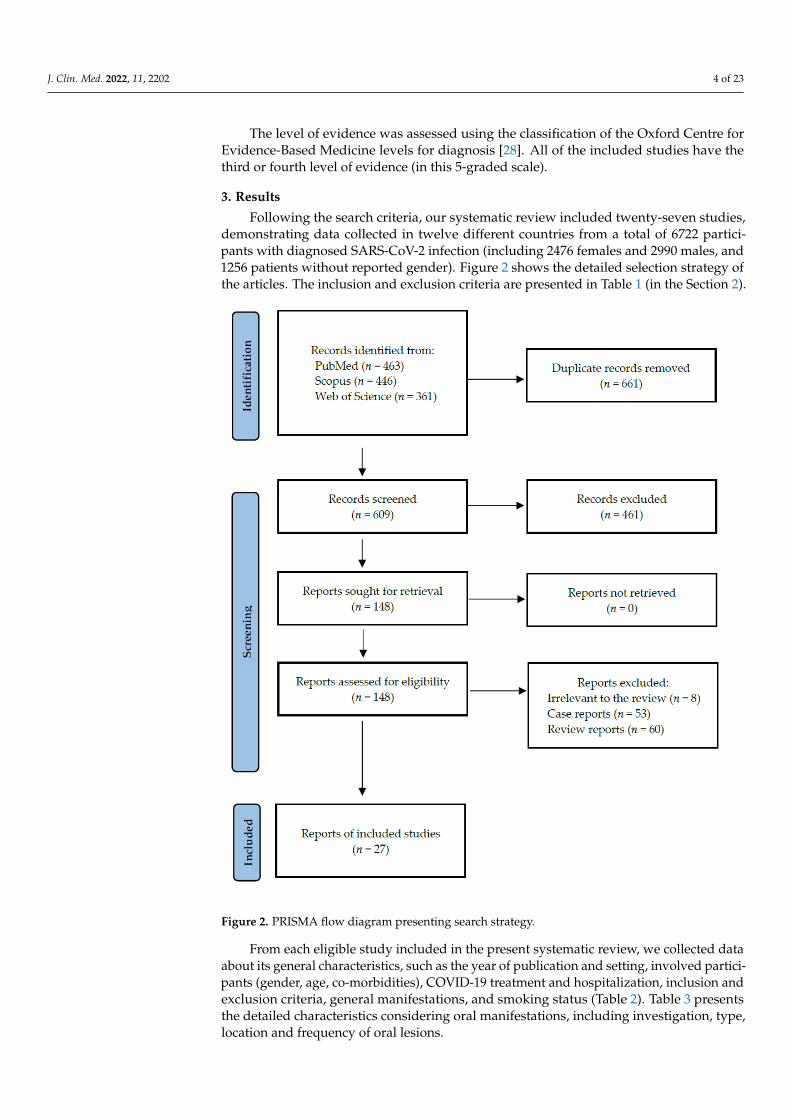
Following the search criteria, our systematic review included twenty-seven studies,demonstrating data collected in twelve different countries from a total of 6722 partici-pants with diagnosed SARS-CoV-2 infection (including 2476 females and 2990 males, and1256 patients without reported gender). Figure 2 shows the detailed selection strategy ofthe articles. The inclusion and exclusion criteria are presented in Table 1 (in the Section 2).
J. Clin. Med. 2022, 11, x FOR PEER REVIEW 5 of 24
Figure 2. PRISMA flow diagram presenting search strategy.
From each eligible study included in the present systematic review, we collected
data about its general characteristics, such as the year of publication and setting, involved
participants (gender, age, co‐morbidities), COVID‐19 treatment and hospitalization, in‐
clusion and exclusion criteria, general manifestations, and smoking status (Table 2). Ta‐
ble 3 presents the detailed characteristics considering oral manifestations, including in‐
vestigation, type, location and frequency of oral lesions.
Figure 2. PRISMA flow diagram presenting search strategy.
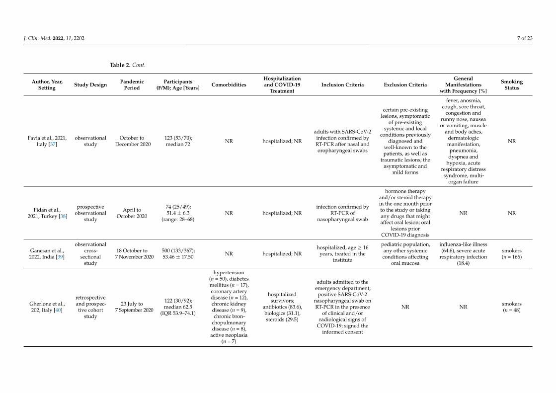
From each eligible study included in the present systematic review, we collected dataabout its general characteristics, such as the year of publication and setting, involved partici-pants (gender, age, co-morbidities), COVID-19 treatment and hospitalization, inclusion andexclusion criteria, general manifestations, and smoking status (Table 2). Table 3 presentsthe detailed characteristics considering oral manifestations, including investigation, type,location and frequency of oral lesions.
J. Clin. Med. 2022, 11, 2202 5 of 23
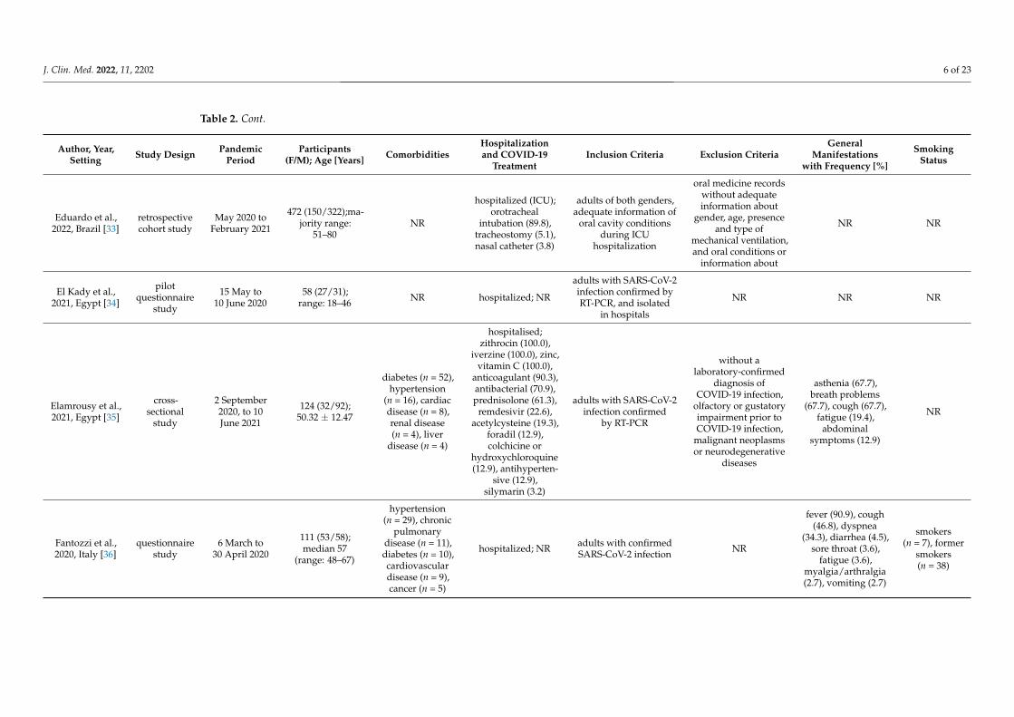
Table 2. General characteristics of included studies.
Author, Year,Setting Study Design Pandemic
PeriodParticipants
(F/M); Age [Years] ComorbiditiesHospitalizationand COVID-19
TreatmentInclusion Criteria Exclusion Criteria
GeneralManifestations
with Frequency [%]
SmokingStatus
Abubakr et al.,2021, Egypt [29]
questionnairestudy
1 May to1 July 2020
573 (408/165);36.19 ± 9.11
(range: 19–50)none non-hospitalized;
NR
Egyptian adults,18–50 years old;
laboratory-confirmedCOVID-19 infection
(PCR test); non-smokers;non-alcoholics;medically free;
mild-to-moderatesymptoms; good oral
hygiene and notsuffering from any oralmanifestations before
the pandemic
failure to complete thewhole questionnaire;poor oral hygiene or
any of the oralsymptoms before the
pandemic; chronicillnesses; smokers;alcoholics; serious
COVID-19 infection,severe respiratoryfailure or required
hospitalization
muscle pain (76.4),malaise (72.8),
headache (70.0),fever (66.0), loss ofsmell (61.8), cough
(55.5), sorethroat (52.4),
dyspnea (51.8),diarrhea (50.3)
non-smokers
Bardellini et al.,2021, Italy [30]
retrospectivecross-sectional
study
March toApril 2020
27 (8/19);4.2 ± 1.7 (range:
3 months–14 years)NR hospitalized; NR
pediatric patients(aged 0–14 years old),laboratory evidence of
COVID-19 infection,signed informed consent
NR
fever > 38 ◦C (55.5),mild febrile
conditions (37.0),cough (37.0),
rhinorrhea (25.9),difficulty in
breathing (18.5)
NA
Biadsee et al.,2020, Israel [31]
questionnairestudy
25 March to15 April 2020
128 (70/58); 36.25(range: 18–73)
hypertension (n = 8),hypothyroidism(n = 4), diabetesmellitus (n = 3),asthma (n = 2)
non-hospitalized;NR
diagnosed by RT-PCRand considered to have
mild symptoms,according to the latest
WHO joint report
questionnaires withmissing information
cough (59.4),weakness (47.7),
myalgia (46.9), fever(42.2), headache(40.6), impaired
sense of smell (38.3),sore throat (26.6),runny nose (26.6),
nasal congestion (22.7),gastrointestinalsymptoms (18.8)
smokers(n = 26)
Bulut et al., 2021,Turkey [32]
questionnairestudy
September 2020to March 2021
200 (125/75);ranges: 20–30: 89(62/27), 31–40: 65(43/22), 41–50: 27(14/13), 51–60: 15(4/11), 61–70: 4 (2/2)
NR
hospitalized (11.5);antiviral drugs (61.7),anticoagulant (16.9),hydroxychloroquine(12.3), antiaggregant(7.1), dexamethasone(5.2), antibiotics (5.2); notuse any medication (27.9)
reported COVID-19in anamnesis age < 18 years presence of
symptoms (87.5) NR
J. Clin. Med. 2022, 11, 2202 6 of 23
Table 2. Cont.
Author, Year,Setting Study Design Pandemic
PeriodParticipants
(F/M); Age [Years] ComorbiditiesHospitalizationand COVID-19
TreatmentInclusion Criteria Exclusion Criteria
GeneralManifestations
with Frequency [%]
SmokingStatus
Eduardo et al.,2022, Brazil [33]
retrospectivecohort study
May 2020 toFebruary 2021
472 (150/322);ma-jority range:
51–80NR
hospitalized (ICU);orotracheal
intubation (89.8),tracheostomy (5.1),nasal catheter (3.8)
adults of both genders,adequate information of
oral cavity conditionsduring ICU
hospitalization
oral medicine recordswithout adequateinformation about
gender, age, presenceand type of
mechanical ventilation,and oral conditions or
information about
NR NR
El Kady et al.,2021, Egypt [34]
pilotquestionnaire
study
15 May to10 June 2020
58 (27/31);range: 18–46 NR hospitalized; NR
adults with SARS-CoV-2infection confirmed byRT-PCR, and isolated
in hospitals
NR NR NR
Elamrousy et al.,2021, Egypt [35]
cross-sectional
study
2 September2020, to 10June 2021
124 (32/92);50.32 ± 12.47
diabetes (n = 52),hypertension
(n = 16), cardiacdisease (n = 8),renal disease(n = 4), liver
disease (n = 4)
hospitalised;zithrocin (100.0),
iverzine (100.0), zinc,vitamin C (100.0),
anticoagulant (90.3),antibacterial (70.9),prednisolone (61.3),
remdesivir (22.6),acetylcysteine (19.3),
foradil (12.9),colchicine or
hydroxychloroquine(12.9), antihyperten-
sive (12.9),silymarin (3.2)
adults with SARS-CoV-2infection confirmed
by RT-PCR
without alaboratory-confirmed
diagnosis ofCOVID-19 infection,
olfactory or gustatoryimpairment prior toCOVID-19 infection,
malignant neoplasmsor neurodegenerative
diseases
asthenia (67.7),breath problems
(67.7), cough (67.7),fatigue (19.4),
abdominalsymptoms (12.9)
NR
Fantozzi et al.,2020, Italy [36]
questionnairestudy
6 March to30 April 2020
111 (53/58);median 57
(range: 48–67)
hypertension(n = 29), chronic
pulmonarydisease (n = 11),diabetes (n = 10),cardiovasculardisease (n = 9),cancer (n = 5)
hospitalized; NR adults with confirmedSARS-CoV-2 infection NR
fever (90.9), cough(46.8), dyspnea
(34.3), diarrhea (4.5),sore throat (3.6),
fatigue (3.6),myalgia/arthralgia(2.7), vomiting (2.7)
smokers(n = 7), former
smokers(n = 38)
J. Clin. Med. 2022, 11, 2202 7 of 23
Table 2. Cont.
Author, Year,Setting Study Design Pandemic
PeriodParticipants
(F/M); Age [Years] ComorbiditiesHospitalizationand COVID-19
TreatmentInclusion Criteria Exclusion Criteria
GeneralManifestations
with Frequency [%]
SmokingStatus
Favia et al., 2021,Italy [37]
observationalstudy
October toDecember 2020
123 (53/70);median 72 NR hospitalized; NR
adults with SARS-CoV-2infection confirmed byRT-PCR after nasal andoropharyngeal swabs
certain pre-existinglesions, symptomatic
of pre-existingsystemic and local
conditions previouslydiagnosed and
well-known to thepatients, as well as
traumatic lesions; theasymptomatic and
mild forms
fever, anosmia,cough, sore throat,
congestion andrunny nose, nauseaor vomiting, muscle
and body aches,dermatologicmanifestation,pneumonia,dyspnea and
hypoxia, acuterespiratory distresssyndrome, multi-
organ failure
NR
Fidan et al.,2021, Turkey [38]
prospectiveobservational
study
April toOctober 2020
74 (25/49);51.4 ± 6.3
(range: 28–68)NR hospitalized; NR
infection confirmed byRT-PCR of
nasopharyngeal swab
hormone therapyand/or steroid therapyin the one month priorto the study or takingany drugs that mightaffect oral lesion; oral
lesions priorCOVID-19 diagnosis
NR NR
Ganesan et al.,2022, India [39]
observationalcross-
sectionalstudy
18 October to7 November 2020
500 (133/367);53.46 ± 17.50 NR hospitalized; NR
hospitalized, age ≥ 16years, treated in the
institute
pediatric population,any other systemicconditions affecting
oral mucosa
influenza-like illness(64.6), severe acute
respiratory infection(18.4)
smokers(n = 166)
Gherlone et al.,202, Italy [40]
retrospectiveand prospec-tive cohort
study
23 July to7 September 2020
122 (30/92);median 62.5
(IQR 53.9–74.1)
hypertension(n = 50), diabetesmellitus (n = 17),coronary arterydisease (n = 12),chronic kidneydisease (n = 9),chronic bron-
chopulmonarydisease (n = 8),
active neoplasia(n = 7)
hospitalizedsurvivors;
antibiotics (83.6),biologics (31.1),steroids (29.5)
adults admitted to theemergency department;
positive SARS-CoV-2nasopharyngeal swab onRT-PCR in the presence
of clinical and/orradiological signs of
COVID-19; signed theinformed consent
NR NR smokers(n = 48)

J. Clin. Med. 2022, 11, 2202 8 of 23
Table 2. Cont.
Author, Year,Setting Study Design Pandemic
PeriodParticipants
(F/M); Age [Years] ComorbiditiesHospitalizationand COVID-19
TreatmentInclusion Criteria Exclusion Criteria
GeneralManifestations
with Frequency [%]
SmokingStatus
Halepas et al.,2021, USA [41]
retrospectivecross-
sectionalstudy
15 March to1 June 2020
47 (23/24);9.0 ± 5.0
(range: 1.3–20.0)NR hospitalized; NR
21 years or younger,fever of prolonged
duration, laboratoryevidence of
inflammation, requiredhospitalization,
multiorgan involvement,confirmed positiveCOVID RT-PCR or
serology test results
NR
multisysteminflammatorysyndrome in
children; fever> 5 days (100.0),
systemic rash (68.1),conjunctivitis (57.5),
vomiting (51.1),diarrhea (38.3),
myocarditis (36.2),cervical lym-
phadenopathy (19.2),cough (14.9),
irritability (14.9),cranial nerve palsy(12.8), pericardial
effusion (12.8),extremity edema
(12.8), arthritis (8.5),rhinorrhea (6.4)
NR
Katz and Yue,2021, USA [42] registry study NR
895 (386/509); 0–9:5.33%, 10–17: 2.47,18–34: 39.9, 35–44:
11.1, 45–54: 1.2,55–64: 11.2, 65–74:12.3, 74–85: 16.5
respiratorydisease, endocrine
disease, obesity,diabetes,
circulatory disease
hospitalized; NR
diagnosis of COVID-19ICD-10-U07.1 and/or
ICD-10-K12.0 (recurrentaphthous stomatitis)
none NRsmokers andnon-smokers
(NR)
Naser et al.,2021, Iraq [43]
prospectivestudy
August 2020to March 2021
338 (138/200);mean 42.1
hypertension(n = 235), diabetesmellitus (n = 218),
heart diseases(n = 108), renal
diseases (n = 69),blood diseases
(n = 37), respiratorydisorders (n = 32),
gastrointestinaldiseases (n = 27),
liver diseases(n = 26)
hospitalized; NR
diagnosed withCOVID-19 + by PCR;age ≥ 10 years, withacute oral or periorallesions either duringadmission or which
appeared later duringtreatment; nonsmoker,
no alcohol consumption
age ≤ 10 years; lesionsappeared or were well
established beforeSARSCoV-2 infection;
not toleratingfollow-up or refusing
to enroll in study
NR non-smokers
J. Clin. Med. 2022, 11, 2202 9 of 23
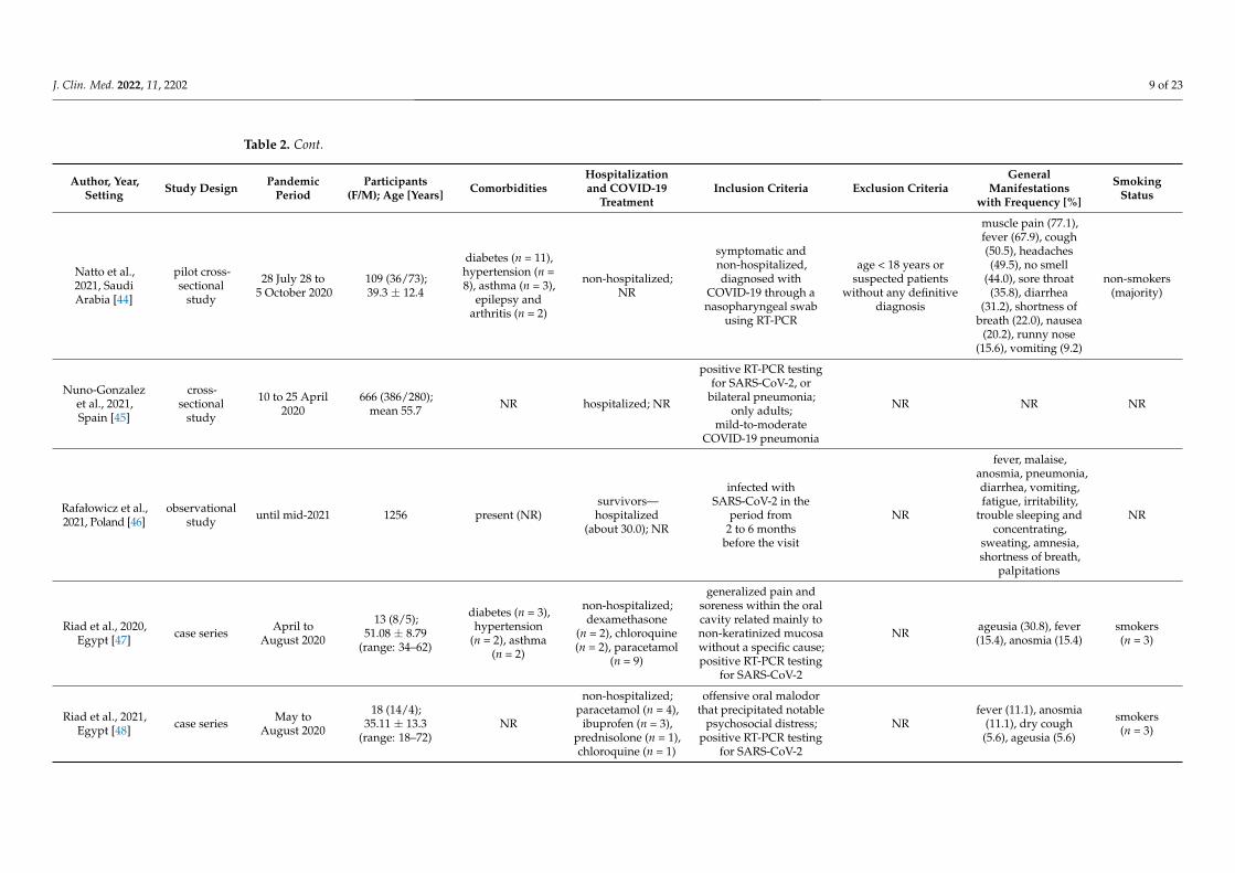
Table 2. Cont.
Author, Year,Setting Study Design Pandemic
PeriodParticipants
(F/M); Age [Years] ComorbiditiesHospitalizationand COVID-19
TreatmentInclusion Criteria Exclusion Criteria
GeneralManifestations
with Frequency [%]
SmokingStatus
Natto et al.,2021, SaudiArabia [44]
pilot cross-sectional
study
28 July 28 to5 October 2020
109 (36/73);39.3 ± 12.4
diabetes (n = 11),hypertension (n =8), asthma (n = 3),
epilepsy andarthritis (n = 2)
non-hospitalized;NR
symptomatic andnon-hospitalized,diagnosed with
COVID-19 through anasopharyngeal swab
using RT-PCR
age < 18 years orsuspected patients
without any definitivediagnosis
muscle pain (77.1),fever (67.9), cough(50.5), headaches(49.5), no smell
(44.0), sore throat(35.8), diarrhea
(31.2), shortness ofbreath (22.0), nausea
(20.2), runny nose(15.6), vomiting (9.2)
non-smokers(majority)
Nuno-Gonzalezet al., 2021,Spain [45]
cross-sectional
study
10 to 25 April2020
666 (386/280);mean 55.7 NR hospitalized; NR
positive RT-PCR testingfor SARS-CoV-2, or
bilateral pneumonia;only adults;
mild-to-moderateCOVID-19 pneumonia
NR NR NR
Rafałowicz et al.,2021, Poland [46]
observationalstudy until mid-2021 1256 present (NR)
survivors—hospitalized
(about 30.0); NR
infected withSARS-CoV-2 in the
period from2 to 6 months
before the visit
NR
fever, malaise,anosmia, pneumonia,diarrhea, vomiting,fatigue, irritability,
trouble sleeping andconcentrating,
sweating, amnesia,shortness of breath,
palpitations
NR
Riad et al., 2020,Egypt [47] case series April to
August 2020
13 (8/5);51.08 ± 8.79
(range: 34–62)
diabetes (n = 3),hypertension
(n = 2), asthma(n = 2)
non-hospitalized;dexamethasone
(n = 2), chloroquine(n = 2), paracetamol
(n = 9)
generalized pain andsoreness within the oralcavity related mainly tonon-keratinized mucosawithout a specific cause;positive RT-PCR testing
for SARS-CoV-2
NR ageusia (30.8), fever(15.4), anosmia (15.4)
smokers(n = 3)
Riad et al., 2021,Egypt [48] case series May to
August 2020
18 (14/4);35.11 ± 13.3
(range: 18–72)NR
non-hospitalized;paracetamol (n = 4),
ibuprofen (n = 3),prednisolone (n = 1),chloroquine (n = 1)
offensive oral malodorthat precipitated notable
psychosocial distress;positive RT-PCR testing
for SARS-CoV-2
NRfever (11.1), anosmia
(11.1), dry cough(5.6), ageusia (5.6)
smokers(n = 3)
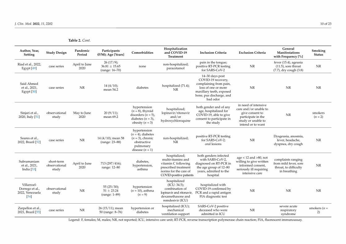
J. Clin. Med. 2022, 11, 2202 10 of 23
Table 2. Cont.
Author, Year,Setting Study Design Pandemic
PeriodParticipants
(F/M); Age [Years] ComorbiditiesHospitalizationand COVID-19
TreatmentInclusion Criteria Exclusion Criteria
GeneralManifestations
with Frequency [%]
SmokingStatus
Riad et al., 2022,Egypt [49] case series April to June
2020
26 (17/9);36.81 ± 15.65(range: 16–70)
none non-hospitalized;paracetamol
pain in the tongue;positive RT-PCR testing
for SARS-CoV-2NR
fever (15.4), ageusia(11.5), sore throat
(7.7), dry cough (3.8)NR
Said Ahmedet al., 2021,Egypt [50]
case series NR 14 (4/10);mean 54.2 diabetes hospitalized (71.4);
NR
14–30 days postCOVID-19 recovery,
complaining from pain,loss of one or more
maxillary teeth, exposedbone, pus discharge, and
bad odor
NR NR NR
Sinjari et al.,2020, Italy [51]
observationalstudy
May to June2020
20 (9/11);mean 69.2
hypertension(n = 8), thyroid
disorders (n = 5),diabetes (n = 3),obesity (n = 3)
hospitalized;lopinavir/ritonavir
and/orhydroxychloroquine
both gender and of anyage, hospitalized for
COVID-19, able to giveconsent to participate in
the study
in need of intensivecare and/or unable to
give consent toparticipate in the
study or unable tointend or to want
NR smokers(n = 2)
Soares et al.,2022, Brazil [52] case series NR 14 (4/10); mean 58
(range: 23–88)
hypertension(n = 4), diabetes(n = 3), chronic
obstructivepulmonary
disease (n = 1)
non-hospitalized;NR
positive RT-PCR testingfor SARS-CoV-2;
oral lesionsNR
Dysgeusia, anosmia,fever, headache,
dyspnea, dry coughNR
Subramaniamet al., 2021,India [53]
short-termobservational
study
April to June2020
713 (297/416);range: 12–80
diabetes,hypertension,
asthma
hospitalized;multivitamins and
vitamin C followingprescribed treatmentnorms for the care ofCOVID-positive patients
both genders infectedwith SARS-CoV-2,
diagnosed on RT-PCR inthe age group of 12–80years, admitted to the
hospital
age < 12 and >80, notwilling to give written
informed consent,seriously ill requiring
intensive care
complaints rangingfrom mild fever, sorethroat, to difficulty
in breathing
NR
Villarroel-Dorrego et al.,
2022, Venezuela[54]
observationalstudy NR
55 (25/30);51 ± 23.24
(range: 1–89)
hypertension(n = 10), asthma
(n = 9)
hospitalized(ICU: 34.5);
combination oflopinavir and ritonavir,dexamethasone and
remdesivir (ICU)
hospitalized withCOVID-19 confirmed byPCR and a rapid antigen
FIA diagnostic test
NR NR NR
Zarpellon et al.,2021, Brazil [55] case series NR 26 (15/11); mean
50 (range: 8–76)hypertension or
diabetes
hospitalized (ICU);mechanical
ventilation support
SARS-CoV-2 positivedeceased who were
admitted in ICUNR
severe acuterespiratorysyndrome
smokers (n =2)
Legend: F, females; M, males; NR, not reported; ICU, intensive care unit; RT-PCR, reverse transcription polymerase chain reaction; FIA, fluorescent immunoassay.
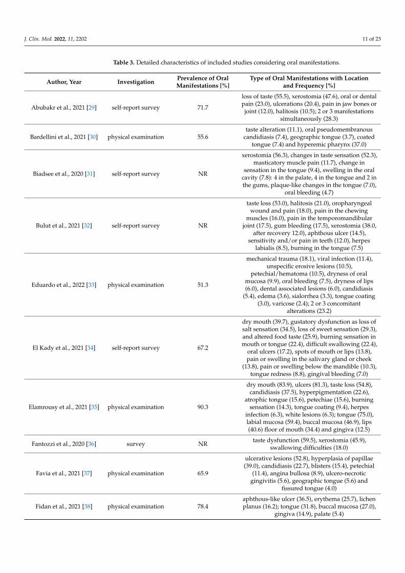
J. Clin. Med. 2022, 11, 2202 11 of 23
Table 3. Detailed characteristics of included studies considering oral manifestations.
Author, Year Investigation Prevalence of OralManifestations [%]
Type of Oral Manifestations with Locationand Frequency [%]
Abubakr et al., 2021 [29] self-report survey 71.7
loss of taste (55.5), xerostomia (47.6), oral or dentalpain (23.0), ulcerations (20.4), pain in jaw bones orjoint (12.0), halitosis (10.5); 2 or 3 manifestations
simultaneously (28.3)
Bardellini et al., 2021 [30] physical examination 55.6taste alteration (11.1), oral pseudomembranous
candidiasis (7.4), geographic tongue (3.7), coatedtongue (7.4) and hyperemic pharynx (37.0)
Biadsee et al., 2020 [31] self-report survey NR
xerostomia (56.3), changes in taste sensation (52.3),masticatory muscle pain (11.7), change in
sensation in the tongue (9.4), swelling in the oralcavity (7.8): 4 in the palate, 4 in the tongue and 2 inthe gums, plaque-like changes in the tongue (7.0),
oral bleeding (4.7)
Bulut et al., 2021 [32] self-report survey NR
taste loss (53.0), halitosis (21.0), oropharyngealwound and pain (18.0), pain in the chewing
muscles (16.0), pain in the temporomandibularjoint (17.5), gum bleeding (17.5), xerostomia (38.0,
after recovery 12.0), aphthous ulcer (14.5),sensitivity and/or pain in teeth (12.0), herpes
labialis (8.5), burning in the tongue (7.5)
Eduardo et al., 2022 [33] physical examination 51.3
mechanical trauma (18.1), viral infection (11.4),unspecific erosive lesions (10.5),
petechial/hematoma (10.5), dryness of oralmucosa (9.9), oral bleeding (7.5), dryness of lips(6.0), dental associated lesions (6.0), candidiasis
(5.4), edema (3.6), sialorrhea (3.3), tongue coating(3.0), varicose (2.4); 2 or 3 concomitant
alterations (23.2)
El Kady et al., 2021 [34] self-report survey 67.2
dry mouth (39.7), gustatory dysfunction as loss ofsalt sensation (34.5), loss of sweet sensation (29.3),and altered food taste (25.9), burning sensation inmouth or tongue (22.4), difficult swallowing (22.4),
oral ulcers (17.2), spots of mouth or lips (13.8),pain or swelling in the salivary gland or cheek
(13.8), pain or swelling below the mandible (10.3),tongue redness (8.8), gingival bleeding (7.0)
Elamrousy et al., 2021 [35] physical examination 90.3
dry mouth (83.9), ulcers (81.3), taste loss (54.8),candidiasis (37.5), hyperpigmentation (22.6),
atrophic tongue (15.6), petechiae (15.6), burningsensation (14.3), tongue coating (9.4), herpes
infection (6.3), white lesions (6.3); tongue (75.0),labial mucosa (59.4), buccal mucosa (46.9), lips(40.6) floor of mouth (34.4) and gingiva (12.5)
Fantozzi et al., 2020 [36] survey NR taste dysfunction (59.5), xerostomia (45.9),swallowing difficulties (18.0)
Favia et al., 2021 [37] physical examination 65.9
ulcerative lesions (52.8), hyperplasia of papillae(39.0), candidiasis (22.7), blisters (15.4), petechial
(11.4), angina bullosa (8.9), ulcero-necroticgingivitis (5.6), geographic tongue (5.6) and
fissured tongue (4.0)
Fidan et al., 2021 [38] physical examination 78.4aphthous-like ulcer (36.5), erythema (25.7), lichenplanus (16.2); tongue (31.8), buccal mucosa (27.0),
gingiva (14.9), palate (5.4)
J. Clin. Med. 2022, 11, 2202 12 of 23
Table 3. Cont.
Author, Year Investigation Prevalence of OralManifestations [%]
Type of Oral Manifestations with Location andFrequency [%]
Ganesan et al., 2022 [39] physical examination NR
alteration in taste sensation (51.2), xerostomia(28.0), erythematous macules with burning
sensation (7.2), atrophic glossitis (4.6), non-specificsolitary ulcers (3.0), candida-like lesions (1.0),white patch (1.0), ductal inflammation (0.4)
Gherlone et al., 2021 [40] physical examination 83.9
salivary gland ectasia (43.0), white tongue (29.0),dry mouth (24.0), masticatory muscle weakness
(19.0), oral ulcers (11.0), temporomandibular jointabnormalities (7.0)
Halepas et al., 2021 [41] physical examination 55.3 red or swollen lips (48.9), strawberry tongue (10.6)
Katz and Yue, 2021 [42] physical examination NR recurrent aphthous stomatitis (0.67; OR = 14.0)
Naser et al., 2021 [43] physical examination NR
loss of taste (79.5), white coats of the tongue (31.6),pain related to the oral cavity (27.8), multiple
aphthous ulceration (24.8), dryness (24.5),dysphagia (23.0), white coats of cheeks and
gingiva (22.4), white coats of the palate (15.6),single aphthous ulceration (9.1)
Natto et al., 2021 [44] physical examination 53.2loss of taste (43.4), erythema/desquamated
gingivitis (7.3), coated tongue (7.3),ulcers/blisters (6.4)
Nuno-Gonzalez et al.,2021 [45] physical examination 11.7
anterior U-shaped lingual papillitis (5.3), aphthousstomatitis (3.2), tongue swelling (3.0), burning
sensation in the mouth (2.4), mucositis (1.8),glossitis with patchy depapillation (1.8),
candidiasis (0.5), enanthema (0.3)
Rafałowicz et al., 2021 [46] physical examination NR
discoloration, ulceration, and hemorrhagicchanges on the oral mucosa (32.0), mycosis located
on the tongue (29.7), unilateral (more oftenleft-sided) aphthous-like lesions on the hard palate
(25.8), atrophic cheilitis (12.5); approx. 60%salivary secretory disorders in the initial period of
infection, which in 6.68% prolonged up to 4months after systemic symptoms disappeared
Riad et al., 2020 [47] physical examination NA
mucositis (intraoral pain, sporadic erythema withminor irritations); all over the mouth (53.8), on the
buccal mucosa (30.8), palate (15.4), andgingiva (7.7)
Riad et al., 2021 [48] physical examination NA
halitosis (the majority with ‘fair’ level of oralhygiene, except for two patients (11.1) with a‘poor’ level, while one patient (5.5) further
complicated by an intraoral ulcer)
Riad et al., 2022 [49] physical examination NA
tongue ulcers (92.3 of them not bleeding, rangedbetween 1 and 7 ulcers per patient, and their size
ranged between 1 and 5 mm corresponding toherpes-like ulcers with scalloped borders); all of
them manifested on dorsum or side of the tongue,while 4 (15.4) on the ventral surface
Said Ahmed et al.,2021 [50] physical examination NA maxillary mucormycosis osteomyelitis
Sinjari et al., 2020 [51] self-report survey NR xerostomia (30.0), impaired taste (25.0), difficultyin swallowing (20.0, burning sensation (15.0)
J. Clin. Med. 2022, 11, 2202 13 of 23
Table 3. Cont.
Author, Year Investigation Prevalence of OralManifestations [%]
Type of Oral Manifestations with Location andFrequency [%]
Soares et al., 2022 [52] physical examination 100.0
petechial, ecchymosis, reddish macules, andchronic ulcers with more than 7 d of evolution,
vesiculobullous eruptions; only the palate (57.1),tongue (28.6), either the lip or palate (14.3)
Subramaniam et al.,2021 [53] physical examination 1.26
ulcers on the buccal mucosa, burning sensation,generalized mucositis on the labial mucosa,
erythema of tongue margins, tongue papillaryatrophy, bilateral angular cheilitis, reddish-white
spots on the palate
Villarroel-Dorrego et al.,2022 [54] physical examination 40.0
alteration or a total loss of taste (60.0), pain orburning in the mouth (36.4), xerostomia (27.3),
candidiasis (12.7), hemorrhagic ulcerative lesions(7.3), multiple ulcerations resembling cancer sores
(5.5), lingual varicose veins (5.5), migratoryglossitis (5.5), enanthems in the labial or cheek
mucosa (3.6), severe angular cheilitis (1.8), whiteplaques (1.8) and lichenoid lesions (1.8), recurrent
cold sore (1.8)
Zarpellon et al., 2021 [55] physical examination(post mortem) NA vesiculo-bullous and ulcerative lesions in the oral
mucosa, such as tongue, lips and buccal mucosa
Legend: NR, not reported; NA, not applicable.
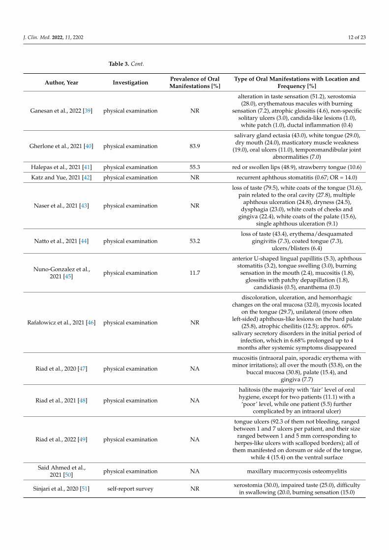
Most studies (involving at least 20 patients) were included in the meta-analysis, theresults of which are presented in the forest plots (Figures 3–6).
J. Clin. Med. 2022, 11, x FOR PEER REVIEW 15 of 24
el‐Dorrego et
al., 2022 [54]
tion mouth (36.4), xerostomia (27.3), candidiasis (12.7), hemorrhagic
ulcerative lesions (7.3), multiple ulcerations resembling cancer
sores (5.5), lingual varicose veins (5.5), migratory glossitis (5.5),
enanthems in the labial or cheek mucosa (3.6), severe angular
cheilitis (1.8), white plaques (1.8) and lichenoid lesions (1.8),
recurrent cold sore (1.8)
Zarpellon et
al., 2021 [55]
physical examina‐
tion
(post mortem)
NA vesiculo‐bullous and ulcerative lesions in the oral mucosa, such
as tongue, lips and buccal mucosa
Legend: NR, not reported; NA, not applicable.
Most studies (involving at least 20 patients) were included in the meta‐analysis, the
results of which are presented in the forest plots (Figures 3–6).
Figure 3. Forest plot presenting the summarised prevalence of oral manifestations among
SARS‐CoV‐2 positive individuals.
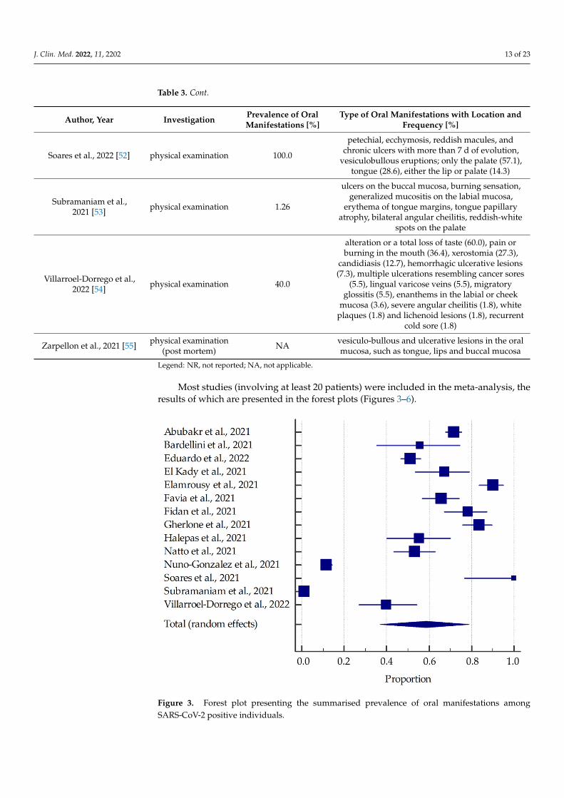
Figure 4. Forest plot presenting the summarized prevalence of xerostomia among SARS‐CoV‐2
positive individuals.
Figure 3. Forest plot presenting the summarised prevalence of oral manifestations amongSARS-CoV-2 positive individuals.
J. Clin. Med. 2022, 11, 2202 14 of 23
J. Clin. Med. 2022, 11, x FOR PEER REVIEW 15 of 24
el‐Dorrego et
al., 2022 [54]
tion mouth (36.4), xerostomia (27.3), candidiasis (12.7), hemorrhagic
ulcerative lesions (7.3), multiple ulcerations resembling cancer
sores (5.5), lingual varicose veins (5.5), migratory glossitis (5.5),
enanthems in the labial or cheek mucosa (3.6), severe angular
cheilitis (1.8), white plaques (1.8) and lichenoid lesions (1.8),
recurrent cold sore (1.8)
Zarpellon et
al., 2021 [55]
physical examina‐
tion
(post mortem)
NA vesiculo‐bullous and ulcerative lesions in the oral mucosa, such
as tongue, lips and buccal mucosa
Legend: NR, not reported; NA, not applicable.
Most studies (involving at least 20 patients) were included in the meta‐analysis, the
results of which are presented in the forest plots (Figures 3–6).
Figure 3. Forest plot presenting the summarised prevalence of oral manifestations among
SARS‐CoV‐2 positive individuals.
Figure 4. Forest plot presenting the summarized prevalence of xerostomia among SARS‐CoV‐2
positive individuals.
Figure 4. Forest plot presenting the summarized prevalence of xerostomia among SARS-CoV-2positive individuals.
J. Clin. Med. 2022, 11, x FOR PEER REVIEW 16 of 24
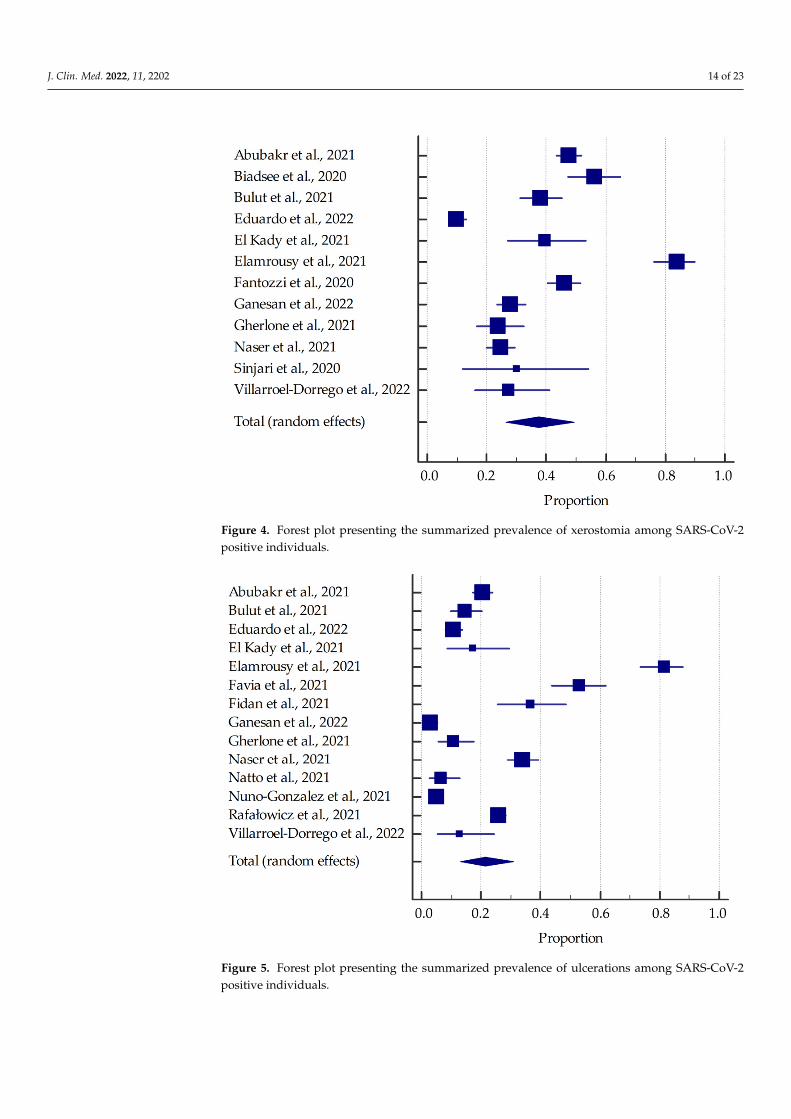
Figure 5. Forest plot presenting the summarized prevalence of ulcerations among SARS‐CoV‐2
positive individuals.
Figure 6. Forest plot presenting the summarized prevalence of taste alterations among
SARS‐CoV‐2 positive individuals.
Based on the included studies reporting the frequency of oral manifestations in
SARS‐CoV‐2 positive individuals, it was determined that the aggregate prevalence was
approximately 58.75% [95% CI: 36.93–78.90%]. Additionally, in this group, the summa‐
rized prevalence of taste alterations, xerostomia and ulcerations were estimated to be
around 54.73% [95% CI: 46.28–63.04%], 37.58% [95% CI: 26.35–49.53%], and 21.43% [95%
CI: 13.17–31.06%], respectively.
4. Discussion
In our discussion, the literature reviewed in regards to oral manifestations of
SARS‐CoV‐2 infection were divided into two subgroups: descriptive studies (surveys,
case series) and analytical studies (observational: cross‐sectional, prospective or retro‐
spective).
Figure 5. Forest plot presenting the summarized prevalence of ulcerations among SARS-CoV-2positive individuals.
J. Clin. Med. 2022, 11, 2202 15 of 23
J. Clin. Med. 2022, 11, x FOR PEER REVIEW 16 of 24
Figure 5. Forest plot presenting the summarized prevalence of ulcerations among SARS‐CoV‐2
positive individuals.
Figure 6. Forest plot presenting the summarized prevalence of taste alterations among
SARS‐CoV‐2 positive individuals.
Based on the included studies reporting the frequency of oral manifestations in
SARS‐CoV‐2 positive individuals, it was determined that the aggregate prevalence was
approximately 58.75% [95% CI: 36.93–78.90%]. Additionally, in this group, the summa‐
rized prevalence of taste alterations, xerostomia and ulcerations were estimated to be
around 54.73% [95% CI: 46.28–63.04%], 37.58% [95% CI: 26.35–49.53%], and 21.43% [95%
CI: 13.17–31.06%], respectively.
4. Discussion
In our discussion, the literature reviewed in regards to oral manifestations of
SARS‐CoV‐2 infection were divided into two subgroups: descriptive studies (surveys,
case series) and analytical studies (observational: cross‐sectional, prospective or retro‐
spective).
Figure 6. Forest plot presenting the summarized prevalence of taste alterations among SARS-CoV-2positive individuals.
Based on the included studies reporting the frequency of oral manifestations inSARS-CoV-2 positive individuals, it was determined that the aggregate prevalence wasapproximately 58.75% [95% CI: 36.93–78.90%]. Additionally, in this group, the sum-marized prevalence of taste alterations, xerostomia and ulcerations were estimated tobe around 54.73% [95% CI: 46.28–63.04%], 37.58% [95% CI: 26.35–49.53%], and 21.43%[95% CI: 13.17–31.06%], respectively.
4. Discussion
In our discussion, the literature reviewed in regards to oral manifestations of SARS-CoV-2infection were divided into two subgroups: descriptive studies (surveys, case series) andanalytical studies (observational: cross-sectional, prospective or retrospective).
4.1. Descriptive Studies
A study conducted by Abubakr et al. [29] surveyed Egyptian patients who presentedwith mild-moderate cases of COVID-19 and suffered from one or more oral manifestationsdue to the virus. As such, 47.6% of the participants experienced xerostomia, 23% sufferedfrom oral or dental pain, 20.4% reported the appearance of ulcerations in the oral cavitywith a significant occurrence in males when compared to females, 12% complained of painin the jaw bones or joints, and 10.5% of patients felt they had halitosis. Moreover, 28.3% ofCOVID-19 patients simultaneously experienced two or three of these oral manifestations.This study also examined oral hygiene status among these infected patients. It confirmedthat patients with poor oral hygiene had ulcerations and oral/dental pain more often thanthose with good oral hygiene. The most prevalent oral manifestation was xerostomia, andthe least pervasive was halitosis.
Similarly, Biadsee et al. [31] examined a web-based questionnaire distributed to 128 pa-tients with COVID-19 who were quarantining in designated hotels in Israel. The followingoral manifestations reported are olfactory dysfunction, taste alterations, dry mouth, facialpain, and masticatory muscle pain. As such, eighty-six patients (67%) had olfactory dys-
J. Clin. Med. 2022, 11, 2202 16 of 23
function, with 19.5% of patients who felt it was heightened from day three to five of theinfection. A total of sixty-seven patients (52%) experienced changes in their taste sensation,specifically toward spicy, salty, sour, and sweet foods, and women were impacted morethan men. Furthermore, seventy-two patients reported dry mouth, strongly correlated toburning mouth and taste alterations. Facial pain was reported by eighteen patients (26%),and fifteen patients (11%) experienced masticatory muscle pain. Additionally, twentypatients described changes to their tongue sensation, nine patients reported the appearanceof plaque on their tongue, and three patients reported oral bleeding. Lastly, oral hygienedid not contribute to these manifestations.
In a study by El Kady et al. [34], a pilot survey on Google Form was conducted onSARS-CoV-2 positive patients, asking the patients to report any oral manifestations listedin the questionnaire, and four categories were created: 1. Gustatory disorders (loss of saltsensation, loss of sweetness, and altered food taste); 2. Symptoms of salivary gland infection(dry mouth, difficulty swallowing, pain or swelling in the submandibular gland area, andpain or swelling in the parotid gland area); 3. Oral mucosal changes (oral ulcers, spotson mouth or lips, tongue redness, gingival bleeding, and burning sensation); 4. Categoryfor patients with no symptoms related to the oral cavity and salivary gland. The resultsreported that 67.2% patients had at least one oral manifestation, with the highest prevalencesymptom of dry mouth at 39.7%. Other symptoms of gustatory dysfunction were 34.5% lossof salt sensation, 29.3% loss of sweetness and 25.9% altered food taste. In this study, thehighest percentage of occurrence of taste alterations among all the reports included in themeta-analysis was found. Salivary gland infection at 22.4% difficult swelling, 13.8% pain orswellings in the salivary gland or cheek, and 10.3% pain or swelling below the mandible.Oral mucosal changes were less prevalent. Loss of salt and sweetness were most associated(27.6%), and dry mouth and gustatory impairment symptoms were often associated (27.6%).The high prevalence of salivary gland related symptoms highlights the protective role andvalue of saliva against viruses.
Moreover, in a survey study by Bulut et al. [32], the possible effects of SARS-CoV-2on the oral tissues were investigated. A total of 200 volunteers, who survived COVID-19,filled out a questionnaire after routine clinical and radiographic examinations, regardingtheir demographic characteristics, general health, oral habits and symptoms in the oralcavity. During the active period of SARS-CoV-2 infection the following observations ofthe oral cavity were recorded; taste loss (53%), halitosis (21%), oropharyngeal wound andpain (18%), pain in temporomandibular joint (17.5%), pain in the chewing muscles (16%),aphthous ulcer (14.5%), sensitivity and/or pain in teeth (12%), herpes labialis (8.5%), andburning in the tongue (7.5%). Xerostomia was observed in 38% of the patients duringthe active period, and of those, 27.6% continued to have xerostomia after. The issue withhalitosis can be due to the wound and pain in the oropharynx region, infection of tonsilsand the soft palate leading to the collection of bacteria and fungi.
In contrast, Riad et al. [49] conducted a case series on patients who sought care in theirhospital for pain in the tongue and had a confirmed SARS-CoV-2 laboratory test. Generalsymptoms experienced by these patients included only one oral manifestation, which isageusia. The onset of tongue ulcers was after five days for 53.8% of patients, 26.9% afterfour days, and 7.7% after two days. The number of ulcers varied considerably amongthese individuals, and their size ranged from 1 to 5 mm. The location of these ulcers waspredominately on the dorsum of the tongue, while a few patients had them on the ventralsurface of the tongue.
In another study by Riad et al. [47], thirteen patients with oral mucositis were exam-ined. These patients complained of generalized pain and soreness within the oral cavity,focused on the nonkeratinized mucosa. The onset of mucositis was recorded at 0–2 daysafter a positive PCR SARS-CoV-2 test was done, and the mean duration was 7–14 days.Intraoral examinations found depapillation of the tongue in all cases, sporadic erythema(53.8%) on the buccal mucosa (30.8%), palate (15.4%) and gingiva (7.7%). Significant asso-ciations of COVID-19 severity and duration of mucositis with pain, and the loss of sense
J. Clin. Med. 2022, 11, 2202 17 of 23
of taste associated with mucositis durations was disclosed. These results support the roleof oral mucosa in being the entryway for the virus. Patients with COVID-19 may easilypresent with mucositis due to cellular damage triggered by the virus, or as an opportunisticinfection due to immune deregulation.
Furthermore, Riad et al. [48] reported eighteen patients who presented a new-onsethalitosis during the course of COVID-19 infection. The issue was brought up due toan offensive oral malodor that precipitated notable psychosocial distress, in which theHalimeter Plus was used for quantitative assessment. Oral hygiene states were assessedwith the Oral Health Assessment Tool finding that most of the patients had a fair level oforal hygiene, therefore suggesting that the epithelial alterations by SARS-CoV-2 on thedorsum of the tongue is the probable reason for the halitosis.
Interestingly, Said Ahmed et al. [50] present a series of fourteen cases that discussthe prevalence of maxillary mucormycosis osteomyelitis in post-COVID-19 patients. Inthis study, nine patients had diabetes before contracting COVID-19, and five patients didnot, but both groups showed signs of hyperglycemia after their quarantine. This casereport concluded that patients with diabetes are at a higher risk of maxillary mucormycosisosteomyelitis due to drugs used to treat COVID-19. It is suggested that these medicationscan cause patients to become immunocompromised, primarily due to their diabetic status.
Rafałowicz et al. [46] studied 1256 patients, and of those presented 6 cases of patientswith the most common long COVID-19 oral cavity symptoms. Amidst these patientsthe following oral cavity changes were reported: 32% discolouration, ulcerations andhemorrhagic changes on the oral mucosa, 29.69% mycosis on the tongue, 25.79% aphthous-like lesion on hard palate, and 12.5% atrophic cheilitis.
Based on the included descriptive studies, xerostomia, olfactory dysfunction andchanges in taste sensation were the most prevalent oral manifestation in SARS-CoV-2positive patients. Halitosis, masticatory muscle pain, and burning sensation were the leastprevalent and were rarely observed.
4.2. Analytical Studies
In an observational study by Sinjari et al. [51], patients hospitalized due to COVID-19participated in a questionnaire to better understand the oral manifestation of these patients.Health status, oral hygiene habits, and symptoms in the oral cavity before and during thehospitalization/COVID-19 manifestation were collected. Additionally, questions to theUnit of Internal Medicine of the hospital, in regards to the clinical conditions of the patients,were collected. As such, 30% of patients reported xerostomia during hospitalization, 25%impaired taste, 15% burning sensation, and 20% difficulty in swallowing. Nuno-Gonzalezet al. [45] examined 666 patients with COVID-19 in a field hospital in Spain to determinethe prevalence of oral and palmoplantar mucocutaneous lesions in infected individuals.The authors established the following results: 25.7% had oral manifestations, including11.5% transient lingual papillitis, 6.6% aphthous stomatitis, 3.9% glossitis with patchy de-papillation, and 3.9% with mucositis. Other common adjuncts to these oral manifestationsare burning sensation, which was felt among 5.3% of patients, and taste disturbances.
Gherlone et al. [40] completed a retrospective and prospective cohort study that in-cluded COVID-19 patients admitted to San Raffaele University Hospital in Milan. The mostfrequent oral manifestations were salivary gland ectasia, dry mouth, TMJ abnormalities,and masticatory muscle weakness. In total, 38% of patients suffered from salivary glandectasia, making it this study’s most common oral manifestation. The majority of cases werefound among the male patients, individuals who had a severe case of COVID-19, and pa-tients who received antibiotics during hospitalization. At follow-up evaluations, dry mouthwas found within 30% of the patients. Diabetes mellitus and COPD are associated withdry mouth; therefore, these medical conditions significantly increase the likelihood of drymouth within these individuals. Masticatory muscle weakness was found among 19% ofpatients, 17% suffered from dysgeusia, 14% anosmia, 7% TMJ abnormalities, 3% facialtingling, and 2% trigeminal neuralgia.
J. Clin. Med. 2022, 11, 2202 18 of 23
Moreover, Fantozzi et al. [36] conducted a retrospective study that included patientsadmitted to the emergency department in Italy and had a confirmed diagnosis of COVID-19.This study determined the prevalence of xerostomia and gustatory/olfactory dysfunctionamong these individuals. Olfactory alterations were the most severe, followed by dysgeusiaand xerostomia. A total of 60% of patients reported taste alterations, 45.9% suffered fromxerostomia, and 41.4% reported smell dysfunctions, and 70% of patients suggested thatthese symptoms occurred before their COVID-19 diagnosis.
In a study by Villarroel-Dorrego et al. [54], 55 hospitalized patients, of which 19 wereadmitted to the intensive care unit, were examined for oral lesions. A total of 40% ofpatients presented with at least one lesion or variation. Ulcers, both hemorrhagic andaphthous-like, were the most common, and it is believed this manifestation is due to thevirus directly causing damage to the oral tissue. Secondary manifestations on the buccalmucosa are commonly seen, specifically candidiasis and recurrent herpetic infection. Effectson the loss of taste (60%), pain or burning in mouth (36.4%), and xerostomia (27.3%) werealso reported.
Similarly, Eduardo et al. [33] conducted a retrospective study on COVID-19 patientsin the intensive care unit, in which 51.3% presented oral lesions and/or saliva alterations.Majority of the lesions in this study are due to the intubation process by an orotracheal tube,causing traumatic injuries (such as intraoral and extra-oral ulcerations) and the medicationsused in treatment, such as anticoagulants and corticosteroid-induced immunosuppression.Petechial and bruising can be from anticoagulants, however it is believed that it can also bepossibly linked with SARS-CoV-2′s role in the microvasculature of the oral cavity. Drynessof the oral cavity is reported frequently as well.
In contrast, Bardellini et al. [30] completed a retrospective study among childreninfected with COVID-19 at a Pediatric Hospital in Brescia, a city in Italy with the highestnumber of COVID-19 cases. The authors reported the following findings among the exam-ined cohort: hyperaemic pharynx, oral pseudomembranous candidiasis, coated tongue,geographic tongue which developed while the individual had a high fever, and lastly,alterations in taste and loss of appetite. Some children also presented with cutaneouslesions such as non-itchy confluent flat papular lesions of the face and limb but did nothave any oral manifestations. Hyperaemic pharynx was the more frequent oral mani-festation presented among the patients. The authors have suggested no consistent oralmanifestations associated with COVID-19 in children, but instead typically found theseclinical presentations in influenza virus infection.
Furthermore, Halepas et al. [41] conducted a cross-sectional study of pediatric pa-tients admitted at Morgan Stanley Children’s Hospital of New York-Presbyterian andtested positive for SARS-CoV-2. This study aimed to determine if multisystem inflamma-tory syndrome increased the incidence of oral manifestations among positive individuals.Findings of this study are: 48.9% of patients had red or swollen lips, and 10.6% had straw-berry tongue. In conclusion, these clinical appearances were present alongside systemicconditions such as full-body rash and conjunctivitis.
In a study by Favia et al. [37], 123 patients were observed for oral lesions, in which65.9% of the oral lesions took place in the early stage of COVID-19. The early stage isdefined as lesions that “appeared together with the onset of general symptoms or withinone week and always before the beginning of COVID-19-specific therapies”. The mostfrequent lesions were single and multiple ulcers (52.8%) with a likelihood of developinginto a larger necrotic area. Blisters were identified in 15.4% cases and of those, about10% were mentioned as early ulcerative lesions in the medical record and exam of thepatients. Several kinds of candidiasis were identified in 28 patients.
Ganesan et al. [39] conducted an observational cross-sectional study in a facilitydedicated to SARS-CoV-2 positive patients. The yielded results are: 51.2% of patients expe-rienced an alteration in taste sensation followed by complete ageusia, 28% of patients hadxerostomia, and 15.4% of patients had mucosal, hard tissue, or bony lesions. Erythematous
J. Clin. Med. 2022, 11, 2202 19 of 23
macules were the most common soft tissue lesions observed, followed by non-specificsolitary ulcers, atrophic glossitis, and candida-like lesions.
Furthermore, in a study by Elamrousy et al. [35], 124 patients were admitted to exploreif oral lesions affect the tongue mainly due to the greater number of cells expressingangiotensin-converting enzyme 2 than in other oral tissues. A total of 90.3% of patientspresented with oral manifestations, of which 62% were asymptomatic. Dry mouth wasexamined in about 84% of the patients. Those that were symptomatic described a painfulor burning sensation. Multiple types of oral lesions were identified, however oral ulcers(mostly aphthous-like ulcers covered with pseudomembrane) counted for the highestamount in 92.8% of patients. The most common sites for oral ulcers were the lip (withhemorrhagic ulcers with crust), tongue, and labial mucosa. Overall, the tongue presentedwith the highest amount of oral lesions at 85.7%, such as ulcers, atrophy, or combinationof lesions with Candida infection. Among all the included studies above, the highestprevalence of xerostomia and ulcerations were observed in this study.
A cross-sectional study was conducted by Natto et al. [44] to determine the prevalenceof oral manifestations in SARS-CoV-2 positive patients. 109 patients were examined ina single medical center with the following results: 43.4% lost their taste, 7.3% had ery-thema/desquamated gingivitis and coated tongue, lastly, 6.4% had visible ulcers/blisters.Loss of taste was the most common oral manifestation, and patients reported that it per-sisted for ten days. Other symptoms appeared for seven days or less, and 79.3% appearedas a single symptom. The most common site for these oral diseases to occur was thedorsum of the tongue (72.4%), followed by vestibules (12.1%) and gingiva, lips, buccalmucosa (8.6%).
Fidan et al. [38] examined 74 SARS-CoV-2 positive patients at their clinic and includedthem in their observational study. The results suggested that aphthous-like ulcers werethe most common oral manifestation in 27 patients. Other findings were also recognized,such as erythema, which occurred in 19 patients, and lichen planus among 12 patients. Themost common site for these clinical appearances was on the tongue (n = 27), and otherareas included the buccal mucosa (n = 20), gingiva (n = 11), and palate (n = 4). In a studyby Naser et al. [43], 338 patients were observed for identifying the most common oral andmaxillofacial lesions. The most common oral conditions were the presence of a white coaton the tongue, pain related to the oral cavity, multiple aphthous ulceration in the oral cavity,dryness of the oral cavity, white coat of the gingiva and cheek, and white coat of the palate.
Surprisingly, in a study by Subramaniam et al. [53], only nine patients out of 713 re-ported oral discomfort due to some form of oral lesions. After oral examination, these orallesions were found to range from herpes simplex ulcers to angular cheilitis. The authorsconclude that they support that oral manifestations could be secondary lesions resultingfrom the deterioration of systemic health, or due to treatments for COVID-19, or could bejust coexisting conditions.
In contrast, in a study by Soares et al. [52], fourteen patients were studied whopresented oral lesions due to COVID-19. The identified oral lesions were divided intotwo groups, and a combination of the two groups also occurred: 1. Lesions presenting asecchymosis, purplish areas, and petechial. These alterations were most commonly observedin the palate and tongue; 2. Vesiculobullous lesions or ulcerations with ischemic aspects,occurring in any location of the oral mucosa, but mainly on the lips, buccal mucosa, andtongue. Furthermore, eight of the fourteen patients presented with lesions only locatedon the palate (57.1%), four patients with tongue lesions, and two patients (14.3%) withlesions on lip or palate (14.3%). Clinical features consisted of petechial, ecchymosis, reddishmacules, chronic ulcers, vesiculobullous eruptions in the lip, palate and buccal mucosa. Inother studies by these authors, clinically and histopathologically it is confirmed that ulcersassociated with COVID-19 have a distinguishing appearance, where ischemic bordersand central area with a fibrinous pseudomembrane is identifiable, and evolves in about21–28 days.
J. Clin. Med. 2022, 11, 2202 20 of 23
A brief report presented by Katz and Yue [42] examined patients with COVID-19 inFlorida that included outpatients and inpatients at different health centers across the state.The study determined if there was an increased odds ratio for COVID-19 patients withrecurrent aphthous stomatitis. The hospital population served as the control group, andthe prevalence of recurrent aphthous stomatitis was 0.148% compared with 0.64% in theCOVID-19 group. The odds ratio was adjusted based on demographics and comorbidities.The authors suggest a strong association between COVID-19 and recurrent aphthousstomatitis; however, more data is required.
Interestingly, Zarpellon et al. [55] conducted a post-mortem study on twenty-six de-ceased patients due to COVID-19. The autopsies included a thorough examination of thehard palate, tongue, jugal and gingival mucosa, anterior tonsillar pillar, and inner lips. Fivepatients displayed ulcerative lesions in the lower lip and vesiculobullous and ulcerativelesions on the tongue and jugal mucosa. Two patients underwent immunohistochemicalstaining, which indicated the presence of herpes simplex virus (HSV-1). One patient under-went a histopathological analysis that concluded the existence of Sarcina ventriculi colonies.The authors concluded that these oral findings are typical for immunocompromised pa-tients and are secondary lesions related to traumatic events or co-infections. Most of theseexamined patients were hospitalized due to severe SARS-CoV-2 infection and receivedmechanical ventilation and, as a consequence, developed oral injuries.
Based on the included analytical studies, alterations in taste sensations, xerostomia,and aphthous-like ulcers were the most prevalent oral manifestation in COVID-19 patients.The most common site for these manifestations is the dorsum of the tongue, in which it isbelieved to be due to epithelial alterations caused by SARS-CoV-2.
4.3. Study Limitations
The main limitations of our systematic review should be considered to be the hetero-geneity of the included studies. Sources of heterogeneity include individual factors, e.g.,age group, gender, race and comorbidities, as well as environmental factors such as hospi-talization, treatment implemented or pandemic period. Furthermore, the variety of selectedstudy design and the methods for the evaluation of determined oral manifestations makesit difficult to compare the meta-analysis results with the individual studies. Unfortunately,the studies did not have adequate timeframes for long-term observation of the evolution ofchanges in the oral mucosa with the development of SARS-CoV-2 infection or improvementof health condition after the hospitalization caused by COVID-19. However, it must beemphasized that the subject is very topical, and at the same time dynamic.
5. Conclusions
Our systematic review suggests higher prevalence of oral manifestations in SARS-CoV-2positive patients, especially xerostomia, ulcerations and taste alterations. However, therelationship between oral health status and SARS-CoV-2 infection cannot be clearly defineddue to confounders such as individual or environmental factors.
Author Contributions: Conceptualization, K.N.; methodology, K.N.; formal analysis, K.N.; investiga-tion and data curation, K.N. and F.P.; writing—original draft preparation, K.N., S.W., N.S. and F.P.;writing—review and editing, K.N. and A.S.; visualization, K.N.; supervision, A.S. All authors haveread and agreed to the published version of the manuscript.
Funding: This research received no external funding.
Institutional Review Board Statement: Not applicable.
Informed Consent Statement: Not applicable.
Data Availability Statement: Data are available on request from the corresponding author.
Conflicts of Interest: The authors declare no conflict of interest.
J. Clin. Med. 2022, 11, 2202 21 of 23
References1. Wu, D.; Wu, T.; Liu, Q.; Yang, Z. The SARS-CoV-2 Outbreak: What We Know. Int. J. Infect. Dis. 2020, 94, 44–48. [CrossRef]2. Dong, E.; Du, H.; Gardner, L. An Interactive Web-Based Dashboard to Track COVID-19 in Real Time. Lancet Infect. Dis. 2020, 20,
533–534. [CrossRef]3. Wartecki, A.; Rzymski, P. On the Coronaviruses and Their Associations with the Aquatic Environment and Wastewater. Water
2020, 12, 1598. [CrossRef]4. Andersen, K.G.; Rambaut, A.; Lipkin, W.I.; Holmes, E.C.; Garry, R.F. The Proximal Origin of SARS-CoV-2. Nat. Med. 2020, 26,
450–452. [CrossRef]5. Yesudhas, D.; Srivastava, A.; Gromiha, M.M. COVID-19 Outbreak: History, Mechanism, Transmission, Structural Studies and
Therapeutics. Infection 2021, 49, 199–213. [CrossRef]6. Somsen, G.A.; van Rijn, C.; Kooij, S.; Bem, R.A.; Bonn, D. Small Droplet Aerosols in Poorly Ventilated Spaces and SARS-CoV-2
Transmission. Lancet Respir. Med. 2020, 8, 658–659. [CrossRef]7. Anderson, E.L.; Turnham, P.; Griffin, J.R.; Clarke, C.C. Consideration of the Aerosol Transmission for COVID-19 and Public
Health. Risk Anal. Off. Publ. Soc. Risk Anal. 2020, 40, 902–907. [CrossRef]8. Setti, L.; Passarini, F.; De Gennaro, G.; Barbieri, P.; Perrone, M.G.; Borelli, M.; Palmisani, J.; Di Gilio, A.; Piscitelli, P.; Miani, A.
Airborne Transmission Route of COVID-19: Why 2 Meters/6 Feet of Inter-Personal Distance Could Not Be Enough. Int. J. Environ.Res. Public Health 2020, 17, 2932. [CrossRef]
9. Sharma, A.; Ahmad Farouk, I.; Lal, S.K. COVID-19: A Review on the Novel Coronavirus Disease Evolution, Transmission,Detection, Control and Prevention. Viruses 2021, 13, 202. [CrossRef]
10. Li, Q.; Guan, X.; Wu, P.; Wang, X.; Zhou, L.; Tong, Y.; Ren, R.; Leung, K.S.M.; Lau, E.H.Y.; Wong, J.Y.; et al. Early TransmissionDynamics in Wuhan, China, of Novel Coronavirus-Infected Pneumonia. N. Engl. J. Med. 2020, 382, 1199–1207. [CrossRef]
11. Wang, D.; Hu, B.; Hu, C.; Zhu, F.; Liu, X.; Zhang, J.; Wang, B.; Xiang, H.; Cheng, Z.; Xiong, Y.; et al. Clinical Characteristicsof 138 Hospitalized Patients With 2019 Novel Coronavirus-Infected Pneumonia in Wuhan, China. JAMA 2020, 323, 1061–1069.[CrossRef]
12. Guan, W.-J.; Ni, Z.-Y.; Hu, Y.; Liang, W.-H.; Ou, C.-Q.; He, J.-X.; Liu, L.; Shan, H.; Lei, C.-L.; Hui, D.S.C.; et al. ClinicalCharacteristics of Coronavirus Disease 2019 in China. N. Engl. J. Med. 2020, 382, 1708–1720. [CrossRef]
13. Chen, N.; Zhou, M.; Dong, X.; Qu, J.; Gong, F.; Han, Y.; Qiu, Y.; Wang, J.; Liu, Y.; Wei, Y.; et al. Epidemiological and ClinicalCharacteristics of 99 Cases of 2019 Novel Coronavirus Pneumonia in Wuhan, China: A Descriptive Study. Lancet Lond. Engl. 2020,395, 507–513. [CrossRef]
14. Stasi, C.; Fallani, S.; Voller, F.; Silvestri, C. Treatment for COVID-19: An Overview. Eur. J. Pharmacol. 2020, 889, 173644. [CrossRef]15. Wang, G.; Zhang, Y.; Zhao, J.; Zhang, J.; Jiang, F. Mitigate the Effects of Home Confinement on Children during the COVID-19
Outbreak. Lancet Lond. Engl. 2020, 395, 945–947. [CrossRef]16. Burrai, J.; Barchielli, B.; Cricenti, C.; Borrelli, A.; D’Amato, S.; Santoro, M.; Vitale, M.; Ferracuti, S.; Giannini, A.M.; Quaglieri, A.
Older Adolescents Who Did or Did Not Experience COVID-19 Symptoms: Associations with Mental Health, Risk Perception andSocial Connection. Int. J. Environ. Res. Public Health 2021, 18, 5006. [CrossRef]
17. Banerjee, D.; Rai, M. Social Isolation in COVID-19: The Impact of Loneliness. Int. J. Soc. Psychiatry 2020, 66, 525–527. [CrossRef]18. Cohen, C.; Cadima, G.; Castellanos, D. Adolescent Well-Being and Coping during COVID-19: A US-Based Survey. J. Pediatr.
Neonatol. 2020, 2, 1007.19. Guan, J.; Wu, C.; Wei, D.; Xu, Q.; Wang, J.; Lin, H.; Wang, C.; Mao, Z. Prevalence and Factors for Anxiety during the COVID-19
Pandemic among College Students in China. Int. J. Environ. Res. Public Health 2021, 18, 4974. [CrossRef]20. Buzzi, C.; Tucci, M.; Ciprandi, R.; Brambilla, I.; Caimmi, S.; Ciprandi, G.; Marseglia, G.L. The Psycho-Social Effects of COVID-19
on Italian Adolescents’ Attitudes and Behaviors. Ital. J. Pediatr. 2020, 46, 69. [CrossRef]21. Dedeilia, A.; Sotiropoulos, M.G.; Hanrahan, J.G.; Janga, D.; Dedeilias, P.; Sideris, M. Medical and Surgical Education Challenges
and Innovations in the COVID-19 Era: A Systematic Review. Vivo Athens Greece 2020, 34, 1603–1611. [CrossRef]22. Nijakowski, K.; Cieslik, K.; Łaganowski, K.; Gruszczynski, D.; Surdacka, A. The Impact of the COVID-19 Pandemic on the
Spectrum of Performed Dental Procedures. Int. J. Environ. Res. Public Health 2021, 18, 3421. [CrossRef]23. Nijakowski, K.; Lehmann, A.; Zdrojewski, J.; Nowak, M.; Surdacka, A. The Effectiveness of the Blended Learning in Conservative
Dentistry with Endodontics on the Basis of the Survey among 4th-Year Students during the COVID-19 Pandemic. Int. J. Environ.Res. Public Health 2021, 18, 4555. [CrossRef]
24. Jin, Y.-H.; Cai, L.; Cheng, Z.-S.; Cheng, H.; Deng, T.; Fan, Y.-P.; Fang, C.; Huang, D.; Huang, L.-Q.; Huang, Q.; et al. A RapidAdvice Guideline for the Diagnosis and Treatment of 2019 Novel Coronavirus (2019-NCoV) Infected Pneumonia (StandardVersion). Mil. Med. Res. 2020, 7, 4. [CrossRef]
25. Chakraborty, T.; Jamal, R.F.; Battineni, G.; Teja, K.V.; Marto, C.M.; Spagnuolo, G. A Review of Prolonged Post-COVID-19Symptoms and Their Implications on Dental Management. Int. J. Environ. Res. Public Health 2021, 18, 5131. [CrossRef]
26. Page, M.J.; McKenzie, J.E.; Bossuyt, P.M.; Boutron, I.; Hoffmann, T.C.; Mulrow, C.D.; Shamseer, L.; Tetzlaff, J.M.; Akl, E.A.;Brennan, S.E.; et al. The PRISMA 2020 Statement: An Updated Guideline for Reporting Systematic Reviews. BMJ 2021, 372, n71.[CrossRef]
27. Study Quality Assessment Tools|NHLBI, NIH. Available online: https://www.nhlbi.nih.gov/health-topics/study-quality-assessment-tools (accessed on 22 August 2020).
J. Clin. Med. 2022, 11, 2202 22 of 23
28. OCEBM Levels of Evidence. Available online: https://www.cebm.net/2016/05/ocebm-levels-of-evidence/ (accessed on22 August 2020).
29. Abubakr, N.; Salem, Z.A.; Kamel, A.H.M. Oral Manifestations in Mild-to-Moderate Cases of COVID-19 Viral Infection in theAdult Population. Dent. Med. Probl. 2021, 58, 7–15. [CrossRef]
30. Bardellini, E.; Bondioni, M.-P.; Amadori, F.; Veneri, F.; Lougaris, V.; Meini, A.; Plebani, A.; Majorana, A. Non-Specific Oraland Cutaneous Manifestations of Coronavirus Disease 2019 in Children. Med. Oral Patol. Oral Cir. Bucal 2021, 26, e549–e553.[CrossRef]
31. Biadsee, A.; Biadsee, A.; Kassem, F.; Dagan, O.; Masarwa, S.; Ormianer, Z. Olfactory and Oral Manifestations of COVID-19:Sex-Related Symptoms-A Potential Pathway to Early Diagnosis. Otolaryngol. Head Neck Surg. 2020, 163, 722–728. [CrossRef]
32. Bulut, D.G.; Turker, N.; Serin, S.; Ustaoglu, G. The Effect of Severe Acute Respiratory Syndrome Coronavirus 2 on the Health ofOral Tissue: A Survey-Based Study. J. Oral Health Oral Epidemiol. 2021, 10, 43–49. [CrossRef]
33. Eduardo, F.d.P.; Bezinelli, L.M.; Gobbi, M.F.; Bergamin, L.G.; de Carvalho, D.L.C.; Corrêa, L. Oral Lesions and Saliva Alterationsof COVID-19 Patients in an Intensive Care Unit: A Retrospective Study. Spec. Care Dent. Off. Publ. Am. Assoc. Hosp. Dent. Acad.Dent. Handicap. Am. Soc. Geriatr. Dent. 2022. [CrossRef]
34. El Kady, D.M.; Gomaa, E.A.; Abdella, W.S.; Ashraf Hussien, R.; Abd ElAziz, R.H.; Khater, A.G.A. Oral Manifestations ofCOVID-19 Patients: An Online Survey of the Egyptian Population. Clin. Exp. Dent. Res. 2021, 7, 852–860. [CrossRef]
35. Elamrousy, W.A.H.; Nassar, M.; Issa, D.R. Prevalence of Oral Lesions in COVID-19 Egyptian Patients. J. Int. Soc. Prev. CommunityDent. 2021, 11, 712–720. [CrossRef]
36. Fantozzi, P.J.; Pampena, E.; Di Vanna, D.; Pellegrino, E.; Corbi, D.; Mammucari, S.; Alessi, F.; Pampena, R.; Bertazzoni, G.;Minisola, S.; et al. Xerostomia, Gustatory and Olfactory Dysfunctions in Patients with COVID-19. Am. J. Otolaryngol. 2020,41, 102721. [CrossRef]
37. Favia, G.; Tempesta, A.; Barile, G.; Brienza, N.; Capodiferro, S.; Vestito, M.C.; Crudele, L.; Procacci, V.; Ingravallo, G.;Maiorano, E.; et al. COVID-19 Symptomatic Patients with Oral Lesions: Clinical and Histopathological Study on 123 Cases of theUniversity Hospital Policlinic of Bari with a Purpose of a New Classification. J. Clin. Med. 2021, 10, 757. [CrossRef]
38. Fidan, V.; Koyuncu, H.; Akin, O. Oral Lesions in COVID-19 Positive Patients. Am. J. Otolaryngol. 2021, 42, 102905. [CrossRef]39. Ganesan, A.; Kumar, S.; Kaur, A.; Chaudhry, K.; Kumar, P.; Dutt, N.; Nag, V.L.; Garg, M.K. Oral Manifestations of COVID-19
Infection: An Analytical Cross-Sectional Study. J. Maxillofac. Oral Surg. 2022. [CrossRef]40. Gherlone, E.F.; Polizzi, E.; Tetè, G.; De Lorenzo, R.; Magnaghi, C.; Rovere Querini, P.; Ciceri, F. Frequent and Persistent Salivary
Gland Ectasia and Oral Disease after COVID-19. J. Dent. Res. 2021, 100, 464–471. [CrossRef]41. Halepas, S.; Lee, K.C.; Myers, A.; Yoon, R.K.; Chung, W.; Peters, S.M. Oral Manifestations of COVID-2019-Related Multisystem
Inflammatory Syndrome in Children: A Review of 47 Pediatric Patients. J. Am. Dent. Assoc. 2021, 152, 202–208. [CrossRef]42. Katz, J.; Yue, S. Increased Odds Ratio for COVID-19 in Patients with Recurrent Aphthous Stomatitis. J. Oral Pathol. Med. Off. Publ.
Int. Assoc. Oral Pathol. Am. Acad. Oral Pathol. 2021, 50, 114–117. [CrossRef]43. Naser, A.I.; Al-Sarraj, M.N.; Deleme, Z.H. Oral and Maxillofacial Lesions in COVID-19 Infection from Mosul Hospital in Iraq:
Epidemiological Study and Approach to Classification and Treatment. J. Oral Res. 2021, 10, 1–14. [CrossRef]44. Natto, Z.S.; Afeef, M.; Khalil, D.; Kutubaldin, D.; Dehaithem, M.; Alzahrani, A.; Ashi, H. Characteristics of Oral Manifestations in
Symptomatic Non-Hospitalized COVID-19 Patients: A Cross-Sectional Study on a Sample of the Saudi Population. Int. J. Gen.Med. 2021, 14, 9547–9553. [CrossRef]
45. Nuno-Gonzalez, A.; Martin-Carrillo, P.; Magaletsky, K.; Martin Rios, M.D.; Herranz Mañas, C.; Artigas Almazan, J.; GarcíaCasasola, G.; Perez Castro, E.; Gallego Arenas, A.; Mayor Ibarguren, A.; et al. Prevalence of Mucocutaneous Manifestations in666 Patients with COVID-19 in a Field Hospital in Spain: Oral and Palmoplantar Findings. Br. J. Dermatol. 2021, 184, 184–185.[CrossRef]
46. Rafałowicz, B.; Wagner, L.; Rafałowicz, J. Long COVID Oral Cavity Symptoms Based on Selected Clinical Cases. Eur. J. Dent. 2021.[CrossRef]
47. Riad, A.; Kassem, I.; Badrah, M.; Klugar, M. The Manifestation of Oral Mucositis in COVID-19 Patients: A Case-Series. Dermatol.Ther. 2020, 33, e14479. [CrossRef]
48. Riad, A.; Kassem, I.; Hockova, B.; Badrah, M.; Klugar, M. Halitosis in COVID-19 Patients. Spec. Care Dent. Off. Publ. Am. Assoc.Hosp. Dent. Acad. Dent. Handicap. Am. Soc. Geriatr. Dent. 2021, 41, 282–285. [CrossRef]
49. Riad, A.; Kassem, I.; Hockova, B.; Badrah, M.; Klugar, M. Tongue Ulcers Associated with SARS-CoV-2 Infection: A Case Series.Oral Dis. 2022, 28 (Suppl. 1), 988–990. [CrossRef]
50. Said Ahmed, W.M.; Elsherbini, A.M.; Elsherbiny, N.M.; El-Sherbiny, M.; Ramzy, N.I.; Arafa, A.F. Maxillary MucormycosisOsteomyelitis in Post COVID-19 Patients: A Series of Fourteen Cases. Diagnostics 2021, 11, 2050. [CrossRef]
51. Sinjari, B.; D’Ardes, D.; Santilli, M.; Rexhepi, I.; D’Addazio, G.; Di Carlo, P.; Chiacchiaretta, P.; Caputi, S.; Cipollone, F. SARS-CoV-2and Oral Manifestation: An Observational, Human Study. J. Clin. Med. 2020, 9, 3218. [CrossRef]
52. Soares, C.D.; Souza, L.L.; de Carvalho, M.G.F.; Pontes, H.A.R.; Mosqueda-Taylor, A.; Hernandez-Guerrero, J.C.; do NascimentoMedeiros, S.D.; de Oliveira Sales, A.; Alves, F.A.; Lopes Pinto, C.A.; et al. Oral Manifestations of Coronavirus Disease 2019(COVID-19): A Comprehensive Clinicopathologic and Immunohistochemical Study. Am. J. Surg. Pathol. 2022, 46, 528–536.[CrossRef]

J. Clin. Med. 2022, 11, 2202 23 of 23
53. Subramaniam, T.; Nikalje, M.R.; Jadhav, S. Oral Manifestations among COVID-19: An Observational Study of 713 Patients. Dent.Res. J. 2021, 18, 67. [CrossRef]
54. Villarroel-Dorrego, M.; Chacón, L.; Rosas, R.; Barrios, V.; Pernía, Y.; Vélez, H. Oral Findings in Patients with COVID-19. ActasDermosifiliogr. 2022, 113, T183–T186. [CrossRef]
55. Zarpellon, A.; Matuck, B.F.; Dolhnikoff, M.; Duarte-Neto, A.N.; Maia, G.; Gomes, S.C.; Sendyk, D.I.; Souza, S.C.O.M.; Mauad, T.;Saldiva, P.H.N.; et al. Oral Lesions and SARS-CoV-2: A Postmortem Study. Oral Dis. 2021. [CrossRef]