Embed Size (px)
Citation preview
Systematic Review and Meta-Analysis
Kidney Dis 2021;7:100–110
Renal Injury by SARS-CoV-2 Infection: A Systematic Review
Mo Wang
a, b Huaying Xiong
a, b Han Chen
a, b Qiu Li
a, b Xiong Zhong Ruan
c, d a
Department of Nephrology, Ministry of Education Key Laboratory of Child Development and Disorders, National Clinical Research Center for Child Health and Disorders, China International Science and Technology Cooperation Base of Child Development and Critical Disorders, Children’s Hospital of Chongqing Medical University, Chongqing, China; b Chongqing Key Laboratory of Pediatrics, Chongqing, China; c Centre for Lipid Research and Key Laboratory of Molecular Biology for Infectious Diseases (Ministry of Education), Institute for Viral Hepatitis, Department of Infectious Diseases, The Second Affiliated Hospital, Chongqing Medical University, Chongqing, China; d Moorhead Research Laboratory, Department of Renal Medicine, University College London, London, UK
Received: July 16, 2020Accepted: October 27, 2020Published online: November 16, 2020
Xiong Zhong RuanCentre for Lipid Research and Key Laboratory of Molecular Biology forInfectious Diseases (Ministry of Education), The Second Affiliated HospitalChongqing Medical University, Chongqing 400016 (China)Xiongzruan @ gmail.com
Qiu LiDepartment of NephrologyChildren’s Hospital of Chongqing Medical UniversityZhongshan 2nd Rd.136, Chongqing 400014 (China)liqiu809 @ 126.com
© 2020 The Author(s)Published by S. Karger AG, Basel
DOI: 10.1159/000512683
KeywordsSARS-CoV-2 · COVID-19 · Angiotensin-converting enzyme 2 · Renal injury · Mechanism
AbstractBackground: SARS-CoV-2 infection can cause renal involve-ment, and severe renal dysfunction is more common among patients with chronic comorbid conditions, especially pa-tients with chronic kidney disease. Angiotensin-converting enzyme 2 (ACE2) has been proven to be the major receptor of SARS-CoV-2 in kidneys, suggesting that ACE2-related changes may be involved in renal injury during the infection. In this review, we systematically reviewed the literature to summarize findings on the mechanism of renal injury caused by SARS-COV-2 infection, in order to provide a theoretical basis for renal protection therapy. Summary: For patients with SARS-CoV-2 infection, renal injury mainly manifests as
increased serum creatinine, variable degrees of proteinuria and hematuria, and radiographic abnormalities of the kid-neys. In this review, we summarize the pathogenesis of renal injury deriving from SARS-CoV-2 infection by focusing on its etiology, pathology, and clinical manifestations. The virus causes kidney injury by either direct infection or systemic ef-fects, including host immune clearance and immune toler-ance disorders, endothelium-mediated vasculitis, thrombus formation, glucose and lipid metabolism disorder, and hy-poxia. Key Messages: Renal injury by SARS-CoV-2 is the re-sult of multiple factors. Via highly expressed ACE2 in renal tissue, SARS-CoV-2 infection fundamentally initiates a mech-anism of renal injury. Systemic effects such as host immune clearance and immune tolerance disorders, endothelial cell injury, thrombus formation, glucose and lipid metabolism disorder, and hypoxia aggravate this renal injury.
© 2020 The Author(s)Published by S. Karger AG, Basel
Mo Wang and Huaying Xiong contributed equally to the work and should be regarded as co-first authors.
This is an Open Access article licensed under the Creative Commons Attribution-NonCommercial-4.0 International License (CC BY-NC) (http://www.karger.com/Services/OpenAccessLicense), applicable to the online version of the article only. Usage and distribution for com-mercial purposes requires written permission.
Renal Injury by SARS-CoV-2 Infection 101Kidney Dis 2021;7:100–110DOI: 10.1159/000512683
Introduction
In December 2019, a cluster of occurrences of an acute respiratory disease, known as novel coronavirus pneu-monia, was first reported in Wuhan, Hubei Province, China. On February 11, 2020, the WHO announced CO-VID-19 to be the official name for the disease, and SARS-CoV-2 to be the name of the new coronavirus that causes the disease [1]. Statistical data show that the outbreak of SARS-CoV-2 infection constitutes an epidemic threat to the world. The exponential increase in patients has led to more than 10 million confirmed cases worldwide, with more than 0.5 million deaths so far [2]. Besides respira-tory syndromes, the disease could lead to multisystem in-volvement, such as myocarditis, gastrointestinal symp-toms, and acute liver injury. There is accumulating evi-dence indicating that SARS-CoV2 infection may lead to acute kidney injury (AKI). Several clinical observations have shown characteristics of renal dysfunction such as increased serum creatinine (SCr), variable degrees of pro-teinuria and hematuria, and even renal fibrosis. There are many conflicting results from clinical phenotypes, and the mechanisms involved remain unclear.
This review aimed to summarize the pathologic fea-tures and clinical manifestations of renal injury caused by SARS-CoV-2 infection, our current understanding of the molecular mechanisms of renal damage caused by SARS-CoV-2 infection, and the potential strategies in clinical management for alleviating renal injury.
Etiology and Pathogenesis
SARS-CoV-2 belongs to the family of Coronaviridae and is an enveloped virus with a single-stranded, positive-sense RNA genome. On transmission electron micros-copy images, the virion of SARS-CoV-2 looks like a solar corona: the virus particle is pleomorphic, spherical, or oval with diameters of approximately 60–140 nm, and the spikes on the envelope range from 8 to 12 nm in length [3]. The single-stranded RNA genome is 29.9 kb in length, in total consisting of 6 major open reading frames, which encode 16 nonstructural proteins and 4 major structural proteins [4]. The structural proteins encoded by the ge-nome of SARS-CoV-2 are spike (S), membrane (M), en-velope (E), and nucleocapsid (N) proteins, which are el-ementary for virion assembly and infection [5].
Lots of reports have demonstrated that angiotensin-converting enzyme 2 (ACE2) is the host cell receptor for SARS-CoV-2. The binding affinity is approximately 10-
to 20-fold higher than for SARS-CoV, which shares the same cellular receptor [6]. ACE2 is a type I membrane protein that is widely expressed in various human tissues. Earlier studies have shown that ACE2 is highly expressed in the testes, gastrointestinal tract, kidneys, heart, lungs, and other tissues, indicating their susceptibility to SARS-CoV-2 infection [7]. A recent study investigated the dif-ferences in ACE2 gene expression according to race, age, sex, and smoking status.
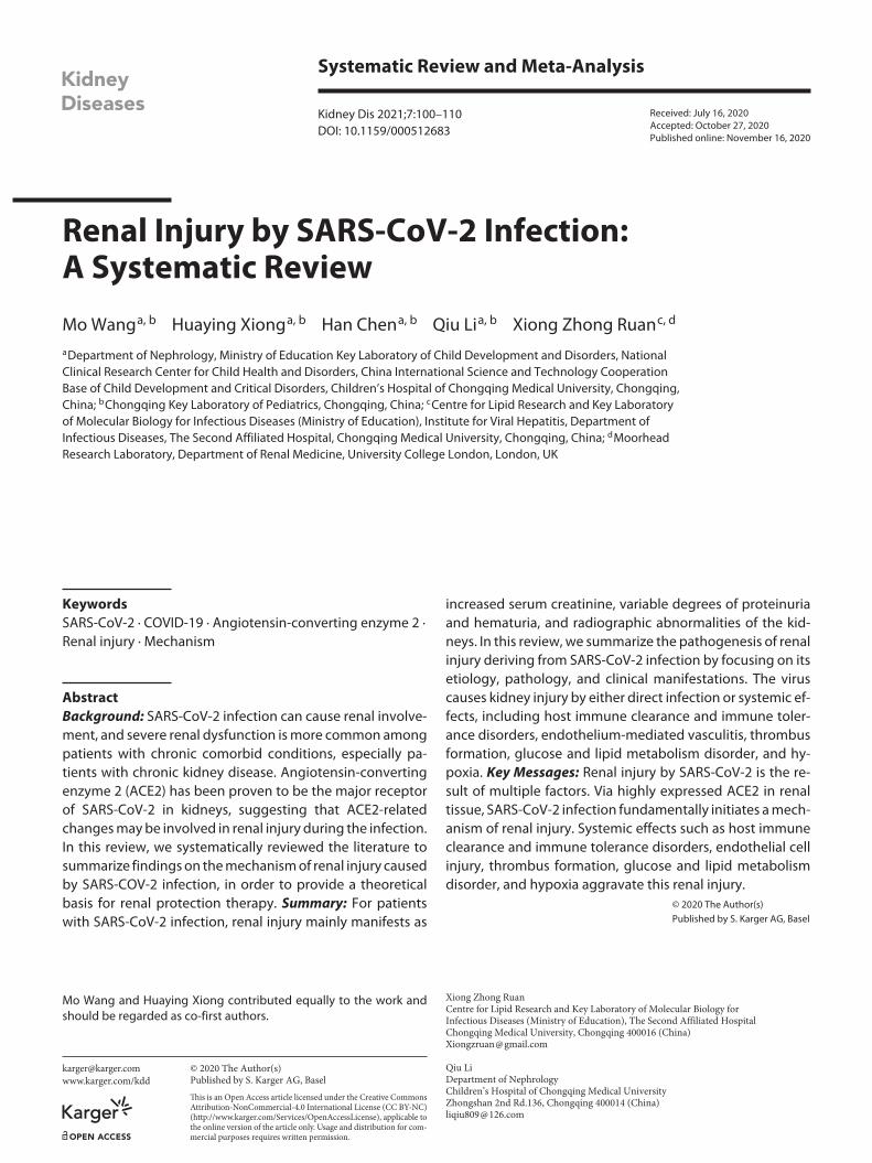
The functions of ACE2 in SARS-CoV-2 infection can be divided into two categories: a peptidase function and a peptidase-independent function. ACE2, a homologue of ACE, is a powerful negative peptide of the renin-angio-tensin system (RAS) and balances the various functions of ACE. The RAS plays a key role in maintaining blood pressure homeostasis and water-salt balance. Renin, cleaving angiotensinogen, produces angiotensin I (Ang I), which is transformed by cleavage of ACE into Ang II. Ang II binds to two G protein-coupled receptors, angio-tensin II receptor type 1 (AT1R) and angiotensin II recep-tor type 2 (AT2R), performing biological functions: vaso-constriction, elevating blood pressure, and promoting in-flammation, oxidative stress and cell apoptosis. Evidence suggests that by degrading Ang II into Ang (1-7), ACE2 negatively regulates the activated RAS, and shows protec-tive effects: vasodilation, as well as suppression of inflam-mation, oxidative stress and cell apoptosis. During SARS-CoV-2 infection, after binding to SARS-CoV-2, the exter-nal domain of ACE2 is cleaved, and the transmembrane domain is internalized, leading to downregulation of ACE2 and an increase in Ang II levels, thereby promoting the “immunoinflammatory storm” [8] (Fig. 1).
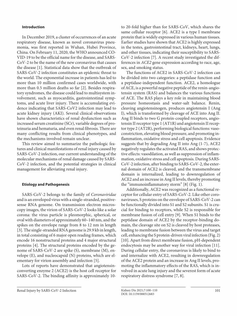
Additionally, ACE2 was recognized as a functional re-ceptor for cellular entry of SARS-CoV-2. Like other coro-naviruses, S proteins on the envelope of SARS-CoV-2 can be functionally divided into S1 and S2 subunits. S1 is cru-cial for binding to receptors, while S2 is responsible for membrane fusion of cell entry [9]. When S1 binds to the peptidase domain of ACE2 by the receptor-binding do-main, the cleavage site on S2 is cleaved by host proteases, leading to membrane fusion between the virus and target cell, enhancing the S protein-driven viral infection (Fig. 2) [10]. Apart from direct membrane fusion, pH-dependent endocytosis may be another way for viral infection [11]. During cellular entry, the coronavirus is likely to bind to and internalize with ACE2, resulting in downregulation of the ACE2 protein and an increase in Ang II levels, pro-moting the inflammatory effects of the RAS, which is in-volved in acute lung injury and the severest form of acute respiratory distress syndrome [7, 8].
Wang/Xiong/Chen/Li/RuanKidney Dis 2021;7:100–110102DOI: 10.1159/000512683
When SARS-CoV-2 invades the body, innate immune cells, as the first line of defense, start the immune re-sponse against the virus including immune clearance and anti-infection immune tolerance. Immune clearance is a
response for the identification and clearance of coronavi-ruses, while anti-infection immune tolerance is a mecha-nism which controls the immune response to avoid im-mune overreaction. An imbalance between immune
● Vasodilatation● Anti-oxidative● Anti-inflammatory● Anti-apoptotic● Anti-fibrosis
● Vasodilatation● Anti-oxidative● Anti-inflammatory● Anti-apoptotic● Anti-fibrosis
● Vasoconstriction● Oxidative stress● Inflammation● Apoptosis● Fibrosis
● Vasoconstriction● Oxidative stress● Inflammation● Apoptosis● Fibrosis
Mas AT1R AT2R Mas AT1R AT2R
ACE2
ACE2 downregulation
SARS-CoV-2
ACE2 shedding
ACE ACE
Angiotensin1-7 Angiotensin
Angiotensin
Angiotensin1-7 Angiotensin
Angiotensin
a b
Fig. 1. Simplified diagram of the renin-angiotensin system. Angio-tensin (Ang) I gets cleaved by angiotensin-converting enzyme (ACE) to form Ang II, which can mediate vasoconstriction and inflammation. ACE2 processes Ang II into Ang (1-7), which gen-erates vasodilation, anti-inflammation, anti-oxidation, and anti-
apoptosis (a). ACE2 is the host cell receptor for SARS-CoV-2. Af-ter binding to SARS-CoV-2, the ACE2 level is downregulated and the Ang II level increases, promoting vasoconstriction, inflamma-tion, oxidative stress, and cell apoptosis (b).
Viral RNA releaseReplication
TranslationTranscription
Assembly
SARS-CoV-2SARS-CoV-2
SARS-CoV-2
SARS-CoV-2
Spike (S) protein
S1
S2
Internalization
Intracellular
Extracellular
ACE2
ACE2
Fig. 2. SARS-CoV-2 binding with angio-tensin-converting enzyme (ACE) 2 and in-ternalization. For cellular entry, SARS-CoV-2 binds to and internalizes with ACE2 by the S1 subunit. Membrane fusion is me-diated via activation of spikes by proteases, and viral RNAs are released into the cyto-plasm, finishing the infection with and rep-lication of SARS-CoV-2.
Renal Injury by SARS-CoV-2 Infection 103Kidney Dis 2021;7:100–110DOI: 10.1159/000512683
clearance and immune tolerance may lead to anti-infec-tion immune intolerance, manifesting as immune over-reaction, which causes organ injury by proinflammatory cytokines and an impaired adaptive immune response.
SARS-CoV-2 infection via ACE2 may lead to local and systemic pathophysiological changes, including cellular immune disorder, cytokine storm, immune compound deposition, endothelial cell injury, thrombus formation, glucose and lipid metabolism disorder, and hypoxia, ag-gravating the renal injury. This review will focus on the mechanism of SARS-CoV-2-induced renal injury.
Renal Histopathologic Features
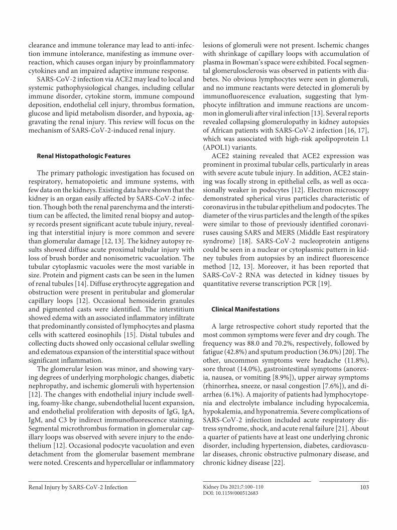
The primary pathologic investigation has focused on respiratory, hematopoietic and immune systems, with few data on the kidneys. Existing data have shown that the kidney is an organ easily affected by SARS-CoV-2 infec-tion. Though both the renal parenchyma and the intersti-tium can be affected, the limited renal biopsy and autop-sy records present significant acute tubule injury, reveal-ing that interstitial injury is more common and severe than glomerular damage [12, 13]. The kidney autopsy re-sults showed diffuse acute proximal tubular injury with loss of brush border and nonisometric vacuolation. The tubular cytoplasmic vacuoles were the most variable in size. Protein and pigment casts can be seen in the lumen of renal tubules [14]. Diffuse erythrocyte aggregation and obstruction were present in peritubular and glomerular capillary loops [12]. Occasional hemosiderin granules and pigmented casts were identified. The interstitium showed edema with an associated inflammatory infiltrate that predominantly consisted of lymphocytes and plasma cells with scattered eosinophils [15]. Distal tubules and collecting ducts showed only occasional cellular swelling and edematous expansion of the interstitial space without significant inflammation.
The glomerular lesion was minor, and showing vary-ing degrees of underlying morphologic changes, diabetic nephropathy, and ischemic glomeruli with hypertension [12]. The changes with endothelial injury include swell-ing, foamy-like change, subendothelial lucent expansion, and endothelial proliferation with deposits of IgG, IgA, IgM, and C3 by indirect immunofluorescence staining. Segmental microthrombus formation in glomerular cap-illary loops was observed with severe injury to the endo-thelium [12]. Occasional podocyte vacuolation and even detachment from the glomerular basement membrane were noted. Crescents and hypercellular or inflammatory
lesions of glomeruli were not present. Ischemic changes with shrinkage of capillary loops with accumulation of plasma in Bowman’s space were exhibited. Focal segmen-tal glomerulosclerosis was observed in patients with dia-betes. No obvious lymphocytes were seen in glomeruli, and no immune reactants were detected in glomeruli by immunofluorescence evaluation, suggesting that lym-phocyte infiltration and immune reactions are uncom-mon in glomeruli after viral infection [13]. Several reports revealed collapsing glomerulopathy in kidney autopsies of African patients with SARS-CoV-2 infection [16, 17], which was associated with high-risk apolipoprotein L1 (APOL1) variants.
ACE2 staining revealed that ACE2 expression was prominent in proximal tubular cells, particularly in areas with severe acute tubule injury. In addition, ACE2 stain-ing was focally strong in epithelial cells, as well as occa-sionally weaker in podocytes [12]. Electron microscopy demonstrated spherical virus particles characteristic of coronavirus in the tubular epithelium and podocytes. The diameter of the virus particles and the length of the spikes were similar to those of previously identified coronavi-ruses causing SARS and MERS (Middle East respiratory syndrome) [18]. SARS-CoV-2 nucleoprotein antigens could be seen in a nuclear or cytoplasmic pattern in kid-ney tubules from autopsies by an indirect fluorescence method [12, 13]. Moreover, it has been reported that SARS-CoV-2 RNA was detected in kidney tissues by quantitative reverse transcription PCR [19].
Clinical Manifestations
A large retrospective cohort study reported that the most common symptoms were fever and dry cough. The frequency was 88.0 and 70.2%, respectively, followed by fatigue (42.8%) and sputum production (36.0%) [20]. The other, uncommon symptoms were headache (11.8%), sore throat (14.0%), gastrointestinal symptoms (anorex-ia, nausea, or vomiting [8.9%]), upper airway symptoms (rhinorrhea, sneeze, or nasal congestion [7.6%]), and di-arrhea (6.1%). A majority of patients had lymphocytope-nia and electrolyte imbalance including hypocalcemia, hypokalemia, and hyponatremia. Severe complications of SARS-CoV-2 infection included acute respiratory dis-tress syndrome, shock, and acute renal failure [21]. About a quarter of patients have at least one underlying chronic disorder, including hypertension, diabetes, cardiovascu-lar diseases, chronic obstructive pulmonary disease, and chronic kidney disease [22].
Wang/Xiong/Chen/Li/RuanKidney Dis 2021;7:100–110104DOI: 10.1159/000512683
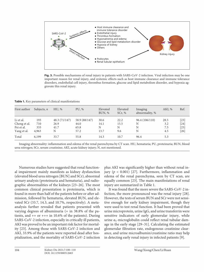
Numerous studies have suggested that renal function-al impairment mainly manifests as kidney dysfunction (elevated blood urea nitrogen [BUN] and SCr), abnormal urinary analysis (proteinuria and hematuria), and radio-graphic abnormalities of the kidneys [23–26]. The most common clinical presentation is proteinuria, which is found in more than half of the patients before or after ad-mission, followed by hematuria, elevated BUN, and ele-vated SCr (33.7, 14.3, and 10.7%, respectively). A meta-analysis further revealed that patients presented with varying degrees of albuminuria (+ in 38.8% of the pa-tients, and ++ or +++ in 10.6% of the patients). During SARS-CoV-2 infection, especially in critically ill patients, AKI was proved to be an important risk factor for mortal-ity [23]. Among those with SARS-CoV-2 infection and AKI, 33.9% of the patients were reported dead after hos-pitalization, and the mortality of SARS-CoV-2 infection
plus AKI was significantly higher than without renal in-jury (p < 0.001) [27]. Furthermore, inflammation and edema of the renal parenchyma, seen by CT scan, are equally common [23]. The main manifestations of renal injury are summarized in Table 1.
It was found that the more severe the SARS-CoV-2 in-fection, the more pronounced was the renal injury [28]. However, the tests of serum BUN and SCr were not sensi-tive enough for early kidney impairment, though they were used to test renal function. It had been proved that urine microprotein, urine IgG, and urine transferrin were sensitive indicators of early glomerular injury, while urine α1-microglobulin could reflect renal tubular dam-age in the early stage [29–31]. Calculating the estimated glomerular filtration rate, endogenous creatinine clear-ance, and urine microalbumin/creatinine ratio may help in detecting early renal injury in infected patients [9].
Table 1. Key parameters of clinical manifestations
First author Subjects, n HU, % PU, % Elevated BUN, %
Elevated SCr, %
Imaging abnormality, %
AKI, % Ref.
Li et al. 193 48.3 (71/147) 58.9 (88/147) 30.6 22.2 96.4 (106/110) 28.5 [23]Cheng et al. 710 26.9 44.0 14.1 15.5 N 3.2 [24]Pei et al. 333 41.7 65.8 N N N 7.5 [25]Yang et al. 4,963 N 57.2 13.7 9.6 N 4.5 [26]
Total 6,199 33.7 55.8 14.3 10.7 96.4 5.3
Imaging abnormality: inflammation and edema of the renal parenchyma by CT scan. HU, hematuria; PU, proteinuria; BUN, blood urea nitrogen; SCr, serum creatinine; AKI, acute kidney injury; N, not mentioned.
SARS-CoV-2
● Host immune clearance and immune tolerance disorder● Endothelial injury● Thrombus formation● Hyponatremia and edema● Glucose and lipid metabolism disorder● Hypoxia of kidney● Others
● Podocytes● Renal tubular epithelium
Kidney injury
Systemic effects
Direct infection
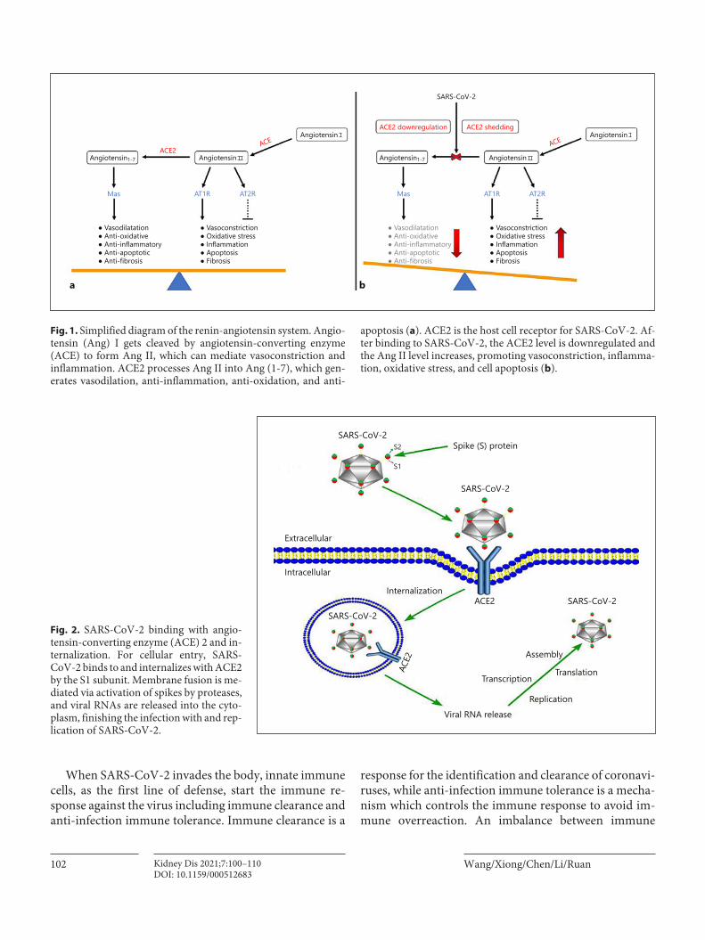
Fig. 3. Possible mechanisms of renal injury in patients with SARS-CoV-2 infection. Viral infection may be one important reason for renal injury, and systemic effects such as host immune clearance and immune tolerance disorders, endothelial cell injury, thrombus formation, glucose and lipid metabolism disorder, and hypoxia ag-gravate this renal injury.
Renal Injury by SARS-CoV-2 Infection 105Kidney Dis 2021;7:100–110DOI: 10.1159/000512683
Potential Mechanisms of Renal Injury
Some studies suggested that kidney damage in patients with SARS-CoV-2 infection was caused by SARS-CoV-2 infection and virus-induced cytokines such as IL-6 and IL-10 [24, 32], but these explanations are incomplete. Re-nal impairment may be caused by multiple mechanisms. Here, we summarize the potential mechanisms of renal injury (Fig. 3).
Direct Renal Infection by SARS-CoV-2The available evidence indicates that SARS-CoV-2
binds to ACE2 through the S1 subunit, directly causing damage to intrinsic renal cells. Human tissue single-cell RNA sequencing data and ACE2 staining revealed that the kidneys and bladder are enriched with ACE2 [12, 33], which results in susceptibility of the renal tissue to SARS-CoV-2. Clusters of coronavirus-like particles are found in the renal tubular epithelium and podocytes, and special SARS-CoV-2 nucleoproteins could be seen in a nuclear or cytoplasmic pattern via indirect fluorescence [12, 18], which suggests that SARS-CoV-2 could directly infect re-nal cells. Autopsy results showed that after infection of the respiratory tract, the virus entered the blood and in-duced viremia [34]. Hence, we infer that the virus could reach the urinary system through blood circulation, bind to and internalize with ACE2 receptors, infect kidney cells expressing the ACE2 receptor, including renal tubu-lar epithelial cells, podocytes, and others. However, ac-cording to current research, the viral load in the kidneys is low and unevenly distributed [12, 34], so it cannot wholly explain the extensive kidney damage.
Systemic Effects of SARS-CoV-2 Infection on the KidneysHost Immune Clearance and Immune Tolerance DisordersThe severity of SARS-COV-2 infection depends on the
balance between clearance of the virus and tolerance of the human immune system. Both disorders of immune clearance and anti-infection immune intolerance may be involved in viral damage to the body.
Macrophages are responsible for the identification and clearance of coronaviruses, and further for activating the downstream inflammatory signaling pathways. Subse-quently, the cellular adaptive immune response is initi-ated. Helper T (Th) cells can be induced to differentiate into Th1 cells and Th2 cells; the former can activate cyto-toxic T cells, which destroy virus-contaminated cells, just like natural killer (NK) cells; then, Th2 cells assist B lym-
phocytes in producing antibodies that inhibit viral repli-cation [35, 36]. This is the immune clearance process of anti-infection after SARS-CoV-2 invasion, accompanied by the production and release of various inflammatory cytokines, which assist the innate and adaptive immune response by participating in the process of the body’s an-tiviral activity.
Having an immune clearance disorder means that the pathogen is not detected and cleared, forming an infec-tious spread. Massive viral replication may result in an excessive intensity of immune cell response, leading to a severe inflammatory response and clinical symptoms. Previous studies have found that during the acute phase of SARS-CoV-2 infection, dendritic cell, monocyte and T-cell responses are broadly suppressed, which suggests weakening of immune response inception and viral clear-ance [36]. Meanwhile, high concentrations of viral nucle-ic acid (RNA), due to the weakened immune clearance, lead to activation of the interferon (IFN)-1 signaling path-way and JAK-STAT signaling pathway, and consequently to the production of a high level of inflammatory cyto-kines, causing damage to the body [37]. Activation of the IFN-1 signaling pathway can lead to the production of an autoreactive adaptive immune response, which can aggra-vate the tissue damage. Furthermore, the host’s anti-infec-tion immune intolerance directly results in an overactive immune response via cellular immune disorder, a cyto-kine storm, and immune compound, leading to multiple organ failure, including the kidneys, liver, and heart.
Usually, virus-infected cells are killed by CD8+ T cells and NK cells. However, patients with SARS-CoV-2 infec-tion were found to have lymphopenia dominated by CD8+ T-cell depletion [38, 39], which is a cellular im-mune disorder, resulting in insufficient killing of virus-infected target cells through cytotoxicity. Besides, be-cause of the feedback mechanism, lymphopenia stimu-lates the overproduction of inflammatory cytokines, which causes severe damage to tissues and organs. As for NK cells, which can nonspecifically kill virus-infected cells, it has been proved that NK cells are strongly acti-vated in acute SARS-CoV-2 infection, and they highly ex-press perforin, NKG2C, and Ksp37 in severe cases. An unsupervised NK-cell response and excessive cytotoxic granules may contribute to tissue injury [40]. Collective-ly, disturbance of the cellular immune system is consid-ered to be one of the reasons for the kidney damage ob-served.
Uncontrolled cytokine production during the above immune process will lead to a cytokine storm, which is a fatal immunopathologic disorder [41]. During infection,
Wang/Xiong/Chen/Li/RuanKidney Dis 2021;7:100–110106DOI: 10.1159/000512683
activation of lymphocytes induces the release of inflam-matory cytokines to destroy the infected cells, but the ex-aggerated cytokine release can lead to extensive endothe-lial dysfunction, disseminated intravascular coagulation, and multiple organ dysfunction syndrome in patients [42]. Studies have suggested that in SARS-CoV-2 pa-tients, cytokine expression – e.g., of IFN-γ, interleukin (IL)-6, IL-10, granulocyte colony-stimulating factor, and monocyte-chemotactic protein 3 – was elevated com-pared with healthy controls [43]. In the kidneys, there is obvious inflammatory infiltration of the renal intersti-tium, which predominantly consists of lymphocytes and plasma cells, with some eosinophils [15]. It also indicates that activated lymphocytes migrate to kidney tissues in order to destroy infected renal cells and release inflamma-tory cytokines, finally resulting in local inflammation and tissue injury. In addition, cytotoxic particles such as per-forin, granulysin, and proinflammatory cytokines which are highly expressed in lymphocytes, also contribute to kidney damage [44].
Besides cellular immunity and a cytokine storm, im-mune complex deposition may also cause kidney damage. Renal pathology in SARS-CoV-2 infected patients sug-gests that the damage is mainly located to the intersti-tium, with minor glomerular lesions. Podocyte-related injuries are the main manifestations of glomerular lesions such as focal segmental glomerulosclerosis and collapsing glomerulopathy [12], which are associated with viral in-fection due to ACE2 expression in podocytes. However, in addition to podocyte damage, severe endothelial dam-age was also observed, including swelling, foamy changes, and subcutaneous transmittal expansion. No inflamma-tory cell infiltration was found in glomeruli, while IgG, IgM, and trace C3 were found in the granular tissues of the capillary wall by direct or indirect immunofluores-cence and electron microscopy [14], suggesting that im-mune complex deposition is one of the causes of glomer-ular injury. Deposition of immune complexes along glo-merular capillaries leads to immune complex-associated nephritis, the mechanism of which may be associated with immune complexes activating the complement sys-tem, leading to kidney injury [45].
Endothelial Cell InjuryElectron microscopic examination shows endothelial
injury to the glomeruli of kidneys, including cell swelling with foam-like changes, subendothelial expansion, and endothelial proliferation [45]. It is supposed that the virus could directly damage endothelial cells by binding to them, especially tubular epithelial cells, which express
high levels of ACE2. Because of vascular endothelial in-jury and a cytokine storm, many critical patients have vasculitis-like manifestations or even gangrene on their extremities. Pathologic examinations reveal small vessel hyperplasia, vessel wall thickening, stenosis of the lumen, occlusion, and focal hemorrhage [44]. Vasculitis may be the underlying mechanism of vascular damage.
Thrombus FormationMost patients with severe SARS-CoV-2 infection have
hypercoagulability and disseminated intravascular coag-ulation, presenting with thrombosis and thrombocytope-nia. In addition, there were two pathological studies of caducous kidneys revealing segmental microthrombi in the glomeruli of SARS-CoV-2 patients [12, 14]. The mechanism of coagulation remains unclear, but studies have demonstrated that endothelial injury leads to up-regulation of tissue factors, thereby activating exogenous coagulation pathways [45]. As the first response of the host immune system to SARS-CoV-2 infection, the com-plement system plays an important role in accelerating platelet adhesion and aggregation, endothelial cell injury, and thrombosis. Thrombocytopenia, common in pa-tients with severe SARS-CoV-2 infection, may also be as-sociated with decreased platelet consumption due to ex-tensive coagulation activation. The kidney is damaged by the formation of extensive microthrombi.
Hyponatremia and EdemaDuring the pandemic of SARS-CoV-2 infection, many
patients have experienced unexplained edemata in the ex-tremities and lungs [46], and some patients developed acute severe hyponatremia [47, 48]. This suggests that these patients have water and salt metabolism disorder, which is a strong risk factor for AKI. The mechanism is unclear, but it was first speculated to be related to dys-function of the RAS. In SARS-CoV-2 infection, ACE2 ex-pression is decreased, which increases Ang II formation, leading to tissue edema. In addition to the RAS disorder, IL-6 is released by monocytes and macrophages, which leads to electrolyte imbalance and increases the circula-tion volume by inducing the nonosmotic release of vaso-pressin. A retrospective study has found that IL-6 is neg-atively associated with hyponatremia, while hyponatre-mia appears to be associated with more adverse outcomes and more severe disease [49].
Glucose and Lipid Metabolism DisorderAmong patients, coexistence with chronic diseases is
a prominent phenomenon, of which hypertension, dia-
Renal Injury by SARS-CoV-2 Infection 107Kidney Dis 2021;7:100–110DOI: 10.1159/000512683
betes, cardiovascular and cerebrovascular diseases, ma-lignant tumors, chronic kidney disease, and other dis-eases more commonly occur [20, 22]. People with dia-betes have significantly higher rates of serious events [50]. The cause is unknown, but we speculate that it may be related to the glucose and lipid metabolism dis-order associated with these chronic diseases, which happens to be a risk factor for kidney damage. It has been reported that tubular ACE2 protein staining is de-creased in patients with diabetes and hypertension compared with healthy persons [51, 52]. During SARS-CoV-2 infection, membranal ACE2 expression is fur-ther reduced due to binding to the virus, which may be a possible explanation for the increased susceptibility to kidney injury observed in patients with diabetes and hypertension.
Hypoxia of the KidneysThe lungs are the main target organ of SARS-CoV-2,
which could lead to hypoxia due to dysfunction of venti-lation and diffusion. In the kidneys, hypoxia may contrib-ute to AKI [53]. Hypoxemia reduces renal blood flow by a number of mechanisms, including stimulation of ad-renergic nerves and disturbances in nitric oxide metabo-lism. Severe hypoxia and ischemia can both result in mi-crovasculature dysfunction. This can impact adjacent in-trinsic cells and capillaries, extending the regions of hypoxia, leading to organ failure [54, 55].
Other EffectsRhabdomyolysis has been reported in SARS-CoV-2
patients [56–58], and the renal anatomy of SARS-CoV-2 patients has shown high levels of creatine phosphokinase staining [12], suggesting that rhabdomyolysis may be in-volved in the occurrence of AKI. Furthermore, hyperten-sion, diarrhea, heart failure, and some drugs could all lead to kidney injury in infected patients.
Management
Currently, there are no specific therapies for the treat-ment of SARS-CoV-2 infection. We should pay more at-tention to the treatment of renal lesions and the protec-tion of renal function of severely infected patients. Given the current clinical studies reporting that patients with combined chronic disease and SARS-CoV-2 infection were easier to develop AKI, these patients need to strengthen management of their fluid balance and closely observe the urine volume, color of urine, any signs of ede-
ma, and blood pressure; avoid the usage of nephrotoxic drugs; and enhance their monitoring of early biological diagnostic indices for identifying AKI, such as blood and/or urine neutrophil gelatinase-associated lipocalin. AKI could be diagnosed following one of the following condi-tions: (1) SCr is increased by ≥26.5 μmol/L within 48 h; (2) SCr has been increased to 1.5 times the baseline value within the previous 7 days; (3) the urine volume is < 0.5 mL/kg/h for 6 h [59, 60].
Possible effective antiviral therapy, symptomatic treat-ment, and promoting renal functional recovery are the principles of renal management. In addition, it has been proved that immunosuppressive drugs such as cyclospo-rin and mycophenolic acid may be good candidates for therapeutic medicines against renal damage by SARS-CoV-2 [61, 62], and specific inhibitors of IL-6 appear to be beneficial in severely infected cases [43]. However, it may be reasonable to further study these agents in con-trolled trials. Tocilizumab, as a needle-mediated mono-clonal antibody against IL-6 receptor, is being tested in a preclinical trial (ChiCTR2000029765). The results of this study are much anticipated and expected. Since ACE2 is a functional receptor of SARS-CoV-2, it has been found that human recombinant soluble ACE2 can competitive-ly bind to SARS-CoV-2, thereby reducing the organ dam-age caused by SARS-CoV-2 entering target cells. In vitro studies have shown that human recombinant soluble ACE2 can significantly block SARS-CoV-2 infection in human renal organs in a dose-dependent manner and is an effective way to protect renal function in the future [63].
For those SARS-CoV-2 patients exhibiting renal im-pairment, it is of great significance to carry out blood pu-rification and other renal replacement therapies in time in case of severe SARS-CoV-2 infection complicated by AKI. Blood purification technologies include plasma-pheresis, adsorption, perfusion, and hemofiltration, espe-cially continuous renal replacement therapy (CRRT), since CRRT has played an important role in the rescue and treatment of patients with SARS, MERS, and other cases of sepsis [64, 65]. CRRT is recommended for use as soon as possible in severely infected patients manifesting macroalbuminuria on admission, as it may remove in-flammatory cytokines and protect renal function, partic-ularly in those patients with elevated SCr levels [23]. The therapeutics listed above would be helpful to patients in-fected with SARS-CoV-2, but their efficacy needs to be further studied and confirmed.
Wang/Xiong/Chen/Li/RuanKidney Dis 2021;7:100–110108DOI: 10.1159/000512683
Conclusions
In SARS-CoV-2 patients, viral infection and replica-tion are probably the main etiologies of renal dysfunction. SARS-CoV-2 may cause renal injury either by direct renal infection or via systemic effects such as host immune clearance and immune tolerance disorders, endothelial cell injury, thrombus formation, glucose and lipid metab-olism disorder, and hypoxia. The mechanism of renal in-jury caused by SARS-CoV-2 has not yet been fully clari-fied. However, our current understanding suggests that the ACE2 signaling pathway plays a key role in mediating renal injury. It is important to monitor kidney injury in the management of SARS-CoV-2. The earlier treatments achieve a better clinical outcome. Patients with AKI are recommended to receive CRRT in order to both protect renal function and remove inflammatory cytokines, which may accelerate the process of disease recovery.
Acknowledgements
We would like to thank Han Chen, Cheng Zhong, Xueying Yang, and Qian Hu for their help in literature and data collection. Besides, we would thank all of the medical staff at the Department of Nephrology, Children’s Hospital of Chongqing Medical Uni-versity, for their support in writing this article.
Statement of Ethics
This study complied with the ethical principles of the Helsinki Declaration of the World Medical Association and was approved by the Ethics Committee of the Children’s Hospital of Chongqing Medical University (reference No. 01/2020).
Conflict of Interest Statement
The authors have no conflicts of interest to disclose.
Funding Sources
Financial support for this work was provided by the COVID-19 Emergency Research Project of Chongqing Medical University (CQMUNCP0310) to Mo Wang as the PI and the National Key R&D Program of China (2018YFC1312700).
Author Contributions
Xiong Zhong Ruan and Qiu Li designed this study and revised the manuscript; Mo Wang, Huaying Xiong, and Han Chen did the literature search and wrote the draft of this paper.
References
1 Wang C, Horby PW, Hayden FG, Gao GF. A novel coronavirus outbreak of global health concern. Lancet. 2020 Feb; 395(10223): 470–3.
2 Coronavirus disease. 2019 (COVID-19) Situ-ation Report – 162 [cited 2020 Jun 30]. Avail-able from: https://www.who.int/docs/de-fault-source/coronaviruse/20200630-covid-19-sitrep-162.pdf?sfvrsn=e00a5466_2.
3 Zheng J. SARS-CoV-2: An Emerging Corona-virus that Causes a Global Threat. Int J Biol Sci. 2020 Mar; 16(10): 1678–85.
4 Zhou M, Zhang X, Qu J. Coronavirus disease 2019 (COVID-19): a clinical update. Front Med. 2020 Apr; 14(2): 126–35.
5 Chen Y, Liu Q, Guo D. Emerging coronavi-ruses: genome structure, replication, and pathogenesis. J Med Virol. 2020 Apr; 92(4):
418–23. 6 Wrapp D, Wang N, Corbett KS, Goldsmith
JA, Hsieh CL, Abiona O, et al. Cryo-EM struc-ture of the 2019-nCoV spike in the prefusion conformation. Science. 2020 Mar; 367(6483):
1260–3. 7 Alenina N, Bader M. ACE2 in Brain Physiol-
ogy and Pathophysiology: Evidence from Transgenic Animal Models. Neurochem Res. 2019 Jun; 44(6): 1323–9.
8 Chen J, Subbarao K. The Immunobiology of SARS*. Annu Rev Immunol. 2007; 25(1): 443–72.
9 Lu R, Zhao X, Li J, Niu P, Yang B, Wu H, et al. Genomic characterisation and epidemiology of 2019 novel coronavirus: implications for virus origins and receptor binding. Lancet. 2020 Feb; 395(10224): 565–74.
10 Yan R, Zhang Y, Li Y, Xia L, Guo Y, Zhou Q. Structural basis for the recognition of the SARS-CoV-2 by full-length human ACE2. Science. 2020 Mar; 367(6485): 1444–8.
11 Lau YL, Peiris JS. Pathogenesis of severe acute respiratory syndrome. Curr Opin Immunol. 2005 Aug; 17(4): 404–10.
12 Su H, Yang M, Wan C, Yi LX, Tang F, Zhu HY, et al. Renal histopathological analysis of 26 postmortem findings of patients with CO-VID-19 in China. Kidney Int. 2020 Jul; 98(1):
219–27.13 Diao B, Wang CH, Wang RS, Feng ZQ, Tan
YJ, Wang HM, et al. Human Kidney Is a Tar-get for Novel Severe Acute Respiratory Syn-drome Coronavirus 2 (SARS-CoV-2) Infec-tion. medRxiv. 2020. Available from: https://doi.org/10.1101/2020.03.04.20031120.
14 Yao XH, Li TY, He ZC, Ping YF, Liu HW, Yu SC, et al. [A pathological report of three CO-VID-19 cases by minimal invasive autopsies]. Zhonghua Bing Li Xue Za Zhi. 2020 May;
49(5): 411–7.15 Larsen CP, Bourne TD, Wilson JD, Saqqa O,
Sharshir MA. Collapsing Glomerulopathy in a Patient with COVID-19. Kidney Int Rep. 2020 Apr; 5(6): 935–9.
16 Peleg Y, Kudose S, D’Agati V, Siddall E, Ah-mad S, Kisselev S, et al. Acute Kidney Injury due to Collapsing Glomerulopathy following COVID-19 Infection. Kidney Int Rep. 2020 Apr; 5(6): 940–5.
17 Wu H, Larsen CP, Hernandez-Arroyo CF, Mohamed MMB, Caza T, Sharshir M, et al. AKI and Collapsing Glomerulopathy Associ-ated with COVID-19 and APOL1 High-Risk Genotype. J Am Soc Nephrol. 2020 Aug;
31(8): 1688–95.18 Xu Z, Shi L, Wang Y, Zhang J, Huang L,
Zhang C, et al. Pathological findings of CO-VID-19 associated with acute respiratory dis-tress syndrome. Lancet Respir Med. 2020 Apr;
8(4): 420–2.
Renal Injury by SARS-CoV-2 Infection 109Kidney Dis 2021;7:100–110DOI: 10.1159/000512683
19 Wichmann D, Sperhake JP, Lütgehetmann M, Steurer S, Edler C, Heinemann A, et al. Au-topsy Findings and Venous Thromboembo-lism in Patients with COVID-19: A Prospec-tive Cohort Study. Ann Intern Med. 2020 Aug; 173(4): 268–77.
20 Fang ZF, Yi F, Wu K, Lai KF, Sun XZ, Zhong NS, et al. Clinical Characteristics of Corona-virus Disease 2019 (COVID-19): An Updated Systematic Review. medRxiv. 2020. Available from: https://doi.org/https://doi.org/10.1101/2020.03.07.20032573.
21 Guan WJ, Ni ZY, Hu Y, Liang WH, Ou CQ, He JX, et al. Clinical characteristics of 2019 novel coronavirus infection in China. me-dRxiv. 2020. Available from: https://doi.org/https://doi.org/10.1101/2020.02.06.2002097.
22 Guan WJ, Liang WH, Zhao Y, Liang HR, Chen ZS, Li YM, et al. Comorbidity and its impact on 1,590 patients with COVID-19 in China: a Nationwide Analysis. medRxiv. 2020. Available from: https://doi.org/10.1101/ 2020.02.25.20027664.
23 Li Z, Wu M, Yao JW, Guo J, Liao X, Song SJ, et al. Caution on Kidney Dysfunctions of 2019-nCoV Patients. medRxiv. 2020. Avail-able from: https://doi.org/10.1101/2020.02.08. 20021212.
24 Cheng YC, Luo R, Wang K, Zhang M, Wang ZX, Dong L, et al. Kidney impairment is as-sociated with in-hospital death of COVID-19 patients. medRxiv. 2020. Available from: https://doi.org/10.1101/2020.02.18.2002324.
25 Pei G, Zhang Z, Peng J, Liu L, Zhang C, Yu C, et al. Renal Involvement and Early Prognosis in Patients with COVID-19 Pneumonia. J Am Soc Nephrol. 2020 Jun; 31(6): 1157–65.
26 Yang X, Jin Y, Li R, Zhang Z, Sun R, Chen D. Prevalence and impact of acute renal impair-ment on COVID-19: a systematic review and meta-analysis. Crit Care. 2020 Jun; 24(1): 356.
27 Yang X, Yu Y, Xu J, Shu H, Xia J, Liu H, et al. Clinical course and outcomes of critically ill patients with SARS-CoV-2 pneumonia in Wuhan, China: a single-centered, retrospec-tive, observational study. Lancet Respir Med. 2020 May; 8(5): 475–81.
28 Hong XW, Chi ZP, Liu GY, Huang H, Guo SQ, Fan JR, et al. Analysis of early renal injury in COVID-19 and diagnostic value of multi-index combined detection. medRxiv. 2020. Available from: https://doi.org/10.1101/2020.03.07.20032599.
29 Chen L, Li JX, Huang XB, Yang B, Wang J, Wang XF. [Determination and its signifi-cance of the ratio of urine microalbumin to urine cretinine in patients with nephrolithia-sis complicated with renal insufficiency]. Bei-jing Da Xue Xue Bao Yi Xue Ban. 2011 Oct;
43(5): 757–60.30 Nordberg J, Allhorn M, Winqvist I, Aker-
ström B, Olsson ML. Quantitative and quali-tative evaluation of plasma and urine α1-microglobulin in healthy donors and patients with different haemolytic disorders and hae-mochromatosis. Clin Chim Acta. 2007 Nov-Dec; 386(1-2): 31–7.
31 Casanova AG, Vicente-Vicente L, Hernán-dez-Sánchez MT, Prieto M, Rihuete MI, Ra-mis LM, et al. Urinary transferrin pre-emp-tively identifies the risk of renal damage posed by subclinical tubular alterations. Biomed Pharmacother. 2020 Jan; 121: 109684.
32 Liu J, Zheng X, Tong Q, Li W, Wang B, Sutter K, et al. Overlapping and discrete aspects of the pathology and pathogenesis of the emerg-ing human pathogenic coronaviruses SARS-CoV, MERS-CoV, and 2019-nCoV. J Med Vi-rol. 2020 May; 92(5): 491–4.
33 Zou X, Chen K, Zou J, Han P, Hao J, Han Z. Single-cell RNA-seq data analysis on the re-ceptor ACE2 expression reveals the potential risk of different human organs vulnerable to 2019-nCoV infection. Front Med. 2020 Apr;
14(2): 185–92.34 Puelles VG, Lütgehetmann M, Lindenmeyer
MT, Sperhake JP, Wong MN, Allweiss L, et al. Multiorgan and Renal Tropism of SARS-CoV-2. N Engl J Med. 2020 Aug; 383(6): 590–2.
35 Esmaeilzadeh A, Elahi R. Immunobiology and immunotherapy of COVID-19: a clini-cally updated overview. J Cell Physiol. 2020 [Online ahead of print].
36 Zhou R, To KK, Wong YC, Liu L, Zhou B, Li X, et al. Acute SARS-CoV-2 Infection Impairs Dendritic Cell and T Cell Responses. Immu-nity. 2020 Oct; 53(4): 864–77.e5.
37 Luo W, Li YX, Jiang LJ, Chen Q, Wang T, Ye DW. Targeting JAK-STAT Signaling to Con-trol Cytokine Release Syndrome in COV-ID-19. Trends Pharmacol Sci. 2020 Aug;
41(8): 531–43.38 Liu J, Li S, Liu J, Liang B, Wang X, Wang H,
et al. Longitudinal characteristics of lym-phocyte responses and cytokine profiles in the peripheral blood of SARS-CoV-2 infect-ed patients. EBioMedicine. 2020 May; 55:
102763.39 Catanzaro M, Fagiani F, Racchi M, Corsini E,
Govoni S, Lanni C. Immune response in CO-VID-19: addressing a pharmacological chal-lenge by targeting pathways triggered by SARS-CoV-2. Signal Transduct Target Ther. 2020 May; 5(1): 84.
40 Maucourant C, Filipovic I, Ponzetta A, Ale-man S, Cornillet M, Hertwig L, et al. Natural killer cell immunotypes related to COVID-19 disease severity. Sci Immunol. 2020 Aug;
5(50):eabd6832.41 Iannaccone G, Scacciavillani R, Del Buono
MG, Camilli M, Ronco C, Lavie CJ, et al. Weathering the Cytokine Storm in COV-ID-19: Therapeutic Implications. Cardiorenal Med. 2020; 10(5): 277–87.
42 Chen G, Zhou Y, Ma J, Xia P, Qin Y, Li X. Is there a role for blood purification therapies targeting cytokine storm syndrome in criti-cally severe COVID-19 patients? Ren Fail. 2020 Nov; 42(1): 483–8.
43 Yang Y, Shen CG, Li JX, Yuan J, Yang MH, Wang FX, et al. Exuberant elevation of IP-10, MCP-3 and IL-1ra during SARS-CoV-2 in-fection is associated with disease severity and
fatal outcome. medRxiv. 2020. Available from: https://doi.org/10.1101/2020.03.02.20029975.
44 Zhang W, Zhao Y, Zhang F, Wang Q, Li T, Liu Z, et al. The use of anti-inflammatory drugs in the treatment of people with severe coronavi-rus disease 2019 (COVID-19): the perspec-tives of clinical immunologists from China. Clin Immunol. 2020 May; 214: 108393.
45 Noris M, Benigni A, Remuzzi G. The case of complement activation in COVID-19 multi-organ impact. Kidney Int. 2020 Aug; 98(2):
314–22.46 Valtueña J, Ruiz-Sánchez D, Volo V, Man-
chado-López P, Garayar-Cantero M. Acral edema during the COVID-19 pandemic. Int J Dermatol. 2020 Sep; 59(9): 1155–7.
47 Aggarwal S, Garcia-Telles N, Aggarwal G, La-vie C, Lippi G, Henry BM. Clinical features, laboratory characteristics, and outcomes of patients hospitalized with coronavirus disease 2019 (COVID-19): early report from the United States. Diagnosis (Berl). 2020 May;
7(2): 91–6.48 Habib MB, Sardar S, Sajid J. Acute symptom-
atic hyponatremia in setting of SIADH as an isolated presentation of COVID-19. IDCases. 2020 Jun; 21:e00859.
49 Berni A, Malandrino D, Parenti G, Maggi M, Poggesi L, Peri A. Hyponatremia, IL-6, and SARS-CoV-2 (COVID-19) infection: may all fit together? J Endocrinol Invest. 2020 Aug;
43(8): 1137–9.50 Kalyanaraman Marcello R, Dolle J, Grami S,
Adule R, Li Z, Tatem K, et al. Characteristics and Outcomes of COVID-19 Patients in New York City’s Public Hospital System. Version 3. medRxiv. 2020 Jun: 2020.05.29.20086645. Available from: https://doi.org/10.1101/2020.05.29.20086645.
51 Mizuiri S, Hemmi H, Arita M, Ohashi Y, Tanaka Y, Miyagi M, et al. Expression of ACE and ACE2 in individuals with diabetic kidney disease and healthy controls. Am J Kidney Dis. 2008 Apr; 51(4): 613–23.
52 Li Y, Xu Q, Ma L, Wu D, Gao J, Chen G, et al. Systematic profiling of ACE2 expression in diverse physiological and pathological condi-tions for COVID-19/SARS-CoV-2. J Cell Mol Med. 2020 Jul; 24(16): 9478–82.
53 Imazio M, Klingel K, Kindermann I, Brucato A, De Rosa FG, Adler Y, et al. COVID-19 pan-demic and troponin: indirect myocardial in-jury, myocardial inflammation or myocardi-tis? Heart. 2020 Aug; 106(15): 1127–31.
54 Fine LG, Norman JT. Chronic hypoxia as a mechanism of progression of chronic kidney diseases: from hypothesis to novel therapeu-tics. Kidney Int. 2008 Oct; 74(7): 867–72.
55 Husain-Syed F, Slutsky AS, Ronco C. Lung-Kidney Cross-Talk in the Critically Ill Patient. Am J Respir Crit Care Med. 2016 Aug; 194(4):
402–14.56 Suwanwongse K, Shabarek N. Rhabdomy-
olysis as a Presentation of 2019 Novel Coronavirus Disease. Cureus. 2020 Apr;
12(4):e7561.
Wang/Xiong/Chen/Li/RuanKidney Dis 2021;7:100–110110DOI: 10.1159/000512683
57 Gefen AM, Palumbo N, Nathan SK, Singer PS, Castellanos-Reyes LJ, Sethna CB. Pediat-ric COVID-19-associated rhabdomyolysis: a case report. Pediatr Nephrol. 2020 Aug; 35(8):
1517–20.58 Valente-Acosta B, Moreno-Sanchez F, Fueyo-
Rodriguez O, Palomar-Lever A. Rhabdomy-olysis as an initial presentation in a patient diagnosed with COVID-19. BMJ Case Rep. 2020 Jun; 13(6):e236719.
59 Huang C, Wang Y, Li X, Ren L, Zhao J, Hu Y, et al. Clinical features of patients infected with 2019 novel coronavirus in Wuhan, China. Lancet. 2020 Feb; 395(10223): 497–506.
60 Khwaja A. KDIGO clinical practice guidelines for acute kidney injury. Nephron Clin Pract. 2012; 120(4):c179–84.
61 Kuba K, Imai Y, Rao S, Gao H, Guo F, Guan B, et al. A crucial role of angiotensin convert-ing enzyme 2 (ACE2) in SARS coronavirus-induced lung injury. Nat Med. 2005 Aug;
11(8): 875–9.62 Tanaka Y, Sato Y, Sasaki T. Suppression of
coronavirus replication by cyclophilin inhibi-tors. Viruses. 2013 May; 5(5): 1250–60.
63 Monteil V, Kwon H, Prado P, Hagelkrüys A, Wimmer RA, Stahl M, et al. Inhibition of SARS-CoV-2 Infections in Engineered Hu-man Tissues Using Clinical-Grade Soluble Human ACE2. Cell. 2020 May; 181(4): 905–913.e7.
64 Chu KH, Tsang WK, Tang CS, Lam MF, Lai FM, To KF, et al. Acute renal impairment in coronavirus-associated severe acute respira-tory syndrome. Kidney Int. 2005 Feb; 67(2):
698–705.65 Arabi YM, Arifi AA, Balkhy HH, Najm H, Al-
dawood AS, Ghabashi A, et al. Clinical course and outcomes of critically ill patients with Middle East respiratory syndrome coronavi-rus infection. Ann Intern Med. 2014 Mar;
160(6): 389–97.