Embed Size (px)
Citation preview

Research Article
Received: 11 June 2011 Revised: 28 January 2012 Accepted: 30 January 2012 Published online in Wiley Online Library
(wileyonlinelibrary.com) DOI 10.1002/jms.2967
ESI-MS and MALLS analysis of quaternarystructure of molluscan hemocyanins†
Pavlina Dolashka,a* Franck Zal,b Aleksandar Dolashki,a Laura Molin,c
Pietro Traldic and Benedetto Salvatod
The understanding of the function of macromolecular complexes is mainly related to a precise knowledge of their structure.Recently, the development of suitable mass spectrometric techniques (electrospray ionization (ESI) and matrix-assisted laserdesorption/ionization (MALDI)) and multi-angle laser light scattering has enabled mass determination of native complexes andof their subunits. By these techniques, the structure and association/dissociation behavior of huge molecules of molluscanOctopus vulgaris, Sepia officinalis and Rapana venosa have been characterized. Molecular masses of the native and dissociatedmolecule of cephalopodan Hcs O. vulgaris (3545 and 359.3 kDa, respectively) and S. officinalis (4134 and 443.8 kDa, respectively)revealed that only one type subunit organizes their molecules, while the presence of two isoforms with different masses (422.8and 400.0 kDa) has been determined for gastropodan R. venosaHc, aggregated into didecamers. The difference of their structuralsubunits was also established after limited proteolysis with TPCK-trypsin. Eight functional units (FUs) with masses of~50kDawere isolated from both subunits of RvH and isoform of Sepia officinalis, while seven FUs were purified from OvH. Furthercharacterization of proteins by ESI-mass spectrometry (MS) and MALDI-MS, methods gave insights into post-translationalmodifications such as glycosylation. Glycosylation of O. vulgaris and S. officinalis Hcs was suggested based on the differences(11.6 and 40.0kDa, respectively) between the masses measured by ESI-MS and those calculated by their gene sequences.Copyright © 2012 John Wiley & Sons, Ltd.
Keywords: Electrospray ionization mass spectrometry (ESI-MS); Glycoproteins; Hemocyanin; Multi-angle laser light scattering (MALLS);Quaternary structure
* Correspondence to: Assoc. Prof. Dr. P. Dolashka, Institute of OrganicChemistry with Centre of Phytochemistry, Bulgarian Academy of Sciences,str. G. Bonchev 9, 1113 Sofia. E-mail: [email protected]
† Submitted on behalf of the 29th Informal Meeting on Mass Spectrometry, 2011.
a Institute of Organic Chemistry, Bulgarian Academy of Sciences, G. Bonchev 9,Sofia 1113, Bulgaria
b 1UPMC Univ. Paris 06, UMR 7144, Equipe Ecophysiologie: Adaptation etEvolution Moléculaires, Station Biologique de Roscoff, 29682, France; 2CNRS,UMR 7144, Station Biologique de Roscoff, 29682, France
c CNR-ISTM, Corso Stati Uniti 4, 35129 Padova, Italy
d Department of Biology, Section of Biophysics and Molecular Physiology,University of Padova, Via Ugo Bassi 58/B, I-35121 Padova, Italy
Abbreviations: Hemocyanins, (Hcs); Functional, units (FUs); Multi-angle, laserlight scattering (MALLS); Electrospray, ionization mass spectrometry (ESI-MS);Electrospray, ionization mass spectrometer (LCT, Micromass, Altrincham, UK);(ESI-TOF), Octopus vulgaris (OvH); Sepia, officinalis (SoH); Rapana, venosa (RvH)and Helix lucorum (HlH); Carcinus, aestuarii (CaH); Scanning, transmissionelectron microscopy (STEM); Matrix, assisted laser desorption/ionization massspectrometer Ultraflex II (Bruker Daltonics, Bremen, Germany) (MALDI-TOF)
INTRODUCTION
Hemocyanins (Hcs) are oxygen-binding proteins, freely dissolved inthe hemolymph, of many arthropods and molluscs.[1–4] Althoughthe Hcs of both phyla use a binuclear copper site for oxygenbinding, they differ substantially for their molecular architectureand arrangement of the subunits constituting the native aggre-gates. Arthropodan Hcs are hexamers or aggregates of hexamers,built up of about 75-kDa subunits containing a pair of copperatoms.[2,3] On the contrary, molluscan Hcs are high molecularweight oligomeric proteins arranged into decamers or didecamerswith molecular mass of 4000 to 9000 kDa.[5–7] They form hollowcylinder shape with diameter of 18nm and a five- or tenfoldsymmetry axis. In cephalopods, the decamers are arranged by tenstructural subunits with molecular weight about 350 kDa.[8,9]
However, in gastropods, two decamers, each arranged by tenstructural subunits with molecular weight of about 350–450kDa,are assembled face to face to form the so-called didecamer.[10–12]
Electron microscopy revealed that native molluscan Hcs exhibit apredominant decameric and didecameric structure.[11–14] Uponincreasing the pH in the absence of divalent cations, all molluscanHcs dissociate in subunits with sedimentation coefficient 11S andmolecular weight range between 250 and 450 kDa according tothe species.[12–14] The number of subunits present in a molluscanspecies varies between 1 and 3. One structural subunit wasidentified in Sepia officinalis (SoH),[15] Aplycia californica (ApH)[14]
and Murex fulvascens (MfH), while two structurally and functionallydistinct Hc isoforms were isolated from Megathura crenulata(KLH),[8] Haliotis tuberculata (HtH),[10,13,16] Nucula nucleus (NnH),[17]
J. Mass. Spectrom. (2012)
Concholepas concholepas (CcH)[18] and Rapana venosa (RvH)[19–21]
Hcs. In the case of Hc of the Roman snail Helix pomatia (HpH)[22]
and Helix lucorum (HlH),[23] three components have been identified(b-HIH, aD-HIH and aN-HIH). They all formed didecamers of homo-geneous decamers, as it was observed for M. crenulata, R. venosa
Copyright © 2012 John Wiley & Sons, Ltd.

P. Dolashka et al.
and H. tuberculata Hcs.[12,24,25] They all show the cylindricalquaternary structure (diameter 35 nm, height 38 nm), typical forgastropodan Hcs, comprising 20 subunits with a molecular massof approximately 450 kDa each. Structural subunits are singlepolypeptide chains folded as to form several globular functionalunits (FUs), with molecular masses in the range of 45–65kDa andeach containing one active site.[26]
On the basis of the mass provided by scanning transmissionelectronmicroscopy (STEM) analysis and subunit masses estimatedby SDS-polyacrylamide gel electrophoresis (SDS-PAGE), the authorswere able to propose a structural model where the one or twodecamers organized the structure of the native Hcs. However, themethods such as multi-angle laser light scattering (MALLS) andelectrospray ionization mass spectrometry (ESI-MS) can detectmore subunits than PAGE.MS is an effective method to measuremolecular masses with high precision and accuracy. Therefore,MS was used to analyse the arthropodan and molluscan Hcs[27–29]
as the methods of choice for complete investigation of subunitcomposition.MALLS and ESI-MS exhibit enough high resolution and are
useful techniques to investigate the heterogeneity of the Hccomplexes. Therefore, in this study, we report the MALLS andESI-MS data for the analysis of the quaternary structure oflarge complexes (Hcs) from mollusks.
EXPERIMENTAL
Hemolymph collection and purification of hemocyanins
The Hc was obtained from the hemolymph collected from marinesnail R. venosa (gastropod) from the Black sea (Varna, Bulgaria),[12,20]
while the hemolymph of O. vulgaris, S. officinalis (cephalopod)were collected at the Zoological Station of Napoli (Italy). Themarine living animals were stored in running sea water beforehemolymph collection. In the case of gastropods, the hemolymphwas collected by cutting the foot muscles of animals, whereas thecephalopods and arthropod hemolymphs have been obtained byincannulation of the aorta vessel. Phenylmethylsulfonyl-fluoride(1% w/v) and Cocktail complete™ (Boheringer, Mannhein) (1 tablet/50ml) were added as protease inhibitors. The hemolymph wasthen filtered on gauze and dialysed against 10-mM Tris/HCl bufferpH 7.5, containing 20-mM CaCl2 for 24 h. To remove blood cellsand precipitated material, after dialysis, the hemolymph wascentrifuged at 20 000g for 30min in a Beckman centrifuge(J2-21). From the clear solutions, Hcs were obtained by sedimenta-tion in a preparative Beckman XL70 ultra-centrifuge (rotor 70TI) at450 000g for 2 h. The dark blue pellets were re-dissolved in50-mM Tris/HCl buffer pH 7.5 containing 20-mM CaCl2.
Preparation of structural subunits
Structural subunits of Hcs were obtained after dissociation by dia-lysing of the native protein against a 0.13M glycine/NaOH buffer,pH 9.2. Structural subunits RvH1 and RvH2 were purified bymeans of an ion exchange Resource 6ml (Pharmacia) column us-ing an FPLC system. Elution was performed as described byDolashka-Angelova et al.[12]
Electron microscopic measurements
Studies of EM specimens were performed using a PhilipsW CM10Transmission ElectronMicroscope with a 30-mmobjective aperture.
wileyonlinelibrary.com/journal/jms Copyright © 20
Samples were adsorbed for 60 s to a glow-discharged pistoform/carbon-coated support film, washed three times with droplets ofdistilled water to remove buffer salts and then negatively stainedwith 1% uranyl acetate. Electron micrographs were routinelyrecorded at an instrumental magnification of 52 000.
Isolation of functional units of RvH2 subunit
Multiunit fragments as well as individual FUs were obtained bylimited proteolysis of 20mg of RvH2 with TPCK-trypsin in a ratio -400/1 (w/w), performed in 50-mM Tris–HCl, pH 8.0, containing1-mM EDTA for 4 h, at a temperature of 37 �C. The componentsof the obtained hydrolysate were separated by anion exchangeon a FPLC system using a Pharmacia Q Sepharose HighPerformance Column (HR 16/10mm), applying a stepwise NaClgradient (0–1.0M) in 50-mM Tris–HCl buffer, pH 8.2. IsolatedFUs were additionally purified on a Hypersil column (250mm4.6mm; 5mm HyPURITY C18, Thermo Quest), eluted with eluentA (0.1% TFA in water) and eluent B (80% acetonitrile in buffer A),using a gradient program of 0% B for 5min and then 0–100% Bin 60min; the flow rate was 0.6ml/min. All fragments (FUs) werecharacterized by 10% SDS-PAGE.
MALLS and ESI- mass spectrometry
100 mL of each Hc was desalted on Centricon PM10 (cut off100 kDa) micro-concentrators (Amicon, Millipore, Bedford MA,USA) in 10-mM ammonium acetate (pH 6.8). Samples were dilutedtwo to three times in the previous buffer to a final concentrationof about 20 pmol/mL for a 1000-kDa complex and continuouslyinfused into the ESI ion source at a flow rate of 5 mL/min. Greatcare was exercised so that the noncovalent interactions survivethe ionization/desorption process. Particularly in order to preservethe integrity of the noncovalent assemblies and to enhance thesensitivity of the detection, the pressure in the interface betweenthe atmospheric source and the high vacuum region wasincreased to 6.5mbar by throttling the pumping line. The acceler-ating voltage applied on the sample cone ranged from 120 to200 V (optimal value was 200 V), and both source and desolvationtemperatures were 100 �C. A solution of 1mg/mL CsI in 50%aqueous isopropanol was used for the calibration of the extendedmass range in the high m/z region. Calibration and sampleacquisitions were performed in the positive ionmode on themassrange m/z 2000–25 000, with a manual pusher value of 250 ns.Data were accumulated over 5 min, smoothed with the SavitzkyGolay method, the background subtracted and the masses finallycalculated. Molecular species were assumed to be representedby series of peaks corresponding to multiply protonated ions.The mass of each species is expressed as a mean of the massescalculated from the series of ions plus standard deviation (SD).Charge state assignments were those that gave minimum SD.The maximum entropy-based software (MaxEnt) was used onlyto find the approximate mass of each subassembly and hencethe charge on each multiply charged peak. Because the MaxEntsoftware fits symmetrical Gaussian peak shapes to the experimen-tal data, it could not be used to establish the accurate mass, sincethe peaks were asymmetrical due to adduct formation.
ESI-MS measurements were performed either on a ESI-TOF(LCT, Micromass, Altrincham, UK) or on a ESI-Q-TOF (Q-TOF II,Micromass, Altrincham, UK) mass spectrometer, and five concen-trations in ammonium acetate buffer were performed at 4 �Cand 6000 trs/min. Purity and homogeneity of the Hcs samples
12 John Wiley & Sons, Ltd. J. Mass. Spectrom. (2012)

Mass spectrometric analyses of hemocyanins
were estimated by mass analysis in denaturing conditions:Hcs were diluted to 10 pmol/ml in a 1:1 water-acetonitrile mixture(v/v) acidified with 1% formic acid. Mass spectra were recorded onthe LCT in the positive ion mode, after calibration with horse heartmyoglobin diluted to 2 pmol/mL in a 1:1 water-acetonitrilemixture (v/v) acidified with 1% formic acid.
MALDI-TOF MS
Matrix-assisted laser desorption/ionization MS (MALDI) analysiswere performed using a MALDI-TOF Ultraflex II (Bruker Daltonics,Bremen, Germany) operating in linear positive ion mode. Ions wereformed by a pulsed UV laser beam (nitrogen laser l=337nm). Theinstrumental parameters were the following: ion source voltage 1:25 kV; ion source voltage 2: 22.20 kV; delay time: 210 ns. Five mL ofeach sample (native molecule of Hcs from S. officinalis, O. vulgarisand R. venosa and their isoforms) was mixed with 5 mL of sinapinicacid matrix solution (saturated solution in H2O/Acetonitrile (50/50;v/v) containing 0.1 % trifluoracetic acid). About 1 mL of this mixturewas deposited on the stainless-steel sample holder. External masscalibrations were done using the Protein Calibration Standard II(Bruker Daltonics), based on the average values of [M+H]+ ofTrypsinogen, Protein A, Albumin-Bovine (BSA) and the averagevalues of [M+H]+ and [M+2H]2+ of Protein A and Albumin-Bovine(BSA) at ‘mass/charge’ (m/z) 23892, 44413, 66432, 22307 and33216, respectively.
Figure 1. Chromatograms of the dissociated molecule of (A) Octopus Hc,(B) Rapana Hc on a Sepharose 6 column, eluted by 10-mM Tris/HCl buffer,pH 9.0.
RESULTS AND DISCUSSION
Determination of protein mass and structure has been a centralfield of investigation since the beginning of biochemistry.Numerous methods have been developed along the decades toobtain information about mass, size, composition, subunitarrangements and physical or biochemical constants.[30]
Commonly used methods in this field are sedimentation velocity(SV), sedimentation equilibrium (SE), gel electrophoresis, size-exclusion chromatography (SEC), STEM, cryoelectron microscopy(cryo-EM), crystallography, primary sequence determination andnuclear magnetic resonance.[8,11,23,30–34] All the cited techniquesare of major interest for investigation of protein mass andstructure: they can either provide high quality information (SE,SV, STEM, crystallography and cryo-EM) or provide rapid andconvenient analysis (gel electrophoresis, SEC). These techniqueswere applied to determine the molecular masses of severalmolluscan and arthropodan Hcs. A molecular weight of3580 kDa was determined for the native molecule of O. dofleiniHc by the SE and a sedimentation coefficient, S20,w
0 , of 51.0S. Thesedimentation coefficient at about of 51S and 63S is very rapidfor the Hcs with one and two subunits, respectively, withmolecular mass about 3000 and 9000 kDa. This was confirmedby dissociation of the native molecule of OvH in 10-mM EDTAbuffer, at pH 8.0, to a single subunit with a molecular weight of359 kDa and a sedimentation coefficient, S20,w
0, of 11.1S.[32]
The size of the native Hc complexes was also studied by SEC,consistently provided a value of 400–450 kDa for severalisoforms.[26] After dissociation of the native Hcs, isolated fromthe hemolymph of molluscs S. officinalis, O. vulgaris and R. venosa,against 0.13M glycin buffer, pH 9.2, they were applied on aSuperose 6 column. Only one fraction was eluted with 50-mMTris–HCl buffer, pH 8.5 on the chromatogram applying dissoci-ated molecule of Octopus Hc and two fractions for Rapana Hc
J. Mass. Spectrom. (2012) Copyright © 2012 John Wiley
(Figure 1A and B, respectively). The absorption spectrum of allisolated isoforms showed three peaks, at 278, 344 and 550 nm,corresponding to aromatic residues, Cu2+-O2- and Cu2+-histidinecoordination centers, respectively (data not shown). However,the masses of subunits are very close, and these techniques giveapproximate values. In this context and to study high molecularmass noncovalent complexes such as invertebrate respiratoryproteins, one would expect to use methods enabling relativelyrapid analysis of samples in native and denaturing conditions,to obtain information about quaternary structure with a highprecision, able to detect slight variations in close molecular species.
MALLS and MS are two complementary approaches permittingto satisfy most of these requirements. MALLS and ESI-MS haveproved to be useful techniques for monitoring interactionsbetween proteins and ligands, association and dissociationkinetics, protein folding and enzymatic mechanisms.[33,34] Thecomplementarity of MALLS solvent versatility and ESI-MS highmass accuracy by Q-TOF instrumentation allows new and originalapproaches for investigation of large noncovalent proteinstructures. Therefore, using these techniques, OvH, SpH andRvH were analysed.
Determination of macromolecular complex mass ofmolluscan hemocyanins by mass spectrometry
Measurements of the molecular masses of the native moleculesand isolated isoforms from above mentioned Hcswere performedby ESI-TOF, ESI-Q-TOF, MALLS and MALDI-MS. MALLS providesmass and size estimation in various aqueous solvents. Beforeanalysis, 100 mL of each Hc and isoforms was desalted on 100 kDamicro-concentrators in 10-mM ammonium acetate (pH 6.8). Innon-denaturing conditions, the mass measurements of native Hcswere performed in the same buffer to preserve their native confor-mation in solution.
Purity and homogeneity of the Hcs samples were estimated bymass analysis in denaturing conditions: Hcs were diluted to 10pmol/ml in a 1:1 water-acetonitrile mixture (v/v) and 1% formic acid.In these conditions, the noncovalent interactions are disrupted,allowing the measurement of the molecular weight of the consti-tutive monomeric polypeptidic chains with a good precision.
SEC-MALLS analyses were performed with a Superose 6-Ccolumn for the native and dissociated molecule of Hc ofS. oficinalis. In Figure 2, an optical density at 280 nm (full curve)and estimated molecular weight (dots) are represented as afunction of the eluted volume. At the native condition, only onefraction was eluted on the column with a mass of Mw=4134
& Sons, Ltd. wileyonlinelibrary.com/journal/jms

Figure 2. SEC-MALLS analysis obtained for (A) the native and (B) dissoci-ated molecule in 130-mM Gly/NaOH, pH 9.6 buffer of hemocyanin S.oficinalis. Analysis was performed with a Superose 6-C columneluted with50-mM Tris–HCl buffer, pH 8.5. Optical density at 280nm (full curve),light scattering at 90� (dash) and estimated molecular weight (dots).(C) ESIM-MS spectra of dissociated molecule of SoH.
Table 1. The molecular masses of Octopus, Rapana and Sepia hemocya
Hemocyanin Subunits/FUs
Native
MALLS
O. vulgaris 17 3 545� 9 35
S. oficinalis 18 4 134� 10 44
R. venosa 2 8 455� 51 56
RvH1 8 42
RvH2 8 40
P. Dolashka et al.
wileyonlinelibrary.com/journal/jms Copyright © 20
� 10 kDa and Rw=19.8� 0.5 nm, corresponding to the nativemolecule of S. oficinalis Hc (Figure 2A). After partial dissociationof the native molecule in 130-mM Gly/NaOH, pH 9.2 buffer, againone fraction with a mass of 443.8� 2 kDa and Rw= 12.5� 1 nm(Figure 2B) was obtained.
Concurrent ESI-MS investigations of Hc were performed toobtain the molecular masses of their constituent chains andsubunits as well as their relative proportions. While MALLS providesmasses in a physiological buffer, ESI-MS measures masses of gas-phase ions. Thus, MALLS and ESI-MS are complementary methodsto explore structure heterogeneity and its relevance in biologicalconditions. Moreover, the experiments were performed on bothinstruments, ESI-TOF and ESI-Q-TOF. Because the quality of the datawas far better on the ESI-Q-TOF than on the ESI-TOF, only themass spectra obtained by ESI-Q-TOF are presented here.
Samples were diluted two to three times in 10-mMammonium acetate (pH 6.8) buffer to a final concentration ofabout 20 pmol/mL for a >1000-kDa complex and continuouslyinfused into the ESI ion source at a flow rate of 5 mL/min. Figure 2Crepresents the spectrum of dissociated molecule of S. oficinalis Hc.Deconvolution of the ESI mass spectrum gives a Mw=423173� 841Da for the subunit. The good agreement between themasses determined by ESI-MS and MALLS shows the reliabilityof themethod and validates the use of ESI-MS for investigationof noncovalent assemblies representative of the native state insolution. Since the known mass of a single subunit is 443kDa,these masses fit unambiguously with the mass of decameric form,organized by 10 subunits, which confirms that only one isoformorganizes the molecule of Octopus Hc.
The representative masses of the native and dissociatedmolecule of SoH are reported in Table 1. The mass spectra ofthe native Sepia in combination with those of dissociatedallowed to analyse the quaternary structure of the molecule,represented in the hemolymph as a decamer.
The same behavior was observed for O. vulgaris Hc. At thenative condition, only one fraction with a Mw=3545� 9 kDaand Rw=16.6� 0.5 nm (Figure 3A) and, after partial dissociation,of 359.3� 1 kDa and Rw=7.9� 1 nm (Figure 3B) were eluted onthe column. The same mass value was obtained by ESI-MS(Mw=343 564� 159Da) after dissociation of the nativemolecule (Figure 3C). Considered together, the results fromthe various approaches to mass determination of Octopusand Sepia Hcs convincingly suggested that the nativemolecules of both Hcs are decamers, composed of identicalsubunits with molecular masses of 359.3 and 423.2 kDa,respectively.
As MALLS gives very precise measurements of molar mass with a1% error or smaller, it was used as a rapid and convenient way todetermine the mass of Rapana Hc complex, which is dissolved in
nins measured by different mass spectrometric techniques
Masses measured by: (kDa)
Dissociated Difference
ESI-MS AAS
9.3� 1 343.6� 0.2 332.011 11.6
3.8� 2 423.2�0.8 383.189 40.0
6.0� 2 211.9� 0.8 -
2.8� 1 315.1� 0.6
0.0� 2
12 John Wiley & Sons, Ltd. J. Mass. Spectrom. (2012)
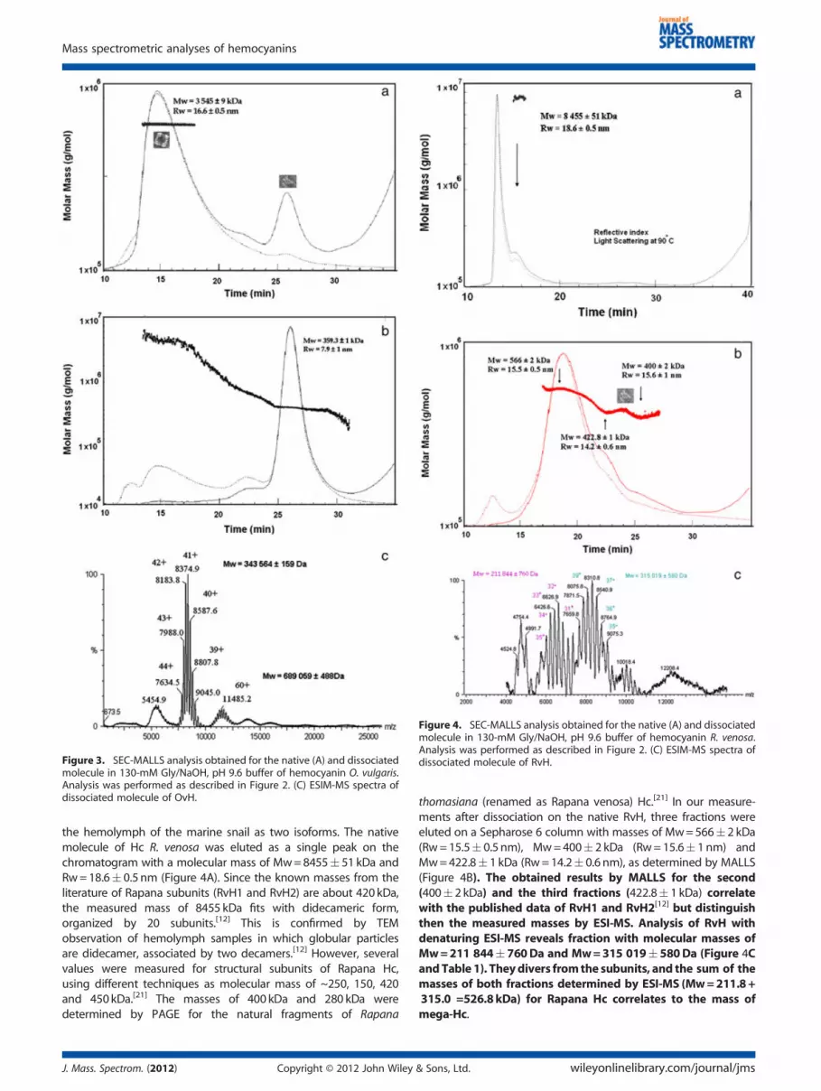
Figure 3. SEC-MALLS analysis obtained for the native (A) and dissociatedmolecule in 130-mM Gly/NaOH, pH 9.6 buffer of hemocyanin O. vulgaris.Analysis was performed as described in Figure 2. (C) ESIM-MS spectra ofdissociated molecule of OvH.
Figure 4. SEC-MALLS analysis obtained for the native (A) and dissociatedmolecule in 130-mM Gly/NaOH, pH 9.6 buffer of hemocyanin R. venosa.Analysis was performed as described in Figure 2. (C) ESIM-MS spectra ofdissociated molecule of RvH.
Mass spectrometric analyses of hemocyanins
the hemolymph of the marine snail as two isoforms. The nativemolecule of Hc R. venosa was eluted as a single peak on thechromatogram with a molecular mass of Mw=8455� 51 kDa andRw=18.6� 0.5 nm (Figure 4A). Since the known masses from theliterature of Rapana subunits (RvH1 and RvH2) are about 420 kDa,the measured mass of 8455 kDa fits with didecameric form,organized by 20 subunits.[12] This is confirmed by TEMobservation of hemolymph samples in which globular particlesare didecamer, associated by two decamers.[12] However, severalvalues were measured for structural subunits of Rapana Hc,using different techniques as molecular mass of ~250, 150, 420and 450kDa.[21] The masses of 400 kDa and 280 kDa weredetermined by PAGE for the natural fragments of Rapana
J. Mass. Spectrom. (2012) Copyright © 2012 John Wiley
thomasiana (renamed as Rapana venosa) Hc.[21] In our measure-ments after dissociation on the native RvH, three fractions wereeluted on a Sepharose 6 column with masses of Mw=566� 2 kDa(Rw=15.5� 0.5 nm), Mw=400� 2 kDa (Rw=15.6� 1nm) andMw=422.8� 1 kDa (Rw=14.2� 0.6 nm), as determined by MALLS(Figure 4B). The obtained results by MALLS for the second(400� 2 kDa) and the third fractions (422.8� 1 kDa) correlatewith the published data of RvH1 and RvH2[12] but distinguishthen the measured masses by ESI-MS. Analysis of RvH withdenaturing ESI-MS reveals fraction with molecular masses ofMw=211 844� 760Da and Mw=315 019�580Da (Figure 4CandTable 1). Theydivers fromthe subunits, and the sum of themasses of both fractions determined by ESI-MS (Mw=211.8+315.0 =526.8kDa) for Rapana Hc correlates to the mass ofmega-Hc.
& Sons, Ltd. wileyonlinelibrary.com/journal/jms

Figure 6. MALDI-TOF spectra on isolated fragment after treatment ofRvH2 subunit of Rapana hemocyanin with trypsin. Chicken egg ovalbu-min (44 400Da) and bovine serum albumin (66 430Da) were used formass scale calibration.
P. Dolashka et al.
Comparison of the measured masses by different methodsof the native and dissociated molecules of Octopus, Sepiaand Rapana Hcs is shown in Table 1. The determined massesof structural subunits of molluscan Hcs by MALLS are about400 kDa. The 400-kDa subunit has existed for ~740 millionyears and is widespread among the Mollusk.[35,36] Moreover,a fraction with mass of 566 kDa, detected for Rapana Hc onMALLS measurements (Figure 4B), was identified until nowonly as a larger mega-Hc subunit in Melanoides tuberculataHc.[37] The unique 550-kDa Hc subunit is one of the largestpolypeptides ever reported, occurring probably only in thesuperfamily Cerithioidea which appeared in the Paleozoic.[38]
The wall of the mega-decamer is apparently constructed accord-ing to the typical scheme, which means that the six wall FUs FU-ato FU-f that exist in the 400-kDa subunit are also present in the550-kDa subunit.[21]
For additional information, RvH was analysed by electronmicroscopical (EM) technique. Typical gastropodan Hcs appear-ing as didecamers tend to bind additional decamers at one orboth ends, thereby forming tridecamers, tubuls and larger multi-decamers.[6,12,13] Representations of the RvH, in Figure 5A, asdidecamers and tridecamer derived from EM analyses. Moreover,the results clearly show that additional decamer linking to thedidecamer forms a tridecamer. The exact association mode of theadditional decamers as a novel mega- Hc tridecamer (Figure 5B)in the Hcs of cerithioid snails Leptoxis, Melanoides and Terebraliawas explained by Lieb et al.[37] Electron microscopy revealeda variable mixture of mega-Hc oligomers in Melanoidestuberculata (Thiaridae)(MtH), with variable proportions of thetwo subunits, however with a clear excess of the 550-kDasubunit. The ‘typical’ tridecamer structures were also foundin KLH2, but in this case, the tridecamer is partially hollow,whereas the cerithioid tridecamer is almost completely filledwith material; it was therefore termed ‘mega-Hc’.Due to the high accuracy ofmolecularmass determination of
the mass spectrometric techniques, they also allow deter-mination of molecular masses of FUs and post-translationalmodifications, such as glycosylation.[38–40] Until now, severalFUs were isolated and analysed after enzymatic cleavage ofHcs.[14,23,31,41,42] The molecular masses of the fractions obtainedafter tryptic digestion of Rapana with trypsin and separatedby a FPLC system using a Q sepharose high-performancecolumn were measured by MALDI-TOF-MS (Figure 6). The ions
Figure 5. Transmission electron microscope images of negatively stainedarrowhead), typical tridecamers build from a decamer (white arrowhead) and
wileyonlinelibrary.com/journal/jms Copyright © 20
at m/z 50585, 55978 and 59123 are due to three FUs,while the ions at m/z 97431 and 108150 are due to two linkedFUs. Some of the eluted peaks on the column containedonly one pure FU. These data confirmed that MS is a veryrapid and useful technique not only to determine the massesof the proteins, but also to gain information on thecomposition of mixture of several fractions. Therefore, themolecular masses of the obtained fractions after trypticdigestion with trypsin of the other Hcs as Octopus and Sepiawere also analysed by MALDI-TOF to be around 50kDa (datanot shown).
Molluscan Hcs are glycosylated, but their glycan content isgenerally not high enough to cause irregular migration on SDS gels.For example, in case of Haliotis tuberculata Hc, ~400 kDa wasdetermined by SDS-PAGE, and 392kDa was later predicted fromthe amino acid sequence.[13,16] From their total yield, the glycansmight well be determined by MS. Therefore, the obtained resultsby ESI-MS andMALLS were successfully applied to predict themassof the oligosaccharides in above mentioned Hcs. The molecularmasses of the isoforms of Octopus and Sepia measured by MS dif-fer from the calculated masses determined by the gene sequences
gastropod hemocyanins. (A) RvH, showing typical didecamers (blacksubunit (dash arrowhead); (B) mega-tridecamers.
12 John Wiley & Sons, Ltd. J. Mass. Spectrom. (2012)

Mass spectrometric analyses of hemocyanins
of S. oficinalis (DQ388569) and O. vulgaris (AY751301) (Table 1).The mass differences of approximately 11.6 and 40.0 kDa in thecase of Octopus and Sepia, respectively, suggest the presence ofglycoforms. The oligosaccharide structures of OvH, SoH and RvHwere determined as complex structures.[38,39]
CONCLUSION
Hcs are glycoproteins, freely dissolved in the hemolymph, ofmany arthropods and molluscs with huge molecular masses of4000 to 9000 kDa.[6–8] The Hcs of both phyla, mollusks and ar-thropods differ substantially for their molecular architecture andarrangement of the subunits constituting the native aggregatesoften represented as noncovalent multimeric proteins.[2,3,5]
Understanding the function of macromolecular complexes is re-lated to a precise knowledge of their structure. Therefore, in thisstudy, the structure, association and dissociation behavior ofmolluscan Hcs from gastropod (R. venosa), cefalopods (O. vulgaris,S. officinalis) were studied by MS and electron spectroscopymethods. This allowed to determine the molecular massesof native and dissociated molecules. ESI-MS, MALLS andMALDI-MS techniques revealed one structural subunit in Sepiaand Octopus Hcs, while two or three components withdifferent masses were measured from Rapan Hc. Based onthe mass values determined by MS, the structure of the abovementioned Hcs was much more precisely determined.Likewise, the carbohydrate content of molluscs OvH and SoH(11.6 and 40.0kDa, respectively) was also suggested by thedifferences of calculated masses of subunits by their genesequences and confirmed by the by MS measurements.
Understanding the function of macromolecular complexes isrelated to a precise knowledge of their structure. Therefore, theobtained results confirmed that using ESI-MS, MALLS andMALDI-MS techniques and also in combination with other meth-ods and techniques as electron microscopy, gene sequences andmodeling precise information of Octopus, Sepia and Rapana Hcswas obtained.
Acknowledgements
This work was supported by a research grant N BG051PO001-3.3-05/0001 scheduled ’science-business “funded by the Opera-tional Programme” Human Resources “,by the Bulgarian NationalScience Fund TK01-496/2009 and CNR (Italy).
References[1] K. E. van Holde, K. I. Miller. Hemocyanins. Adv. Protein Chem. 1995,
47, 1.[2] T. Burmester. Evolutionary history and diversity of arthropod hemo-
cyanins. Micron 2004, 35, 121.[3] P. Dolashka-Angelova, A. Dolashki, S. Stevanovic, R. Hristova, B.
Atanasov, P. Nicolov, W. Voelter. Structure and stability of arthropodanhemocyanin Limulus polyphemus. Spectrochim. Acta. 2005, 61, 1207.
[4] S. Stoeva, P. Dolashka, R. Hristova, N. Genov, W. Voelter. Subunitcomposition and N-terminal analysis of arthropod hemocyanins.Comp. Biochem. Physiol. B 1999, 122, 69.
[5] E. V. Orlova, P. Dube, J. R. Harris, E. Beckman, F. Zemlin, J. Markl, M.van Heel. Structure of keyhole limpet hemocyanin type 1 (KLH1) at15Å resolution by electron cryomicroscopy and angular reconstitu-tion. J. Mol. Biol. 1997, 271, 417.
[6] U. Meissner, P. Dube, J. R. Harris, H. Stark, J. Markl. Structure of amolluscan hemocyanin didecamer (HtH1 from Haliotis tuberculata) at12Å resolution by cryoelectron microscopy. J. Mol. Biol. 2000, 298, 21.
J. Mass. Spectrom. (2012) Copyright © 2012 John Wiley
[7] J. Schütz, P. Dolashka-Angelova, R. Abrashev, P. Nicolov, W. Voelter.Isolation and spectroscopic characterization of the structural subu-nits of keyhole limpet hemocyanin. Biochim. Biophys. Acta 2001,1546, 325.
[8] C. Gatsogiannis, A. Moeller, F. Depoix, U. Meissner, J. Markl. Nautiluspompilius hemocyanin: 9Å cryo-EM structure and molecular modelreveal the subunit pathway and the interfaces between the 70 func-tional units. J. Mol. Biol. 2007, 374, 465.
[9] O. Lambert, N. Boisset, J. C. Taveau, J. N. Amy. Three-DimensionalReconstruction of Sepia officinalis Hemocyanin from Frozen-Hydrated Specimens. Arch. Biochem. Biophys. 1995, 316, 950.
[10] H. Keller, B. Lieb, B. Altenhein, D. Gebauer, S. Richter, S. Stricker, J.Markl. Abalone (Haliotis tuberculata) hemocyanin type 1 (HtH1) orga-nization of the � 400 kDa subunit, and amino acid sequence of itsfunctional units f, g, and h. Eur. J. Biochem. 1999, 264, 27.
[11] C. Gatsogiannis, J. Markl. Keyhole limpet hemocyanin: 9Å CryoEMstructure and molecular model of the KLH1 didecamer reveal theinterfaces and intricate topology of the 160 functional units. J. Mol.Biol. 2009, 385, 963.
[12] P. Dolashka-Angelova, H. Schwarz, A. Dolashki, M. Beltramini, B.Salvato, M. Schick, M. Saeed, W. Voelter. Oligomeric stability ofRapana venosa hemocyanin (RvH) and its structural subunits.Biochim. Biophys. Acta 2003, 1646, 77.
[13] B. Lieb, B. Altenhein, R. Lehnert, W. Gebauer, J. Markl. Subunit orga-nization of the abalone Haliotis tuberculata hemocyanin type 2(HtH2) and cDNA sequence encoding its functional units d, e, f, gand h. Eur. J. Biochem. 1999, 265, 134.
[14] B. Lieb, V. Boisguérin, W. Gebauer, J. Markl. cDNA sequence, proteinstructure, and evolution of the single hemocyanin from Aplysia cali-fornica, an opisthobranch gastropod. J. Mol. Evol. 2004, 59, 536.
[15] J. Lamy, V. You, J. C. Taveau, N. Boisset, J. N. Lamy. Intramolecularlocalization of the functional units of Sepia officinalis hemocyaninby immunoelectron microscopy. J. Mol. Biol. 1998, 284, 1051.
[16] B. Lieb, B. Altenhein, J. Markl. The sequence of a gastropod hemo-cyanin (HtH1 from Haliotis tuberculata). J. Biol. Chem. 2000,275, 5675.
[17] S. Bergmann, L. Markl, B. Lieb. The first complete cDNA sequence ofthe hemocyanin from a bivalve, the protobranch Nucula nucleus. J.Mol. Evol. 2007, 64, 500.
[18] P. De Ioannes, B. Moltedo, H. Oliva, R. Pacheco, F. Faunes, A. DeIoannes, M. Becker. Hemocyanin of the molluscan Concholepas con-cholepas exhibits an unusual heterodecameric array of subunits. J.Biol. Chem. 2004, 279, 26134.
[19] E. Dainese, D. Svergun, M. Beltramini, P. Di Muro, B. Salvato. Low-resolution structure of the proteolytic fragments of the Rapana venosahemocyanin in solution. Arch. Biochem. Biophys. 2000, 373, 154.
[20] S. Stoeva, P. Dolashka, K. Pervanova, N. Genov, W. Voelter. Multido-main structure of the Rapana thomasiana (Gastropod) hemocyaninstructural subunit RHSS1. Comp. Biochem. Physiol. 1997, 118, 927.
[21] W. Gebauer, S. Stoeva, W. Voelter, E. Dainese, B. Salvato, M. Beltramini,J. Markl. Hemocyanin subunit organization of the gastropod Rapanathomasiana. Arch. Biochem. Biophys. 1999, 372, 128.
[22] E. Wood, M. Chaplin, C. Gielens, J. De Sadeleer, G. Préaux, R. Lontie.Relative molecular mass of the polypeptide chain of bc-haemocyaninof Helix pomatia and carbohydrate composition of the functionalunits. Comp. Biochem. Physiol. 1985, 82, 179.
[23] L. Velkova, I. Dimitrov, H. Schwarz, S. Stevanovic, W. Voelter,B. Salvato, P. Dolashka-Angelova. Structure of hemocyanin fromgarden snail Helix vulgaris. Comp. Biochem. Physiology B. 2010,157, 116.
[24] J. R. Harris, W. Gebauer, F. U. Guderian, J. Markl. Keyhole limpethemocyanin (KLH) I: Reassociation from Immucothel followed byseparation of KLH1 and KLH2. Micron 1997, 28, 31.
[25] B. Altenhein, J. Markl, B. Lieb. Gene structure and hemocyaninisoform HtH2 from the mollusc Haliotis tuberculata indicate earlyand late intron hot spots. Gene 2002, 301, 53.
[26] K. I. Miller, M. E. Cuff, W. F. Lang, P. Varga-Weisz, K. Field, K. E. vanHolde. Sequence of the Octopus dofleini hemocyanin subunit: struc-tural and evolutionary implications. J. Mol. Biol. 1998, 278, 855.
[27] K. J. Light-Wahl, B. L. Schwartz, R. D. Smith. Observation of noncova-lent quarternary associations of proteins by electrospray ionizationmass spectrometry. J. Am. Chem. Soc. 1994, 116, 5271.
[28] S. Sanglier, E. Leize, A. Van Dorsselaer. Comparative ESI-MS Study of2.2 MDa Native Hemocyanins from Deep-Sea and Shore Crabs:FromProtein Oligomeric State to Biotope. J. Am. Soc. Mass Spectrom. 2003,14, 419.
& Sons, Ltd. wileyonlinelibrary.com/journal/jms

P. Dolashka et al.
[29] H. Kayser, K. Mann, G. Machaidze, M. Nimtz, P. Ringler, S. A. Müller, U.Aebi. Isolation, Characterisation and Molecular Imaging of a High-Molecular-Weight Insect Biliprotein, a Member of the HexamericArylphorin Protein Family. J. Mol. Biol. 2009, 389, 74.
[30] A. A. Sousa, R. D. Leapman. Quantitative STEM mass measurement ofbiological macromolecules in a 300 kV TEM. J. Microsc. 2007, 228, 1, 25.
[31] B. Salvato, A. Ghiretti-Magaldi, F. Ghiretti. Hemocyanin of Octopusvulgaris. The molecular weight of the minimal functional subunit in3M urea. Biochemistry 1979, 18, 2731.
[32] I. Karen, K. I. Miller, K. E. van Holde. The structure of Octopus dofleinihemocyanin. Comp. Biochem. Physiol. B 1982, 73, 1013.
[33] M. Bruneaux, M. Rousselot, E. Leize, F. H. Lallier, F. Zal. The StructuralAnalysis of Large Noncovalent Oxygen Binding Proteins by MALLSand ESI-MS: A Review on Annelid Hexagonal Bilayer Hemoglobinand Crustacean Hemocyanin. Curr. Prot. Pept. Sci. 2008, 9, 150.
[34] M. Przybylski, M. O. Glocker. Electrospray mass spectrometry of bio-molecular complexes with noncovalent interactions: new analyticalperspectives for supramolecular chemistry and molecular recogni-tion. Angew. Chem. Int. Ed Engl. 1996, 35, 806.
[35] S. Bergmann, B. Lieb, P. Ruth, J. Markl. The hemocyanin from a livingfossil, the cephalopod Nautilus pompilius: protein structure, gene or-ganization, and evolution. J. Mol. Evol. 2006, 62, 362.
[36] B. Lieb, B. Altenhein, J. Markl, A. Vincent, van Olden, E., van Holde,K. E., K. I. Miller. Structures of two molluscan hemocyanin genes:Significance for gene evolution. PNAS. 2001, 98, 4546.
wileyonlinelibrary.com/journal/jms Copyright © 20
[37] B. Lieb, W. Gebauer, C. Gatsogiannis, F. Depoix, N. Hellmann,M. Harasewych, G. Ellen, E. Strong, J. Markl. Molluscanmega-hemocyanin: an ancient oxygen carrier tuned by a~550 kDa polypeptide. Frontiers in Zoology. 2010, 7, 14.
[38] K. Sandra, P. Dolashka-Angelova, B. Devreese, J. Van Beeumen. Newinsights in Rapana venosa hemocyani N-glycosylation resulting fromon-line mass spectrometric analyses. Glycobiology 2007, 17, 141.
[39] P. Dolashka-Angelova, L. Velkova, S. Shishkov, K. Kostova, A.Dolashki, I. Dimitrov, B. Atanasov, B. Devreese, W. Voelter,J. Van Beeumen. Glycan structures and antiviral effect of thestructural subunit RvH2 of Rapana hemocyanin. Carbohydr. Res.2010, 345, 2361
[40] P. Dolashka-Angelova, B. Lieb, L. Velkova, N. Heilen, K. Sandra,L. Nikolaeva-Glomb, A. Dolashki, A. S. Galabov, J. V. Beeumen,S. Stevanovic, W. Voelter, B. Devreese. Identification of glycosylatedsites in Rapana hemocyanin by mass spectrometry and gene se-quence, and their antiviral effect. Bioconjug. Chem. 2009, 20, 1315.
[41] P. Dolashka-Angelova, S. Stefanovic, A. Dolashki, B. Devreese, B.Tzvetkova, W. Voelter, J. Van Beeumen, B. Salvato. A challengig in-sight on the structural unit 1 of molluscan Rapana venosa hemocya-nin. Arch. Biochem. Biophys. 2007, 459, 50.
[42] F. Zal, F. Chausson, E. Leize, A. Van Dorsselaer; F. H. Lallier, B. N.Green. Quadrupole time-of-flight mass spectrometry of the nativehemocyanin of the deep-sea crab Bythograea thermydron. Biomacro-molecules 2002, 3, 229.
12 John Wiley & Sons, Ltd. J. Mass. Spectrom. (2012)


