Embed Size (px)
Citation preview

J. exp. Bio/. 124, 73-91 (1986) 7 3Printed in Great Britain © The Company of Biologists Limited 1986
MODULATION OF THE CALCIUM CURRENT OFMOLLUSCAN NEURONES BY NEUROTRANSMITTERS
BY H. M. GERSCHENFELD, CONSTANCE HAMMONDAND DANIELE PAUPARDIN-TRITSCH
Laboratoire de Neurobiologie, Ecole Normale Superieure, 46 Rue d'Ulm,75230 Paris Cedex 05, France
SUMMARY
In identified neurones of the snail Helix aspersa, the Ca2+ current can bemodulated by biogenic amines and peptides in different ways.
1. A reversible increase of Ca2+ current is evoked by 1 ^mol I"1 serotonin (5-HT)in a group of identified snail ventral neurones (D6, D7, El). This 5-HT-inducedenhancement of Ca2+ current is probably mediated by cyclic GMP. Neither cyclicAMP nor forskolin mimic the effect of 5-HT, but both the intracellular injectionof cyclic GMP and the application of zaprinast (an inhibitor of a cyclic-GMP-dependent phosphodiesterase) induce a Ca2+ current increase. Moreover, when amaximal Ca2+ current increase is induced by cyclic GMP, 5-HT becomes ineffectiveand vice versa.
2. Decreases in the Ca2+ current can be generated by two mechanisms, (a) Areversible decrease of the Ca2+ current is evoked by both dopamine (1 /zmoll"1) andthe neuropeptide FMRFamide (1 ̂ moll"1) on different identified neurones. Intra-cellular injections of either cyclic AMP or cyclic GMP do not mimic the effect ofdopamine or FMRFamide. Moreover, the intracellular injection of EGTA does notaffect the Ca2+ current decrease induced by these transmitters, (b) An 'irreversible'decrease of the Ca2+ current of the D2 neurone is elicited by 5-20/imoll"1
cholecystokinin octapeptide (CCK8). Intracellular Ca2+ plays a key role in theintracellular mediation of this effect since the intracellular injection of EGTAprevents the CCK 8-induced decrease of the Ca2+ current.
INTRODUCTION
In recent years, a convincing body of knowledge has been accumulated indicatingthat neurotransmitters can modulate voltage-dependent channels such as those forCa2+.
The first observations supporting this idea were made in cardiac muscle fibres ofboth amphibians and mammals. In these, stimulation of beta-adrenergic receptors byadrenaline and its agonists caused an increase of the plateau component of the actionpotential of the cardiac muscle fibres, resulting from an increase of the Ca2+ current(Vassort et al. 1969; Reuter & Sholz, 1977; see reviews by Reuter, 1983; Reuter,Kokubun & Prod'hom, this volume; Tsien, 1983; McCleskey et al. this volume).
Keywords: synapse, neurotransmission, serotonin, dopamine, FMRFamide, cholecystokinin.

74 H. M. GERSCHENFELD AND OTHERS
This beta-adrenergic effect on the Ca2+ current was mimicked by the application ofcyclic 3',5'-adenosine monophosphate (cyclic AMP) or its analogues (see Tsien,1977; Reuter, Cachelin, De Peyer & Kobuku, 1983; Brum et al. 1983) and also byinjection of the catalytic unit of a cyclic-AMP-dependent protein kinase (Osterriederet al. 1982; Brum et al. 1983). More recently, patch-clamp studies of cardiac cellshave shown that beta-adrenergic agents evoke an increase of the average number offunctional Ca2+ channels per cell (Reuter et al. 1983; Bean, Nowycky & Tsien,1984) and a slowing of the time course of both the activation and inactivation of thesechannels (Bean et al. 1984; Brum, Osterrieder & Trautwein, 1984; see the chaptersby Reuter et al. and McCleskey et al. in this volume).
In other preparations, however, it was observed that the transmitter-inducedincrease of the Ca2+-dependent spike duration did not involve a change of the Ca2+
current. For instance, serotonin (5-hydroxytryptamine, 5-HT) was found to increasethe calcium spike duration of sensory neurones in Aplysia californica, and of someidentified neurones in Helix aspersa, by evoking a decrease in a voltage-dependentK+ conductance (Klein & Kandel, 1978, 1980; Paupardin-Tritsch, Deterre &Gerschenfeld, 1981). Similar effects were obtained by applying dopamine (DA) toidentified snail neurones (Paupardin-Tritsch, Colombaioni, Deterre & Gerschenfeld,1985a) or two small cardioexcitatory peptides (SCPA and SCPB) to Aplysia sensoryneurones (Abrams et al. 1984). These effects were also mimicked eitherby theintracellular injection of cyclic AMP or by agents which increased the intracellularcyclic AMP concentration (Klein & Kandel, 1978, 1980; Deterre, Paupardin-Tritsch, Bockaert & Gerschenfeld, 1981, 1982). A similar increase in the duration ofthe action potential of Aplysia neurones could also be evoked by the intracellularinjection of the catalytic unit of a cyclic-AMP-dependent protein kinase (Kaczmareket al. 1980; Castellucci et al. 1982). The patch-clamp study of sensory Aplysianeurones revealed that both 5-HT and cyclic AMP elicit the closing of a populationof 'background' K+ channels (called the S-channels) which constitute the predomi-nant open channel population when these cells are held at potentials near OmV(Siegelbaum, Camardo & Kandel, 1982; see Siegelbaum, Belardetti, Camardo &Shuster, this volume). The application of the catalytic unit of a cyclic-AMP-dependent protein kinase to inside-out membrane patches from the same neuronesalso closes the S-channels (Shuster, Camardo, Siegelbaum & Kandel, 1985; seeSiegelbaum et al. this volume). In voltage-clamped Aplysia sensory neurones, theclosing of the S-channels by 5-HT is reflected in the decrease of a specific outwardcurrent component, the S-current (Klein, Camardo & Kandel, 1982). 5-HT, DAand the neuropeptide FMRF amide were also able to decrease the S-current ofidentified snail neurones (Paupardin-Tritsch et al. 1985a, and unpublished results;Colombaioni, Paupardin-Tritsch, Vidal & Gerschenfeld, 1985).
In both cardiac muscle cells and peripheral neurones, neurotransmitters have alsobeen reported to decrease the duration of the Ca + current. In cardiac muscle fibres,muscarinic agonists have been reported to decrease the plateau phase of their Ca +-dependent action potential (Giles & Xoble, 1976; see Tsien & Siegelbaum, 1980).This effect appears to be associated with a decrease in Ca conductance (Hino &

Ca2+ current modulation by transmitters 75
Ochi, 1980) and would involve cyclic 3'5'-guanosine monophosphate (cyclic GMP)as a second messenger (Trautwein, Taniguchi & Noma, 1982). In the case ofperipheral neurones, noradrenaline has been observed to decrease the duration of theCa """-dependent spike recorded in the soma of both rat sympathetic neurones (Horn& McAfee, 1980; Galvan & Adams, 1982) and chick embryo dorsal root sensoryneurones in culture (Dunlap & Fischbach, 1980). The ionic mechanism involved inthis effect of noradrenaline on both types of neurones is the same and consists of adecrease of the Ca2+ current resulting from a decrease in Ca2+ conductance, withoutapparent alteration of the Ca2+ current kinetics (Dunlap & Fischbach, 1980; Galvan& Adams, 1982).
GAB A (y-aminobutyric acid), serotonin, dopamine, enkephalin and somatostatinwere also found to decrease the duration of the action potential of cultured dorsal rootneurones from chick embryos (Dunlap & Fischbach, 1980; Canfield & Dunlap,1984). In other vertebrate neurones, some neuropeptides were, in addition, found todecrease the Ca2+-dependent spike. Thus, it was observed that met-enkephalinshortens the duration of the Ca2+-dependent spike of amphibian Rohon—Beardneurones (Bixby & Spitzer, 1983) and leu-enkephalin, and other opioid peptides,decrease the duration of the somatic action potential of mouse sensory ganglionneurones in culture (Werz & Macdonald, 1982). In none of these cases have the ionicmechanisms of peptide action been analysed in detail.
In this article, we will review recent work from our own laboratory on theneurotransmitter-induced modulation of the Ca2+ current of identified neurones ofthe snail, Helix aspersa. In this preparation, biogenic amines and peptides werefound to exert at least three different actions on the neuronal Ca2+ current: (1) 5-HTevokes, in a group of identified snail ventral neurones, a reversible increase of theCa2+ current probably mediated by cyclic GMP (Paupardin-Tritsch, Hammond &Gerschenfeld, 19856, 1986); (2) both dopamine and the neuropeptide FMRFamideelicit a reversible decrease of the Ca2+ current mediated by an unknown intracellularmessenger which is not Ca2+, cyclic AMP or cyclic GMP (Paupardin-Tritsch et al.1985a; Colombaioni et al. 1985; see also Akopyan, Iljin & Chemeris, 1984); and (3)the cholecystokinin octapeptide (CCK 8) induces an irreversible decrease of the Ca +
current of an identified neurone through an intracellular mechanism in whichintracellular Ca2+ appears to play an important role (D. Paupardin-Tritsch, C.Hammond & H. M. Gerschenfeld, unpublished results).
ACTION POTENTIAL AND MEMBRANE CURRENTS IN SNAIL NEURONES
The electrophysiological experiments that will be reviewed in this article were allperformed on in vitro preparations of isolated circumoesophageal ganglia of the snailHelix aspersa. Some large neurones of these ganglia could be recognized from onepreparation to another by their position inside the ganglia (see Kerkut et al. 1975;Paupardin-Tritsch et al. 1986).
The action potential recorded in the cell body of these large molluscan neuronesinvolves Na+, Ca2+ and K+ ions. Many of the transmembrane currents carried by
76 H. M. GERSCHENFELD AND OTHERS
these ions can be specifically blocked by different pharmacological agents. Thus,the blockade of some membrane K+-conductance components by the presence of30 mmol I"1 tetraethylammonium (TEA) ions in snail saline (TEA-saline) prolongsthe spike of these neurones by unmasking a Ca2+-dependent plateau phase (seeFig. 1A).
To find which specific voltage-dependent membrane current of an identified snailneurone is modulated by the neurotransmitters, the neurones were voltage-clampedand the different current components were studied after isolating them pharmaco-logically. In our experiments, we first bathed the neurones in a saline containing1 fimol I"1 tetrodotoxin (TTX) and 30 mmol P 1 TEA. Then, holding the membranepotential at — SOmV, the neurone was depolarized to between 0 and +20 mV usingcurrent pulses of 60ms-l s duration. In these conditions, a composite current wasgenerally recorded: an initial inward component being followed by a large outwardcurrent. In the presence of TTX, the inward current of molluscan neurones is Ca2+-dependent (see reviews by Kostyuk, 1980; Hagiwara & Byerly, 1981), their outwardone corresponding to different K+-current components, namely: IDR, the K+
current associated with delayed rectification: 1^, an early transient K+ current; Ic,the Ca2+-dependent K+ current (see reviews by D. Adams, Smith & Thompson,1980; P. Adams, 1982) and sometimes the S-current (Klein ef al. 1982).
To study the effects of neurotransmitters on the Ca2+ current, it was essential toisolate the inward current from the different outward current components. More-over, it was necessary to ensure that the action of the neurotransmitters was notexerted on those outward K+-current components which do not depend on Ca .For this purpose, both the inward current and the Ca2+-dependent K+ currents wereblocked by bathing the snail neurones in a saline containing l^imolP1 TTX and30 mmol r 1 TEA and in which Ca2+ was replaced with Mg2* (TTX/TEA/Mg2*-saline). To study the remaining outward K+ currents, the membrane potential washeld at — 50 mV and the cell depolarized by pulses of 1-1-2s to between +10 and+ 20 mV.
Once it was known that the neurotransmitter did not affect the Ca2+-independentoutward K+ currents, its effects on the inward current through the Ca2+ channelswere then studied. To separate this inward current from the other current com-ponents, and to facilitate it, the neurones were bathed in a saline containing both1/zmoir1 TTX and 30 mmol T1 TEA and in which Caz+ was replaced with Ba2+
(TTX/TEA/Ba2+-saline). In these voltage-clamp experiments the neurones weregenerally held at — 50 mV and depolarized to between 0 and +20 mV by pulses of50—60 ms duration.
A SEROTONIN-INDUCED INCREASE OF THE CALCIUM CURRENT PROBABLY
MEDIATED BY CYCLIC GMP
In a group of identified neurones (neurones D6, D7), located in the ventral face ofthe parietal ganglion of the snail, Helix aspersa, bathed in a TEA-saline, 5-HT
1) was found to evoke an increase of the duration of the Ca2+-dependent

Ca2+ current modulation by transmitters
B
77
5-HT Cyclic GMP
Control \ \ 10 m s
10mv|
|20nA|
10 ms
0
-50mV J Cyclic GMP
Fig. 1. Effects of serotonin (5-HT) and cyclic GMP on the action potential and inwardcurrent of identified snail ventral pallial neurones. (A) In a D6 neurone bathed in a TEA-saline, the somatic action potential shows a Ca2+-dependent plateau phase (Control).Application of 1 ymo\ I"1 5-HT increases the duration of the spike. (B) In the same snailD4 neurone bathed in a TEA-saline, intracellular injection of cyclic GMP evokes anincrease of the duration of the Ca2+-dependent spike similar to that induced by 5-HT inA. (C) Inward current recorded from a D6 neurone bathed in TTX/TEA/Ba2+-saline(see text for details). The cell, held at -50mV, is depolarized for 60ms to OmV.Application of l/imoll"1 5-HT evokes a 30% increase in the amplitude of the peakinward current. (D) Inward current recorded in a D6 neurone from another preparation.The cell, bathed in a TTX/TEA/Baz+-saline, was held at - 5 0 mV and depolarized to-I-10 mV with a 60-ms pulse. Intracellular injection of cyclic GMP also evokes an increaseof the inward current.
action potential (Fig. 1A). This effect showed a latency of 1-3min and could beeasily reversed by washing out the neurotransmitter for 5—10 min. In cases in whichthe 5-HT application was maintained for more than 15 min, no desensitizationphenomena were observed.
The application of 1 ^mol I"1 5-HT to the same neurones did not affect any TEA-insensitive, Ca -independent outward current components (not illustrated). Incontrast, the application of 1 /imol I"1 5-HT evoked a 30% increase in the amplitudeof the inward current of the ventral parietal neurones bathed in a TTX/TEA/Ba2+-saline (Fig. 1C).
78 H. M. GERSCHENFELD AND OTHERS
To further confirm that 5-HT specifically increased the inward current throughthe Ca2+ channels, and to dismiss a possible effect of 5-HT on a hidden outwardcurrent component, the antibiotic nystatin was used to exchange completely theintracellular K+ for Cs+ (see Tillotson, 1979). In neurones loaded with Cs+ andbathed in a Cs+-containing Tris saline in which Ca2+ was replaced by Ba2+, i.e. inconditions in which Cs+ was the only intracellular monovalent cation and both K+
and Na+ were absent from the extracellular medium, 5-HT still markedly decreasedthe inward current.
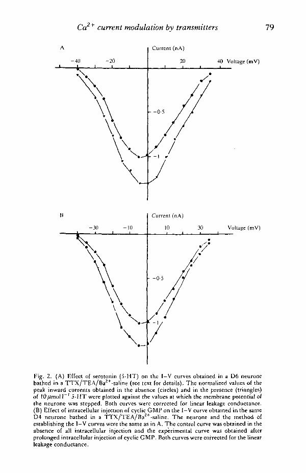
Analysis of the I-V curves relating the inward current amplitudes to the mem-brane potential at which they were recorded in ventral parietal neurones bathed in aTTX/TEA/Ba +-saline also showed that 5-HT exclusively increased the inwardcurrent (Fig. 2A) throughout the membrane potential range explored. Moreover,the potential level at which the inward current became maximal either in the presenceor in the absence of 5-HT in the bath was the same and at this potential level theinward current increase induced by 5-HT was maximal.
From these results it was concluded that 5-HT enhanced the inward current of theidentified ventral parietal neurones by increasing the membrane Ca2+ conductance.
The possibility that an intracellular second messenger could be involved in thegeneration of the 5-HT-induced of Ca2+ current in the snail ventral parietal neuroneswas suggested by the relatively long latency of the effect of 5-HT on the Ca2+-dependent action potential.
In the case of cardiac muscle cells, beta-adrenergic agonists were observed toenhance the Ca2+ current by increasing the intracellular concentration of cyclic AMP(see Introduction). It was found, however, that cyclic AMP did not intervene in the5-HT-induced increase in Ca + current of the snail ventral neurones. Thus, intra-cellular injections of cyclic AMP modified neither the duration of the action potentialof these neurones nor the outward or inward currents recorded in them. Moreover,neither the inward current nor the 5-HT-induced increase of this current werealtered by forskolin, a potent adenylate cyclase stimulating agent (Seamon & Daly,1981; see Deterre et al. 1982, for its effects on molluscan neurones).
The possibility of a participation of intracellular Ca2+ in the mechanism of theeffect of 5-HT was also examined. It was found that the intracellular injection ofEGTA in the identified ventral parietal neurones did not affect the 5-HT-inducedincrease of the inward current. To confirm that the cell loading with EGTA waseffective, parallel experiments were carried out in which intracellular injections ofEGTA were shown to block the Ca2+-dependent afterhyperpolarization that followsa train of spikes. These findings thus suggested that the effect of 5-HT on the inwardcurrent is not related to changes in the intracellular Ca + concentration.
Since DeRiemer et al. (1985) had reported that either the intracellular injectionof protein kinase C or the stimulation of this enzyme by phorbol ester TPA (12-0-tetradecanoyl-phorbol-13 acetate) evoked an increase of the inward current ofneuroendocrine bag cells of Aplysia, the effect of phorbol ester TPA was also testedon the identified ventral parietal snail neurones. The application of lOOnmoll"1
phorbol ester TPA to these neurones bathed in a TTX/TEA/Ba2+-saline also
Ca current modulation by transmitters 79
40 Voltage (mV)
Voltage (mV)
Fig. 2. (A) Effect of serotonin (5-HT) on the I-V curves obtained in a D6 neuronebathed in a TTX/TEA/Ba2+-saline (see text for details). The normalized values of thepeak inward currents obtained in the absence (circles) and in the presence (triangles)of lO^mol I"1 5-HT were plotted against the values at which the membrane potential ofthe neurone was stepped. Both curves were corrected for linear leakage conductance.(B) Effect of intracellular injection of cyclic GMP on the I-V curve obtained in the sameD4 neurone bathed in a TTX/TEA/Ba2+-saline. The neurone and the method ofestablishing the I-V curves were the same as in A. The control curve was obtained in theabsence of all intracellular injection and the experimental curve was obtained afterprolonged intracellular injection of cyclic GMP. Both curves were corrected for the linearleakage conductance.
80 H. M. GERSCHENFELD AND OTHERS
evoked an increase in the inward current. In this case, the increase in the current wasvery small at the peak and much larger at the end of the pulse. Therefore, phorbolester TPA apparently acted by reducing the inward current inactivation. Moreover,the application of 5-HT during the continued presence of phorbol ester was still ableto induce an increase in inward current, even if the phorbol ester TPA-induceddepression of the apparent inward current inactivation persisted.
The most interesting results were obtained with cyclic GMP which, unlike cyclicAMP, mimicked well the effects of 5-HT on the inward current of the ventral parietalneurones. The intracellular injection of cyclic GMP into these cells evoked anincrease of the spike duration similar to that caused by 5-HT (Fig. IB). Moreover,the intracellular injection of cyclic GMP in the ventral parietal neurones bathed in aTTX/TEA/Ba +-saline markedly increased their inward current (Fig. ID). When aprolonged 5-HT application evoked a maximal increase in the inward current, theintracellular injection of cyclic GMP was unable to induce a further increase in theinward current. Conversely, when cyclic GMP induced a maximal increase in theinward current, the application of 5-HT became ineffective.
The effects of 5-HT and cyclic GMP on the I-V curves obtained from the samesingle ventral parietal neurones were also similar (Fig. 2). As with 5-HT, cyclicGMP was observed to induce an increase in the inward current at all the potentialsexamined (Fig. 2B). Moreover, the potential level at which the inward currentbecame maximal both in control conditions and during the cyclic GMP injectionswas the same, the cyclic GMP effect being maximal at this potential level.
We examined next whether the inhibitors of phosphodiesterases which elicited anincrease of the intracellular cyclic GMP concentration in other cells (for instance,vascular muscle cells, see Winquist et al. 1984) could also induce a similar enhance-ment of the inward current in the identified ventral parietal neurones. We failed toobserve any overt action of isobutylmethylxanthine (IBMX) on the inward current.In contrast, zaprinast (M&B22948, see Winquist et al. 1984) was found to evoke anincrease of the inward current similar to that evoked by 5-HT. However, zaprinastnot only was unable to potentiate the effect of a low concentration of 5-HT on theCa2+ current but it actually blocked the 5-HT-induced increase of the current.
In more recent experiments, it could be observed that the intracellular injection ofa cyclic GMP-dependent protein kinase previously activated by cyclic GMP evokeda marked increase of the Ca + current of the identified snail ventral neurones.Moreover, the injection of this enzyme, in the absence of any previous activationof it by cyclic GMP, was found to potentiate the effect of very low concentrationsof both 5-HT and zaprinast which, by themselves, were ineffective on the inwardcurrent (D. Paupardin-Tritsch, C. Hammond, H. M. Gerschenfeld, A. Nairn &P. Greengard, unpublished results).
Considered together, all these results on the identified snail ventral parietalneurones indicate that the 5-HT-induced increase in duration of their somatic Ca2+
action potential is due to an increase in membrane Caz+ conductance. Moreover,they rule out the possibility that the 5-HT-induced increase of the Ca2+ currentcould be mediated by cyclic AMP, intracellular Ca2+ or diacylglycerol via the
Ca2+ current modulation by transmitters 81
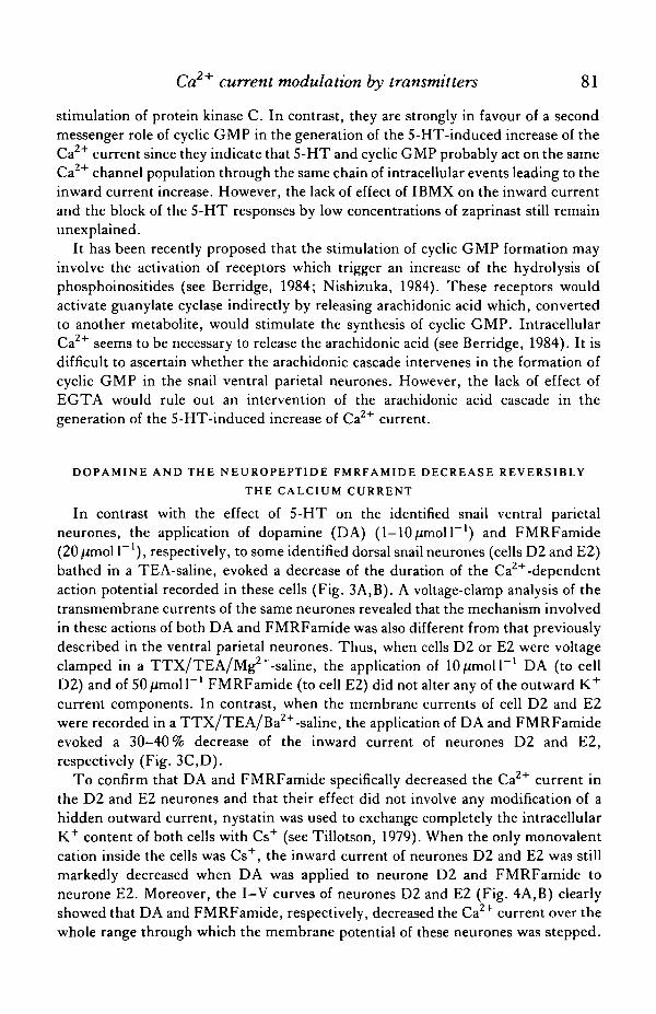
stimulation of protein kinase C. In contrast, they are strongly in favour of a secondmessenger role of cyclic GMP in the generation of the 5-HT-induced increase of theCa + current since they indicate that 5-HT and cyclic GMP probably act on the sameCa channel population through the same chain of intracellular events leading to theinward current increase. However, the lack of effect of IBMX on the inward currentand the block of the 5-HT responses by low concentrations of zaprinast still remainunexplained.
It has been recently proposed that the stimulation of cyclic GMP formation mayinvolve the activation of receptors which trigger an increase of the hydrolysis ofphosphoinositides (see Berridge, 1984; Nishizuka, 1984). These receptors wouldactivate guanylate cyclase indirectly by releasing arachidonic acid which, convertedto another metabolite, would stimulate the synthesis of cyclic GMP. IntracellularCa2+ seems to be necessary to release the arachidonic acid (see Berridge, 1984). It isdifficult to ascertain whether the arachidonic cascade intervenes in the formation ofcyclic GMP in the snail ventral parietal neurones. However, the lack of effect ofEGTA would rule out an intervention of the arachidonic acid cascade in thegeneration of the 5-HT-induced increase of Ca2+ current.
DOPAMINE AND THE NEUROPEPTIDE FMRFAMIDE DECREASE REVERSIBLYTHE CALCIUM CURRENT
In contrast with the effect of 5-HT on the identified snail ventral parietalneurones, the application of dopamine (DA) (1-10/imolF1) and FMRFamide(20/imol I"1), respectively, to some identified dorsal snail neurones (cells D2 and E2)bathed in a TEA-saline, evoked a decrease of the duration of the Ca2+-dependentaction potential recorded in these cells (Fig. 3A,B). A voltage-clamp analysis of thetransmembrane currents of the same neurones revealed that the mechanism involvedin these actions of both DA and FMRFamide was also different from that previouslydescribed in the ventral parietal neurones. Thus, when cells D2 or E2 were voltageclamped in a TTX/TEA/Mg2+-saline, the application of lO^molP1 DA (to cellD2) and of 50/imoir1 FMRFamide (to cell E2) did not alter any of the outward K+
current components. In contrast, when the membrane currents of cell D2 and E2were recorded in a TTX/TEA/Ba2+-saline, the application of DA and FMRFamideevoked a 30-40% decrease of the inward current of neurones D2 and E2,respectively (Fig. 3C,D).
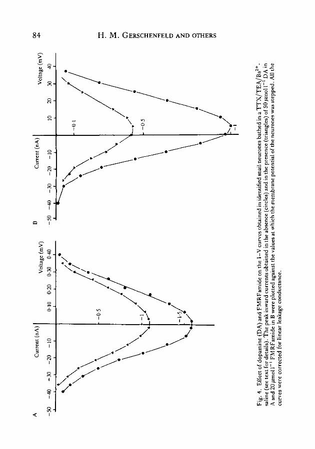
To confirm that DA and FMRFamide specifically decreased the Ca2+ current inthe D2 and E2 neurones and that their effect did not involve any modification of ahidden outward current, nystatin was used to exchange completely the intracellularK+ content of both cells with Cs+ (see Tillotson, 1979). When the only monovalentcation inside the cells was Cs+, the inward current of neurones D2 and E2 was stillmarkedly decreased when DA was applied to neurone D2 and FMRFamide toneurone E2. Moreover, the I-V curves of neurones D2 and E2 (Fig. 4A,B) clearlyshowed that DA and FMRFamide, respectively, decreased the Ca2+ current over thewhole range through which the membrane potential of these neurones was stepped.

82 H. M. GERSCHENFELD AND OTHERS
B
k Control
D
Fig. 3. Effect of 10/imoll ' dopamine (DA) and FMRFamide on the somatic actionpotential and the inward current of identified snail neurones. (A) The duration of theCa2+-dependent action potential of a D2 neurone bathed in a TEA-saline (Control) isdecreased by the application of lO^moir ' DA. (B) Bath application of 10/imoll"1
FMRFamide to an E2 neurone bathed in a TEA-saline evokes a marked decrease in theduration of the Ca +-dependent spike. (C) Inward current recordings in a D2 neuronebathed in a TTX/TEA/Ba2+-saline (see text for details). The neurone was held at— 50 mV and depolarized for 60 ms to OmV and then repolarized to — 75 mV. Bathapplication of lOfimoll"1 DA decreases the inward current and the inward tail current.(D) Inward current recordings in an E2 neurone bathed in a TTX/TEA/Baz+-saline.The cell, initially held at — 60mV, was depolarized for 60ms to +10mV and thenrepolarized to —75 mV. Application of 20^mol I"1 FMRFamide decreases the amplitudeof both the inward current and the tail inward current.
The potential level at which the Ca2+ current became maximal either in the presenceor in the absence of the transmitters in the bath was the same. Furthermore, thedecrease of the inward current induced by DA and FMRFamide was maximal at thispotential level. It can therefore be concluded that the main effect of DA on cell D2and of FMRFamide on cell E2 was to decrease their Ca2+ conductance.
Experiments were then performed to investigate the possible intervention in theactions of DA or FMRFamide of the three second messengers already mentioned inthe case of the 5-HT-induced increase in the Ca2+ current: cyclic AMP, cyclic GMPand Ca2+ ions. The intracellular injection of either cyclic AMP or cyclic GMP in D2or E2 neurones affected neither the Ca """-dependent spike duration nor the inwardcurrent of these neurones. Biochemical single-cell assay of adenylate cyclase activity

Ca2+ current modulation by transmitters 83
on single D2 and E2 neurones showed that DA did not stimulate the enzyme activityin neurone D2 and that FMRFamide did not activate the enzyme in cell E2.
Finally, injection of EGTA into neurones D2 and E2 for periods of 30—60 min didnot affect the Caz+ current decreases induced by DA and FMRFamide, respectively,in these neurones.
Therefore, it can be concluded that DA and FMRFamide both induce a similardecrease in the duration of the Ca +-dependent action potential of neurones D2 andE2 by reversibly decreasing the Ca2+ current. We have not at present any definitiveproof that the same mechanism underlies both actions. It is certain, however, thatboth actions probably involve an intracellular mechanism in which cyclic AMP,cyclic GMP or a decrease in the intracellular Ca2+ concentration do not seem to beinvolved.
A CCK8-INDUCED IRREVERSIBLE DECREASE OF CALCIUM CURRENT
In addition to 5-HT, DA and FMRFamide, a CCK8-like neuropeptide has beendetected in molluscan nervous systems and has been localized by immunocyto-chemical methods in identified cells of the snail Helix aspersa (Osborne, Cuello &Dockeray, 1982).
It has been reported that CCK8 exerts depolarizing excitatory effects on ver-tebrate central neurones (see e.g. Brooks & Kelly, 1985; Willetts, Urban, Murase &Randic, 1985). Moreover, in some dorsal horn motoneurones, CCK8 was found toevoke a reversible decrease in duration of the Ca2+-dependent action potential(Willetts et al. 1985). In some snail neurones, CCK8 may evoke an inward current,but more interestingly, on neurone D2 (in which DA has been observed to evoke areversible decrease of Ca2+ current, see above), CCK8 was also found to induce adecrease of Ca2+ current. However, in contrast with DA, the effect of CCK 8 on theCa2+ current was not reversed by washing out the peptide.
Moreover, CCK 8 acted on neurone D2 at much lower concentrations than DA(5-20 nmol F 1 ) . In a D2 neurone bathed in a TEA-saline, the application of20 nmol 1~' CCK 8 caused, in 2 min, a marked decrease of the duration of the somaticCa2+-dependent action potential (Fig. 5A). This decrease progressed further withtime (Fig. 5B). In our experimental procedure, CCK 8 was generally removed 9 minafter the onset of its application, but it could be observed that the Ca2+-dependentspike was still decreasing in duration during the first 2—3 min of washing out and thenremained unchanged even if the washing was prolonged for as long as 45 min.
The transmembrane currents were then analysed in voltage-clamped D2 neuronesof many preparations. No effect of CCK 8 was observed on the TEA-insensitive,Ca2+-independent outward current. When a D2 neurone was bathed in a TTX/TEA/Ba2+-saline in which NaCl had been replaced by Tris-Cl and to which3mmoll~1 4-aminopyridine (4-AP) had been added, it could be observed that theapplication of 5 nmol I"1 CCK 8 also reduced the amplitude of its inward current.The time course of the action of the peptide at this concentration on the inwardcurrent was slower than that observed on the Ca2+ action potential. After 6—9 min of
A -50
Cur
rent
(n
A)
-40
-30
-20
-10
Vol
tage
(ra
V)
0-10
0-
20
0-30
0-
40
Cur
rent
(nA
)V
olta
ge (
mV
)
20
30
40-5
0 -4
0 -3
0 -2
0 -1
0
00 o so C/l o z '-tl w r a > o w CO
Fig
. 4.
E
ffec
t of
dop
amin
e (D
A)
and
FM
RF
amid
e on
the
I-V
cur
ves
obta
ined
in
iden
tifie
d sn
ail n
euro
nes
bath
ed i
n a
TT
X/T
EA
/Ba2
+-
salin
e (s
ee te
xt f
or d
etai
ls).
Th
e pe
ak i
nwar
d cu
rren
ts o
btai
ned
in th
e ab
senc
e (c
ircl
es)
and
in th
e pr
esen
ce (
tria
ngle
s) o
f 50
fjm
o\ 1
~' D
A i
nA
and
20 ̂
mol
1~'
FM
RF
amid
e in
B w
ere
plot
ted
agai
nst
the
valu
es a
t whi
ch t
he m
embr
ane
pote
ntia
l of
the
neu
rone
s w
as s
tepp
ed.
All
the
curv
es w
ere
corr
ecte
d fo
r li
near
lea
kage
con
duct
ance
.
Ca2+ current modulation by transmitters 85
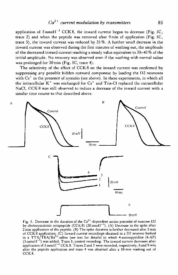
application of 5nmoll ' CCK8, the inward current began to decrease (Fig. SC,trace 2) and when the peptide was removed after 9 min of application (Fig. 5C,trace 3), the inward current was reduced by 25%. A further small decrease in theinward current was observed during the first minutes of washing out, the amplitudeof the decreased inward current reaching a steady value equivalent to 30-40 % of theinitial amplitude. No recovery was observed even if the washing with normal salinewas prolonged for 30 min (Fig. 5C, trace 4).
The selectivity of the effect of CCK8 on the inward current was confirmed bysuppressing any possible hidden outward component by loading the D2 neuroneswith Cs+ in the presence of nystatin (see above). In these experiments, in which allthe intracellular K+ was exchanged for Cs+ and Tris-Cl replaced the extracellularNaCl, CCK8 was still observed to induce a decrease of the inward current with asimilar time course to that described above.
Control
10 ms
I 50 mV
Fig. 5. Decrease in the duration of the Ca2+ -dependent action potential of neurone D2by cholecystokinin octapeptide (CCK8) (ZOnmoll"1). (A) Decrease in the spike after2 min application of the peptide. (B) The spike duration is further decreased after 5 minof CCK 8 application. (C) Inward current recordings obtained in a D2 neurone bathedin a TTX/TEA/Ba2+-saline (see text for details) to which 4-aminopyridine (4.-AP)(3 mmoll" ) was added. Trace 1, control recording. The inward current decreases afterapplication of 5 nmol P 1 CCK 8. Traces 2 and 3 were recorded, respectively, 6 and 9 minafter the peptide application and trace 4 was obtained after a 10-min washing out ofCCK 8.

86 H. M. GERSCHENFELD AND OTHERS
Analysis of the I—V curves relating the net currents evoked by CCK 8 to themembrane potentials at which they were recorded in D2 neurones bathed in TEA/TTX/Ba2+-saline containing 3mmolP' 4-AP confirmed that CCK 8 specificallydecreased the inward current of the D2 neurone by decreasing its Ca2+ conductance.
The latency and the peculiar time course of the CCK 8-induced decrease of inwardcurrent suggested the intervention of a second messenger, as in the precedingexamples of Caz+ current modulation. As has already been shown during the study ofthe DA-induced decrease of Ca2+ current in the D2 neurone (see above), theintracellular injection of either cyclic AMP or cyclic GMP inside this cell was foundnot to affect the Ca + current. But, in contrast with what had been observed in thecase of the DA-induced decrease in the Ca2+ current, the intracellular injection ofEGTA prevented the Ca2+ current decrease evoked by CCK 8. Nevertheless, theintracellular injection of EGTA was not able to reverse the CCK 8 effect when it wasperformed once the Ca2+ current had already been decreased by the application ofCCK 8.
It can therefore be concluded that CCK 8 evokes, in neurone D2, an irreversibledecrease in the Ca + current and that an increase in the intracellular Ca2+ con-centration appears to be a necessary initial event for the generation of the Ca2+
current modulation by the peptide.
DUALITIES OF THE NEUROTRANSMITTER-INDUCED MODULATIONS OF THE
CALCIUM ACTION POTENTIAL OF SNAIL NEURONES
In the case of cardiac muscle cells (see Introduction), the target of the neuro-transmitter-induced modulations of the Ca2+-dependent action potential was theCa + current. In the case of molluscan neurones, the situation is more complexbecause the increase in the duration of the Ca2+-dependent action potential mayresult from a decrease of a specific K+ current (the S-current), in the absence of anyalteration to the Ca2+ current.
The duality of action of dopamine on snail neurones illustrates this complexitywell. DA acts on identified snail neurones in two different ways: it may eitherprolong the duration of the Ca +-dependent action potential of some neuronesby decreasing a cyclic-AMP-dependent K+ current or it may decrease the Ca2+-dependent spike duration of other neurones by decreasing the neuronal Ca2+ current(Paupardin-Tritsch et al. 1985a).
This duality of effects may even take place at the level of one single neurone. Forinstance, it has been observed that both DA and FMRFamide can induce, inidentified snail neurones, a decrease of both the S-current and the Ca2+ current.These current modulations have been observed to exert opposite effects on the Ca2+-dependent action potential. However, in these cells, in which DA or FMRFamidehad a dual effect, these transmitters always elicited a decrease in the duration of theCa2+-dependent action potential (Paupardin-Tritsch et al. 1985a). The mechanismunderlying the predominance of the decrease in the Ca2+ current on the actionpotential duration is unknown.

Co2"1" current modulation by transmitters 87
In other snail neurones, it was also observed that a neurotransmitter could evoketwo different alterations of the membrane conductance having agonistic effects onthe Ca +-dependent spike. For instance, in a group of identified Aplysia neurones,the RB cells, 5-HT probably increases the Ca2+ current and also evokes a slowinward current (Pellmar, 1984), probably due to a cyclic-AMP-dependent decreasein K+ conductance.
MULTIPLICITY OF NEUROTRANSMITTERS AND SECOND MESSENGERSMODULATING THE CALCIUM CURRENT
It is important to emphasize that as for the neurotransmitters which modulate theCa +-dependent spike duration, the second messengers mediating the modulation ofthe Ca + current can also display multiple actions. Thus, whereas in cardiac musclecells cyclic AMP mediates the increase in Ca2+ current and cyclic GMP seems tointervene in the decrease of the Ca + current, in some molluscan neurones cyclicAMP mediates a decrease in K+ conductance whereas cyclic GMP is probablyinvolved in an increase in the Ca2+ current of other identified neurones.
A second intracellular messenger system also probably intervenes in the neuro-transmitter-induced decrease in the Ca2+ current of vertebrate neurones. In sym-pathetic ganglion neurones, the noradrenaline-induced decrease in Ca2+ currentappears to be modulated by alpha-adrenergic receptors (Horn & McAffee, 1980).Since activation of alpha-receptors has been reported to induce inhibition of adeny-late cyclase activity (Jakobs, 1979), it has been postulated that the decrease in theCa2+ current is linked to a fall in intracellular cyclic AMP concentration (McAffeeet al. 1981). Moreover, more recently Holz, Rane & Dunlap (1986) have reportedthat GABA- and noradrenaline-induced decreases in Ca2+ current in embryonicspinal ganglion cells in culture were blocked both by pertussis toxin and byintracellular administration of GDP-B-S [guanosine 5'-O-(2-thiodiphosphate)], anon-hydrolysable analogue of GDP which competitively inhibits GTP binding.These results strongly suggest that the neurotransmitter-induced depression of theCa current of the spinal ganglion neurones involves the intervention of a GTP-binding protein.
In the case of an identified snail neurone, the D2 cell, two different transmitterswere found to decrease the Ca2+ current, apparently using two different secondmessenger systems. The mechanism of the DA-induced reversible decrease in Ca2+
current is still unknown, but intracellular Ca2+ does not seem to be involved. Themechanism of the irreversible decrease in the Ca +-current elicited by CCK 8probably involves the participation of intracellular Ca +.
PHYSIOLOGICAL IMPLICATIONS
The modulation of the Ca2+ current at neuronal synaptic endings plays animportant role in the regulation of the transmitter release by them. Since it is notpossible to measure directly the Ca + current in a large majority of presynaptic
88 H. M. GERSCHENFELD AND OTHERS
endings, the cell bodies of neurones producing a Ca2+ spike and endowed withtransmitter receptors can constitute rather useful models to analyse events whichmay be involved in the modulation of the transmitter release at the neuronal endings.Thus, Klein & Kandel (1978, 1980) have shown that the S-HT-induced increase induration of the soma action potential of Aplysia sensory neurones could account forthe mechanism of the presynaptic facilitation evoked by 5-HT or for the stimulationof some interneurones which underlies the 'sensitization' of the gill withdrawalreflex. It is possible, following the same line of thought, that a 5-HT-inducedincrease in Ca2+ current at the endings of the snail ventral parietal neurones couldalso evoke a reversible presynaptic facilitation of their transmitter release.
In contrast, the decrease in Ca + conductance in chick embryo sensory ganglionneurones evoked by catecholamines, GABA and certain peptides (Dunlap &Fischbach, 1980) would account for the presynaptic inhibition at some of the dorsalroot ganglion cell endings in the spinal cord (see Mudge, Leeman & Fischbach,1979). A similar parallelism between decrease in the Ca2+ conductance of the somamembrane and presynaptic inhibition has been described in neurone L10 of Aplysia(Shapiro, Castellucci & Kandel, 1980). It can then be postulated that if DA isreleased near the endings of neurone D2 it could also be involved in presynapticinhibition phenomena. Moreover, the CCK8-induced decrease in Ca2+ currentcould also underlie a long-term presynaptic inhibition of neurones, such as snailneurone D2, showing CCK8-induced responses.
REFERENCES
ABRAMS, T. W., CASTELLUCCI, V. F., CAMARDO, J. S., KANDEL, E. R. & LLOYD, P. E. (1984).
Two endogenous peptides modulate the gill and siphon withdrawl reflex in Aplysia by pre-synaptic facilitation involving cAMP-dependent closure of a serotonin-sensitive channel. Proc.natn.Acad. Sci. U.SA. 81, 7956-7960.
ADAMS, D. J., SMITH, S. J. & THOMPSON, S. H. (1980). Ionic currents in molluscan neurones.A. Rev. Neurvsci. 3, 141-167.
ADAMS, P. R. (1982). Voltage-dependent conductances of vertebrate neurones. Trends Neurvsci.46, 116-119.
AKOPYAN, A. R., ILJIN, V. & CHEMERIS, N. K. (1984). Dopamine-induced modulation of Ca-component of action potential in neuronal soma of L. stagnalis mollusc. Biofizika 29, 284—288(in Russian).
BEAN, B. P., NOWYCKY, M. C. & TSIEN, R. W. (1984). /3-Adrenergic modulation of calciumchannels in frog ventricular heart cells. Nature, Loud. 307, 371-375.
BERRIDGE, M. J. (1984). Inositol trisphosphate and diacylglycerol as second messengers. Biochem.J. 220, 345-360.
BLXBY, J. L. & SPITZER, N. C. (1983). Enkephalin reduces calcium action potentials in Rohon-Beard neurons in-vivo.J. Neurvsci. 3, 1014-1018.
BROOKS, P. A. & KELLY, J. S. (1985). Cholecystokinin as a potent excitant of neurons of thedentate gyrus. Ann. N.Y. Acad. Sci. 448, 361-374.
BRUM, G., FLOCKERZI, V., HOFMAN, F., OSTERRIEDER, W. &TRAUTWEIN, W. (1983). Injection of
catalytic subunit of cAMP-dependent protein kinase into isolated myocytes. Pfliigers Arch ges.Physiol. 398, 146-154.
BRUM, G., OSTERRIEDER, W. & TRAUTWEIN, W. (1984). jS-adrenergic increase in the calciumconductance of cardiac myocytes studied with the patch-clamp. Pfliigers Arch. ges. Physiol. 401,111-118.

Co2"1" current modulation by transmitters 89
CANFIELD, D. R. & DUNLAP, K. (1984). Pharmacological characterization of amine receptors onembryonic chick sensory neurones. Br.jf. Pharmac. 82, 557-561.
CASTELLUCCI, V. F., KANDEL, E. R., SCHWARTZ, J. H., WILSON, F. D., NAIRN, A. C. &
GREENGARD, P. (1982). Intracellular injection of the catalytic unit of cyclic AMP-dependentprotein kinase stimulates facilitation of transmitter release underlying behavioral sensitization.Proc. natn. Acad. Sci. U.SA. 77, 7492-7496.
COLOMBAIONI, L., PAUPARDIN-TRTTSCH, D., VIDAL, P. P. & GERSCHENFELD, H. M. (1985). The
neuropeptide FMRFamide decreases both the Ca2+-conductance and a cAMP-dependent K+-conductance in identified snail neurons..?. Neurosci. 9, 2533-2538.
DERIEMER, S. A., STRONG, J. A., ALBERT, K. A., GREENGARD, P. & KACZMAREK, L. K. (1985).
Enhancement of calcium current in Aplysia neurones by phorbol ester and protein kinase C.Nature, Lond. 313, 313-316.
DETERKE, P., PAUPARDIN-TRITSCH, D., BOCKAERT, J. & GERSCHENFELD, H. M. (1981). Role of
cyclic AMP in a serotonin-evoked slow inward current in snail neurones. Nature, Lond. 290,783-785.
DETERRE, P., PAUPARDIN-TRITSCH, D., BOCKAERT, J. & GERSCHENFELD, H. M. (1982). cAMP-
mediated decrease in K+ conductance evoked by serotonin and dopamine in the same neuron:a biochemical and physiological single-cell study. Proc. natn. Acad. Set. U.SA. 79, 7934—7938.
DUNLAP, K. & FISCHBACH, G. D. (1980). Neurotransmitters decrease the calcium conductanceactivated by depolarization of embryonic chick sensory neurones. J. Physiol., Lond. 317,519-535.
GALVAN, M. & ADAMS, P. R. (1982). Control of calcium current in rat sympathetic neurons bynorepinephrine. Brain Res. 244, 135-144.
GILES, W. & NOBLE, S. J. (1976). Changes in membrane current in bullfrog atrium produced byacetylcholine. J. Physiol., Lond. 261, 103-123.
HAGIWARA, S. & BYERLY, L. (1981). Calcium channel. A. Rev. Neurosci. 4, 69-125.HlNO, N. & OCHI, R. (1980). Effect of acetylcholine on membrane currents in guinea-pig papillary
muscle. J. Physiol., Lond. 307, 183-197.HOLZ, G. G., IV, RANE, S. G. & DUNLAP, K. (1986). GTP-binding proteins mediate transmitter
inhibition of voltage-dependent calcium channels. Nature, Lond. 319, 670-672.HORN, J. P. & MCAFFEE, D. A. (1980). Alpha adrenergic inhibition of calcium-dependent
potentials in rat sympathetic neurones..7. Physiol., Lond. 301, 109-204.JAKOBS, K. H. (1979). Inhibition of adenylate cyclase by hormones and neurotransmitters. J . cell.
molec. Endocr. 16, 147-150.KACZMAREK, I. K., JENNINGS, K. R., STRUMWASSER, F., NAIRN, A. C , WALTER, A. C. &
GREENGARD, P. (1980). Microinjection of catalytic subunit of cAMP-dependent protein kinaseenhances calcium action potentials of bag cell neurons in cell culture. Proc. natn. Acad. Sci.U.SA. 77, 7487-7491.
KERKUT, G. A., LAMBERT, J. D. C , GAYTON, R. J., LOKER, J. E. & WALKER, R. J. (1975).
Mapping of nerve cells in the suboesophageal ganglion of Helix aspersa. Contp. Biochem. Physiol.50A, 1-25.
KLEIN, M., CAMARDO, J. S. & KANDEL, E. R. (1982). Serotonin modulates a new potassiumcurrent in the sensory neurons that show presynaptic facilitation in Aplysia. Proc. natn. Acad.Sci. U.SA. 79, 5713-5717.
KLEIN, M. & KANDEL, E. R. (1978). Presynaptic modulation of voltage dependent Ca2+ current:mechanism for behavioral sensitization in Aplysia califomica. Proc. natn. Acad. Sci. U.SA. 75,3512-3516.
KLEIN, M. & KANDEL, E. R. (1980). Mechanism of calcium current modulation underlyingpresynaptic facilitation and behavioral sensitization in Aplysia. Pmc. natn. Acad. Sci. U.SA. 77,6912-6916.
KOSTYUK, P. G. (1980). Calcium ionic channels in electrically excitable membrane. Neurosci. 5,945-959.
MCAFFEE, D. A., HENON, B. K., HORN, J. P. & YAROWSKY, P. (1981). Calcium currentsmodulated by adrenergic receptors in sympathetic neurons. Fedn Proc. FednAm. Socs exp. Biol.40, 2246-2249.
MUDGE, A. W., LEEMAN, S. E. & FISCHBACH, G. D. (1979). Enkephalin inhibits release ofsubstance P from sensory neurons in culture and decreases action potential duration. Proc. natn.Acad. Sci. U.SA. 76, 526-530.

90 H. M. GERSCHENFELD AND OTHERS
NlSHIZUKA, N. (1984). Turnover of inositol phospholipids and signal transduction. Science 225,1365-1370.
OSBORNE, N. N., CUELLO, A. C. & DOCKERAY, G. J. (1982). Substance P, cholecystokinin andserotonin in a giant neuron. Science 216, 409-411.
OSTERRIEDER, W., BRUM, G., HESCHELER, J., TRAUTWEIN, W., FLOCKERZI, V. & HOFMAN, F.(1982). Injection of subunits of cyclic AMP-dependent protein kinase into cardiac myocytesmodulates Ca2+ current. Nature, Land. 298, 576-578.
PAUPARDIN-TRTTSCH, D., COLOMBAIONI, L., DETERRE, P. & GERSCHENFELD, H. M. (1985a).
Two different mechanisms of calcium spike modulation by dopamine. J. Neurosci. 9,2522-2532.
PAUPARDIN-TRITSCH, D., DETERRE, P. & GERSCHENFELD, H. M. (1981). Relationship betweentwo voltage-dependent serotonin responses of molluscan neurones. Brain Res. 217, 201-206.
PAUPARDIN-TRITSCH, D., HAMMOND, C. & GERSCHENFELD (19856). A serotonin-induced increaseof calcium current in identified molluscan neurons mimicked by cGMP but not by cAMP.Neurosci. Abstr. 11, 466.
PAUPARDiN-TRrrscH, D., HAMMOND, C. & GERSCHENFELD, H. M. (1986). Serotonin and cyclicGMP both increase the calcium current of identified molluscan neurons. J . Neurosci. (in press).
PELLMAR, T . C. (1984). Enhancement of inward current by serotonin in neurons of Aplysia.J.Neurobiol. 15, 13-25.
REUTER, H. (1983). Calcium channel modulation by neurotransmitters, enzymes and drugs.Nature, Land. 301, 569-574.
REUTER, H., CACHEUN, A. B., D E PEYER, J. E. & KOBUKU, S. (1983). Modulation of calciumchannels in cultured cardiac cells by isoproterenol and 8-bromo-cAMP. Cold Spring Harb.Symp. quant. Biol. 48, 193-200.
REUTER, H. & SCHOLZ, H. (1977). The regulation of the calcium conductance of cardiac muscle byadrenaline. J . Physiol, Land. 264, 49-62.
SEAMON, K. B. & DALY, J. W. (1981). Forskolin: unique diterpene activator of cyclic AMPgenerating system. J. cyclic Nucleotide Res. 7, 201-224.
SHAPIRO, E., CASTELLUCCI, V. F. & KANDEL, E. R. (1980). Presynaptic inhibition in Aplysiainvolves a decrease in the Ca2+ current of the presynaptic neuron. Proc. natn. Acad. Sci. U.SA.77, 1185-1189.
SHUSTER, M. J., CAMARDO, J. S., SIEGELBAUM, S. A. & KANDEL, E. R. (1985). Cyclic AMP-
dependent protein kinase closes the serotonin-sensitive K+ channels of Aplysia sensory neuronesin cell-free membrane patches. Nature, Lond. 313, 392-395.
SIEGELBAUM, S. A., CAMARDO, J. S. & KANDEL, E. R. (1982). Serotonin and cyclic AMP closesingle K+ channels in Aplysia sensory neurones. Nature, Lond. 299, 413-417.
TlLLOTSON, D. (1979). Inactivation of Ca2+-conductance dependent on entry of Ca2+-ions inmolluscan neurones. Proc. natn. Acad. Sci. U.SA. 76, 1497-1500.
TRAUTWEIN, W., TANIGUCHI, J. & NOMA, A. (1982). The effects of intracellular cyclic nucleotideand calcium on the action potential and acetylcholine response of isolated cardiac cells. PflugersArch. ges. Physiol. 392, 307-314.
TsiEN, R. W. (1977). Cyclic AMP and contractile activity in the heart. Adv. cyclic Nucleotide Res.8, 363-420.
TSIEN, R. W. (1983). Calcium channels in excitable cell membranes. A. Rev. Physiol. 45, 381-358.TSIEN, R. W. & SIEGELBAUM, S. A. (1980). Excitable tissues: the heart. In Physiology of
Membrane Disorders (ed. T. E. Andreoli & J. F. Hoffman), pp. 517-538. New York: PlenumPress.
VASSORT, G., ROUGIER, O., GARNIER, D., SAUVTAT, M. P., CORABOEUF, E. & GARGOUIL, Y. M.
(1969). Effect of adrenaline on inward membrane currents during the action potential. PflugersArch ges. Physiol. 309, 70-81.
WERZ, A. M. & MACDONALD, A. (1982). Opioid peptides decrease calcium-dependent actionpotential duration of mouse dorsal root ganglion neurons in cell culture. Brain Res. 239,315-321.

Ca2+ current modulation by transmitters 91
WILLETTS, J., URBAN, L., MURASE, K. & RANDIC, M. (1985). Actions of cholecystokininoctapeptide on rat spinal dorsal horn neurones. Ann. N.Y. Acad. Sci. 448, 385-402.
WINQUIST, R. J., FAISON, E. P., WALDMAN, S. A., SCHWARTZ, K., MURAD, F. & RAPPORT, R. M.
(1984). Atrial natriuretic factor elicits an endothelium-independent relaxation and activatesparticulate guanylate cyclase in vascular smooth muscle. Prvc. natn. Acad. Sci. U.SA. 81,7661-7664.


