Embed Size (px)
Citation preview

Electron Transfer Pathways in Cytochrome c Oxidase
M. Fátima Lucas1, Denis L. Rousseau2, and Victor Guallar1,*
1ICREA, Life Science Program. Barcelona Supercomputing Center, Jordi Girona, 29, Barcelona,08028, Spain2Department of Physiology and Biophysics, Albert Einstein College of Medicine of YeshivaUniversity, 1300 Morris Park Avenue, Bronx, New York, 10461, USA
AbstractMixed quantum mechanical/molecular mechanics calculations, were used to explore the electronpathway of the terminal electron transfer enzyme, Cytochrome c Oxidase. This enzyme catalyzesthe reduction of molecular oxygen to water in a multiple step process. Density functionalcalculations on the three redox centers allowed for the characterization of the electron transfermechanism, following the sequence CuA → heme a → heme a3. This process is largely affectedby the presence of positive charges, confirming the possibility of a proton coupled electrontransfer. An extensive mapping of all residues involved in the electron transfer, between the CuAcenter (donor) and the O2 reduction site heme a3-CuB (receptor), was obtained by selectivelyactivating/deactivating different quantum regions. The method employed, called QM/MM e-pathway, allowed the identification of key residues along the possible electron transfer paths,consistent with experimental data. In particular, the role of arginines 481 and 482 appears crucialin the CuA → heme a and in the heme a → heme a3 electron transfer processes.
KeywordsCytochrome c oxidase; electron transfer; QM/MM e-pathway; heme; bioenergetics
1. IntroductionCytochrome c Oxidases (ferrocytochrome-c:oxygen oxidoreductase, EC 1.9.3.1, hereafterknown as CcO) act as the terminal enzymes of aerobic respiration in eukaryotic organismsand many bacteria. In the terminal reaction, CcO takes electrons from a soluble iron-containing electron transfer (ET) protein, cytochrome c, and passes them on to dioxygen(O2) which is reduced to water following the reaction: O2 + 4H+ + 4e− → 2H2O [1]. Thefree energy obtained from the reduction of O2 is converted to an electrochemical proton
© 2011 Elsevier B.V. All rights reserved.*To whom correspondence should be addressed: Life Science Program. Barcelona Supercomputing Center, Jordi Girona, 29,Barcelona, 08028 - Spain. Telephone: +34-934137727. Fax: +34-934137721. [email protected]'s Disclaimer: This is a PDF file of an unedited manuscript that has been accepted for publication. As a service to ourcustomers we are providing this early version of the manuscript. The manuscript will undergo copyediting, typesetting, and review ofthe resulting proof before it is published in its final citable form. Please note that during the production process errors may bediscovered which could affect the content, and all legal disclaimers that apply to the journal pertain.SUPPORTING INFORMATION PARAGRAPH. Equivalent figures to the manuscript concerning the heme a3 low-spin state can befound here. A figure showing the heme a to heme a3 transfer region.CuA geometrical data for reduced and oxidized states. Movie ofthe spontaneous proton transfer process in CuB reduced state, with formation of a water molecule. This material is available free ofcharge on the journal’s web site.
NIH Public AccessAuthor ManuscriptBiochim Biophys Acta. Author manuscript; available in PMC 2012 October 1.
Published in final edited form as:Biochim Biophys Acta. 2011 October ; 1807(10): 1305–1313. doi:10.1016/j.bbabio.2011.03.003.
NIH
-PA Author Manuscript
NIH
-PA Author Manuscript
NIH
-PA Author Manuscript

gradient, across mitochondrial or bacterial membranes, that is subsequently used for theadenosine triphosphate (ATP) synthesis by the membrane bound H+-ATP synthase.
CcO are members of a large super-family of heme/copper containing proteins composedfrom 3 to 5 subunits in bacteria and up to 13 in mammalian mitochondria. However, onlysubunits I and II are essential for the function of the enzyme, which is consistent with thehigh degree of sequence conservation in these subunits for the various terminal oxidases.Subunit I contains two heme centers. The first of these, low-spin heme a, is thought to act asan electron input device to the second heme a3. The high-spin heme a3 is part of thebinuclear center, with a copper atom (CuB), and is the site of dioxygen reduction. Subunit II,which contains another copper center (CuA) with two Cu atoms, has been shown to be theprimary acceptor of electrons from reduced cytochrome c [2, 3]. Due to its location, withinthe membrane, the CuA site has constituted a challenge to full characterization because ofthe spectral interference from the heme groups and the inherent difficulties of studying amembrane bound enzyme. [4, 5] Only with the determination of the crystal structure of CcO,as well as of an engineered CuA domain, was it possible to ascertain the dinuclear nature ofthis center [6–8]. EPR spectral analyses have shown that CuA is a mixed-valence [Cu(II)/Cu(I)] complex in which an unpaired electron is equally distributed over the two Cu ions[9](confirming the hypothesis suggested in 1962[10]). A high degree of delocalization onto twobridging cysteine sulfur atoms (Cys252 and Cys256 – Rhodobacter sphaeroides CcOnumbering, used throughout and unless mentioned otherwise the residues are present insubunit I) is observed and some spin density on a nitrogen atom of a bonded imidazolegroup (HisII260). In addition to these bonds, a weak axial interaction with a sulfur atomfrom a methionine (MetII263) and a carbonyl oxygen from a glutamate (Glu254) residue canalso be found with the two copper atoms in the CuA center [11].
Electron transfer from cytochrome c occurs by electrostatic binding to subunit II, whichshould be followed by electron transfer through the CuA center to heme a or heme a3, seeFigure 1. Heme a is coordinated by two axial histidine ligands (His102 and His421)provided from different helices. On the other hand, heme a3 has one axial histidine ligand(His419). Neither of the hemes is covalently bound to the enzyme (besides the coordinationchemistry to the axial histidines) and although the generally accepted view that heme a is anintermediary between CuA and heme a3[12, 13] the possibility for a direct electron transferfrom CuA to the heme a3-CuB site has not been excluded [14].
Considering the ET to begin with an electron transfer from CuA to heme a, several proposalsexist in the literature with different initiating residues. After leaving the copper atom somesuggest that the electron should proceed through His260 [6, 15, 16] via, possibly, arginines481 and/or 482 of subunit I to the propionate ring A of heme a, B panel in Figure 1.Alternatively, the electron transfer can start at Cys252 [15, 17] or even Cys256 [16] andfavoring these two paths is the large degree of electron delocalization on the sulfur atoms.Likewise, from heme a to heme a3 several hypotheses exist for ET. Most start at His421then following through the only residue between the two ligand histidines, Phe420 or bymeans of Val417, Arg418 and Tyr415 as possible intermediates [6, 14]. Another possibilityconsiders a direct jump from the methyl group of the D ring of heme a to its counterpart onthe heme a3 [18] or even a through-space jump between two neighboring CH groups ineither hemes [19]. There is also experimental work suggesting the presence of tryptophanand/or tyrosine radicals in the vicinity of the heme groups, associated with the formation ofreactive intermediates [20–24]. Finally, it has also been considered that additionally to thepath from CuA to CuB via heme a, a direct pathway to heme a3 can not be ruled out [14]. Inspite of all the research done in this area, the exact path for electron transfer remainsunknown.
Lucas et al. Page 2
Biochim Biophys Acta. Author manuscript; available in PMC 2012 October 1.
NIH
-PA Author Manuscript
NIH
-PA Author Manuscript
NIH
-PA Author Manuscript

The most common method to study this kind of process consists of mutational experiments.However, even though this technique, in principle, points out the importance of someresidues within a protein its results can also be misleading since mutations can producechanges in the original structure that may mask the real cause for the loss/gain of activity or,in this particular case, electron transfer. Alternatively, computational tools can provide aquick and cheap alternative in mapping electron transfer pathways with the key advantage ofnot introducing significant perturbations on the system [25–27]. Much effort has also beendedicated to the assessment of the time evolution of an electron transfer process by diverseapproaches as described in the following work and references within [28, 29]. In the presentwork, however, we will explore the electron transfer pathway, in Cytochrome c Oxidase, bymeans of a new computation technique [30], that has already shown to give good results[31]. This method, QM/MM e-pathway, consists basically in activating-deactivatingdifferent regions in the quantum region, by means of a QM/MM scheme, which allows us tofollow the progress of an electron and so describe its path between a donor and acceptor.The theoretical analysis indicates a mechanism for the electron transfer that follows thesequence CuA → heme a → heme a3, which is significantly affected by electrostaticperturbations of the active site - i.e. to proton translocation. We also identify the keyresidues along the electron transfer pathway between these redox centers.
2. Computational Methods2.1 QM/MM
The subjacent concept to mixed Quantum Mechanics/Molecular Mechanics (QM/MM) is touse different levels of theory to model diverse parts of the system. The main advantage ofthese methods consists of the ability of adequately describing large systems such as proteins[32–38]. The reactive part (for example the active site) is usually treated with a robust highlevel method and so providing the possibility of a full quantum treatment, essential for bondbreaking/forming processes or electron transfer. The remaining part of the system isincluded by means of a lower level as Molecular Mechanics offering the adequate structuralconstraints as well as the electrostatics and the van der Waals interactions with the corereacting region. In this work, the QM/MM calculations have been carried out with the QSite4.5 program which is part of the Schrödinger Suite [39]. The B3LYP hybrid densityfunctional method has been used for the QM part of the system [40–44]. A mixed basis setwas used with the double ζ basis set 6–31G* [45] for all atoms except the metals where thelacvp* pseudopotential [46] was employed. The OPLS-AA [47, 48] force field was used forthe treatment of the Molecular Mechanics part.
2.2 QM-MM e-pathwayThe QM/MM scheme is fundamental for the electron transfer search path algorithm that willbe described shortly here. The methodology is based on the idea that regions contained inMM part can not allocate explicit electron density given that the smallest unit is the atom.So, if an unpaired electron is present in the system it must necessarily occupy the quantumregion. This said, the strategy consist in including in the QM part all residues that areconsidered to be in the “transfer region” (TR), that is the amino-acids contained between theredox centers. The donor and acceptor must be in the oxidized from, meaning that theelectron has already left the donor but still hasn’t reached the acceptor. The first stepconsists of building the MM parameters for both donor and acceptor in the oxidized form,that is to say each redox center is fully (QM) optimized separately in the correct state andplaced in the MM region, keeping their QM geometries and ESP charges. Thus, these groupsare removed from the quantum section and only the transfer region is used where an extraelectron is injected and a doublet state specified. A single point calculation (at the HF/6-31G* level to improve spin localization) is performed and the residue that contains the
Lucas et al. Page 3
Biochim Biophys Acta. Author manuscript; available in PMC 2012 October 1.
NIH
-PA Author Manuscript
NIH
-PA Author Manuscript
NIH
-PA Author Manuscript

extra spin density is identified. This residue is then removed from the QM region and asecond iteration is performed. The spin density must now find another host and thisprocedure is repeated until a complete path between the donor and acceptor is found. Thismethod was previously tested and proved to produce electron transfer pathways consistentwith previous experimental and theoretical results at an inexpensive computational cost [30,31].
2.3 System setupWe have started by selecting an adequate crystal structure, the wild type 2.3 Å resolutionCytochrome c Oxidases from Rhodobacter sphaeroides [49]. From the crystal, we modeledthe oxidized ferric/cupric state (O), an intermediate known to accept electrons fromcytochrome c, a reduction that is expected to be accompanied by the uptake of protons bythe enzyme. The O state was modeled with an hydroxyl group as the sixth ligand of heme a3and a second hydroxyl group added to the CuB center . Preparation of the system includedsolvation of the protein with a water box of at least 12 Å layer of waters (~97×92×108 Å3)and neutralization using a 0.150 mM NaCl salt. Due to its proximity to the hemepropionates, Asp407 was neutralized (all other ionic groups were kept charged). Moleculardynamics simulations were run using NAMD v2.6 [50]. The protein was described with theCHARMM22 force field [51] while the parameters for the redox centers have been takenfrom the following references [52–54]. The system was initially minimized to removepossible bad contacts, then heated to 300 K and equilibrated during 300 ps. A 2 fs time stepwas used with lengths for all bonds containing hydrogen fixed using the SHAKE algorithm[55]. Throughout all metal centers and ligands were maintained frozen.
For the QM/MM calculations the system was reduced, maintaining only an 8 Å water layeraround the protein resulting in a total of 29088 atoms. Each individual redox center wasfully optimized separately, at both reduced and oxidized states. The CuA site contains a totalof 104 atoms including the sequence Cys252, Ser253, Glu254, Leu255 and Cys256. Theside chains of Met263, His217 and His260 were also added and, naturally, the two copperatoms. This leads to a total charge of 0 and doublet for the oxidized state and total charge −1and singlet state for the reduced CuA site. The QM section of the heme a center is comprisedof 96 atoms with the two side chains of histidines 102 and 421 and the heme, including thepropionates but excluding hydroxyethylfarnesyl group beyond C11 atom. The total chargefor this system is −2, for the reduced state, with two negative charges coming from thepropionates, two from the porphyrin nitrogen atoms and +2 from the iron. The reduced lowspin iron is in a singlet state. The oxidized state exhibits a −1 global charge with a +3 ironatom in doublet state. The heme a3-CuB site contains 136 atoms in the QM section,including the heme truncated in a similar manner as heme a, the histidine 419 side chain andthe iron atom for the heme center. The CuB site includes the side chains of the threehistidines 284, 333 and 334 as well as the covalently linked (to His284) tyrosine 288.Additionally, two OH− groups are coordinated to the iron and copper atoms. In oxidizedstate the high spin iron has 5 unpaired electrons (sextet state) and the copper is +2 with anextra uncoupled electron leading to a total charge of the QM partition of −1. The reducedstates with a total charge of −2 have been explored considering either the iron or the copperreduced giving way to a total sextet state with the charges of either Fe3+/Cu+ or Fe2+/Cu2+.These centers were all fully optimized individually at the DFT/6-31G* level andconvergence criteria were set to default values. Once the structures were fully optimized, thepoint charges were localized at each atomic center and were derived from molecularelectrostatic potential (ESP). These structures and charges were used in all QM/MMcalculations always maintaining the redox centers frozen when present in the MM partitionand non-bounded cutoff of 100 Å were used to ensure a correct electrostatic description.
Lucas et al. Page 4
Biochim Biophys Acta. Author manuscript; available in PMC 2012 October 1.
NIH
-PA Author Manuscript
NIH
-PA Author Manuscript
NIH
-PA Author Manuscript

Energy calculations were done by combining all these sites into one quantum region andsingle point calculation performed.
When studying the electron transfer pathway, the initial transfer region (between the redoxcenters) that contained 329 atoms was allowed to relax through 7 cycles of QM optimizationin neutral state (24 hours of 32 processors), previous to the addition of the extra electron (afull optimization was computationally limited given the size of the system). This wasnecessary as we noticed that without this step the spin density was extremely delocalized,probably due to a poor force field structure. This situation changed upon only 3–4 cycles ofoptimization leading to highly localized spin densities. These were, in most cases, very welllimited to a certain part of the residues, either the side chain or the backbone.
The boundary regions between the quantum and classical atoms were treated using acapping (link) atom, namely a hydrogen atom to satisfy the valence of the quantum chemicalsystem [56]. Attention was paid to ensure that any relevant part of the protein was includedin the QM partition. For example, in the case of the transfer regions it was essential that thespin would not localize on any of the frontier residues so avoiding any incorrect descriptionof these limiting residues. Additionally, special care was place in not breaking any π-conjugated system (peptide bond, etc,) with a hydrogen atom link. Although the DFT wavefunction is strictly speaking not an eigenfunction of the operator S2, the evaluation of <S2>still provides a useful control of the quality of wave function. Minor spin contaminationoccurs however never exceeding 0.37 % in the case where all metal centres are included.When the transfer region is considered the value of <S2> never exceeds 0.762 (ideal value ofa pure doublet spin eigenstate is 0.750).
3. ResultsIn the first part, results from the study of the redox centers will be presented. By combiningall active redox sites in the quantum region we were able to map the electron progress. Theoccupied orbitals are described and the energetics presented. The second part focuses on thetransfer region between these centers by applying the QM/MM e-pathway procedure. In thiscase, all metals are removed from the quantum section and kept as point charges in themolecular mechanics section leaving only the connecting amino-acids in the so calledtransfer region. The highest affinity residues are identified and a complete path between CuAto heme a3-CuB is offered.
3.1 Redox centersIn order to obtain an estimate of the relative energies for the different redox centers weperformed geometry optimizations. These calculations were done separately for each site(CuA, heme a and heme a3-CuB) given that a full optimization with all centers would beprohibitive. The CuA has been modeled to include in the quantum region both metals and theresidues 217, 252, 253, 254, 255, 256, 260 and 263. Both hemes include the iron atom andthe porphyrin ring with the hydroxyethyl farnesyl group truncated between the C11 and C12atoms. In heme a the two binding histidines were added. For the heme a3-CuB site allresidues bounded to both the iron and copper atoms were included, namely the histidines419, 284 (and the covalently linked tyrosine 288 to this histidine) 333 and 334 as well as thetwo OH− groups, as the heme a3 and CuB ligands used to model the O state. Finally, singlepoint calculations were performed combining all sites in the quantum region and an electronadded or removed depending on the desired final state.
Once cytochrome c electrostatically binds to subunit II, an electron is expected to move tothe CuA center. This will be our starting point. We are interested in depicting the momentwhen the electron leaves from CuA to the next site and for this reason the CuA site will be in
Lucas et al. Page 5
Biochim Biophys Acta. Author manuscript; available in PMC 2012 October 1.
NIH
-PA Author Manuscript
NIH
-PA Author Manuscript
NIH
-PA Author Manuscript

its reduced geometry while both heme sites are in their oxidized states. The structures wereoptimized at the DFT level and the charges calculated. The calculations were performedconsidering the enzyme in the oxidized state (O) but the model applies to other parts of thecycle as well. We have optimized the CuA center in both the oxidized and reduced states andobserved, as would be expected [57, 58], minor differences between the two structures. TheCu-Cu distance varies from 2.80 Å to 2.73 corresponding to the oxidized (delocalized mixedvalence state) and reduced state respectively. Minor changes take place in the Cu-S (Cys)distances where we observe a slight increase (inferior to 0.1 Å) in bond length from oxidizedto reduced state (all distances can be viewed in supporting information, SI). We find the ESPpartial charges on copper to be 0.43 and 0.53 for the oxidized structure while the reducedexhibits 0.39 and 0.43. The atomic spin densities were determined to be 0.13 and 0.28 forthe two copper atoms in the mixed valence state with spin density distributed also on theligand sulfur atoms (0.13 Cys256 and 0.33 Cys252) and residual density on the histidinenitrogens (0.020 and 0.043). Only minor spin contamination was detected (0.755). Naturally,no spin density is found in the reduced state. The next center to be optimized was heme awhich presents, in the oxidized state, an Fe3+ low spin center with one unpaired electron.Concerning the heme a3, three situations have been explored: low spin (equivalent to theheme a) and high spin, both coupled and uncoupled to the copper CuB atom. According toexperimental evidence [59, 60] the iron atom should exhibit a high spin state but ourcalculations show that the lowest energy state to be the low spin. However, care must betaken as the theory level seems to play an important role in accessing the correct spin state.In the case of a cobalt system, for instance, it has been recently reported [61] that the lowestenergy spin state obtained with DFT was not necessarily the correct one. Given thisdiscrepancy between experimental and computational results we have decided that it wouldbe important to perform all studies in the high and low spin states. The results show that thechanges in geometry driven by the spin state do not introduce any differences in the electrontransfer processes and that only minor energy changes are observed between the redoxcenters. For this reason, and given the experimental evidence, all calculations shown withinthis paper, were performed in the high spin uncoupled state (the low spin state results areshown in SI).
The distance between Fea3 (high spin uncoupled state) and the CuB atom is 4.75 Å for theoxidized state, in agreement with experimental data [16, 49, 62]. Considering a QM regioncontaining both hemes a and a3 as well as the two copper centers (CuA and CuB) we havecarried out single point calculations on the previously optimized structures. We intended tocompare the relative energies when the electron travels from center to center maintaining theinitial structure. For this, we have constrained the initial guess to the spin population ofinterest in each fragment by assigning the desired charge and spin multiplicity. That is tosay, for example, if we wish to observe the spin density localized on the CuA center, the firstpoint of the electron path, we must assign a +1 charge to both copper atoms and a spinmultiplicity of 1. This guides Jaguar (37) to generate a high quality initial guesswavefunction biased to a reduced state on the CuA center. By repeating this procedure on allmetals we are now able to compare the relative energies and the affinity of each center. InFigure 2 we can observe the spin-density plot for CcO when the electron is present in theCuA site. We do not detect any spin density in the CuA site given that the extra electronactually “fills” a hole present in the oxidized state where both copper atoms have a formalcharge of +1 with complete d10 orbitals.
Heme a and a3 contain an Fe3+ d5 ion. The strong octahedral field induced by the heme aligands gives way to low spin complexes and so the unpaired electron shall occupy one ofthe T2g orbitals. The highest occupied orbital is located at the bisection of the axis in theporphyrin plane (nitrogen atoms), corresponding to the dxy orbital. The high spin iron atomfrom heme a3, with an unpaired electron in each d orbitals presents a spherical spin density,
Lucas et al. Page 6
Biochim Biophys Acta. Author manuscript; available in PMC 2012 October 1.
NIH
-PA Author Manuscript
NIH
-PA Author Manuscript
NIH
-PA Author Manuscript

together with residual spin density in each of the six octahedral ligands. The atomic spindensities for both these atoms are 1.12 for low spin heme a-Fe3+ and 4.05 for high spinheme a3-Fe3+. In the CuB center, the Cu dxz orbital interacts with one of the oxygen p orbitallobes of the coordinated OH− group and the σ* nitrogen orbitals from the three ligatedhistidines.
We are now interested in observing the changes in spin density and energies as the electronflows from the CuA center to the acceptors. With CuA relaxed to the reduced state but hemea and a3 still in the oxidized geometries we are able to identify the moment when theelectron leaves the donor. Figure 3A shows the system where the electron has moved toheme a leaving a hole in CuA. The spin density of CuA center, in the mixed-valence [Cu(II)/Cu(I)] state, shows a spin density equally distributed over the two dxz Cu orbitals, as wouldbe expected [9]. Spin density is also observed on both sulfur p orbitals from Cys252 andCys256 and the nitrogen atom from His217 and His260.
The reduced heme a presents an Fe2+ metal center which does not show any spin density (nounpaired electrons); the heme a3 site displays an identical spin plot as the previous case, leftA panel in Figure 3. The same procedure was repeated for heme a3 and, in this case, we canobserve that the spin density corresponding to the 4 unpaired electrons remaining on thereduced state exhibit a modified spin plot. The change in the shape of the spin density to aless spherical contour reflects the uneven occupation of the d orbitals (Fig. 3B). On the otherhand, it is very clear that the electron has left the heme a, now in the Fe3+ state, confirmedby the spin density present on this center. The spin population on the CuA site is identical inboth situations.
Finally, we have constrained the initial guess so that the electron should be localized on theCuB atom. Figure 4 depicts this situation corresponding to the final state in the electrontransfer chain. Little spin density is observed on the CuB site as a consequence of the extraelectron “filling” the hole from the oxidized state. Also the OH− group bounded to coppercontains less evidence of spin density.
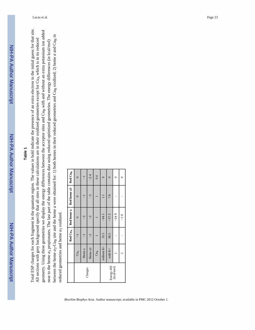
The summation of all partial charges, for each section of the quantum region, is shown inTable 1; the total partial charges agree with the above orbital analysis. The labels: Red CuA,Red heme a, Red heme a3 and Red CuB refer to the calculation where initial spin populationguess has been constrained to the reduced CuA, heme a, heme a3 and CuB, respectively. Forthe initially constrained CuA reduced state, we observe a −1 charge. The same fragment inthe other situations exhibits a total charge of zero, in agreement with an oxidized CuA centerwhich contains a Cu2+ and Cu+ atoms and negative charges from the two cysteines and theGluII254 residue adding up to 0 formal charge. From Table 1 we can also see that for theoxidized heme a center the total charge is about −1 while in the reduced state it is −2. Thischarge is again consistent with the iron +2/+3 oxidation state, corresponding to the oxidized/reduced states, plus the −4 charge from the porphyrin group (2 from the propionates and 2from the deprotonation of the porphyrin nitrogens). In the heme a3-CuB site it is also patentfrom the charge distribution when the electron is present in the heme and when it moves tothe copper center. Thus, summing up the ESP charges for the different redox centersconfirms the localization of the electrons observed in the previous orbital analysis. In theheme a3-CuB site case there is a larger degree of delocalization, as it had been observed bythe spin density plots, due to the close proximity of the redox centers. In agreement with ourfindings the perturbed heme a3 spectra observed by Ji et al. [60] could be explained by thedelocalization of this single electron between the heme a3 and CuB centers. This oneelectron reduced state, termed the E state, has been reported to have an electron distributionamong the redox centers [63–65].
Lucas et al. Page 7
Biochim Biophys Acta. Author manuscript; available in PMC 2012 October 1.
NIH
-PA Author Manuscript
NIH
-PA Author Manuscript
NIH
-PA Author Manuscript

In the second part of table 1 (upper section), the differences in energy between the all sitesand the CuB site are shown. In the first row (without K+) we can observe that the reducedheme a state is found ca. 80 kcal/mol lower than the reduced CuA site indicative of a higherelectron affinity for the heme. Likewise, the reduced heme a3 and CuB sites lie about 35kcal/mol below the reduced CuA. The highest energy lying fragment is the CuA, whichindicates a lower affinity, consistent with the relay function attributed to this center.According to these values the electron is more stable in the heme a, so that we would notexpect the electron to proceed to heme a3.
Based on Belevich et al. [66], the electron transfer from heme a to the binuclear centershould follow a proton transfer to an unidentified protonatable site above the hemes. For thisreason, we have perturbed the system in order to model the presence of an extra proton inthe vicinity of the a3 propionates. A hydrogen bonded water molecule, present in the middleof the carboxylate groups, was altered to a potassium atom (included in the classic part) andthe energies recalculated without any further optimization. These differences between theenergies of each reduced fragment and CuA are present in the last row of Table 1 (with K+).We can see that (despite the simplicity of the calculation) the presence of a positive chargein the proximity of the a3 propionates has an accentuated stabilizing effect on the heme a3and CuB sites. The heme a now lies ~84 kcal/mol below the CuA only 4 kcal/mol morestable while the heme a3 presents a drop in energy grater than to 24 kcal/mol. The drop iseven more enhanced for the CuB center, where another 31 kcal/mol are observed.
These modeling efforts aimed to address the electron affinity at the initial stage of theelectron transfer, when CuA is reduced and the other redox centers are oxidized; thecalculations were performed keeping the oxidized geometries for the hemes and CuB centers(and no additional K+ ion). The final electron location however, will depend on the affinityof the centers in the reduced optimized geometries. Thus, additional calculations usingreduced geometries were necessary. We began by optimizing heme a and heme a3 to theirreduced geometries. In these conditions heme a now lies 25 kcal/mol below heme a3-CuB(~44 kcal/mol when both sites are in their oxidized geometries). Contrary to the first case,when both heme groups were present in their oxidized geometries, we could not localize theelectron in heme a3 (heme a always recover it, indicating its high electronic affinity) butonly in CuB. Thus, in the last two rows in table one we could only fill the heme a and CuBfields. Next, we optimized the system considering the CuB site also in its reduced state. Herewe observe a chemical change with a spontaneous proton transfer from the OH− groupbounded to the iron atom to the hydroxyl coordinated to the Cu atom, forming a watermolecule at the copper center (a movie for this process can be found in SI). This electron-proton coupled transfer leads to differences in the electronic affinity of each center wherenow CuB and heme a have degenerate affinities (last row in table 1). These results seem topoint that the electron and proton transfer might be coupled in order for the electron to reachthe final CuB metal center, an extended topic of discussion in cytochrome c oxidasechemistry [67].
3.2 Electron transfer pathwaysIn this section, the potential electronic paths between CuA and CuB are investigated usingthe QM-MM e-pathway scheme. The goal consists of identifying all possible active residuesbetween the donor and acceptor. QM/MM calculations were performed to optimize thegeometry of all the residues around the metal atoms in their oxidized form. The ESP chargeswere calculated at the DFT level of theory and were used on these atoms when included inthe MM region. The system was then ready for application of the QM-MM e-pathwayscheme.
Lucas et al. Page 8
Biochim Biophys Acta. Author manuscript; available in PMC 2012 October 1.
NIH
-PA Author Manuscript
NIH
-PA Author Manuscript
NIH
-PA Author Manuscript

3.2.1 CuA to heme a electron transfer—With both CuA and heme a in their oxidizedstates and geometries included in the MM region, 18 residues have been selected to beintroduced in the QM area, as well as the propionate groups from both heme groups,counting a total of over 300 atoms. The residues: Gln251, Cys252, Ser253, Glu254, Leu255,Cys256, Gly257, Ile258, Ser259, His260, Ala261, Tyr262, Met263, Pro480, Arg481,Arg482, Tyr483 and Ile484 were elected in order to include all amino-acids in theintermediary area, between CuA and heme a, and taking into account information from theliterature about residues known to be important for this electron transfer pathway as depictedin Figure 5.
The first residue to be identified, with the highest electron affinity, is Arg482 and smalleramounts spread through propionate D from both heme a and heme a3, pale blue in Figure 6.When we remove the side chain of Arg482 from the quantum region, the spin density is nowobserved in the side chain of Arg481 and some small amount on the propionate groups,purple in Figure 6. The next residue to be identified is in the vicinity of the CuA center,Ser253 (and also some on Glu254, displayed in orange in Figure 6) which lies immediatelynext to Cys252. Once this residue is removed, the spin density moves to the backbone ofboth Arg481 and Arg482 (green in Figure 6). Finally, the full removal of these two residuesleads to spin density on the backbone of Pro480 (yellow in Figure 6), completing a possibleelectron transfer pathway between CuA and heme a. We should note that in thesecalculations the electron is extremely localized, normally found in π conjugated molecularorbitals of each residue.
Interestingly, not including the heme a3 propionate groups in the quantum region at thebeginning shifts the first spin density to Arg481. Arg482 appears as the second residue.Proceeding with this study in the same manner as the previous situation, the presence/absence of the a3 propionates does not induce any more changes. Not including thepropionates into the quantum region involves a fix charge description at the propionateoxygens, resulting into a less flexible modeling of the system polarization. The shift of spindensity from Arg481 to Arg482 indicates the plasticity of the system in diverting theelectron flow as a response to the electrostatic environment, in agreement with the previousresults where we perturbed the system by adding a positive charge (the potassium additionshown in table 1).
3.2.2 Heme a to heme a3 electron transfer—Once the electron transfer path betweenCuA and heme a was established, we proceeded by changing the transfer region to beincluded in the QM part as to incorporate the adequate residues between the two hemegroups (see figure 6 SI). All residues bridging the porphyrin rings were added, as well as thetwo arginines 481 and 482 and the two propionate groups. The first residue to be highlightedis Arg481 side chain as well as Arg482 and the D propionate groups of the two hemes (redcolor in Fig. 7). Removing the side chain of Arg481 highlights the bridging Phe420 and thetwo heme histidines (419 and 421) (blue). We have also investigated if the insertion of thetwo tryptophans (172 and 280)[20–23] in the vicinity of the two heme D propionates hadany effect on the spin density of the residues within the transfer region. When including bothtryptophans we observe very small amounts of spin density on Trp172, but most of the spinis still located on Arg481, as was the case when these residues were not included.Furthermore, we have repeated the calculations with the inclusion of a potassium ionhalfway between the heme a3 propionates (as done for the energy calculations previously)which leads to no changes in the spin populations with the arginines still showing thehighest affinity for the electron. The complete electron transfer pathway can be seen inFigure 7.
Lucas et al. Page 9
Biochim Biophys Acta. Author manuscript; available in PMC 2012 October 1.
NIH
-PA Author Manuscript
NIH
-PA Author Manuscript
NIH
-PA Author Manuscript

4. DiscussionThe energetic analysis suggest that the reaction from CuA to the binuclear center shouldoccur in two sequential steps via heme a. Our calculations show that for the oxidized Ostate, the most favorable site for the electron is the intermediate heme a site. As seen in table1 adding one electron to the heme a center results in 45 kcal/mol lower energy than theaddition to the heme a3-CuB. When considering the reduced geometries however, thisenergy gap is reduced to degeneracy between both centers. This degeneracy, however, isonly accomplished when a proton is coupled to the electron transfer. When reducing the CuBsite we observe a spontaneous proton transfer from the Fe-OH group to the Cu-OH one,indicating that the electron transfer from heme a to the heme a3-CuB site is coupled to aproton transfer. We should emphasize here that our models do not include an extra proton inthe system. Even in absence of the extra proton, the reduced CuB abstracts one proton fromthe iron hydroxyl group. While this might not be a real chemical step in the process (whereprotons are pumped to the binuclear center), it clearly indicates the necessity of a proton tostabilize the electron transfer. It might also indicate that the hydroxyl group bound at theiron might be part of the proton transfer pathway to the copper center. Furthermore, theimportance of a proton transfer along the CuB reduction is further confirmed by the strongstabilizing effect of a positively charged environment in the proximity of the propionate a3(table 1), a result in agreement with the principle of electroneutrality introduced by Mitchelland Rich [67].
The results show the importance of Cys252, Ser253, Glu254, Pro480, R482 and the Dpropionate group of heme a in the first electron transfer between CuA and heme a. Mostlikely, not all these residues should be involved in the electron transfer by physically hostingthe electron. Some (or even all of them) might simply act as bridge mediators between alonger through space jump [68]. Electron tunneling in biological systems has been thesubject of much research through the years [69]. According to some experimental studies,His260 seemed to be the preferential residue for the beginning of the pathway which doesnot concur with our findings [70]. Nevertheless, experimentally observed spin density onboth sulfur cysteine atoms support Cys252 as a possible precursor for this ET process. Thehighly conserved subunit I Arg482 is expected to be part of the electron transfer to heme apropionates [71]. For example, a reduction from 93000s−1 to 50s−1 in the electron transferrate from CuA and heme a has been observed in the R482P mutant [72]. The same studyconcludes that the Arg482 side chain and the peptide backbone largely influence the rate ofelectron transfer to heme a. Similar observations on the role of Arg482 have been made byother studies mapping tunneling ET pathways by means of semiempirical methods [18].According to our results Arg482 should have an active part in the CuA to heme a electronicmigration and the backbone is expected to play an important part as well. This pointdemonstrates the capabilities of our new technique, QM/MM e-pathway, to map importantresidues along electron transfer pathways. It has also been seen, by adding/deleting thepropionate groups to the quantum region, that the affinity of the arginine pair for spindensity is dependent on the environment. A small change in the electrostatic descriptionswitches the propensity of the process between the two arginines, which might open apossibility to alter the final destination of the electron flow to either heme a or heme a3. Theactive role for the propionate groups seen here, in the ET, acting as a vehicle of electronsand not only as an electrostatic anchor for binding, has also been underlined previously inseveral heme systems [30, 31, 73–75].
The second step involves the electron transfer from heme a to heme a3. In the first QM/MMiteration, we find Arg482, Arg481 and the two propionate groups. Thus, one could imaginea direct propionate-propionate ET assisted (bridge mediated) by the arginine. Arg481 couldalso act as an intermediate, a vehicle for the electrons in their passage between propionates.
Lucas et al. Page 10
Biochim Biophys Acta. Author manuscript; available in PMC 2012 October 1.
NIH
-PA Author Manuscript
NIH
-PA Author Manuscript
NIH
-PA Author Manuscript

However, mutational studies seem to favor the first mechanism. Mutating the arginine into alysine, R481K, shows normal activity while in most of the other cases the activity is reducedto less than 10% [72]. Lysine side chain does not present a conjugate system, as opposed toarginine, and it is not a good electronic conductor. The distance between the two hemecenters is within that of a direct electron transfer between the metal centers. Phe420, aphenylalanine in between both irons, appears in the second QM/MM e-pathway iteration.Mutation of Phe420 leads to a partially active enzyme which is consistent with this residueacting only as bridge mediated direct metal-metal ET [76]. Theoretical kinetic studies shouldconfirm the different rates for these two ET mechanisms [77, 78].
Finally, it has been shown that the ion pair D-propionate (from heme a)-Arg481 can beaffected by the proton pumping from the D-channel [79]. Our results supports that electrontransfer through heme a might be associated with proton/electron cooperative coupling atthis center, which could explain the presence of heme a in oxidases as a possibleintermediate towards the binuclear site [80].
5. ConclusionsQM/MM methods, together with a novel algorithm (QM/MM e-pathway) were used to mapthe electron transfer process between the three redox centers in Cytochrome c Oxidase. Ourcalculations confirmed a two step ET process following the CuA → heme a → heme a3sequence. On the first step Arg482 delivers the electrons to the metal center by means oftheir propionate groups. For the heme to heme electron transfer we support the existence oftwo different pathways[18]. One would involve directly the heme a propionate-heme a3propionate contact, mediated by Arg481. The second pathway would involve directly themetal centers, mediated by Phe420. The overall process as seen through the energeticanalysis is very sensitive to the additional presence of positive charges in the vicinity of thepropionate groups, supporting a proton-electron coupled mechanism.
Supplementary MaterialRefer to Web version on PubMed Central for supplementary material.
AcknowledgmentsFunding Statement
Computational resources were provided by the Barcelona Supercomputing Center. Work was supported by fundsfrom the Barcelona Supercomputer Center and through the Spanish Ministry of Education and Science through theproject CTQ2007-62122/BQU. MFL was supported by The Fundação para a Ciência e Tecnologia – grant SFRH/BPD/47901/2008. DLR is supported by NIH Grant GM074982.
REFERENCES1. Wikström M. Proton pump coupled to cytochrome c oxidase in mitochondria. Nature. 1977;
266:271–273. [PubMed: 15223]2. Brzezinski P, Sundahl M, Ädelroth P, Wilson M, El-Agez B, Wittung P, Malmström B. Triplet-state
quenching in complexes between Zn-cytochrome c and cytochrome oxidase or its CuA domain.Biophys. Chem. 1995; 54:191–197. [PubMed: 7756569]
3. Szundi I, Cappuccio JA, Borovok N, Kotlyar AB, Einarsdóttir O. Photoinduced electron transfer inthe cytochrome c/cytochrome c oxidase complex using thiouredopyrenetrisulfonatelabeledcytochrome c. Optical multichannel detection. Biochemistry. 2001; 40:2186–2193. [PubMed:11329287]
4. Johansson MP, Kaila VRI, Laakkonen L. Charge parameterization of the metal centers incytochrome c oxidase. J. Comp. Chem. 2008; 29:753–767. [PubMed: 17876762]
Lucas et al. Page 11
Biochim Biophys Acta. Author manuscript; available in PMC 2012 October 1.
NIH
-PA Author Manuscript
NIH
-PA Author Manuscript
NIH
-PA Author Manuscript

5. Olsson MHM, Ryde U. Geometry, Reduction Potential, and Reorganization Energy of the BinuclearCuA Site, Studied by Density Functional Theory. J. Am. Chem. Soc. 2001; 123:7866–7876.[PubMed: 11493060]
6. Iwata S, Ostermeier C, Ludwig B, Michel H. Structure at 2.8 Å resolution of cytochrome c oxidasefrom Paracoccus denitrificans. Nature. 1995; 376:660–669. [PubMed: 7651515]
7. Wilmanns M, Lappalainen P, Kelly M, Sauer-Eriksson E, Saraste M. Crystal structure of themembrane-exposed domain from a respiratory quinol oxidase complex with an engineered dinuclearcopper center(cytochrome oxidase/cupredoxin fold). Proc. Natl. Acad. Sci. USA. 1995; 92:11955–11959. [PubMed: 8618822]
8. Tsukihara T, Aoyama H, Yamashita E, Tomizaki T, Yamaguchi H, Shinzawa-Itoh K, Nakashima R,Yaono R, S Y. Structures of metal sites of oxidized bovine heart cytochrome c oxidase at 2.8 A.Science. 1995; 269:1069–1074. [PubMed: 7652554]
9. Fee JA, Sanders D, Slutter CE, Doan PE, Aasa R, Karpefors M, Vänngård T. Multifrequency EPREvidence for a Binuclear CuA Center in Cytochrome c Oxidase: Studies with a 63Cu- and 65Cu-Enriched, Soluble Domain of the Cytochrome ba3 Subunit II from Thermus thermophilus.Biochem. Biophys. Res. Commun. 1995; 212:77–83. [PubMed: 7612020]
10. Beinert H, Griffiths DE, Wharton DC, Sands RH. Properties of the Copper Associated withCytochrome Oxidase as Studied by Paramagnetic Resonance Spectroscopy. J. Biol. Chem. 1962;237:2337–2346. [PubMed: 13866633]
11. Andrew C, Fraczkiewicz R, Czernuszewicz R, Lappalainen P, Saraste M, Sanders-Loehr J.Identification and Description of Copper-Thiolate Vibrations in the Dinuclear CuA Site ofCytochrome c Oxidase. J. Am. Chem. Soc. 1996; 118:10436–10445.
12. Hill BC. Modeling the sequence of electron transfer reactions in the single turnover of reduced,mammalian cytochrome c oxidase with oxygen. J. Biol. Chem. 1994; 269:2419–2425. [PubMed:8300568]
13. Soulimane T, Buse G, Bourenkov G, Bartunik H, Huber R, Than M. Structure and mechanism ofthe aberrant ba3-cytochrome c oxidase from Thermus thermophilus. EMBO J. 2000; 19:1766–1776. [PubMed: 10775261]
14. Regan J, Ramirez B, Winkler J, Gray H, Malmström B. Pathways for Electron Tunneling inCytochrome c Oxidase. J. Bioenerg. Biomembr. 2004; 30:35–39. [PubMed: 9623803]
15. Ramirez B, Malmstrom B, Winkler J, Gray H. The currents of life: The terminal electron- transfercomplex of respiration. Proc. Natl. Acad. Sci. USA. 1995; 92:11949–11951. [PubMed: 8618820]
16. Yoshikawa S, Shinzawa-Itoh K, Nakashima R, Yaono R, Yamashita E, Inoue N, Yao M, Fei M,Libeu C, Mizushima T, Yamaguchi H, Tomizaki T, Tsukihara T. Redox-Coupled CrystalStructural Changes in Bovine Heart Cytochrome c Oxidase. Science. 1998; 280:1723–1729.[PubMed: 9624044]
17. Williams K, Gamelin D, LaCroix L, Houser R, Tolman W, Mulder T, de Vries S, Hedman B,Hodgson K, Solomon E. Influence of Copper-Sulfur Covalency and Copper-Copper Bonding onValence Delocalization and Electron Transfer in the CuA Site of Cytochrome c Oxidase II. J. Am.Chem. Soc. 1997; 119:613–614.
18. Medvedev D, Daizadeh I, Stuchebrukhov A. Electron Transfer Tunneling Pathways in BovineHeart Cytochrome c Oxidase. J. Am. Chem. Soc. 2000; 122:6571–6582.
19. Tan M-L, Balabin I, Onuchic J. Dynamics of Electron Transfer Pathways in Cytochrome cOxidase. Biophys. J. 2004; 86:1813–1819. [PubMed: 14990507]
20. Rigby S, Junemann S, Rich PR, Heathcote P. Reaction of Bovine Cytochrome c Oxidase withHydrogen Peroxide Produces a Tryptophan Cation Radical and a Porphyrin Cation Radical.Biochemistry. 2000; 39:5921. [PubMed: 10821663]
21. Svistunenko D, Wilson M, Cooper C. Tryptophan or Tyrosine? On the nature of the amino acidradical formed following hydrogen peroxide treatment of cytochrome c oxidase. Biochem.Biophys. Acta. 2004; 1655:372. [PubMed: 15100053]
22. Wiertz F, Richter O, Ludwig B, Vries S. Kinetic resolution of a tryptophan-radical intermediate inthe reaction cycle of Paracoccus denitrificans cytochrome c oxidase. J. Biol.l Chem. 2007;282:31580.
Lucas et al. Page 12
Biochim Biophys Acta. Author manuscript; available in PMC 2012 October 1.
NIH
-PA Author Manuscript
NIH
-PA Author Manuscript
NIH
-PA Author Manuscript

23. MacMillan F, K. B, Angerer H, Michel H. The sole of tryptophan 272 in the paracoccusdenitrificans cytochrome c oxidase. Febs Letters. 2006; 580:1345. [PubMed: 16460733]
24. Rich PR, Rigby SEJ, Heathcote P. Radicals associated with the catalytic intermediates of bovinecytochrome c oxidase. Biochimica et Biophysica Acta (BBA) - Bioenergetics. 2002; 1554:137–146.
25. Balabin I, Onuchic J. Dynamically Controlled Protein Tunneling Paths in Photosynthetic ReactionCenters. Science. 2000; 290:114–117. [PubMed: 11021791]
26. Balabin I, Onuchic J. Connection between Simple Models and Quantum Chemical Models forElectron-Transfer Tunneling Matrix Element Calculations: A Dyson's Equations-Based Approach.J. Phys. Chem. 1996; 100:11573–11580.
27. Gehlen J, Daizadeh I, Stuchebrukhov A, Marcus R. Tunneling matrix element in Ru-modified bluecopper proteins: pruning the protein in search of electron transfer pathways. Inorg. Chim. Acta.1996; 243:271–282.
28. Warshel A. Dynamics of reactions in polar solvents. Semiclassical trajectory studies of electron-transfer and proton-transfer reactions. J. Phys. Chem. 1982; 86:2218–2224.
29. Warshel A, Parson WW. Computer Simulations of Electron-Transfer Reactions in Solution and inPhotosynthetic Reaction Centers. Annu. Rev. Phys. Chem. 1991; 42:279–309. [PubMed: 1747189]
30. Guallar V, Wallrapp F. Mapping protein electron transfer pathways with QM/MM methods. J.Roy. Soc. Interf. 2008; 5:S233.
31. Wallrapp F, Masone D, Guallar V. Electron transfer in the P450cam/PDX complex. The QM/MMe-pathway. J.Phys. Chem. A. 2008; 112:12989–12994. [PubMed: 18823106]
32. Mo Y, Gao J. Ab initio QM/MM simulations with a molecular orbital-valence bond (MOVB)method: application to an SN2 reaction in water. J. Comp. Chem. 2000; 21:1458–1469.
33. Vreven T, Morokuma K, Farkas Ö, Schlegel HB, Frisch MJ. Geometry optimization with QM/MM, ONIOM, and other combined methods. I. Microiterations and constraints. J. Comp. Chem.2003; 24:760–769. [PubMed: 12666168]
34. Reuter N, Dejaegere A, Maigret B, Karplus M. Frontier bonds in QM/MM methods: A comparisonof different approaches. J. Phys. Chem. A. 2000; 104:1720–1735.
35. Friesner RA, Guallar V. Ab Initio Quantum Chemical and Mixed Quantum Mechanics/MolecularMechanics (QM/MM) Methods for Studying Enzymatic Catalysis. Ann. Rev. Phys. Chem. 2005;56
36. Mulholland AJ. Modelling enzyme reaction mechanisms, specificity and catalysis. Drug Discov.Today. 2005; 10:1393–1402. [PubMed: 16253878]
37. Senn HM, Thiel W. QM/MM methods for biological systems, Atomistic Approaches in ModernBiology: From Quantum Chemistry to Molecular Simulations. 2007; vol. 268:173–290.
38. Lin H, Truhlar DG. QM/MM: what have we learned, where are we, and where do we go fromhere? Theor. Chem. Acc. 2007; 117:185–199.
39. Schrödinger Inc.. QSite 4.5. Portland Oregon: 2007.40. Becke AD. Completely numerical calculations on diatomic molecules in the local-density
approximation. Phys Rev A. 1986; 33:2786–2788. [PubMed: 9896968]41. Becke AD. Density-Functional Exchange-Energy Approximation With Correct Asymptotic-
Behavior. Phys. Rev. A. 1988; 38:3098–3100. [PubMed: 9900728]42. Becke AD. A New Mixing of Hartree-Fock and Local Density-Functional Theories. J. Chem. Phys.
1993; 98:1372–1377.43. Becke AD. Density-Functional Thermochemistry. 3. The Role of Exact Exchange. J. Chem. Phys.
1993; 98:5648–5652.44. Lee C, Yang W, Parr RG. Development of the Colle-Salvetti correlation-energy formula into a
functional of the electron density. Phys. Rev. B. 1988; 37:785–789.45. Hehre WJ, Ditchfield R, Pople JA. Self Consistent Molecular Orbital Methods. XII. Further
Extensions of Gaussian---Type Basis Sets for Use in Molecular Orbital Studies of OrganicMolecules. J. Chem. Phys. 1972; 56:2257–2261.
46. Hay PJ, Wadt WR. Abinitio Effective Core Potentials for Molecular Calculations - Potentials for Kto Au Including the Outermost Core Orbitals. J. Chem. Phys. 1985; 82:299–310.
Lucas et al. Page 13
Biochim Biophys Acta. Author manuscript; available in PMC 2012 October 1.
NIH
-PA Author Manuscript
NIH
-PA Author Manuscript
NIH
-PA Author Manuscript

47. Jorgensen WL, Maxwell DS, TiradoRives J. Development and testing of the OPLS all-atom forcefield on conformational energetics and properties of organic liquids. J. Am. Chem. Soc. 1996;118:11225–11236.
48. Kaminski GA, Friesner RA, Tirado-Rives J, Jorgensen WL. Evaluation and Reparametrization ofthe Opls-Aa Force Field for Proteins Via Comparison With Accurate Quantum ChemicalCalculations on Peptides. J. Phys. Chem. B. 2001; 105:6474–6487.
49. Svensson-Ek M, Abramson J, Larsson G, Tornroth S, Brzezinski P, Iwata S. The X-ray CrystalStructures of Wild-type and EQ(I-286) Mutant Cytochrome c Oxidases from Rhodobactersphaeroides. J. Mol. Biol. 2002; 321:329–339. [PubMed: 12144789]
50. Phillips JC, Braun R, Wang W, Gumbart J, Tajkhorshid E, Villa E, Chipot C, Skeel RD, Kale L,Schulten K. Scalable molecular dynamics with NAMD. J. Comp. Chem. 2005; 26:1781–1802.[PubMed: 16222654]
51. MacKerell AD, Bashford D, Bellott M, Dunbrack RL, Evanseck JD, Field MJ, Fischer S, Gao J,Guo H, Ha S, Joseph-McCarthy D, Kuchnir L, Kuczera K, Lau FTK, Mattos C, Michnick S, NgoT, Nguyen DT, Prodhom B, Reiher WE, Roux B, Schlenkrich M, Smith JC, Stote R, Straub J,Watanabe M, Wiorkiewicz-Kuczera J, Yin D, Karplus M. All-atom empirical potential formolecular modeling and dynamics studies of proteins. J. Phys. Chem. B. 1998; 102:3586–3616.
52. Olkhova E, Hutter MC, Lill MA, Helms V, Michel H. Dynamic Water Networks in Cytochrome cOxidase from Paracoccus denitrificans Investigated by Molecular Dynamics Simulations. Biophys.J. 2003; 86:1873–1889. [PubMed: 15041635]
53. Olkhova E, Helms V, Michel H. Titration Behavior of Residues at the Entrance of the D- Pathwayof Cytochrome c Oxidase from Paracoccus denitrificans Investigated by Continuum ElectrostaticCalculations. Biophys. J. 2005; 89:2324–2331. [PubMed: 16192282]
54. Fadda E, Yu C-H, Pomès Rg. Electrostatic control of proton pumping in cytochrome c oxidase.Biochimica et Biophysica Acta (BBA) - Bioenergetics. 2008; 1777:277–284.
55. Ryckaert J-P, Ciccotti G, Berendsen HJC. Numerical integration of the cartesian equations ofmotion of a system with constraints: molecular dynamics of n-alkanes. J. Comp. Phys. 1977;23:327–341.
56. Singh UC, Kollman PA. A combined ab initio quantum mechanical and molecular mechanicalmethod for carrying out simulations on complex molecular systems: Applications to the CH3Cl +Cl- exchange reaction and gas phase protonation of polyethers. J. Comp. Chem. 1986; 7:718–730.
57. Gamelin DR, Randall DW, Hay MT, Houser RP, Mulder TC, Canters GW, de Vries S, TolmanWB, Lu Y, Solomon EI. Spectroscopy of Mixed-Valence CuA-Type Centers: Ligand-FieldControl of Ground-State Properties Related to Electron Transfer. J. Am. Chem. Soc. 1998;120:5246–5263.
58. Gorelsky SI, Xie X, Chen Y, Fee JA, Solomon EI. The Two-State Issue in the Mixed- ValenceBinuclear CuA Center in Cytochrome c Oxidase and N2O Reductase. J. Am. Chem. Soc. 2006;128:16452–16453. [PubMed: 17177365]
59. Han S, Takahashi S, Rousseau DL. Time dependence of the catalytic intermediates in cytochromec oxidase. J. Biol. Chem. 2000; 275:1910–1919. [PubMed: 10636892]
60. Ji H, Yeh S-Y, Rousseau DL. Modulation of the Electron Redistribution in Mixed ValenceCytochrome c Oxidase by Protein Conformational Changes. J. Biol. Chem. 2004; 279:9392–9399.[PubMed: 14660573]
61. Rado M, Srebro M, Broclawik E. Conformational Stability and Spin States of Cobalt(II)Acetylacetonate: CASPT2 and DFT Study. J. Chem. Theory Comput. 2009; 5:1237–1244.
62. Tsukihara T, Yoshikawa S. Crystal Structural Studies of a Membrane Protein Complex,Cytochrome c Oxidase from Bovine Heart. Acta Cryst. 1998; A54:895–904.
63. Moody AJ, Brandt U, Rich PR. Single electron reduction of 'slow' and 'fast' cytochrome-c oxidase.FEBS letters. 1991; 293:101–105. [PubMed: 1660000]
64. Brunori M, Giuffre A, Sarti P. Cytochrome c oxidase, ligands and electrons. J. Inorg. Biochem.2005; 99:324–336. [PubMed: 15598510]
65. Michel H, Behr J, Harrenga A, Kannt A. CYTOCHROME C OXIDASE: Structure andSpectroscopy. Annual Review of Biophysics and Biomolecular Structure. 1998; 27:329–356.
Lucas et al. Page 14
Biochim Biophys Acta. Author manuscript; available in PMC 2012 October 1.
NIH
-PA Author Manuscript
NIH
-PA Author Manuscript
NIH
-PA Author Manuscript

66. Belevich I, Verkhovsky M, Wikstrom M. Proton-coupled Electron Transfer Drives the ProtonPump of Cytochrome c Oxidase. Nature. 2006; 440:829–832. [PubMed: 16598262]
67. Mitchell R, Rich PR. Proton uptake by cytochrome c oxidase on reduction and on ligand binding.Biochem. Biophys. Acta. 1994; 1186:19–26. [PubMed: 8011665]
68. Beratan DN, Onuchic JN, Winkler JR, Gray HB. Electron-Tunneling Pathways In Proteins.Science. 1992; 258:1740–1741. [PubMed: 1334572]
69. Page CC, Moser CC, Chen X, Dutton PL. Natural engineering principles of electron tunnelling inbiological oxidation-reduction. Nature. 1999; 402:47–52. [PubMed: 10573417]
70. Wang K, Geren L, Zhen Y, Ma L, Ferguson-Miller S, Durham B, Millett F. Mutants of the CuASite in Cytochrome c Oxidase of Rhodobacter sphaeroides: II. Rapid Kinetic Analysis of ElectronTransfer. Biochemistry. 2002; 41:2298–2304. [PubMed: 11841222]
71. Brzezinski P. Internal Electron-Transfer Reactions in Cytochrome c Oxidase. Biochemistry. 1996;35:5611–5615. [PubMed: 8639518]
72. Qian J, Mills D, Geren L, Wang K, Hoganson C, Schmidt B, Hiser C, Babcock GT, Durham B,Millett F, Ferguson-Miller S. Role of the Conserved Arginine Pair in Proton and Electron Transferin Cytochrome c Oxidase. Biochemistry. 2004; 43:5748–5756. [PubMed: 15134449]
73. Sharpe M, Ferguson-Miller S. A chemically explicit model for the mechanism of proton pumpingin heme–copper oxidases. J. Bioenerg. Biomembr. 2008; 40:541–549. [PubMed: 18830692]
74. Mills DA, Xu S, Geren L, Hiser C, Qin L, Sharpe MA, McCracken J, Durham B, Millett F,Ferguson-Miller S. Proton-Dependent Electron Transfer from CuA to Heme a and Altered EPRSpectra in Mutants Close to Heme a of Cytochrome Oxidase†. Biochemistry. 2008; 47:11499–11509. [PubMed: 18847227]
75. Egawa T, Lee HJ, Gennis RB, Yeh S-R, Rousseau DL. Critical structural role of R481 incytochrome c oxidase from Rhodobacter sphaeroides. Biochimica et Biophysica Acta (BBA) -Bioenergetics. 2009; 1787:1272–1275.
76. Hosler JP, Ferguson-Miller S, Calhoun MW, Thomas JW, Hill J, Lemieux L, Ma J, Georgiou C,Fetter J, Shapleigh J, Tecklenburg M, Babcock GT, Gennis R. Insight into the active-site structureand function of cytochrome oxidase by analysis of site-directed mutants of bacterial cytochromeaa3 and cytochrome bo. J. Bioenerg. Biomembr. 1993; 25:121–136. [PubMed: 8389745]
77. Pilet E, Jasaitis A, Liebl U, Vos MH. Electron transfer between hemes in mammalian cytochromec oxidase. Proc. Natl. Acad. Sci. USA. 2004; 101:16198–16203. [PubMed: 15534221]
78. Jasaitis A, Rappaport F, Pilet E, Liebl U, Vos MH. Activationless electron transfer through thehydrophobic core of cytochrome c oxidase. Proc. Natl. Acad. Sci. USA. 102:10882–10886.(PNAS August 2, 2005 vol. no. ).
79. Wikstrom M, Ribacka C, Molin M, Laakkonen L, Verkhovsky M, Puustinen A. Gating of protonand water transfer in the respiratory enzyme cytochrome c oxidase. Proc. Natl. Acad. Sci. USA.2005; 102:10478–10481. [PubMed: 16014708]
80. Papa S, Capitanio N, Capitanio G, Palese LL. Protonmotive cooperativity in cytochrome c oxidase.Biochim. Biophys. Acta. 2004; 1658:95–105. [PubMed: 15282180]
Lucas et al. Page 15
Biochim Biophys Acta. Author manuscript; available in PMC 2012 October 1.
NIH
-PA Author Manuscript
NIH
-PA Author Manuscript
NIH
-PA Author Manuscript

Figure 1.Overall structure of Cytochrome c Oxidase from Rhodobacter sphaeroides. A - Possibleelectron transfer path shown. The redox active and inactive cofactors of CcO are displayedas well as the D- and K-pathways used for proton uptake. B - Relative position of allresidues proposed to participate in ET process.
Lucas et al. Page 16
Biochim Biophys Acta. Author manuscript; available in PMC 2012 October 1.
NIH
-PA Author Manuscript
NIH
-PA Author Manuscript
NIH
-PA Author Manuscript

Figure 2.Unpaired electrons spin-density plot for a system where the CuA site is reduced and bothheme a and heme a3-CuB are oxidized.
Lucas et al. Page 17
Biochim Biophys Acta. Author manuscript; available in PMC 2012 October 1.
NIH
-PA Author Manuscript
NIH
-PA Author Manuscript
NIH
-PA Author Manuscript

Figure 3.Unpaired electron spin-density plots for a system where: the CuA and CuB sites are oxidizedand A) heme a reduced and heme a3 are oxidized. B) heme aoxidized and heme a3 reduced.
Lucas et al. Page 18
Biochim Biophys Acta. Author manuscript; available in PMC 2012 October 1.
NIH
-PA Author Manuscript
NIH
-PA Author Manuscript
NIH
-PA Author Manuscript

Figure 4.Unpaired electron spin-density plots for a system where the CuA and both heme a and hemea3 are oxidized and CuB is reduced.
Lucas et al. Page 19
Biochim Biophys Acta. Author manuscript; available in PMC 2012 October 1.
NIH
-PA Author Manuscript
NIH
-PA Author Manuscript
NIH
-PA Author Manuscript

Figure 5.All residues included in the QM section are shown. The heme propionates have also beenincluded but the remaining part of the heme (part shown in grey) was included in the MMregion as well as the rest of the protein. Each residue’s backbone is shown in “think” coloredrepresentation with the respective sidechain in “thin” grey.
Lucas et al. Page 20
Biochim Biophys Acta. Author manuscript; available in PMC 2012 October 1.
NIH
-PA Author Manuscript
NIH
-PA Author Manuscript
NIH
-PA Author Manuscript

Figure 6.Electron transfer pathway for Cytochrome c Oxidase between CuA and heme a.
Lucas et al. Page 21
Biochim Biophys Acta. Author manuscript; available in PMC 2012 October 1.
NIH
-PA Author Manuscript
NIH
-PA Author Manuscript
NIH
-PA Author Manuscript

Figure 7.Electron transfer pathway for CcO between heme a and heme a3.
Lucas et al. Page 22
Biochim Biophys Acta. Author manuscript; available in PMC 2012 October 1.
NIH
-PA Author Manuscript
NIH
-PA Author Manuscript
NIH
-PA Author Manuscript
NIH
-PA Author Manuscript
NIH
-PA Author Manuscript
NIH
-PA Author Manuscript
Lucas et al. Page 23
Tabl
e 1
Tota
l ESP
cha
rges
for e
ach
frag
men
t in
the
quan
tum
regi
on. T
he v
alue
s in
bold
indi
cate
the
pres
ence
of a
n ex
tra e
lect
ron
in th
e in
itial
gue
ss fo
r tha
t site
.A
ll se
ctio
ns w
ith g
rey
back
grou
nd sp
ecify
that
all
site
s in
thes
e ca
lcul
atio
ns a
re in
thei
r oxi
dize
d ge
omet
ries e
xcep
t for
Cu A
whi
ch is
in it
s red
uced
geom
etry
. Usi
ng th
ese
geom
etrie
s we
disp
lay
the
ener
gy d
iffer
ence
s bet
wee
n th
e ac
cept
or si
tes a
nd C
u B w
ith a
nd w
ithou
t an
extra
pot
assi
um io
n ad
ded
near
to th
e he
me
a 3 p
ropi
onat
es. T
he la
st p
art o
f the
tabl
e co
ntai
ns d
ata
usin
g re
duce
d op
timiz
ed g
eom
etrie
s. Th
e en
ergy
diff
eren
ces (
in k
cal/m
ol)
betw
een
the
hem
e a 3
-Cu B
site
and
the
hem
e a
wer
e ob
tain
ed fo
r: 1)
bot
h he
mes
in th
eir r
educ
ed g
eom
etrie
s and
Cu B
oxi
dize
d; 2
) hem
e a
and
Cu B
inre
duce
d ge
omet
ries a
nd h
eme
a 3 o
xidi
zed.
Red
Cu A
Red
hem
e a
Red
hem
e a3
Red
Cu B
Cha
rges
Cu A
−1
00
0
Hem
e a
−1
−2
−1
−1
Hem
e a3
−2
−2
−3
−2.4
Cu B
11
10.
4
Ener
gy d
iff.
(kca
l/mol
)
with
out K
+35
.5−44.1
1.1
0
with
K+
66.5
−17.2
7.8
0
1-
−24.9
-0
2-
−1.0
-0
Biochim Biophys Acta. Author manuscript; available in PMC 2012 October 1.


![[8] Cytochrome-c oxidase from Saccharomyces cerevisiae](https://img.dokumen.tips/doc/110x75/635b7a829d85dc43cb0777af/8-cytochrome-c-oxidase-from-saccharomyces-cerevisiae.jpg)


