Embed Size (px)
Citation preview

IntroductionSoluble N-ethylmaleimide sensitive fusion protein attachmentprotein receptors (SNAREs) play a fundamental role inmembrane fusion in the secretory and degradative pathways ofeukaryotic cells. SNAREs were originally classified into twocategories: vesicular SNAREs (v-SNAREs), localized in thedonor membrane, and target SNAREs (t-SNAREs), localizedin the acceptor compartment (Chen and Scheller, 2001; Galliand Haucke, 2001; Rothman, 2002). SNAREs are smallproteins composed mainly of a C-terminal membraneanchoring domain and an α-helical domain (the SNARE motif)that mediates membrane fusion. In some cases this domain ispreceded by a regulatory N-terminal extension (Filippini et al.,2001). The SNARE motif mediates pairing of SNAREs,leading to a parallel α-helical bundle, made of four chains,called the SNARE complex (Poirier et al., 1998; Sutton et al.,1998). The structure of the synaptic SNARE complex showedthat the interaction between these proteins is mediated byhydrophobic interactions, except at the central region (ionic‘zero’ layer) where an arginine residue from the v-SNAREsynaptobrevin 2 (also called vesicle-associated membraneprotein 2, VAMP2) forms hydrogen bonds with threeglutamines, each from a distinct SNARE motif (one fromsyntaxin 1 and two from SNAP-25) (Sutton et al., 1998).Accordingly, residues in the ionic layer are very well conservedand are crucial for SNARE complex formation and/or stability(Katz and Brennwald, 2000; Ossig et al., 2000; Scales et al.,2001). These observations led a new classification of SNAREsinto R- and Q-SNAREs, which correspond in general to v- and
t-SNAREs, respectively (Fasshauer et al., 1998). The t-SNAREis now referred to as the cis-complex of target SNAREs,composed of one heavy chain and two light chains, each oneproviding a helix to the four-helix bundle of the core SNAREcomplex (Fukuda et al., 2000). The correct topology ofSNAREs – one v-SNARE on a vesicle interacting with itscognate three-component t-SNARE on the target membrane –is crucial for accurate SNARE complex formation (Parlati etal., 2000).
Membrane trafficking is a highly dynamic process. Themolecular machinery mediating membrane fusion iscontinuously cycling between distinct intracellularcompartments. This is especially true for v-SNAREs. Forexample, exocytic SNAREs located on vesicles that have fusedwith the plasma membrane need to be rapidly internalized fromthe cell surface in order to participate in other rounds of fusion.Moreover, one v-SNARE might participate in several fusionsteps, as illustrated by cellubrevin, which mediates exocytosisand endosome-to-TGN trafficking (Galli et al., 1994; Mallardet al., 2002). Similarly, the yeast v-SNAREs Snc1 and Snc2are involved in exocytosis and in retrograde transport toendosomes and to the Golgi complex (Gurunathan et al., 2000;Paumet et al., 2001).
Despite this continuous trafficking, SNAREs are localizedat steady state in specific intracellular compartments, and thespecificity of membrane fusion is ensured. Identifying themechanism responsible for compartmental specificity is amajor issue in this field. The emerging picture is that there arecomplementary layers of regulation that are essential for
2805
SNARE proteins are key mediators of membrane fusion.Their function in ensuring compartmental specificity ofmembrane fusion has been suggested by in vitro studies butnot demonstrated in vivo. We show here that ectopicexpression of the plasma membrane t-SNARE heavy chainsyntaxin 1 in the endoplasmic reticulum induces theredistribution of its cognate vesicular SNAREs, TI-VAMPand cellubrevin, and its light chain t-SNARE SNAP-23.These effects were prevented by co-expressing nSec1.Expression of syntaxin 1 alone impaired the cell surfaceexpression of TI-VAMP and cellubrevin but not therecycling of transferrin receptor. TI-VAMP, cellubrevinand SNAP-23 associated in vivo with exogenous syntaxin 1.
Redistribution of TI-VAMP in the ER of syntaxin-1-expressing cells was microtubule dependent and impairedthe trafficking of CD63, a cargo of TI-VAMP-containingvesicles. We conclude that the destination of v-SNAREs isdriven by their specific interaction with cognate t-SNAREs.Our in vivo data provide strong support for the theory thathighly specific v-SNARE–t-SNARE interactions controlcompartmental specificity of membrane fusion.
Supplementary data available online
Key words: Membrane fusion, SNARE proteins, TI-VAMP,Cellubrevin, Syntaxin
Summary
Ectopic expression of syntaxin 1 in the ER redirectsTI-VAMP- and cellubrevin-containing vesiclesSonia Martinez-Arca 1, Véronique Proux-Gillardeaux 1, Philipp Alberts 1, Daniel Louvard 2 and Thierry Galli 1,*1Membrane Traffic and Neuronal Plasticity, INSERM U536, Institut du Fer-à-Moulin, 75005 Paris, France2Morphogenesis and Cell Signaling, CNRS UMR 144, Institut Curie, 75005 Paris, France*Author for correspondence (e-mail: [email protected])
Accepted 10 March 2003Journal of Cell Science 116, 2805-2816 © 2003 The Company of Biologists Ltddoi:10.1242/jcs.00467
Research Article

2806
the establishment and maintenance of organellecompartmentalization in eukaryotic cells. Among theseregulators there are members of the Rab GTPase family andtheir effectors (Pfeffer., 2001; Siniossoglou and Pelham, 2001),tethering factors (Shorter et al., 2002) and the SNAREsthemselves (McNew et al., 2000; Scales et al., 2000). Studiesusing an in vitro fusion assay suggested that compartmentalspecificity could be achieved to a large extent by the inherentspecificity of cognate interactions between SNAREs (McNewet al., 2000).
In this study, we sought to test the hypothesis that the steadystate subcellular localization and destination of v-SNAREs,and therefore the specificity of membrane fusion, depend onthe ability of v-SNAREs to interact with their cognate t-SNAREs and thus would ultimately depend on the localizationof t-SNAREs. If this was true, interfering with the targeting ofa t-SNARE should in turn affect the distribution of its specificv-SNARE partners without altering the localization of its non-cognate SNAREs. To test this hypothesis, we took advantageof the fact that expression of the plasma membrane t-SNAREsyntaxin 1 in non-neuronal cells leads to the accumulation ofthis protein in the Golgi apparatus (at short time points aftertransfection), followed by its redistribution to the ER (afterlonger times of transfection). Whereas co-transfection ofsyntaxin 1 and nSec1/Munc18-1 restores normal plasmamembrane targeting of syntaxin 1 (Perez-Branguli et al., 2002;Rowe et al., 2001; Rowe et al., 1999). Our data show thatectopic expression of syntaxin 1 in the ER redirected itscognate v-SNAREs without affecting non-cognate ones.Therefore our results support the proposal that the localizationof a v-SNARE may be linked to the distribution of its cognatet-SNAREs and provide in vivo evidence for SNARE-mediatedspecificity of membrane fusion.
Materials and MethodsAntibodies and clonesMouse monoclonal antibody clone 158.2 directed against TI-VAMPwill be described elsewhere (Muzerelle et al., 2003). Mousemonoclonal antibodies anti-transferrin receptor (68.4, from I.Trowbridge, Salk Institute, La Jolla, CA, USA), green fluorescentprotein (GFP; clone 7.1 and 13.1, Roche, Indianapolis, IN), syntaxin1 (HPC-1, from C. Barnstable, Yale University, New Haven, CT),Vti1b (clone 7), syntaxin 4 (clone 49), syntaxin 6 (clone 30) (fromTransduction Labs, Lexington, KY), Na+/K+ ATPase a-1 (UpstateBiotechnology, Waltham, MA) and CD63 [Clone AD1 (Furuno et al.,1996)] have been described previously. Monoclonal biotinylated anti-CD63 antibody was from Ancell (Bayport, MN). Monoclonalantibody anti-α-tubulin was from Amersham Biosciences(Piscataway, NJ). Rabbit polyclonal antibodies anti-syntaxin 1 andanti-calreticulin were from Synaptic Systems (Göttingen, Germany)and Affinity Bioreagents (Golden, CO), respectively. Rabbitpolyclonal antibody anti-syntaxin 7 was kindly provided by W. Wong(Institute of Molecular and Cell Biology, Singapore). Rabbit serumsanti-TI-VAMP (TG18), SNAP-23 [TG7 (Galli et al., 1998)],cellubrevin [TG2 (Galli et al., 1998)], VAMP8 [TG15 (Paumet et al.,2000)] and VAMP4 [TG19/20 (Mallard et al., 2002)] were purified byaffinity chromatography.
The human cDNA of endobrevin, originally cloned from CaCo2cells, was obtained from ATCC (EST176564) (Paumet et al., 2000).The N-terminal GFP fusion protein GFP-TIVAMP has been describedpreviously (Martinez-Arca et al., 2000). For the N-terminal GFPfusion proteins GFP-cellubrevin (GFP-Cb) and GFP-endobrevin
(GFP-Eb) the cDNAs of cellubrevin (Galli et al., 1998) andendobrevin were cloned into the pEGFP-C3 vector (Clontech, PaloAlto, CA). For the C-terminal GFP-fusion constructs TIVAMP-GFPand Cb-GFP, we generated a superecliptic variant of the eclipticpHLuorin (G. Miesenbock, Sloan Kettering Memorial Hospital, NY)containing two mutations (F64L and S65T) that lead to enhancedfluorescence (Sankaranarayanan and Ryan, 2000). The cDNAs of ratsyntaxin 1A and nSec1 have been described previously (Bennett etal., 1992; Garcia et al., 1994). The cDNA of rat syntaxin 7 was fromR. Jahn [Max-Planck Institute, Göttingen, FRG (Antonin et al.,2000)].
Cell culture and transfection HeLa cells were cultured and transfected as described previously(Martinez-Arca et al., 2000) or with Lipofectamine 2000 (Invitrogen,Carlsbad, CA) following the manufacturer’s instructions. MDCK cellswere grown in DMEM with 10% FCS. Stable MDCK clonesexpressing GFP-TIVAMP, GFP-Cb or GFP-Eb were produced byelectroporation and selected with G418 (0.4 mg/ml). MDCK cloneswere transfected with Lipofectamine 2000. For the nocodazoletreatment MDCK cells plated the day before were treated with 5 µMnocodazole for 1 hour at 37°C before transfection. The drug was leftin the medium until fixation of the cells.
Immunoprecipitation HeLa cells were lysed 24 hours after transfection in TSE (50 mM TrispH 8.0, 10 mM EDTA, 150 mM NaCl) plus 1% Triton X-100 withprotease inhibitors for 1 hour at 4°C under continuous shaking. Thesupernatant resulting from centrifugation at 20,000 g for 30 minutes,adjusted to a protein concentration of 1 mg/ml, immunoprecipitatedat 4°C overnight, followed by the addition of 50 µl of magnetic beads(Dynabeads, Dynal, Compiègne, France). After 3 hours at 4°C themagnetic beads were washed four times with TSE plus 1% Triton X-100 and eluted with sample buffer. Proteins were resolved by SDS-PAGE and detected by western blotting with the ECL system(SuperSignal West Pico Chemiluminescent Substrate, Pierce,Rockford, IL).
Antibody uptake and transferrin recyclingHeLa cells transfected with Cb-GFP or TIVAMP-GFP plus syntaxin1 were incubated in the presence of 5 µg/ml anti-GFP monoclonalantibody in culture medium for 1 hour at 37°C, washed extensivelywith PBS, fixed with 4% PFA and processed for immunofluorescence.For the transferrin recycling assay, cells were starved for 30 minutesin DMEM/15 mM HEPES pH 7.5, incubated in the same medium withbiotinylated human transferrin (SIGMA, St Louis, MI) for 1 hour at37°C, washed extensively and either fixed immediately (transferrinuptake) or left at 37°C for 1 hour before fixation (transferrin release).For the detection of the CD63 molecules at the plasma membrane,cells were incubated for 1 hour at 4°C with the monoclonal anti-CD63AD1 antibody (dilution 1/50) in DMEM/15 mM HEPES pH 7.5,washed in PBS and fixed.
Immunocytochemistry and confocal microscopyCells were fixed with 4% PFA and processed for immunofluorescenceas described previously (Coco et al., 1999). For theimmunofluorescence with anti-SNAP-23 and with anti-Na+/K+
ATPase, cells were fixed/permeabilized with cold methanol asdescribed elsewhere (Faigle et al., 2000). Secondary antibodies(Molecular Probes, Eugene, OR) were coupled to Cy3 and Alexa 488for double labeling and to Cy3 and Cy5 for triple labeling with GFP-fused proteins. Confocal laser scanning microscopy was performedusing a SP2 confocal microscope (Leica, Mannheim, FRG). Images
Journal of Cell Science 116 (13)

2807Compartmental specificity of membrane fusion
were acquired by sequential excitation with 488 nm, 543 nm and 633nm laser beams. Low magnification images were obtained using a 63×lens (zoom 2-3), the image size being set to 1024×1024 pixels. Thehigh magnification images were obtained using a 100× lens (zoom 10-12), the image size being set to 256×256 pixels. Images wereassembled using Adobe Photoshop (Adobe Systems, San Jose, CA).
Results Mislocalization of syntaxin 1 induces a parallelmislocalization of cognate but not of non-cognateSNAREsOur working hypothesis was that the intracellular distributionof v-SNAREs is regulated by their ability to participate inSNARE complexes with cognate t-SNAREs. To test thishypothesis, we transfected syntaxin 1 in HeLa cells.Intracellular retention of exogenous syntaxin 1 also occurredin these cells, as previously shown in other non-neuronal cells(Rowe et al., 2001; Rowe et al., 1999), because when syntaxin
1 was transfected alone it accumulated in the ER from 6 hoursafter transfection onwards, extensively colocalizing with theER marker calreticulin (Fig. 1A). However, syntaxin 1 wastransported to the plasma membrane in cells co-transfectedwith nSec1 (Fig. 1B, Fig. 4, Fig. 6, Fig. 8). The v-SNAREscellubrevin and tetanus-neurotoxin-insensitive vesicle-associated membrane protein (Galli et al., 1998) [TI-VAMP,also called VAMP7 (Advani et al., 1998)] and synaptobrevin-like gene 1 [SybL1 (D’Esposito et al., 1996)] interactspecifically with syntaxin 1 (Chilcote et al., 1995; Martinez-Arca et al., 2000), so we reasoned that if our hypothesis wascorrect, then the intracellular distribution of cellubrevin and TI-VAMP would be altered in HeLa cells expressing syntaxin 1.By contrast, v-SNAREs of the endocytic pathway, likeendobrevin/VAMP8, which has not been shown to interactspecifically with syntaxin 1 in vivo, should not be affected.
This was, in fact, the case. In cells co-transfected withsyntaxin 1 and nSec1, endogenous TI-VAMP displayed atypical vesicular pattern, the same as in non-transfected
Fig. 1. Cognate v-SNAREs were mistargeted in HeLa cells expressing syntaxin 1. (A) HeLa cells transfected with syntaxin 1 were fixed anddouble stained for syntaxin 1 and for the endogenous ER marker calreticulin. Insets show confocal images acquired at high magnification of asyntaxin-1-expressing cell. Note the colocalization between syntaxin 1 and calreticulin. (B,C) HeLa cells transfected with syntaxin 1 or co-transfected with syntaxin 1 and nSec1 as indicated were fixed and double stained for syntaxin 1 and endogenous TI-VAMP, cellubrevin (Cb) orendobrevin (Eb). Transfected cells are marked with an asterisk. A non-transfected cell in C (low panel) is indicated with a triangle. Note thatTI-VAMP vesicular staining was lost in cells expressing syntaxin 1 alone but not in those co-expressing syntaxin 1 and nSec1. In syntaxin-1-expressing cells the perinuclear enrichment characteristic of cellubrevin (arrows) was also lost. By contrast, endobrevin vesicular staining wasunaffected. Bar, 6 µm. Bar in the inset, 2.5 µm.
2808
cells, whereas in cells transfected with syntaxin 1 alone thevesicular pattern was changed to a diffuse reticular stainingreminiscent of the ER (Fig. 1B). In the same way,endogenous cellubrevin displayed a vesicular pattern with astrong concentration in the perinuclear region in non-transfected cells or in cells co-transfected with syntaxin 1and nSec1, whereas in syntaxin-1-expressing cellscellubrevin was widespread throughout the cytoplasm, andits perinuclear enrichment was lost (Fig. 1C and data notshown). Notably, this drastic change in distribution uponsyntaxin 1 expression in HeLa cells was not seen for non-cognate v-SNAREs of syntaxin 1, such as endobrevin(Fig. 1C).
A detailed study of the compartment to which endogenousTI-VAMP and cellubrevin were redistributed in syntaxin-1-transfected HeLa cells was hampered both proteins beingexpressed at only a low level, and it is only their normallocalization to vesicular structures that facilitates theirdetection. However, when the vesicular distribution was lostin syntaxin-1-expressing cells, the pattern of both v-SNAREsbecame diffuse, as expected for membrane proteinsredistributing from discrete punctate structures to a muchlarger surface such as the ER. Therefore, to further analyzewhether or not TI-VAMP and cellubrevin colocalized withsyntaxin 1 in the ER, we used stable MDCK cell linesexpressing GFP-tagged versions of TI-VAMP, cellubrevinand endobrevin. Expression of syntaxin 1 in MDCK cellsalso results in its intracellular retention, whereas co-transfection with nSec1 restores its delivery to the surface
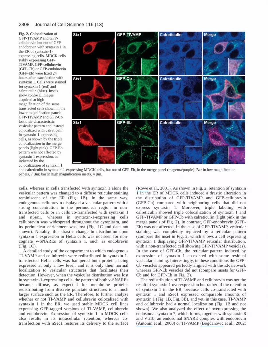
(Rowe et al., 2001). As shown in Fig. 2, retention of syntaxin1 in the ER of MDCK cells induced a drastic alteration inthe distribution of GFP-TIVAMP and GFP-cellubrevin(GFP-Cb) compared with neighboring cells that did notexpress syntaxin 1. Moreover, triple labeling withcalreticulin showed triple colocalization of syntaxin 1 andGFP-TIVAMP or GFP-Cb with calreticulin (light pink in themerge panels of Fig. 2). In contrast, GFP-endobrevin (GFP-Eb) was not affected. In the case of GFP-TIVAMP, vesicularstaining was completely replaced by a reticular pattern(compare the inset in Fig. 2, which shows a cell expressingsyntaxin 1 displaying GFP-TIVAMP reticular distribution,with a non-transfected cell showing GFP-TIVAMP vesicles).In the case of GFP-Cb, the reticular pattern induced byexpression of syntaxin 1 co-existed with some residualvesicular staining. Interestingly, in these conditions the GFP-Cb vesicles appeared perfectly aligned with the ER networkwhereas GFP-Eb vesicles did not (compare insets for GFP-Cb and for GFP-Eb in Fig. 2).
The redistribution of TI-VAMP and cellubrevin was not theresult of syntaxin 1 overexpression but rather of the retentionof syntaxin 1 in the ER, because cells co-transfected withsyntaxin 1 and nSec1 expressed comparable amounts ofsyntaxin 1 (Fig. 1B, Fig. 3B), and yet, in this case, TI-VAMPand cellubrevin had a normal localization (Fig. 1B and notshown). We also analyzed the effect of overexpressing theendosomal syntaxin 7, which forms, together with syntaxin 8and Vti1b, an endosomal SNARE complex with endobrevin(Antonin et al., 2000) or TI-VAMP (Bogdanovic et al., 2002;
Journal of Cell Science 116 (13)
Fig. 2. Colocalization ofGFP-TIVAMP and GFP-cellubrevin but not of GFP-endobrevin with syntaxin 1 inthe ER of syntaxin-1-expressing cells. MDCK cellsstably expressing GFP-TIVAMP, GFP-cellubrevin(GFP-Cb) or GFP-endobrevin(GFP-Eb) were fixed 24hours after transfection withsyntaxin 1. Cells were stainedfor syntaxin 1 (red) andcalreticulin (blue). Insetsshow confocal imagesacquired at highmagnification of the sametransfected cells shown in thelower magnification panels.GFP-TIVAMP and GFP-Cblost their characteristicvesicular pattern and insteadcolocalized with calreticulinin syntaxin-1-expressingcells, as shown by the triplecolocalization in the mergepanels (light pink). GFP-Ebpattern was not affected bysyntaxin 1 expression, asindicated by thecolocalization of syntaxin 1and calreticulin in syntaxin-1-expressing MDCK cells, but not of GFP-Eb, in the merge panel (magenta/purple). Bar in low magnificationpanels, 7 µm; bar in high magnification insets, 4 µm.

2809Compartmental specificity of membrane fusion
Wade et al., 2001). However, in MDCK cells, exogenoussyntaxin 7 was distributed to endosomes, as previouslyreported for the endogenous protein (Mullock et al., 2000)and colocalizes partially with TI-VAMP and endobrevin butnot with cellubrevin (Supplementary Fig. 1 available atjcs.biologists.org/supplemental). The lack of mislocalizationof syntaxin 7 upon overexpression, which is most probablycaused by MDCK cells endogenously expressing thisSNARE and the machinery for its correct localizationand function (including its putative SM protein) did notallow further study of its role in cognate v-SNAREdistribution.
Cognate v-SNAREs are retained in the ER of syntaxin-1-expressing cells through their specific interaction withsyntaxin 1The mislocalization of TI-VAMP and cellubrevin in syntaxin-1-expressing cells suggested that both v-SNAREs might beretained in the ER through a direct and specific interactionwith syntaxin 1. Therefore, we searched for syntaxin-1-containing SNARE complexes. SNAREs are able to formpromiscuous interactions in vitro (Fasshauer et al., 1999; Yanget al., 1999), although this is not the case in vivo or in vitroin conditions where SNAREs are inserted in a membranerather than in solution (McNew et al., 2000; Parlati et al.,2000; Scales et al., 2000). Nevertheless, we first sought toclarify this point and verified that in our experimentalconditions SNARE complexes did not form in the detergentextract of transfected HeLa cells. To test this, HeLa cells wereeither co-transfected with GFP-Cb plus syntaxin 1 ortransfected with each construct alone. Lysates from co-transfected cells and a 1:1 mixture of lysates from cellsexpressing each protein alone were immunoprecipitated withantibodies against GFP or syntaxin 1. As shown in Fig. 3A,we could only detect co-immunoprecipitation of GFP-Cb andsyntaxin 1 when the cells were co-transfected, indicating thatthe complexes were formed inside the cell and not during theextraction procedure.
We then obtained cells expressing either syntaxin 1 alone(retaining syntaxin 1 at the ER, Fig. 1) or together withnSec1 (expressing syntaxin 1 normally at the plasmamembrane, see Fig. 1B) and searched for endogenousSNAREs co-immunoprecipitating with syntaxin 1. nSec1was efficiently expressed by the co-transfected cells but itdid not co-immunoprecipitate with anti-syntaxin 1antibodies owing to the instability of the nSec1–syntaxin 1complex (Garcia et al., 1995). As expected, SNAREs that donot normally interact with syntaxin 1, such as VAMP4,endobrevin and Vti1b, were not found in the anti-syntaxin 1immunoprecipitates under any condition (Fig. 3B). Bycontrast, the plasma membrane SNARE synaptosomalassociated protein of 23 kDa (SNAP-23) was efficientlyrecovered, supporting the specificity of the interactions. Asexpected, SNAP-23 co-immunoprecipitated with syntaxin 1when syntaxin 1 was correctly sorted to the plasmamembrane, although to a lesser extent than when syntaxin 1was retained in the ER. By contrast, TI-VAMP andcellubrevin could only be detected in anti-syntaxin 1immunoprecipitates when this t-SNARE was mistargeted tothe ER. This is most probably because when syntaxin 1 is
co-expressed with nSec1 the latter negatively regulates theavailability of the former to participate in ternary SNAREcomplexes (Perez-Branguli et al., 2002; Yang et al., 2000).To detect co-immunoprecipitation of cognate v-SNAREs,and because SNARE complexes are short-lived (Peng andGallwitz, 2002), it would be necessary to pre-treat the cellswith N-ethylmaleimide (NEM) (Galli et al., 1998).
Fig. 3. TI-VAMP and cellubrevin interacted specifically in the ERwith ectopic syntaxin 1. (A) HeLa cells were either co-transfectedwith GFP-Cb and Stx1 (1), or transfected with each cDNA alone(2,3), as indicated. 24 hours after transfection, cells were lysed andthe extract from co-transfected cells was immediatelyimmunoprecipitated (1), whereas extracts from cells transfected withonly GFP-Cb or Stx1 were mixed and then immunoprecipitated(2+3) with the antibodies indicated. (B) HeLa cells were transfectedwith syntaxin 1 (1) or co-transfected with syntaxin 1 and nSec1 (2).24 hours after transfection, cells were lysed and processed forimmunoprecipitation with either anti-syntaxin 1 mouse monoclonalantibody (1 and 2) or with control mouse immunoglobulins (1C and2C). Proteins were resolved by SDS-PAGE, and western blots wereprobed with the antibodies indicated. Note that cellubrevin, SNAP-23 and TI-VAMP but not VAMP4, endobrevin or Vti1b wererecovered in the syntaxin 1 immunoprecipitate. SM, startingmaterial.

2810
Mislocalization of syntaxin 1 induces the relocalization ofits cognate light chain SNAP-23SNARE complex formation is controlled both by the strictspecificity of the recognition between v- and t-SNAREs and bythe correct topology of this interaction: namely one v-SNAREin one membrane and a t-SNARE complex formed by onesyntaxin and two light chains in the other membrane (Parlatiet al., 2000). Our biochemical data regarding SNAP-23suggested that when syntaxin 1 is retained in the ER, themembrane of this compartment fulfilled the requirements to bea target membrane for TI-VAMP and cellubrevin vesicles (i.e.the presence of syntaxin 1 and the two light chains providedby SNAP-23). To confirm this point, we analyzed theintracellular distribution of SNAP-23 in syntaxin-1-expressingcells. As shown in Fig. 4A, in non-transfected cells or in cellsco-transfected with syntaxin 1 plus nSec1, SNAP-23 displayedthe expected cell surface staining (arrows, Fig. 4A) and theintracellular vimentin-associated labeling previously described(Faigle et al., 2000) (data not shown); by contrast, in cellsexpressing syntaxin 1 this plasma membrane pattern wascompletely lost, and a significant pool of SNAP-23 colocalizedwith syntaxin 1 (arrowhead, Fig. 4A). As a control, weanalyzed the distribution of the plasma membrane proteinNa+/K+ ATPase, which does not interact with SNAREs. Asexpected, the surface localization of Na+/K+ ATPase was notaffected by the expression of syntaxin 1 (Fig. 4B). Altogether,these data suggest that the ectopic expression of syntaxin 1 in
the ER induced the mislocalization of its cognate light chainby a direct interaction.
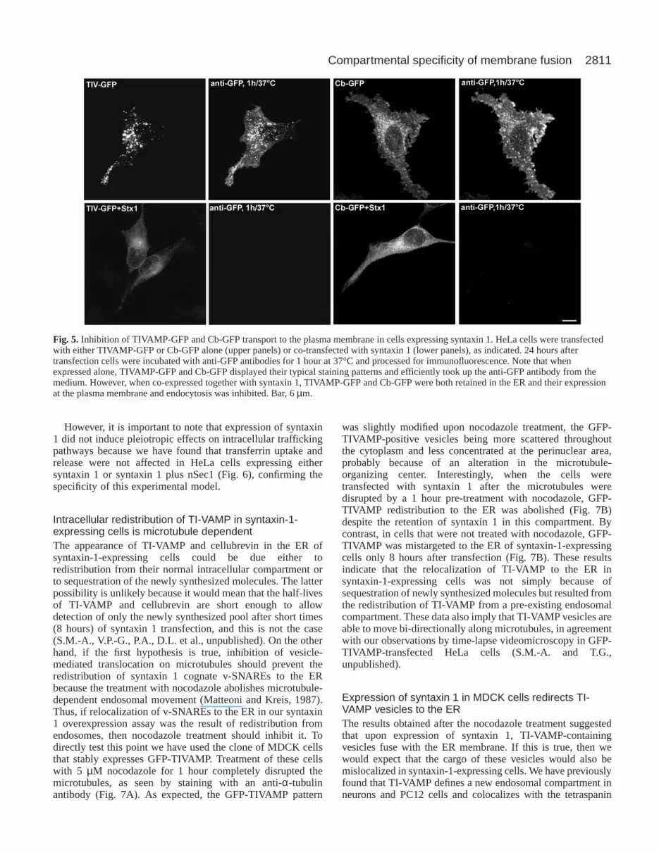
Transport to the plasma membrane of TI-VAMP andcellubrevin is impaired in syntaxin-1-expressing cellsThe mislocalization of endogenous TI-VAMP and cellubrevinupon ectopic expression of syntaxin 1, together with theirspecific interaction with syntaxin 1 in the ER, suggested thatunder these conditions both proteins might not be able to reachthe cell surface. To further analyze this point, we designedGFP fusion proteins of TI-VAMP and cellubrevin with theGFP tag fused to the C-terminus, so that we could monitortheir appearance at the plasma membrane and endocytosis byincubating living cells with antibodies directed against GFPand measuring antibody uptake. Cells transfected withTIVAMP-GFP or Cb-GFP alone displayed typical plasmamembrane and vesicular staining patterns and efficientlybound the anti-GFP antibody in the culture medium (Fig. 5,upper panels), suggesting that TIVAMP-GFP and Cb-GFPreached the plasma membrane, as is the case for theendogenous cellubrevin (Galli et al., 1994). In contrast, whenTIVAMP-GFP or Cb-GFP was co-transfected with syntaxin 1,both proteins were retained in the ER and failed to bind tothe extracellular anti-GFP antibody (Fig. 5, lower panels),suggesting that transport to the plasma membrane andendocytosis was abolished.
Journal of Cell Science 116 (13)
Fig. 4. SNAP-23 was redistributed tothe ER in cells expressing syntaxin 1.HeLa cells transfected with syntaxin 1or co-transfected with syntaxin 1 andnSec1 as indicated were fixed anddouble stained for syntaxin 1 andendogenous SNAP-23 (A) or theplasma membrane marker Na+/K+
ATPase (B). Note the colocalization ofSNAP-23 and ectopic syntaxin 1(arrowhead) and the absence of SNAP-23 from the plasma membrane in cellsexpressing syntaxin 1 alone and theplasma membrane staining of SNAP-23 in non-transfected cells or in cellsco-transfected with syntaxin 1 plusnSec1 (arrows). By contrast,endogenous Na+/K+ ATPase is notrelocalized from the plasma membranein syntaxin-1-expressing cells. Bar,7 µm.
2811Compartmental specificity of membrane fusion
However, it is important to note that expression of syntaxin1 did not induce pleiotropic effects on intracellular traffickingpathways because we have found that transferrin uptake andrelease were not affected in HeLa cells expressing eithersyntaxin 1 or syntaxin 1 plus nSec1 (Fig. 6), confirming thespecificity of this experimental model.
Intracellular redistribution of TI-VAMP in syntaxin-1-expressing cells is microtubule dependentThe appearance of TI-VAMP and cellubrevin in the ER ofsyntaxin-1-expressing cells could be due either toredistribution from their normal intracellular compartment orto sequestration of the newly synthesized molecules. The latterpossibility is unlikely because it would mean that the half-livesof TI-VAMP and cellubrevin are short enough to allowdetection of only the newly synthesized pool after short times(8 hours) of syntaxin 1 transfection, and this is not the case(S.M.-A., V.P.-G., P.A., D.L. et al., unpublished). On the otherhand, if the first hypothesis is true, inhibition of vesicle-mediated translocation on microtubules should prevent theredistribution of syntaxin 1 cognate v-SNAREs to the ERbecause the treatment with nocodazole abolishes microtubule-dependent endosomal movement (Matteoni and Kreis, 1987).Thus, if relocalization of v-SNAREs to the ER in our syntaxin1 overexpression assay was the result of redistribution fromendosomes, then nocodazole treatment should inhibit it. Todirectly test this point we have used the clone of MDCK cellsthat stably expresses GFP-TIVAMP. Treatment of these cellswith 5 µM nocodazole for 1 hour completely disrupted themicrotubules, as seen by staining with an anti-α-tubulinantibody (Fig. 7A). As expected, the GFP-TIVAMP pattern
was slightly modified upon nocodazole treatment, the GFP-TIVAMP-positive vesicles being more scattered throughoutthe cytoplasm and less concentrated at the perinuclear area,probably because of an alteration in the microtubule-organizing center. Interestingly, when the cells weretransfected with syntaxin 1 after the microtubules weredisrupted by a 1 hour pre-treatment with nocodazole, GFP-TIVAMP redistribution to the ER was abolished (Fig. 7B)despite the retention of syntaxin 1 in this compartment. Bycontrast, in cells that were not treated with nocodazole, GFP-TIVAMP was mistargeted to the ER of syntaxin-1-expressingcells only 8 hours after transfection (Fig. 7B). These resultsindicate that the relocalization of TI-VAMP to the ER insyntaxin-1-expressing cells was not simply because ofsequestration of newly synthesized molecules but resulted fromthe redistribution of TI-VAMP from a pre-existing endosomalcompartment. These data also imply that TI-VAMP vesicles areable to move bi-directionally along microtubules, in agreementwith our observations by time-lapse videomicroscopy in GFP-TIVAMP-transfected HeLa cells (S.M.-A. and T.G.,unpublished).
Expression of syntaxin 1 in MDCK cells redirects TI-VAMP vesicles to the ERThe results obtained after the nocodazole treatment suggestedthat upon expression of syntaxin 1, TI-VAMP-containingvesicles fuse with the ER membrane. If this is true, then wewould expect that the cargo of these vesicles would also bemislocalized in syntaxin-1-expressing cells. We have previouslyfound that TI-VAMP defines a new endosomal compartment inneurons and PC12 cells and colocalizes with the tetraspanin
Fig. 5. Inhibition of TIVAMP-GFP and Cb-GFP transport to the plasma membrane in cells expressing syntaxin 1. HeLa cells were transfectedwith either TIVAMP-GFP or Cb-GFP alone (upper panels) or co-transfected with syntaxin 1 (lower panels), as indicated. 24 hours aftertransfection cells were incubated with anti-GFP antibodies for 1 hour at 37°C and processed for immunofluorescence. Note that whenexpressed alone, TIVAMP-GFP and Cb-GFP displayed their typical staining patterns and efficiently took up the anti-GFP antibody from themedium. However, when co-expressed together with syntaxin 1, TIVAMP-GFP and Cb-GFP were both retained in the ER and their expressionat the plasma membrane and endocytosis was inhibited. Bar, 6 µm.

2812
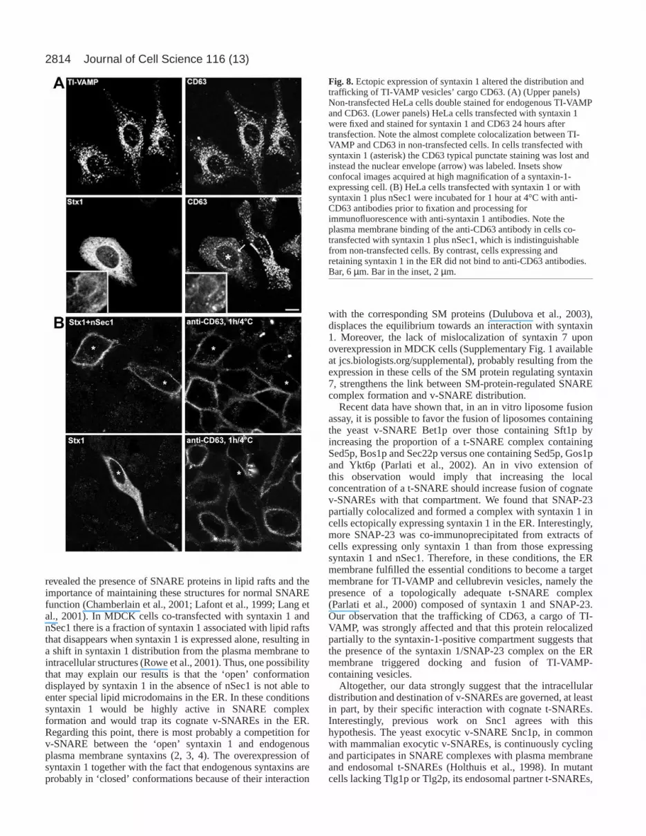
protein CD63 (Berditchevski, 2001; Coco et al., 1999). Wefound the same result in HeLa cells, as shown by the extensivecolocalization of TI-VAMP and CD63 (Fig. 8A, upper panels).Therefore, we investigated the fate of CD63 molecules in cellsexpressing and retaining syntaxin 1 in the ER. As shown in Fig.8A (lower panels), the classic vesicular pattern of CD63 waspartially lost in cells transfected with syntaxin 1. In theseconditions, CD63-positive vesicles were less abundant, moreheterogenous in size and shape and, importantly, the nuclearenvelope and fine reticular structures characteristic of the ERwere labeled with the anti-CD63 antibody (arrow in Fig. 8A).Furthermore, high magnification confocal images showedpartial colocalization of CD63 with the syntaxin 1 retained inthe ER (inset in Fig. 8A). This change in distribution is not theresult of a block in CD63 export from the ER, because the sameresult was obtained when cells expressing syntaxin 1 wereincubated with cycloheximide for 3 hours prior to fixation (datanot shown). Furthermore, we have checked that CD63 does notcolocalize with calreticulin in untransfected cells or cellsexpressing syntaxin 1 plus nSec1 (S.M.-A., V.P.G., P.A., D.L.et al., unpublished). In normal conditions, the tetraspaninprotein CD63 cycles through the cell surface (Kobayashi et al.,2000). Therefore, to confirm the effect of the ER retention ofsyntaxin 1 on CD63 distribution, we performed an antibodybinding assay at 4°C to detect CD63 molecules at the plasmamembrane. As expected, in non-transfected cells or in cellstransfected with syntaxin 1 plus nSec1, the anti-CD63 antibodywas efficiently bound, as a result of the presence of CD63 atthe cell surface (Fig. 8B, upper panels). Strikingly, in cellsexpressing syntaxin 1 there was no detectable binding of theantibody (Fig. 8B, lower panels), supporting the redistributionof CD63 in these conditions. Moreover, when the anti-CD63antibody was incubated with living cells at 37°C for 90 minutes,
it was efficiently internalized in non-transfected cells or in cellstransfected with syntaxin 1 plus nSec1, whereas no internalizedantibody was detected in cells transfected with syntaxin 1 alone(S.M.-A., V.P.G., P.A., D.L. et al., unpublished).
DiscussionIn the present study, we sought to analyze in vivo the linkbetween the intracellular distribution and destination of v-SNAREs and the compartmental specificity of fusion. Ourresults show that in the absence of nSec1 the t-SNARE heavychain syntaxin 1, normally found at the neuronal plasmamembrane, is localized to the ER and induces an activeredistribution of its cognate light chain SNAP-23 and cognatev-SNAREs, cellubrevin and TI-VAMP but does not alter thedistribution of non-cognate v-SNAREs, such as endobrevin.
The redistribution of TI-VAMP and cellubrevin to the ER infibroblasts expressing syntaxin 1 points to a direct relationshipbetween SNARE complex formation and v-SNAREdistribution and destination for the following reasons. (1) Itwas due to syntaxin 1 mistargeting and not simply to syntaxin1 overexpression because syntaxin 1 was expressed at the samelevel in the presence and absence of nSec1 and yet no effecton cognate v-SNARE distribution was seen in cells co-transfected with syntaxin 1 and nSec1. (2) It was not the resultof the retention of the newly synthesized v-SNARE proteinsbut of an active microtubule-dependent redistribution of thepre-existing v-SNARE molecules. (3) It recapitulated thespecificity found in v-/t-SNARE interactions because onlysyntaxin 1 cognate v-SNAREs were affected by itsmistargeting, and we detected a bona fide interaction betweenthe syntaxin 1 retained in the ER and the redistributed TI-VAMP and cellubrevin by co-immunoprecipitation. (4) The
Journal of Cell Science 116 (13)
Fig. 6. Ectopic expression of syntaxin 1 did not affect transferrin recycling. HeLa cells were transfected with syntaxin 1 (upper panels) or co-transfected with syntaxin 1 and nSec1 (lower panels). 24 hours later cells were allowed to internalize transferrin for 1 hour at 37°C, washed andeither fixed immediately (uptake) or incubated for a further 1 hour at 37°C before fixation (release). Notice that both the uptake and the releaseof transferrin were comparable in both kinds of transfected cells and indistinguishable from non-transfected cells, independently of theintracellular distribution of syntaxin 1. Bar, 5 µm.

2813Compartmental specificity of membrane fusion
effect of the expression of syntaxin 1 on the redistribution ofCD63 suggests that the vesicles containing TI-VAMP andCD63 docked and fused with the syntaxin-1-containing ER
membrane. At steady state, the effect of the expression ofsyntaxin 1 in the ER on CD63 localization was only partial(Fig. 8A). This could be due to the fact that TI-VAMP andCD63 may not necessarily be transported in the same vesiclesall along their trafficking pathway. In contrast, the inhibitionof the expression of CD63 at the plasma membrane was verystrong (Fig. 8B), thus demonstrating that the trafficking ofCD63 was strongly impaired. The expression of syntaxin 1 innon-neuronal cells was also shown to induce a relocalizationof Golgi markers (Rowe et al., 2001). However, theredistribution of TI-VAMP and cellubrevin observed insyntaxin-1-expressing cells (this study) was not merely theresult of the disassembly of the Golgi because Brefeldin A, adrug that induces the collapse of the Golgi complex (Fujiwaraet al., 1988), has no effect on the localization of TI-VAMP(Advani et al., 1999).
It is noteworthy that, despite the mislocalization ofcellubrevin and TI-VAMP, cells ectopically expressingsyntaxin 1 are still capable of internalizing and recyclingtransferrin. This indicates that the mislocalization of syntaxin1 to the ER does not have pleiotropic effects on all membranefusion steps. This result is rather unexpected because itsuggests that a v-SNARE other than cellubrevin participates intransferrin recycling. In this regard, it is important to note thatendobrevin colocalizes with internalized transferrin (Wong etal., 1998) (S.M.-A. and T.G., unpublished) as does cellubrevin(Galli et al., 1994), which suggests that cellubrevin andendobrevin may have overlapping functions. The participationof endobrevin in this transport pathway could explain why thetreatment of cells with tetanus toxin only inhibited one third ofthe total release of apo-transferrin (Galli et al., 1994) and thelack of a major phenotype in cellubrevin-knockout mice (Yanget al., 2001). Irrespective of the molecular mechanismunderlying transferrin recycling in syntaxin-1-transfected cells,these results support the specificity of the v-/t-SNAREinteraction in vivo and rule out the possibility that the observedeffect on TI-VAMP and cellubrevin could be because of ageneral defect in intracellular trafficking.
nSec1 belongs to the Sec/Munc18 (SM) family of proteins(Rizo and Sudhof, 2002) and is essential for synaptic vesicleexocytosis (Verhage et al., 2000). It binds tightly to the closedconformation of syntaxin 1 (Dulubova et al., 1999) andcompetes with SNARE complex formation (Yang et al., 2000).As discussed by Rowe et al., nSec1 may act as a chaperone-likeprotein, allowing syntaxin 1 to proceed through the secretorypathway by keeping it in a ‘closed’ conformation, unable tointeract with its partner SNAREs (Rowe et al., 2001), andpreventing the formation of non-productive SNARE complexes.Our data suggest that when syntaxin 1 is overexpressed in theabsence of nSec1, it may display an ‘open’ conformation(Dulubova et al., 1999) that is unable to exit the ER but ableto interact with its cognate partners, resulting in the finalredistribution of its cognate v-SNAREs to the ER. These resultspoint to the importance of the balance between syntaxin 1 andnSec1 for the correct functionality of both proteins, as has beenshown to be the case in other systems such as chromaffin cellsand Drosophila(Voets et al., 2001; Wu et al., 1998). The effectof syntaxin 1 expression reported here may also explain theblockade of membrane transport observed in these conditions(Rowe et al., 1999), because functional exocytic v-SNAREswould be sequestered in the ER. Moreover, several studies have
Fig. 7.TI-VAMP relocalization in syntaxin-1-expressing cells wasmicrotubule dependent. (A) MDCK cells stably expressing GFP-TIVAMP incubated with or without 5 µM nocodazole for 1 hourwere fixed and stained for α-tubulin. Note that in the presence ofnocodazol, microtubules were completely disrupted. (B) MDCKcells stably expressing GFP-TIVAMP incubated with or without5 µM nocodazole for 1 hour prior to transfection with syntaxin 1were fixed and stained for syntaxin 1 8 hours after transfection. Incontrol conditions (Cont), the expression of syntaxin 1 and itsretention in the ER induced a relocalization of GFP-TIVAMPtowards syntaxin-1-positives structures in the ER (arrows). Bycontrast, in the absence of functional microtubules (+Noc) thelocalization of GFP-TIVAMP in transfected cells wasindistinguishable from non-transfected cells. Bar, 4 µm.
2814
revealed the presence of SNARE proteins in lipid rafts and theimportance of maintaining these structures for normal SNAREfunction (Chamberlain et al., 2001; Lafont et al., 1999; Lang etal., 2001). In MDCK cells co-transfected with syntaxin 1 andnSec1 there is a fraction of syntaxin 1 associated with lipid raftsthat disappears when syntaxin 1 is expressed alone, resulting ina shift in syntaxin 1 distribution from the plasma membrane tointracellular structures (Rowe et al., 2001). Thus, one possibilitythat may explain our results is that the ‘open’ conformationdisplayed by syntaxin 1 in the absence of nSec1 is not able toenter special lipid microdomains in the ER. In these conditionssyntaxin 1 would be highly active in SNARE complexformation and would trap its cognate v-SNAREs in the ER.Regarding this point, there is most probably a competition forv-SNARE between the ‘open’ syntaxin 1 and endogenousplasma membrane syntaxins (2, 3, 4). The overexpression ofsyntaxin 1 together with the fact that endogenous syntaxins areprobably in ‘closed’ conformations because of their interaction
with the corresponding SM proteins (Dulubova et al., 2003),displaces the equilibrium towards an interaction with syntaxin1. Moreover, the lack of mislocalization of syntaxin 7 uponoverexpression in MDCK cells (Supplementary Fig. 1 availableat jcs.biologists.org/supplemental), probably resulting from theexpression in these cells of the SM protein regulating syntaxin7, strengthens the link between SM-protein-regulated SNAREcomplex formation and v-SNARE distribution.
Recent data have shown that, in an in vitro liposome fusionassay, it is possible to favor the fusion of liposomes containingthe yeast v-SNARE Bet1p over those containing Sft1p byincreasing the proportion of a t-SNARE complex containingSed5p, Bos1p and Sec22p versus one containing Sed5p, Gos1pand Ykt6p (Parlati et al., 2002). An in vivo extension ofthis observation would imply that increasing the localconcentration of a t-SNARE should increase fusion of cognatev-SNAREs with that compartment. We found that SNAP-23partially colocalized and formed a complex with syntaxin 1 incells ectopically expressing syntaxin 1 in the ER. Interestingly,more SNAP-23 was co-immunoprecipitated from extracts ofcells expressing only syntaxin 1 than from those expressingsyntaxin 1 and nSec1. Therefore, in these conditions, the ERmembrane fulfilled the essential conditions to become a targetmembrane for TI-VAMP and cellubrevin vesicles, namely thepresence of a topologically adequate t-SNARE complex(Parlati et al., 2000) composed of syntaxin 1 and SNAP-23.Our observation that the trafficking of CD63, a cargo of TI-VAMP, was strongly affected and that this protein relocalizedpartially to the syntaxin-1-positive compartment suggests thatthe presence of the syntaxin 1/SNAP-23 complex on the ERmembrane triggered docking and fusion of TI-VAMP-containing vesicles.
Altogether, our data strongly suggest that the intracellulardistribution and destination of v-SNAREs are governed, at leastin part, by their specific interaction with cognate t-SNAREs.Interestingly, previous work on Snc1 agrees with thishypothesis. The yeast exocytic v-SNARE Snc1p, in commonwith mammalian exocytic v-SNAREs, is continuously cyclingand participates in SNARE complexes with plasma membraneand endosomal t-SNAREs (Holthuis et al., 1998). In mutantcells lacking Tlg1p or Tlg2p, its endosomal partner t-SNAREs,
Journal of Cell Science 116 (13)
Fig. 8. Ectopic expression of syntaxin 1 altered the distribution andtrafficking of TI-VAMP vesicles’ cargo CD63. (A) (Upper panels)Non-transfected HeLa cells double stained for endogenous TI-VAMPand CD63. (Lower panels) HeLa cells transfected with syntaxin 1were fixed and stained for syntaxin 1 and CD63 24 hours aftertransfection. Note the almost complete colocalization between TI-VAMP and CD63 in non-transfected cells. In cells transfected withsyntaxin 1 (asterisk) the CD63 typical punctate staining was lost andinstead the nuclear envelope (arrow) was labeled. Insets showconfocal images acquired at high magnification of a syntaxin-1-expressing cell. (B) HeLa cells transfected with syntaxin 1 or withsyntaxin 1 plus nSec1 were incubated for 1 hour at 4°C with anti-CD63 antibodies prior to fixation and processing forimmunofluorescence with anti-syntaxin 1 antibodies. Note theplasma membrane binding of the anti-CD63 antibody in cells co-transfected with syntaxin 1 plus nSec1, which is indistinguishablefrom non-transfected cells. By contrast, cells expressing andretaining syntaxin 1 in the ER did not bind to anti-CD63 antibodies.Bar, 6 µm. Bar in the inset, 2 µm.

2815Compartmental specificity of membrane fusion
the steady-state distribution of Snc1p was affected (Lewis etal., 2000). Moreover, Snc1p is also redirected to a haze oftransport vesicles in a mutant yeast strain in which Tlg1p andTlg2p accumulated on the same structures (Siniossoglouand Pelham, 2001). Exocytic v-SNAREs, such as Snc1,synaptobrevin 2, cellubrevin and TI-VAMP, are continuouslycycling between the plasma membrane and endosomes. Ourresults suggest that when a t-SNARE is constitutively active(i.e. syntaxin 1 in the absence of nSec1 in this study) then morev-/t-SNARE complexes form and the v-SNAREs localize, to agreat extent, to the membrane where the cognate t-SNARE isexpressed. In contrast, in normal conditions (i.e. co-expressionof syntaxin 1 and nSec1 in this study or wild-type fibroblasts)the level of SNARE complexes formed (and thereforerecovered in detergent extracts) is low, and TI-VAMP andcellubrevin localize to endosomal vesicles. This suggests thatthe steady-state subcellular localization of v-SNAREs is theresult of equilibrium between two states: one corresponding tov-SNAREs on the donor vesicles and the other to v-SNAREsin the target membrane. The lack of nSec1 displaced thisequilibrium towards the second state.
Our findings have important implications for howcompartmental specificity is achieved during membranefusion. Indeed, we have shown that the ectopic expression ofsyntaxin 1 induces an illegitimate rerouteing of vesicles, thefusion of which is mediated by the cognate v-SNAREs ofsyntaxin 1, TI-VAMP and cellubrevin. These results showthat the highly controlled and specific v-/t-SNAREinteraction is essential to define the destination of membranecarriers in vivo in mammalian cells. In conclusion, our resultssuggest that the exquisite regulation of the v-/t-SNAREinteraction that ensures compartmental specificity ofmembrane fusion is also one of the factors accounting for theaccuracy of the dynamic intracellular distribution anddestination of v-SNAREs.
We thank R. Rudge and L. Johannes for critical reading of themanuscript, G. Miesenbock and J. Rothman for generous gift ofpHLuorin and R. Jahn for the syntaxin 7 cDNA. S.M.-A. is arecipient of a post-doctoral fellowship from the Spanish Ministeriode Educación y Cultura. V.P.G. is a recipient of a post-doctoralfellowship from Association pour la Recherche sur le Cancer(ARC). P.A. is a recipient of a doctoral fellowship from Societé deSecours des Amis des Sciences. This work was supported in part bygrants from Human Frontier Science Program (RGY0027/2001-B101), European Community (QLG3-CT-2001-02430_RetrogradeSignaling), Association pour la Recherche sur le Cancer (ARCN°5873) and Ministère de la Recherche (ACI-JC5254) to T.G.
ReferencesAdvani, R. J., Bae, H. R., Bock, J. B., Chao, D. S., Doung, Y. C., Prekeris,
R., Yoo, J. S. and Scheller, R. H.(1998). Seven novel mammalian SNAREproteins localize to distinct membrane compartments. J. Biol. Chem.273,10317-10324.
Advani, R. J., Yang, B., Prekeris, R., Lee, K. C., Klumperman, J. andScheller, R. H. (1999). VAMP-7 mediates vesicular transport fromendosomes to lysosomes. J. Cell Biol.146, 765-775.
Antonin, W., Holroyd, C., Fasshauer, D., Pabst, S., vonMollard, G. F. andJahn, R. (2000). A SNARE complex mediating fusion of late endosomesdefines conserved properties of SNARE structure and function. EMBO J.19, 6453-6464.
Bennett, M. K., Calakos, N. and Scheller, R. H.(1992). Syntaxin: a synapticprotein implicated in docking of synaptic vesicles at presynaptic activezones. Science257, 255-259.
Berditchevski, F.(2001). Complexes of tetraspanins with integrins: more thanmeets the eye. J. Cell Sci.114, 4143-4151.
Bogdanovic, A., Bennett, N., Kieffer, S., Louwagie, M., Morio, T., Garin,J., Satre, M. and Bruckert, F. (2002). Syntaxin 7, Syntaxin 8, Vti1 andVAMP7 form an active SNARE complex for early macropinocyticcompartment fusion in Dictyostelium discoideum. Biochem. J. 368, 29-39.
Chamberlain, L. H., Burgoyne, R. D. and Gould, G. W.(2001). SNAREproteins are highly enriched in lipid rafts in PC12 cells: Implications for thespatial control of exocytosis. Proc. Natl. Acad. Sci. USA98, 5619-5624.
Chen, Y. A. and Scheller, R. H.(2001). Snare-mediated membrane fusion.Nat. Rev. Mol. Cell Biol.2, 98-106.
Chilcote, T. J., Galli, T., Mundigl, O., Edelmann, L., McPherson, P. S.,Takei, K. and de Camilli, P. (1995). Cellubrevin and synaptobrevins:similar subcellular localization and biochemical properties in PC12 cells. J.Cell Biol. 129, 219-231.
Coco, S., Raposo, G., Martinez, S., Fontaine, J. J., Takamori, S., Zahraoui,A., Jahn, R., Matteoli, M., Louvard, D. and Galli, T. (1999). Subcellularlocalization of tetanus neurotoxin-insensitive vesicle-associated membraneprotein (VAMP)/VAMP7 in neuronal cells: Evidence for a novel membranecompartment. J. Neurosci.19, 9803-9812.
D’Esposito, M., Ciccodicola, A., Gianfrancesco, F., Esposito, T., Flagiello,L., Mazzarella, R., Schlessinger, D. and D’Urso, M.(1996). Asynaptobrevin-like gene in the Xq28 pseudoautosomal region undergoes Xinactivation. Nat. Genet.13, 227-229.
Dulubova, I., Sugita, S., Hill, S., Hosaka, M., Fernandez, I., Sudhof, T. C.and Rizo, J.(1999). A conformational switch in syntaxin during exocytosis:role of munc18. EMBO J.18, 4372-4382.
Dulubova, I., Yamaguchi, T., Arac, D., Li, H. M., Huryeva, I., Min, S. W.,Rizo, J. and Sudhof, T. C.(2003). Convergence and divergence in themechanism of SNARE binding by Sec1/Munc18-like proteins. Proc. Natl.Acad. Sci. USA100, 32-37.
Faigle, W., Colucci-Guyon, E., Louvard, D., Amigorena, S. and Galli, T.(2000). Vimentin filaments in fibroblasts are a reservoir for SNAP23, acomponent of the membrane fusion machinery. Mol. Biol. Cell 11, 3485-3494.
Fasshauer, D., Sutton, R. B., Brunger, A. T. and Jahn, R.(1998). Conservedstructural features of the synaptic fusion complex: SNARE proteinsreclassified as Q- and R-SNAREs. Proc. Natl. Acad. Sci. USA95, 15781-15786.
Fasshauer, D., Antonin, W., Margittai, M., Pabst, S. and Jahn, R.(1999).Mixed and Non-cognate SNARE complexes. Characterization of assemblyand biophysical properties. J. Biol. Chem.274, 15440-15446.
Filippini, F., Rossi, V., Galli, T., Budillon, A., D’Urso, M. and D’Esposito,M. (2001). Longins: a new evolutionary conserved VAMP family sharing anovel SNARE domain. Trends Biochem. Sci.26, 407-409.
Fujiwara, T., Oda, K., Yokota, S., Takatsuki, A. and Ikehara, Y.(1988).Brefeldin A causes disassembly of the Golgi complex and accumulation ofsecretory proteins in the endoplasmic reticulum. J. Biol. Chem.263, 18545-18552.
Fukuda, R., McNew, J. A., Weber, T., Parlati, F., Engel, T., Nickel, W.,Rothman, J. E. and Sollner, T. H.(2000). Functional architecture of anintracellular membrane t-SNARE. Nature407, 198-202.
Furuno, T., Teshima, R., Kitani, S., Sawada, J. and Nakanishi, M.(1996).Surface expression of CD63 antigen (AD1 antigen) in P815 mastocytomacells by transfected IgE receptors. Biochem. Biophys. Res. Commun.219,740-744.
Galli, T. and Haucke, V. (2001). Cycling of synaptic vesicles: how far? Howfast! Sci STKE2001, RE1.
Galli, T., Chilcote, T., Mundigl, O., Binz, T., Niemann, H. and de Camilli,P. (1994). Tetanus toxin-mediated cleavage of cellubrevin impairsexocytosis of transferrin receptor-containing vesicles in CHO cells. J. CellBiol. 125, 1015-1024.
Galli, T., Zahraoui, A., Vaidyanathan, V. V., Raposo, G., Tian, J. M.,Karin, M., Niemann, H. and Louvard, D. (1998). A novel tetanusneurotoxin-insensitive vesicle-associated membrane protein in SNAREcomplexes of the apical plasma membrane of epithelial cells. Mol. Biol. Cell9, 1437-1448.
Garcia, E. P., Gatti, E., Butler, M., Burton, J. and de Camilli, P.(1994). Arat brain Sec1 homologue related to Rop and UNC18 interacts with syntaxin.Proc. Natl. Acad. Sci. USA91, 2003-2007.
Garcia, E. P., McPherson, P. S., Chilcote, T. J., Takei, K. and de Camilli,P. (1995). rbSec1A and B colocalize with syntaxin 1 and SNAP-25throughout the axon, but are not in a stable complex with syntaxin. J. CellBiol. 129, 105-120.

2816
Gurunathan, S., Chapman-Shimshoni, D., Trajkovic, S. and Gerst, J. E.(2000). Yeast exocytic v-SNAREs confer endocytosis. Mol. Biol. Cell 11,3629-3643.
Holthuis, J. C. M., Nichols, B. J., Dhruvakumar, S. and Pelham, H. R. B.(1998). Two syntaxin homologues in the TGN/endosomal system of yeast.EMBO J.17, 113-126.
Katz, L. and Brennwald, P. (2000). Testing the 3Q: 1R ‘’rule’’: Mutationalanalysis of the ionic “zero’’ layer in the yeast exocytic SNARE complexreveals no requirement for arginine. Mol. Biol. Cell11, 3849-3858.
Kobayashi, T., Vischer, U. M., Rosnoblet, C., Lebrand, C., Lindsay, M.,Parton, R. G., Kruithof, E. K. O. and Gruenberg, J. (2000). Thetetraspanin CD63/lamp3 cycles between endocytic and secretorycompartments in human endothelial cells. Mol. Biol. Cell11, 1829-1843.
Lafont, F., Verkade, P., Galli, T., Wimmer, C., Louvard, D. and Simons, K.(1999). Raft association of SNAP receptors acting in apical trafficking inMadin-Darby canine kidney cells. Proc. Natl. Acad. Sci. USA96, 3734-3738.
Lang, T., Bruns, D., Wenzel, D., Riedel, D., Holroyd, P., Thiele, C. andJahn, R. (2001). SNAREs are concentrated in cholesterol-dependentclusters that define docking and fusion sites for exocytosis. EMBO J.20,2202-2213.
Lewis, M. J., Nichols, B. J., PrescianottoBaschong, C., Riezman, H. andPelham, H. R. B.(2000). Specific retrieval of the exocytic SNARE Snc1pfrom early yeast endosomes. Mol. Biol. Cell11, 23-38.
Mallard, F., Tang, B. L., Galli, T., Tenza, D., Saint-Pol, A., Yue, X., Antony,C., Hong, W., Goud, B. and Johannes, L.(2002). Early/recyclingendosomes-to-TGN transport involves two SNARE complexes and a Rab6isoform. J. Cell Biol.156, 653-654.
Martinez-Arca, S., Alberts, P., Zahraoui, A., Louvard, D. and Galli, T.(2000). Role of tetanus neurotoxin insensitive vesicle-associated membraneprotein (TI-VAMP) in vesicular transport mediating neurite outgrowth. J.Cell Biol. 149, 889-899.
Matteoni, R. and Kreis, T. E. (1987). Translocation and clustering ofendosomes and lysosomes depends on microtubules. J. Cell Biol.105, 1253-1265.
McNew, J. A., Parlati, F., Fukuda, R., Johnston, R. J., Paz, K., Paumet,F., Sollner, T. H. and Rothman, J. E.(2000). Compartmental specificityof cellular membrane fusion encoded in SNARE proteins. Nature407, 153-159.
Mullock, B. M., Smith, C. W., Ihrke, G., Bright, N. A., Lindsay, M.,Parkinson, E. J., Brooks, D. A., Parton, R. G., James, D. E., Luzio, J. P.et al. (2000). Syntaxin 7 is localized to late endosome compartments,associates with Vamp 8, and is required for late endosome-lysosome fusion.Mol. Biol. Cell11, 3137-3153.
Muzerelle, A., Alberts, P., Martinez-Arca, S., Jeannequin, O., Lafaye, P.,Mazié, J.-C., Galli, T. and Gaspar, P. (2003). Tetanus neurotoxin-insensitive vesicle-associated membrane protein localizes to a presynapticmembrane compartment in selected terminal subsets of the rat brain.Neuroscience(in press).
Ossig, R., Schmitt, H. D., deGroot, B., Riedel, D., Keranen, S., Ronne, H.,Grubmuller, H. and Jahn, R. (2000). Exocytosis requires asymmetry inthe central layer of the SNARE complex. EMBO J.19, 6000-6010.
Parlati, F., McNew, J. A., Fukuda, R., Miller, R., Sollner, T. H. andRothman, J. E. (2000). Topological restriction of SNARE-dependentmembrane fusion. Nature407, 194-198.
Parlati, F., Varlamov, O., Paz, K., McNew, J. A., Hurtado, D., Sollner, T.H. and Rothman, J. E. (2002). Distinct SNARE complexes mediatingmembrane fusion in Golgi transport based on combinatorial specificity.Proc. Natl. Acad. Sci. USA99, 5424-5429.
Paumet, F., le Mao, J., Martin, S., Galli, T., David, B., Blank, U. and Roa,M. (2000). Soluble NSF attachment protein receptors (SNAREs) in RBL-2H3 mast cells: functional role of syntaxin 4 in exocytosis and identificationof a vesicle-associated membrane protein 8-containing secretorycompartment. J. Immunol.164, 5850-5857.
Paumet, F., Brugger, B., Parlati, F., McNew, J. A., Sollner, T. H. andRothman, J. E. (2001). A t-SNARE of the endocytic pathway must beactivated for fusion. J. Cell Biol.155, 961-968.
Peng, R. W. and Gallwitz, D.(2002). Sly1 protein bound to Golgi syntaxinSed5p allows assembly and contributes to specificity of SNARE fusioncomplexes. J. Cell Biol.157, 645-655.
Perez-Branguli, F., Muhaisen, A. and Blasi, J.(2002). Munc 18a binding tosyntaxin 1A and 1B isoforms defines its localization at the plasmamembrane and blocks SNARE assembly in a three-hybrid system assay.Mol. Cell. Neurosci.20, 169-180.
Pfeffer, S.(2001). Rab GTPases: specifying and deciphering organelle identityand function. Trends Cell Biol.11, 487-491.
Poirier, M. A., Xiao, W. Z., Macosko, J. C., Chan, C., Shin, Y. K. andBennett, M. K. (1998). The synaptic SNARE complex is a parallel four-stranded helical bundle. Nat. Struct. Biol.5, 765-769.
Rizo, J. and Sudhof, T. C.(2002). Snares and munc18 in synaptic vesiclefusion. Nat. Rev. Neurosci.3, 641-653.
Rothman, J. E. (2002). The machinery and principles of vesicle transport inthe cell. Nat. Medicine8, 1059-1062.
Rowe, J., Corradi, N., Malosio, M. L., Taverna, E., Halban, P.,Meldolesi, J. and Rosa, P.(1999). Blockade of membrane transport anddisassembly of the Golgi complex by expression of syntaxin 1A inneurosecretion-incompetent cells: prevention by rbSEC1. J. Cell Sci.112,1865-1877.
Rowe, J., Calegari, F., Taverna, E., Longhi, R. and Rosa, P.(2001). Syntaxin1a is delivered to the apical and basolateral domains of epithelial cells: therole of munc-18 proteins. J. Cell Sci.114, 3323-3332.
Sankaranarayanan, S. and Ryan, T. A.(2000). Real-time measurements ofvesicle-SNARE recycling in synapses of the central nervous system. Nat.Cell Biol. 2, 197-204.
Scales, S. J., Chen, Y. A., Yoo, B. Y., Patel, S. M., Doung, Y. C. and Scheller,R. H. (2000). SNAREs contribute to the specificity of membrane fusion.Neuron26, 457-464.
Scales, S. J., Yoo, B. Y. and Scheller, R. H.(2001). The ionic layer is requiredfor efficient dissociation of the snare complex by alpha-SNAP and NSF.Proc. Natl. Acad. Sci. USA98, 14262-14267.
Shorter, J., Beard, M., Seemann, J., Dirac-Svejstrup, A. and Warren, G.(2002). Sequential tethering of Golgins and catalysis of SNAREpinassembly by the vesicle-tethering protein p115. J. Cell Biol.157, 45-62.
Siniossoglou, S. and Pelham, H. R. B.(2001). An effector of ypt6p binds thesnare tlg1p and mediates selective fusion of vesicles with late golgimembranes. EMBO J.20, 5991-5998.
Sutton, R. B., Fasshauer, D., Jahn, R. and Brunger, A. T.(1998). Crystalstructure of a SNARE complex involved in synaptic exocytosis at 2.4angstrom resolution. Nature395, 347-353.
Verhage, M., Maia, A. S., Plomp, J. J., Brussaard, A. B., Heeroma, J. H.,Vermeer, H., Toonen, R. F., Hammer, R. E., vandenBerg, T. K., Missler,M. et al. (2000). Synaptic assembly of the brain in the absence ofneurotransmitter secretion. Science287, 864-869.
Voets, T., Toonen, R. F., Brian, E. C., deWit, H., Moser, T., Rettig, J.,Sudhof, T. C., Neher, E. and Verhage, M.(2001). Munc18-1 promoteslarge dense-core vesicle docking. Neuron31, 581-591.
Wade, N., Bryant, N. J., Connolly, L. M., Simpson, R. J., Luzio, J. P., Piper,R. C. and James, D. E.(2001). Syntaxin 7 complexes with mouse Vps10ptail interactor 1b, Syntaxin 6, vesicle-associated membrane protein(VAMP)8, and VAMP7 in B16 melanoma cells. J. Biol. Chem.276, 19820-19827.
Wong, S. H., Zhang, T., Xu, Y., Subramaniam, V. N., Griffiths, G. andHong, W. J. (1998). Endobrevin, a novel synaptobrevin/VAMP-like proteinpreferentially associated with the early endosome. Mol. Biol. Cell9, 1549-1563.
Wu, M. N., Littleton, J. T., Bhat, M. A., Prokop, A. and Bellen, H. J.(1998).ROP, the DrosophilaSec1 homolog, interacts with syntaxin and regulatesneurotransmitter release in a dosage- dependent manner. EMBO J.17, 127-139.
Yang, B., Gonzalez, L., Prekeris, R., Steegmaier, M., Advani, R. J. andScheller, R. H.(1999). SNARE interactions are not selective – Implicationsfor membrane fusion specificity. J. Biol. Chem.274, 5649-5653.
Yang, B., Steegmaier, M., Gonzalez, L. C. and Scheller, R. H.(2000).nSec1 binds a closed conformation of syntaxin1A. J. Cell Biol.148, 247-252.
Yang, C. M., Mora, S., Ryder, J. W., Coker, K. J., Hansen, P., Allen, L. A.and Pessin, J. E.(2001). VAMP3 null mice display normal constitutive,insulin- and exercise-regulated vesicle trafficking. Mol. Cell. Biol.21, 1573-1580.
Journal of Cell Science 116 (13)


