Embed Size (px)
Citation preview
Development/Plasticity/Repair
NT-3 Replacement with Brain-Derived Neurotrophic FactorRedirects Vestibular Nerve Fibers to the Cochlea
Lino Tessarollo,1 Vincenzo Coppola,1 and Bernd Fritzsch2
1Neural Development Group, Mouse Cancer Genetics Program, National Cancer Institute, Frederick, Maryland 21701, and 2Department of BiomedicalSciences, Creighton University, Omaha, Nebraska 68178
Survival of inner ear sensory neurons depends on two neurotrophins, BDNF and NT-3, and their respective receptors, TrkB and TrkC.Because both receptors are present in the same neuron, it has been suggested that BDNF and NT-3 are functionally redundant inpromoting neuronal survival. Knock-in of one ligand into the locus of the other one confirmed this hypothesis for the cochlea, leavingopen the question of why two neurotrophins are required for proper innervation of the mammalian ear. Here, we show that the precisespatiotemporal pattern of expression of the two neurotrophins is essential for proper patterning of the inner ear innervation. Miceexpressing BDNF under the control of the NT-3 promoter develop exuberant projections of vestibular sensory neurons to the basal turnof the cochlea. This projection can be enhanced by combining the transgene with a null mutation of BDNF. However, vestibular fibersrerouted into the cochlea do not reach hair cells and remain outside the organ of Corti, suggesting a chemotactic role for neurotrophinson these fibers. Our data provide genetic evidence that neurotrophins in the ear exert both survival and axon guidance roles.
Key words: cochlea; ear; guidance; neurotrophic; neurotropic; vestibular
IntroductionThe inner ear of vertebrates is composed of three different sen-sory systems dedicated to the perception of sound, gravity, andvarious head movements (Lewis et al., 1985). Keeping the infor-mation gathered by these different systems distinct requires thatconnections of the ear with the brain develop such that each of thesix end organs communicates its information selectively to spe-cific areas of the brain for sensory information processing (Mak-lad and Fritzsch, 2003b; Newlands and Perachio, 2003; Ryugoand Parks, 2003). Therefore, connections of the cochlea have todevelop so that specific areas are mapped in a precise topologicalmanner onto auditory nuclei and higher-auditory centers (Rubeland Fritzsch, 2002) to ensure appropriate development of thetonotopic neuronal network that decodes direction-specific in-formation (Kapfer et al., 2002). Recent evidence on the distribu-tion of sensory neurons connecting various end organs suggeststhat matching sensory neurons to specific epithelia and centraltargets may not rely on mechanisms known from other sensorysystems (Maklad and Fritzsch, 2003a). At the moment, it is fair tosay that the molecular basis for the development of specific innerear connections is still unknown (Fritzsch, 2003).
One aspect unique to the development of the mammalian earis a complicated spatiotemporal pattern of expression of two neu-rotrophins, BDNF and NT-3 (Ernfors et al., 1992; Pirvola et al.,1992; Farinas et al., 2001) and an overlapping distribution of their
specific receptors, TrkB and TrkC (Ernfors et al., 1992; Ylikoski etal., 1993; Farinas et al., 2001). What exactly the function of twoneurotrophins is in the mammalian ear remains somewhat un-clear. Initial data suggested a hair cell-specific projection (Ernforset al., 1995). Closer analyses of single null neurotrophin mutantsshowed that the developmental dynamics of neurotrophin ex-pression leads to specific loss of sensory neurons to parts or entiresensory organs in which the other neurotrophin cannot compen-sate for its absence (Bianchi et al., 1996; Fritzsch et al., 1997). Ithas been suggested that the two neurotrophins of the ear may befunctionally replaceable because their specific function appearsto be related to their respective expression profile rather than anyspecifc signaling capacity (Farinas et al., 2001). Indeed, recentstudies using replacement of either neurotrophin by the otherindicates functional equivalence of either neurotrophin for thecochlea (Coppola et al., 2001; Agerman et al., 2003). Interest-ingly, although replacement of NT-3 with BDNF rescues the sur-vival of the cochlea basal turn sensory neurons, lost in NT-3 nullmutant mice, it also generates an exuberant innervation of thebasal turn (Coppola et al., 2001).
Using this mouse model (Coppola et al., 2001), we now pro-vide evidence that the delayed expression of BDNF in the basalturn of the cochlea is crucial to avoid redirection of vestibularfibers into the cochlea to generate an exuberant innervation. Wealso provide evidence that in the ear neurotrophins are not nec-essary for initial pathfinding. Together, these findings suggestthat in the ear there are two distinct molecular guidance phasesthat control the pattern of innervation.
Materials and MethodsMice. NT-3 tgBDNF�/� is a homozygous null mutation for the NT-3 genethat expresses BDNF under control of the NT-3 promoter. The
Received Dec. 15, 2003; revised Jan. 20, 2004; accepted Jan. 22, 2004.This work was supported by Grant RO1 DC005590 (B.F.) from the National Institute on Deafness and Other
Communication Disorders.Correspondence should be addressed to Dr. B. Fritzsch, Department of Biomedical Sciences, Creighton University,
Omaha, NE 68178. E-mail: [email protected]:10.1523/JNEUROSCI.5514-03.2004
Copyright © 2004 Society for Neuroscience 0270-6474/04/242575-10$15.00/0
The Journal of Neuroscience, March 10, 2004 • 24(10):2575–2584 • 2575
NT-3 tgBDNF knock-in mice, as well as the BDNF-deficient mice used inthis study, were as described previously (Liebl et al., 2000; Coppola et al.,2001). Both NT-3 tgBDNF and BDNF mutant mice were backcrossed in aC57Bl/6 background, respectively, for 10 and 15 generations. Double andmost of the single mutants were obtained from intercrosses of NT-3tgBDNF/BDNF double heterozygous mice. Timed embryos were obtained byovernight mating of doubly heterozygotes. The noon when the vaginalplug was observed was considered embryonic day (E) 0.5. Gravid uteriwere removed from timed pregnant females at different stages of gesta-tion (E11.5, 12.5, 13.5), and embryos were dissected and fixed overnightin 4% paraformaldehyde (PFA) and 0.1 M phosphate buffer, pH 7.2.Anesthetized neonate animals were perfused with 4% PFA for retrogradelabeling experiments.
All animals were treated in accordance withthe guidelines provided by the Animal Care andUse Committee of the National Cancer Insti-tute at Frederick, Maryland.
Analysis of inner ear innervation. We have an-alyzed a total of 10 ears from E11.5 mutants, 16ears from E12.5 mutants, 24 ears from E13.5mutants, and 54 newborn mutant ears. Amongthese ears were a total of 14 doubly null mutantears, which we compared with 18 single BDNFnull mutants and 18 single NT-3 tgBDNF nullmutants at E11.5, 12.5, and 13.5 and postnatalday (P) 0 (Table 1). Analysis consisted of label-ing the afferent fibers to the ear from the brain-stem and cerebellum using differently coloredlipophilic tracers, PTIR 271 and PTIR 278(Fritzsch et al., 2002). After insertion of the dye-soaked filter strips in the ear, the brainstem, orthe cerebellum (rhombomere 1 during earlyembryonic development), the fixed heads wereincubated for 2 or 4 d at 36°C until the dye haddiffused into the fine terminals. The inner earswere then dissected, and ganglia and sensoryepithelia were mounted flat in glycerol. Imageswere captured using a cooled CCD camera. Inaddition, ears were analyzed by confocal mi-croscopy (Radiance 2000; Bio-Rad, Hercules,CA). Image stacks of flat mounted cochlea andvestibular end organs were taken and collapsedin the z-axis to reveal the entire pattern of in-nervation.
Brains were dissected, embedded in gelatin,and sectioned at 100 �m as described previ-ously (Maklad and Fritzsch, 2002). Mountedsections were analyzed using confocal micros-copy as described above and previously (Mak-lad and Fritzsch, 2003a).
ResultsWe previously showed that loss of cochlearinnervation caused by NT-3 deficiency canbe rescued by BDNF expression under theNT-3 promoter (Coppola et al., 2001;Fritzsch et al., 2004). Paradoxically, wehave also observed an extensive outgrowthof fibers selectively to the basal turn of thecochlea, despite some minor spiral gan-glion neuron loss. Therefore, we have in-vestigated whether these additional fibers came from sourcesother than the cochlea-specific spiral ganglion neurons. We fo-cused on the vestibular afferents because only these ear afferentscan reach the cerebellum and, thus, can readily be distinguishedin retrograde labeling experiments (Dino et al., 2001; Barmack,2003; Maklad and Fritzsch, 2003a; Newlands and Perachio,
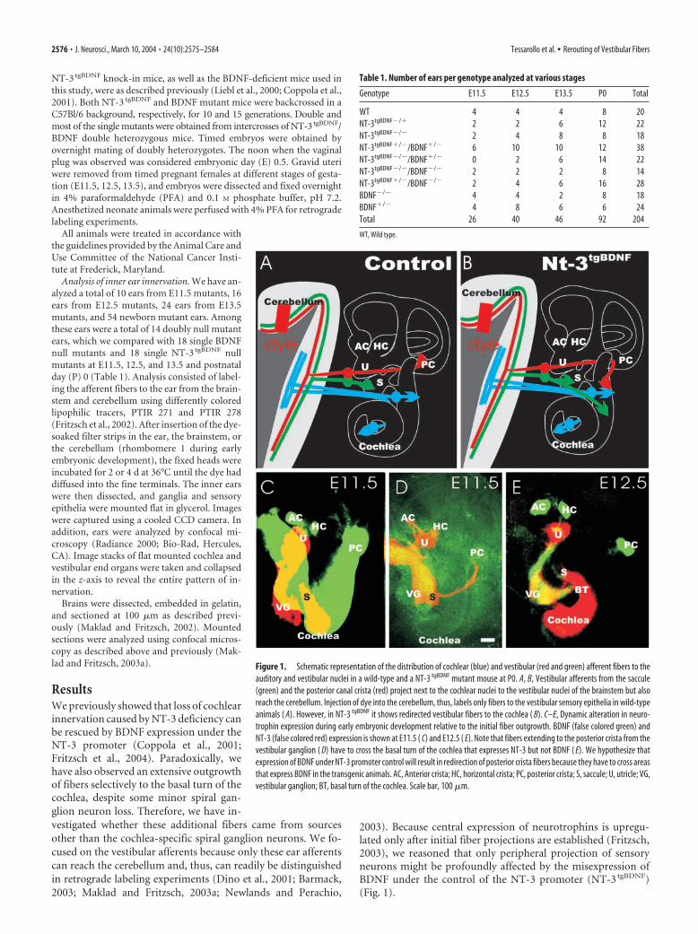
2003). Because central expression of neurotrophins is upregu-lated only after initial fiber projections are established (Fritzsch,2003), we reasoned that only peripheral projection of sensoryneurons might be profoundly affected by the misexpression ofBDNF under the control of the NT-3 promoter (NT-3 tgBDNF)(Fig. 1).
Table 1. Number of ears per genotype analyzed at various stages
Genotype E11.5 E12.5 E13.5 P0 Total
WT 4 4 4 8 20NT-3tgBDNF�/� 2 2 6 12 22NT-3tgBDNF�/� 2 4 8 8 18NT-3tgBDNF�/�/BDNF�/� 6 10 10 12 38NT-3tgBDNF�/�/BDNF�/� 0 2 6 14 22NT-3tgBDNF�/�/BDNF�/� 2 2 2 8 14NT-3tgBDNF�/�/BDNF�/� 2 4 6 16 28BDNF�/� 4 4 2 8 18BDNF�/� 4 8 6 6 24Total 26 40 46 92 204
WT, Wild type.
Figure 1. Schematic representation of the distribution of cochlear (blue) and vestibular (red and green) afferent fibers to theauditory and vestibular nuclei in a wild-type and a NT-3 tgBDNF mutant mouse at P0. A, B, Vestibular afferents from the saccule(green) and the posterior canal crista (red) project next to the cochlear nuclei to the vestibular nuclei of the brainstem but alsoreach the cerebellum. Injection of dye into the cerebellum, thus, labels only fibers to the vestibular sensory epithelia in wild-typeanimals ( A). However, in NT-3 tgBDNF it shows redirected vestibular fibers to the cochlea ( B). C–E, Dynamic alteration in neuro-trophin expression during early embryonic development relative to the initial fiber outgrowth. BDNF (false colored green) andNT-3 (false colored red) expression is shown at E11.5 ( C) and E12.5 ( E). Note that fibers extending to the posterior crista from thevestibular ganglion ( D) have to cross the basal turn of the cochlea that expresses NT-3 but not BDNF ( E). We hypothesize thatexpression of BDNF under NT-3 promoter control will result in redirection of posterior crista fibers because they have to cross areasthat express BDNF in the transgenic animals. AC, Anterior crista; HC, horizontal crista; PC, posterior crista; S, saccule; U, utricle; VG,vestibular ganglion; BT, basal turn of the cochlea. Scale bar, 100 �m.
2576 • J. Neurosci., March 10, 2004 • 24(10):2575–2584 Tessarollo et al. • Rerouting of Vestibular Fibers
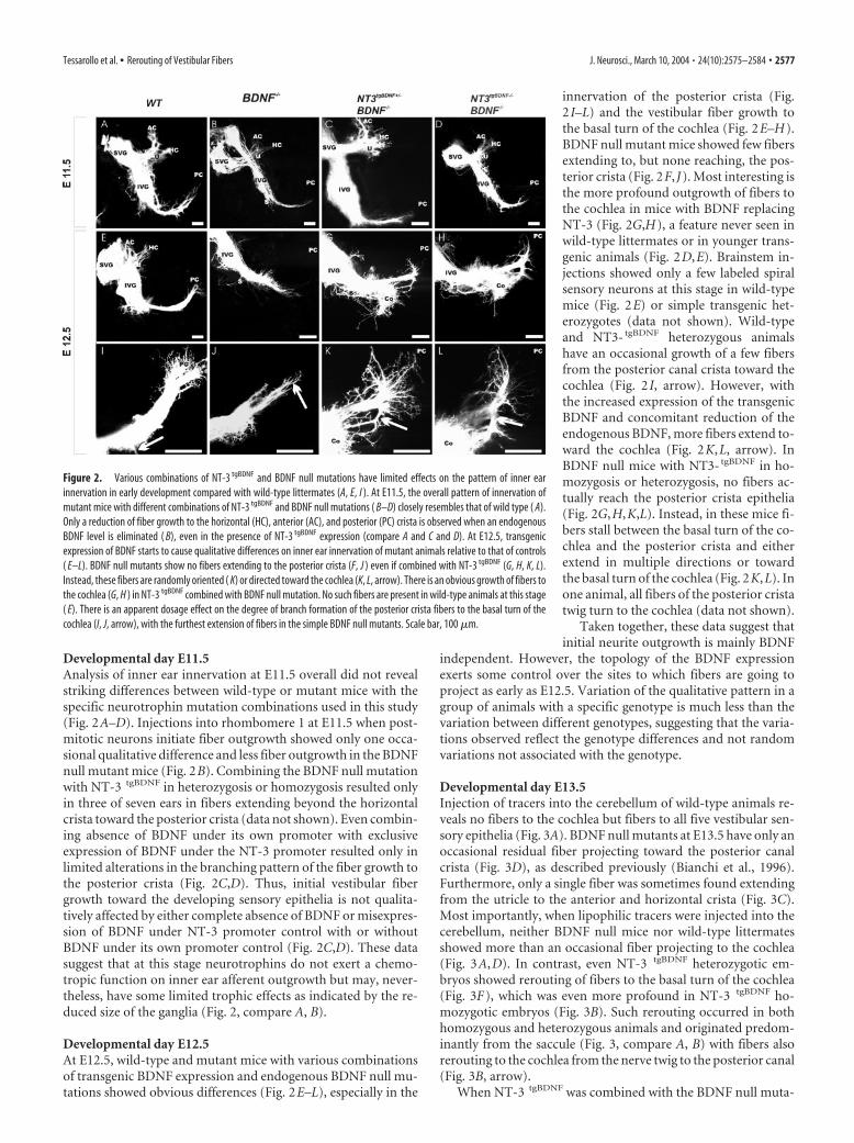
Developmental day E11.5Analysis of inner ear innervation at E11.5 overall did not revealstriking differences between wild-type or mutant mice with thespecific neurotrophin mutation combinations used in this study(Fig. 2A–D). Injections into rhombomere 1 at E11.5 when post-mitotic neurons initiate fiber outgrowth showed only one occa-sional qualitative difference and less fiber outgrowth in the BDNFnull mutant mice (Fig. 2B). Combining the BDNF null mutationwith NT-3 tgBDNF in heterozygosis or homozygosis resulted onlyin three of seven ears in fibers extending beyond the horizontalcrista toward the posterior crista (data not shown). Even combin-ing absence of BDNF under its own promoter with exclusiveexpression of BDNF under the NT-3 promoter resulted only inlimited alterations in the branching pattern of the fiber growth tothe posterior crista (Fig. 2C,D). Thus, initial vestibular fibergrowth toward the developing sensory epithelia is not qualita-tively affected by either complete absence of BDNF or misexpres-sion of BDNF under NT-3 promoter control with or withoutBDNF under its own promoter control (Fig. 2C,D). These datasuggest that at this stage neurotrophins do not exert a chemo-tropic function on inner ear afferent outgrowth but may, never-theless, have some limited trophic effects as indicated by the re-duced size of the ganglia (Fig. 2, compare A, B).
Developmental day E12.5At E12.5, wild-type and mutant mice with various combinationsof transgenic BDNF expression and endogenous BDNF null mu-tations showed obvious differences (Fig. 2E–L), especially in the
innervation of the posterior crista (Fig.2 I–L) and the vestibular fiber growth tothe basal turn of the cochlea (Fig. 2E–H).BDNF null mutant mice showed few fibersextending to, but none reaching, the pos-terior crista (Fig. 2F, J). Most interesting isthe more profound outgrowth of fibers tothe cochlea in mice with BDNF replacingNT-3 (Fig. 2G,H), a feature never seen inwild-type littermates or in younger trans-genic animals (Fig. 2D,E). Brainstem in-jections showed only a few labeled spiralsensory neurons at this stage in wild-typemice (Fig. 2E) or simple transgenic het-erozygotes (data not shown). Wild-typeand NT3- tgBDNF heterozygous animalshave an occasional growth of a few fibersfrom the posterior canal crista toward thecochlea (Fig. 2 I, arrow). However, withthe increased expression of the transgenicBDNF and concomitant reduction of theendogenous BDNF, more fibers extend to-ward the cochlea (Fig. 2K,L, arrow). InBDNF null mice with NT3- tgBDNF in ho-mozygosis or heterozygosis, no fibers ac-tually reach the posterior crista epithelia(Fig. 2G,H,K,L). Instead, in these mice fi-bers stall between the basal turn of the co-chlea and the posterior crista and eitherextend in multiple directions or towardthe basal turn of the cochlea (Fig. 2K,L). Inone animal, all fibers of the posterior cristatwig turn to the cochlea (data not shown).
Taken together, these data suggest thatinitial neurite outgrowth is mainly BDNF
independent. However, the topology of the BDNF expressionexerts some control over the sites to which fibers are going toproject as early as E12.5. Variation of the qualitative pattern in agroup of animals with a specific genotype is much less than thevariation between different genotypes, suggesting that the varia-tions observed reflect the genotype differences and not randomvariations not associated with the genotype.
Developmental day E13.5Injection of tracers into the cerebellum of wild-type animals re-veals no fibers to the cochlea but fibers to all five vestibular sen-sory epithelia (Fig. 3A). BDNF null mutants at E13.5 have only anoccasional residual fiber projecting toward the posterior canalcrista (Fig. 3D), as described previously (Bianchi et al., 1996).Furthermore, only a single fiber was sometimes found extendingfrom the utricle to the anterior and horizontal crista (Fig. 3C).Most importantly, when lipophilic tracers were injected into thecerebellum, neither BDNF null mice nor wild-type littermatesshowed more than an occasional fiber projecting to the cochlea(Fig. 3A,D). In contrast, even NT-3 tgBDNF heterozygotic em-bryos showed rerouting of fibers to the basal turn of the cochlea(Fig. 3F), which was even more profound in NT-3 tgBDNF ho-mozygotic embryos (Fig. 3B). Such rerouting occurred in bothhomozygous and heterozygous animals and originated predom-inantly from the saccule (Fig. 3, compare A, B) with fibers alsorerouting to the cochlea from the nerve twig to the posterior canal(Fig. 3B, arrow).
When NT-3 tgBDNF was combined with the BDNF null muta-
Figure 2. Various combinations of NT-3 tgBDNF and BDNF null mutations have limited effects on the pattern of inner earinnervation in early development compared with wild-type littermates (A, E, I ). At E11.5, the overall pattern of innervation ofmutant mice with different combinations of NT-3 tgBDNF and BDNF null mutations ( B–D) closely resembles that of wild type ( A).Only a reduction of fiber growth to the horizontal (HC), anterior (AC), and posterior (PC) crista is observed when an endogenousBDNF level is eliminated ( B), even in the presence of NT-3 tgBDNF expression (compare A and C and D). At E12.5, transgenicexpression of BDNF starts to cause qualitative differences on inner ear innervation of mutant animals relative to that of controls( E–L). BDNF null mutants show no fibers extending to the posterior crista (F, J ) even if combined with NT-3 tgBDNF (G, H, K, L).Instead, these fibers are randomly oriented ( K) or directed toward the cochlea (K, L, arrow). There is an obvious growth of fibers tothe cochlea (G, H ) in NT-3 tgBDNF combined with BDNF null mutation. No such fibers are present in wild-type animals at this stage( E). There is an apparent dosage effect on the degree of branch formation of the posterior crista fibers to the basal turn of thecochlea (I, J, arrow), with the furthest extension of fibers in the simple BDNF null mutants. Scale bar, 100 �m.
Tessarollo et al. • Rerouting of Vestibular Fibers J. Neurosci., March 10, 2004 • 24(10):2575–2584 • 2577
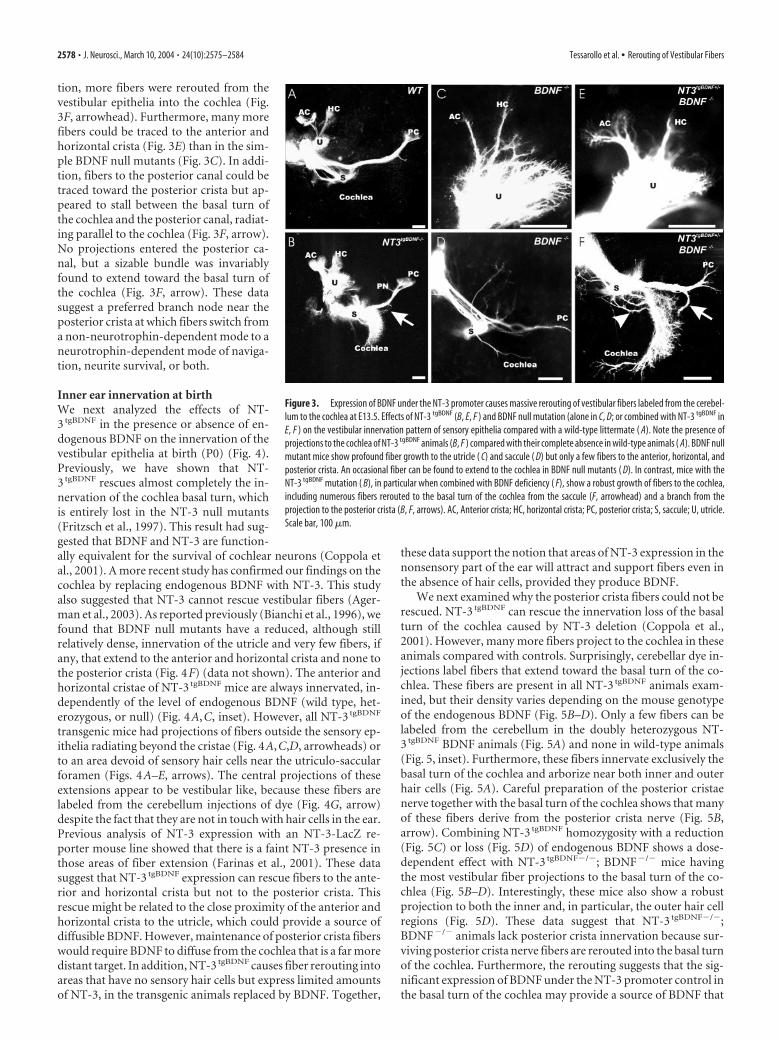
tion, more fibers were rerouted from thevestibular epithelia into the cochlea (Fig.3F, arrowhead). Furthermore, many morefibers could be traced to the anterior andhorizontal crista (Fig. 3E) than in the sim-ple BDNF null mutants (Fig. 3C). In addi-tion, fibers to the posterior canal could betraced toward the posterior crista but ap-peared to stall between the basal turn ofthe cochlea and the posterior canal, radiat-ing parallel to the cochlea (Fig. 3F, arrow).No projections entered the posterior ca-nal, but a sizable bundle was invariablyfound to extend toward the basal turn ofthe cochlea (Fig. 3F, arrow). These datasuggest a preferred branch node near theposterior crista at which fibers switch froma non-neurotrophin-dependent mode to aneurotrophin-dependent mode of naviga-tion, neurite survival, or both.
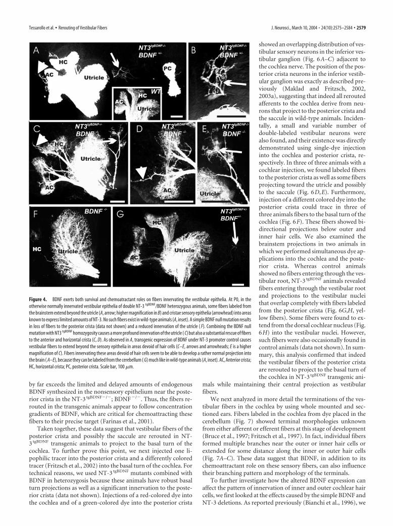
Inner ear innervation at birthWe next analyzed the effects of NT-3 tgBDNF in the presence or absence of en-dogenous BDNF on the innervation of thevestibular epithelia at birth (P0) (Fig. 4).Previously, we have shown that NT-3 tgBDNF rescues almost completely the in-nervation of the cochlea basal turn, whichis entirely lost in the NT-3 null mutants(Fritzsch et al., 1997). This result had sug-gested that BDNF and NT-3 are function-ally equivalent for the survival of cochlear neurons (Coppola etal., 2001). A more recent study has confirmed our findings on thecochlea by replacing endogenous BDNF with NT-3. This studyalso suggested that NT-3 cannot rescue vestibular fibers (Ager-man et al., 2003). As reported previously (Bianchi et al., 1996), wefound that BDNF null mutants have a reduced, although stillrelatively dense, innervation of the utricle and very few fibers, ifany, that extend to the anterior and horizontal crista and none tothe posterior crista (Fig. 4F) (data not shown). The anterior andhorizontal cristae of NT-3 tgBDNF mice are always innervated, in-dependently of the level of endogenous BDNF (wild type, het-erozygous, or null) (Fig. 4A,C, inset). However, all NT-3 tgBDNF
transgenic mice had projections of fibers outside the sensory ep-ithelia radiating beyond the cristae (Fig. 4A,C,D, arrowheads) orto an area devoid of sensory hair cells near the utriculo-saccularforamen (Figs. 4A–E, arrows). The central projections of theseextensions appear to be vestibular like, because these fibers arelabeled from the cerebellum injections of dye (Fig. 4G, arrow)despite the fact that they are not in touch with hair cells in the ear.Previous analysis of NT-3 expression with an NT-3-LacZ re-porter mouse line showed that there is a faint NT-3 presence inthose areas of fiber extension (Farinas et al., 2001). These datasuggest that NT-3 tgBDNF expression can rescue fibers to the ante-rior and horizontal crista but not to the posterior crista. Thisrescue might be related to the close proximity of the anterior andhorizontal crista to the utricle, which could provide a source ofdiffusible BDNF. However, maintenance of posterior crista fiberswould require BDNF to diffuse from the cochlea that is a far moredistant target. In addition, NT-3 tgBDNF causes fiber rerouting intoareas that have no sensory hair cells but express limited amountsof NT-3, in the transgenic animals replaced by BDNF. Together,
these data support the notion that areas of NT-3 expression in thenonsensory part of the ear will attract and support fibers even inthe absence of hair cells, provided they produce BDNF.
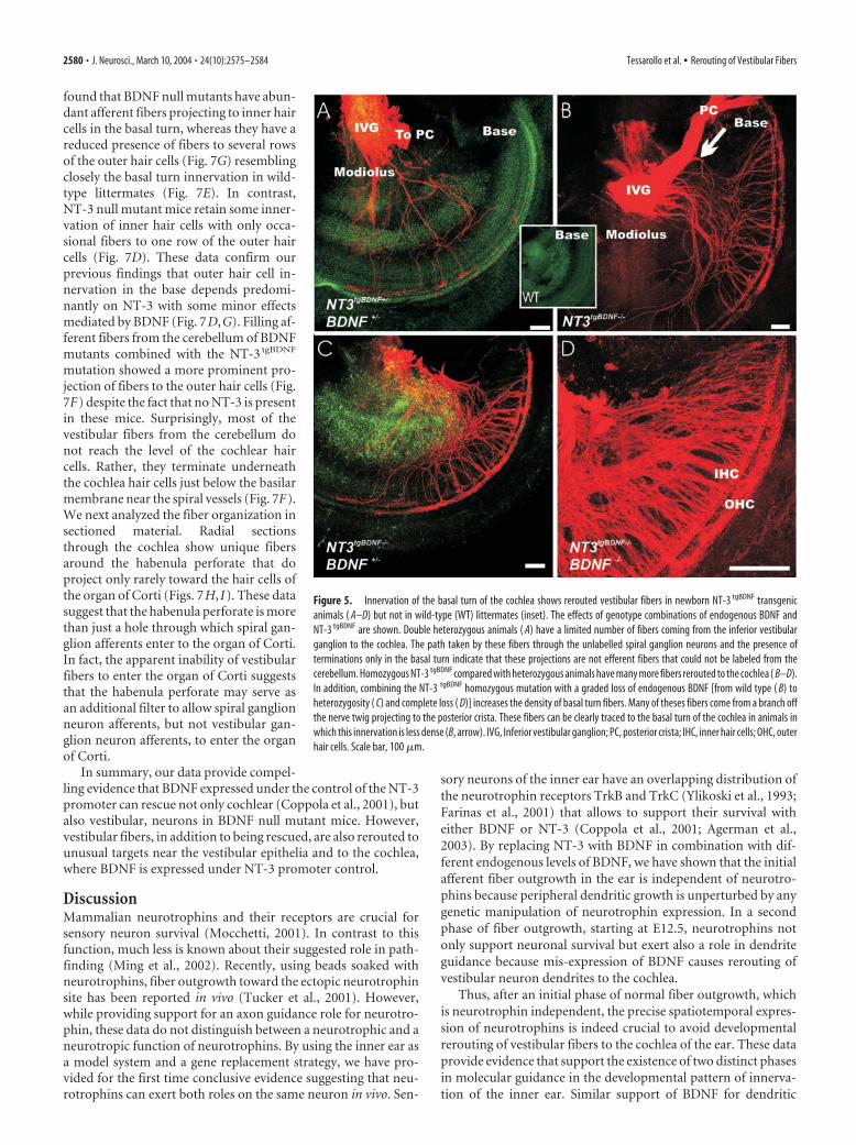
We next examined why the posterior crista fibers could not berescued. NT-3 tgBDNF can rescue the innervation loss of the basalturn of the cochlea caused by NT-3 deletion (Coppola et al.,2001). However, many more fibers project to the cochlea in theseanimals compared with controls. Surprisingly, cerebellar dye in-jections label fibers that extend toward the basal turn of the co-chlea. These fibers are present in all NT-3 tgBDNF animals exam-ined, but their density varies depending on the mouse genotypeof the endogenous BDNF (Fig. 5B–D). Only a few fibers can belabeled from the cerebellum in the doubly heterozygous NT-3 tgBDNF BDNF animals (Fig. 5A) and none in wild-type animals(Fig. 5, inset). Furthermore, these fibers innervate exclusively thebasal turn of the cochlea and arborize near both inner and outerhair cells (Fig. 5A). Careful preparation of the posterior cristaenerve together with the basal turn of the cochlea shows that manyof these fibers derive from the posterior crista nerve (Fig. 5B,arrow). Combining NT-3 tgBDNF homozygosity with a reduction(Fig. 5C) or loss (Fig. 5D) of endogenous BDNF shows a dose-dependent effect with NT-3 tgBDNF�/�; BDNF�/� mice havingthe most vestibular fiber projections to the basal turn of the co-chlea (Fig. 5B–D). Interestingly, these mice also show a robustprojection to both the inner and, in particular, the outer hair cellregions (Fig. 5D). These data suggest that NT-3 tgBDNF�/�;BDNF�/� animals lack posterior crista innervation because sur-viving posterior crista nerve fibers are rerouted into the basal turnof the cochlea. Furthermore, the rerouting suggests that the sig-nificant expression of BDNF under the NT-3 promoter control inthe basal turn of the cochlea may provide a source of BDNF that
Figure 3. Expression of BDNF under the NT-3 promoter causes massive rerouting of vestibular fibers labeled from the cerebel-lum to the cochlea at E13.5. Effects of NT-3 tgBDNF (B, E, F ) and BDNF null mutation (alone in C, D; or combined with NT-3 tgBDNF inE, F ) on the vestibular innervation pattern of sensory epithelia compared with a wild-type littermate ( A). Note the presence ofprojections to the cochlea of NT-3 tgBDNF animals (B, F ) compared with their complete absence in wild-type animals ( A). BDNF nullmutant mice show profound fiber growth to the utricle ( C) and saccule ( D) but only a few fibers to the anterior, horizontal, andposterior crista. An occasional fiber can be found to extend to the cochlea in BDNF null mutants ( D). In contrast, mice with theNT-3 tgBDNF mutation ( B), in particular when combined with BDNF deficiency ( F), show a robust growth of fibers to the cochlea,including numerous fibers rerouted to the basal turn of the cochlea from the saccule (F, arrowhead) and a branch from theprojection to the posterior crista (B, F, arrows). AC, Anterior crista; HC, horizontal crista; PC, posterior crista; S, saccule; U, utricle.Scale bar, 100 �m.
2578 • J. Neurosci., March 10, 2004 • 24(10):2575–2584 Tessarollo et al. • Rerouting of Vestibular Fibers
by far exceeds the limited and delayed amounts of endogenousBDNF synthesized in the nonsensory epithelium near the poste-rior crista in the NT-3 tgBDNF�/�; BDNF�/�. Thus, the fibers re-routed in the transgenic animals appear to follow concentrationgradients of BDNF, which are critical for chemoattracting thesefibers to their precise target (Farinas et al., 2001).
Taken together, these data suggest that vestibular fibers of theposterior crista and possibly the saccule are rerouted in NT-3 tgBDNF transgenic animals to project to the basal turn of thecochlea. To further prove this point, we next injected one li-pophilic tracer into the posterior crista and a differently coloredtracer (Fritzsch et al., 2002) into the basal turn of the cochlea. Fortechnical reasons, we used NT-3 tgBDNF mutants combined withBDNF in heterozygosis because these animals have robust basalturn projections as well as a significant innervation to the poste-rior crista (data not shown). Injections of a red-colored dye intothe cochlea and of a green-colored dye into the posterior crista
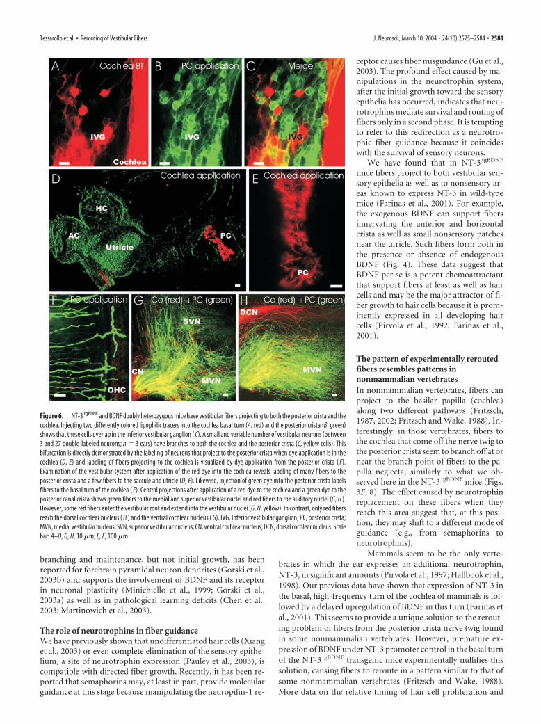
showed an overlapping distribution of ves-tibular sensory neurons in the inferior ves-tibular ganglion (Fig. 6A–C) adjacent tothe cochlea nerve. The position of the pos-terior crista neurons in the inferior vestib-ular ganglion was exactly as described pre-viously (Maklad and Fritzsch, 2002,2003a), suggesting that indeed all reroutedafferents to the cochlea derive from neu-rons that project to the posterior crista andthe saccule in wild-type animals. Inciden-tally, a small and variable number ofdouble-labeled vestibular neurons werealso found, and their existence was directlydemonstrated using single-dye injectioninto the cochlea and posterior crista, re-spectively. In three of three animals with acochlear injection, we found labeled fibersto the posterior crista as well as some fibersprojecting toward the utricle and possiblyto the saccule (Fig. 6D,E). Furthermore,injection of a different colored dye into theposterior crista could trace in three ofthree animals fibers to the basal turn of thecochlea (Fig. 6F). These fibers showed bi-directional projections below outer andinner hair cells. We also examined thebrainstem projections in two animals inwhich we performed simultaneous dye ap-plications into the cochlea and the poste-rior crista. Whereas control animalsshowed no fibers entering through the ves-tibular root, NT-3 tgBDNF animals revealedfibers entering through the vestibular rootand projections to the vestibular nucleithat overlap completely with fibers labeledfrom the posterior crista (Fig. 6G,H, yel-low fibers). Some fibers were found to ex-tend from the dorsal cochlear nucleus (Fig.6H) into the vestibular nuclei. However,such fibers were also occasionally found incontrol animals (data not shown). In sum-mary, this analysis confirmed that indeedthe vestibular fibers of the posterior cristaare rerouted to project to the basal turn ofthe cochlea in NT-3 tgBDNF transgenic ani-
mals while maintaining their central projection as vestibularfibers.
We next analyzed in more detail the terminations of the ves-tibular fibers in the cochlea by using whole mounted and sec-tioned ears. Fibers labeled in the cochlea from dye placed in thecerebellum (Fig. 7) showed terminal morphologies unknownfrom either afferent or efferent fibers at this stage of development(Bruce et al., 1997; Fritzsch et al., 1997). In fact, individual fibersformed multiple branches near the outer or inner hair cells orextended for some distance along the inner or outer hair cells(Fig. 7A–C). These data suggest that BDNF, in addition to itschemoattractant role on these sensory fibers, can also influencetheir branching pattern and morphology of the terminals.
To further investigate how the altered BDNF expression canaffect the pattern of innervation of inner and outer cochlear haircells, we first looked at the effects caused by the simple BDNF andNT-3 deletions. As reported previously (Bianchi et al., 1996), we
Figure 4. BDNF exerts both survival and chemoatractant roles on fibers innervating the vestibular epithelia. At P0, in theotherwise normally innervated vestibular epithelia of double NT-3 tgBDNF/BDNF heterozygous animals, some fibers labeled fromthe brainstem extend beyond the utricle (A, arrow; higher magnification in B) and cristae sensory epithelia (arrowhead) into areasknown to express limited amounts of NT-3. No such fibers exist in wild-type animals (A, inset). A simple BDNF null mutation resultsin loss of fibers to the posterior crista (data not shown) and a reduced innervation of the utricle ( F). Combining the BDNF nullmutation with NT3 tgBDNF homozygosity causes a more profound innervation of the utricle ( C) but also a substantial rescue of fibersto the anterior and horizontal crista (C, D). As observed in A, transgenic expression of BDNF under NT-3 promoter control causesvestibular fibers to extend beyond the sensory epithelia in areas devoid of hair cells (C–E, arrows and arrowheads; E is a highermagnification of C). Fibers innervating these areas devoid of hair cells seem to be able to develop a rather normal projection intothe brain ( A–E), because they can be labeled from the cerebellum ( G) much like in wild-type animals (A, inset). AC, Anterior crista;HC, horizontal crista; PC, posterior crista. Scale bar, 100 �m.
Tessarollo et al. • Rerouting of Vestibular Fibers J. Neurosci., March 10, 2004 • 24(10):2575–2584 • 2579
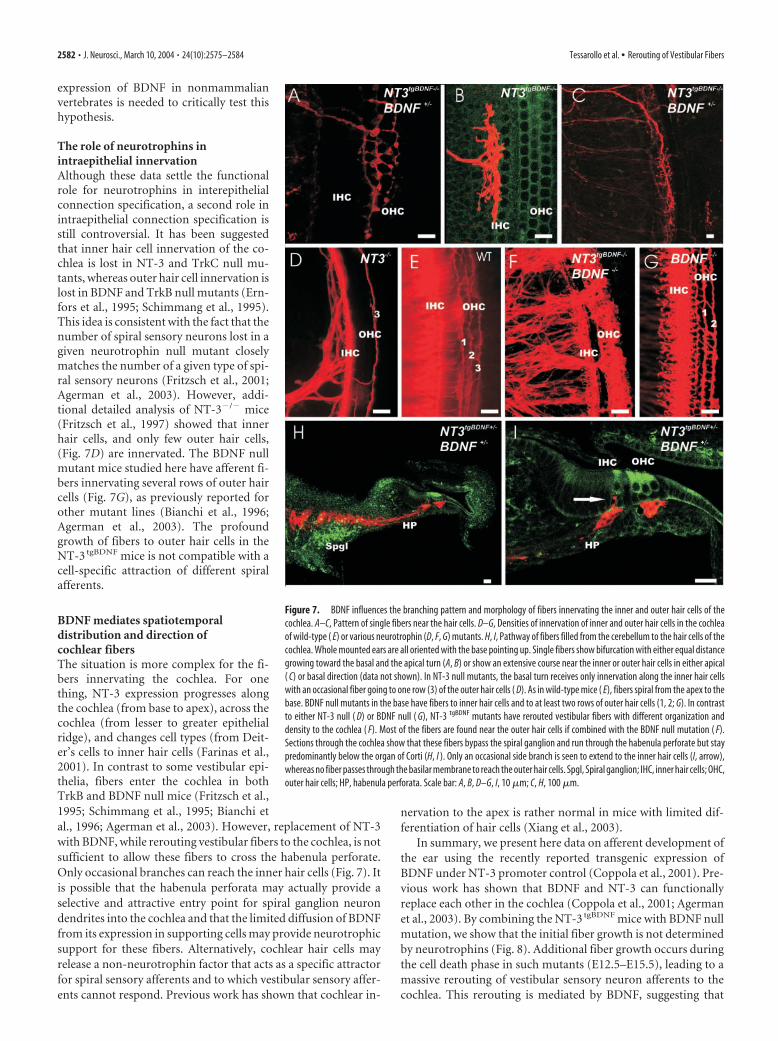
found that BDNF null mutants have abun-dant afferent fibers projecting to inner haircells in the basal turn, whereas they have areduced presence of fibers to several rowsof the outer hair cells (Fig. 7G) resemblingclosely the basal turn innervation in wild-type littermates (Fig. 7E). In contrast,NT-3 null mutant mice retain some inner-vation of inner hair cells with only occa-sional fibers to one row of the outer haircells (Fig. 7D). These data confirm ourprevious findings that outer hair cell in-nervation in the base depends predomi-nantly on NT-3 with some minor effectsmediated by BDNF (Fig. 7D,G). Filling af-ferent fibers from the cerebellum of BDNFmutants combined with the NT-3 tgBDNF
mutation showed a more prominent pro-jection of fibers to the outer hair cells (Fig.7F) despite the fact that no NT-3 is presentin these mice. Surprisingly, most of thevestibular fibers from the cerebellum donot reach the level of the cochlear haircells. Rather, they terminate underneaththe cochlea hair cells just below the basilarmembrane near the spiral vessels (Fig. 7F).We next analyzed the fiber organization insectioned material. Radial sectionsthrough the cochlea show unique fibersaround the habenula perforate that doproject only rarely toward the hair cells ofthe organ of Corti (Figs. 7H, I). These datasuggest that the habenula perforate is morethan just a hole through which spiral gan-glion afferents enter to the organ of Corti.In fact, the apparent inability of vestibularfibers to enter the organ of Corti suggeststhat the habenula perforate may serve asan additional filter to allow spiral ganglionneuron afferents, but not vestibular gan-glion neuron afferents, to enter the organof Corti.
In summary, our data provide compel-ling evidence that BDNF expressed under the control of the NT-3promoter can rescue not only cochlear (Coppola et al., 2001), butalso vestibular, neurons in BDNF null mutant mice. However,vestibular fibers, in addition to being rescued, are also rerouted tounusual targets near the vestibular epithelia and to the cochlea,where BDNF is expressed under NT-3 promoter control.
DiscussionMammalian neurotrophins and their receptors are crucial forsensory neuron survival (Mocchetti, 2001). In contrast to thisfunction, much less is known about their suggested role in path-finding (Ming et al., 2002). Recently, using beads soaked withneurotrophins, fiber outgrowth toward the ectopic neurotrophinsite has been reported in vivo (Tucker et al., 2001). However,while providing support for an axon guidance role for neurotro-phin, these data do not distinguish between a neurotrophic and aneurotropic function of neurotrophins. By using the inner ear asa model system and a gene replacement strategy, we have pro-vided for the first time conclusive evidence suggesting that neu-rotrophins can exert both roles on the same neuron in vivo. Sen-
sory neurons of the inner ear have an overlapping distribution ofthe neurotrophin receptors TrkB and TrkC (Ylikoski et al., 1993;Farinas et al., 2001) that allows to support their survival witheither BDNF or NT-3 (Coppola et al., 2001; Agerman et al.,2003). By replacing NT-3 with BDNF in combination with dif-ferent endogenous levels of BDNF, we have shown that the initialafferent fiber outgrowth in the ear is independent of neurotro-phins because peripheral dendritic growth is unperturbed by anygenetic manipulation of neurotrophin expression. In a secondphase of fiber outgrowth, starting at E12.5, neurotrophins notonly support neuronal survival but exert also a role in dendriteguidance because mis-expression of BDNF causes rerouting ofvestibular neuron dendrites to the cochlea.
Thus, after an initial phase of normal fiber outgrowth, whichis neurotrophin independent, the precise spatiotemporal expres-sion of neurotrophins is indeed crucial to avoid developmentalrerouting of vestibular fibers to the cochlea of the ear. These dataprovide evidence that support the existence of two distinct phasesin molecular guidance in the developmental pattern of innerva-tion of the inner ear. Similar support of BDNF for dendritic
Figure 5. Innervation of the basal turn of the cochlea shows rerouted vestibular fibers in newborn NT-3 tgBDNF transgenicanimals ( A–D) but not in wild-type (WT) littermates (inset). The effects of genotype combinations of endogenous BDNF andNT-3 tgBDNF are shown. Double heterozygous animals ( A) have a limited number of fibers coming from the inferior vestibularganglion to the cochlea. The path taken by these fibers through the unlabelled spiral ganglion neurons and the presence ofterminations only in the basal turn indicate that these projections are not efferent fibers that could not be labeled from thecerebellum. Homozygous NT-3 tgBDNF compared with heterozygous animals have many more fibers rerouted to the cochlea ( B–D).In addition, combining the NT-3 tgBDNF homozygous mutation with a graded loss of endogenous BDNF [from wild type ( B) toheterozygosity ( C) and complete loss ( D)] increases the density of basal turn fibers. Many of theses fibers come from a branch offthe nerve twig projecting to the posterior crista. These fibers can be clearly traced to the basal turn of the cochlea in animals inwhich this innervation is less dense (B, arrow). IVG, Inferior vestibular ganglion; PC, posterior crista; IHC, inner hair cells; OHC, outerhair cells. Scale bar, 100 �m.
2580 • J. Neurosci., March 10, 2004 • 24(10):2575–2584 Tessarollo et al. • Rerouting of Vestibular Fibers
branching and maintenance, but not initial growth, has beenreported for forebrain pyramidal neuron dendrites (Gorski et al.,2003b) and supports the involvement of BDNF and its receptorin neuronal plasticity (Minichiello et al., 1999; Gorski et al.,2003a) as well as in pathological learning deficits (Chen et al.,2003; Martinowich et al., 2003).
The role of neurotrophins in fiber guidanceWe have previously shown that undifferentiated hair cells (Xianget al., 2003) or even complete elimination of the sensory epithe-lium, a site of neurotrophin expression (Pauley et al., 2003), iscompatible with directed fiber growth. Recently, it has been re-ported that semaphorins may, at least in part, provide molecularguidance at this stage because manipulating the neuropilin-1 re-
ceptor causes fiber misguidance (Gu et al.,2003). The profound effect caused by ma-nipulations in the neurotrophin system,after the initial growth toward the sensoryepithelia has occurred, indicates that neu-rotrophins mediate survival and routing offibers only in a second phase. It is temptingto refer to this redirection as a neurotro-phic fiber guidance because it coincideswith the survival of sensory neurons.
We have found that in NT-3 tgBDNF
mice fibers project to both vestibular sen-sory epithelia as well as to nonsensory ar-eas known to express NT-3 in wild-typemice (Farinas et al., 2001). For example,the exogenous BDNF can support fibersinnervating the anterior and horizontalcrista as well as small nonsensory patchesnear the utricle. Such fibers form both inthe presence or absence of endogenousBDNF (Fig. 4). These data suggest thatBDNF per se is a potent chemoattractantthat support fibers at least as well as haircells and may be the major attractor of fi-ber growth to hair cells because it is prom-inently expressed in all developing haircells (Pirvola et al., 1992; Farinas et al.,2001).
The pattern of experimentally reroutedfibers resembles patterns innonmammalian vertebratesIn nonmammalian vertebrates, fibers canproject to the basilar papilla (cochlea)along two different pathways (Fritzsch,1987, 2002; Fritzsch and Wake, 1988). In-terestingly, in those vertebrates, fibers tothe cochlea that come off the nerve twig tothe posterior crista seem to branch off at ornear the branch point of fibers to the pa-pilla neglecta, similarly to what we ob-served here in the NT-3 tgBDNF mice (Figs.3F, 8). The effect caused by neurotrophinreplacement on these fibers when theyreach this area suggest that, at this posi-tion, they may shift to a different mode ofguidance (e.g., from semaphorins toneurotrophins).
Mammals seem to be the only verte-brates in which the ear expresses an additional neurotrophin,NT-3, in significant amounts (Pirvola et al., 1997; Hallbook et al.,1998). Our previous data have shown that expression of NT-3 inthe basal, high-frequency turn of the cochlea of mammals is fol-lowed by a delayed upregulation of BDNF in this turn (Farinas etal., 2001). This seems to provide a unique solution to the rerout-ing problem of fibers from the posterior crista nerve twig foundin some nonmammalian vertebrates. However, premature ex-pression of BDNF under NT-3 promoter control in the basal turnof the NT-3 tgBDNF transgenic mice experimentally nullifies thissolution, causing fibers to reroute in a pattern similar to that ofsome nonmammalian vertebrates (Fritzsch and Wake, 1988).More data on the relative timing of hair cell proliferation and
Figure 6. NT-3 tgBDNF and BDNF doubly heterozygous mice have vestibular fibers projecting to both the posterior crista and thecochlea. Injecting two differently colored lipophilic tracers into the cochlea basal turn (A, red) and the posterior crista (B, green)shows that these cells overlap in the inferior vestibular ganglion ( C). A small and variable number of vestibular neurons (between3 and 27 double-labeled neurons; n � 3 ears) have branches to both the cochlea and the posterior crista (C, yellow cells). Thisbifurcation is directly demonstrated by the labeling of neurons that project to the posterior crista when dye application is in thecochlea (D, E) and labeling of fibers projecting to the cochlea is visualized by dye application from the posterior crista ( F).Examination of the vestibular system after application of the red dye into the cochlea reveals labeling of many fibers to theposterior crista and a few fibers to the saccule and utricle (D, E). Likewise, injection of green dye into the posterior crista labelsfibers to the basal turn of the cochlea ( F). Central projections after application of a red dye to the cochlea and a green dye to theposterior canal crista shows green fibers to the medial and superior vestibular nuclei and red fibers to the auditory nuclei (G, H ).However, some red fibers enter the vestibular root and extend into the vestibular nuclei (G, H, yellow). In contrast, only red fibersreach the dorsal cochlear nucleus ( H ) and the ventral cochlear nucleus ( G). IVG, Inferior vestibular ganglion; PC, posterior crista;MVN, medial vestibular nucleus; SVN, superior vestibular nucleus; CN, ventral cochlear nucleus; DCN, dorsal cochlear nucleus. Scalebar: A–D, G, H, 10 �m; E, F, 100 �m.
Tessarollo et al. • Rerouting of Vestibular Fibers J. Neurosci., March 10, 2004 • 24(10):2575–2584 • 2581
expression of BDNF in nonmammalianvertebrates is needed to critically test thishypothesis.
The role of neurotrophins inintraepithelial innervationAlthough these data settle the functionalrole for neurotrophins in interepithelialconnection specification, a second role inintraepithelial connection specification isstill controversial. It has been suggestedthat inner hair cell innervation of the co-chlea is lost in NT-3 and TrkC null mu-tants, whereas outer hair cell innervation islost in BDNF and TrkB null mutants (Ern-fors et al., 1995; Schimmang et al., 1995).This idea is consistent with the fact that thenumber of spiral sensory neurons lost in agiven neurotrophin null mutant closelymatches the number of a given type of spi-ral sensory neurons (Fritzsch et al., 2001;Agerman et al., 2003). However, addi-tional detailed analysis of NT-3�/� mice(Fritzsch et al., 1997) showed that innerhair cells, and only few outer hair cells,(Fig. 7D) are innervated. The BDNF nullmutant mice studied here have afferent fi-bers innervating several rows of outer haircells (Fig. 7G), as previously reported forother mutant lines (Bianchi et al., 1996;Agerman et al., 2003). The profoundgrowth of fibers to outer hair cells in theNT-3 tgBDNF mice is not compatible with acell-specific attraction of different spiralafferents.
BDNF mediates spatiotemporaldistribution and direction ofcochlear fibersThe situation is more complex for the fi-bers innervating the cochlea. For onething, NT-3 expression progresses alongthe cochlea (from base to apex), across thecochlea (from lesser to greater epithelialridge), and changes cell types (from Deit-er’s cells to inner hair cells (Farinas et al.,2001). In contrast to some vestibular epi-thelia, fibers enter the cochlea in bothTrkB and BDNF null mice (Fritzsch et al.,1995; Schimmang et al., 1995; Bianchi etal., 1996; Agerman et al., 2003). However, replacement of NT-3with BDNF, while rerouting vestibular fibers to the cochlea, is notsufficient to allow these fibers to cross the habenula perforate.Only occasional branches can reach the inner hair cells (Fig. 7). Itis possible that the habenula perforata may actually provide aselective and attractive entry point for spiral ganglion neurondendrites into the cochlea and that the limited diffusion of BDNFfrom its expression in supporting cells may provide neurotrophicsupport for these fibers. Alternatively, cochlear hair cells mayrelease a non-neurotrophin factor that acts as a specific attractorfor spiral sensory afferents and to which vestibular sensory affer-ents cannot respond. Previous work has shown that cochlear in-
nervation to the apex is rather normal in mice with limited dif-ferentiation of hair cells (Xiang et al., 2003).
In summary, we present here data on afferent development ofthe ear using the recently reported transgenic expression ofBDNF under NT-3 promoter control (Coppola et al., 2001). Pre-vious work has shown that BDNF and NT-3 can functionallyreplace each other in the cochlea (Coppola et al., 2001; Agermanet al., 2003). By combining the NT-3 tgBDNF mice with BDNF nullmutation, we show that the initial fiber growth is not determinedby neurotrophins (Fig. 8). Additional fiber growth occurs duringthe cell death phase in such mutants (E12.5–E15.5), leading to amassive rerouting of vestibular sensory neuron afferents to thecochlea. This rerouting is mediated by BDNF, suggesting that
Figure 7. BDNF influences the branching pattern and morphology of fibers innervating the inner and outer hair cells of thecochlea. A–C, Pattern of single fibers near the hair cells. D–G, Densities of innervation of inner and outer hair cells in the cochleaof wild-type ( E) or various neurotrophin (D, F, G) mutants. H, I, Pathway of fibers filled from the cerebellum to the hair cells of thecochlea. Whole mounted ears are all oriented with the base pointing up. Single fibers show bifurcation with either equal distancegrowing toward the basal and the apical turn (A, B) or show an extensive course near the inner or outer hair cells in either apical( C) or basal direction (data not shown). In NT-3 null mutants, the basal turn receives only innervation along the inner hair cellswith an occasional fiber going to one row (3) of the outer hair cells ( D). As in wild-type mice ( E), fibers spiral from the apex to thebase. BDNF null mutants in the base have fibers to inner hair cells and to at least two rows of outer hair cells (1, 2; G). In contrastto either NT-3 null ( D) or BDNF null ( G), NT-3 tgBDNF mutants have rerouted vestibular fibers with different organization anddensity to the cochlea ( F). Most of the fibers are found near the outer hair cells if combined with the BDNF null mutation ( F).Sections through the cochlea show that these fibers bypass the spiral ganglion and run through the habenula perforate but staypredominantly below the organ of Corti (H, I ). Only an occasional side branch is seen to extend to the inner hair cells (I, arrow),whereas no fiber passes through the basilar membrane to reach the outer hair cells. Spgl, Spiral ganglion; IHC, inner hair cells; OHC,outer hair cells; HP, habenula perforata. Scale bar: A, B, D–G, I, 10 �m; C, H, 100 �m.
2582 • J. Neurosci., March 10, 2004 • 24(10):2575–2584 Tessarollo et al. • Rerouting of Vestibular Fibers
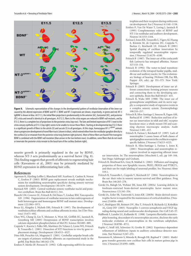
neurite growth is primarily regulated in the ear by BDNF,whereas NT-3 acts predominantly as a survival factor (Fig. 8).This finding suggests that growth of afferents to regenerating haircells (Kawamoto et al., 2003) may be primarily mediated byBDNF expression in differentiating hair cells.
ReferencesAgerman K, Hjerling-Leffler J, Blanchard MP, Scarfone E, Canlon B, Nosrat
C, Ernfors P (2003) BDNF gene replacement reveals multiple mecha-nisms for establishing neurotrophin specificity during sensory nervoussystem development. Development 130:1479 –1491.
Barmack NH (2003) Central vestibular system: vestibular nuclei and poste-rior cerebellum. Brain Res Bull 60:511–541.
Bianchi LM, Conover JC, Fritzsch B, DeChiara T, Lindsay RM, YancopoulosGD (1996) Degeneration of vestibular neurons in late embryogenesis ofboth heterozygous and homozygous BDNF null mutant mice. Develop-ment 122:1965–1973.
Bruce LL, Kingsley J, Nichols DH, Fritzsch B (1997) The development ofvestibulocochlear efferents and cochlear afferents in mice. Int J Dev Neu-rosci 15:671– 692.
Chen WG, Chang Q, Lin Y, Meissner A, West AE, Griffith EC, Jaenisch R,Greenberg ME (2003) Derepression of BDNF transcription involvescalcium-dependent phosphorylation of MeCP2. Science 302:885– 889.
Coppola V, Kucera J, Palko ME, Martinez-De Velasco J, Lyons WE, FritzschB, Tessarollo L (2001) Dissection of NT3 functions in vivo by gene re-placement strategy. Development 128:4315– 4327.
Dino MR, Perachio AA, Mugnaini E (2001) Cerebellar unipolar brush cellsare targets of primary vestibular afferents: an experimental study in thegerbil. Exp Brain Res 140:162–170.
Ernfors P, Merlio JP, Persson H (1992) Cells expressing mRNA for neuro-
trophins and their receptors during embryonicrat development. Eur J Neurosci 4:1140 –1158.
Ernfors P, Van De Water T, Loring J, Jaenisch R(1995) Complementary roles of BDNF andNT-3 in vestibular and auditory development.Neuron 14:1153–1164.
Farinas I, Jones KR, Tessarollo L, Vigers AJ, HuangE, Kirstein M, de Caprona DC, Coppola V,Backus C, Reichardt LF, Fritzsch B (2001)Spatial shaping of cochlear innervation bytemporally regulated neurotrophin expres-sion. J Neurosci 21:6170 – 6180.
Fritzsch B (1987) The inner ear of the coelacanthfish Latimeria has tetrapod affinities. Nature327:153–154.
Fritzsch B (1992) The water-to-land transition:evolution of the tetrapod basilar papilla, mid-dle ear and auditory nuclei. In: The evolution-ary biology of hearing (Webster DB, Fay RR,Popper AN, eds), pp 351–375. New York:Springer.
Fritzsch B (2003) Development of inner ear af-ferent connections: forming primary neuronsand connecting them to the developing sen-sory epithelia. Brain Res Bull 60:423– 433.
Fritzsch B, Wake MH (1988) The inner ear ofgymnophione amphibians and its nerve sup-ply: a comparative study of regressive events ina complex sensory system. Zoomorphology108:210 –217.
Fritzsch B, Silos-Santiago I, Smeyne R, Fagan AM,Barbacid M (1995) Reduction and loss of in-ner ear innervation in trkB and trkC receptorknockout mice: a whole mount DiI and scan-ning electron microscopic analysis. AuditNeurosci 1:401– 417.
Fritzsch B, Farinas I, Reichardt LF (1997) Lack ofneurotrophin 3 causes losses of both classes ofspiralganglionneuronsinthecochlea inaregion-specific fashion. J Neurosci 17:6213–6225.
Fritzsch B, Silos-Santiago I, Farinas I, Jones K(2001) Neurotrophins and neurotrophin re-ceptors involved in supporting afferent inner
ear innervation. In: The neurotrophins (Mocchetti I, ed), pp 149 –164.San Diego: Salzburger and Graham.
Fritzsch B, Muirhead KA, Gray B, Maklad A (2002) Diffusion and imagingproperties of three new lipophilic tracers, PKH2, PKH26 and PTIR271,and their use for triple labeling of neuronal profiles. Soc Neurosci Abstr519.1.
Fritzsch B, Tessarollo L, Coppola V, Reichardt LF (2004) Neurotrophins inthe ear: their roles in sensory neuron survival and fiber guidance. ProgBrain Res 146:265–278.
Gorski JA, Balogh SA, Wehner JM, Jones KR (2003a) Learning deficits inforebrain-restricted brain-derived neurotrophic factor mutant mice.Neuroscience 121:341–354.
Gorski JA, Zeiler SR, Tamowski S, Jones KR (2003b) Brain-derived neuro-trophic factor is required for the maintenance of cortical dendrites. J Neu-rosci 23:6856 – 6865.
Gu C, Rodriguez ER, Reimert DV, Shu T, Fritzsch B, Richards LJ, KolodkinAL, Ginty DD (2003) Neuropilin-1 conveys semaphorin and VEGF sig-naling during neural and cardiovascular development. Dev Cell 5:45–57.
Hallbook F, Lundin LG, Kullander K (1998) Lampetra fluviatilis neurotro-phin homolog, descendant of a neurotrophin ancestor, discloses the earlymolecular evolution of neurotrophins in the vertebrate subphylum.J Neurosci 18:8700 – 8711.
Kapfer C, Seidl AH, Schweizer H, Grothe B (2002) Experience-dependentrefinement of inhibitory inputs to auditory coincidence-detector neu-rons. Nat Neurosci 5:247–253.
Kawamoto K, Ishimoto S, Minoda R, Brough DE, Raphael Y (2003) Math1gene transfer generates new cochlear hair cells in mature guinea pigs invivo. J Neurosci 23:4395– 4400.
Figure 8. Schematic representation of the changes in the developmental pattern of vestibular innervation of the inner earcaused by the altered expression of BDNF and NT-3. BDNF and NT-3 expression are shown, respectively, in green and red; NT-3tgBDNF is shown in blue. At E11.5, the initial fiber projection is predominantly to the anterior (AC), horizontal (HC), and posterior(PC) crista and overall is identical in all genotypes. At E12.5, fibers to the crista organs are reduced in BDNF null mutants, and byE13.5, there is a complete loss of projections to the posterior crista (top row). The late and limited expression of NT-3 next to thecrista sensory epithelia at E13.5 (top right) seems to be unable to rescue those fibers. Starting at developmental day E12.5 thereis a significant growth of fibers to the utricle (U) and saccule (S) in all genotypes. At this stage, homozygous NT-3 tgBDNF animalsshow a progressive development of novel fiber tracts (shown in blue), which extend either from the vestibular ganglion directly tothe cochlea (Co) or emanate from the posterior crista twig (bottom right pictures). More of these fibers are found if the transgenicBDNF is combined with the BDNF null mutation (blue arrows in the two bottom rows). In addition, some fibers that do not reachor innervate the posterior crista reroute to the basal turn of the cochlea (bottom right).
Tessarollo et al. • Rerouting of Vestibular Fibers J. Neurosci., March 10, 2004 • 24(10):2575–2584 • 2583
Lewis ER, Leverenz EL, Bialek WS (1985) The vertebrate inner ear. BocaRaton, FL: CRC.
Liebl DJ, Klesse LJ, Tessarollo L, Wohlman T, Parada LF (2000) Loss ofbrain-derived neurotrophic factor-dependent neural crest-derived sen-sory neurons in neurotrophin-4 mutant mice. Proc Natl Acad Sci USA97:2297–2302.
Maklad A, Fritzsch B (2002) The developmental segregation of posteriorcrista and saccular vestibular fibers in mice: a carbocyanine tracer studyusing confocal microscopy. Dev Brain Res 135.
Maklad A, Fritzsch B (2003a) Partial segregation of posterior crista and sac-cular fibers to the nodulus and uvula of the cerebellum in mice, and itsdevelopment. Brain Res Dev Brain Res 140:223–236.
Maklad A, Fritzsch B (2003b) Development of vestibular afferent projectionsinto the hindbrain and their central targets. Brain Res Bull 60:497–510.
Martinowich K, Hattori D, Wu H, Fouse S, He F, Hu Y, Fan G, Sun YE (2003)DNA methylation-related chromatin remodeling in activity-dependentBDNF gene regulation. Science 302:890 – 893.
Ming GL, Wong ST, Henley J, Yuan XB, Song HJ, Spitzer NC, Poo MM(2002) Adaptation in the chemotactic guidance of nerve growth cones.Nature 417:411– 418.
Minichiello L, Korte M, Wolfer D, Kuhn R, Unsicker K, Cestari V, Rossi-Arnaud C, Lipp HP, Bonhoeffer T, Klein R (1999) Essential role forTrkB receptors in hippocampus-mediated learning. Neuron 24:401– 414.
Mocchetti I (2001) The neurotrophins. San Diego: Salzburger and Graham.Newlands SD, Perachio AA (2003) Central projections of the vestibular
nerve: a review and single fiber study in the Mongolian gerbil. Brain ResBull 60:475– 495.
Pauley S, Wright TJ, Pirvola U, Ornitz D, Beisel K, Fritzsch B (2003) Expres-sion and function of FGF10 in mammalian inner ear development. DevDyn 227:203–215.
Pirvola U, Ylikoski J, Palgi J, Lehtonen E, Arumae U, Saarma M (1992)Brain-derived neurotrophic factor and neurotrophin 3 mRNAs in theperipheral target fields of developing inner ear ganglia. Proc Natl Acad SciUSA 89:9915–9919.
Pirvola U, Hallbook F, Xing-Qun L, Virkkala J, Saarma M, Ylikoski J (1997)Expression of neurotrophins and Trk receptors in the developing, adult,and regenerating avian cochlea. J Neurobiol 33:1019 –1033.
Rubel EW, Fritzsch B (2002) Auditory system development: primary audi-tory neurons and their targets. Annu Rev Neurosci 25:51–101.
Ryugo DK, Parks TN (2003) Primary innervation of the avian and mamma-lian cochlear nucleus. Brain Res Bull 60:435– 456.
Schimmang T, Minichiello L, Vazquez E, San Jose I, Giraldez F, Klein R,Represa J (1995) Developing inner ear sensory neurons require TrkBand TrkC receptors for innervation of their peripheral targets. Develop-ment 121:3381–3389.
Tucker KL, Meyer M, Barde YA (2001) Neurotrophins are required fornerve growth during development. Nat Neurosci 4:29 –37.
Xiang M, Maklad A, Pirvola U, Fritzsch B (2003) Brn3c null mutant miceshow long-term, incomplete retention of some afferent inner ear inner-vation. BMC Neurosci 4:2.
Ylikoski J, Pirvola U, Moshnyakov M, Palgi J, Arumae U, Saarma M (1993)Expression patterns of neurotrophin and their receptor mRNAs in the ratinner ear. Hear Res 65:69 –78.
2584 • J. Neurosci., March 10, 2004 • 24(10):2575–2584 Tessarollo et al. • Rerouting of Vestibular Fibers


