Embed Size (px)
Citation preview
Developmental Cell
Article
Sec1/Munc18 Protein StabilizesFusion-Competent Syntaxinfor Membrane Fusion in Arabidopsis CytokinesisMisoon Park,1 Sonja Touihri,1 Isabel Muller,1,3 Ulrike Mayer,2 and Gerd Jurgens1,*1Zentrum fur Molekularbiologie der Pflanzen (ZMBP), Entwicklungsgenetik2ZMBP, Mikroskopie
University of Tubingen, Auf der Morgenstelle 3, 72076 Tubingen, Germany3Present address: Regierung von Oberbayern, Technischer Umweltschutz, Gentechnik, 80538, Munchen, Germany
*Correspondence: [email protected]
DOI 10.1016/j.devcel.2012.03.002
SUMMARY
Intracellular membrane fusion requires complexesof syntaxins with other SNARE proteins and regula-tory Sec1/Munc18 (SM) proteins. In membranefusion mediating, e.g., neurotransmitter release orglucose-stimulated insulin secretion in mammals,SM proteins preferentially interact with the inactiveclosed, rather than the active open, conformation ofsyntaxin or with the assembled SNARE complex.Other membrane fusion processes such as vacuolarfusion in yeast involve like membranes carrying cis-SNARE complexes, and the role of SM protein isunknown. We investigated syntaxin-SM proteininteraction in membrane fusion of Arabidopsis cyto-kinesis, which involves cytokinesis-specific syntaxinKNOLLE and SM protein KEULE. KEULE interactedwith an open conformation of KNOLLE that comple-mented both knolle and keule mutants. This interac-tion occurred at the cell division plane and requiredthe KNOLLE linker sequence between helix Hc andSNARE domain. Our results suggest that in cytoki-nesis, SM protein stabilizes the fusion-competentopen form of syntaxin, thereby promoting trans-SNARE complex formation.
INTRODUCTION
Intracellular membrane fusion requires membrane-anchored
SNARE (soluble N-ethylmaleimide-sensitive factor attachment
receptor) proteins and regulatory Sec1/Munc18 (SM) proteins.
When a transport vesicle arrives at a target membrane compart-
ment, a trans-SNARE complex forms between v(esicle)-SNARE
protein on the vesicle and syntaxin and additional t(arget)-
SNARE proteins on the target membrane (Sudhof and Rothman,
2009). In addition, SMprotein interacts with the cognate syntaxin
in its inactive closed, rather than fusion-competent open, confor-
mation or with the assembled SNARE complex by binding to an
N-peptide sequence of syntaxin (Dulubova et al., 1999, 2007;
Misura et al., 2000; Yamaguchi et al., 2002). Vacuole fusion in
Develo
yeast represents a different kind of membrane fusion in which
like membranes carrying cis-SNARE complexes fuse with one
another through priming by Sec18p/NSF ATPase followed by
trans-SNARE complex formation. This process is driven by
HOPS complex, which contains the SM protein Vps33p (Wick-
ner, 2010). How Vps33p regulates vacuole fusion is still
unknown. In Arabidopsis, two of six SM proteins (Sanderfoot
et al., 2000) have been functionally related to cognate syntaxins
or SNARE complexes. VPS45 positively regulates the SYP41-
SYP61-VTI12 SNARE complex in vacuolar trafficking at the
TGN/EE (trans-Golgi network/early endosomes) (Bassham
et al., 2000; Dettmer et al., 2006; Zouhar et al., 2009). SM protein
KEULE (KEU) is involved in cytokinesis, interacting with the cyto-
kinesis-specific syntaxin KNOLLE (KN), and also in root-hair
development independently of KN (Lukowitz et al., 1996; Lauber
et al., 1997; Waizenegger et al., 2000; Assaad et al., 2001). In
plant cytokinesis, membrane vesicles derived from the TGN
accumulate at the plane of cell division where they fuse with
one another to form the cell plate that matures into a new stretch
of plasma membrane between the daughter nuclei (Jurgens,
2005). Recently, Touihri et al. (2011) found out that two domains
of KN syntaxin—the SNARE domain and the adjacent linker
sequence—are essential for membrane fusion in cytokinesis.
Here we report that KN-KEU interaction requires the linker
sequence, occurs at the cell division plane and involves the
fusion-competent open conformation of KN. Our results suggest
a model of SM protein action in Arabidopsis cytokinesis that is
different from SM protein action in other membrane fusion
events in that SM protein stabilizes the fusion-competent open
form of syntaxin, thereby promoting trans-SNARE complex
formation.
RESULTS
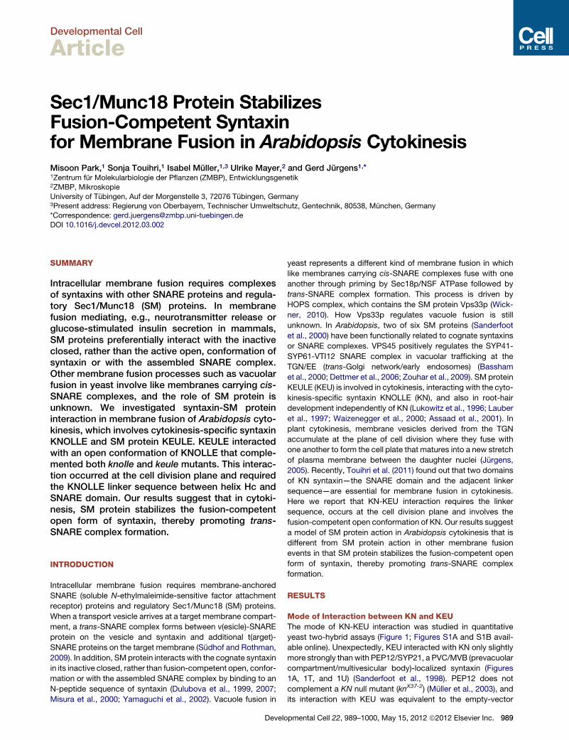
Mode of Interaction between KN and KEUThe mode of KN-KEU interaction was studied in quantitative
yeast two-hybrid assays (Figure 1; Figures S1A and S1B avail-
able online). Unexpectedly, KEU interacted with KN only slightly
more strongly thanwith PEP12/SYP21, a PVC/MVB (prevacuolar
compartment/multivesicular body)-localized syntaxin (Figures
1A, 1T, and 1U) (Sanderfoot et al., 1998). PEP12 does not
complement a KN null mutant (knX37-2) (Muller et al., 2003), and
its interaction with KEU was equivalent to the empty-vector
pmental Cell 22, 989–1000, May 15, 2012 ª2012 Elsevier Inc. 989
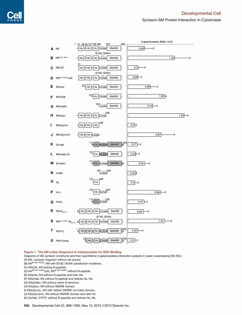
Figure 1. The KN Linker Sequence Is Indispensable for KEU BindingDiagrams of AD-syntaxin constructs and their quantitative b-galactosidase interaction analysis in yeast coexpressing BD-KEU.
(A) KN, cytosolic fragment without tail anchor.
(B) KNIE182,183AA, KN with IE182,183AA substitution mutations.
(C) KND30, KN lacking N-peptide.
(D) KNIE182,183AAD30, KNIE182,183AA without N-peptide.
(E) KNDHa, KN without N-peptide and helix Ha.
(F) KNDHab, KN without N-peptide and helices Ha, Hb.
(G) KNDHabc, KN without entire N terminus.
(H) KNDSyn, KN without SNARE domain.
(I) KNDSynDL, KN with neither SNARE nor linker domain.
(J) KNDSynDHc, KN without SNARE domain and helix Hc.
(K) SDHab, SYP31 without N-peptide and helices Ha, Hb.
Developmental Cell
Syntaxin-SM Protein Interaction in Cytokinesis
990 Developmental Cell 22, 989–1000, May 15, 2012 ª2012 Elsevier Inc.
Figure 2. KN Directly Interacts with KEU
Pull-down assays. Purified recombinant GST, GST:KN and GST:KNIE182,183AA
proteins (lacking the C-terminal membrane anchor) were tested for interaction
with agarose beads-trapped 63HA:KEU (A) or GFP:SNAP33 (B) isolated from
plant extracts. Input (IN), flow-through (FL), and precipitate (IP) were subjected
to immunoblot (IB) analysis with antibodies against HA, GFP, or GST. Note that
GST:KNIE182,183AA interacts more strongly than GST:KN with both 63HA:KEU
and GFP:SNAP33 (arrows). Asterisks, free GST; kDa (kilodalton), molecular
weight markers (left); Input (%), loading volumes relative to the input; Rel. of IB,
relative signal intensity (plant input signal = 100% for IB-HA and IB-GFP; input
signal of GST:KN = 100% for IB-GST).
See also Figure S2.
Developmental Cell
Syntaxin-SM Protein Interaction in Cytokinesis
control (not shown). Syntaxins adopt two alternative conforma-
tions: a closed conformation in which the three N-terminal
helices (Habc domain) fold back onto the SNARE domain, form-
ing a four-helical bundle, and an open conformation in which the
two regions are physically apart (Margittai et al., 2003b; Sutton
et al., 1998). The open form of syntaxin can be experimentally
(L) KNDHab-LS, KNDHab with SYP31 linker domain.
(M) SDHab-L, SDHab with KN linker domain.
(N) Linker, linker domain of KN.
(O) Hc, helix Hc of KN.
(P) Hc-L, helix Hc, and linker domain of KN.
(Q) HcS-L, helix Hc of SYP31, and KN linker domain.
(R) KN-NPEP12, KN with N-peptide of PEP12 (amino acids 1–20 of PEP12 replace
(S) KNIE182,183AA-NPEP12, KN with IE182,183AA substitution mutations and N-pep
(T) PEP12, cytosolic fragment without tail anchor.
(U) PEP12DHa, PEP12 without N-peptide and helix Ha. Domains of PEP12 and
constructs lack the C-terminal membrane anchor (tail anchor) to allow for nuclea
three times. Bars in the graph represent SD (n = 12).
See also Figures S1 and S2.
Develo
stabilized by two point mutations in the linker between helix Hc
and the SNARE domain (Dulubova et al., 1999; D’Andrea-
Merrins et al., 2007). Homologous substitutions introduced into
KN (KNIE182,183AA) increased the interaction with KEU �3-fold
(Figure 1B), which correlated with its increased susceptibility to
limited trypsin proteolysis, suggestive of a more open structure
(Figure S1C). To verify this mode of interaction between KN
and KEU by a different approach, we performed modified pull-
down assays. GST-fused KN or KNIE182,183AA protein was puri-
fied from bacterial cells and then incubated with 63HA:KEU
that had been isolated from plant extracts and trapped on
agarose beads. Consistent with the results in yeast interaction
analysis, GST:KN interacted only weakly with 63HA:KEU
whereas GST:KNIE182,183AA displayed stronger interaction (Fig-
ure 2A). Thus, KEU interacts with KN directly and KEU appears
to prefer the open conformation of KN for binding. To determine
whether the open conformation of KN is competent to interact
with its SNARE partners, we performed comparable pull-down
assays of the two recombinant KN variants with GFP:SNAP33
purified from transgenic plants in the same manner as 63HA:
KEU. GST:KNIE182,183AA interacted more strongly than GST:KN
with SNAP33, which is a KN-interacting Arabidopsis SNAP25
homolog also involved in cytokinesis (Heese et al., 2001) (Fig-
ure 2B). Taken together, our results suggest that the open
conformation of KN is competent to interact with both its SNARE
partner(s) and the SM protein KEU.
The Linker Sequence of KN Is Essential for Bindingof KEUTo identify KN region(s) essential for interaction with KEU,
successively larger deletions were generated from the N-
terminus of KN based on its predicted secondary structure (Fig-
ure 1) (Lukowitz et al., 1996). Elimination of amino acids 1–30 had
no dramatic effect on KN-KEU interaction (Figure 1C), whereas
removal of one or more helices of the Habc domain strongly
increased the interaction (Figures 1E–1G). Truncated KN lacking
the SNARE domain (KNDSyn) also showed strong binding to
KEU (Figure 1H). Furthermore, this interaction was abolished
by removal of the linker (KNDSynDL), but not the adjacent helix
Hc (KNDSynDHc, Figures 1I and 1J), demonstrating the essential
role of the linker. The linker between helix Hc and the SNARE
domain is highly flexible, presumably allowing for conformational
changes of syntaxins (Margittai et al., 2003a). We replaced the
KN linker with the unrelated linker of the Golgi-localized
Sed5p/syntaxin 5 ortholog SYP31 (Figure S2) (Uemura et al.,
2004). Neither truncated SYP31 protein (SDHab) nor the
d amino acids 1–42 of KN).
tide of PEP12.
SYP31 are marked by light and dark gray color, respectively. Note that all
r uptake. Amino acid positions are indicated. The measurement was repeated
pmental Cell 22, 989–1000, May 15, 2012 ª2012 Elsevier Inc. 991
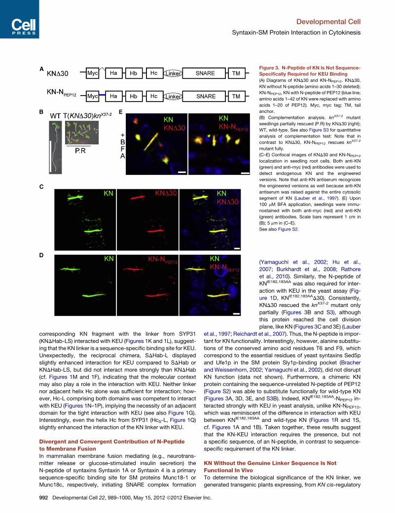
Figure 3. N-Peptide of KN Is Not Sequence-
Specifically Required for KEU Binding
(A) Diagrams of KND30 and KN-NPEP12. KND30,
KN without N-peptide (amino acids 1–30 deleted);
KN-NPEP12, KN with N-peptide of PEP12 (blue line;
amino acids 1–42 of KN were replaced with amino
acids 1–20 of PEP12). Myc, myc tag; TM, tail
anchor.
(B) Complementation analysis. knX37-2 mutant
seedlings partially rescued (P.R) by KND30 (right);
WT, wild-type. See also Figure S3 for quantitative
analysis of complementation test: Note that in
contrast to KND30, KN-NPEP12 rescues knX37-2
mutant fully.
(C–E) Confocal images of KND30 and KN-NPEP12
localization in seedling root cells. Both anti-KN
(green) and anti-myc (red) antibodies were used to
detect endogenous KN and the engineered
versions. Note that anti-KN antiserum recognizes
the engineered versions as well because anti-KN
antiserum was raised against the entire cytosolic
segment of KN (Lauber et al., 1997). (E) Upon
100 mM BFA application, seedlings were immu-
nostained with both anti-myc (red) and anti-KN
(green) antibodies. Scale bars represent 1 cm in
(B); 5 mm in (C–E).
See also Figure S2.
Developmental Cell
Syntaxin-SM Protein Interaction in Cytokinesis
corresponding KN fragment with the linker from SYP31
(KNDHab-LS) interacted with KEU (Figures 1K and 1L), suggest-
ing that the KN linker is a sequence-specific binding site for KEU.
Unexpectedly, the reciprocal chimera, SDHab-L displayed
slightly enhanced interaction for KEU compared to SDHab or
KNDHab-LS, but did not interact more strongly than KNDHab
(cf. Figures 1M and 1F), indicating that the molecular context
may also play a role in the interaction with KEU. Neither linker
nor adjacent helix Hc alone was sufficient for interaction; how-
ever, Hc-L comprising both domains was competent to interact
with KEU (Figures 1N–1P), implying the necessity of an adjacent
domain for the tight interaction with KEU (see also Figure 1G).
Interestingly, even the helix Hc from SYP31 (HcS-L, Figure 1Q)
slightly enhanced the interaction of the KN linker with KEU.
Divergent and Convergent Contribution of N-Peptideto Membrane FusionIn mammalian membrane fusion mediating (e.g., neurotrans-
mitter release or glucose-stimulated insulin secretion) the
N-peptide of syntaxins Syntaxin 1A or Syntaxin 4 is a primary
sequence-specific binding site for SM proteins Munc18-1 or
Munc18c, respectively, initiating SNARE complex formation
992 Developmental Cell 22, 989–1000, May 15, 2012 ª2012 Elsevier Inc.
(Yamaguchi et al., 2002; Hu et al.,
2007; Burkhardt et al., 2008; Rathore
et al., 2010). Similarly, the N-peptide of
KNIE182,183AA was also required for inter-
action with KEU in the yeast assay (Fig-
ure 1D, KNIE182,183AAD30). Consistently,
KND30 rescued the knX37-2 mutant only
partially (Figures 3B and S3), although
this protein reached the cell division
plane, like KN (Figures 3C and 3E) (Lauber
et al., 1997; Reichardt et al., 2007). Thus, the N-peptide is impor-
tant for KN functionality. Interestingly, however, alanine substitu-
tions of the conserved amino acid residues T6 and F9, which
correspond to the essential residues of yeast syntaxins Sed5p
and Ufe1p in the SM protein Sly1p-binding pocket (Bracher
and Weissenhorn, 2002; Yamaguchi et al., 2002), did not disrupt
KN function (data not shown). Furthermore, a chimeric KN
protein containing the sequence-unrelated N-peptide of PEP12
(Figure S2) was able to substitute functionally for wild-type KN
(Figures 3A, 3D, 3E, and S3B). Indeed, KNIE182,183AA-NPEP12 in-
teracted strongly with KEU in yeast analysis, unlike KN-NPEP12,
which was reminiscent of the difference in interaction with KEU
between KNIE182,183AA and wild-type KN (Figures 1R and 1S,
cf. Figures 1A and 1B). Taken together, these results suggest
that the KN-KEU interaction requires the presence, but not
a specific sequence, of an N-peptide, in contrast to sequence-
specific requirement of the KN linker.
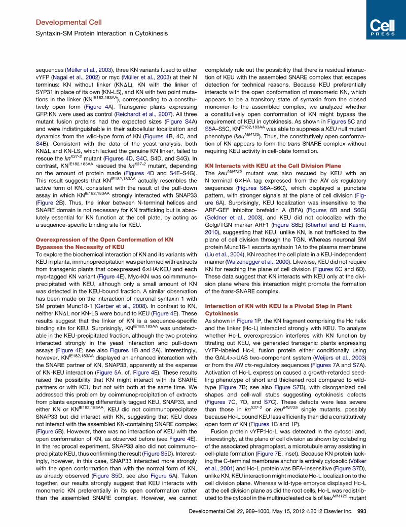
KN Without the Genuine Linker Sequence Is NotFunctional In VivoTo determine the biological significance of the KN linker, we
generated transgenic plants expressing, from KN cis-regulatory
Developmental Cell
Syntaxin-SM Protein Interaction in Cytokinesis
sequences (Muller et al., 2003), three KN variants fused to either
vYFP (Nagai et al., 2002) or myc (Muller et al., 2003) at their N
terminus: KN without linker (KNDL), KN with the linker of
SYP31 in place of its own (KN-LS), and KN with two point muta-
tions in the linker (KNIE182,183AA), corresponding to a constitu-
tively open form (Figure 4A). Transgenic plants expressing
GFP:KN were used as control (Reichardt et al., 2007). All three
mutant fusion proteins had the expected sizes (Figure S4A)
and were indistinguishable in their subcellular localization and
dynamics from the wild-type form of KN (Figures 4B, 4C, and
S4B). Consistent with the data of the yeast analysis, both
KNDL and KN-LS, which lacked the genuine KN linker, failed to
rescue the knX37-2 mutant (Figures 4D, S4C, S4D, and S4G). In
contrast, KNIE182,183AA rescued the knX37-2 mutant, depending
on the amount of protein made (Figures 4D and S4E–S4G).
This result suggests that KNIE182,183AA actually resembles the
active form of KN, consistent with the result of the pull-down
assay in which KNIE182,183AA strongly interacted with SNAP33
(Figure 2B). Thus, the linker between N-terminal helices and
SNARE domain is not necessary for KN trafficking but is abso-
lutely essential for KN function at the cell plate, by acting as
a sequence-specific binding site for KEU.
Overexpression of the Open Conformation of KNBypasses the Necessity of KEUTo explore the biochemical interaction of KN and its variants with
KEU in planta, immunoprecipitation was performedwith extracts
from transgenic plants that coexpressed 63HA:KEU and each
myc-tagged KN variant (Figure 4E). Myc-KN was coimmmuno-
precipitated with KEU, although only a small amount of KN
was detected in the KEU-bound fraction. A similar observation
has been made on the interaction of neuronal syntaxin 1 with
SM protein Munc18-1 (Gerber et al., 2008). In contrast to KN,
neither KNDL nor KN-LS were bound to KEU (Figure 4E). These
results suggest that the linker of KN is a sequence-specific
binding site for KEU. Surprisingly, KNIE182,183AA was undetect-
able in the KEU-precipitated fraction, although the two proteins
interacted strongly in the yeast interaction and pull-down
assays (Figure 4E; see also Figures 1B and 2A). Interestingly,
however, KNIE182,183AA displayed an enhanced interaction with
the SNARE partner of KN, SNAP33, apparently at the expense
of KN-KEU interaction (Figure 5A, cf. Figure 4E). These results
raised the possibility that KN might interact with its SNARE
partners or with KEU but not with both at the same time. We
addressed this problem by coimmunoprecipitation of extracts
from plants expressing differentially tagged KEU, SNAP33, and
either KN or KNIE182,183AA. KEU did not coimmunoprecipitate
SNAP33 but did interact with KN, suggesting that KEU does
not interact with the assembled KN-containing SNARE complex
(Figure 5B). However, there was no interaction of KEU with the
open conformation of KN, as observed before (see Figure 4E).
In the reciprocal experiment, SNAP33 also did not coimmuno-
precipitate KEU, thus confirming the result (Figure S5D). Interest-
ingly, however, in this case, SNAP33 interacted more strongly
with the open conformation than with the normal form of KN,
as already observed (Figure S5D, see also Figure 5A). Taken
together, our results strongly suggest that KEU interacts with
monomeric KN preferentially in its open conformation rather
than the assembled SNARE complex. However, we cannot
Develo
completely rule out the possibility that there is residual interac-
tion of KEU with the assembled SNARE complex that escapes
detection for technical reasons. Because KEU preferentially
interacts with the open conformation of monomeric KN, which
appears to be a transitory state of syntaxin from the closed
monomer to the assembled complex, we analyzed whether
a constitutively open conformation of KN might bypass the
requirement of KEU in cytokinesis. As shown in Figures 5C and
S5A–S5C, KNIE182,183AA was able to suppress a KEU null mutant
phenotype (keuMM125). Thus, the constitutively open conforma-
tion of KN appears to form the trans-SNARE complex without
requiring KEU activity in cell-plate formation.
KN Interacts with KEU at the Cell Division PlaneThe keuMM125 mutant was also rescued by KEU with an
N-terminal 63HA tag expressed from the KN cis-regulatory
sequences (Figures S6A–S6C), which displayed a punctate
pattern, with stronger signals at the plane of cell division (Fig-
ure 6A). Surprisingly, KEU localization was insensitive to the
ARF-GEF inhibitor brefeldin A (BFA) (Figures 6B and S6G)
(Geldner et al., 2003), and KEU did not colocalize with the
Golgi/TGN marker ARF1 (Figure S6E) (Stierhof and El Kasmi,
2010), suggesting that KEU, unlike KN, is not trafficked to the
plane of cell division through the TGN. Whereas neuronal SM
protein Munc18-1 escorts syntaxin 1A to the plasma membrane
(Liu et al., 2004), KN reaches the cell plate in a KEU-independent
manner (Waizenegger et al., 2000). Likewise, KEU did not require
KN for reaching the plane of cell division (Figures 6C and 6D).
These data suggest that KN interacts with KEU only at the divi-
sion plane where this interaction might promote the formation
of the trans-SNARE complex.
Interaction of KN with KEU Is a Pivotal Step in PlantCytokinesisAs shown in Figure 1P, the KN fragment comprising the Hc helix
and the linker (Hc-L) interacted strongly with KEU. To analyze
whether Hc-L overexpression interferes with KN function by
titrating out KEU, we generated transgenic plants expressing
vYFP-labeled Hc-L fusion protein either conditionally using
the GAL4>>UAS two-component system (Weijers et al., 2003)
or from the KN cis-regulatory sequences (Figures 7A and S7A).
Activation of Hc-L expression caused a growth-retarded seed-
ling phenotype of short and thickened root compared to wild-
type (Figure 7B; see also Figure S7B), with disorganized cell
shapes and cell-wall stubs suggesting cytokinesis defects
(Figures 7C, 7D, and S7C). These defects were less severe
than those in knX37-2 or keuMM125 single mutants, possibly
becauseHc-L boundKEU less efficiently than did a constitutively
open form of KN (Figures 1B and 1P).
Fusion protein vYFP:Hc-L was detected in the cytosol and,
interestingly, at the plane of cell division as shown by colabeling
of the associated phragmoplast, a microtubule array assisting in
cell-plate formation (Figure 7E, inset). Because KN protein lack-
ing the C-terminal membrane anchor is entirely cytosolic (Volker
et al., 2001) and Hc-L protein was BFA-insensitive (Figure S7D),
unlike KN, KEU interactionmightmediate Hc-L localization to the
cell division plane. Whereas wild-type embryos displayed Hc-L
at the cell division plane as did the root cells, Hc-L was redistrib-
uted to the cytosol in themultinucleated cells of keuMM125mutant
pmental Cell 22, 989–1000, May 15, 2012 ª2012 Elsevier Inc. 993
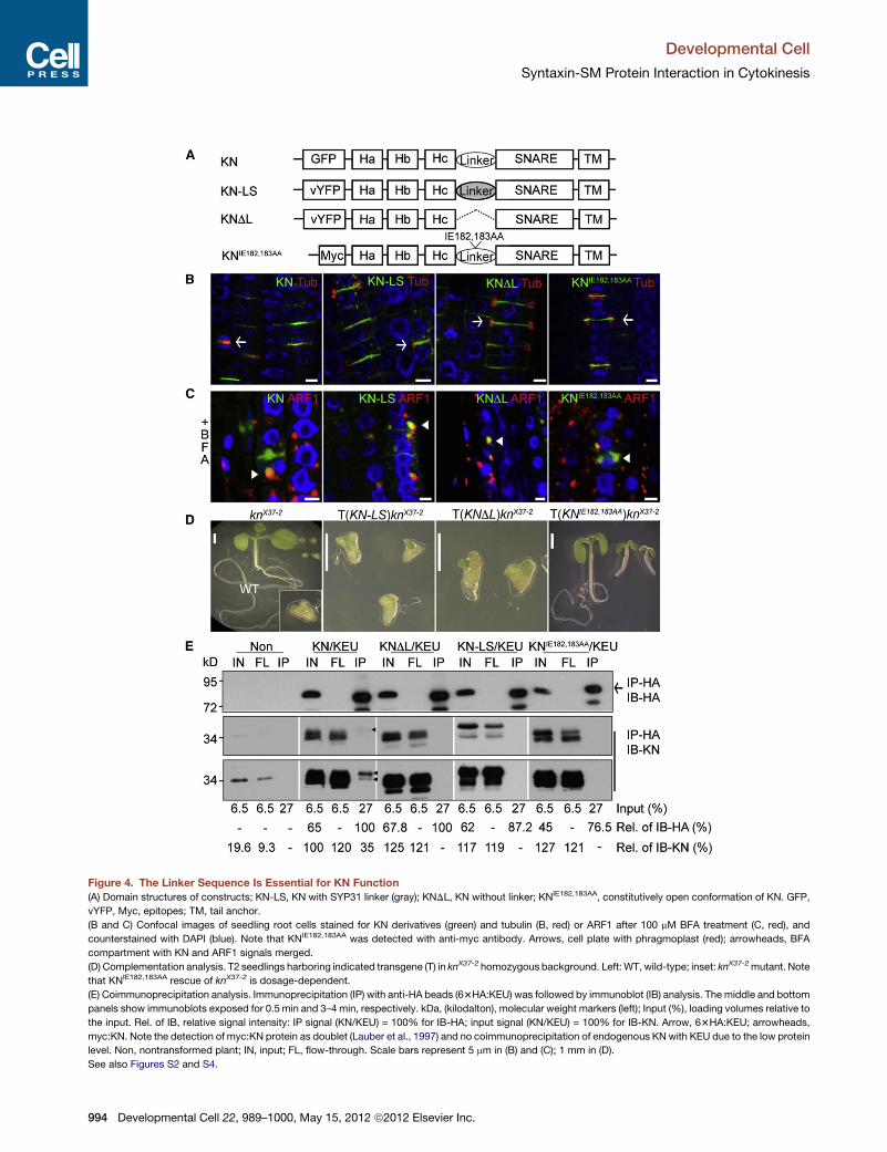
Figure 4. The Linker Sequence Is Essential for KN Function
(A) Domain structures of constructs; KN-LS, KN with SYP31 linker (gray); KNDL, KN without linker; KNIE182,183AA, constitutively open conformation of KN. GFP,
vYFP, Myc, epitopes; TM, tail anchor.
(B and C) Confocal images of seedling root cells stained for KN derivatives (green) and tubulin (B, red) or ARF1 after 100 mM BFA treatment (C, red), and
counterstained with DAPI (blue). Note that KNIE182,183AA was detected with anti-myc antibody. Arrows, cell plate with phragmoplast (red); arrowheads, BFA
compartment with KN and ARF1 signals merged.
(D) Complementation analysis. T2 seedlings harboring indicated transgene (T) in knX37-2 homozygous background. Left:WT, wild-type; inset: knX37-2mutant. Note
that KNIE182,183AA rescue of knX37-2 is dosage-dependent.
(E) Coimmunoprecipitation analysis. Immunoprecipitation (IP) with anti-HA beads (63HA:KEU) was followed by immunoblot (IB) analysis. The middle and bottom
panels show immunoblots exposed for 0.5 min and 3–4 min, respectively. kDa, (kilodalton), molecular weight markers (left); Input (%), loading volumes relative to
the input. Rel. of IB, relative signal intensity: IP signal (KN/KEU) = 100% for IB-HA; input signal (KN/KEU) = 100% for IB-KN. Arrow, 63HA:KEU; arrowheads,
myc:KN. Note the detection of myc:KN protein as doublet (Lauber et al., 1997) and no coimmunoprecipitation of endogenous KNwith KEU due to the low protein
level. Non, nontransformed plant; IN, input; FL, flow-through. Scale bars represent 5 mm in (B) and (C); 1 mm in (D).
See also Figures S2 and S4.
Developmental Cell
Syntaxin-SM Protein Interaction in Cytokinesis
994 Developmental Cell 22, 989–1000, May 15, 2012 ª2012 Elsevier Inc.
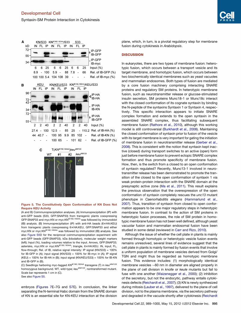
Figure 5. The Constitutively Open Conformation of KN Does Not
Require KEU Activity
(A and B) Coimmunoprecipitation analyses. (A) Immunoprecipitation (IP) with
anti-GFP beads (S33, GFP:SNAP33) from transgenic plants coexpressing
GFP:SNAP33 and myc:KN or myc:KNIE182,183AA was followed by immunoblot
(IB) analysis. (B) Immunoprecipitation (IP) with anti-HA beads (63HA:KEU)
from transgenic plants coexpressing 63HA:KEU, GFP:SNAP33 and either
myc:KN or myc:KNIE182,183AA was followed by immunoblot (IB) analysis. See
also Figure S5D for the reciprocal coimmunoprecipitation experiment with
anti-GFP beads (GFP:SNAP33). kDa (kilodalton), molecular weight markers
(left); Input (%), loading volumes relative to the input. Arrows, GFP:SNAP33;
asterisks, myc:KN or myc:KNIE182,183AA; triangle, 63HA:KEU. IN, input; FL,
flow-through; Rel. of IB, relative signal intensity: IP signal (KN/S33) = 100%
for IB-GFP in (A); input signal (KN/S33) = 100% for IB-myc in (A); IP signal
(KEU) = 100% for IB-HA in (B); input signal (KN/KEU/S33) = 100% for IB-KN
and IB-GFP in (B).
(C) Seedlings harboring myc-tagged KNIE182,183AA transgene (T) in keuMM125
homozygous background. WT, wild-type; keuMM125, nontransformed mutant.
Scale bar represents 1 cm in (C).
See also Figure S5.
Developmental Cell
Syntaxin-SM Protein Interaction in Cytokinesis
embryos (Figures 7E–7G and S7E). In conclusion, the linker
separating the N-terminal Habc domain from the SNARE domain
of KN is an essential site for KN-KEU interaction at the division
Develo
plane, which, in turn, is a pivotal regulatory step for membrane
fusion during cytokinesis in Arabidopsis.
DISCUSSION
In eukaryotes, there are two types of membrane fusion: hetero-
typic fusion, which occurs between a transport vesicle and its
target membrane, and homotypic fusion, which occurs between
two biochemically identical membranes such as yeast vacuoles
and mammalian endosomes. Both types of fusion are mediated
by a core fusion machinery comprising interacting SNARE
proteins and regulatory SM proteins. In heterotypic membrane
fusion, such as neurotransmitter release or glucose-stimulated
insulin secretion, SM proteins Munc18-1 or Munc18c interact
with the closed conformation of its cognate syntaxin by binding
the N-peptide of the syntaxins Syntaxin 1 or Syntaxin 4, respec-
tively. This specific interaction appears to initiate SNARE
complex formation and extends to the open syntaxin in the
assembled SNARE complex, thus facilitating subsequent
membrane fusion (Rathore et al., 2010), although this working
model is still controversial (Burkhardt et al., 2008). Maintaining
the closed conformation of syntaxin prior to fusion of the vesicle
with its target membrane is very important for gating the initiation
of membrane fusion in neurotransmitter release (Gerber et al.,
2008). This is consistent with the notion that syntaxin kept inac-
tive (closed) during transport switches to an active (open) form
just beforemembrane fusion to prevent ectopic SNARE complex
formation and thus promote specificity of membrane fusion.
How, then, is the switch from a closed to an open conformation
of syntaxin regulated? Recently, Munc13-1 involved in neuro-
transmitter release has been demonstrated to promote the tran-
sition of the closed to the open conformation of syntaxin 1 via
weak protein-protein interaction with the SNARE domain at the
presynaptic active zone (Ma et al., 2011). This result explains
the previous observation that the overexpression of the open
conformation of syntaxin completely rescues the unc13 mutant
phenotype in Caenorhabditis elegans (Hammarlund et al.,
2007). Thus, transition of syntaxin from closed to open confor-
mation appears to be one major regulatory steps in heterotypic
membrane fusion. In contrast to the action of SM proteins in
heterotypic fusion processes, the role of SM protein in homo-
typicmembrane fusion has not beenwell defined, although yeast
vacuolar fusion and mammalian endosomal fusion have been
studied in some detail (reviewed in Carr and Rizo, 2010).
Although the issue of whether the cell plate in plants is mainly
formed through homotypic or heterotypic vesicle fusion events
remains unresolved, several lines of evidence suggest that the
cell plate in plants is mainly formed by fusion events that involve
a uniform population of membrane vesicles derived from Golgi/
TGN and might thus be regarded as homotypic membrane
fusion. This evidence includes: (1) morphologically identical
membrane vesicles �60 nm in diameter are aligned properly in
the plane of cell division in knolle or keule mutants but fail to
fuse with one another (Waizenegger et al., 2000); (2) inhibition
of the secretory, but not the endocytic, pathway entails cytoki-
nesis defects (Reichardt et al., 2007); (3) KN is newly synthesized
during mitosis (Lauber et al., 1997), delivered to the plane of cell
division, not to the plasmamembrane, via the secretory pathway
and degraded in the vacuole shortly after cytokinesis (Reichardt
pmental Cell 22, 989–1000, May 15, 2012 ª2012 Elsevier Inc. 995
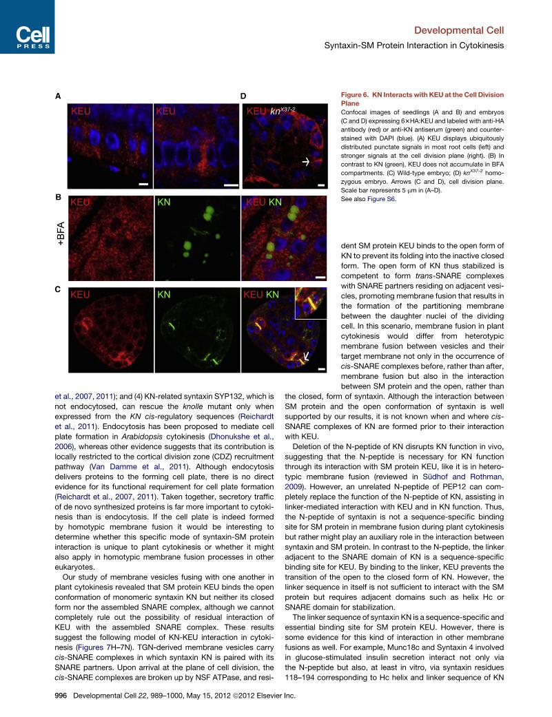
Figure 6. KN Interacts with KEU at the Cell Division
Plane
Confocal images of seedlings (A and B) and embryos
(C and D) expressing 63HA:KEU and labeled with anti-HA
antibody (red) or anti-KN antiserum (green) and counter-
stained with DAPI (blue). (A) KEU displays ubiquitously
distributed punctate signals in most root cells (left) and
stronger signals at the cell division plane (right). (B) In
contrast to KN (green), KEU does not accumulate in BFA
compartments. (C) Wild-type embryo; (D) knX37-2 homo-
zygous embryo. Arrows (C and D), cell division plane.
Scale bar represents 5 mm in (A–D).
See also Figure S6.
Developmental Cell
Syntaxin-SM Protein Interaction in Cytokinesis
et al., 2007, 2011); and (4) KN-related syntaxin SYP132, which is
not endocytosed, can rescue the knolle mutant only when
expressed from the KN cis-regulatory sequences (Reichardt
et al., 2011). Endocytosis has been proposed to mediate cell
plate formation in Arabidopsis cytokinesis (Dhonukshe et al.,
2006), whereas other evidence suggests that its contribution is
locally restricted to the cortical division zone (CDZ) recruitment
pathway (Van Damme et al., 2011). Although endocytosis
delivers proteins to the forming cell plate, there is no direct
evidence for its functional requirement for cell plate formation
(Reichardt et al., 2007, 2011). Taken together, secretory traffic
of de novo synthesized proteins is far more important to cytoki-
nesis than is endocytosis. If the cell plate is indeed formed
by homotypic membrane fusion it would be interesting to
determine whether this specific mode of syntaxin-SM protein
interaction is unique to plant cytokinesis or whether it might
also apply in homotypic membrane fusion processes in other
eukaryotes.
Our study of membrane vesicles fusing with one another in
plant cytokinesis revealed that SM protein KEU binds the open
conformation of monomeric syntaxin KN but neither its closed
form nor the assembled SNARE complex, although we cannot
completely rule out the possibility of residual interaction of
KEU with the assembled SNARE complex. These results
suggest the following model of KN-KEU interaction in cytoki-
nesis (Figures 7H–7N). TGN-derived membrane vesicles carry
cis-SNARE complexes in which syntaxin KN is paired with its
SNARE partners. Upon arrival at the plane of cell division, the
cis-SNARE complexes are broken up by NSF ATPase, and resi-
996 Developmental Cell 22, 989–1000, May 15, 2012 ª2012 Elsevier Inc.
dent SM protein KEU binds to the open form of
KN to prevent its folding into the inactive closed
form. The open form of KN thus stabilized is
competent to form trans-SNARE complexes
with SNARE partners residing on adjacent vesi-
cles, promoting membrane fusion that results in
the formation of the partitioning membrane
between the daughter nuclei of the dividing
cell. In this scenario, membrane fusion in plant
cytokinesis would differ from heterotypic
membrane fusion between vesicles and their
target membrane not only in the occurrence of
cis-SNARE complexes before, rather than after,
membrane fusion but also in the interaction
between SM protein and the open, rather than
the closed, form of syntaxin. Although the interaction between
SM protein and the open conformation of syntaxin is well
supported by our results, it is not known when and where cis-
SNARE complexes of KN are formed prior to their interaction
with KEU.
Deletion of the N-peptide of KN disrupts KN function in vivo,
suggesting that the N-peptide is necessary for KN function
through its interaction with SM protein KEU, like it is in hetero-
typic membrane fusion (reviewed in Sudhof and Rothman,
2009). However, an unrelated N-peptide of PEP12 can com-
pletely replace the function of the N-peptide of KN, assisting in
linker-mediated interaction with KEU and in KN function. Thus,
the N-peptide of syntaxin is not a sequence-specific binding
site for SM protein in membrane fusion during plant cytokinesis
but rather might play an auxiliary role in the interaction between
syntaxin and SM protein. In contrast to the N-peptide, the linker
adjacent to the SNARE domain of KN is a sequence-specific
binding site for KEU. By binding to the linker, KEU prevents the
transition of the open to the closed form of KN. However, the
linker sequence in itself is not sufficient to interact with the SM
protein but requires adjacent domains such as helix Hc or
SNARE domain for stabilization.
The linker sequence of syntaxin KN is a sequence-specific and
essential binding site for SM protein KEU. However, there is
some evidence for this kind of interaction in other membrane
fusions as well. For example, Munc18c and Syntaxin 4 involved
in glucose-stimulated insulin secretion interact not only via
the N-peptide but also, at least in vitro, via syntaxin residues
118–194 corresponding to Hc helix and linker sequence of KN
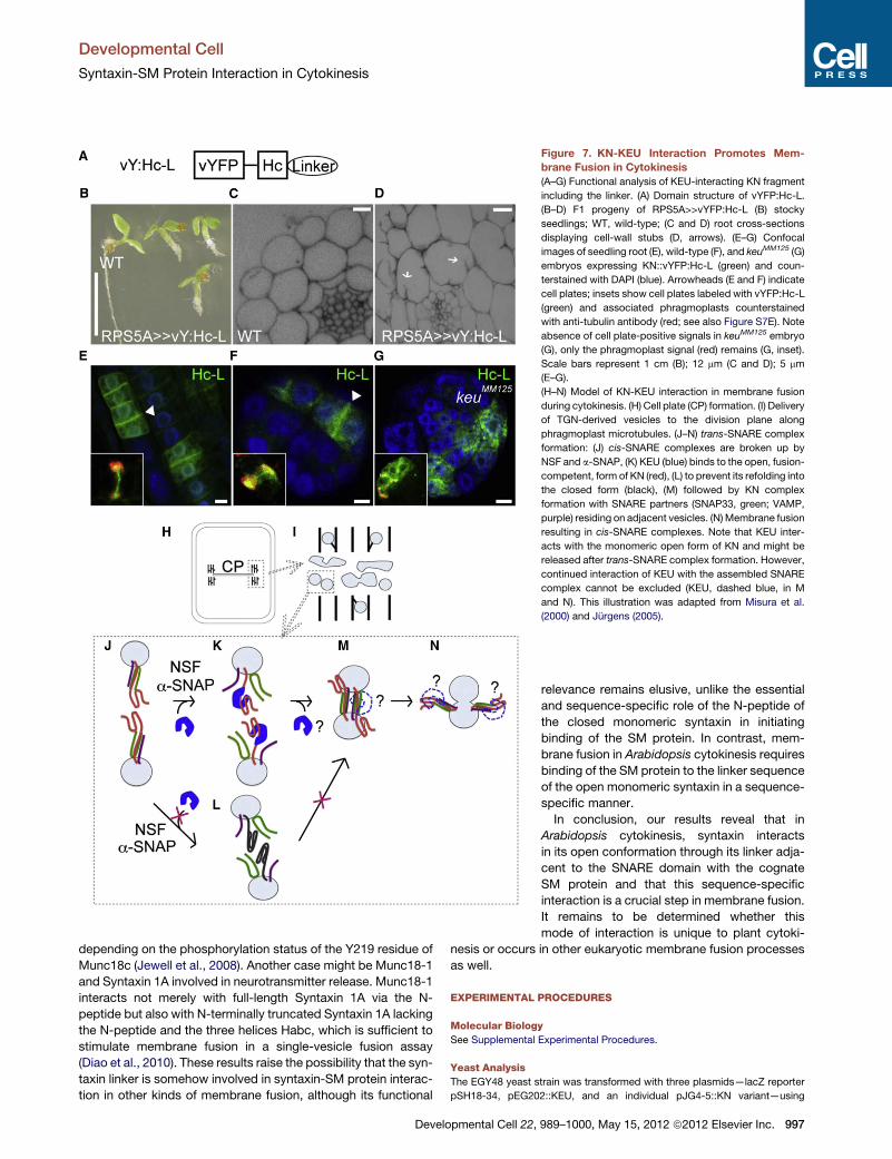
Figure 7. KN-KEU Interaction Promotes Mem-
brane Fusion in Cytokinesis
(A–G) Functional analysis of KEU-interacting KN fragment
including the linker. (A) Domain structure of vYFP:Hc-L.
(B–D) F1 progeny of RPS5A>>vYFP:Hc-L (B) stocky
seedlings; WT, wild-type; (C and D) root cross-sections
displaying cell-wall stubs (D, arrows). (E–G) Confocal
images of seedling root (E), wild-type (F), and keuMM125 (G)
embryos expressing KN::vYFP:Hc-L (green) and coun-
terstained with DAPI (blue). Arrowheads (E and F) indicate
cell plates; insets show cell plates labeled with vYFP:Hc-L
(green) and associated phragmoplasts counterstained
with anti-tubulin antibody (red; see also Figure S7E). Note
absence of cell plate-positive signals in keuMM125 embryo
(G), only the phragmoplast signal (red) remains (G, inset).
Scale bars represent 1 cm (B); 12 mm (C and D); 5 mm
(E–G).
(H–N) Model of KN-KEU interaction in membrane fusion
during cytokinesis. (H) Cell plate (CP) formation. (I) Delivery
of TGN-derived vesicles to the division plane along
phragmoplast microtubules. (J–N) trans-SNARE complex
formation: (J) cis-SNARE complexes are broken up by
NSF and a-SNAP, (K) KEU (blue) binds to the open, fusion-
competent, form of KN (red), (L) to prevent its refolding into
the closed form (black), (M) followed by KN complex
formation with SNARE partners (SNAP33, green; VAMP,
purple) residing on adjacent vesicles. (N)Membrane fusion
resulting in cis-SNARE complexes. Note that KEU inter-
acts with the monomeric open form of KN and might be
released after trans-SNARE complex formation. However,
continued interaction of KEU with the assembled SNARE
complex cannot be excluded (KEU, dashed blue, in M
and N). This illustration was adapted from Misura et al.
(2000) and Jurgens (2005).
Developmental Cell
Syntaxin-SM Protein Interaction in Cytokinesis
depending on the phosphorylation status of the Y219 residue of
Munc18c (Jewell et al., 2008). Another case might be Munc18-1
and Syntaxin 1A involved in neurotransmitter release. Munc18-1
interacts not merely with full-length Syntaxin 1A via the N-
peptide but also with N-terminally truncated Syntaxin 1A lacking
the N-peptide and the three helices Habc, which is sufficient to
stimulate membrane fusion in a single-vesicle fusion assay
(Diao et al., 2010). These results raise the possibility that the syn-
taxin linker is somehow involved in syntaxin-SM protein interac-
tion in other kinds of membrane fusion, although its functional
Developmental Cell 22,
relevance remains elusive, unlike the essential
and sequence-specific role of the N-peptide of
the closed monomeric syntaxin in initiating
binding of the SM protein. In contrast, mem-
brane fusion in Arabidopsis cytokinesis requires
binding of the SM protein to the linker sequence
of the open monomeric syntaxin in a sequence-
specific manner.
In conclusion, our results reveal that in
Arabidopsis cytokinesis, syntaxin interacts
in its open conformation through its linker adja-
cent to the SNARE domain with the cognate
SM protein and that this sequence-specific
interaction is a crucial step in membrane fusion.
It remains to be determined whether this
mode of interaction is unique to plant cytoki-
nesis or occurs in other eukaryotic membrane fusion processes
as well.
EXPERIMENTAL PROCEDURES
Molecular Biology
See Supplemental Experimental Procedures.
Yeast Analysis
The EGY48 yeast strain was transformed with three plasmids—lacZ reporter
pSH18-34, pEG202::KEU, and an individual pJG4-5::KN variant—using
989–1000, May 15, 2012 ª2012 Elsevier Inc. 997
Developmental Cell
Syntaxin-SM Protein Interaction in Cytokinesis
a polyethylene glycol (PEG) transformation method (33.3% [w/v] PEG3500,
100 mM LiAc, 0.3 mg/ml ssDNA). Quantitative b-galactosidase analysis was
performed as follows. In brief, cell mass was suspended in reaction solution
(buffer H [100 mM HEPES pH 7, 150 mM NaCl, 2 mM MgCl2, 1% [w/v] BSA],
6.1% [v/v] chloroform, 0.006% [w/v] SDS) followed by addition of ortho-nitro-
phenyl-b-galactoside (ONPG, 4 mg/ml in buffer H, Sigma-Aldrich). The Miller
Units of each sample were calculated [(1,000 3 OD420)/(culture volume 3
time3 OD600)] (Miller, 1972). Each measurement was done with four indepen-
dent colonies and repeated three times.
Trypsin Treatment
T7-tagged KN and KNIE182,183AA proteins were purified from BL21(DE3)pLys
bacterial cells, using anti-T7-beads (Merck-Novagen) according to the manu-
facturer’s instructions, suspended in cold PBS, immediately followed by the
addition of trypsin (1 M stock solution in PBS; always freshly prepared,
Sigma-Aldrich). The reaction was stopped by addition of 13 Laemmli buffer
and boiling for 5 min at the 95�C.
Plant Material, Growth Condition, and Transformation
Arabidopsis thaliana wild-type—Columbia (Col-O) or Landsberg (Ler)—plants
were grown on soil at 18�C or 23�C in long-day conditions (Mayer et al., 1991).
Wild-type or heterozygous plants either of knX37-2 or of keuMM125 were trans-
formed with Agrobacterium tumefaciens, using the floral-dip method (Clough
and Bent, 1998). KN::myc:KN (Muller et al., 2003) or KN::GFP:KN (Reichardt
et al., 2007) plants were crossed with KN::63HA:KEU plants for the coimmu-
noprecipitation or immunofluorescence analyses. RPS5A::GAL4 (Weijers
et al., 2003) plants were crossed with T1 plants of UAS::vYFP:Hc-L for pheno-
typic analysis. 35S (CauliflowerMosaic Virus promoter)::GFP:SNAP33 line was
provided by Dr. Liliane Sticher and crossed with the transgenic plants bearing
myc:KN, myc:KNIE182,183AA or KN::63HA:KEU for the coimmunoprecipitation
experiment.
Genetic Analysis
T1 plants grown on soil from bulk-harvested seeds were selected for trans-
formants by spraying three times with a 1:1,000 diluted BASTA (183 g/l glufo-
sinate; AgrEvo, Dusseldorf, Germany). Selected BASTA-resistant plants were
genotyped. PCR-genotyping: knX37-2, primers X37-2CIII and X37-2DIII, which
amplify a 0.5-kb fragment from knX37-2 and a 1.5-kb fragment from wild-type
KN (Muller et al., 2003); keuMM125, primers KEU in 14 and KEU in 17 giving
a 0.4-kb fragment from keuMM125 and a 0.5-kb fragment of wild-type KEU;
vYFP:KN-LS or vYFP:KNDL, primers vYFPforward and KNreverse giving an �1-
kb fragment of each transgene; myc:KNIE182,183AA, myc:KND30, or myc:KN-
NPEP12, primers mycforward and KNreverse giving an �1-kb fragment of each
transgene; 63HA:KEU, primers HAforward and KEU400 giving an�0.6-kb frag-
ment. Genomic DNA was isolated using a cetyltrimethylammonium bromide
(CTAB)-based miniscale protocol as previously reported (Assaad et al.,
2001). For seedling observation, seeds were germinated on solid medium
(2.15 g/l MS salts, 1 g/l MES, 1% [w/v] sucrose, pH 5.6) in the same growth
condition as described above. Segregation of antibiotics resistance was
counted on phosphinothricin (PPT, 15 mg/l)-supplemented medium.
Coimmunoprecipitation
Five grams of inflorescence or 5-day-old seedlings was ground thoroughly in
liquid nitrogen and suspended in lysis buffer (50mMTris pH 7.5, 150mMNaCl,
0.5% [v/v] Triton X-100) supplemented with an EDTA-free protease inhibitors
cocktail (Roche Diagnostics). After 30 min incubation on ice, cell debris-
removed supernatants were incubated with anti-HA (Sigma-Aldrich) or anti-
GFP (GFP-trap, Chromotek) beads for 2 hr in the cold room with mild rotation.
The beads were rinsed four times with washing buffer (50 mM Tris pH 7.5,
200 mM NaCl [for the interaction analysis of KN variants/GFP:SNAP33,
150 mM NaCl was used instead], 0.2% Triton X-100) supplemented with
EDTA-free protease inhibitors cocktail, and resuspended in washing buffer
and Laemmli buffer.
Pull-Down Assay
HA-tagged KEULE (63HA:KEU) or GFP-tagged SNAP33 (GFP:SNAP33) was
purified using anti-HA or anti-GFP beads as described in coimmunoprecipita-
tion method. GST, GST:KN, and GST:KNIE182,183AA proteins were purified from
998 Developmental Cell 22, 989–1000, May 15, 2012 ª2012 Elsevier
BL21(DE3)pLys bacterial cells, using anti-GST-beads and 10 mM reduced
L-glutathione (Sigma-Aldrich) according to the manufacturer’s instructions.
Equal amount of eluted GST variants was incubated with agarose beads-trap-
ped 63HA:KEULE or GFP:SNAP33 in the cold roomwith mild rotation in buffer
(50 mM HEPES 7.5; 150 mM NaCl; 1 mM MgCl2; 1 mM EDTA; 0.5% Triton
X-100) supplemented with protease inhibitors cocktail. After overnight incuba-
tion, beads were rinsed five times with washing buffer (50 mM HEPES 7.5;
200 mM NaCl; 1 mM MgCl2; 1 mM EDTA; 0.2% Triton X-100) supplemented
with protease inhibitors cocktail and subjected to immunoblot analysis.
Immunoblots and Immunofluorescence Analysis
For immunoblot analysis of proteins from yeast cells, individual yeast trans-
formants were grown overnight. The cell mass was suspended in 13 Laemmli
buffer and immediately frozen in liquid nitrogen. After incubation at 95�C for
20 min, supernatants were immuno-analyzed. For detection of BD-fused
KEU and AD-fused KN variants, anti-LexA (1:1,000, Santa Cruz Biotechnol-
ogies) and POD-conjugated anti-HA antibodies (1:1,000, Roche Diagnostics)
were used, respectively.
For immunoblot analysis of proteins from plants, inflorescences from T1
plants or entire T2 seedlings were harvested. Total proteins were extracted
with lysis buffer supplemented with an EDTA-free protease inhibitors cocktail.
Antibodies of anti-KN (rabbit, 1:5,000) (Lauber et al., 1997), anti-tubulin
(mouse, 1:1,000, Sigma-Aldrich), anti-myc 9E10 (mouse, 1:1,000, Santa
Cruz), POD-conjugated anti-HA, anti-YFP (rabbit, 1:1,000, a gift from S. de
Vries), anti-GFP (rabbit, 1:1,000, Invitrogen), and POD-conjugated anti-GST
(rabbit, 1:2,000, Sigma-Aldrich) were used to detect the indicated proteins.
POD-conjugated secondary antibodies (1:7,000, Sigma) were used in all
immunoblot analyses except for ‘‘IP-GFP/IB-myc’’ shown in Figure S5D, which
was detected with alkaline phosphatase (AP)-conjugated secondary antibody
(1:7,000, Novagen). Membranes were developed using both conventional
method (AGFA film developing system) and a chemiluminescence detection
system (Fusion Fx7 Imager, PEQlab, Germany).
Immunostaining of seedling roots was performed as previously reported
(Volker et al., 2001). In brief,�5-day-old seedlings were fixed in 4% (w/v) para-
formaldehyde and labeled with the indicated antibodies. For brefeldin A (BFA)
treatment,�5-day-old seedlings were treated with 100 mMbrefeldin A (50 mM
stock solution in 1:1 DMSO/EtOH, Invitrogen) for 1 hr followed by fixation.
For immunofluorescence, anti-KN (rabbit, 1:2,000), anti-tubulin (rat, 1:1,000,
Abcam, Boston), anti-HA (mouse, 1:1,000, BAbCO, Richmond, CA), anti-myc
9E10 (mouse, 1:1,000, Santa Cruz), two sorts of anti-ARF1 (anti-ARF1 for
Figures 3C and S4B [rabbit, 1:1,000] [Pimpl et al., 2000]: anti-ARF1 for Figures
S6E and S7D [rabbit, 1:1,000, Agrisera]), anti-mouse FITC (1:600, Dianova),
anti-rabbit-Cy3TM (1:600, Dianova), and anti-rabbit-Alexa488 (1:600, Invitro-
gen) were used.
Embryos were prepared as previously reported (Heese et al., 2001). In brief,
embryos were fixed in 4% paraformaldehyde plus 1% Triton X-100 and then
squeezed out of the seed coat on gelatin-coated slides. Fluorescence images
were taken using a confocal laser scanning microscope (Leica) and Leica TCS
software.
Phenotypic Analysis
Seedling sectioning was performed as previously reported (Heese et al., 2001).
In brief, �7-day-old seedlings were fixed in 4% paraformaldehyde, dehy-
drated in a series of ethanol, and embedded in LR-white resin (London Resin).
Sections were cut in 3- to 5-mm thick slices using a cryomicrotome (Supercut
2065) and stained with toluidine blue.
Software
Sequences were analyzed using Vector NTI (Invitrogen). Images were pro-
cessed using Adobe Photoshop CS3 and Adobe illustrator CS3. Quantification
of signal intensity in immunoblot analysis was done using ImageJ program
(NIH).
SUPPLEMENTAL INFORMATION
Supplemental Information includes seven figures, Supplemental Experi-
mental Procedures, and one table and can be found with this article online
at doi:10.1016/j.devcel.2012.03.002.
Inc.
Developmental Cell
Syntaxin-SM Protein Interaction in Cytokinesis
ACKNOWLEDGMENTS
We thank Ulrike Hiller for technical assistance, Sacco de Vries (Wageningen
University, Netherlands) and Liliane Sticher (University of Fribourg,
Switzerland) for providing materials, and Martin Bayer, Niko Geldner, Rita
Groß-Hardt and our group members for critical reading of the manuscript.
This work was funded by the Korea Research Foundation through fellowship
C00066 to M.P. and the Deutsche Forschungsgemeinschaft through grant
Ju 179/14-1. M.P. performed most of the experiments. S.T. did the genetic
study and immunofluorescence analysis of KND30 and 63HA:KEULE in
embryos. I.M. performed the functional study of KN-NPEP12 and generated
the transgenic plants bearing KNIE182,183AA. U.M. contributed to the genetic
complementation analysis of KN engineered versions. G.J. initially developed
this project. M.P. and G.J. analyzed and discussed the data and wrote the
paper.
Received: May 20, 2011
Revised: February 1, 2012
Accepted: March 2, 2012
Published online: May 14, 2012
REFERENCES
Assaad, F.F., Huet, Y., Mayer, U., and Jurgens, G. (2001). The cytokinesis gene
KEULE encodes a Sec1 protein that binds the syntaxin KNOLLE. J. Cell Biol.
152, 531–543.
Bassham, D.C., Sanderfoot, A.A., Kovaleva, V., Zheng, H., and Raikhel, N.V.
(2000). AtVPS45 complex formation at the trans-Golgi network. Mol. Biol.
Cell 11, 2251–2265.
Bracher, A., and Weissenhorn, W. (2002). Structural basis for the Golgi
membrane recruitment of Sly1p by Sed5p. EMBO J. 21, 6114–6124.
Burkhardt, P., Hattendorf, D.A., Weis, W.I., and Fasshauer, D. (2008).
Munc18a controls SNARE assembly through its interaction with the syntaxin
N-peptide. EMBO J. 27, 923–933.
Carr, C.M., and Rizo, J. (2010). At the junction of SNARE and SM protein func-
tion. Curr. Opin. Cell Biol. 22, 488–495.
Clough, S.J., and Bent, A.F. (1998). Floral dip: a simplified method for
Agrobacterium-mediated transformation of Arabidopsis thaliana. Plant J. 16,
735–743.
D’Andrea-Merrins, M., Chang, L., Lam, A.D., Ernst, S.A., and Stuenkel, E.L.
(2007). Munc18c interaction with syntaxin 4 monomers and SNARE complex
intermediates in GLUT4 vesicle trafficking. J. Biol. Chem. 282, 16553–16566.
Dettmer, J., Hong-Hermesdorf, A., Stierhof, Y.D., and Schumacher, K. (2006).
Vacuolar H+-ATPase activity is required for endocytic and secretory trafficking
in Arabidopsis. Plant Cell 18, 715–730.
Dhonukshe, P., Baluska, F., Schlicht, M., Hlavacka, A., Samaj, J., Friml, J., and
Gadella, T.W.J., Jr. (2006). Endocytosis of cell surface material mediates cell
plate formation during plant cytokinesis. Dev. Cell 10, 137–150.
Diao, J., Su, Z., Lu, X., Yoon, T.Y., Shin, Y.K., and Ha, T. (2010). Single-vesicle
fusion assay reveals Munc18-1 binding to the SNARE core is sufficient for
stimulating membrane fusion. ACS Chem. Neurosci. 1, 168–174.
Dulubova, I., Sugita, S., Hill, S., Hosaka, M., Fernandez, I., Sudhof, T.C., and
Rizo, J. (1999). A conformational switch in syntaxin during exocytosis: role of
munc18. EMBO J. 18, 4372–4382.
Dulubova, I., Khvotchev, M., Liu, S., Huryeva, I., Sudhof, T.C., and Rizo, J.
(2007). Munc18-1 binds directly to the neuronal SNARE complex. Proc. Natl.
Acad. Sci. USA 104, 2697–2702.
Geldner, N., Anders, N., Wolters, H., Keicher, J., Kornberger, W., Muller, P.,
Delbarre, A., Ueda, T., Nakano, A., and Jurgens, G. (2003). The Arabidopsis
GNOM ARF-GEF mediates endosomal recycling, auxin transport, and auxin-
dependent plant growth. Cell 112, 219–230.
Gerber, S.H., Rah, J.C., Min, S.W., Liu, X., deWit, H., Dulubova, I., Meyer, A.C.,
Rizo, J., Arancillo, M., Hammer, R.E., et al. (2008). Conformational switch of
syntaxin-1 controls synaptic vesicle fusion. Science 321, 1507–1510.
Develo
Hammarlund, M., Palfreyman, M.T., Watanabe, S., Olsen, S., and Jorgensen,
E.M. (2007). Open syntaxin docks synaptic vesicles. PLoS Biol. 5, e198.
Heese, M., Gansel, X., Sticher, L., Wick, P., Grebe, M., Granier, F., and
Jurgens, G. (2001). Functional characterization of the KNOLLE-interacting
t-SNARE AtSNAP33 and its role in plant cytokinesis. J. Cell Biol. 155, 239–249.
Hu, S.H., Latham, C.F., Gee, C.L., James, D.E., and Martin, J.L. (2007).
Structure of the Munc18c/Syntaxin4 N-peptide complex defines universal
features of the N-peptide binding mode of Sec1/Munc18 proteins. Proc.
Natl. Acad. Sci. USA 104, 8773–8778.
Jewell, J.L., Oh, E., Bennett, S.M., Meroueh, S.O., and Thurmond, D.C. (2008).
The tyrosine phosphorylation of Munc18c induces a switch in binding speci-
ficity from syntaxin 4 to Doc2beta. J. Biol. Chem. 283, 21734–21746.
Jurgens, G. (2005). Plant cytokinesis: fission by fusion. Trends Cell Biol. 15,
277–283.
Lauber, M.H., Waizenegger, I., Steinmann, T., Schwarz, H., Mayer, U., Hwang,
I., Lukowitz, W., and Jurgens, G. (1997). The Arabidopsis KNOLLE protein is
a cytokinesis-specific syntaxin. J. Cell Biol. 139, 1485–1493.
Liu, J., Ernst, S.A., Gladycheva, S.E., Lee, Y.Y., Lentz, S.I., Ho, C.S., Li, Q., and
Stuenkel, E.L. (2004). Fluorescence resonance energy transfer reports proper-
ties of syntaxin1a interaction with Munc18-1 in vivo. J. Biol. Chem. 279,
55924–55936.
Lukowitz, W., Mayer, U., and Jurgens, G. (1996). Cytokinesis in the
Arabidopsis embryo involves the syntaxin-related KNOLLE gene product.
Cell 84, 61–71.
Ma, C., Li, W., Xu, Y., and Rizo, J. (2011). Munc13 mediates the transition from
the closed syntaxin-Munc18 complex to the SNARE complex. Nat. Struct.
Mol. Biol. 18, 542–549.
Margittai, M., Fasshauer, D., Jahn, R., and Langen, R. (2003a). The Habc
domain and the SNARE core complex are connected by a highly flexible linker.
Biochemistry 42, 4009–4014.
Margittai, M., Widengren, J., Schweinberger, E., Schroder, G.F., Felekyan, S.,
Haustein, E., Konig, M., Fasshauer, D., Grubmuller, H., Jahn, R., and Seidel,
C.A. (2003b). Single-molecule fluorescence resonance energy transfer reveals
a dynamic equilibrium between closed and open conformations of syntaxin 1.
Proc. Natl. Acad. Sci. USA 100, 15516–15521.
Mayer, U., Torres Ruiz, R.A., Berleth, T., Misera, S., and Jurgens, G. (1991).
Mutations affecting body organization in the Arabidopsis embryo. Nature
353, 402–407.
Miller, J.H. (1972). Assay of beta-galactosidase. Experiments in Molecular
Biology (Cold Spring Harbor, New York: CSH Laboratory Press), pp. 352–355.
Misura, K.M.S., Scheller, R.H., andWeis,W.I. (2000). Three-dimensional struc-
ture of the neuronal-Sec1-syntaxin 1a complex. Nature 404, 355–362.
Muller, I., Wagner, W., Volker, A., Schellmann, S., Nacry, P., Kuttner, F.,
Schwarz-Sommer, Z., Mayer, U., and Jurgens, G. (2003). Syntaxin specificity
of cytokinesis in Arabidopsis. Nat. Cell Biol. 5, 531–534.
Nagai, T., Ibata, K., Park, E.S., Kubota, M., Mikoshiba, K., and Miyawaki, A.
(2002). A variant of yellow fluorescent protein with fast and efficient maturation
for cell-biological applications. Nat. Biotechnol. 20, 87–90.
Pimpl, P., Movafeghi, A., Coughlan, S., Denecke, J., Hillmer, S., and Robinson,
D.G. (2000). In situ localization and in vitro induction of plant COPI-coated vesi-
cles. Plant Cell 12, 2219–2236.
Rathore, S.S., Bend, E.G., Yu, H., Hammarlund, M., Jorgensen, E.M., and
Shen, J. (2010). Syntaxin N-terminal peptide motif is an initiation factor for
the assembly of the SNARE-Sec1/Munc18 membrane fusion complex. Proc.
Natl. Acad. Sci. USA 107, 22399–22406.
Reichardt, I., Stierhof, Y.D., Mayer, U., Richter, S., Schwarz, H., Schumacher,
K., and Jurgens, G. (2007). Plant cytokinesis requires de novo secretory traf-
ficking but not endocytosis. Curr. Biol. 17, 2047–2053.
Reichardt, I., Slane, D., El Kasmi, F., Knoll, C., Fuchs, R., Mayer, U., Lipka, V.,
and Jurgens, G. (2011). Mechanisms of functional specificity among plasma-
membrane syntaxins in Arabidopsis. Traffic 12, 1269–1280.
Sanderfoot, A.A., Ahmed, S.U., Marty-Mazars, D., Rapoport, I., Kirchhausen,
T., Marty, F., and Raikhel, N.V. (1998). A putative vacuolar cargo receptor
pmental Cell 22, 989–1000, May 15, 2012 ª2012 Elsevier Inc. 999
Developmental Cell
Syntaxin-SM Protein Interaction in Cytokinesis
partially colocalizes with AtPEP12p on a prevacuolar compartment in
Arabidopsis roots. Proc. Natl. Acad. Sci. USA 95, 9920–9925.
Sanderfoot, A.A., Assaad, F.F., and Raikhel, N.V. (2000). The Arabidopsis
genome. An abundance of soluble N-ethylmaleimide-sensitive factor adaptor
protein receptors. Plant Physiol. 124, 1558–1569.
Stierhof, Y.D., and El Kasmi, F. (2010). Strategies to improve the antigenicity,
ultrastructure preservation and visibility of trafficking compartments in
Arabidopsis tissue. Eur. J. Cell Biol. 89, 285–297.
Sudhof, T.C., and Rothman, J.E. (2009). Membrane fusion: grappling with
SNARE and SM proteins. Science 323, 474–477.
Sutton, R.B., Fasshauer, D., Jahn, R., and Brunger, A.T. (1998). Crystal struc-
ture of a SNARE complex involved in synaptic exocytosis at 2.4 A resolution.
Nature 395, 347–353.
Touihri, S., Knoll, C., Stierhof, Y.D., Muller, I., Mayer, U., and Jurgens, G.
(2011). Functional anatomy of the Arabidopsis cytokinesis-specific syntaxin
KNOLLE. Plant J. 68, 755–764.
Uemura, T., Ueda, T., Ohniwa, R.L., Nakano, A., Takeyasu, K., and Sato, M.H.
(2004). Systematic analysis of SNARE molecules in Arabidopsis: dissection of
the post-Golgi network in plant cells. Cell Struct. Funct. 29, 49–65.
Van Damme, D., Gadeyne, A., Vanstraelen, M., Inze, D., Van Montagu, M.C.,
De Jaeger, G., Russinova, E., and Geelen, D. (2011). Adaptin-like protein
1000 Developmental Cell 22, 989–1000, May 15, 2012 ª2012 Elsevie
TPLATE and clathrin recruitment during plant somatic cytokinesis occurs via
two distinct pathways. Proc. Natl. Acad. Sci. USA 108, 615–620.
Volker, A., Stierhof, Y.D., and Jurgens, G. (2001). Cell cycle-independent
expression of the Arabidopsis cytokinesis-specific syntaxin KNOLLE results
in mistargeting to the plasma membrane and is not sufficient for cytokinesis.
J. Cell Sci. 114, 3001–3012.
Waizenegger, I., Lukowitz, W., Assaad, F.F., Schwarz, H., Jurgens, G., and
Mayer, U. (2000). The Arabidopsis KNOLLE and KEULE genes interact to
promote vesicle fusion during cytokinesis. Curr. Biol. 10, 1371–1374.
Weijers, D., Van Hamburg, J.P., Van Rijn, E., Hooykaas, P.J., and Offringa, R.
(2003). Diphtheria toxin-mediated cell ablation reveals interregional communi-
cation during Arabidopsis seed development. Plant Physiol. 133, 1882–1892.
Wickner, W. (2010). Membrane fusion: five lipids, four SNAREs, three chaper-
ones, two nucleotides, and a Rab, all dancing in a ring on yeast vacuoles.
Annu. Rev. Cell Dev. Biol. 26, 115–136.
Yamaguchi, T., Dulubova, I., Min, S.W., Chen, X., Rizo, J., and Sudhof, T.C.
(2002). Sly1 binds to Golgi and ER syntaxins via a conserved N-terminal
peptide motif. Dev. Cell 2, 295–305.
Zouhar, J., Rojo, E., and Bassham, D.C. (2009). AtVPS45 is a positive regulator
of the SYP41/SYP61/VTI12 SNARE complex involved in trafficking of vacuolar
cargo. Plant Physiol. 149, 1668–1678.
r Inc.