Embed Size (px)
Citation preview
R
O
Dp
SPa
Pb
Pc
d
1e
f
Eg
P
a
A
R
R
2
A
K
B
P
M
T
M
sdc
h1
ARTICLE IN PRESSPOR-365; No. of Pages 7
reports of practical oncology and radiotherapy x x x ( 2 0 1 4 ) xxx–xxx
Available online at www.sciencedirect.com
ScienceDirect
jou rn al hom ep age: ht tp : / /www.e lsev ier .com/ locate / rpor
riginal research article
osimetric effect of tissue heterogeneity for 125Irostate implants
usana Maria Oliveiraa,b,c,∗, Nuno José Teixeiraa,d, Lisete Fernandesd,e,f,edro Telesg, Pedro Vazg
Faculdade de Ciências Médicas, Universidade Nova de Lisboa, Campo Mártires da Pátria, 130, 1169-056 Lisbon,ortugalQuadrantes Faro – Unidade de Radioterapia do Algarve, Rua da Associacão Oncológica do Algarve, 8000-316 Faro,ortugalMedicalConsult, SA, Campo Grande, 56-8◦A, 1700-093 Lisbon, PortugalEscola Superior de Tecnologia da Saúde de Lisboa, Instituto Politécnico de Lisboa, Av. D. João II, lote 4.69.01,900-096 Lisbon, PortugalInstituto Gulbenkian de Ciência, Rua da Quinta Grande, 6, 2780-156 Oeiras, PortugalCentro de Biodiversidade, Genómica Integrativa e Funcional, Faculdade de Ciências, Universidade de Lisboa,difício ICAT, Campus FCUL, Campo Grande, 1740-016 Lisbon, PortugalIST/ITN, Instituto Superior Técnico, Universidade Técnica de Lisboa, Estrada Nacional 10, 2695-006 Bobadela LRS,ortugal
r t i c l e i n f o
rticle history:
eceived 3 October 2013
eceived in revised form
6 November 2013
ccepted 19 March 2014
eywords:
rachytherapy
rostate cancer
a b s t r a c t
Aim: To use Monte Carlo (MC) together with voxel phantoms to analyze the tissue hetero-
geneity effect in the dose distributions and equivalent uniform dose (EUD) for 125I prostate
implants.
Background: Dose distribution calculations in low dose-rate brachytherapy are based on
the dose deposition around a single source in a water phantom. This formalism does not
take into account tissue heterogeneities, interseed attenuation, or finite patient dimensions
effects. Tissue composition is especially important due to the photoelectric effect.
Materials and methods: The computed tomographies (CT) of two patients with prostate cancer
were used to create voxel phantoms for the MC simulations. An elemental composition and
onte Carlo
issue heterogeneity
density were assigned to each structure. Densities of the prostate, vesicles, rectum and
bladder were determined through the CT electronic densities of 100 patients. The same
Please cite this article in press as: Oliveira SM, et al. Dosimetric effect of tissue heterogeneity for 125I prostate implants. Rep Pract Oncol Radiother(2014), http://dx.doi.org/10.1016/j.rpor.2014.03.004
odel-based calculation algorithms simulations were performed considering the same phantom as pure water. Results were
compared via dose–volume histograms and EUD for the prostate and rectum.
Results: The mean absorbed doses presented deviations of 3.3–4.0% for the prostate and of
2.3–4.9% for the rectum, when comparing calculations in water with calculations in the
Abbreviations: LDRBT, low dose-rate brachytherapy; AAPM TG, American Association of Physicists in Medicine Task Group; PS, planningystem; MC, Monte Carlo; CT, computerized tomography; MBDCA, model-based dose calculation algorithm; DVH, dose–volume histogram;DVH, differential dose–volume histogram; EUD, equivalent uniform dose; TCP, tumor control probability (TCP); NTCP, normal tissueomplication probability; EBRT, external beam radiotherapy; OAR, organ at risk; HT, heterogeneous; W, water.∗ Corresponding author at: MedicalConsult, SA, Campo Grande, 56-8◦A, 1700-093 Lisbon, Portugal. Tel.: +351 963295939.
E-mail addresses: [email protected], [email protected] (S.M. Oliveira).
ttp://dx.doi.org/10.1016/j.rpor.2014.03.004507-1367/© 2014 Greater Poland Cancer Centre. Published by Elsevier Urban & Partner Sp. z o.o. All rights reserved.

ARTICLE IN PRESSRPOR-365; No. of Pages 7
2 reports of practical oncology and radiotherapy x x x ( 2 0 1 4 ) xxx–xxx
heterogeneous phantom. In the calculations in water, the prostate D90 was overestimated
by 2.8–3.9% and the rectum D0.1cc resulted in dose differences of 6–8%. The EUD resulted in
an overestimation of 3.5–3.7% for the prostate and of 7.7–8.3% for the rectum.
Conclusions: The deposited dose was consistently overestimated for the simulation in water.
In order to increase the accuracy in the determination of dose distributions, especially
around the rectum, the introduction of the model-based algorithms is recommended.
© 2014 Greater Poland Cancer Centre. Published by Elsevier Urban & Partner Sp. z o.o. All
rights reserved.
68 years old, median of prostate volume of 58.2 cm3, and amedian Gleason score of 7. In order to evaluate the tissue het-erogeneity influence in the dose distributions, a comparison
1. Background
Low dose-rate brachytherapy (LDRBT), using 125I and 103Pdpermanent implants, has become very popular in the treat-ment of early stage prostate cancer. The American Associationof Physicists in Medicine (AAPM) Task Group No. 43 (TG-43)1
and the updated report (TG-43U1)2 recommended a water-based dose calculation formalism for this low-energy emittingsources. The dose deposition is described around a singlesource in a spherical water phantom and then interpolatedin order to obtain tables of absorbed dose to be used inthe planning systems (PS). However, the influence of tissueand applicator heterogeneities, interseed attenuation, or finitepatient dimensions can significantly change the absorbeddose values in the PS.3 Moreover, for low-energy sources,the photoelectric effect predominates and differences in themass-energy absorption coefficients between water and othertissues may result in significant differences in dose distribu-tions.
Chibani et al.4 investigated the effects of seed anisotropyand interseed attenuation for 103Pd and 125I prostate implantsusing Monte Carlo (MC) methods for two idealized and two realprostate implants. Absolute total dose differences betweenfull MC simulations and point-source dose-kernel superpo-sition were as high as 7.4% for the idealized model and6.1% for the clinical model for the 103Pd implants and 4.4%for the idealized and 4.6% for the clinical for the 125I. Car-rier et al.5 found deviations of 6.8% for the prostate D90
parameter (dose achieving 90% of the target volume) whencomparing a clinical technique to a full MC simulation,of which 4.3% were due to the interseed attenuation and2.5% to the tissue composition. Hanada et al.6 comparedthe TG-43U1 parameters, � and gL(r), using MC simulations,for water and prostate tissue. The comparison of the D90
prostate parameter showed a dose underestimation of 1.7%for the prostate tissue relative to water. CT-based studiescomparing homogeneous water phantom with a heteroge-neous phantom revealed a dose underestimation of 2.8 Gyin D90
7 and a decrease of 5.6% in the tissue irradiatedvolume.8
In order to overcome these issues, new model-baseddose calculation algorithms (MBDCA) are now available forbrachytherapy. These algorithms account for heterogeneitycorrections. The recently released AAPM report TG-1863 pro-vides guidance for the use of these algorithms in terms of thedose-specification medium, voxel-by-voxel interaction correc-tion cross sections, and a commissioning process.
Please cite this article in press as: Oliveira SM, et al. Dosimetric effect of tis(2014), http://dx.doi.org/10.1016/j.rpor.2014.03.004
2. Aim
The purpose of this work was to understand the importance ofthese MDCAs in terms of the tissue heterogeneity correction.Dose distributions of LDRBT treatments of prostate cancerwith 125I permanent implants using Monte Carlo methodswere performed in a water medium and in a heteroge-neous medium with the density and tissue composition ofthe prostate and surrounding tissues, and the values com-pared. For the simulations, we used two anthropomorphicvoxel phantoms extracted from the computed tomography(CT) of two patients with prostate cancer. Dose depositionwas evaluated on a voxel-by-voxel basis for the prostateand the rectum and compared via dose–volume histograms(DVH), equivalent uniform dose (EUD), tumor control prob-ability (TCP) and normal tissues complication probability(NTCP).
3. Materials and methods
3.1. Monte Carlo dose calculations
The simulations were performed using the MCNPX code ver-sion 27a9 and the default photon scattering cross sectiontables from the National Nuclear Data Center’s ENDF/B-VI.8library10 based on EPDL97.11 CT DICOM images of two patientswith prostate cancer were segmented using the ImageJ version1.44p12 software and converted into the MCNPX code in orderto create two voxel phantoms. A CT of a patient with a smallprostate (prostate A: 31 cm3) and a big prostate (prostate B:109 cm3) were chosen. The size of each voxel is the same asthe CT voxel: 0.94 mm × 0.94 mm × 5 mm. To each structure ofinterest, a given density and elemental composition (Table 1)were assigned. The elemental composition of the skin, blad-der, rectum, prostate, spinal cord, bones and muscle, as wellas skin density, were taken from the ICRP publication 89.13
Elemental compositions of the spinal cord and residual tis-sue, as well as the respective densities, and muscle and bonedensities were taken from the ICRU 44 report.14 Finally, thedensities of the prostate, vesicles, rectum and bladder weredetermined through the CT electronic densities of 100 patientswith prostate cancer. These patients had a median age of
sue heterogeneity for 125I prostate implants. Rep Pract Oncol Radiother

ARTICLE IN PRESSRPOR-365; No. of Pages 7
reports of practical oncology and radiotherapy x x x ( 2 0 1 4 ) xxx–xxx 3
Table 1 – Elemental composition and density assigned to each segmented structure in the voxel phantoms.
Medium/tissue Elemental composition (% by mass) Density (g/cm3)
H C N O Na Mg P S Cl K Ca
Water2 11.2 88.8 0.9982
Skin13 10.0 20.4 4.2 64.5 0.2 0.1 0.2 0.3 0.1 1.10013
Bladder13 10.5 9.6 2.6 76.1 0.2 0.2 0.2 0.3 0.3 1.014a
Rectum13,b 10.6 11.5 2.2 75.1 0.1 0.1 0.1 0.2 0.1 0.932a
Prostate13 10.5 25.6 2.7 60.2 0.1 0.2 0.3 0.2 0.2 1.027a
Vesicles13,c 10.5 25.6 2.7 60.2 0.1 0.2 0.3 0.2 0.2 0.989a
Spinal cord14,d 10.7 14.5 2.2 71.2 0.2 0.4 0.2 0.3 0.3 1.04014
Bone13,e 3.5 16.0 4.2 44.5 0.3 0.2 9.5 0.3 21.5 1.92014
Muscle13 10.2 14.3 3.4 71.0 0.1 0.2 0.3 0.1 0.4 1.05014
Residual tissue14,f 11.4 59.8 0.7 27.8 0.1 0.1 0.1 0.95014
a This study.b Considered alimentary tract stomach and intestine elemental composition.c Considered prostate tissue elemental composition.d Considered brain elemental composition and density.
wp
wbwpsacoBdM
3
DDtCw
EecuesfNt
gr
g
e Adult mineral bone.f Adipose tissue.
as performed by considering all the mentioned structures asure water in the simulations.
The geometry description in the MC simulations of seedsere based on the Amersham model 6711seed manufactured
y General Electric Health Care, taken from Dolan et al.15 Seedsere placed in a modified peripheral loading, first they wereositioned in the periphery of the prostate and then someeeds were added in the central portion to compensate the lowbsorbed doses in the center. A dose prescription of 145 Gy wasonsidered, with an initial source activity of 0.7 mCi. A totalf 93 seeds were used for prostate A and of 204 for prostate. The MCNPX F6 tally, a track-length estimator, was used toeterminate the energy deposition in each voxel in units ofeV g−1 photon−1 after the simulation of 1E8 particles.
.2. EUD evaluation
ose distributions were evaluated by isodose visualization,VH and the AAPM TG-13716 dose reporting parameters for
he prostate (V100, V150 and D90) and rectum (V100 and D0.1cc).omparison between heterogeneous and water calculationsas also performed via the EUD.
The EUD concept was first developed by Niemierko17 forxternal Beam Radiotherapy (EBRT). It provides a method forvaluating non-uniform dose distributions based on models oflonogen survival. The EUD was later generalized for the eval-ation of normal tissues.18,19 It is defined as a radiobiologicalffective dose that, if delivered uniformly, would result in theame biological effect as a non-uniform dose distribution or,or normal tissues, would lead to the same NTCP. Here, theTCP is calculated based on the Lyman’s model20 along with
he method of effective volume by Kutcher and Burman.21
Using the differential dose–volume histogram (dDVH) of aiven dose distribution, the generalized EUD to compute theectum EUD is given by:
Please cite this article in press as: Oliveira SM, et al. Dosimetric effect of tis(2014), http://dx.doi.org/10.1016/j.rpor.2014.03.004
EUD =(
N∑i=1
viDai
)1/a
(1)
where N is the number of elements in the dDVH, vi isthe fractional organ volume receiving a dose Di and a is atissue-specific parameter that describes the volume effect.The parameter a is negative for tumors and approaches aminimum for a → −∞, and is positive for normal tissues,approaching a maximum dose for a → +∞ (serial organs).
For brachytherapy treatments, dose distributions arehighly non-uniform and the mean dose does not reflect thebiological effectiveness. As such, changes in dose distributionsoccurring above or below the mean dose lead to changes in thetreatment outcome. Moreover, the brachytherapy treatment ishighly influenced by the dose-rate, repair of sublethal damageand clonogen proliferation effects. Wang and Li22 describedthe EUD as a numerical value for any delivery scheme withrespect to that for EBRT delivered in 2 Gy fractions, allowingfor the comparison of different radiotherapy modalities, asEBRT and LDRBT. For tumors, EUD that results in the survivingfraction S is calculated by:
EUD = − ln (S) + · d − 1.4 · (�/d)
(2)
where and characterize intrinsic radiosensitivity, � is theeffective tumor cell repopulation rate (� = ln(2)/Tpot); Tpot is thetumor-cell potential doubling time, and d is the dose per frac-tion (d = 2 Gy).
The LQ formalism and survival for LDRBT treatments havebeen abundantly discussed.23–25 The surviving fraction forLDRBT is calculated as follows:
S = e−(˛·D−ˇ·G·D2−�·Teff ) (3)
sue heterogeneity for 125I prostate implants. Rep Pract Oncol Radiother
where D is the total dose delivered, G is a protraction factorto account for the repair of sublethal damage, and Teff is theeffective treatment time at which the cell killing rate is too lowto compete with cell repopulation.
ARTICLE IN PRESSRPOR-365; No. of Pages 7
4 reports of practical oncology and radiotherapy x x x ( 2 0 1 4 ) xxx–xxx
Table 2 – Radiobiological parameters values used for theEUD, TCP and NTCP calculation for prostate and rectum.
Parameter Value
� (125I) 0.00048 h−1
Prostatea19 −10˛27 0.15 Gy−1
Tpot28,29 42 days, 1008 h
�27 2.6 h−1
˛/ˇ30 2.7 GyK27 3.0 × 10⁶ cells (intermediate-risk patient group)k26 0.8154
Rectum˛/ˇ31 5.5 Gy�32 0.6 h−1
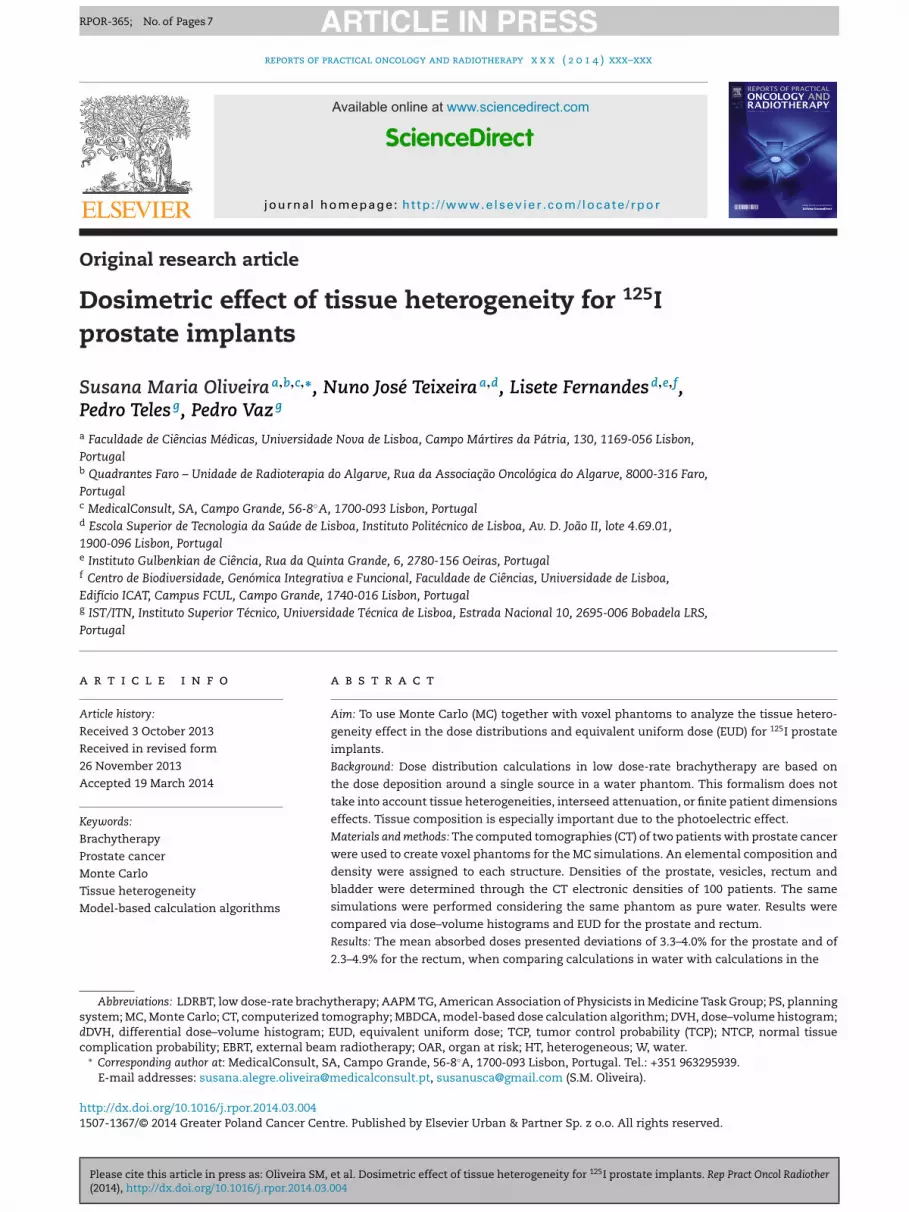
Fig. 1 – DVH obtained for the prostate A with the MonteCarlo simulation of 1E8 particles. Straight line: calculationsin the heterogeneous phantom (HT); dashed line:
with the simulations, are shown in Tables 3 and 4.Regarding the prostate results, the D90 parameter overesti-
mation was of 3.91% for prostate A and of 2.79% for prostate
a33 4.3m33 0.19TD50
33 81.9 Gy
In order to account for dose distributions heterogeneity, theoverall surviving fraction, S, was calculated through the dDVHfor each dose distribution17,22:
S =N∑
i=1
vi · S(Di) (4)
where S is the weighted average of the survival fractions takenover all N near-homogeneously irradiated sub-volumes of thetarget, and vi is the fractional dose bin Di in the dDVH.To com-pute the tumor control probability (TCP) from S, the Poissonstatistics is used:
TCP = e−K·S (5)
where K is the tumor clonogen cell number.For normal tissues, Luxton et al.26 derived an analytical
approximation to the phenomenological LKB model20,21 inorder to calculate the EUD for a given NTCP. The expressionto determinate rectum NTCP is as follows:
NTCP = 12
ek·u−(k2·u2/2) (6)
the parameter k was derived by Luxton et al.26 in order to fit theLyman equation, and u is a variable from the Lyman model,20
given by:
u = D − TD50
m · TD50, (7)
where m is a dimensionless ‘scaling’ parameter, and TD50 isthe whole organ dose for which NTCP is 50%.
The parameter values used to compute the EUD, TCP andNTCP in this work are shown in Table 2.
4. Results
Please cite this article in press as: Oliveira SM, et al. Dosimetric effect of tis(2014), http://dx.doi.org/10.1016/j.rpor.2014.03.004
4.1. Dose–volume histograms (DVH) results
The DVHs obtained when comparing the results in the hetero-geneous phantom (HT) and in water (W) in the two considered
calculations in water (W).
prostates are shown in Figs. 1–4. For prostate A, the results areshown in Fig. 1 for the prostate target volume and Fig. 2 for therectum A as an organ at risk (OAR). For prostate B, Fig. 3 showsthe prostate DVHs and Fig. 4, the rectum B. Maximum statis-tical errors were under 1% for prostates A and B, and around7% for rectum A and 5% for rectum B. Statistical errors forthe rectum are higher due to the lower doses (less particles)traversing this structure.
The results show that in the calculations where tissue het-erogeneities are not taken into account, the absorbed dose isoverestimated. A mean absorbed dose overestimation of 3.96%was obtained for prostate A and of 3.30% for prostate B. Forthe rectum, the same differences in the mean absorbed dosewere of 4.86% for rectum A and of 2.28% for rectum B. Clin-ical endpoints regarding the AAPM TG-13716 dose reportingparameters, both for the prostate and the rectum, obtained
sue heterogeneity for 125I prostate implants. Rep Pract Oncol Radiother
Fig. 2 – DVH obtained for the rectum A with the MonteCarlo simulation of 1E8 particles. Straight line: calculationsin the heterogeneous phantom (HT); dashed line:calculations in water (W).

ARTICLE IN PRESSRPOR-365; No. of Pages 7
reports of practical oncology and radiotherapy x x x ( 2 0 1 4 ) xxx–xxx 5
Table 3 – AAPM TG-13716 dose reporting parameters obtained for the prostates A and B with the simulations in theheterogeneous phantom (HT) and in water (W).
Dose parameter endpoint Prostate A Prostate B
W HT W HT
V100 > 95% 97% 97% 97% 97%V150 ≤ 50% 46% 37% 44% 36%D90 > 90 Gy 186 Gy 179 Gy 184 Gy 179 Gy
Table 4 – AAPM TG-13716 dose reporting parameters obtained for the rectums A and B with the simulations in theheterogeneous phantom (HT) and in water (W).
Dose parameter endpoint Rectum A Rectum B
W HT W HT
BrfB
4
TcrFpT
e3BTwasa
FCic
V100 < 2 cc 0.06 cc
D0.1 < 150% 101%
, when comparing the heterogeneous (HT) and water (W)esults. Concerning the rectum D0.1 parameter, a dose dif-erence of 8% was found in rectum A and of 6% in rectum.
.2. Equivalent uniform dose (EUD) results
he overestimation of the clinical dose parameters in the cal-ulations in water relative to the heterogeneous medium iseflected in the EUD determined through the DVHs shown inigs. 1–4 and Eq. (1) for the rectum and Eqs. (2)–(4) for therostate. Results for the EUD, TCP and NTCP are shown inables 5 and 6 for the prostate and rectum, respectively.
Comparing the results obtained in water and in the het-rogeneous phantom, an equivalent dose overestimation of.53% was obtained for prostate A and of 3.70% for prostate. These EUD values are reflected in a difference of 1% in theCP. For rectal lower doses, differences are more significant,ith the equivalent dose overestimated by 7.69% in rectum A
Please cite this article in press as: Oliveira SM, et al. Dosimetric effect of tis(2014), http://dx.doi.org/10.1016/j.rpor.2014.03.004
nd by 8.33% in rectum B. NTCP are very low for the rectumtructure and a difference of 0.02% was obtained for rectum And of 0.01% for rectum B.
ig. 3 – DVH obtained for the prostate B with the Montearlo simulation of 1E8 particles. Straight line: calculations
n the heterogeneous phantom (HT); dashed line:alculations in water (W).
0.00 cc 0.00 cc 0.00 cc93% 92% 86%
5. Discussion
For LDRBT, the photoelectric effect plays a significant role, asthe dose distributions are highly dependent on the atomicnumber of the irradiated tissue. Regarding, as an example,the prostate D90 parameter, a dose overestimation between2.8 and 3.9% was found, when comparing with the results inwater.
Landry et al.34 obtained an overestimation of 2.0% in D90
when comparing the dose transported in a prostate tissue withelemental composition equivalent to ours but scored in waterwith simulations in water. Chibani et al.4 found a dose over-estimation of 2.5% for the effects of anisotropy and interseedattenuation for the same D90 parameter comparing a full MCsimulation with a line-source kernel superposition method forthe 125I sources. Considering these two effects, a water-basedcalculation with the dose deposition described around a sin-gle source can lead to a dose overestimation above 6% for D90.On the other hand, Carrier et al.5 when taking into account
sue heterogeneity for 125I prostate implants. Rep Pract Oncol Radiother
both tissue heterogeneity and interseed attenuation found adose overestimation of 7.0% for D90, when comparing a clinicaltechnique with a MC simulation.
Fig. 4 – DVH obtained for the rectum B with the Monte Carlosimulation of 1E8 particles. Straight line: calculations in theheterogeneous phantom (HT); dashed line: calculations inwater (W).

ARTICLE IN PRESSRPOR-365; No. of Pages 7
6 reports of practical oncology and radiotherapy x x x ( 2 0 1 4 ) xxx–xxx
Table 5 – EUD and TCP obtained for prostates A and B with the simulations in the heterogeneous phantom (HT) and inwater (W).
Prostate A Prostate B
W HT W HT
EUD (Gy) 88 85 84 81TCP (%) 100.0 99.9 99.9 99.8
Table 6 – EUD and NTCP obtained for rectums A and B with the simulations in the heterogeneous phantom (HT) and inwater (W).
Rectum A Rectum B
W HT W HT
26
0.04
r
EUD (Gy) 28
NTCP (%) 0.06
In addition to the tissue heterogeneity and interseed atten-uation effects, there are other sources of uncertainty relatedto a water dose deposition formalism. For example, prostaticcalcifications are present to some degree in many cases andtend to increase with advancing age.35 The calcium content,due to its high cross section, increases the absorption of the125I X-rays. Meigooni et al.36 had shown that changing the cal-cium content from 1.7% to 2.3% in a solid water phantom willchange the conversion factors of a water equivalent materialto water for 125I sources up to 5%.
This study showed that taking into account the tissue het-erogeneity instead of considering the whole body as purewater may change the dose deposition recorded in the LDRBTtreatments with 125I sources. All together, the combinationof the tissue composition with other factors as the interseedattenuation and presence of calcification in the prostate mayconsiderably change the clinical dosimetry parameters. It isproven, for example, that the D90 correlates with the clini-cal outcome, and it is specially sensitive if it is near 140 Gy.37
The implementation of the MBDCA in the clinical dosimetry ofthe LDRBT will allow to overcome most of these uncertainties,enabling greater accuracy in its dose recording. However, therecommendation of the AAPM report TG-186,3 should be care-fully followed and changes in dose prescription would requiremore clinical trials.
6. Conclusions
MC simulations were performed in voxelized pelvic phantomsbased on the TC pelvic DICOM images of two patients withprostate cancer. A specific elemental composition and den-sity were assigned to each different structure in order to createanthropomorphic phantoms. Simulations in these phantomswere compared to simulations considering a simple waterphantom with the same body contour to estimate differencesin dose distributions due to the tissue heterogeneity effect.Considering either a physical DVH analysis or a biologicalEUD evaluation, simulations in water overestimate the actualabsorbed dose distributions. Mean adsorbed dose overestima-tions between 3.3 and 4.0% were found for the prostate and
Please cite this article in press as: Oliveira SM, et al. Dosimetric effect of tis(2014), http://dx.doi.org/10.1016/j.rpor.2014.03.004
between 2.3 and 4.9% for the rectum. Regarding the EUD evalu-ation, an overestimation between 3.5 and 3.7% for the prostateand between 7.7 and 8.3% for the rectum was obtained. It isexpected that the new MBDCAs that are being introduced in
26 240.04 0.03
the LDRBT PS could reduce these deviations allowing for moreprecise dose distribution evaluations in patients with prostatecancer.
Conflict of interest
None declared.
Financial disclosure
None declared.
e f e r e n c e s
1. Nath R, Anderson LL, Luxton G, Weaver KA, Williamson JF,Meigooni AS. Dosimetry of interstitial brachytherapy sources:recommendations of the AAPM Radiation TherapyCommittee Task Group No. 43. Med Phys 1995;22:209–34.
2. Rivard MJ, Coursey BM, DeWerd LA, Hanson WF, Huq MS,Ibbott GS, et al. Update of AAPM Task Group No. 43 Report: arevised AAPM protocol for brachytherapy dose calculations.Med Phys 2004;31:633–74.
3. Beaulieu L, Tedgren AC, Carrier JF, Davis SD, Mourtada F,Rivard MJ, et al. Report of the Task Group 186 on model-baseddose calculation methods in brachytherapy beyond the TG-43formalism: current status and recommendations for clinicalimplementation. Med Phys 2012;39:6208–36.
4. Chibani O, Williamson JF, Todor D. Dosimetric effects of seedanisotropy and interseed attenuation for 103Pd and 125Iprostate implants. Med Phys 2005;32:2557–66.
5. Carrier JF, D’Amours M, Verhaegen F, Reniers B, Martin AG,Vigneault E, et al. Postimplant dosimetry using Monte Carlodose calculation engine: a new clinical standard. Int J RadiatOncol Biol Phys 2007;68:1190–8.
6. Hanada T, Yorozu A, Ohashi T, Shigematsu N, Saito K,Maruyama K. The effects of tissue composition of theprostate on the dose calculation for 125I brachytherapy.Kitasato Med J 2011;41:136–44.
7. Hanada T, Yorozu A, Ohashi T, Shigematsu N, Maruyama K.Evaluation of the dosimetric parameters for 125Ibrachytherapy determined in prostate medium using CT
sue heterogeneity for 125I prostate implants. Rep Pract Oncol Radiother
images. J Radiat Res 2010;51:553–61.8. Demarco JJ, Smathers JB, Burnison CM, Ncube QK, Solberg TD.
CT-based dosimetry calculations for 125I prostate implants.Int J Radiat Oncol Biol Phys 1999;45:1347–53.

ARTICLE IN PRESSRPOR-365; No. of Pages 7
radio
1
1
1
1
1
1
1
1
1
1
2
2
2
2
2
2
2
2
2
2
3
3
3
3
3
3
3
brachytherapy sources. Med Phys 2006;33:3988–92.37. Stock RG, Stone NN, Tabert A, Iannuzzi C, DeWyngaert JK. A
reports of practical oncology and
9. X-5 Monte Carlo Team. MCNP: a general Monte Carlo N-particletransport code ver 5 vol 1. Overview and theory LA-UR-03-1987.Los Alamos, NM, USA: Los Alamos National Laboratory; 2003[revised 2008].
0. National Nuclear Data Center. Cross Section Evaluation WorkingGroup. ENDF-201: ENDF/B-VI summary documentation. 8th ed.Upton, NY: Brookhaven National Laboratory reportBNL-NCS-17541; 2000.
1. Cullen DE, Hubbell JH, Kissel L. EPDL97: the evaluated PhotonData Library, ‘97 Version. Lawrence Livermore National Laboratory.Report No UCRL-50400 vol 6 rev 5. Gaithersburg, MD: NationalInstitute of Standards and Technology; 1997.
2. Rasband WS. ImageJ. Bethesda, MD, USA: U.S. NationalInstitutes of Health; 1997–2012 http://imagej.nih.gov/ij/
3. International Commission on Radiological Protection. Basicanatomical and physiological data for use in radiological protection:reference values. ICRP Publication No. 89. Oxford: Pergamon;2002.
4. International Commission on Radiation Units andMeasurements. Tissue substitutes in radiation dosimetry andmeasurement, ICRU Report No. 44. Bethesda, MD: ICRUPublications; 1989.
5. Dolan J, Zuofeng L, Williamson JF. Monte Carlo andexperimental dosimetry of an 125I brachytherapy seed. MedPhys 2006;33:4675–84.
6. Nath R, Bice WS, Butler WM, Chen Z, Meigooni AS, NarayanaV, et al. AAPM Task Group No. 137 report: AAPMrecommendations on dose prescription and reportingmethods for permanent interstitial brachytherapy forprostate cancer. Med Phys 2009;36:5310–22.
7. Nimierko A. Reporting and analyzing dose distributions: aconcept of equivalent uniform dose. Med Phys 1997;24:103–10.
8. Nimierko A. A generalized concept of equivalent uniformdose (EUD). Med Phys 1999:26 [Abstract].
9. Wu Q, Mohan R, Niemierko A, Schmidt-Ullrich R.Optimization of intensity-modulated radiotherapy plansbased on the equivalent uniform dose. Int J Radiat Oncol BiolPhys 2002;52:224–35.
0. Lyman JT. Complication probability as assessed fromdose–volume histograms. Radiat Res 1985;104:S13–9.
1. Kutcher GJ, Burman C. Calculation probability factors fornon-uniform normal tissue irradiation: the effective volumemethod. Int J Radiat Oncol Biol Phys 1989;16:1623–30.
2. Wang JZ, Li XA. Evaluation of external beam radiotherapyand brachytherapy for localized prostate cancer using
Please cite this article in press as: Oliveira SM, et al. Dosimetric effect of tis(2014), http://dx.doi.org/10.1016/j.rpor.2014.03.004
equivalent uniform dose. Med Phys 2003;30:34–40.3. Dale RG. The application of the linear-quadratic dose–effect
equation to fractionated and protracted radiotherapy. Br JRadiol 1985;58:515–28.
therapy x x x ( 2 0 1 4 ) xxx–xxx 7
4. Dale RG. Radiobiological assessment of permanent implantsusing tumor repopulation factors in linear-quadratic mode.Br J Radiol 1989;62:241–4.
5. Antipas V, Dale RG, Coles IP. A theoretical investigation intothe role of tumour radiosensitivity, clonogen repopulation,tumour shrinkage and radionuclide RBE in permanentbrachytherapy implants of 125I and 103Pd. Phys Med Biol2001;46:2557–69.
6. Luxton G, Keall PJ, King CR. A new formula for normal tissuecomplication probability (NTCP) as a function of equivalentuniform dose (EUD). Phys Med Biol 2008;53:23–36.
7. Wang JZ, Guerrero M, Li XA. How low is the alpha/beta ratiofor prostate cancer? Int J Radiat Oncol Biol Phys2003;55:194–203.
8. Haustermans KM, Hofland I, Van Poppel H, Oyen R, Van deVoorde W, Begg AC, et al. Cell kinetic measurements inprostate cancer. Int J Radiat Oncol Biol Phys 1997;37:1067–70.
9. Hausterman K, Fowler JF. A comment on proliferation rates inhuman prostate cancer. Int J Radiat Oncol Biol Phys 2000;48:303.
0. Oliveira SM, Teixeira NJ, Fernandes L. What do we knowabout the ˛/ for prostate cancer? Med Phys 2012;39:3189–201.
1. Brenner D. Fractionation and late rectal toxicity. Int J RadiatOncol Biol Phys 2004;60:1013–5.
2. Brenner D, Armour E, Corry P, Hall E. Sublethal damage repairtimes for a late-responding tissue relevant to brachytherapy(and external-beam radiotherapy): implications for newbrachytherapy protocols. Int J Radiat Oncol Biol Phys1998;41:135–8.
3. Rancati T, Fiorino C, Gagliardi G, Cattaneo GM, Sanguineti G,Borca VC, et al. Fitting late rectal bleeding data usingdifferent NTCP models: results from an Italian multi-centricstudy (AIROPROS0101). Radiother Oncol 2004;73:21–32.
4. Landry G, Renier B, Murrer L, Lutgens L, Van Gurp EB, PignolJP, et al. Sensitivity of low energy brachytherapy Monte Carlodose calculations to uncertainties in human tissuecomposition. Med Phys 2010;37:5188–98.
5. Tvedt KE, Kopstad G, Haugen OA, Halgunset J. Subcellularconcentrations of calcium, zinc, and magnesium in benignnodular hyperplasia of the human prostate: X-raymicroanalysis of freeze-dried cryosections. Cancer Res1987;47:323–8.
6. Meigooni AS, Awan SB, Thompson NS, Dini SA. Updated SolidWaterTM to water conversion factors for 125I and 103Pd
sue heterogeneity for 125I prostate implants. Rep Pract Oncol Radiother
dose–response study for I-125 prostate implants. Int J RadiatOncol Biol Phys 1998;41:101–8.


![Specific [125I]brain natriuretic peptide-26 binding sites in rat and pig kidneys](https://img.dokumen.tips/doc/110x75/6361ca65c78c28495d0b9470/specific-125ibrain-natriuretic-peptide-26-binding-sites-in-rat-and-pig-kidneys.jpg)


