Embed Size (px)
Citation preview
TH
EJ
OU
RN
AL
OF
CE
LL
BIO
LO
GY
©
The Rockefeller University Press $8.00The Journal of Cell Biology, Vol. 167, No. 5, December 6, 2004 841–849http://www.jcb.org/cgi/doi/10.1083/jcb.200406048
JCB: ARTICLE
JCB 841
DNA replication checkpoint control of Wee1 stability by vertebrate Hsl7
Ayumi Yamada, Brad Duffy, Jennifer A. Perry, and Sally Kornbluth
Department of Pharmacology and Cancer Biology, Duke University Medical Center, Durham, NC 27710
2/M checkpoints prevent mitotic entry uponDNA damage or replication inhibition by tar-geting the Cdc2 regulators Cdc25 and Wee1.
Although Wee1 protein stability is regulated by DNA-responsive checkpoints, the vertebrate pathways control-ling Wee1 degradation have not been elucidated. Inbudding yeast, stability of the Wee1 homologue, Swe1,is controlled by a regulatory module consisting of theproteins Hsl1 and Hsl7 (histone synthetic lethal 1 and 7),which are targeted by the morphogenesis checkpoint toprevent Swe1 degradation when budding is inhibited.We report here the identification of
Xenopus
Hsl7 as a
G
positive regulator of mitosis that is controlled, instead,by an entirely distinct checkpoint, the DNA replicationcheckpoint. Although inhibiting Hsl7 delayed mitosis,Hsl7 overexpression overrode the replication check-point, accelerating Wee1 destruction. Replication check-point activation disrupted Hsl7–Wee1 interactions, butbinding was restored by active polo-like kinase. Thesedata establish Hsl7 as a component of the replicationcheckpoint and reveal that similar cell cycle controlmodules can be co-opted for use by distinct checkpointsin different organsims.
Introduction
Entry into mitosis in all eukaryotic cells is controlled by theCdc2 (cell division control protein 2)–cyclin B kinase complex.Activation of this complex is restrained in the presence of dam-aged or incompletely replicated DNA by checkpoint pathwaysthat act to maintain inhibitory phosphorylations of Cdc2 on Thr14 and Tyr 15 (Elledge, 1996; Lew and Kornbluth, 1996).These sites are phosphorylated by related kinases, Wee1 andMyt1, and are dephosphorylated by the Cdc25 phosphatase.Accordingly, checkpoint pathways act to inhibit Cdc25 func-tion and ensure the continued Cdc2-suppressive activity ofWee1 (and possibly Myt1). Recently, it has been reported thatWee1 protein stability is controlled by DNA-responsive check-point pathways in extracts prepared from
Xenopus
eggs. In-triguingly, it was found that Wee1 is stabilized during DNAreplication (or following DNA damage) and that it is degradedwithin nuclei at the time of mitotic entry (Michael and New-port, 1998). In
Xenopus laevis
it has been reported that Wee1ubiquitination and proteasomal degradation are driven by aSkp1–cullin–F box (SCF) E3 ligase complex containing a
novel F-box protein, Tome-1 (trigger of mitotic entry; Ayad etal., 2003). Human Wee1A, however, is reportedly ubiquiti-nated by an SCF complex containing
�
-TrCP (
�
transducinrepeat containing protein; Watanabe et al., 2004). This differencemay reflect either species differences or the fact that an embry-onic form of Wee1 was studied in
Xenopus
whereas a somaticform of Wee1 was analyzed in human cells. Other vertebratefactors responsible for promoting Wee1 degradation or linkingit to DNA-responsive checkpoints have not yet been identified.
In the yeast
Saccharomyces cerevisiae
, the stability of theWee1 homologue, Swe1 is under the control of a checkpointpathway monitoring budding and the status of the actin cyto-skeleton (the morphogenesis checkpoint; Sia et al., 1996). Thischeckpoint is thought to prevent mitotic entry, at least in part,through stabilization of Swe1 (Sia et al., 1998). Looking to thissystem for candidate Wee1 regulators, we were struck by therequirement for an evolutionarily conserved methyltransferase,Hsl7 (histone synthetic lethal 7), in promoting Wee1 degrada-tion. The morphogenesis checkpoint regulates interaction ofHsl7 with an associated kinase, Hsl1, to prevent Swe1 degrada-tion. Consistent with these observations, overexpression ofHsl7 can override the morphogenesis checkpoint, allowing mitoticentry even when budding is inhibited (McMillan et al., 1999).These data place
S. cerevisiae
Hsl7 as a central regulator ofSwe1 protein stability. In contrast, the Hsl7 homologue (Skb1)in the fission yeast,
Schizosaccharomyces pombe
, appears to
A. Yamada and B. Duffy contributed equally to this work.Correspondence to Sally Kornbluth: [email protected] used in this paper:
�
-TrCP,
�
transducin repeat containing pro-tein; ELB, egg lysis buffer;
GVBD, germinal vesicle breakdown; Hsl7, histonesynthetic lethal 7; JBP1, Janus kinase binding protein 1; Plk1, Polo-like kinase;Plx1,
Xenopus
Polo-like kinase;
SCF, Skp1–cullin–F box.
on March 4, 2014
jcb.rupress.orgD
ownloaded from
Published December 6, 2004
JCB • VOLUME 167 • NUMBER 5 • 2004842
have a cell cycle role diametrically opposed to that of
S. cerevi-siae
Hsl7. Specifically, Skb1 has been reported to inhibit,rather than promote, mitotic entry through direct binding to themitotic Cdc2–cyclin complex (Gilbreth et al., 1998). In addi-tion to its activity in cell cycle regulation, Skb1 has been re-ported to regulate morphogenesis (in association with a PAKfamily kinase) and to be involved in the hyperosmotic stressresponse, which stimulates Skb1 methyltransferase activity (Yanget al., 1999; Bao et al., 2001).
Although Hsl7 homologues have been reported in verte-brates, including humans, analyses of their potential role(stimulatory or inhibitory) in controlling mitotic entry hasbeen hampered by a potentially distinct requirement for verte-brate Hsl7 as a component of the methylosome regulatingmRNA splicing (Friesen et al., 2001). Therefore, to analyzeHsl7 cell cycle function we turned to the
Xenopus
egg extract,which can undergo multiple cell cycles in vitro without anyde novo mRNA transcription, allowing an analysis of cell cy-cle regulation in a system free of confounding effects onmRNA processing (Friesen et al., 2001). We report here that
Xenopus
Hsl7 controls entry into M phase by controlling theintranuclear stability of Wee1. Moreover, just as overproduc-tion of Hsl7 can override the morphogenesis checkpoint inbudding yeast, overproduction of
Xenopus
Hsl7 can short-cir-cuit the DNA replication checkpoint, allowing mitotic entryin the presence of incompletely replicated DNA. These datastrongly suggest that the Hsl7–Wee1 cell cycle control mod-ule can be used for controlling entry into mitosis in verte-brates, as well as in
S. cerevisiae
, but that this module has
been co-opted by vertebrates to respond to a different check-point signal than in yeast.
Results
Identification of
Xenopus
Hsl7
To identify potential
Xenopus
Hsl7 homologues, we searchedthe Washington University
Xenopus
EST database, and iden-tified five Hsl7-related EST clones of various lengths. AfterDNA sequencing, we identified a full-length
Xenopus
Hsl7cDNA (hereafter referred to as xHsl7) bearing 26% identity/44% similarity to yeast Hsl7 and 83% identity/92% similar-ity to human Hsl7 (known as JBP1 [Janus kinase bindingprotein 1]/PRMT5; Genbank/EMBL/DDBJ accession no.AY535008) (Fig. 1). We were further encouraged to pursueanalysis of this clone as we found that recombinant
S. cerevi-siae
Hsl7 protein readily bound
Xenopus
Wee1 in eggextracts (Fig. 2 A). This suggested that the Hsl7–Wee1 inter-action might be conserved and that the
Xenopus
proteinmight also play a role in regulating Wee1 function and mi-totic entry.
Overexpression of xHsl7 accelerates entry into mitosis
As a first step in evaluating xHsl7 function, we wished tooverexpress xHsl7 in cycling extracts of
Xenopus
eggs, whichare able to oscillate between S and M phases of the cell cycle.For this purpose, we supplemented translationally competent(though transcriptionally inactive) cycling egg extracts with
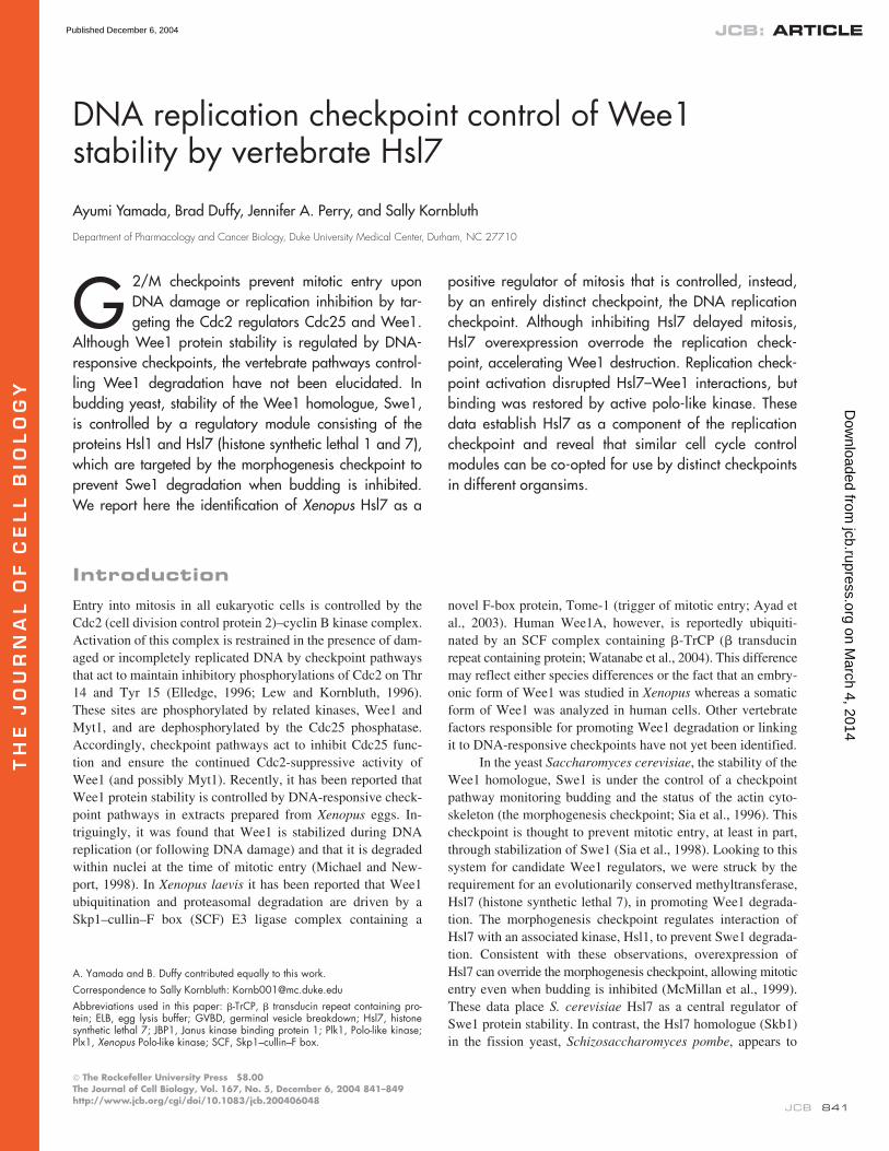
Figure 1. Amino acid sequence alignment ofXenopus, human and yeast Hsl7 proteins. Theamino acid sequences of Hsl7 orthologs werealigned using the ClustalW alignment parame-ters in the program MacVector. Dark shadingindicates identities and light shading indicatessimilarities.
on March 4, 2014
jcb.rupress.orgD
ownloaded from
Published December 6, 2004
HSL7 REGULATION OF DNA REPLICATION CHECKPOINT • YAMADA ET AL.
843
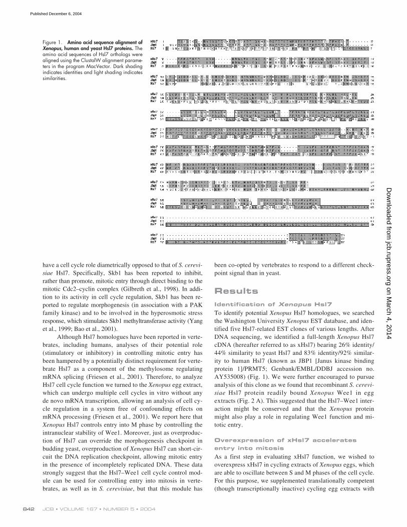
excess mRNA encoding xHsl7, allowing production of xHsl7to levels approximately twofold that found endogenously (un-published data). Under these conditions, we found that excessxHsl7 could markedly accelerate entry into mitosis, as shownby both microscopic monitoring of nuclear envelope break-down and assays of histone H1-directed Cdc2 kinase activity(Fig. 2 B). Interestingly, this acceleration was absent in ex-tracts that had not been supplemented with nuclei, suggestingthat the effects of xHsl7 on the cell cycle were nuclear depen-dent (Fig. 2 C). (Note that the extract shown here is distinctfrom the one used in Fig. 2 B. In this and subsequent figures,the absolute timing of mitosis differed from extract to extract,but the relative rates of mitotic entry under different treatmentconditions remained constant.) The different response of ex-tracts containing or lacking nuclei to xHsl7 addition did notreflect any differences in the intrinsic stability of xHsl7 pro-tein under these conditions, since xHsl7 was equally stable inthe presence and absence of nuclei (Fig. 2 D). In addition, anxHsl7 mutant altered at well-conserved residues required forprotein methyltransferase activity was as potent as wild typein accelerating mitotic entry, consistent with recent reportsthat methyltransferase activity is dispensable for Hsl7-medi-ated cell cycle regulation in
S. cerevisiae
(unpublished data;Ma, 2000; Theesfeld et al., 2003).
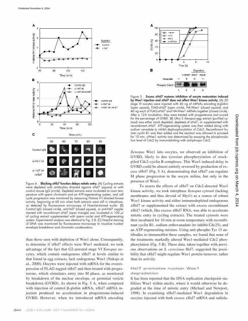
Depletion or inhibition of Hsl7 delays the entry into mitosis
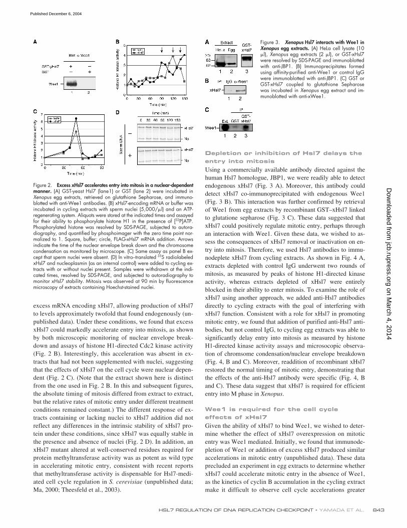
Using a commercially available antibody directed against thehuman Hsl7 homologue, JBP1, we were readily able to detectendogenous xHsl7 (Fig. 3 A). Moreover, this antibody coulddetect xHsl7 co-immunoprecipitated with endogenous Wee1(Fig. 3 B). This interaction was further confirmed by retrievalof Wee1 from egg extracts by recombinant GST–xHsl7 linkedto glutatione sepharose (Fig. 3 C). These data suggested thatxHsl7 could positively regulate mitotic entry, perhaps throughan interaction with Wee1. Given these data, we wished to as-sess the consequences of xHsl7 removal or inactivation on en-try into mitosis. Therefore, we used Hsl7 antibodies to immu-nodeplete xHsl7 from cycling extracts. As shown in Fig. 4 A,extracts depleted with control IgG underwent two rounds ofmitosis, as measured by peaks of histone H1-directed kinaseactivity, whereas extracts depleted of xHsl7 were entirelyblocked in their ability to enter mitosis. To examine the role ofxHsl7 using another approach, we added anti-Hsl7 antibodiesdirectly to cycling extracts with the goal of interfering withxHsl7 function. Consistent with a role for xHsl7 in promotingmitotic entry, we found that addition of purified anti-Hsl7 anti-bodies, but not control IgG, to cycling egg extracts was able tosignificantly delay entry into mitosis as measured by histoneH1-directed kinase activity assays and microsocopic observa-tion of chromsome condensation/nuclear envelope breakdown(Fig. 4, B and C). Moreover, readdition of recombinant xHsl7restored the normal timing of mitotic entry, demonstrating thatthe effects of the anti-Hsl7 antibody were specific (Fig. 4, Band C). These data suggest that xHsl7 is required for efficiententry into M phase in
Xenopus
.
Wee1 is required for the cell cycle effects of xHsl7
Given the ability of xHsl7 to bind Wee1, we wished to deter-mine whether the effect of xHsl7 overexpression on mitoticentry was Wee1 mediated. Initially, we found that immunode-pletion of Wee1 or addition of excess xHsl7 produced similaraccelerations in mitotic entry (unpublished data). These dataprecluded an experiment in egg extracts to determine whetherxHsl7 could accelerate mitotic entry in the absence of Wee1,as the kinetics of cyclin B accumulation in the cycling extractmake it difficult to observe cell cycle accelerations greater
Figure 2. Excess xHsl7 accelerates entry into mitosis in a nuclear-dependentmanner. (A) GST-yeast Hsl7 (lane1) or GST (lane 2) were incubated inXenopus egg extracts, retrieved on glutathione Sepharose, and immuno-blotted with anti-Wee1 antibodies. (B) xHsl7-encoding mRNA or buffer wasincubated in cycling extracts with sperm nuclei (5,000/�l) and an ATP-regenerating system. Aliquots were stored at the indicated times and assayedfor their ability to phosphorylate histone H1 in the presence of [32P]ATP.Phosphorylated histone was resolved by SDS-PAGE, subjected to autora-diography, and quantified by phosphoimager with the zero time point nor-malized to 1. Square, buffer; circle, FLAG-xHsl7 mRNA addition. Arrowsindicate the time of the nuclear envelope break down and the chromosomecondensation as monitored by microscope. (C) Same assay as panel B ex-cept that sperm nuclei were absent. (D) In vitro–translated 35S radiolabeledxHsl7 and nucleoplasmin (as an internal control) were added to cycling ex-tracts with or without nuclei present. Samples were withdrawn at the indi-cated times, resolved by SDS-PAGE, and subjected to autoradiography tomonitor xHsl7 stability. Mitosis was observed at 90 min by fluorescencemicroscopy of extracts containing Hoechst-stained nuclei.
Figure 3. Xenopus Hsl7 interacts with Wee1 inXenopus egg extracts. (A) HeLa cell lysate (10�l), Xenopus egg extracts (2 �l), or GST-xHsl7were resolved by SDS-PAGE and immunoblottedwith anti-JBP1. (B) Immunoprecipitates formedusing affinity-purified anti-Wee1 or control IgGwere immunoblotted with anti-JBP1. (C) GST orGST-xHsl7 coupled to glutathione Sepharosewas incubated in Xenopus egg extract and im-munoblotted with anti-xWee1.
on March 4, 2014
jcb.rupress.orgD
ownloaded from
Published December 6, 2004
JCB • VOLUME 167 • NUMBER 5 • 2004844
than those seen with depletion of Wee1 alone. Consequently,to determine if xHsl7 effects were Wee1 mediated, we tookadvantage of the fact that G2-arrested stage VI
Xenopus
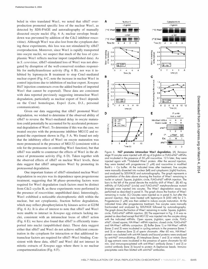
oo-cytes, which contain endogenous xHsl7 at levels similar tothat found in egg extracts, lack endogenous Wee1 (Nakajo etal., 2000). Oocytes were injected with mRNA for the overex-pression of FLAG-tagged xHsl7 and then treated with proges-terone, which stimulates entry into M phase, as monitoredby breakdown of the nuclear envelope, or germinal vesiclebreakdown (GVBD). As shown in Fig. 5 A, when comparedwith injection of control
�
-globin mRNA, xHsl7 mRNA in-jection produced no acceleration in progesterone-inducedGVBD. However, when we introduced mRNA encoding
Xenopus
Wee1 into oocytes, we observed an inhibition ofGVBD, likely to due tyrosine phosphorylation of stock-piled Cdc2–cyclin B complexes. This Wee1-induced delay inGVBD could be almost entirely reversed by production of ex-cess xHsl7 (Fig. 5 A), demonstrating that xHsl7 can regulateM phase progression in the oocyte milieu, but only in thepresence of Wee1.
To assess the effects of xHsl7 on Cdc2-directed Wee1kinase activity, we took interphase
Xenopus
cytosol (lackingmembranes and thus devoid of Myt1) as a source of robustWee1 kinase activity and either immunodepleted endogenousxHsl7 or supplemented the extract with excess recombinantxHsl7 (which, like excess xHsl7 RNA, was able to acceleratemitotic entry in cycling extracts). The treated cytosols werethen incubated for 10 min at room temperature with recombi-nant cyclin B1, sodium orthovanadate (to inhibit Cdc25), andan ATP-regenerating mixture. Using anti-phospho Tyr 15 an-tibodies to immunoblot these samples, we found that none ofthe treatments markedly altered Wee1-mediated Cdc2 phos-phorylation (Fig. 5 B). These data, taken together with previ-ous observations on
S. cerevisiae
Hsl7, suggested the possi-bility that xHsl7 might regulate Wee1 protein turnover, ratherthan its activity.
Hsl7 promotes nuclear Wee1 degradation
It has been reported that the DNA replication checkpoint sta-bilizes Wee1 within nuclei, where it would otherwise be de-graded at the time of mitotic entry (Michael and Newport,1998). In examining xHsl7-mediated Wee1 degradation inoocytes injected with both excess xHsl7 mRNA and radiola-
Figure 4. Blocking xHls7 function delays mitotic entry. (A) Cycling extractswere depleted with antibodies directed against xHsl7 (square) or withcontrol mouse IgG (circle). Depleted extracts were incubated at room tem-perature with sperm chromatin and an ATP-regenerating system, and cellcycle progression was monitored by measuring Histone H1-directed kinaseactivity, beginning at 60 min when both extracts were still in interphase,as detected by fluorescence microscopy of Hoechst-stained nuclei. (B)Control IgG (closed circle), anti-Hsl7 (closed square), or anti-Hsl7 supple-mented with recombinant xHsl7 (open triangle) was incubated in 100 �lof cycling extract supplemented with sperm nuclei and ATP-regeneratingsystem. Experimental analysis was identical to Fig. 2 B. (C) Hoechst stainingof DNA was monitored by fluorescence microscopy to visualize nuclearenvelope breakdown and chromatin condensation.
Figure 5. Excess xHsl7 restores inhibition of oocyte maturation inducedby Wee1 injection and xHsl7 does not affect Wee1 kinase activity. (A) 50stage VI oocytes were injected with 40 ng of mRNAs encoding �-globin(open square), FLAG-xHsl7 (open circle), HA-Wee1 (closed square), and40 ng each of FLAG-xHsl7 and HA-Wee1 mRNAs together (closed circle).After a 12-h incubation, they were treated with progesterone and scoredfor the percentage of GVBD. (B) Ultra S Xenopus egg extract (purified cy-tosol) was either mock depleted, depleted of xHsl7, or supplemented withrecombinant xHsl7. ATP-regenerating system was then added along withsodium vanadate to inhibit dephosphorylation of Cdc2. Recombinant hu-man cyclin B1 was then added and the reaction was allowed to proceedfor 10 min. xWee1 activity was determined by assaying the phosphoryla-tion level of Cdc2 by immunoblotting with anti-phospo Cdc2.
on March 4, 2014
jcb.rupress.orgD
ownloaded from
Published December 6, 2004
HSL7 REGULATION OF DNA REPLICATION CHECKPOINT • YAMADA ET AL.
845
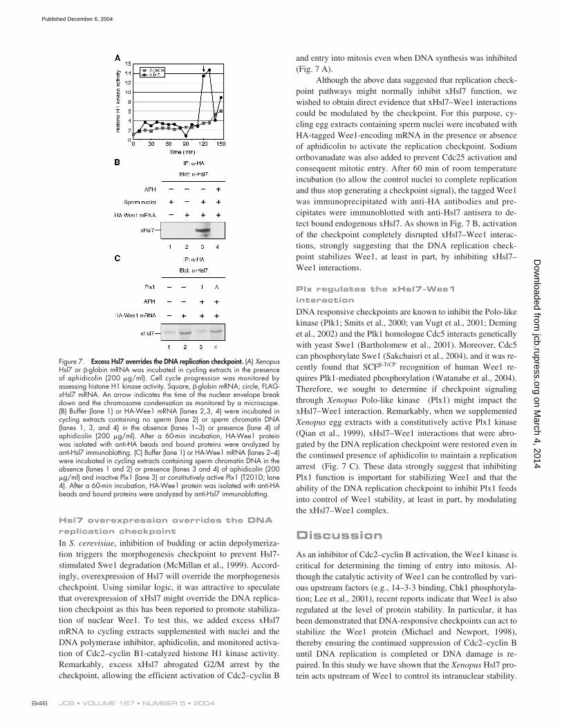
beled in vitro translated Wee1, we noted that xHsl7 over-production promoted specific loss of the nuclear Wee1, asdetected by SDS-PAGE and autoradiography of manuallydissected oocyte nuclei (Fig. 6 A; nuclear envelope break-down was prevented by addition of the Cdc2 inhibitor rosco-vitine). Although Wee1 was also lost from the cytoplasm dur-ing these experiments, this loss was not stimulated by xHsl7overproduction. Moreover, since Wee1 is rapidly transportedinto oocyte nuclei, we suspect that much of the loss of cyto-plasmic Wee1 reflects nuclear import (unpublished data). Asin
S. cerevisiae
, xHsl7-stimulated loss of Wee1 was not abro-gated by disruption of the well-conserved residues responsi-ble for methyltransferase activity (Fig. 6 B), nor was it in-hibited by leptomycin B treatment to stop Crm1-mediatednuclear export (Fig. 6 C; note the increase in nuclear Wee1 incontrol injections due to inhibition of nuclear export.
Xenopus
Hsl7 injection counteracts even the added burden of importedWee1 that cannot be exported). These data are consistentwith data reported previously suggesting intranuclear Wee1degradation, particularly as nuclear export of Swe1 dependson the Crm1 homologue, Expo1 (Lew, D.J., personalcommunication).
Given our data suggesting that xHsl7 promoted Wee1degradation, we wished to determine if the observed ability ofxHsl7 to reverse the Wee1-mediated delay in oocyte matura-tion could potentially be accounted for by accelerated proteaso-mal degradation of Wee1. To determine if this was the case, wetreated oocytes with the proteasome inhibitor MG132 and re-peated the experiment shown in Fig. 5 A. We found not onlythat the inhibitory effect of Wee1 on oocyte maturation wasmore pronounced in the presence of MG132 (consistent with arole for the proteasome in controlling Wee1 function), but thatxHsl7 was unable to counteract the effects of Wee1 in the ab-sence of proteasome activity (Fig. 6 D). Taken together withthe observed effects of xHsl7 on nuclear Wee1 levels, thesedata suggest that xHsl7 antagonizes Wee1 by promoting itsproteasomal degradation.
One important feature of xHsl7-stimulated nuclear Wee1degradation in oocytes was its dependence upon progesteronetreatment, suggesting that M phase–promoting factors wererequired for Wee1 degradation (such factors must be distinctfrom Cdc2–cyclin B, as these experiments were performed inthe presence of roscovitine; unpublished data). Interestingly,Wee1 exhibited a noticeable electrophoretic shift within thenuclear, but not cytoplasmic, fraction before degradation,which may reflect phosphorylation by kinases active at G2/M(Fig. 6 A). It is also of interest to note that xHsl7 and Wee1were unable to interact in
Xenopus
egg extracts lacking nu-clei, consistent with an intranuclear locus of xHsl7 action(Fig. 6 E); we have also found that xHsl7 is efficiently trans-ported into nuclei (unpublished data). These data suggesteither that xHsl7 and Wee1 do not achieve sufficient concen-tration in the cytoplasm for interaction or that additional in-tranuclear factors are required for xHsl7–Wee1 binding. Con-sistent with these data, xHsl7 and Wee1 did not interact inmitotic extracts of
Xenopus
eggs where there is no nuclearcompartmentalization (Fig. 6 F).
Figure 6. Hsl7 promotes intranuclear Wee1 degradation. (A) Xenopusstage VI oocytes were injected with 40 ng of �-globin or FLAG-xHsl7 mRNAsand incubated in the presence of 50 �M roscovitine. 12 h later, they wereinjected again with 35S-labeled Wee1 protein. After the second injection,they were treated with progesterone (1 �M) and roscovitine in modifiedBarth’s � Ca buffer. At the indicated times after treatment, five oocyteswere manually dissected into nuclear (left) and cytoplasmic (right) fractions,and analyzed by SDS-PAGE and autoradiography. The graph represents aquantitation of the data above showing the fraction of Wee1 remaining innuclei or cytosol. Square, �-globin; circle, FLAG-xHsl7 mRNA injection. Thebars to the left of the panel denote the mobility shift of Wee1. (B) 40 ngmRNAs of FLAG-xHsl7 (circle) and FLAG-xHsl7 metyltransferase mutant(triangle) were injected into oocytes. The Wee1 degradation assay wasperformed as described in panel A. The graph shows the fraction of Wee1remaining in nuclei. (C) Oocytes were injected as in panel A, but after thesecond injection, they were treated with leptomycin B (200 nM) for 2 h.Progesterone (1 �M) was then added to induce oocyte maturation. At theindicated times after progesterone treatment, five oocytes were manuallyfractionated and analyzed by SDS-PAGE followed by autoradiography.The graph shows the fraction of Wee1 remaining in nuclei. Square, �-globin;circle, FLAG-xHsl7 mRNA injection. (D) The experiment in Fig. 5 A was re-peated as described except that MG132 was injected into the oocytes alongwith the indicated mRNAs. Open square, �-globin; open circle, FLAG-xHsl7; closed square, HA-Wee1 mRNA injection; closed circle, FLAG-xHsl7and HA-Wee1 mRNAs coinjection. (E) Buffer (lane 1) or HA-Wee1 mRNA(lanes 2 and 3) were incubated in cycling extracts in the presence (lanes 1and 3) or absence (lane 2) of sperm chromatin. After 60 min, HA-Wee1protein was isolated with anti-HA beads and bound proteins were analyzedby anti-Hsl7 immunoblotting. (F) Interphase (lanes 1 and 3) or mitotic (lane2) egg extracts were incubated in the presence of sperm chromatin for 60min, and immunoprecipitated with anti-Wee1 antibody (lanes 1 and 2) orcontrol antibody (lane 3) bound to protein A–Sepharose beads. Immuno-precipitates were then blotted with anti-Hsl7 antibodies.
on March 4, 2014
jcb.rupress.orgD
ownloaded from
Published December 6, 2004
JCB • VOLUME 167 • NUMBER 5 • 2004846
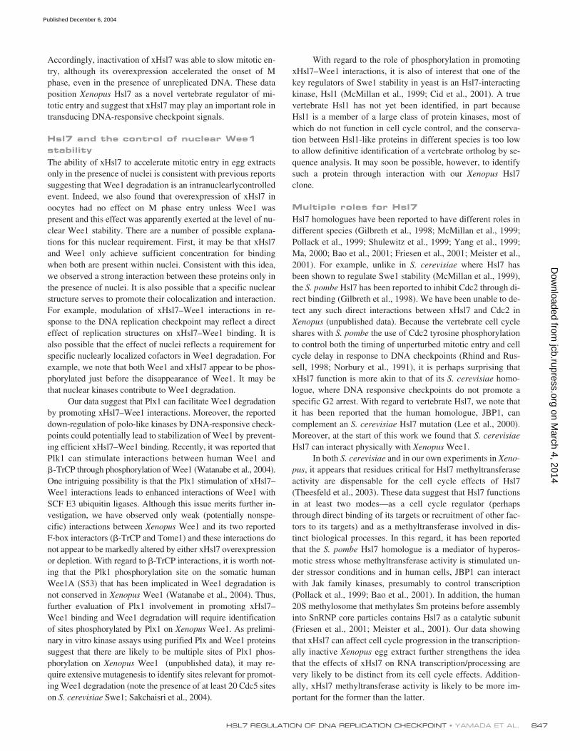
Hsl7 overexpression overrides the DNA replication checkpoint
In
S. cerevisiae
, inhibition of budding or actin depolymeriza-tion triggers the morphogenesis checkpoint to prevent Hsl7-stimulated Swe1 degradation (McMillan et al., 1999). Accord-ingly, overexpression of Hsl7 will override the morphogenesischeckpoint. Using similar logic, it was attractive to speculatethat overexpression of xHsl7 might override the DNA replica-tion checkpoint as this has been reported to promote stabiliza-tion of nuclear Wee1. To test this, we added excess xHsl7mRNA to cycling extracts supplemented with nuclei and theDNA polymerase inhibitor, aphidicolin, and monitored activa-tion of Cdc2–cyclin B1-catalyzed histone H1 kinase activity.Remarkably, excess xHsl7 abrogated G2/M arrest by thecheckpoint, allowing the efficient activation of Cdc2–cyclin B
and entry into mitosis even when DNA synthesis was inhibited(Fig. 7 A).
Although the above data suggested that replication check-point pathways might normally inhibit xHsl7 function, wewished to obtain direct evidence that xHsl7–Wee1 interactionscould be modulated by the checkpoint. For this purpose, cy-cling egg extracts containing sperm nuclei were incubated withHA-tagged Wee1-encoding mRNA in the presence or absenceof aphidicolin to activate the replication checkpoint. Sodiumorthovanadate was also added to prevent Cdc25 activation andconsequent mitotic entry. After 60 min of room temperatureincubation (to allow the control nuclei to complete replicationand thus stop generating a checkpoint signal), the tagged Wee1was immunoprecipitated with anti-HA antibodies and pre-cipitates were immunoblotted with anti-Hsl7 antisera to de-tect bound endogenous xHsl7. As shown in Fig. 7 B, activationof the checkpoint completely disrupted xHsl7–Wee1 interac-tions, strongly suggesting that the DNA replication check-point stabilizes Wee1, at least in part, by inhibiting xHsl7–Wee1 interactions.
Plx regulates the xHsl7–Wee1 interaction
DNA responsive checkpoints are known to inhibit the Polo-likekinase (Plk1; Smits et al., 2000; van Vugt et al., 2001; Deminget al., 2002) and the Plk1 homologue Cdc5 interacts geneticallywith yeast Swe1 (Bartholomew et al., 2001). Moreover, Cdc5can phosphorylate Swe1 (Sakchaisri et al., 2004), and it was re-cently found that SCF
�
-TrCP
recognition of human Wee1 re-quires Plk1-mediated phosphorylation (Watanabe et al., 2004).Therefore, we sought to determine if checkpoint signalingthrough
Xenopus
Polo-like kinase (Plx1) might impact thexHsl7–Wee1 interaction. Remarkably, when we supplemented
Xenopus
egg extracts with a constitutively active Plx1 kinase(Qian et al., 1999), xHsl7–Wee1 interactions that were abro-gated by the DNA replication checkpoint were restored even inthe continued presence of aphidicolin to maintain a replicationarrest (Fig. 7 C). These data strongly suggest that inhibitingPlx1 function is important for stabilizing Wee1 and that theability of the DNA replication checkpoint to inhibit Plx1 feedsinto control of Wee1 stability, at least in part, by modulatingthe xHsl7–Wee1 complex.
Discussion
As an inhibitor of Cdc2–cyclin B activation, the Wee1 kinase iscritical for determining the timing of entry into mitosis. Al-though the catalytic activity of Wee1 can be controlled by vari-ous upstream factors (e.g., 14–3-3 binding, Chk1 phosphoryla-tion; Lee et al., 2001), recent reports indicate that Wee1 is alsoregulated at the level of protein stability. In particular, it hasbeen demonstrated that DNA-responsive checkpoints can act tostabilize the Wee1 protein (Michael and Newport, 1998),thereby ensuring the continued suppression of Cdc2–cyclin Buntil DNA replication is completed or DNA damage is re-paired. In this study we have shown that the
Xenopus
Hsl7 pro-tein acts upstream of Wee1 to control its intranuclear stability.
Figure 7. Excess Hsl7 overrides the DNA replication checkpoint. (A) XenopusHsl7 or �-globin mRNA was incubated in cycling extracts in the presenceof aphidicolin (200 �g/ml). Cell cycle progression was monitored byassessing histone H1 kinase activity. Square, �-globin mRNA; circle, FLAG-xHsl7 mRNA. An arrow indicates the time of the nuclear envelope breakdown and the chromosome condensation as monitored by a microscope.(B) Buffer (lane 1) or HA-Wee1 mRNA (lanes 2,3, 4) were incubated incycling extracts containing no sperm (lane 2) or sperm chromatin DNA(lanes 1, 3, and 4) in the absence (lanes 1–3) or presence (lane 4) ofaphidicolin (200 �g/ml). After a 60-min incubation, HA-Wee1 proteinwas isolated with anti-HA beads and bound proteins were analyzed byanti-Hsl7 immunoblotting. (C) Buffer (lane 1) or HA-Wee1 mRNA (lanes 2–4)were incubated in cycling extracts containing sperm chromatin DNA in theabsence (lanes 1 and 2) or presence (lanes 3 and 4) of aphidicolin (200�g/ml) and inactive Plx1 (lane 3) or constitutively active Plx1 (T201D; lane4). After a 60-min incubation, HA-Wee1 protein was isolated with anti-HAbeads and bound proteins were analyzed by anti-Hsl7 immunoblotting.
on March 4, 2014
jcb.rupress.orgD
ownloaded from
Published December 6, 2004
HSL7 REGULATION OF DNA REPLICATION CHECKPOINT • YAMADA ET AL.
847
Accordingly, inactivation of xHsl7 was able to slow mitotic en-try, although its overexpression accelerated the onset of Mphase, even in the presence of unreplicated DNA. These dataposition
Xenopus
Hsl7 as a novel vertebrate regulator of mi-totic entry and suggest that xHsl7 may play an important role intransducing DNA-responsive checkpoint signals.
Hsl7 and the control of nuclear Wee1 stability
The ability of xHsl7 to accelerate mitotic entry in egg extractsonly in the presence of nuclei is consistent with previous reportssuggesting that Wee1 degradation is an intranuclearlycontrolledevent. Indeed, we also found that overexpression of xHsl7 inoocytes had no effect on M phase entry unless Wee1 waspresent and this effect was apparently exerted at the level of nu-clear Wee1 stability. There are a number of possible explana-tions for this nuclear requirement. First, it may be that xHsl7and Wee1 only achieve sufficient concentration for bindingwhen both are present within nuclei. Consistent with this idea,we observed a strong interaction between these proteins only inthe presence of nuclei. It is also possible that a specific nuclearstructure serves to promote their colocalization and interaction.For example, modulation of xHsl7–Wee1 interactions in re-sponse to the DNA replication checkpoint may reflect a directeffect of replication structures on xHsl7–Wee1 binding. It isalso possible that the effect of nuclei reflects a requirement forspecific nuclearly localized cofactors in Wee1 degradation. Forexample, we note that both Wee1 and xHsl7 appear to be phos-phorylated just before the disappearance of Wee1. It may bethat nuclear kinases contribute to Wee1 degradation.
Our data suggest that Plx1 can facilitate Wee1 degradationby promoting xHsl7–Wee1 interactions. Moreover, the reporteddown-regulation of polo-like kinases by DNA-responsive check-points could potentially lead to stabilization of Wee1 by prevent-ing efficient xHsl7–Wee1 binding. Recently, it was reported thatPlk1 can stimulate interactions between human Wee1 and
�
-TrCP through phosphorylation of Wee1 (Watanabe et al., 2004).One intriguing possibility is that the Plx1 stimulation of xHsl7–Wee1 interactions leads to enhanced interactions of Wee1 withSCF E3 ubiquitin ligases. Although this issue merits further in-vestigation, we have observed only weak (potentially nonspe-cific) interactions between
Xenopus
Wee1 and its two reportedF-box interactors (
�
-TrCP and Tome1) and these interactions donot appear to be markedly altered by either xHsl7 overexpressionor depletion. With regard to
�
-TrCP interactions, it is worth not-ing that the Plk1 phosphorylation site on the somatic humanWee1A (S53) that has been implicated in Wee1 degradation isnot conserved in
Xenopus
Wee1 (Watanabe et al., 2004). Thus,further evaluation of Plx1 involvement in promoting xHsl7–Wee1 binding and Wee1 degradation will require identificationof sites phosphorylated by Plx1 on
Xenopus
Wee1. As prelimi-nary in vitro kinase assays using purified Plx and Wee1 proteinssuggest that there are likely to be multiple sites of Plx1 phos-phorylation on
Xenopus
Wee1 (unpublished data), it may re-quire extensive mutagenesis to identify sites relevant for promot-ing Wee1 degradation (note the presence of at least 20 Cdc5 siteson
S. cerevisiae
Swe1; Sakchaisri et al., 2004).
With regard to the role of phosphorylation in promotingxHsl7–Wee1 interactions, it is also of interest that one of thekey regulators of Swe1 stability in yeast is an Hsl7-interactingkinase, Hsl1 (McMillan et al., 1999; Cid et al., 2001). A truevertebrate Hsl1 has not yet been identified, in part becauseHsl1 is a member of a large class of protein kinases, most ofwhich do not function in cell cycle control, and the conserva-tion between Hsl1-like proteins in different species is too lowto allow definitive identification of a vertebrate ortholog by se-quence analysis. It may soon be possible, however, to identifysuch a protein through interaction with our
Xenopus
Hsl7clone.
Multiple roles for Hsl7Hsl7 homologues have been reported to have different roles indifferent species (Gilbreth et al., 1998; McMillan et al., 1999;Pollack et al., 1999; Shulewitz et al., 1999; Yang et al., 1999;Ma, 2000; Bao et al., 2001; Friesen et al., 2001; Meister et al.,2001). For example, unlike in S. cerevisiae where Hsl7 hasbeen shown to regulate Swe1 stability (McMillan et al., 1999),the S. pombe Hsl7 has been reported to inhibit Cdc2 through di-rect binding (Gilbreth et al., 1998). We have been unable to de-tect any such direct interactions between xHsl7 and Cdc2 inXenopus (unpublished data). Because the vertebrate cell cycleshares with S. pombe the use of Cdc2 tyrosine phosphorylationto control both the timing of unperturbed mitotic entry and cellcycle delay in response to DNA checkpoints (Rhind and Rus-sell, 1998; Norbury et al., 1991), it is perhaps surprising thatxHsl7 function is more akin to that of its S. cerevisiae homo-logue, where DNA responsive checkpoints do not promote aspecific G2 arrest. With regard to vertebrate Hsl7, we note thatit has been reported that the human homologue, JBP1, cancomplement an S. cerevisiae Hsl7 mutation (Lee et al., 2000).Moreover, at the start of this work we found that S. cerevisiaeHsl7 can interact physically with Xenopus Wee1.
In both S. cerevisiae and in our own experiments in Xeno-pus, it appears that residues critical for Hsl7 methyltransferaseactivity are dispensable for the cell cycle effects of Hsl7(Theesfeld et al., 2003). These data suggest that Hsl7 functionsin at least two modes—as a cell cycle regulator (perhapsthrough direct binding of its targets or recruitment of other fac-tors to its targets) and as a methyltransferase involved in dis-tinct biological processes. In this regard, it has been reportedthat the S. pombe Hsl7 homologue is a mediator of hyperos-motic stress whose methyltransferase activity is stimulated un-der stressor conditions and in human cells, JBP1 can interactwith Jak family kinases, presumably to control transcription(Pollack et al., 1999; Bao et al., 2001). In addition, the human20S methylosome that methylates Sm proteins before assemblyinto SnRNP core particles contains Hsl7 as a catalytic subunit(Friesen et al., 2001; Meister et al., 2001). Our data showingthat xHsl7 can affect cell cycle progression in the transcription-ally inactive Xenopus egg extract further strengthens the ideathat the effects of xHsl7 on RNA transcription/processing arevery likely to be distinct from its cell cycle effects. Addition-ally, xHsl7 methyltransferase activity is likely to be more im-portant for the former than the latter.
on March 4, 2014
jcb.rupress.orgD
ownloaded from
Published December 6, 2004
JCB • VOLUME 167 • NUMBER 5 • 2004848
Hsl7–Wee1 as a checkpoint control moduleIn budding yeast, degradation of Swe1 involves the hierarchi-cal association of Hsl1, then Hsl7, then Swe1 to the mother/budneck (Shulewitz et al., 1999; Longtine et al., 2000). When bud-ding is prevented, the morphogenesis checkpoint is activated,leading to inactivation of the Hsl1–Hsl7–Swe1 cell cycle con-trol module and consequent stabilization of Swe1 (McMillan etal., 1999). This is accomplished, at least in part, by impairingproper recruitment of Hsl7 to the septin cortex. Although thesespecifics of Swe1 degradation are obviously not directly appli-cable to Wee1 degradation in animal cells that do not repro-duce by budding, our data indicate that Hsl7 can also be used tocontrol Wee1 stability in vertebrates. In effect, these data dem-onstrate that cell cycle control “modules” can be co-opted foruse by different checkpoint signaling pathways. In S. cerevi-siae, Hsl7 does, indeed, regulate Swe1 stability, but this regula-tory module is under the control of pathways responding toperturbations in morphogenesis. In vertebrate cells, control ofWee1 stability is important for the operation of DNA-respon-sive checkpoints that operate to control the state of Cdc2 tyro-sine phosphorylation.
Materials and methodsCloning of Xenopus Hsl7 cDNAUsing the protein sequence of the human Hsl7 homologue, JBP1, in a BLASTsearch against the Washington University Xenopus EST database we identi-fied 5 EST clones homologous to JBP1. After sequencing, it was determinedthat the EST clone with GenBank/EMBL/DDBJ accession number BF613396contained the full-length Xenopus homologue of JBP1, xHsl7.
Plasmid construction and RNA synthesisGST-xHsl7 was constructed by inserting the Xenopus EST clone into theBglII and HindIII sites of pGEX’KG, resulting in addition of nine amino ac-ids to the linker between GST and xHsl7. FLAG-xHsl7 was amplified byPCR using FLAG-xHsl7/BglII (5�-TTT AGA TCT ATG GAC TAC AAG GACGAC GAT GAC AAG ATG GCG GCA GGT G-3�) and xHsl7/NotI (5�-TTG CGG CCG CAA TCA CAG GCC AAT TG-3�). The amplified frag-ment was digested with BglII and NotI and subcloned into pSP64T, yield-ing pSP64T/FLAG-xHsl7. The metyltransferase mutant of xHsl7 (Arg364to Ala) was made using the Quick Change Site-Directed Mutagensis Kit(Stratagene); the primers used were 5�-GTG CTC GGA GCA GGC GCGGGA CCC CTT-3� and 5�-AAG GGG TCC CGC GCC TGC TCC GAGCAC-3�. To prepare pSP64T/HA-Wee1, Wee1 was PCR amplified usingxWee1/BglII (5�-AAG GTG AGA TCT ATG AGA ATG GCC-3�) andxWee1/NotI (5�-TTG CGG CCG CAA TTA ATA CCC TCC GC-3�). ThePCR fragment was digested with BglII and NotI, and subcloned into pEBB-HA vector. Then, HA-Wee1 was amplified again by PCR using HA/BamHI (5�-CTT GGA TCC ATG GCT TCT AGC TAT CCT TAT GAC-3�) andxWee1/NotI, digested with BamHI and NotI, and subcloned into pSP64T.Constructs were digested with XbaI and mRNAs prepared using Strat-agene’s mCAP RNA capping kit.
Purification of recombinant xHsl7 and Plx1GST-xHsl7 in BL21 E. coli. was grown to an OD600 of 0.5 at 37�C. It wasthen transferred to 18�C and grown to an OD600 of 0.8 and induced with50 �l 0.5 M isopropyl-1-thio-�-D-galactopyranoside per liter for 18 h. Pro-teins were prepared as in Walsh et al. (2003). To obtain cleaved xHsl7,GST-xHsl7 beads were washed twice and incubated with 500 �l of throm-bin cleavage buffer (20 mM Tris, pH 8.4, 150 mM NaCl, and 2.5 mMCaCl2) with 10 U thrombin for 1 h at room temp. Cleaved protein was di-alyzed into XB buffer (100 mM KCl, 50 mM sucrose, 10 mM Hepes, pH7.7, 1 mM MgCl2, and 0.1 mM CaCl2). For binding assay of GST-xHsl7and Wee1 protein, GST-xHsl7 or GST beads were washed twice with egglysis buffer (ELB; pH 7.7; 250 mM sucrose, 50 mM KCl, 10 mM Hepes,2.5 mM MgCl2, and 1 mM DTT) and then incubated with interphase eggextract for 1 h at 4�C. The beads were washed five times in ELB and
bound proteins were eluted with SDS-PAGE sample buffer, resolved bySDS-PAGE and blotted with anti-Wee1 (Zymed Laboratories). Plx1 andconstitutively active Plx1 (T201D) were purified as described previously(Qian et al., 1998). GST-Hsl7 (yeast) was a gift from the lab of DanielLew (Duke University, Durham, NC).
Xenopus egg extract and immunodepletion of Hsl7Xenopus egg extracts (cycling, interphase [S], and ultra S) and spermchromatin were prepared as described previously (Murray, 1991). For im-munodepletion of xHsl7 from egg extract, 10 �g of anti-JBP1(Hsl7) or con-trol mouse IgG1 (Abcam) was bound to 40 �l of a 1:1 mixture of proteinA–Sepharose 4B beads and Sepharose 4B rat anti–mouse IgG1 beadsand then washed twice with egg lysis buffer. These beads were then incu-bated under constant agitation with 100 �l of egg extract for 30 min at4�C. The depletion procedure was then repeated with a new set of beadsto allow for maximal depletion.
Histone H1 kinase assay2 �l of cycling extracts was added to 28 �l of H1 kinase reaction mix(final concentrations, 10 mM Hepes KOH [pH 7.2], 5 mM MgCl2, 50mM NaCl, 83 �M ATP, 4.2 mM DTT, 5 �g of histone H1) and 2 �Ci[�-32P]ATP. The reactions were incubated at room temperature for 10 minand resolved by 12.5% SDS-PAGE. The bands corresponding to histoneH1 were quantified with a phosphorimager (Molecular Dynamics).
Oocyte injection and Wee1 degradation assayStage VI oocytes were prepared as described previously (Swenson et al.,1989). 40 nl of mRNAs (40 ng) encoding �-globin or FLAG-xHsl7 wereinjected into oocytes. Injected oocytes were incubated in 50 �M rosco-vitine. After 12 h, they were injected with 40 nl of 35S-labeled in vitro–translated Wee1 and treated with 1 �M progesterone and roscovitine inmodified Barth’s � Ca buffer. Five oocytes were separated into nuclearand cytoplasmic fractions by manual dissection under mineral oil. Nucleiwere resuspended in SDS-PAGE sample buffer. Cytoplasmic fractionswere suspended in buffer (20 mM Hepes KOH, pH 7.5, 20 mM �-glyc-erophosphate, 15 mM MgCl2, 20 mM EGTA, 1 mM PMSF, and 5 ng/�laprotinin/leupeptin) and spun for 5 min at 13,000 g to remove insolublematerial. The fractions were separated on 10% SDS-PAGE and the re-maining Wee1 in these fractions was quantified by phosphorimager.
Co-immunoprecipitation of xHsl7 and Wee1For co-immunoprecipitation of Wee1 and xHsl7 in interphase extracts, 10�g anti-Wee1 or control rabbit IgG was bound to 20 �l of proteinA–Sepharose 4B beads, washed twice with ELB, and then incubated withinterphase egg extract for 1 h at 4�C. Beads were then washed three timeswith ELB, eluted with SDS-PAGE sample buffer, resolved by SDS-PAGE,and blotted with anti-JBP1 (BD Biosciences). For co-immunoprecipitation ofxHsl7 and Wee1 in cycling extracts, 7.5 �g of HA-Wee1 mRNA wasadded to 150 �l of extract with or without 5,000/�l sperm chromatin and0.5 mM Na3VO4 and incubated at room temperature for 60 min. In somecases, 0.2 mg/ml aphidicolin was added. Extracts were then diluted withbuffer (10 mM Hepes KOH, pH 7.5, 0.1 M KCl, 0.1% Nonidet P-40, and10 mM �-glycerophosphate) and incubated with 20 �l of anti-HA affinitymatrix (Roche) at 4�C. After 2 h, beads were washed twice and elutedwith SDS-PAGE sample buffer. Bound fractions were subjected to SDS-PAGE followed by anti-JBP1 immunoblotting.
Cdc2 Tyr15 phosphorylationTo measure the effect of Hsl7 on Cdc2 tyrosine 15 phosphorylation rates,100 �l aliquots of egg were either depleted with anti-JBP1 sera or withpreimmune sera. In addition, 100 �l aliquots of cytosol were supple-mented with 10 �l of XB buffer or 10 �l of recombinant xHsl7 in XBbuffer. Samples were incubated for 10 min at 4�C with an ATP-regenerat-ing system with or without 0.5 mM Na3VO4. Kinase assays were initiatedby addition of 1 �l of cyclin B1�13 and proceeded for 10 min be-fore SDS-PAGE sample buffer addition. 2 �l of each sample was then re-solved by SDS-PAGE and blotted with anti-phospho–Cdc2 (Cell SignalingTechnology).
MicroscopyDNA was stained with Hoechst 33258 and nuclear morphology was ex-amined using a Zeiss Axioskop with a 40� Plan-Neofluar air objectivewith an NA of 0.75. Images were captured using a Pentamax cooledcharge-coupled device camera (Princeton Instruments), interfaced withMetaMorph software (Universal Imaging Corp.) and levels were adjustedusing Adobe Photoshop 7.0.
on March 4, 2014
jcb.rupress.orgD
ownloaded from
Published December 6, 2004
HSL7 REGULATION OF DNA REPLICATION CHECKPOINT • YAMADA ET AL. 849
We thank M.Yoshida for leptomycin B and J. Maller for Plx1 cDNA. Wethank D.J. Lew for many helpful discussions
This work was supported by National Institutes of Health grantRO1GM067225 to S. Kornbluth. A. Yamada was supported by the YamadaScience Foundation, and is currently supported by the Uehara Memorial Foun-dation. B. Duffy is a predoctoral fellow of the Pharma Foundation.
Submitted:8 June 2004Accepted:28 October 2004
ReferencesAyad, N.G., S. Rankin, M. Murakami, J. Jebanathirajah, S. Gygi, and M.W.
Kirschner. 2003. Tome-1, a trigger of mitotic entry, is degraded duringG1 via the APC. Cell. 113:101–113.
Bao, S., Y. Qyang, P. Yang, H. Kim, H. Du, G. Bartholomeusz, J. Henkel, R. Pi-mental, F. Verde, and S. Marcus. 2001. The highly conserved proteinmethyltransferase, Skb1, is a mediator of hyperosmotic stress responsein the fission yeast Schizosaccharomyces pombe. J. Biol. Chem. 276:14549–14552.
Bartholomew, C.R., S.H. Woo, Y.S. Chung, C. Jones, and C.F. Hardy. 2001.Cdc5 interacts with the Wee1 kinase in budding yeast. Mol. Cell. Biol.21:4949–4959.
Cid, V.J., M.J. Shulewitz, K.L. McDonald, and J. Thorner. 2001. Dynamic lo-calization of the Swe1 regulator Hsl7 during the Saccharomyces cerevi-siae cell cycle. Mol. Biol. Cell. 12:1645–1669.
Deming, P.B., K.G. Flores, C.S. Downes, R.S. Paules, and W.K. Kaufmann.2002. ATR enforces the topoisomerase II-dependent G2 checkpointthrough inhibition of Plk1 kinase. J. Biol. Chem. 277:36832–36838.
Elledge, S.J. 1996. Cell cycle checkpoints: preventing an identity crisis. Sci-ence. 274:1664–1672.
Friesen, W.J., S. Paushkin, A. Wyce, S. Massenet, G.S. Pesiridis, G. VanDuyne, J. Rappsilber, M. Mann, and G. Dreyfuss. 2001. The methylo-some, a 20S complex containing JBP1 and pICln, produces dimeth-ylarginine-modified Sm proteins. Mol. Cell. Biol. 21:8289–8300.
Gilbreth, M., P. Yang, G. Bartholomeusz, R.A. Pimental, S. Kansra, R. Ga-diraju, and S. Marcus. 1998. Negative regulation of mitosis in fissionyeast by the shk1 interacting protein skb1 and its human homolog,Skb1Hs. Proc. Natl. Acad. Sci. USA. 95:14781–14786.
Lee, J., A. Kumagai, and W.G. Dunphy. 2001. Positive regulation of Wee1 byChk1 and 14-3-3 proteins. Mol. Biol. Cell. 12:551–563.
Lee, J.H., J.R. Cook, B.P. Pollack, T.G. Kinzy, D. Norris, and S. Pestka. 2000.Hsl7p, the yeast homologue of human JBP1, is a protein methyltrans-ferase. Biochem. Biophys. Res. Commun. 274:105–111.
Lew, D.J., and S. Kornbluth. 1996. Regulatory roles of cyclin dependent kinasephosphorylation in cell cycle control. Curr. Opin. Cell Biol. 8:795–804.
Longtine, M.S., C.L. Theesfeld, J.N. McMillan, E. Weaver, J.R. Pringle, andD.J. Lew. 2000. Septin-dependent assembly of a cell cycle-regulatorymodule in Saccharomyces cerevisiae. Mol. Cell. Biol. 20:4049–4061.
Ma, X.J. 2000. Cell-cycle regulatory proteins Hsl7p/Skb1p belong to the proteinmethyltransferase superfamily. Trends Biochem. Sci. 25:11–12.
McMillan, J.N., M.S. Longtine, R.A. Sia, C.L. Theesfeld, E.S. Bardes, J.R.Pringle, and D.J. Lew. 1999. The morphogenesis checkpoint in Saccha-romyces cerevisiae: cell cycle control of Swe1p degradation by Hsl1pand Hsl7p. Mol. Cell. Biol. 19:6929–6939.
Meister, G., C. Eggert, D. Buhler, H. Brahms, C. Kambach, and U. Fischer.2001. Methylation of Sm proteins by a complex containing PRMT5 andthe putative U snRNP assembly factor pICln. Curr. Biol. 11:1990–1994.
Michael, W.M., and J. Newport. 1998. Coupling of mitosis to the completion ofS phase through Cdc34-mediated degradation of Wee1. Science. 282:1886–1889.
Murray, A.W. 1991. Cell cycle extracts. Methods Cell Biol. 36:581–605.
Nakajo, N., S. Yoshitome, J. Iwashita, M. Iida, K. Uto, S. Ueno, K. Okamoto,and N. Sagata. 2000. Absence of Wee1 ensures the meiotic cell cycle inXenopus oocytes. Genes Dev. 14:328–338.
Norbury, C., J. Blow, and P. Nurse. 1991. Regulatory phosphorylation of thep34cdc2 protein kinase in vertebrates. EMBO J. 10:3321–3329.
Pollack, B.P., S.V. Kotenko, W. He, L.S. Izotova, B.L. Barnoski, and S. Pestka.1999. The human homologue of the yeast proteins Skb1 and Hsl7p inter-acts with Jak kinases and contains protein methyltransferase activity. J.Biol. Chem. 274:31531–31542.
Qian, Y.W., E. Erikson, C. Li, and J.L. Maller. 1998. Activated polo-like kinasePlx1 is required at multiple points during mitosis in Xenopus laevis. Mol.Cell. Biol. 18:4262–4271.
Qian, Y.W., E. Erikson, and J.L. Maller. 1999. Mitotic effects of a constitutively
active mutant of the Xenopus polo-like kinase Plx1. Mol. Cell. Biol. 19:8625–8632.
Rhind, N., and P. Russell. 1998. Tyrosine phosphorylation of cdc2 is requiredfor the replication checkpoint in Schizosaccharomyces pombe. Mol. Cell.Biol. 18:3782–3787.
Sakchaisri, K., S. Asano, L.R. Yu, M.J. Shulewitz, C.J. Park, J.E. Park, Y.W.Cho, T.D. Veenstra, J. Thorner, and K.S. Lee. 2004. Coupling morpho-genesis to mitotic entry. Proc. Natl. Acad. Sci. USA. 101:4124–4129.
Shulewitz, M.J., C.J. Inouye, and J. Thorner. 1999. Hsl7 localizes to a septinring and serves as an adapter in a regulatory pathway that relieves tyro-sine phosphorylation of Cdc28 protein kinase in Saccharomyces cerevi-siae. Mol. Cell. Biol. 19:7123–7137.
Sia, R.A., H.A. Herald, and D.J. Lew. 1996. Cdc28 tyrosine phosphorylationand the morphogenesis checkpoint in budding yeast. Mol. Biol. Cell.7:1657–1666.
Sia, R.A., E.S. Bardes, and D.J. Lew. 1998. Control of Swe1p degradation bythe morphogenesis checkpoint. EMBO J. 17:6678–6688.
Smits, V.A., R. Klompmaker, L. Arnaud, G. Rijksen, E.A. Nigg, and R.H. Me-dema. 2000. Polo-like kinase-1 is a target of the DNA damage check-point. Nat. Cell Biol. 2:672–676.
Swenson, K.I., J.R. Jordan, E.C. Beyer, and D.L. Paul. 1989. Formation of gapjunctions by expression of connexins in Xenopus oocyte pairs. Cell. 57:145–155.
Theesfeld, C.L., T.R. Zyla, E.G. Bardes, and D.J. Lew. 2003. A monitor for budemergence in the yeast morphogenesis checkpoint. Mol. Biol. Cell. 14:3280–3291.
van Vugt, M.A., V.A. Smits, R. Klompmaker, and R.H. Medema. 2001. Inhibi-tion of Polo-like kinase-1 by DNA damage occurs in an ATM- or ATR-dependent fashion. J. Biol. Chem. 276:41656–41660.
Walsh, S., S.S. Margolis, and S. Kornbluth. 2003. Phosphorylation of the cyclinb1 cytoplasmic retention sequence by mitogen-activated protein kinaseand Plx. Mol. Cancer Res. 1:280–289.
Watanabe, N., H. Arai, Y. Nishihara, M. Taniguchi, T. Hunter, and H. Osada.2004. M-phase kinases induce phospho-dependent ubiquitination of so-matic Wee1 by SCFbeta-TrCP. Proc. Natl. Acad. Sci. USA. 101:4419–4424.
Yang, P., R. Pimental, H. Lai, and S. Marcus. 1999. Direct activation of the fis-sion yeast PAK Shk1 by the novel SH3 domain protein, Skb5. J. Biol.Chem. 274:36052–36057.
on March 4, 2014
jcb.rupress.orgD
ownloaded from
Published December 6, 2004