Embed Size (px)
Citation preview

The Rockefeller University Press, 0021-9525/98/08/963/11 $2.00The Journal of Cell Biology, Volume 142, Number 4, August 24, 1998 963–973http://www.jcb.org 963
Differential Modulation of SERCA2 Isoforms by Calreticulin
Linu M. John,* James D. Lechleiter,
‡
and Patricia Camacho
§
*Department of Biomedical Engineering, University of Virginia Health Sciences Center, Charlottesville, Virginia 22908;
‡
Department of Molecular Medicine, Institute of Biotechnology, University of Texas Health Science Center at San Antonio, San Antonio, Texas 78245; and
§
Department of Physiology, University of Texas Health Science Center at San Antonio, San Antonio, Texas 78284
Abstract.
In
Xenopus
laevis
oocytes, overexpression of calreticulin suppresses inositol 1,4,5-trisphosphate-
induced Ca
2
1
oscillations in a manner consistent with inhibition of Ca
2
1
uptake into the endoplasmic reticu-lum. Here we report that the alternatively spliced iso-forms of the sarcoendoplasmic reticulum Ca
2
1
-ATPase (SERCA)2 gene display differential Ca
2
1
wave proper-ties and sensitivity to modulation by calreticulin. We demonstrate by glucosidase inhibition and site-directed mutagenesis that a putative glycosylated residue (N1036) in SERCA2b is critical in determining both the
selective targeting of calreticulin to SERCA2b and iso-form functional differences. Calreticulin belongs to a novel class of lectin ER chaperones that modulate im-mature protein folding. In addition to this role, we sug-gest that these chaperones dynamically modulate the conformation of mature glycoproteins, thereby affect-ing their function.
Key words: calreticulin • Ca
2
1
-ATPases • Ca
2
1
waves • confocal imaging • ER lectin chaperones
C
alreticulin
, calnexin, and calmegin represent anovel class of lectin chaperones that modulateprotein folding in the ER, ensuring that immature
polypeptides achieve their correct mature folding confor-mation (Bergeron et al., 1994; Hammond et al., 1994;Hammond and Helenius, 1994; Helenius, 1994; Nigamet al., 1994; Nauseef et al., 1995; Williams, 1995; Heleniuset al., 1997). In brief, the molecular events associated withthe modulation of protein folding involve the recognitionand binding of calreticulin and calnexin to the monogluco-sylated form of misfolded glycoproteins in the ER lumen(Ou et al., 1993; Hammond et al., 1994; Peterson et al.,1995; Otteken and Moss, 1996; Rodan et al., 1996; Zapunet al., 1997). After chaperone dissociation, polypeptidesthat have not achieved their mature conformation are re-glucosylated by the action of the UDP-glucosyl trans-ferase, which acts as the folding sensor (Hebert et al.,1995; Sousa and Parodi, 1995; Ware et al., 1995). This re-glucosylation allows cyclic association and dissociation ofthe chaperones from their targets (Helenius et al., 1997).
Ligand-mediated activation of the inositol 1,4,5-tris-
phosphate receptor (IP
3
R)
1
causes release of Ca
2
1
fromintracellular stores (Berridge, 1993; Putney and Bird, 1993;Pozzan et al., 1994; Bezprozvanny and Ehrlich, 1995;Clapham, 1995; Furuichi and Mikoshiba, 1995). At inter-mediate IP
3
concentrations, Ca
2
1
release causes oscilla-tions and waves in
Xenopus laevis
oocytes (Parker andYao, 1991; DeLisle and Welsch, 1992; Lechleiter andClapham, 1992) and other cells (Cornell-Bell et al., 1990;Boitano et al., 1992; Dani et al., 1992; Mahoney et al.,1993; Rooney and Thomas, 1993; Nathanson et al., 1995;Robb-Gaspers and Thomas, 1995; Simpson and Russell,1996). The cyclic nature of these oscillations is possible be-cause of the operation of two fundamental processes.First, the probability of opening the IP
3
-bound IP
3
R isgoverned by cytosolic Ca
2
1
such that at low Ca
2
1
concen-trations, the probability of opening is increased, but athigh Ca
2
1
concentrations channel inactivation occurs (Iino,1990; Parker and Ivorra, 1990; Bezprozvanny et al., 1991;Finch et al., 1991). Second, Ca
2
1
sequestration from the cy-tosol by Ca
2
1
-sensitive ATPases can remove the inhibitoryeffect of high cytosolic Ca
2
1
on the IP
3
R (MacLennan etal., 1997). Consistent with this fact, we have previouslydemonstrated that overexpression of sarcoendoplasmicreticulum Ca
2
1
-ATPases (SERCAs) 1 and 2b causes a
Address all correspondence to Patricia Camacho, Ph.D., Department ofPhysiology, University of Texas Health Science Center at San Antonio,7703 Floyd Curl Drive, San Antonio, TX 78284-7756. Tel.: (210) 567-6558.Fax: (210) 567-4410. E-mail: [email protected]
1.
Abbreviations used in this paper
: DNJ, deoxynojirimicin; GFP, greenfluorescent protein; IP
3
R, Inositol 1,4,5-trisphosphate receptor; SERCA,Sarco endoplasmic reticulum calcium ATPase.
on January 28, 2016jcb.rupress.org
Dow
nloaded from
Published August 24, 1998

The Journal of Cell Biology, Volume 142, 1998 964
two- to threefold increase in the frequency of Ca
2
1
waves(Camacho and Lechleiter, 1993; Camacho and Lechleiter,1995). Three genes encode a family of structurally relatedCa
2
1
-ATPases (MacLennan et al., 1985; Brandl et al.,1986; Gunteski-Hamblin et al., 1988; Lytton and MacLen-nan, 1988; Burk et al., 1989). By overexpressing SERCAisoforms in COS cells, Lytton and coworkers demon-strated that all SERCAs are activated by a rise in cytosolicCa
2
1
,
and that isoforms differ in their sensitivity to Ca
2
1
(Lytton et al., 1992). SERCA3, a selectively expressed iso-form (Wu et al., 1995), is the least sensitive to Ca
2
1
(
K
d
z
1
m
M), while SERCA1, the skeletal muscle isoform (Wu etal., 1995), has an intermediate sensitivity (
K
d
z
400 nM). TheSERCA2 gene produces two alternatively spliced productsthat differ in their Ca
2
1
sensitivity, turnover rates of Ca
2
1
transport, and ATP hydrolysis (Lytton et al., 1992; Ver-boomen et al., 1992; Verboomen et al., 1994). SERCA2a,the cardiac isoform (Wu et al., 1995), has an intermediatesensitivity to cytosolic Ca
2
1
(
K
d
z
400 nM), and is func-tionally indistinguishable from SERCA1 (Lytton et al.,1992). In contrast, the SERCA2b isoform, which is expressedin all nonmuscle cells (Wu et al., 1995), has the highestsensitivity to Ca
2
1
(
K
d
z
200 nM) and the lowest transportcapacity of all SERCAs (Lytton et al., 1992). In the presentstudy, we report functional differences in terms of Ca
2
1
wave properties when either SERCA2a or SERCA2b iso-forms are overexpressed. Unlike SERCA2a, SERCA2b hasan additional 46 amino acids at its COOH terminus(Gunteski-Hamblin et al., 1988). Thus, unlike all othermembers of this family of Ca
2
1
-ATPases, SERCA2b hasan additional eleventh transmembrane segment that placesits COOH terminus in the ER lumen (Bayle et al., 1995). Inthis COOH terminus, asparagine residue N1036 forms partof a glycosylation consensus signal. Recent evidence suggeststhat this residue may be glycosylated (Bayle et al., 1995).
We have previously demonstrated that calreticulin over-expression in
Xenopus laevis
oocytes modulates IP
3
-mediated Ca
2
1
release. This modulation is characterizedby a sustained elevation in cytosolic Ca
2
1
without repeti-tive oscillations in Ca
2
1
release (Camacho and Lechleiter,1995). Even in those oocytes that display Ca
2
1
oscilla-tions, the latter are of lower amplitude and frequency (Ca-macho and Lechleiter, 1995). Modulation of Ca
2
1
releaseby calreticulin survives despite deletion of the high-capac-ity/low-affinity Ca
2
1
binding domain (
D
C mutant), sug-gesting that high-capacity Ca
2
1
buffering by calreticulinis not responsible for inhibition of Ca
2
1
oscillations. The
D
C mutant contains both the N- and P-domains of cal-reticulin (Michalak et al., 1992; Camacho and Lechleiter,1995). The proline-rich P-domain, which is responsible forlectin activity (Krause and Michalak, 1997), is shared withcalnexin and calmegin (Ohsako et al., 1994; Tjoelker etal., 1994; Watanabe et al., 1994). Here we test the hypothe-sis that calreticulin inhibits IP
3
-mediated Ca
2
1
oscilla-tions by interacting with the putative glycosylated residueat the COOH terminus of SERCA2b, thereby modulatingthe folding state, and thus Ca
2
1
uptake by SERCA2b.Since SERCA2a lacks this luminal COOH terminus, wetest the hypothesis that differences in Ca
2
1
uptake be-tween the two isoforms are due to an interaction with cal-reticulin. By pharmacologically inhibiting glucosidases, weimplicate the lectin activity of calreticulin in modulating
Ca
2
1
pump activity of SERCA2b. Furthermore, by site-directed mutagenesis we demonstrate that the residueN1036 of SERCA2b is critical in determining the functionaldifferences between the products of the SERCA2 gene.
Materials and Methods
Expression Vector Construction
All cDNAs were subcloned between the 5
9
and 3
9
untranslated regions of
Xe-nopus
laevis
b
-globin as previously described (Camacho and Lechleiter,1995). To overexpress SERCA2a, we used PCR to amplify the full open read-ing frame from the cDNA encoding rat SERCA2a (Gunteski-Hamblin et al.,1988; clone RS 8-17, gift of G. Shull, University of Cincinnati College ofMedicine, Department of Microbiology and Molecular Genetics). Theforward primer in the PCR reaction had the sequence 5
9
-ATGCGGATC-CGCCATGGAGAACGCTCACACAAAGACCG-3
9
and encoded for aBamHI site at the NH
2
terminus, while the reverse primer with the sequence5
9
-ATCGAAGCTTCGGTTACTCCAGTATTGCAGGC-3
9
incorporateda HindIII site at the 3
9
end of the SERCA2a-encoding cDNA. After ampli-fication, the PCR product was gel-isolated, digested with BamHI and HindII,and subcloned into the vector pGEM-HE Not. Because the plasmid RS 8-17encoding SERCA2a had a missing adenosine (nucleotide 1490) that wouldcreate an open reading frame shift at the NH
2
terminus, the fragment Bam-HI
→
EcoRI was substituted with the identical fragment from SERCA2b.Since the cDNAs encoding SERCA2a and SERCA2b are identical until nu-cleotide 3,484, the resulting plasmid pHN-SERCA2a contains the cDNA en-coding SERCA2a between the 5
9
UT and the 3
9
UT of
Xenopus laevis
b
-globin. DNA sequencing was used to corroborate the addition of themissing adenosine nucleotide. The construction of
Xenopus
expression ve-ctors for
D
C and SERCA2b has previously been described (Camacho andLechleiter, 1995).
A general-purpose
Xenopus
expression vector encoding a fusion of GFPwith any desired cDNA was made as follows: on the first round of construc-tion, the EcoRI fragment from pRSETB-GFP S65T (gift of R. Tsien, Uni-versity of California San Diego, Department of Cellular and Molecular Med-icine, La Jolla, CA) was subcloned into plasmid pGEM-HE-Not digestedwith EcoRI treated with calf intestinal phosphatase (Boehringer MannheimCorp., Indianapolis, IN) to dephosphorylate the ends. The resulting plasmidpHNb-GFP-S65T contains the GFP (S65T) mutant and multiple cloningsites (MCS) from pRSETB. In the second round of construction, we usedthis template to PCR-amplify GFP-S65T without a stop codon. The forwardprimer used in the PCR reaction had the sequence 5
9
-ATTCGAGCTCGG-TACCCAGCTTGCTTGTTC-3
9
, is complementary to the 5
9
UT of
Xenopuslaevis
b
globin, and also contains a SstI and KpnI restriction site ofpGEM-HE-Not. The reverse primer had the sequence 5
9
-GAGCTC-GAGCTCGGATCCTTTGTATAGTTCATCCATGCC-3
9
,
and encodesthe last seven amino acids at the COOH terminus of GFP (except the stopcodon) followed by the BamHI, SstI, and XhoI restriction sites from theMCS of pRSETB. The amplified PCR fragment GFP-S65T without the stopcodon was digested with EcoRI, gel-isolated, and subcloned into plasmidpGEM-HE-Not. This vector called pHN-GFP-S65T(
D
TAA) was sequencedto corroborate deletion of the stop codon, and was used to prepare the fusionconstructs of GFP (S65T) at the NH
2
terminus of SERCA2a, SERCA2b,and the SERCA2bN1036A. A BamHI fragment from pHN-GFP-S65T(
D
TAA) containing the GFP (S65T) minus the stop codon was sub-cloned into pHN-GFP-SERCA2a previously digested with BamHI, and wastreated with calf intestinal phosphatase. Thus, the cDNA encoding GFP-S65T(
D
TAA) would be fused in frame with the SERCA2a cDNA. The anal-ogous procedure was followed in the construction of pHN-GFP(S65T)-SERCA2b as well as pHN-GFP (S65T)-S2bN1036A.
In Vitro Transcriptions and Oocyte Protocols
Synthetic mRNA was prepared as previously described (Camacho andLechleiter, 1995). In brief, plasmids were linearized with NotI (restrictionenzymes from GIBCO BRL, Gaithersburg, MD) except for plasmid pHN-
D
C, which was linearized with NheI. Transcription from the T7 promoterwas carried out using reagents from the Megascript™ high-yield transcrip-tion kit and capped with m
7
G(5
9
)ppp(5
0
) (both from Ambion, Austin,TX). All synthetic mRNAs were resuspended at a concentration of 1.5–2.0
m
g/
m
l and stored in aliquots of 3
m
l at
2
80
8
C until used. Stage VI–defolliculated oocytes were injected with a 50-nl bolus of mRNA using astandard positive pressure injector (Nanoject; Drummond Scientific Co.,
on January 28, 2016jcb.rupress.org
Dow
nloaded from
Published August 24, 1998

John et al.
Calreticulin Modulation of SERCA2 Isoforms
965
Broomall, PA). After mRNA injection, oocytes were cultured for 5–7 duntil Ca
2
1
imaging was performed. The culture media contained 50%L-15 Media (GIBCO BRL) supplemented with antibiotics, and waschanged daily. Unhealthy oocytes were also discarded daily.
Western Blot Analysis
Oocyte extracts used in Western blots were prepared from pools of 10 oo-cytes as previously described (Camacho and Lechleiter, 1995). The finalpellet of each extract was resuspended in 50 ml of 1% SDS per oocyteequivalent, and was stored frozen in aliquots of one oocyte equivalenteach. One oocyte equivalent of each fraction was loaded on an SDS gel,stained with Coomassie blue, and scanned on a UMAX Powerlook IIscanner. Two invariant adjacent protein bands of z40 kD that appear ineach extract were used as densitometric standards. The average of all den-sitometric readings was used to normalize the sample volume to load onSDS PAGE gels. To detect the DC mutant, samples were run on a 12%gel, and to detect the SERCA2 and GFP-tagged SERCA2 proteins, thesamples were run on 8% gels by SDS-PAGE. To visualize the SERCA an-tigen, the membranes were probed with the polyclonal rabbit anti-SERCA antibody (C-4 Ab in Fig. 1 c and N1 Ab in Figs. 2 b and 7 a; bothantibodies were a gift from J. Lytton, University of Calgary Health Sci-ences Centre, Department of Biochemistry and Molecular Biology, Cal-gary, Alberta, Canada). To detect the DC mutant of calreticulin (see Figs.5 b and 6 d), oocyte fractions were probed with a primary rabbit anti-KDEL Ab that recognizes the COOH-terminal six amino acids of calreti-culin (gift of M. Michalak, University of Alberta, Department of Bio-chemistry, Edmonton, Alberta, Canada). Note that this mutant containsthe last six amino acids of calreticulin, including the KDEL ER retentionsignal, and thus it can be detected with this antibody (Camacho and Lech-leiter, 1995). Alkaline phosphatase–conjugated secondary antibodies wereused in all Western blots (Jackson ImmunoResearch Laboratories, Inc.,West Grove, PA), and colorimetric detection was accomplished by NBT/BCIP (NitroBlue Tetrazolium/5-Bromo-4-Chloro-3-Indolyl Phosphate;Promega Corp., Madison, WI).
Confocal Imaging of Intracellular Ca21
Ca21 wave activity was imaged as previously described (Camacho andLechleiter, 1995). In brief, oocytes were injected with either CalciumGreen I or Calcium Orange (Molecular Probes, Inc., Eugene, OR) as indi-cated 30 –60 min before each experiment. The fluorescent indicator wasdelivered by positive pressure injection in a 50-nl bolus and designed toreach z12.5 mM final concentration assuming a 1:20 dilution of a 1-ml oo-cyte volume. Unless otherwise stated in the figure legend, images were ac-quired with a MRC 600UV confocal laser scanning microscope (Bio-RadLaboratories, Hercules, CA) at zoom 1.5 attached to a Diaphot invertedmicroscope with a 103 (0.5 NA) UVfluor objective lens (Nikon, Inc.,Melville, NY) at 0.5-s intervals using Time Course/Ratiometric Software(TCSM; Bio-Rad Laboratories). The confocal aperture was set at its larg-est diameter. Images were analyzed with ANALYZE software (MayoClinic/Foundation, Rochester, MN) on a Sun Sparc2 or a Silicon GraphicsO2 workstation. Ca21 increases were reported as DF/F, which represents(Fpeak 2 Frest) / Frest. Ca21 wave activity was induced by injecting a 50-nlbolus of 6 mM IP3 (z300 nM final; Calbiochem-Novabiochem Corp., LaJolla, CA). All images were acquired in extracellular medium containing96 mM NaCl, 2 mM KCl, 2 mM MgCl2, 5 mM Hepes (pH 7.5; GIBCOBRL), and 1 mM EGTA (Sigma Chemical Co.) without extracellularCa21. GFP fluorescence in Figs. 2 c and 7 b was acquired using a NoranOZ confocal laser scanning microscope using a 603 water immersion ob-jective (1.2 NA) at zoom 1.0.
ImmunofluorescenceOocytes were saved individually for immunofluorescence to detect ex-pression and targeting of DC and SERCAs. Oocytes were fixed in 4%paraformaldehyde, 3% sucrose solution for 2 h at 48C. To remove fixative,the oocytes were washed twice in 20% sucrose, 0.1 M phosphate bufferfollowed by incubation with shaking for 2 h at 48C. Oocytes were embed-ded in acrylamide (Hausen and Dreyer, 1991) as previously described(Camacho and Lechleiter, 1995), frozen in a dry ice–EtOH bath, and sec-tioned in 20-mm slices at 2208C. Oocyte slices were mounted on glassslides, and nonspecific binding was blocked with a 2% blocking reagent(Boehringer Mannheim Corp., Indianapolis, IN) and 10% horse serum in13 TBS solution. Slices were incubated for 1–2 h with a rabbit anti–human calreticulin polyclonal Ab (1:30 dilution, antibody PA3-900; Affin-
ity Bioreagents, Golden, CO). After washing the primary antibody threetimes in 13 TBST, oocyte slices were incubated with lissamine-rhodamineanti-rabbit secondary antibody (1:30 dilution, Jackson ImmunoResearchLaboratories, Inc., West Grove, PA). The secondary antibody was washedagain as described above, and the slides were mounted in media contain-ing buffered (pH 8.5) polyvinyl alcohol (Mowiol™; Calbiochem-Novabio-chem Corp.) prepared according to Osborn and Weber (1982). Anti-fad-ing agent (n-propyl gallate; Sigma Chemical Co.) was added to themounting medium at a concentration of 20 g/liter (Longing et al., 1993).To label SERCAs 2a, 2b, and SERCA2bN1036A mutant, we used a rabbitpolyclonal Ab C-4 that recognizes all rat SERCA pumps (gift of J. Lyt-ton). The oocyte slices were incubated with C-4 Ab for 60 min at 1:500 di-lution in 1% blocking reagent and 10% Donkey serum (Jackson Immu-noResearch Laboratories). After four washes of 3 min each in TBST,incubation with FITC-conjugated goat anti–rabbit IgG (H1L; JacksonImmunoResearch Laboratories) was performed for 60 min at a 1:17 dilu-tion. After washing four times for 3 min each in 13 TBST, a second block-ing step was added (1% blocking reagent, 10% donkey serum) for 30 min.After a final wash cycle, the slices were mounted as described above.Stained sections were imaged with a MRC 600UV confocal microscope(Bio-Rad Laboratories) adapted to a Diaphot 200 inverted microscopeand a 1003 (1.3NA) oil immersion objective (Nikon, Inc.) using a fluoros-cein and rhodamine filter set (Bio-Rad’s T1 / T2A filter cubes), and werecombined with an argon/krypton laser (Coherent).
Statistical AnalysisStatistical significance was determined by using a one-tailed Student’st-test or a Chi squared test as appropriate.
Results
Functional Differences Between SERCA2aand SERCA2b
To compare the modulation of IP3-mediated Ca21 releaseby SERCA2a and SERCA2b, we overexpressed each iso-form by injecting synthetic mRNAs (50 nl, 2 mg/ml) intoXenopus laevis oocytes (cDNAs encoding rat SERCAs;Gunteski-Hamblin et al., 1988). Confocal imaging of Ca21
wave activity was performed 5–7 d later as previously de-scribed (Camacho and Lechleiter, 1995). In control oo-cytes (H2O replacing mRNA), IP3 injection (z300 nM fi-nal) initiates a tidal wave of Ca21 release that envelopesthe entire oocyte, and is followed by low-frequency oscilla-tions (Camacho and Lechleiter, 1993; Camacho andLechleiter, 1995). In contrast, similar injections of IP3 intooocytes overexpressing SERCA2 isoforms result in high-frequency Ca21 waves (shorter period between waves)without an initial Ca21 tide. Confocal images of intracellu-lar Ca21 release for two oocytes overexpressing eitherSERCA2a or SERCA2b obtained at low magnificationare shown in Fig. 1 a (103 objective, top). Ca21 wave pro-files in SERCA2b-overexpressing oocytes (n 5 30) arecharacterized by broad wave widths and sharply delin-eated wave fronts. In contrast, the wave profiles inSERCA2a-overexpressing oocytes (n 5 19) have shorterwave widths, and the leading wave fronts are poorly delin-eated. These differences in individual Ca21 wave charac-teristics are more clearly observed at a higher magnifica-tion (403 objective) in oocytes overexpressing eitherSERCA2a (n 5 4) or SERCA2b (n 5 4; Fig. 1 a, bottom).Immunofluorescence images with a SERCA-specific anti-body shows that both Ca21 ATPases are targeted to thesame yolk-free ER corridors in the oocyte (Fig. 1 b). Theimmunofluorescence pattern is identical in SERCA2a andSERCA2b-overexpressing oocytes. A control oocyte (H2O
on January 28, 2016jcb.rupress.org
Dow
nloaded from
Published August 24, 1998
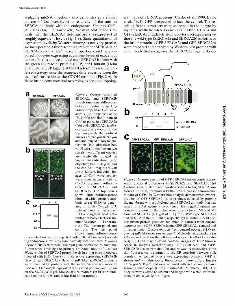
The Journal of Cell Biology, Volume 142, 1998 966
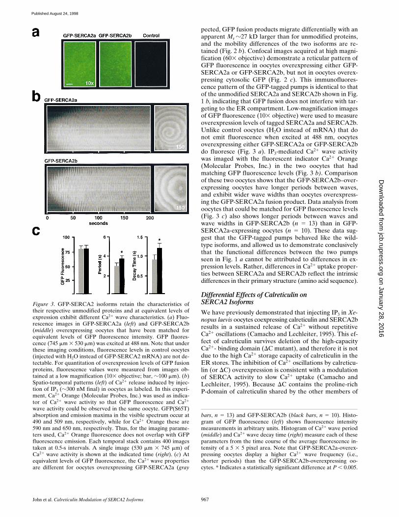
replacing mRNA injection) also demonstrates a similarpattern of low-intensity cross-reactivity of the anti-ratSERCA antibody with the endogenous Xenopus Ca21-ATPases (Fig. 1 b, lower left). Western blot analysis re-veals that the SERCA2 isoforms are overexpressed atroughly equivalent levels (Fig. 1 c). Since quantitation ofexpression levels by Western blotting is not very precise,we incorporated a fluorescent tag into either SERCA2a orSERCA2b so that Ca21 wave properties could be com-pared in oocytes expressing equivalent levels of exogenouspumps. To this end we labeled each SERCA2 isoform withthe green fluorescent protein (GFP) S65T mutant (Heimet al., 1995). GFP tagging at the NH2 terminus was the pre-ferred strategy since the sequence differences between thetwo isoforms reside at the COOH terminus (Fig. 2 a). Inthese fusion constructs and according to accepted topolog-
ical maps of SERCA proteins (Clarke et al., 1990; Bayleet al., 1995), GFP is expected to face the cytosol. The re-sulting fusion constructs were expressed in the oocyte byinjecting synthetic mRNAs encoding GFP-SERCA2a andGFP-SERCA2b. Extracts from oocytes overexpressing ei-ther the wild-type (SERCA2a and SERCA2b) isoforms orthe fusion proteins (GFP-SERCA2a and GFP-SERCA2b)were prepared and analyzed by Western blot probing withan antibody that recognizes the SERCA2 antigens. As ex-
Figure 1. Overexpression ofSERCA2a and SERCA2breveals functional differencesbetween isoforms in IP3-induced repetitive Ca21 waveactivity. (a) Comparison of theIP3 (z300 nM final)-inducedCa21 response in a SERCA2a(left) and a SERCA2b (right)-overexpressing oocyte. In thetop two panels the confocalimages are 745 mm 3 745 mmand are imaged at low magni-fication (103 objective; bar,z100 mm). In the bottom twopanels, two different oocytesare confocally imaged athigher magnification (403objective; bar, z20 mm) andthe confocal images are 240mm 3 180 mm. Individual im-ages of Ca21 wave activitywere taken at peak activity.(b) Confocal immunofluores-cence of SERCA2a andSERCA2b. The top panelsshow immunofluorescenceobtained with a primary anti-body to rat SERCAs gener-ated in rabbit (C-4, gift of J.Lytton) and a secondaryFITC-conjugated goat anti–rabbit antibody (Jackson Im-munoResearch Laborato-ries). The bottom panels arecontrols. The left panelshows immunofluorescence
of a control oocyte (not injected with SERCA2 message) reveal-ing endogenous levels of cross-reactivity with the native Xenopusoocyte SERCA2b protein. The right panel shows control immuno-fluorescence omitting the primary antibody. Bar, z10 mm. (c)Western blot of SERCA2 protein levels in either control oocytesinjected with H2O (lane 1) or oocytes overexpressing SERCA2b(lane 2) and SERCA2a (lane 3) mRNAs. SERCA2 productswere detected by probing with the same C-4 primary antibodyused in b. One oocyte equivalent was loaded per lane and run onan 8% SDS PAGE gel. Molecular size markers (in kD) are indi-cated on the left (Hi range; Bio-Rad Laboratories).
Figure 2. Overexpression of GFP-SERCA2 fusion constructs re-veals functional differences in SERCA2a and SERCA2b. (a)Cartoon view of the fusion constructs used to tag SERCA iso-forms at the NH2 terminus with the S65T increased fluorescencemutant of GFP. (b) Western blot analysis demonstrates overex-pression of GFP-SERCA2 fusion products detected by probingthe membrane with a polyclonal anti-SERCA2 antibody that wasraised in rabbit against a recombinant His-tagged fragment en-compassing most of the cytoplasmic loop between M4 and M5from rat SERCA2 (N1, gift of J. Lytton). Wild-type SERCA2aand SERCA2b (lanes 1 and 3, respectively) migrated z27 kD be-low fusion protein products contained in extracts from oocytesoverexpressing GFP-SERCA2a and GFP-SERCA2b (lanes 2 and4, respectively). Oocyte extracts from control oocytes (H2O re-placing mRNA) were run on lane 5. Molecular size markers (inkD) are indicated on the left (Kaleidoscope; Bio-Rad Laborato-ries). (c) High magnification confocal images of GFP fluores-cence in oocytes overexpressing GFP-SERCA2a and GFP-SERCA2b fusion proteins (left and middle, respectively). Notethat fluorescence is confined to the ER corridors between yolkplatelets. A control oocyte overexpressing cytosolic GFP isshown (right). In this oocyte, fluorescence is more diffuse. Imagesare 52 mm 3 36 mm, and were acquired with an OZ confocal laserscanning microscope (Noran Instruments, Middleton, WI). Theoocytes were excited at 488 nm and imaged with a 603 water im-mersion objective. Bar, z10 mm.
on January 28, 2016jcb.rupress.org
Dow
nloaded from
Published August 24, 1998
John et al. Calreticulin Modulation of SERCA2 Isoforms 967
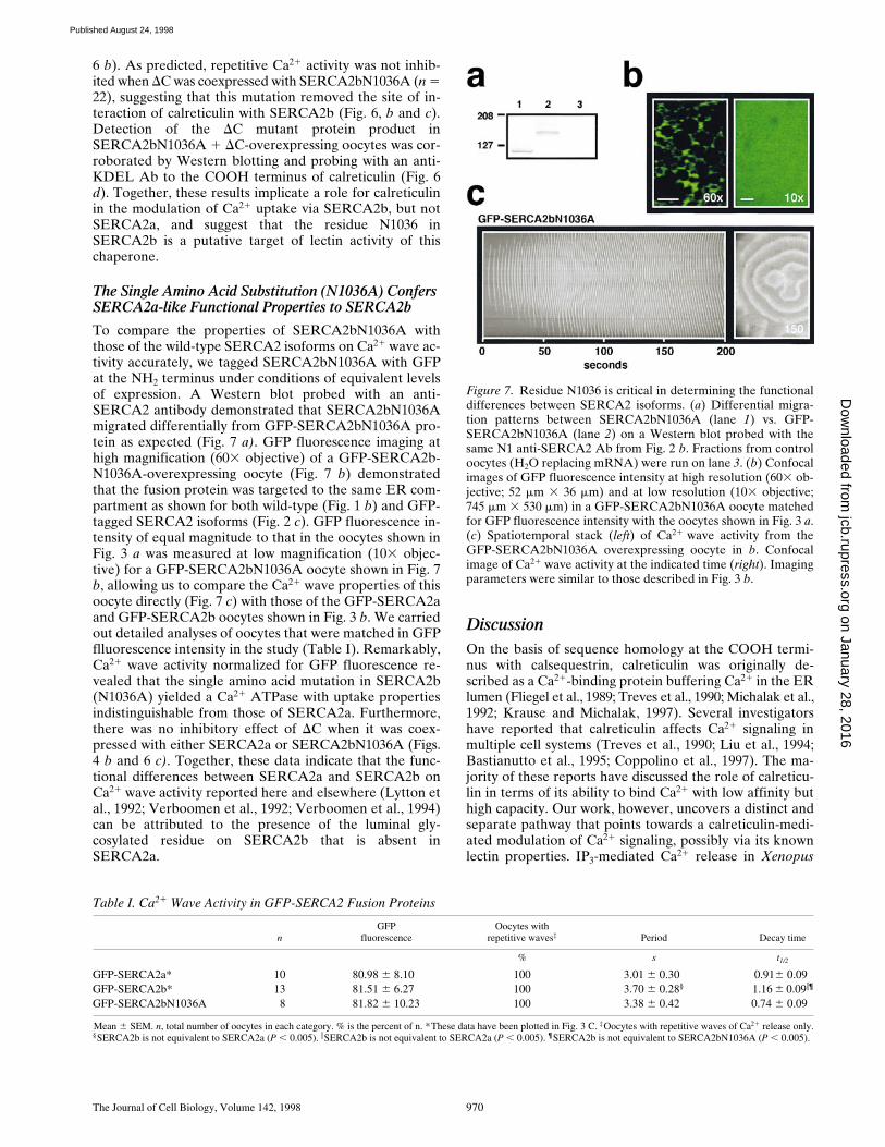
pected, GFP fusion products migrate differentially with anapparent Mr z27 kD larger than for unmodified proteins,and the mobility differences of the two isoforms are re-tained (Fig. 2 b). Confocal images acquired at high magni-fication (603 objective) demonstrate a reticular pattern ofGFP fluorescence in oocytes overexpressing either GFP-SERCA2a or GFP-SERCA2b, but not in oocytes overex-pressing cytosolic GFP (Fig. 2 c). This immunofluores-cence pattern of the GFP-tagged pumps is identical to thatof the unmodified SERCA2a and SERCA2b shown in Fig.1 b, indicating that GFP fusion does not interfere with tar-geting to the ER compartment. Low-magnification imagesof GFP fluorescence (103 objective) were used to measureoverexpression levels of tagged SERCA2a and SERCA2b.Unlike control oocytes (H2O instead of mRNA) that donot emit fluorescence when excited at 488 nm, oocytesoverexpressing either GFP-SERCA2a or GFP-SERCA2bdo fluoresce (Fig. 3 a). IP3-mediated Ca21 wave activitywas imaged with the fluorescent indicator Ca21 Orange(Molecular Probes, Inc.) in the two oocytes that hadmatching GFP fluorescence levels (Fig. 3 b). Comparisonof these two oocytes shows that the GFP-SERCA2b–over-expressing oocytes have longer periods between waves,and exhibit wider wave widths than oocytes overexpress-ing the GFP-SERCA2a fusion product. Data analysis fromoocytes that could be matched for GFP fluorescence levels(Fig. 3 c) also shows longer periods between waves andwave widths in GFP-SERCA2b (n 5 13) than in GFP-SERCA2a-expressing oocytes (n 5 10). These data sug-gest that the GFP-tagged pumps behaved like the wild-type isoforms, and allowed us to demonstrate conclusivelythat the functional differences between the two pumpsseen in Fig. 1 a cannot be attributed to differences in ex-pression levels. Rather, differences in Ca21 uptake proper-ties between SERCA2a and SERCA2b reflect the intrinsicdifferences in their primary structure (amino acid sequence).
Differential Effects of Calreticulin onSERCA2 Isoforms
We have previously demonstrated that injecting IP3 in Xe-nopus laevis oocytes coexpressing calreticulin and SERCA2bresults in a sustained release of Ca21 without repetitiveCa21 oscillations (Camacho and Lechleiter, 1995). This ef-fect of calreticulin survives deletion of the high-capacityCa21- binding domain (DC mutant), and therefore it is notdue to the high Ca21 storage capacity of calreticulin in theER stores. The inhibition of Ca21 oscillations by calreticu-lin (or DC) overexpression is consistent with a modulationof SERCA activity to slow Ca21 uptake (Camacho andLechleiter, 1995). Because DC contains the proline-richP-domain of calreticulin shared by the other members of
Figure 3. GFP-SERCA2 isoforms retain the characteristics oftheir respective unmodified proteins and at equivalent levels ofexpression exhibit different Ca21 wave characteristics. (a) Fluo-rescence images in GFP-SERCA2a (left) and GFP-SERCA2b(middle) overexpressing oocytes that have been matched forequivalent levels of GFP fluorescence intensity. GFP fluores-cence (745 mm 3 530 mm) was excited at 488 nm. Note that underthese imaging conditions, fluorescence levels in control oocytes(injected with H2O instead of GFP-SERCA2 mRNA) are not de-tectable. For quantitation of overexpression levels of GFP fusionproteins, fluorescence values were measured from images ob-tained at a low magnification (103 objective; bar, z100 mm). (b)Spatio-temporal patterns (left) of Ca21 release induced by injec-tion of IP3 (z300 nM final) in oocytes as labeled. In this experi-ment, Ca21 Orange (Molecular Probes, Inc.) was used as indica-tor of Ca21 wave activity so that GFP fluorescence and Ca21
wave activity could be observed in the same oocyte. GFP(S65T)absorption and emission maxima in the visible spectrum occur at490 and 509 nm, respectively, while for Ca21 Orange these are590 nm and 650 nm, respectively. Thus, for the imaging parame-ters used, Ca21 Orange fluorescence does not overlap with GFPfluorescence emission. Each temporal stack contains 400 imagestaken at 0.5-s intervals. A single image (530 mm 3 745 mm) ofCa21 wave activity is shown at the indicated time (right). (c) Atequivalent levels of GFP fluorescence, the Ca21 wave propertiesare different for oocytes overexpressing GFP-SERCA2a (gray
bars, n 5 13) and GFP-SERCA2b (black bars, n 5 10). Histo-gram of GFP fluorescence (left) shows fluorescence intensitymeasurements in arbitrary units. Histogram of Ca21 wave period(middle) and Ca21 wave decay time (right) measure each of theseparameters from the time course of the average fluorescence in-tensity of a 5 3 5 pixel area. Note that GFP-SERCA2a-overex-pressing oocytes display a higher Ca21 wave frequency (i.e.,shorter periods) than the GFP-SERCA2b-overexpressing oo-cytes. * Indicates a statistically significant difference at P , 0.005.
on January 28, 2016jcb.rupress.org
Dow
nloaded from
Published August 24, 1998

The Journal of Cell Biology, Volume 142, 1998 968
this class of ER chaperones (Ohsako et al., 1994; Tjoelkeret al., 1994; Watanabe et al., 1994), and because lectin ac-tivity resides in the P-domain (Krause and Michalak,1997), we tested whether DC modulation of SERCA2bwas responsible for the decreased rate of Ca21 transportcharacteristic of this pump (Lytton et al., 1992). Further-more, since SERCA2a does not have a glycosylation motiffacing the lumen of the ER, we also decided to testwhether the functional differences between the two iso-forms are due to a lack of interaction of calreticulin withSERCA2a. To this end, we coexpressed DC withSERCA2b or with SERCA2a (Fig. 4). A high percentage
of SERCA2b-overexpressing oocytes exhibited robusthigh-frequency IP3-mediated Ca21 oscillations (94%; n 532; Fig. 4 a). The number of oocytes showing repetitiveCa21 activity was significantly reduced when SERCA2b
Figure 4. DC inhibits Ca21 oscillations when coexpressed withSERCA2b, but not when coexpressed with SERCA2a. (a and b)Comparison of the IP3 (z300 nM)-induced Ca21 wave activityin a SERCA2b-overexpressing oocyte with the Ca21 wave activ-ity of a SERCA2b 1 DC-coexpressing oocyte. (c and d) Compar-ison of the IP3 (z300 nM)-induced Ca21 wave activity in aSERCA2a-overexpressing oocyte with the Ca21 wave activity ofa SERCA2a 1 DC-coexpressing oocyte. Individual confocal im-ages (745 mm 3 745 mm) of Ca21 wave activity were taken at theindicated times. The bottom trace in each panel represents thechange in fluorescence (DF/F) shown as a function of time for a5 3 5 pixel area (white square in the first panel of each image).
Figure 5. DC inhibition of repetitive Ca21 waves and reversal ofthe DC effect by glucosidase inhibitors. (a) The percent of oo-cytes exhibiting repetitive Ca21 oscillations is significantly re-duced in oocytes coexpressing DC with SERCA2b (black bars),but not in oocytes coexpressing DC with SERCA2a (gray bars;**P , 0.01, Chi-squared test). Of those oocytes that did displayrepetitive Ca21 oscillations, the interwave period (middle histo-gram) and decay time (right histogram) were significantly increasedin oocytes coexpressing DC with SERCA2b when compared withcontrol oocytes overexpressing SERCA2b alone (*P , 0.005). Nosignificant differences were found between oocytes coexpressingDC with SERCA2a as compared with control oocytes overex-pressing SERCA2a alone in either interwave period or in decaytime of individual waves. Note that there is a change in scale val-ues for the ordinate in histograms of wave period and decay timewith respect to Fig. 3 c. The larger scale in this figure reflects thelonger period and longer decay time of Ca21 waves in SERCA2b 1DC-overexpressing oocytes. (b) Western blot analysis demon-strates overexpression of the DC mutant of CRT in fractions fromoocytes coexpressing this calreticulin mutant with SERCA2a(lane 1) and SERCA2b (lane 2). Oocyte extracts from control oo-cytes (H2O replacing mRNA) were run on lane 3. The membranewas probed with a primary anti-CRT KDEL Ab that recognizesthe last six amino acids at the COOH terminus of rabbit CRT(gift of Michalak). (c) Glucosidase inhibition antagonizes the ef-fects of DC overexpression on oscillatory Ca21 waves. Period be-tween waves in SERCA2b 1 DC-overexpressing oocytes is signifi-cantly decreased (n 5 13) in oocytes injected with 1 mM final DNJ(Toronto Research Chemicals, North York, Ontario, Canada)when compared with uninjected SERCA2b 1 DC control oocytes(n 5 18). **Indicates statistical significance at P , 0.025. In thesame groups of oocytes, decay time of individual Ca21 waves is alsoreduced, although the differences are not statistically significant.
on January 28, 2016jcb.rupress.org
Dow
nloaded from
Published August 24, 1998

John et al. Calreticulin Modulation of SERCA2 Isoforms 969
was coexpressed with DC (66%, n 5 32, P , 0.01). In theremaining oocytes (34%), injections of IP3 caused sus-tained release of Ca21 without repetitive Ca21 waves (Fig.4 b). Detailed analysis of Ca21 waves revealed that even inthose SERCA2b 1 DC-coexpressing oocytes that had re-petitive Ca21 waves, the interwave periods were significantlylonger (P , 0.005), and wave widths were significantlybroader (P , 0.005) than those oocytes overexpressingSERCA2b alone (Fig. 5 a). In the majority of SERCA2a-overexpressing oocytes (95%, n 5 20), injections of IP3caused high-frequency Ca21 oscillations such as those ob-served in the oocyte shown in Fig. 4 a. In contrast to the in-hibition of Ca21 oscillations seen in oocytes coexpressingSERCA2b 1 DC, all SERCA2a 1 DC–overexpressing oo-cytes tested (100%, n520) exhibited high-frequency Ca21
oscillations (Fig. 4 d). Detailed analysis revealed that therewere no statistically significant differences in Ca21 waveproperties between SERCA2a and SERCA2a 1 DC–overexpressing oocytes (Fig. 5 a). Western blot analysis re-vealed that the DC mutant was coexpressed with eitherSERCA2a or SERCA2b in this set of experiments, sug-gesting that the lack of modulation of SERCA2a by DCcannot be attributed to a lack of DC expression (Fig. 5 b).Correct targeting of DC to the ER was confirmed by con-focal immunofluorescence performed on oocyte slicesprobed with an anti-calreticulin Ab as previously de-scribed (Camacho and Lechleiter, 1995; data not shown).Together these results indicate that the differential Ca21
transport by the SERCA2 isoforms must be due to thepresence of the luminal-facing COOH terminus extensionof SERCA2b, conferring susceptibility of SERCA2b tomodulation by calreticulin.
Residue N1036 of SERCA2b is Implicated in the Inhibitiory Effect of Calreticulin
Deoxynojirimycin (DNJ) is a specific inhibitor of glucosi-dases I and II, and prevents calreticulin and calnexin bind-ing to monoglucosylated target residues (Hebert et al.,1995; Peterson et al., 1995). If modulation of SERCA2bCa21 uptake by calreticulin results from a lectin effect,then this glucosidase inhibitor should antagonize the ef-fects of DC overexpression. To test this hypothesis, we in-jected DNJ into oocytes that were overexpressingSERCA2b 1 DC (n 5 18) 30–60 min before imaging. DNJinjection (z1 mM final) resulted in significantly (P ,0.025) decreased interwave periods in SERCA2b 1 DC–overexpressing oocytes (n 5 13; Fig. 5 c). A similar trendwas observed in terms of a decrease in the decay time ofindividual waves in these oocytes. No significant differ-ences were observed in the percent of oocytes displayingrepetitive Ca21 wave activity between control and DNJ-injected oocytes.
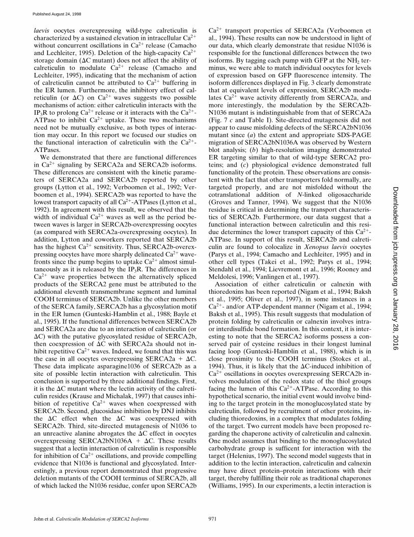
Unlike SERCA2a, SERCA2b has a potential glycosy-lated residue (N1036) at the COOH terminus (Fig. 6 a). Ifthe sustained Ca21 release and inhibition of oscillatoryCa21 waves is due to an interaction of calreticulin (or DC)with this SERCA2b residue, then site-directed mutagene-sis to an unreactive alanine (SERCA2bN1036A mutant)should remove this potential site of calreticulin interac-tion. Consequently, when coexpressed with SERCA2b-N1036A, DC should no longer have an effect on repetitive
Ca21 wave activity. To test this hypothesis, we overex-pressed either SERCA2bN1036A by itself, or coexpressedit with DC (SERCA2bN1036A 1 DC oocytes). Overexpres-sion of SERCA2bN1036A alone resulted in high-fre-quency Ca21 oscillations in all oocytes tested (n 5 23; Fig.
Figure 6. Site-directed mutagenesis of SERCA2b residue N1036creates a protein that is no longer responsive to DC coexpression,and that resembles SERCA2a. (a) Amino acid sequence compar-ison between the COOH terminus of SERCA2a and SERCA2b.The eleventh transmembrane segment of SERCA2b is shown(hatched). The consensus N-linked glycosylation motif is under-lined, and the mutated residue N1036A is indicated in bold. (b)Comparison of Ca21 wave activity in two oocytes overexpressingSERCA2bN1036A (top) or SERCA2bN1036A 1 DC (bottom).(c) The left histogram shows percent of oocytes exhibiting repeti-tive Ca21 oscillations when SERCA2bN1036A is expressed aloneor with DC. Of those oocytes that displayed repetitive Ca21 oscil-lations, no significant differences were found in either interwaveperiod (middle histogram) or in decay time (right histogram) be-tween oocytes coexpressing DC with SERCA2bN1036A and con-trol oocytes overexpressing SERCA2bN1036A alone. These re-sults are similar to those observed for SERCA2a and SERCA2a1 DC-overexpressing oocytes (see Fig. 4 b). (d) Western blotanalysis demonstrates overexpression of DC in fractions fromSERCA2bN1036A 1 DC oocytes (lane 1). No detectable CRTproduct was observed in extracts from control oocytes (H2O re-placing mRNA) (lane 2). The membrane was probed with theanti-CRT KDEL primary Ab from Fig. 4 c.
on January 28, 2016jcb.rupress.org
Dow
nloaded from
Published August 24, 1998
The Journal of Cell Biology, Volume 142, 1998 970
6 b). As predicted, repetitive Ca21 activity was not inhib-ited when DC was coexpressed with SERCA2bN1036A (n 522), suggesting that this mutation removed the site of in-teraction of calreticulin with SERCA2b (Fig. 6, b and c).Detection of the DC mutant protein product inSERCA2bN1036A 1 DC-overexpressing oocytes was cor-roborated by Western blotting and probing with an anti-KDEL Ab to the COOH terminus of calreticulin (Fig. 6d). Together, these results implicate a role for calreticulinin the modulation of Ca21 uptake via SERCA2b, but notSERCA2a, and suggest that the residue N1036 inSERCA2b is a putative target of lectin activity of thischaperone.
The Single Amino Acid Substitution (N1036A) ConfersSERCA2a-like Functional Properties to SERCA2b
To compare the properties of SERCA2bN1036A withthose of the wild-type SERCA2 isoforms on Ca21 wave ac-tivity accurately, we tagged SERCA2bN1036A with GFPat the NH2 terminus under conditions of equivalent levelsof expression. A Western blot probed with an anti-SERCA2 antibody demonstrated that SERCA2bN1036Amigrated differentially from GFP-SERCA2bN1036A pro-tein as expected (Fig. 7 a). GFP fluorescence imaging athigh magnification (603 objective) of a GFP-SERCA2b-N1036A-overexpressing oocyte (Fig. 7 b) demonstratedthat the fusion protein was targeted to the same ER com-partment as shown for both wild-type (Fig. 1 b) and GFP-tagged SERCA2 isoforms (Fig. 2 c). GFP fluorescence in-tensity of equal magnitude to that in the oocytes shown inFig. 3 a was measured at low magnification (103 objec-tive) for a GFP-SERCA2bN1036A oocyte shown in Fig. 7b, allowing us to compare the Ca21 wave properties of thisoocyte directly (Fig. 7 c) with those of the GFP-SERCA2aand GFP-SERCA2b oocytes shown in Fig. 3 b. We carriedout detailed analyses of oocytes that were matched in GFPflluorescence intensity in the study (Table I). Remarkably,Ca21 wave activity normalized for GFP fluorescence re-vealed that the single amino acid mutation in SERCA2b(N1036A) yielded a Ca21 ATPase with uptake propertiesindistinguishable from those of SERCA2a. Furthermore,there was no inhibitory effect of DC when it was coex-pressed with either SERCA2a or SERCA2bN1036A (Figs.4 b and 6 c). Together, these data indicate that the func-tional differences between SERCA2a and SERCA2b onCa21 wave activity reported here and elsewhere (Lytton etal., 1992; Verboomen et al., 1992; Verboomen et al., 1994)can be attributed to the presence of the luminal gly-cosylated residue on SERCA2b that is absent inSERCA2a.
DiscussionOn the basis of sequence homology at the COOH termi-nus with calsequestrin, calreticulin was originally de-scribed as a Ca21-binding protein buffering Ca21 in the ERlumen (Fliegel et al., 1989; Treves et al., 1990; Michalak et al.,1992; Krause and Michalak, 1997). Several investigatorshave reported that calreticulin affects Ca21 signaling inmultiple cell systems (Treves et al., 1990; Liu et al., 1994;Bastianutto et al., 1995; Coppolino et al., 1997). The ma-jority of these reports have discussed the role of calreticu-lin in terms of its ability to bind Ca21 with low affinity buthigh capacity. Our work, however, uncovers a distinct andseparate pathway that points towards a calreticulin-medi-ated modulation of Ca21 signaling, possibly via its knownlectin properties. IP3-mediated Ca21 release in Xenopus
Figure 7. Residue N1036 is critical in determining the functionaldifferences between SERCA2 isoforms. (a) Differential migra-tion patterns between SERCA2bN1036A (lane 1) vs. GFP-SERCA2bN1036A (lane 2) on a Western blot probed with thesame N1 anti-SERCA2 Ab from Fig. 2 b. Fractions from controloocytes (H2O replacing mRNA) were run on lane 3. (b) Confocalimages of GFP fluorescence intensity at high resolution (603 ob-jective; 52 mm 3 36 mm) and at low resolution (103 objective;745 mm 3 530 mm) in a GFP-SERCA2bN1036A oocyte matchedfor GFP fluorescence intensity with the oocytes shown in Fig. 3 a.(c) Spatiotemporal stack (left) of Ca21 wave activity from theGFP-SERCA2bN1036A overexpressing oocyte in b. Confocalimage of Ca21 wave activity at the indicated time (right). Imagingparameters were similar to those described in Fig. 3 b.
Table I. Ca21 Wave Activity in GFP-SERCA2 Fusion Proteins
nGFP
fluorescenceOocytes with
repetitive waves‡ Period Decay time
% s t1/2
GFP-SERCA2a* 10 80.98 6 8.10 100 3.01 6 0.30 0.916 0.09GFP-SERCA2b* 13 81.51 6 6.27 100 3.70 6 0.28§ 1.16 6 0.09i¶
GFP-SERCA2bN1036A 8 81.82 6 10.23 100 3.38 6 0.42 0.74 6 0.09
Mean 6 SEM. n, total number of oocytes in each category. % is the percent of n. *These data have been plotted in Fig. 3 C. ‡Oocytes with repetitive waves of Ca21 release only.§SERCA2b is not equivalent to SERCA2a (P , 0.005). iSERCA2b is not equivalent to SERCA2a (P , 0.005). ¶SERCA2b is not equivalent to SERCA2bN1036A (P , 0.005).
on January 28, 2016jcb.rupress.org
Dow
nloaded from
Published August 24, 1998
John et al. Calreticulin Modulation of SERCA2 Isoforms 971
laevis oocytes overexpressing wild-type calreticulin ischaracterized by a sustained elevation in intracellular Ca21
without concurrent oscillations in Ca21 release (Camachoand Lechleiter, 1995). Deletion of the high-capacity Ca21
storage domain (DC mutant) does not affect the ability ofcalreticulin to modulate Ca21 release (Camacho andLechleiter, 1995), indicating that the mechanism of actionof calreticulin cannot be attributed to Ca21 buffering inthe ER lumen. Furthermore, the inhibitory effect of cal-reticulin (or DC) on Ca21 waves suggests two possiblemechanisms of action: either calreticulin interacts with theIP3R to prolong Ca21 release or it interacts with the Ca21-ATPase to inhibit Ca21 uptake. These two mechanismsneed not be mutually exclusive, as both types of interac-tion may occur. In this report we focused our studies onthe functional interaction of calreticulin with the Ca21-ATPases.
We demonstrated that there are functional differencesin Ca21 signaling by SERCA2a and SERCA2b isoforms.These differences are consistent with the kinetic parame-ters of SERCA2a and SERCA2b reported by othergroups (Lytton et al., 1992; Verboomen et al., 1992; Ver-boomen et al., 1994). SERCA2b was reported to have thelowest transport capacity of all Ca21-ATPases (Lytton et al.,1992). In agreement with this result, we observed that thewidth of individual Ca21 waves as well as the period be-tween waves is larger in SERCA2b-overexpressing oocytes(as compared with SERCA2a-overexpressing oocytes). Inaddition, Lytton and coworkers reported that SERCA2bhas the highest Ca21 sensitivity. Thus, SERCA2b-overex-pressing oocytes have more sharply delineated Ca21 wave-fronts since the pump begins to uptake Ca21 almost simul-taneously as it is released by the IP3R. The differences inCa21 wave properties between the alternatively splicedproducts of the SERCA2 gene must be attributed to theadditional eleventh transmembrane segment and luminalCOOH terminus of SERCA2b. Unlike the other membersof the SERCA family, SERCA2b has a glycosylation motifin the ER lumen (Gunteski-Hamblin et al., 1988; Bayle etal., 1995). If the functional differences between SERCA2band SERCA2a are due to an interaction of calreticulin (orDC) with the putative glycosylated residue of SERCA2b,then coexpression of DC with SERCA2a should not in-hibit repetitive Ca21 waves. Indeed, we found that this wasthe case in all oocytes overexpressing SERCA2a 1 DC.These data implicate asparagine1036 of SERCA2b as asite of possible lectin interaction with calreticulin. Thisconclusion is supported by three additional findings. First,it is the DC mutant where the lectin activity of the calreti-culin resides (Krause and Michalak, 1997) that causes inhi-bition of repetitive Ca21 waves when coexpressed withSERCA2b. Second, glucosidase inhibition by DNJ inhibitsthe DC effect when the DC was coexpressed withSERCA2b. Third, site-directed mutagenesis of N1036 toan unreactive alanine abrogates the DC effect in oocytesoverexpressing SERCA2bN1036A 1 DC. These resultssuggest that a lectin interaction of calreticulin is responsiblefor inhibition of Ca21 oscillations, and provide compellingevidence that N1036 is functional and glycosylated. Inter-estingly, a previous report demonstrated that progressivedeletion mutants of the COOH terminus of SERCA2b, allof which lacked the N1036 residue, confer upon SERCA2b
Ca21 transport properties of SERCA2a (Verboomen etal., 1994). These results can now be understood in light ofour data, which clearly demonstrate that residue N1036 isresponsible for the functional differences between the twoisoforms. By tagging each pump with GFP at the NH2 ter-minus, we were able to match individual oocytes for levelsof expression based on GFP fluorescence intensity. Theisoform differences displayed in Fig. 3 clearly demonstratethat at equivalent levels of expression, SERCA2b modu-lates Ca21 wave activity differently from SERCA2a, andmore interestingly, the modulation by the SERCA2b-N1036 mutant is indistinguishable from that of SERCA2a(Fig. 7 c and Table I). Site-directed mutagenesis did notappear to cause misfolding defects of the SERCA2bN1036mutant since (a) the extent and appropriate SDS-PAGEmigration of SERCA2bN1036A was observed by Westernblot analysis; (b) high-resolution imaging demonstratedER targeting similar to that of wild-type SERCA2 pro-teins; and (c) physiological evidence demonstrated fullfunctionality of the protein. These observations are consis-tent with the fact that other transporters fold normally, aretargeted properly, and are not misfolded without thecotranslational addition of N-linked oligosaccharide(Groves and Tanner, 1994). We suggest that the N1036residue is critical in determining the transport characteris-tics of SERCA2b. Furthermore, our data suggest that afunctional interaction between calreticulin and this resi-due determines the lower transport capacity of this Ca21-ATPase. In support of this result, SERCA2b and calreti-culin are found to colocalize in Xenopus laevis oocytes(Parys et al., 1994; Camacho and Lechleiter, 1995) and inother cell types (Takei et al., 1992; Parys et al., 1994;Stendahl et al., 1994; Lievremont et al., 1996; Rooney andMeldolesi, 1996; Vanlingen et al., 1997).
Association of either calreticulin or calnexin withthioredoxins has been reported (Nigam et al., 1994; Bakshet al., 1995; Oliver et al., 1997), in some instances in aCa21- and/or ATP-dependent manner (Nigam et al., 1994;Baksh et al., 1995). This result suggests that modulation ofprotein folding by calreticulin or calnexin involves intra-or interdisulfide bond formation. In this context, it is inter-esting to note that the SERCA2 isoforms possess a con-served pair of cysteine residues in their longest luminalfacing loop (Gunteski-Hamblin et al., 1988), which is inclose proximity to the COOH terminus (Stokes et al.,1994). Thus, it is likely that the DC-induced inhibition ofCa21 oscillations in oocytes overexpressing SERCA2b in-volves modulation of the redox state of the thiol groupsfacing the lumen of this Ca21-ATPase. According to thishypothetical scenario, the initial event would involve bind-ing to the target protein in the monoglucosylated state bycalreticulin, followed by recruitment of other proteins, in-cluding thioredoxins, in a complex that modulates foldingof the target. Two current models have been proposed re-garding the chaperone activity of calreticulin and calnexin.One model assumes that binding to the monoglucosylatedcarbohydrate group is sufficent for interaction with thetarget (Helenius, 1997). The second model suggests that inaddition to the lectin interaction, calreticulin and calnexinmay have direct protein–protein interactions with theirtarget, thereby fulfilling their role as traditional chaperones(Williams, 1995). In our experiments, a lectin interaction is
on January 28, 2016jcb.rupress.org
Dow
nloaded from
Published August 24, 1998
The Journal of Cell Biology, Volume 142, 1998 972
suggested by the demonstration that the N-glycosylationsite in SERCA2b is necessary for calreticulin-mediated in-hibition of Ca21 oscillations. At the present time we can-not rule out the possibility that after initial binding to themonoglucosylated residue, the P-domain interacts directlywith a motif in SERCA2b to suppress IP3-mediated Ca21
oscillations. Overexpression of calreticulin or any otherdeletion mutant of calreticulin that we have tested, includ-ing the DC mutant, does not interfere with the extent ofcoexpression of SERCA2 pumps (Camacho and Lechlei-ter, 1995), suggesting that the chaperone does not inducemisfolding and degradation of SERCA2b. Optimal levelsof expression of SERCA2b (as indicated by the appear-ance of high-frequency Ca21 waves) appear only 7–9 d af-ter mRNA injection. This observation together with datafrom other laboratories (Gill et al., 1996) in which the for-mation of functional Ca21 pools after thapsigargin treat-ment requires 3–6 h, suggests that the synthesis of newSERCA protein is very slow. Thus, we suggest the possi-bility that calreticulin not only functions as a lectin chaper-one that modulates folding of integral membrane glyco-proteins during protein processing and maturation, but, asis the case of SERCA2b described here, it may also dy-namically modulate the conformation of mature proteinswith immediate functional consequences. This interpreta-tion is further supported by the ability of the glucosidaseinhibitor DNJ to reverse effects of the DC mutant coex-pression with SERCA2b on the modulation of Ca21 waveactivity. Since the DNJ treatment was acute (30-min expo-sure only), our data are consistent with an action of CRTon the monoglucosylated form of a fully mature protein.In conclusion, the results presented here provide a newconceptual framework to understand how ER luminalproteins control Ca21 release and uptake. Based on thelectin properties of calreticulin, we suggest that this novelclass of chaperones can dynamically modulate the confor-mation of integral membrane glycoproteins, thereby af-fecting their function.
We wish to thank David Castle, Jonathan Lytton, Enrico Nasi, and LlewelynRoderick for critical reading of the manuscript. We thank Marek Micha-lak, Gary Shull, and Roger Tsien for the gifts of CRT, SERCA2, and GFP(SGST) cDNAs, respectively. Antibodies used in this study were gener-ously provided by M. Michalak (CRT KDEL Ab) and by J. Lytton (C-4Ab and N1 Ab).
This work was supported by National Institutes of Health Grant RO1GM55372 to P. Camacho.
Received for publication 8 May 1998 and in revised form 24 June 1998.
References
Baksh, S., K. Burns, C. Andrin, and M. Michalak. 1995. Interaction of calreticu-lin with protein disulfide isomerase. J. Biol. Chem. 270:31338–31344.
Bastianutto, C., E. Clementi, F. Codazzi, P. Podini, F. De Giorgi, R. Rizzuto, J.Meldolesi, and T. Pozzan. 1995. Overexpression of calreticulin increases theCa21 capacity of rapidly exchanging Ca21 stores and reveals aspects of theirluminal microenvironment and function. J. Cell Biol. 130:847–855.
Bayle, D., D. Weeks, and G. Sachs. 1995. The membrane topology of the ratsarcoplasmic and endoplasmic reticulum calcium ATPases by in vitro trans-lation scanning. J. Biol. Chem. 270:25678–25684.
Bergeron, J.J.M., M.B. Brenner, D.Y. Thomas, and D.B. Williams. 1994. Calnexin:a membrane-bound chaperone of the endoplasmic reticulum. Trends Bio-chem. Sci. 19:124–128.
Berridge, M.J. 1993. Inositol trisphosphate and calcium signaling. Nature. 361:315–325.
Bezprozvanny, I., and B.E. Ehrlich. 1995. The inositol 1,4,5-trisphosphate re-ceptor. J. Membr. Biol. 145:205–216.
Bezprozvanny, I., J. Watras, and B.E. Ehrlich. 1991. Bell-shaped calcium-response
curves of Ins(1,4,5)P3- and calcium-gated channels from endoplasmic reticu-lum of cerebellum. Nature. 351:751–754.
Boitano, S., E.R. Dirksen, and M.J. Sanderson. 1992. Intercellular propagationof calcium waves mediated by inositol trisphosphate. Science. 258:292–295.
Brandl, C.J., N.M. Green, B. Korczak, and D.H. MacLennan. 1986. Two Ca21
ATPase genes: homologies and mechanistic implications of deduced aminoacid sequences. Cell. 44:597–607.
Burk, S.E., J. Lytton, D.H. MacLennan, and G.E. Shull. 1989. cDNA cloning,functional expression, and mRNA tissue distribution of a third organellarCa21 pump. J. Biol. Chem. 264:18561–18568.
Camacho, P., and J.D. Lechleiter. 1993. Increased frequency of calcium wavesin Xenopus laevis oocytes that express a calcium-ATPase. Science. 260:226–229.
Camacho, P., and J.D. Lechleiter. 1995. Calreticulin inhibits repetitive calciumwaves. Cell. 82:765–771.
Clapham, D.E. 1995. Ca21 signaling. Cell. 80:259–268.Clarke, D.M., T.W. Loo, and D.H. MacLennan. 1990. The epitope for mono-
clonal antibody A20 (amino acids 870–890) is located on the luminal surfaceof the Ca21 ATPase of the sarcoplasmic reticulum. J. Biol. Chem. 265:17405–17408.
Coppolino, M.G., M.J. Woodside, N. Demaurex, S. Grinstein, R. St-Arnaud,and S. Dedhar. 1997. Calreticulin is essential for integrin-mediated calciumsignaling and cell adhesion. Nature. 386:843–847.
Cornell-Bell, A.H., S.M. Finkbeiner, M.S. Cooper, and S.J. Smith. 1990.Glutamate induces calcium waves in cultured astrocytes: long-range glial sig-naling. Science. 247:470–473.
Dani, J.W., A. Chernjavsky, and S.J. Smith. 1992. Neuronal activity triggers cal-cium waves in hippocampal astrocyte networks. Neuron. 8:429–440.
DeLisle, S., and M.J. Welsch. 1992. Inositol trisphosphate is required for propa-gation of calcium waves in Xenopus oocytes. J. Biol. Chem. 267:7963–7966.
Finch, E.A., T.J. Turner, and S.M. Goldin. 1991. Calcium as a coagonist of ino-sitol 1,4,5-trisphosphate-induced calcium release. Science. 252:443–446.
Fliegel, L., K. Burns, D.H. MacLennan, R.A.F. Reithmeier, and M. Michalak.1989. Molecular cloning of the high affinity calcium binding protein (calreti-culin) of skeletal muscle sarcoplasmic reticulum. J. Biol. Chem. 264:21522–21528.
Furuichi, T., and K. Mikoshiba. 1995. Inositol 1,4,5-trisphosphate receptor me-diated Ca21 signaling in the brain. J. Neurochem. 64:953–960.
Gill, D.L., R.T. Waldron, K.E. Rys-Sikora, C.A. Ufret-Vincenty, M.N. Graber,C.J. Favre, and A. Alfonso. 1996. Calcium pools, calcium entry, and cellgrowth. Biosci. Rep. 16:139–157.
Groves, J.D., and M.J.A. Tanner. 1994. Role of N-glycosylation in the expres-sion of human band 3-mediated anion transport. Mol. Membr. Biol. 11:31–38.
Gunteski-Hamblin, A.-M., J. Greeb, and G.E. Shull. 1988. A novel Ca21 pumpexpressed in brain, kidney, and stomach is encoded by an alternative tran-script of the slow-twitch muscle sarcoplasmic reticulum Ca21-ATPase gene.J. Biol. Chem. 263:15032–15040.
Hammond, C., I. Braakman, and A. Helenius. 1994. Role of N-linked oligosac-charide recognition, glucose trimming, and calnexin in glycoprotein foldingand quality control. Proc. Natl. Acad. Sci. USA. 91:913–917.
Hammond, C., and A. Helenius. 1994. Quality control in the in the secretorypathway: retention of a misfolded viral membrane glycoprotein involves cy-cling between the ER, intermediate compartment and Golgi apparatus. J.Cell. Biol. 126:41–52.
Hausen, P., and C. Dreyer. 1991. The use of polyacrylamide as an embeddingmedium for immunohistochemical studies of embryonic tissues. Stain Tech-nol. 56:287–293.
Hebert, D., B. Foellmer, and A. Helenius. 1995. Glucose trimming and regluco-sylation determine glycoprotein association with calnexin in the endoplasmicreticulum. Cell. 8:425–433.
Heim, R., A. Cubitt, and R. Tsien. 1995. Improved green fluorescence. Nature.373:663–664.
Helenius, A. 1994. How N-linked oligosaccharides affect glycoprotein foldingin the endoplasmic reticulum. Mol. Biol. Cell. 5:253–265.
Helenius, A., E.S. Trombetta, D.N. Hebert, and J.F. Simmons. 1997. Calnexin,calreticulin, and the folding of glycoproteins. Trends Cell Biol. 7:193–200.
Iino, M. 1990. Biphasic Ca21 dependence of inositol 1,4,5-trisphosphate-inducedCa21 release in smooth muscle cells of the guinea pig taenia caeci. J. Gen.Physiol. 95:1103–1122.
Krause, K.-H., and M. Michalak. 1997. Calreticulin. Cell. 88:439–443.Lechleiter, J.D., and D.E. Clapham. 1992. Molecular mechanisms of intracellu-
lar calcium excitability in X. laevis oocytes. Cell. 69:283–294.Lievremont, J.-P., A.-M. Hill, D. Tran, J.-F. Coquil, N. Stelly, and J.-P. Mauger.
1996. Intracellular calcium stores and inositol 1,4,5-trisphosphate receptor inrat liver cells. Biochem. J. 314:189–197.
Liu, N., R.E. Fine, E. Simons, and R.J. Johnson. 1994. Decreasing calreticulinexpression lowers the Ca21 response to bradykinin and increases sensitivityto ionomycin in NG-108-15 cells. J. Biol. Chem. 269:28635–28639.
Longing, A., C. Souchier, M. French, and P.-A. Bryon. 1993. Comparison ofanti-fading agents used in fluorescence microscopy: image analysis and laserconfocal microscopy study. J. Histochem. Cytochem. 41:1833–1840.
Lytton, J., and D.H. MacLennan. 1988. Molecular cloning of cDNAs from kid-ney coding for two alternatively spliced products of the cardiac Ca21-ATP-ase gene. J. Biol. Chem. 263:15024–15031.
Lytton, J., M. Westlin, S.E. Burk, G.E. Shull, and D.H. MacLennan. 1992.Functional comparisons between isoforms of the sarcoplasmic or endoplas-
on January 28, 2016jcb.rupress.org
Dow
nloaded from
Published August 24, 1998
John et al. Calreticulin Modulation of SERCA2 Isoforms 973
mic reticulum family of calcium pumps. J. Biol. Chem. 267:14483–14489.MacLennan, D.H., C.J. Brandl, B. Korczak, and N.M. Green. 1985. Amino-acid
sequence of a Ca21/Mg21-dependent ATPase from rabbit muscle sarcoplas-mic reticulum, deduced from its complementary cDNA sequence. Nature.316:696–700.
MacLennan, D.H., W.J. Rice, and N.M. Green. 1997. The mechanism of Ca21
transport by sarco(endo)plasmic reticulim Ca21-ATPases. J. Biol. Chem.272:28815–28818.
Mahoney, M.G., L.L. Slakey, P.K. Hepler, and D.J. Gross. 1993. Independentmodes of propagation of calcium waves in smooth muscle cells. J. Cell Sci.104:1101–1107.
Michalak, M., R.E. Milner, K. Burns, and M. Opas. 1992. Calreticulin. Bio-chem. J. 285:681–692.
Nathanson, M.H., A.D. Burgstahler, A. Mennone, M.B. Fallon, C.B. Gonzalez,and J.C. Saez. 1995. Ca21 waves are organized among hepatocytes in the in-tact organ. Am. J. Physiol. 269:G167–G171.
Nauseef, W.M., S.J. McCormick, and R.A. Clark. 1995. Calreticulin functionsas a molecular chaperone in the biosynthesis of myeloperoxidase. J. Biol.Chem. 270:4741–4747.
Nigam, S., A. Goldberg, S. Ho, M. Rhode, K. Bush, and M. Sherman. 1994. Aset of endoplasmic reticulum proteins possessing properties of molecularchaperones includes Ca21-binding proteins and members of the thioredoxinsuperfamily. J. Biol. Chem. 269:1744–1749.
Ohsako, S., Y. Hayashi, and D. Bunick. 1994. Molecular cloning and sequenc-ing of calnexin-t. J. Biol. Chem. 269:14140–14148.
Oliver, J.D., F.J. Van Der Wal, N.J. Bulleid, and S. High. 1997. Interaction ofthe thiol-dependent reductase ERp57 with nascent glycoproteins. Science.275:86–88.
Osborn, M., and K. Weber. 1982. Immunofluorescence and immunocytochemi-cal procedures with affinity purified antibodies: tubulin-containing struc-tures. Methods Cell Biol. 24:97–132.
Otteken, A., and B. Moss. 1996. Calreticulin interacts with newly synthesizedhuman immunodeficiency virus type I envelope glycoprotein, suggesting achaperone function similar to that of calnexin. J. Biol. Chem. 271:97–103.
Ou, W.-J., P. Cameron, D.Y. Thomas, and J.J. Bergeron. 1993. Association offolding intermediates of glycoproteins with calnexin during protein matura-tion. Nature. 364:771–776.
Parker, I., and I. Ivorra. 1990. Inhibition by Ca21 of inositol trisphosphate-mediated Ca21 liberation: A possible mechanism for oscillatory release ofCa21. Proc. Natl. Acad. Sci. USA. 87:260–264.
Parker, I., and Y. Yao. 1991. Regenerative release of calcium from functionallydiscrete subcellular stores by inositol trisphosphate. Proc. R. Soc. Lond. Ser.B. 246:269–274.
Parys, J.B., S.M. McPherson, L. Mathews, K.P. Campbell, and F.J. Longo. 1994.Presence of inositol 1,4,5-trisphosphate receptor, calreticulin, and calseques-trin in eggs of sea urchins and Xenopus laevis. Dev. Biol. 161:466–476.
Peterson, J.R., A. Ora, P.N. Van, and A. Helenius. 1995. Transient, lectin-likeassociation of calreticulin with folding intermediates of cellular and viral gly-coproteins. Mol. Biol. Cell. 6:1173–1184.
Pozzan, T., R. Rizzuto, P. Volpe, and J. Meldolesi. 1994. Molecular and cellularphysiology of intracellular calcium stores. Physiol. Rev. 74:595–636.
Putney, J.W.J., and J. Bird. 1993. The inositol phosphate-calcium signaling sys-tem in nonexcitable cells. Endocr. Rev. 14:610–631.
Robb-Gaspers, L.D., and A.P. Thomas. 1995. Coordination of Ca21 signalingby intercellular propagation of Ca21 waves in the intact liver. J. Biol. Chem.270:8102–8107.
Rodan, A.R., J.F. Simons, E.S. Trombetta, and A. Helenius. 1996. N-linked oli-
gosaccharides are necessary and sufficient for association of glycosylatedforms of bovine RNase with calnexin and calreticulin. EMBO (Eur. Mol.Biol. Organ.) J. 15:6921–6930.
Rooney, E., and J. Meldolesi. 1996. The endoplasmic reticulum in PC12 cells. J.Biol. Chem. 271:29304–29311.
Rooney, T.A., and A.P. Thomas. 1993. Intracellular calcium waves generatedby Ins(1,4,5)P3-dependent mechanisms. Cell Calcium. 14:674–690.
Simpson, P., and J. Russell. 1996. Mitochondria support inositol 1,4,5-trisphos-phate-mediated Ca21 waves in cultured oligodendrocytes. J. Biol. Chem.271:33493–33501.
Sousa, M., and A.J. Parodi. 1995. The molecular basis for the recognition ofmisfolded glycoproteins by the UDP-Glc glycoprotein glucosyltransferase.EMBO (Eur. Mol. Biol. Organ.) J. 14:4196–4203.
Stendahl, O., K.H. Krause, J. Krishcher, P. Jerstrom, J.M. Theler, R.A. Clark,J.L. Carpentier, and D.P. Lew. 1994. Redistribution of intracellular Ca21
stores during phagocytosis in human neutrophils. Science. 265:1439–1441.Stokes, D.L., W.R. Taylor, and N.M. Green. 1994. Structure, transmembrane
topology and helix packing of P-type ion pumps. FEBS Lett. 346:32–38.Takei, K., H. Stukenbrok, A. Metcalf, G.A. Mignery, T.C. Sudhof, P. Volpe,
and P. DeCammilli. 1992. Ca21 stores in Purkinje neurons: endoplasmicreticulum subcompartments demonstrated by the heterogeneous distribu-tion of the InsP3 receptor, Ca21-ATPase, and calsequestrin. J. Neurosci. 12:489–505.
Tjoelker, L.W., C.E. Seyfried, R.L.J. Eddy, M.G. Byers, T.B. Shows, J. Cal-deron, R.B. Schreiber, and P.W. Gray. 1994. Human, mouse, and rat cal-nexin cDNA cloning: identification of potential calcium binding motifs andgene localization to human chromosome 5. Biochemistry. 33:3229–3236.
Treves, S., M. De Mattei, M. Lanfredi, A. Villa, N.M. Green, D.H. MacLennan,J. Meldolesi, and T. Pozzan. 1990. Calreticulin is a candidate for a calseques-trin-like function in Ca21-storage compartments (calciosomes) of liver andbrain. Biochem. J. 271:473–480.
Vanlingen, S., J.B. Parys, L. Missiaen, H. De Smedt, F. Wuytack, and R.Casteels. 1997. Distribution of inositol 1,4,5-trisphosphate receptor isoforms,SERCA isoforms and Ca21 binding proteins in RBL-2H3 rat basophilic leu-kemia cells. Cell Calcium. 22:475–486.
Verboomen, H., F. Wuytack, H. De Smedt, B. Himpens, and R. Casteels. 1992.Functional difference between SERCA2a and SERCA2b Ca21 pumps andtheir modulation by phospholamban. Biochem. J. 286:591–595.
Verboomen, H., F. Wuytack, L. Van Den Boshc, L. Mertens, and R. Casteels.1994. The functional importance of the C-terminal tail in the gene 2 organel-lar Ca21 transport ATPase (SERCA2a/b). Biochem. J. 303:979–984.
Ware, F.E., A. Vassilakos, P.A. Peterson, M.R. Jackson, M.A. Lehrman, andD.B. Williams. 1995. The molecular chaperone calnexin binds Glc1-Man9GlcNAc2 oligosaccharide as an initial step in recognizing unfolded gly-coproteins. J. Biol. Chem. 270:4697–4704.
Watanabe, D., K. Yamada, Y. Nishina, Y. Tajima, U. Koshimizu, A. Nagata,and Y. Nishimune. 1994. Molecular cloning of a novel Ca21 binding protein(calmegin) specifically expressed during male meiotic germ cell develop-ment. J. Biol. Chem. 269:7744–7749.
Williams, D.B. 1995. Calnexin: a molecular chaperone with a taste for carbohy-drate. Biochem. Cell Biol. 73:123–132.
Wu, K.-D., W.-S. Lee, J. Wey, D. Bungard, and J. Lytton. 1995. Localizationand quantification of endoplasmic reticulum Ca21-ATPase isoform tran-scripts. Am. J. Physiol. 269:C775–C784.
Zapun, A., S.M. Petrescu, P.M. Rudd, R.A. Dwek, D.Y. Thomas, and J.J.M.Bergeron. 1997. Conformation-independent binding of monoglucosylated ri-bonuclease B to calnexin. Cell. 88:29–38.
on January 28, 2016jcb.rupress.org
Dow
nloaded from
Published August 24, 1998


