Embed Size (px)
Citation preview
Cloning and partial characterization of a Boophilus microplus(Acari: Ixodidae) calreticulinq
Carlos Alexandre Sanchez Ferreira,a Itabajara Da Silva Vaz Jr.,a,b S�eergio Silva da Silva,a,c
Karen L. Haag,a,d Jesus G. Valenzuela,e and Aoi Masudaa,f,*
a Centro de Biotecnologia do Estado do Rio Grande do Sul, Universidade Federal do Rio Grande do Sul, Caixa Postal 15005,
Campus do Vale, Porto Alegre, RS, Brazilb Faculdade de Veterin�aaria, Universidade Federal do Rio Grande do Sul, Porto Alegre, RS, Brazil
c Departamento de Veterin�aaria Preventiva, Faculdade de Veterin�aaria, Universidade Federal de Pelotas, Cap~aao do Le~aao, RS, Brazild Departamento de Gen�eetica, Instituto de Biocieencias, Universidade Federal do Rio Grande do Sul, Porto Alegre, RS, Brazil
e Laboratory of Parasitic Diseases, National Institute of Allergy and Infectious Diseases, National Institutes of Health, Bethesda, MD, USAf Departamento de Biologia Molecular e Biotecnologia, Instituto de Biocieencias, Universidade Federal do Rio Grande do Sul, Porto Alegre, RS, Brazil
Received 9 October 2001; accepted 4 June 2002
Abstract
We report the cloning, sequence characterization and expression analysis of a calreticulin (CRT) coding cDNA of Boophilus
microplus. CRT is a calcium-binding protein involved in multiple cell functions and possibly implicated in parasites host immune
system evasion. The CRT cDNA sequence and its molecular characterization are described. Sequence similarity and phylogenetic
analyses indicate a close relationship to other arthropod CRT sequences. The CRT cDNA was also expressed in a procariotic system
and the recombinant protein (rBmCRT) was used to raise antibodies in a rabbit. Expression analyses of the corresponding gene in
different developmental stages and tissues were performed by RT-PCR and Western-blot, which indicated a ubiquitous expression
of the B. microplus calreticulin gene and demonstrated its presence in saliva. Sera of tick-infested bovines suggested that this protein
may not be able to induce an IgG-based humoral response in its natural host.
Index Descriptors and Abbreviations: Boophilus microplus; Tick; Gene expression; Parasite–host relationship; CRT, calreticulin; PBS,
phosphate-buffered saline; SDS, sodium dodecyl sulfate; PAGE, polyacrylamide gel electrophoresis; RT, reverse transcription;
PCR, polymerase chain reaction; pfu, plaque forming units; ER, endoplasmic reticulum; IgG, immunoglobulin G. � 2002 Elsevier
Science (USA). All rights reserved.
1. Introduction
Ticks are blood-sucking arthropods that infest a widearray of species (Sauer et al., 1995), including humansand some animals of economic importance, and causeimportant losses to livestock production (Bowman et al.,1996). One-host ixodidae ticks, in particular, expendmost of their life feeding on the host, therefore theymust be able to deal with all problems of haemostaticand immunologic origin the vertebrate blood may bringto a parasite (Ribeiro, 1989; Sauer et al., 1995). Salivary
secretions are well recognized to perform such modula-tory events in the tick–host relationship (Ribeiro, 1989,1995; Wikel, 1999), but the purification and furthercharacterization of these activities are many times un-feasible due to the small amounts of saliva that areavailable from ticks. An alternative approach would bethe identification of tick salivary genes in bacterial ex-pression systems and search for their biological func-tions using recombinant proteins.Calreticulin (CRT) is a calcium-binding protein,
known to perform several functions in mammals (Mic-halak et al., 1999). CRT is also secreted by ticks intotheir hosts (Jaworski et al., 1995) and its involvement inhost immune system modulation has been suggested(Jaworski et al., 1995; Kovacs et al., 1998). The proteinis divided into three domains: a N-terminal domain
Experimental Parasitology 101 (2002) 25–34
www.academicpress.com
qThe sequence data reported herein have been deposited in
GenBank under Accession No. AF420211.* Corresponding author. Fax: +55-51-331-67309.
E-mail address: [email protected] (A. Masuda).
0014-4894/02/$ - see front matter � 2002 Elsevier Science (USA). All rights reserved.
PII: S0014 -4894 (02 )00032-2
(N-domain), which is the most conserved domainamong all CRTs (Michalak et al., 1999); an internaldomain (P-domain), which binds Caþ2 with high affinity(Baksh and Michalak, 1991); and a C-terminal domain(C-domain), which is highly acidic and exhibits a high-capacity of Caþ2 binding (Baksh and Michalak, 1991).Boophilus microplus is a one-host tick that usually in-
fests cattle and is responsible for economic losses rangingaround billions of dollars per year (Bowman et al., 1996).However, as to how this parasitic relationship is accom-plished, very little is known. The characterization of sal-ivary gland-associated molecules may improve ourknowledge on the mechanisms involved in tick–host in-teraction and how they could be inhibited.We report herethe isolation, sequence characterization, and expressionanalysis of a salivary calreticulin coding cDNA of B. mi-croplus. We have also tested the immunogenicity of thecorresponding protein (BmCRT) under infestations, andby immunization with the recombinant protein.
2. Material and methods
2.1. Ticks and harvest of saliva
B. microplus females, eggs, and larvae (Porto Alegrestrain) were maintained in an incubator at 28 �C and85% relative humidity, and their parasitic life wascompleted in calves, housed in individual pens on slattedfloors. Partially engorged adult female ticks were ob-tained by direct detachment from the calves, kept in awet chamber, and salivation was induced by injection of5ll of 2% pilocarpine solution. Saliva was then collectedfor a period of 2 h directly from tick mouthparts andstored at )70 �C until use.
2.2. Antigen preparation
Fully and partially engorged female ticks were washedwith phosphate-buffered saline pH 7.2 (PBS) plus 500 IUpenicillin/ml. The dorsal surface was dissected with ascalped blade. Salivary glands, guts, ovaries, and fat bodieswere separated with fine-tipped forceps and washed inPBS. These materials were kept frozen at )70 �C until use.The frozen tissues were thawed and protein extracts
prepared according to Da Silva Vaz Jr. et al. (1994). Theprotein concentrations of the extracts were measuredusing the Bradford method (1976) with bovine serumalbumin as standard.
2.3. Antibody production
Antibodies against salivary gland were raised in arabbit by inoculation of 100lg salivary gland extractemulsified with an equal volume of Freund’s completeadjuvant. Three additional boosters were given every
three weeks with 100lg of antigen emulsified in Fre-und’s incomplete adjuvant. Fifteen days after the lastbooster serum was collected.
2.4. Synthesis and screening of the salivary gland cDNAlibrary
An unidirectional cDNA library was synthesized fromsalivary gland poly(A)þ RNA of partially engorged adultfemales using the UNIZAP vector (Stratagene), accord-ing to the instructions of the manufacturer. The poly(A)þ
RNA was obtained using the Micro-Fast Track Kit (In-vitrogen). The immunological screening was performedusing the anti-salivary gland serum. Nine thousand re-combinant cDNA clones were screened on nitrocellulosemembranes (Schleicher & Sch€uull) resulting in a singlepositive CRT-similar clone, named Bmsg1. Membraneswere blocked with blotto (5% cow non-fat dry milk inPBS) for 1 h at room temperature, and then incubatedwith anti-salivary gland antibodies at room temperaturefor 18 h. After three washes with blotto, goat anti-rabbitIgG antibody conjugated to alkaline phosphatase (Sig-ma) diluted 1:5000 in blotto was incubated for 1 h at roomtemperature. After three washes with PBS and once withdevelopment buffer (5mM MgCl2, 100mM NaCl,100mM Tris, pH 9.5), membranes were stained with 5-bromo-4-chloro-3-indolylphosphate (BCIP) and nitro-blue tetrazolium (NBT).The Bmsg1 cDNA was used to screen the same
cDNA library in order to obtain the full cDNA codingregion of BmCRT. Nine thousand recombinant plaqueforming units (pfu) were screened on nitrocellulosemembranes (Schleicher & Sch€uull) using the NucleicAcids ECL Kit (Amersham–Pharmacia), according tothe instructions of the manufacturer.All positive clones isolated were excised into the
pBluescript II (Stratagene) plasmid and their insertsanalyzed by sequencing and digestion with restrictionenzymes.
2.5. DNA sequencing and analysis
DNA sequencing of all cDNAs isolated was per-formed on an ABI-PRISM 377 automated DNA se-quencer (Perkin–Elmer) at Molecular Genetics Facility,University of Georgia, Athens, Georgia, USA.TheFASTAalgorithm (PearsonandLipman, 1988)was
used to analyze the nucleotide and deduced amino acidsequence homologies with previously reported sequenceswithin databases. Multiple alignment of CRT sequenceswasperformedwithCLUSTALW(Thompsonetal., 1994).
2.6. Phylogenetic analysis
CRT sequences were aligned with the BmCRT de-duced protein sequence using the CLUSTALW program
26 C.A.S. Ferreira et al. / Experimental Parasitology 101 (2002) 25–34
(Thompson et al., 1994). An unrooted neighbor-joiningphylogenetic tree (Saitou andNei, 1987) was created withthe proportion of pair-wise nucleotide differences (p-dis-tances) using the MEGA program (Kumar et al., 1993).Bootstrap support was assessed using 500 replicates. TheGenBank accession numbers are: Amblyomma america-num, AAC79094; Aplysia californica, JH0795; Arabidop-sis thaliana, O04151; Beta vulgaris, O81919; Bos taurus(bovine brain isoform 2), S36799;Caenorhabditis elegans,P27798; Chlamydomonas reinhardtii, Q9STD3;Danio re-rio, AAF13700; Dictyostelium discoideum, Q23858; Di-rofilaria immitis, AAD03405; Drosophila melanogasterP29413; Euglena gracilis, Q9ZNY3; Homo sapiens (hu-man), NP_004334; Leishmania donovani, U49191.1; Li-tomosoides sigmodontis, CAA04877; Mus musculus(mouse), NP_031617; Necator americanus, CAA07254;Onchocerca volvulus, P11012; Oryctolagus cuniculus(rabbit), P15253; Oryza sativa, Q9SLY8; Prunus armeni-aca, Q9XF98; Rana rugosa, S71343; Rattus norvegicus(rat), NP_071794; Ricinus communis, P93508; Schisto-soma japonicum, AAC00515; Schistosoma mansoni,Q06814; Strongylocentrotus purpuratus, AAD55725;Trypanosoma cruzi, AAD22175; Tritrichomonas suis,CAB92410; Zea mays, S58170.
2.7. Construction of a plasmid expressing the matureBmCRT
The coding region of mature BmCRT was subclonedinto the pGEX-4T1 vector (Amersham–Pharmacia),using the restriction sites of BstZ17 (nucleotides 79–84;indicated in Fig. 1) and Xho I (pBluescript). As there is asite for XhoI within Bmcrt, a partial cleavage was per-formed to obtain the correct fragment to be cloned,which produces a recombinant protein that lacks thesignal peptide plus four amino acids of the matureprotein (see Fig. 1). The fragment was cloned within thesites of XhoI and SmaI of pGEX-4T1. Correct cloningwas confirmed by sequencing, and the recombinantplasmid named pGEX-CRT.The glutathione–Sepharose 4B (Amersham–Pharma-
cia) affinity chromatography was used to purifyrBmCRT. Lysogens of BL21/pGEX-CRT were preparedafter growth in Luria–Bertani medium. Recombinantprotein expression was induced with IPTG 0.1mM. Cellpellet from 2000ml culture were suspended in 20ml ofPBS and frozen at )70 �C. Cells were thawed and dis-rupted in a French press. Triton X-100 was added to thesupernatant to a final concentration of 1%. The super-natant was then loaded on the column that had beenequilibrated and washed with buffer A (140mM NaCl,2.7mM KCl, 10mM Na2HPO4, 1.8mM KH2PO4), andthe fusion protein was then incubated with thrombinovernight at 23.5 �C. The GST portion and the remainingfusion protein in the column were eluted with buffer B(50mM Tris–HCl+ 10mM glutathione, pH 8.0). Protein
purity was monitored by a 10% SDS–PAGE (Laemmli,1970) stained with Coomassie blue G-250.
2.8. RNA poly(A)þ purification and RT-PCR
RNA poly(A)þ was purified from B. microplus tissuesand bovine blood collected with citrate, and stored at)70 �C. The RNA poly(A)þ purification was performedwith the Quick Prep Micro mRNA Purification Kit(Amersham–Pharmacia), as described by the manufac-turer. Five hundred ng of RNA poly(A)þ were submittedto reverse transcription (RT) at 37 �C in the presence ofoligo-dT (Amersham–Pharmacia) and M-MLV ReverseTranscriptase (Gibco-Life Technologies) also accordingto the instructions of the manufacturer. PCR amplifica-tions were performed using 1/20 of the RT reaction,10 pmol of each primer (pfsg1 and prsg1) and 2.5U of
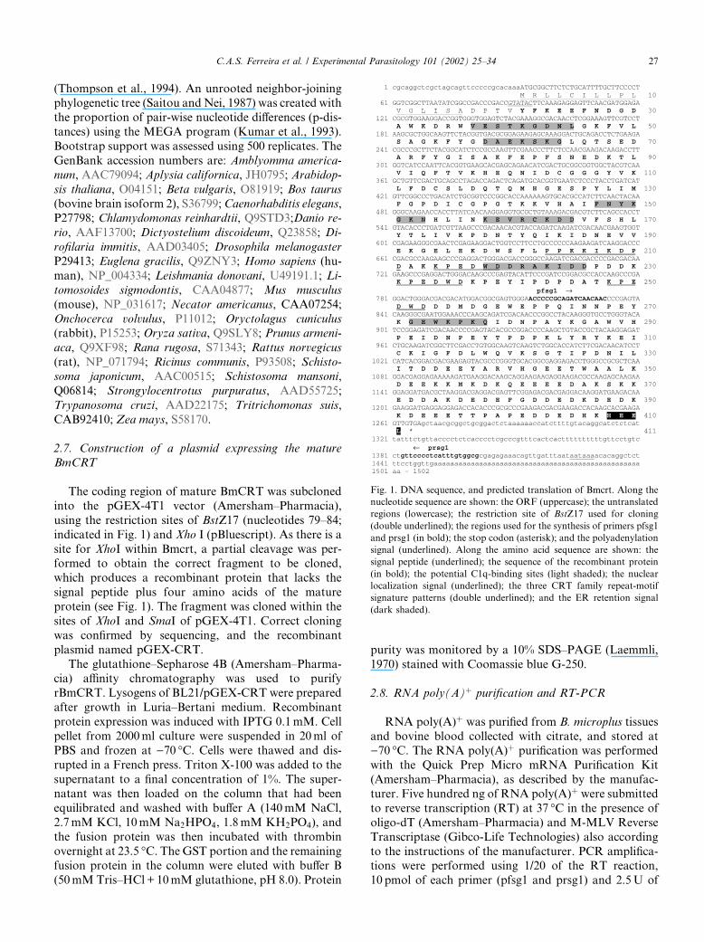
Fig. 1. DNA sequence, and predicted translation of Bmcrt. Along the
nucleotide sequence are shown: the ORF (uppercase); the untranslated
regions (lowercase); the restriction site of BstZ17 used for cloning
(double underlined); the regions used for the synthesis of primers pfsg1
and prsg1 (in bold); the stop codon (asterisk); and the polyadenylation
signal (underlined). Along the amino acid sequence are shown: the
signal peptide (underlined); the sequence of the recombinant protein
(in bold); the potential C1q-binding sites (light shaded); the nuclear
localization signal (underlined); the three CRT family repeat-motif
signature patterns (double underlined); and the ER retention signal
(dark shaded).
C.A.S. Ferreira et al. / Experimental Parasitology 101 (2002) 25–34 27
TaqDNA polimerase (Cenbiot) in a final volume of 50ll.Samples were denatured for 10min at 94 �C and ampli-fication was achieved through 35 cycles of 30 s at 94 �C,30 s at 52 �C, and 30 s at 72 �C, with a final extension cycleof 30 s at 52 �C and 10min at 72 �C. Negative controlsfor the RT reactions and PCR amplifications were alwaysincluded into the assay. Amplicons were visualizedby agarose gel electrophoresis and ethidium bromidestaining.
2.9. Immunogenicity of rBmCRT
rBmCRT was further purified by 10% SDS–PAGEelectrophoresis and one rabbit was subcutaneously in-oculated with four doses of approximately 100lg ofprotein emulsified in Freund’s incomplete adjuvant.Fifteen days after the last booster serum was collected.One bovine was also immunized with the same rBmCRTpreparation. In the initial four inoculations 150lg ofrBmCRT were used; in the following boosters theamount of protein were raised to 250lg (fifth booster),350lg (sixth booster), and 400lg (seventh and eighthboosters).Six bovines were artificially infested repeatedly 12
times: 6 times with 18,000 B. microplus larvae followedby 6 times with 800 larvae, and sera were collected aftereach infestation. Rhippicephalus sanguineus infested andnon-infested dog sera were obtained at the VeterinaryHospital of Universidade Federal do Rio Grande doSul.
2.10. Western-blot
For Western-blot analysis, tissue extracts and purifiedrBmCRT were resuspended in sample buffer containing2% SDS, 250mM Tris, pH 6.8, 0.025% bromophenolblue, 5% glycerol, 10% b-mercaptoethanol, and 5Murea,separated in SDS–PAGE 10% gel electrophoresis andtransferred to nitrocellulose at 70V for 1 h at 4 �C in12mM carbonate buffer pH 9.9 (Dunn, 1986). The ni-trocellulose sheet was blocked with blotto for 2 h at roomtemperature. In the Western-blot shown in Fig. 5, theanti-rBmCRT rabbit serum (1:2000) was incubated inblotto overnight at 4 �C, and the secondary antibodyconjugated to alkaline phosphatase and developmentprocedure was the same used in the immunologicalscreening.In the Western-blots shown in Fig. 6 the protocol for
the SDS–PAGE, protein transfer to nitrocellulose andblocking were the same as described above. In Fig. 6aand c the antigen used was rBmCRT at a concentrationof 12lg per nitrocellulose strip (strips of 4mm;30lg=cm), and in Fig. 6b the antigen used was a par-tially engorged salivary glands extract at a concentrationof 44lg per strip ð110lg=cmÞ. The B. microplus infestedbovines sera were diluted 1:20, the R. sanguineus infested
dog sera 1:50 and the anti-rBmCRT rabbit serum 1:400.Prior to the overnight incubation at 4 � C with the an-tigens, all sera were diluted in an E. coli BL21 strainlysate expressing the pGEX-4T1 vector and incubatedfor 2 h at room temperature for absorption of anti-E. coli and anti-vector derived protein antibodies.Preparation of the E. coli BL21 strain lysate was per-formed according to Rott et al. (2000). The secondaryantibodies used were conjugated to peroxidase (anti-bovine IgG, Sigma, diluted 1:2000; anti-dog IgG, Sigma,diluted 1:2000), and after the 1 h incubation three wa-shes with PBS were performed and the developmentbuffer (5mg 3,30-diaminobenzidine in 30ml PBS plus150ll H2O2 30% and 100ll CoCl2 1%) was added.
3. Results
3.1. Isolation of the cDNA clones
A cDNA clone (Bmsg1) was obtained by immuno-logical screening from a salivary gland library using arabbit anti-salivary gland serum (as described in Section2). The Bmsg1 sequence was determined, which encodesa 1 kbp cDNA fragment with a 254 amino acids ORFwith high similarity to CRT sequences. As no ATGcodon that could code for an initiating methionine wasevident and a N-terminal region was apparently missing(deduced from sequence comparison), Bmsg1 DNA wasthen used to screen the same library in order to obtain afull-length coding sequence cDNA. Twenty-four posi-tive clones were obtained out of 9000 recombinant pfu,and four of them were sequenced at their ends based oninsert size and restriction endonucleases digestion pat-tern. All of them presented the same sequence, whichwere identical to Bmsg1 at the C-terminus. One of them,named Bmcrt, was then fully sequenced and its deducedprotein sequence also was shown to be highly similar toCRTs from other organisms (Fig. 2), and to contain aputative full-length coding sequence. Fig. 1 shows the1502 bp sequence of Bmcrt and its deduced amino acidsequence.
3.2. Sequence, similarity, and phylogenetic analysis
Bmcrt possesses an ORF of 1233 bp that encodes aprotein of 47.7 kDa. A probable signal peptide of 16predominantly hydrophobic amino acids is present atthe N-terminus, which begins at a putative initiationcodon (Fig. 1). The presence of the signal peptide wascorroborated by the N-terminal microsequencing of anative salivary BmCRT-similar protein, which showedthat the mature protein began at the aspartic residue 17(Carlos Termignoni, personal communication). Thepredicted molecular mass and pI for the mature proteinare 46 kDa and 4.48, respectively. BmCRT possesses six
28 C.A.S. Ferreira et al. / Experimental Parasitology 101 (2002) 25–34
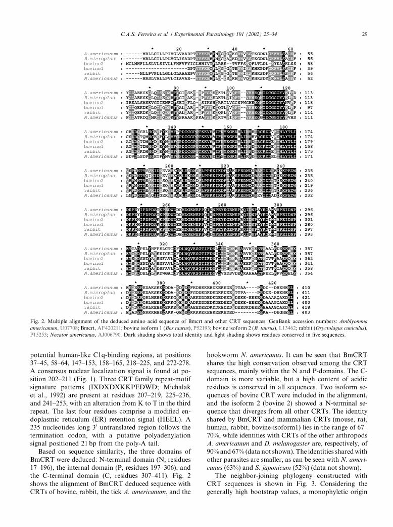
potential human-like C1q-binding regions, at positions37–45, 58–64, 147–153, 158–165, 218–225, and 272-278.A consensus nuclear localization signal is found at po-sition 202–211 (Fig. 1). Three CRT family repeat-motifsignature patterns (IXDXDXKKPEDWD; Michalaket al., 1992) are present at residues 207–219, 225–236,and 241–253, with an alteration from K to T in the thirdrepeat. The last four residues comprise a modified en-doplasmic reticulum (ER) retention signal (HEEL). A235 nucleotides long 30 untranslated region follows thetermination codon, with a putative polyadenylationsignal positioned 21 bp from the poly-A tail.Based on sequence similarity, the three domains of
BmCRT were deduced: N-terminal domain (N, residues17–196), the internal domain (P, residues 197–306), andthe C-terminal domain (C, residues 307–411). Fig. 2shows the alignment of BmCRT deduced sequence withCRTs of bovine, rabbit, the tick A. americanum, and the
hookworm N. americanus. It can be seen that BmCRTshares the high conservation observed among the CRTsequences, mainly within the N and P-domains. The C-domain is more variable, but a high content of acidicresidues is conserved in all sequences. Two isoform se-quences of bovine CRT were included in the alignment,and the isoform 2 (bovine 2) showed a N-terminal se-quence that diverges from all other CRTs. The identityshared by BmCRT and mammalian CRTs (mouse, rat,human, rabbit, bovine-isoform1) lies in the range of 67–70%, while identities with CRTs of the other arthropodsA. americanum and D. melanogaster are, respectively, of90%and 67% (data not shown). The identities sharedwithother parasites are smaller, as can be seen with N. ameri-canus (63%) and S. japonicum (52%) (data not shown).The neighbor-joining phylogeny constructed with
CRT sequences is shown in Fig. 3. Considering thegenerally high bootstrap values, a monophyletic origin
Fig. 2. Multiple alignment of the deduced amino acid sequence of Bmcrt and other CRT sequences. GenBank accession numbers: Amblyomma
americanum, U07708; Bmcrt, AF420211; bovine isoform 1 (Bos taurus), P52193; bovine isoform 2 (B. taurus), L13462; rabbit (Oryctolagus cuniculus),
P15253; Necator americanus, AJ006790. Dark shading shows total identity and light shading shows residues conserved in five sequences.
C.A.S. Ferreira et al. / Experimental Parasitology 101 (2002) 25–34 29
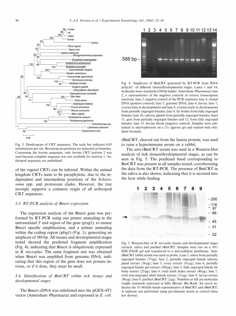
of the vegetal CRTs can be inferred. Within the animalkingdom CRTs seem to be paraphyletic, due to the in-dependent and intermediate positions of the Schisto-soma spp. and protozoan clades. However, the treestrongly supports a common origin of all arthropodCRT sequences.
3.3. RT-PCR analysis of Bmcrt expression
The expression analysis of the Bmcrt gene was per-formed by RT-PCR using one primer annealing in theuntranslated 30 end region of the gene (prsg1), to ensureBmcrt specific amplification, and a primer annealingwithin the coding region (pfsg1) (Fig. 1), generating anamplicon of 588 bp. All tissues and developmental stagestested showed the predicted fragment amplification(Fig. 4), indicating that Bmcrt is ubiquitously expressedin B. microplus. The same fragment size was obtainedwhen Bmcrt was amplified from genomic DNA, indi-cating that this region of the gene does not possess in-trons, or if it does, they must be small.
3.4. Identification of BmCRT within tick tissues anddevelopmental stages
The Bmcrt cDNA was subcloned into the pGEX-4T1vector (Amersham–Pharmacia) and expressed in E. coli.
rBmCRT, cleaved out from the fusion protein, was usedto raise a hyperimmune serum on a rabbit.The anti-rBmCRT serum was used in a Western-blot
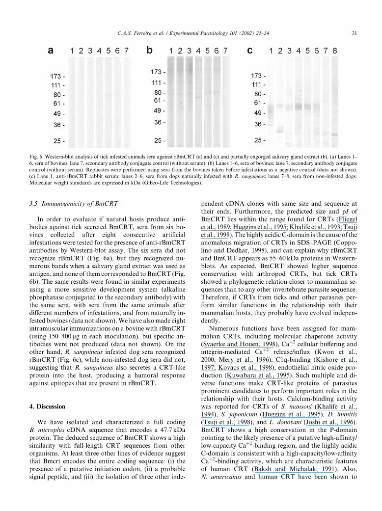
analysis of tick tissues/developmental stages, as can beseen in Fig. 5. The predicted band corresponding toBmCRT was present in all samples tested, corroboratingthe data from the RT-PCR. The presence of BmCRT inthe saliva is also shown, indicating that it is secreted intothe host while feeding.
Fig. 3. Dendrogram of CRT sequences. The scale bar indicates 0.05
substitutions per site. Bootstrap proportions are indicated at branches.
Concerning the bovine sequences, only bovine CRT isoform 2 was
used because complete sequence was not available for isoform 1. Ar-
thropod sequences are underlined.
Fig. 4. Amplicons of BmCRT generated by RT-PCR from RNA
poly(A)þ of different tissues/developmental stages. Lanes 1 and 14,
molecular mass standards (100 bp ladder, Amersham–Pharmacia); lane
2, a representative of the negative controls of reverse transcriptase
reactions; lane 3, negative control of the PCR reactions; lane 4, cloned
DNA (positive control); lane 5, genomic DNA; lane 6, larvae; lane 7,
ovaries (late in development) and lane 8, ovaries (early in development)
from partially engorged females; lane 9, fat bodies from fully engorged
females; lane 10, salivary glands from partially engorged females; lanes
11, guts from partially engorged females and 12, from fully engorged
females; lane 13, bovine blood (negative control). Samples were sub-
mitted to electrophoresis on a 2% agarose gel and stained with ethi-
dium bromide.
Fig. 5. Western-blot of B. microplus tissues and developmental stages
extracts, saliva and purified rBmCRT. Samples were run on a 10%
SDS–PAGE gel and transferred to a nitrocellulose membrane. Anti-
rBmCRT rabbit serum was used as probe. Lane 1, saliva from partially
engorged females ð75lgÞ; lane 2, partially engorged female salivarygland extract ð36lgÞ; lane 3, ovary extract ð61lgÞ; lane 4, partiallyengorged female gut extract ð180lgÞ; lane 5, fully engorged female fatbody extract ð23lgÞ; lane 6, total adult males extract ð40lgÞ; lane 7,total non-engorged adult female extract ð15lgÞ; lane 8, larvae extractð96lgÞ; lane 9, purified rBmCRT ð2lgÞ. Numbers at left are molecularweight standards expressed in kDa (Broad—Bio-Rad). An arrow in-
dicates the 55–60 kDa bands representative of BmCRT and rBmCRT.
A replicate was performed using pre-immune serum as control (data
not shown).
30 C.A.S. Ferreira et al. / Experimental Parasitology 101 (2002) 25–34
3.5. Immunogenicity of BmCRT
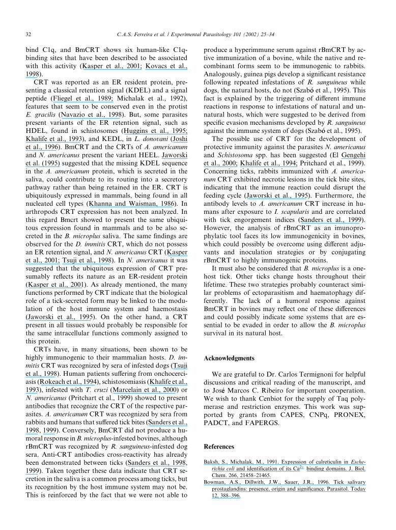
In order to evaluate if natural hosts produce anti-bodies against tick secreted BmCRT, sera from six bo-vines collected after eight consecutive artificialinfestations were tested for the presence of anti-rBmCRTantibodies by Western-blot assay. The six sera did notrecognize rBmCRT (Fig. 6a), but they recognized nu-merous bands when a salivary gland extract was used asantigen, and none of them corresponded to BmCRT (Fig.6b). The same results were found in similar experimentsusing a more sensitive development system (alkalinephosphatase conjugated to the secondary antibody) withthe same sera, with sera from the same animals afterdifferent numbers of infestations, and from naturally in-fested bovines (data not shown).We have also made eightintramuscular immunizations on a bovine with rBmCRT(using 150–400 lg in each inoculation), but specific an-tibodies were not produced (data not shown). On theother hand, R. sanguineus infested dog sera recognizedrBmCRT (Fig. 6c), while non-infested dog sera did not,suggesting that R. sanguineus also secretes a CRT-likeprotein into the host, producing a humoral responseagainst epitopes that are present in rBmCRT.
4. Discussion
We have isolated and characterized a full codingB. microplus cDNA sequence that encodes a 47.7 kDaprotein. The deduced sequence of BmCRT shows a highsimilarity with full-length CRT sequences from otherorganisms. At least three other lines of evidence suggestthat Bmcrt encodes the entire coding sequence: (i) thepresence of a putative initiation codon, (ii) a probablesignal peptide, and (iii) the isolation of three other inde-
pendent cDNA clones with same size and sequence attheir ends. Furthermore, the predicted size and pI ofBmCRT lies within the range found for CRTs (Fliegelet al., 1989; Huggins et al., 1995; Khalife et al., 1993; Tsujiet al., 1998). Thehighly acidicC-domain is the cause of theanomalous migration of CRTs in SDS–PAGE (Coppo-lino and Dedhar, 1998), and can explain why rBmCRTand BmCRT appears as 55–60 kDa proteins in Western-blots. As expected, BmCRT showed higher sequenceconservation with arthropod CRTs, but tick CRTsshowed a phylogenetic relation closer to mammalian se-quences than to any other invertebrate parasite sequence.Therefore, if CRTs from ticks and other parasites per-form similar functions in the relationship with theirmammalian hosts, they probably have evolved indepen-dently.Numerous functions have been assigned for mam-
malian CRTs, including molecular chaperone activity(Svaerke and Houen, 1998), Caþ2 cellular buffering andintegrin-mediated Caþ2 release/influx (Kwon et al.,2000; Mery et al., 1996), C1q-binding (Kishore et al.,1997; Kovacs et al., 1998), endothelial nitric oxide pro-duction (Kuwabara et al., 1995). Such multiple and di-verse functions make CRT-like proteins of parasitesprominent candidates to perform important roles in therelationship with their hosts. Calcium-binding activitywas reported for CRTs of S. mansoni (Khalife et al.,1994), S. japonicum (Huggins et al., 1995), D. immitis(Tsuji et al., 1998), and L. donovani (Joshi et al., 1996).BmCRT shows a high conservation in the P-domainpointing to the likely presence of a putative high-affinity/low-capacity Caþ2-binding region, and the highly acidicC-domain is consistent with a high-capacity/low-affinityCaþ2-binding activity, which are characteristic featuresof human CRT (Baksh and Michalak, 1991). Also,N. americanus and human CRT have been shown to
Fig. 6. Western-blot analysis of tick infested animals sera against rBmCRT (a) and (c) and partially engorged salivary gland extract (b). (a) Lanes 1–
6, sera of bovines; lane 7, secondary antibody conjugate control (without serum). (b) Lanes 1–6, sera of bovines; lane 7, secondary antibody conjugate
control (without serum). Replicates were performed using sera from the bovines taken before infestations as a negative control (data not shown).
(c) Lane 1, anti-rBmCRT rabbit serum; lanes 2–6, sera from dogs naturally infested with R. sanguineus; lanes 7–8, sera from non-infested dogs.
Molecular weight standards are expressed in kDa (Gibco-Life Technologies).
C.A.S. Ferreira et al. / Experimental Parasitology 101 (2002) 25–34 31
bind C1q, and BmCRT shows six human-like C1q-binding sites that have been described to be associatedwith this activity (Kasper et al., 2001; Kovacs et al.,1998).CRT was reported as an ER resident protein, pre-
senting a classical retention signal (KDEL) and a signalpeptide (Fliegel et al., 1989; Michalak et al., 1992),features that seem to be conserved even in the protistE. gracilis (Navazio et al., 1998). But, some parasitespresent variants of the ER retention signal, such asHDEL, found in schistosomes (Huggins et al., 1995;Khalife et al., 1993), and KEDL, in L. donovani (Joshiet al., 1996). BmCRT and the CRTs of A. americanumand N. americanus present the variant HEEL. Jaworskiet al. (1995) suggested that the missing KDEL sequencein the A. americanum protein, which is secreted in thesaliva, could contribute to its routing into a secretorypathway rather than being retained in the ER. CRT isubiquitously expressed in mammals, being found in allnucleated cell types (Khanna and Waisman, 1986). Inarthropods CRT expression has not been analyzed. Inthis regard Bmcrt showed to present the same ubiqui-tous expression found in mammals and to be also se-creted in the B. microplus saliva. The same findings areobserved for the D. immitis CRT, which do not possessan ER retention signal, and N. americanus CRT (Kasperet al., 2001; Tsuji et al., 1998). In N. americanus it wassuggested that the ubiquitous expression of CRT pre-sumably reflects its nature as an ER-resident protein(Kasper et al., 2001). As already mentioned, the manyfunctions performed by CRT indicate that the biologicalrole of a tick-secreted form may be linked to the modu-lation of the host immune system and haemostasis(Jaworski et al., 1995). On the other hand, a CRTpresent in all tissues would probably be responsible forthe same intracellular functions commonly assigned tothis protein.CRTs have, in many situations, been shown to be
highly immunogenic to their mammalian hosts. D. im-mitis CRT was recognized by sera of infested dogs (Tsujiet al., 1998). Human patients suffering from onchocerci-asis (Rokeach et al., 1994), schistosomiasis (Khalife et al.,1993), infested with T. cruzi (Marcelain et al., 2000) orN. americanus (Pritchart et al., 1999) showed to presentantibodies that recognize the CRT of the respective par-asites. A. americanum CRT was recognized by sera fromrabbits and humans that suffered tick bites (Sanders et al.,1998, 1999). Conversely, BmCRT did not produce a hu-moral response inB.microplus-infested bovines, althoughrBmCRT was recognized by R. sanguineus-infested dogsera. Anti-CRT antibodies cross-reactivity has alreadybeen demonstrated between ticks (Sanders et al., 1998,1999). Taken together these data indicate that CRT se-cretion in the saliva is a common process among ticks, butits recognition by the host immune system may not be.This is reinforced by the fact that we were not able to
produce a hyperimmune serum against rBmCRT by ac-tive immunization of a bovine, while the native and re-combinant forms seem to be immunogenic to rabbits.Analogously, guinea pigs develop a significant resistancefollowing repeated infestations of R. sanguineus whiledogs, the natural hosts, do not (Szab�oo et al., 1995). Thisfact is explained by the triggering of different immunereactions in response to infestations of natural and un-natural hosts, which were suggested to be derived fromspecific evasion mechanisms developed by R. sanguineusagainst the immune system of dogs (Szab�oo et al., 1995).The possible use of CRT for the development of
protective immunity against the parasites N. americanusand Schistosoma spp. has been suggested (El Gengehiet al., 2000; Khalife et al., 1994; Pritchard et al., 1999).Concerning ticks, rabbits immunized with A. america-num CRT exhibited necrotic lesions in the tick bite sites,indicating that the immune reaction could disrupt thefeeding cycle (Jaworski et al., 1995). Furthermore, theantibody levels to A. americanum CRT increase in hu-mans after exposure to I. scapularis and are correlatedwith tick engorgement indices (Sanders et al., 1999).However, the analysis of rBmCRT as an imunopro-phylatic tool faces its low immunogenicity in bovines,which could possibly be overcome using different adju-vants and inoculation strategies or by conjugatingrBmCRT to highly immunogenic proteins.It must also be considered that B. microplus is a one-
host tick. Other ticks change hosts throughout theirlifetime. These two strategies probably counteract simi-lar problems of ectoparasitism and haematophagy dif-ferently. The lack of a humoral response againstBmCRT in bovines may reflect one of these differencesand could possibly indicate some systems that are es-sential to be evaded in order to allow the B. microplussurvival in its natural host.
Acknowledgments
We are grateful to Dr. Carlos Termignoni for helpfuldiscussions and critical reading of the manuscript, andto Jos�ee Marcos C. Ribeiro for important cooperation.We wish to thank Cenbiot for the supply of Taq poly-merase and restriction enzymes. This work was sup-ported by grants from CAPES, CNPq, PRONEX,PADCT, and FAPERGS.
References
Baksh, S., Michalak, M., 1991. Expression of calreticulin in Esche-
richia coli and identification of its Ca2þ binding domains. J. Biol.
Chem. 266, 21458–21465.
Bowman, A.S., Dillwith, J.W., Sauer, J.R., 1996. Tick salivary
prostaglandins: presence, origin and significance. Parasitol. Today
12, 388–396.
32 C.A.S. Ferreira et al. / Experimental Parasitology 101 (2002) 25–34
Bradford, M.M., 1976. A rapid and sensitive method for the
quantitation of microgram quantities of proteins utilizing the
principle of protein-dye binding. Anal. Biochem. 72, 248–254.
Coppolino, M.G., Dedhar, S., 1998. Molecules in focus: calreticulin.
Int. J. Biochem. Cell Biol. 30, 553–558.
Da Silva Vaz Jr., I., Ozaki, L.S., Masuda, A., 1994. Serum of Boophilus
microplus infested cattle reacts with different tick tissues. Vet.
Parasitol. 52, 71–78.
Dunn, S.D., 1986. Effects of the modification of transfer buffer
composition and the renaturation of proteins in gels on the
recognition of proteins on Western-blot by monoclonal antibodies.
Anal. Biochem. 157, 144–153.
El Gengehi, N., El Ridi, R., Tawab, N.A., El Demellawy, M.,
Mangold, B.L., 2000. A Schistosoma mansoni 62-kDa band is
identified as an irradiated cell antigen and characterized as
calreticulin. J. Parasitol. 86, 993–1000.
Fliegel, L., Burns, K., MacLennan, D.H., Reithmeier, R.A.F.,
Michalak, M., 1989. Molecular cloning of the high-affinity calci-
um-binding protein (calreticulin) of skeletal muscle sarcoplasmic
reticulum. J. Biol. Chem. 264, 21522–21528.
Huggins, M.C., Gibbs, J., Moloney, N.A., 1995. Cloning of a
Schistosoma japonicum gene encoding an antigen with homology
to calreticulin. Mol. Biochem. Parasitol. 71, 81–87.
Jaworski, D.C., Simmen, F.A., Lamoreaux, W., Coons, L.B., Muller,
M.T., Needham, G.R., 1995. A secreted calreticulin protein in
ixodid tick (Amblyomma americanum) saliva. J. Insect Physiol. 41,
369–375.
Joshi, M., Pogue, G.P., Duncan, R.C., Lee, N.S., Singh, N.K., Atreya,
C.D., Dwyer, D.M., Nakhasi, H.L., 1996. Isolation and charac-
terization of Leishmania donovani calreticulin gene and its conser-
vation of the RNA binding activity. Mol. Biochem. Parasitol. 81,
53–64.
Kasper, G., Brown, A., Eberl, M., Vallar, L., Kieffer, N., Berry, N.,
Girdwood, K., Eggleton, P., Quinnell, R., Pritchard, D.I., 2001. A
calreticulin-like molecule from the human hookworm Necator
americanus interacts with C1q and the cytoplasmic signaling
domains of some integrins. Parasite Immunol. 23, 141–152.
Khalife, J., Liu, J.L., Pierce, R., Porchet, E., Godin, C., Capron, A.,
1994. Characterization and localization of Schistosoma mansoni
calreticulin Sm58. Parasitology 108, 527–532.
Khalife, J., Trotein, F., Schacht, A., Godin, C., Pierce, R.J., Capron,
A., 1993. Cloning of the gene encoding a Schistosoma mansoni
antigen homologous to human Ro/SS-A autoantigen. Mol. Bio-
chem. Parasitol. 57, 193–202.
Khanna, N.C., Waisman, D.M., 1986. Development of radioimmu-
noassay for quantitation of calregulin in bovine tissues. Biochem-
istry 25, 1078–1082.
Kishore, U., Sontheimer, R.D., Sastry, K.N., Zaner, K.S., Zappi,
E.G., Hughes, G.R.V., Khamashta, M.A., Strong, P., Reid,
K.B.M., Eggleton, P., 1997. Release of calreticulin from neutro-
phils may alter C1q-mediated immune functions. Biochem. J. 322,
543–550.
Kovacs, H., Campbell, I.D., Strong, P., Johnson, S., Ward, F.J., Reid,
K.B.M., Eggleton, P., 1998. Evidence that C1q binds specifically to
CH2 -like immunoglobulin c motifs present in the autoantigen
calreticulin and interferes with complement activation. Biochem-
istry 37, 17865–17874.
Kumar, S., Tamura, K., Nei, M., 1993. In: MEGA: Molecular
Evolutionary Genetic Analysis, Version 1.01. Pennsylvania State
University, University Park, PA, p. 16802.
Kuwabara, K., Pinsky, D.J., Schmidt, A.M., Benedict, C., Brett, J.,
Ogawa, S., Broekman, M.J., Marcus, A.J., Sciacca, R.R., Micha-
lak, M., Wang, F., Pan, Y.C., Grunfeld, S., Patton, S., Malinski,
T., Stern, D.M., Ryan, J., 1995. Calreticulin, an antithrombotic
agent which binds to vitamin K-dependent coagulation factors,
stimulates endothelial nitric oxide production, and limits throm-
bosis in canine coronary arteries. J. Biol. Chem. 270, 8179–8187.
Kwon, M.S., Park, C.S., Choi, K., Park, C.S., Ahnn, J., Kim, J., Eom,
S.H., Kaufman, S.J., Song, W.K., 2000. Calreticulin couples
calcium release and calcium influx in integrin-mediated calcium
signaling. Mol. Biol. Cell 11, 1433–1443.
Laemmli, E.K., 1970. Cleavage of structural proteins during assembly
of the head of bacteriophage T4. Nature 227, 680–685.
Marcelain, K., Colombo, A., Molina, M.C., Ferreira, L., Lorca, M.,
Aguill�oon, J.C., Ferreira, A., 2000. Development of an immunoen-zymatic assay for the detection of human antibodies against
Trypanosoma cruzi calreticulin, an immunodominant antigen. Acta
Trop. 75, 291–300.
Mery, L., Mesaeli, N., Michalak, M., Opas, D.P., Lew, D.P., Krause,
K., 1996. Overexpression of calreticulin increases Ca2þ storage and
decreases store-operated Ca2þ influx. J. Biol. Chem. 271, 9332–
9339.
Michalak, M., Corbett, E.F., Mesaeli, N., Nakamura, K., Opas, M.,
1999. Calreticulin: one protein, one gene, many functions. Bio-
chem. J. 344, 281–292.
Michalak, M., Milner, R.E., Burns, K., Opas, M., 1992. Calreticulin.
Biochem. J. 285, 681–692.
Navazio, L., Nardi, M.C., Pancaldi, S., Dainese, P., Baldan, B.,
Fitchette-Lain�ee, A.C., Faye, L., Meggio, F., Martin, W., Mariani,
P., 1998. Functional conservation of calreticulin in Euglena gracilis.
J. Eukaryot. Microbiol. 45, 307–313.
Pearson, W.R., Lipman, D.J., 1988. Improved tools for biological
sequence comparison. Proc. Natl. Acad. Sci. USA 85, 2444–2448.
Pritchard, D.I., Brown, A., Kasper, G., Mcelroy, P., Loukas, A.,
Hewitt, C., Berry, C., F€uullkrug, R., Beck, E., 1999. A hookworm
allergen which strongly resembles calreticulin. Parasite Immunol.
21, 439–450.
Ribeiro, J.M.C., 1989. Role saliva in tick/host interactions. Exp. Appl.
Acarol. 7, 15–20.
Ribeiro, J.M.C., 1995. How ticks make a living? Parasitol. Today 11,
91–93.
Rokeach, L.A., Zimmerman, P.A., Unnasch, T.R., 1994. Epitopes of
the Onchocerca volvulus RAL1 antigen, a member of the calreti-
culin family of proteins, recognized by sera from patients with
onchocerciasis. Infect. Immun. 62, 3696–3704.
Rott, M.B., Fern�aandez, V., Farias, S., Ceni, J., Ferreira, H.B., Haag,
K.L., Zaha, A., 2000. Comparative analysis of two different
subunits of antigen B from Echinococcus granulosus: gene se-
quences, expression in Escherichia coli and serological evaluation.
Acta Trop. 75, 331–340.
Saitou, N., Nei, M., 1987. The neighbor-joining method: a new method
for the reconstruction of phylogenetic trees. Mol. Biol. Evol. 4,
406–425.
Sanders, M.L., Glass, G.E., Nadelman, R.B., Wormser, G.P., Scott,
A.L., Raha, S., Ritchie, B.C., Jaworski, D.C., Schwartz, B.S., 1999.
Antibody levels to recombinant tick calreticulin increase in humans
after exposure to Ixodes scapularis (Say) and are correlated with
tick engorgement indices. Am. J. Epidemiol. 149, 777–784.
Sanders, M.L., Jaworski, D.C., Sanchez, J.L., DeFraites, R.F., Glass,
G.E., Scott, A.L., Raha, S., Ritchie, B.C., Needham, G.R.,
Schwartz, B.S., 1998. Antibody to a cDNA-derived calreticulin
protein from Amblyomma americanum as a biomarker of tick
exposure in humans. Am. J. Trop. Med. Hyg. 59, 279–285.
Sauer, J.R., McSwain, J.L., Bowman, A.S., Essenberg, R.C., 1995.
Tick salivary gland physiology. Annu. Rev. Entomol. 40, 245–267.
Svaerke, C., Houen, G., 1998. Chaperone properties of calreticulin.
Acta Chem. Scand. 52, 942–949.
Szab�oo, M.P.J., Mukai, L.S., Rosa, P.C.S., Bechara, G.H., 1995.
Differences in the acquired resistance of dogs, hamsters, and guinea
pigs to repeated infestations with adult ticks Rhipicephalus
sanguineus (ACARI: IXODIDAE). Braz. J. Vet. Res. Animal Sci.
32, 43–50.
Thompson, J.D., Higgins, D.G., Gibson, T.J., 1994. CLUSTAL W:
improving the sensitivity of progressive multiple sequence align-
C.A.S. Ferreira et al. / Experimental Parasitology 101 (2002) 25–34 33

ment through sequence weighting, position-specific gap penalties
and weight matrix choice. Nucleic Acids Res. 22, 4673–4680.
Tsuji, N., Morales, T.H., Ozols, V.V., Carmody, A.B., Chandrashekar,
R., 1998. Molecular characterization of a calcium-binding protein
from the filarial parasite Dirofilaria immitis. Mol. Biochem.
Parasitol. 97, 69–79.
Wikel, S.K., 1999. Modulation of the host immune system by
ectoparasitic arthropods. Bioscience 49, 311–320.
34 C.A.S. Ferreira et al. / Experimental Parasitology 101 (2002) 25–34


