Embed Size (px)
Citation preview

DISEASES OF AQUATIC ORGANISMSDis Aquat Org
Vol. 73: 175–192, 2007 Published January 18
INTRODUCTION
Batrachochytrium dendrobatidis is a fungus belong-ing to the Phylum Chytridiomycota, Class Chytridio-mycetes, Order Chytridiales and is the highly infec-tious aetiological agent responsible for a potentially
© Inter-Research 2007 · www.int-res.com*Email: [email protected]
ABSTRACT: Batrachochytrium dendrobatidis is a fun-gus belonging to the Phylum Chytridiomycota, ClassChytridiomycetes, Order Chytridiales, and is the highlyinfectious aetiological agent responsible for a poten-tially fatal disease, chytridiomycosis, which is currentlydecimating many of the world’s amphibian popula-tions. The fungus infects 2 amphibian orders (Anuraand Caudata), 14 families and at least 200 species andis responsible for at least 1 species extinction. Whilstthe origin of the agent and routes of transmission arebeing debated, it has been recognised that successfulmanagement of the disease will require effective sam-pling regimes and detection assays. We have devel-oped a range of unique sampling protocols togetherwith diagnostic assays for the detection of B. dendro-batidis in both living and deceased tadpoles andadults. Here, we formally present our data and discussthem in respect to assay sensitivity, specificity,repeatability and reproducibility. We suggest thatcompliance with the recommended protocols willavoid the generation of spurious results, thereby pro-viding the international scientific and regulatory com-munity with a set of validated procedures which willassist in the successful management of chytridiomyco-sis in the future.
KEY WORDS: Chytridiomycosis · Batrachochytriumdendrobatidis · Amphibian declines · Diagnostic assays ·Sampling protocols
Resale or republication not permitted withoutwritten consent of the publisher
FEATURE ARTICLE
Diagnostic assays and sampling protocols for thedetection of Batrachochytrium dendrobatidis
A. D. Hyatt,1,* D. G. Boyle1, V. Olsen1, D. B. Boyle1, L. Berger2, D. Obendorf3,A. Dalton4, K. Kriger5, M. Hero5, H. Hines6, R. Phillott7, R. Campbell7,
G. Marantelli8, F. Gleason9, A. Colling1
1Australian Animal Health Laboratory, CSIRO Livestock Industries, Private Bag 24, Geelong, Victoria 3220, Australia2School of Veterinary and Biomedical Sciences, James Cook University, Townsville, Queensland 4811, Australia
37 Bonnington Road, West Hobart, Tasmania 7000, Australia4School of Zoology, University of Tasmania, Private Bag 5, Hobart, Tasmania 7001, Australia
5Centre for Innovative Conservation Strategies, Griffith University, Queensland 9726, Australia6Queensland Parks and Wildlife Service, PO Box 64, Bellbowrie, Queensland 4070, Australia7School of Tropical Biology, James Cook University, Townsville, Queensland 4811, Australia
8The Amphibian Research Centre, Western Treatment Plant, New Farm Road, Werribee, Victoria 3030, Australia9School of Biological Sciences A12, University of Sydney, Sydney, New South Wales 2006, Australia
Chytridiomycosis, which is caused by the fungus Batracho-chytrium dendrobatidis, is the most infectious and deadly dis-ease ever documented in amphibians. It affects 93 species onall continents except Asia. Alex Hyatt and co-workers havedeveloped validated diagnostic assays and sampling proto-cols for the specific identification of the fungus. This willgreatly facilitate research aimed at decreasing its devastatingeffects. Photo: Veronica Olsen
OPENPEN ACCESSCCESS

Dis Aquat Org 73: 175–192, 2007
fatal amphibian disease, chytridiomycosis. The fungusis responsible for many amphibian deaths and popula-tion declines in Australia, New Zealand, USA, CentralAmerica, South America, Spain and Germany (Bergeret al. 1998, Lips 1999, Pessier et al. 1999, Mutschmannet al. 2000, Bosch et al. 2001, Bradley et al. 2002, Greenet al. 2002, Ron et al. 2003, Weldon et al. 2004). Thedisease is one of the processes referred to by Stuart etal. (2004) threatening 48% of the rapidly decliningamphibian species with extinction. Chytridiomycosishas been recognised by the Office International desEpizooties (OIE) ad hoc Group on Amphibian Diseasesas one of the 2 pathogens of particular importance (theother one being viruses belonging to the family Iri-doviridae and genus Ranavirus) in international tradein amphibians (www.oie.int/aac/eng/FDC%20reports/Oct%202006%20report%20(English).pdf). While adecision on whether the OIE should develop standardsfor international trade in amphibians is pending,chytridiomycosis would be one of the primary candi-dates for an amphibian disease to be listed by the OIE(E. M. Bernoth, Canberra, pers. comm.). In Australiathe disease has been listed as a ‘key threatening pro-cess’ under the Commonwealth Environment Protec-tion and Biodiversity Conservation Act 1999 (EBPCAct). Consequential to this listing an Australian federal‘Threat Abatement Plan’ (‘a plan that providesresearch, management and other actions necessary toreduce the key threatening process concerned to anacceptable level in order to maximise the chances ofthe long-term survival in nature of native species andecological communities affected by the process’) hasbeen prepared to provide a national strategy to man-age the impacts of the disease on the environment(www.deh.gov.au/biodiversity/invasive/diseases/).
Effective management of chytridiomycosis willdepend on different countries and regions recognisingthe disease as a ‘threatening process’ (a disease thatthreatens, or may threaten the survival, abundance, orevolutionary development of a native species or eco-logical community) and implementing strategies for itscontrol. Briefly, managerial strategies will ultimatelyinvolve the detection of infected populations of bothlaboratory-housed and free-ranging animals, identifi-cation of infected geographical areas and control ofhuman-mediated movement of animals from one loca-tion to another. To accomplish these demanding objec-tives, strategies must be underpinned with the use ofvalidated sampling protocols and sensitive and specificdiagnostic assays.
To date a range of assays exists, including histo-pathology (Berger et al. 1998, Briggs & Burgin 2003),histochemistry (Berger et al. 2002, Van Ells et al. 2003,Olsen et al. 2004), electron microscopy (Berger et al.2002) and real-time PCR TaqMan assay (Boyle et al.
2004). This study assesses the above diagnostic assaysand sampling protocols (developed in our laboratory) interms of sensitivity, specificity, repeatability and repro-ducibility. Quantitation of these performance criteriawill enable the assays and sampling protocols to beused in a range of functions, whereby the performancelimitations of the above and the interpretative power ofthe resultant data are known. Such functions include(1) identification of Batrachochytrium dendrobatidis inadults and tadpoles from both captive and ‘free-ranging’animals, (2) estimation of prevalence of infection of‘free-ranging’ and captive populations, (3) identifica-tion of infected animals or groups towards implement-ing disease control measures, (4) demonstration ofdisease-free zones via surveying and (5) demonstrationof eradication of infection from individuals undergoingtreatment. These purposes are, in the main, analogousto those defined by the OIE (www.oie.int/VCDA/eng/en_background_vcda.htm) for assays intended forthe detection and for minimising the translocation ofpathogens via the international movement of livestockand associated commodities.
We also describe the comparative advantages anddisadvantages of the diagnostic assays (histopathol-ogy, histochemistry and a quantitative real-time PCRTaqMan assay) and the different sampling protocols(toe clipping, water baths and filters, and swabs) per seand in relation to interpretation of data relating to indi-viduals and populations. Finally, we outline the correctimplementation of the diagnostic and sampling proce-dures, which will enable their successful repetition andreproduction in most laboratories, thereby minimisingthe possibility of generating spurious results.
MATERIALS AND METHODS
Husbandry. Frogs were housed, fed and euthanizedas described previously (Berger et al. 2004) in accor-dance with procedures approved by the CSIRO (Aus-tralian Animal Health Laboratory) Animal Ethics Com-mittee (AEC No. 990 and AEC No. 1033). Frogs wereacclimatized at 23°C for 2 wk prior to infection. All ani-mals used in these studies were known to be suscepti-ble to the Batrachochytrium dendrobatidis isolatesdetailed below (Berger 2001).
Cultivation of Batrachochytrium dendrobatidis.B. dendrobatidis isolates used in these studies wereAAHL 98 1810/3 from a sick, wild adult lacelid Nicti-myces dayi and AAHL 00 545 from a wild-caughtcaptive metamorph of Litoria lesueuri. Zoospore sus-pensions for infection of frogs were prepared by har-vesting from 4 d agar plate cultures using a weak saltsolution (DS) and counting in a haemocytometer asdescribed by Boyle et al. (2004).
176

Hyatt et al.: Detection and sampling of Batrachochytrium dendrobatidis
Animal experiments. Expt 1 — Histology vs. TaqManPCR, and toe clipping versus bathing: The 4 to 7 moold captive-bred great barred frogs Mixophyes fascio-latus (n = 30) were infected with 104 zoospores fromBatrachochytrium dendrobatidis isolate AAHL 981810/3 by bath inoculation for 20 h. Uninfected controls(n = 12) were similarly bathed in DS solution. Frogswere sampled weekly by placing them in a container of10 ml of DS water (bathing) for either 15, 30, or 60 min,followed by toe clipping (× 2) for TaqMan assays andhistological examination (see following subsection). Allanimals were housed in individual containers.
Expt 2 — Comparison of bathing, toe clipping andswabbing: Due to difficulties in sourcing animals, thespecies used in Expt 1 were substituted with 5 mo oldcaptive-bred green tree frogs Litoria caerulae. A totalof 21 frogs were infected with 104 zoospores fromBatrachochytrium dendrobatidis isolate AAHL 00 545by bath inoculation for 4 h. Uninfected controls weresimilarly bathed in DS solution. Frogs were sampled atDays 4, 7, 10, 14, 17, 21, 24 and 35 by toe clipping andeither bathing (30 min) in 10 ml DS solution or swab-bing the frog using fine-tip swabs (Medical Wire &Equipment Co. MW 100–100). Various swabs includ-ing wooden toothpicks and sponge and cotton wool-based applicators were previously tested for efficiencyof DNA recovery. The best swabs were found to bethe fine-tip swabs mentioned above (data not shown).The underside of the legs, feet and drink patch werecomprehensively swabbed (3 to 5 times), and the swabwas then broken off into a 1.5 ml Eppendorf tube. Thebath water from half the samples was immediatelyfiltered through a 0.45 µm filter (Sartorius MinisartNo. 16555).
Expt 3 — Comparison of excised tadpole oral discsand swabs: Free-ranging Litoria ewingi and L. tasman-iensis tadpoles collected in Tasmania were swabbed asdescribed below, and oral discs were excised and airdried on filter paper. The animals were ‘free-ranging’and came from non-infected areas (D. Obendorf pers.comm.). All procedures described in this paper whichinvolved cutting/excising incorporated the singleusage of new (single use) scalpel blades and took placeon disposable Petri dishes.
Sampling protocols: Toe clips for use in real-timeTaqMan PCR were harvested into 1.5 ml tubes andstored at –80°C prior to DNA extraction. Toe clips to beprocessed for histology were fixed in 10% neutralbuffered formalin.
Toe clips, oral discs and swabs: In the field, tadpolemouths were swabbed using fine-tip swabs; then theoral discs were dissected and air dried onto filterpaper. In the laboratory, the air-dried oral disc wasexcised from the filter paper and extracted in 50 µlPrepMan Ultra (Applied Biosystems).
When swabbing frogs, the underside of the legs, feetand drink patch were comprehensively swabbed (3 to5 times), and the swab was then broken off into a1.5 ml tube. The mouthparts of tadpoles were swabbedby inserting the swab into the mouth and twirling theswab several times. Swabs were extracted in 50 µlPrepMan Ultra.
Water bath and filters: DNA was extracted from thebath water by pelleting the Batrachochytrium dendro-batidis in a bench centrifuge for 15 min at 1700 × g.The supernatant was decanted, and the pellet wasresuspended by vortexing in the remaining liquidbefore transferring it to a 1.5 ml tube and microfugingfor 3 min. The supernatant was removed, and theresulting pellet was resuspended in 25 µl PrepManUltra. Analyses of samples spiked with zoosporesshowed that the majority were recovered from suchpellets (data not shown). Alternatively, bath water wasimmediately filtered through a 0.45 µm filter, the filterwas removed, and DNA was extracted in 200 µl Prep-Man Ultra.
Control experiments (not detailed in this paper)showed that failure to centrifuge the bath water within8 h resulted in poor zoospore recovery. To investigatethe fate of the zoospores in solution, zoospore numberswere counted in a BD FACSCalibur flow cytometer(BD Biosciences) over 31 h at room temperature andover 72 h at 4°C. TruCount tubes (BD Biosciences No.340334) were used for quantification, and CellquestProwas used for analysis.
Expt 4 — Pooling versus individual swab assays:Costs of testing environmental samples using the Taq-Man assay can be reduced by pooling swabs. How-ever, this may compromise the sensitivity of the assayto unacceptable levels. We tested the reduction in sen-sitivity by spiking 1 swab with 1, 10, or 100 zoosporesand then combining 1 spiked swab with 4 or 9 cleanswabs. An increased volume of PrepMan Ultra wasused for extraction of DNA—200 µl for 5 swabs and400 µl for 10 swabs. Each test was performed in tripli-cate and repeated 10 times.
Expt 5 — Storage: Inevitably there is a delay be-tween sampling in the field and assaying the swab inthe laboratory. We studied the long-term stability ofzoospores on swabs by spiking with 5000 zoosporesand storing at room temperature, –4 and –20°C for 1, 3,6 and 18 mo before DNA extraction of the swab withPrepMan Ultra as described below. Each test wasperformed in triplicate and repeated twice.
We also examined the effect of storage of swabs inethanol on zoospore recovery. Swabs were spikedwith 1, 10, or 100 zoospores and stored in 1 ml of70% ethanol for 14 d in 1.5 ml tubes. Before extrac-tion, the tubes were vortexed for 15 s; the swab wasremoved and allowed to air dry before extraction
177

Dis Aquat Org 73: 175–192, 2007
with PrepMan Ultra. The ethanol solution was pel-leted for 1 min in a microfuge, the ethanol wasremoved, and the pellet was allowed to air drybefore extraction in 50 µl PrepMan Ultra. Alterna-tively, the swab was removed from the ethanol andsoaked in DS solution overnight before extraction ofthe swab and DS solution as described above. Eachtest was performed in triplicate and repeated 10times.
Protocols for diagnostic assays. Histopathologyand immunohistochemistry: Toe clips were collectedat 6 time points from each of 42 animals (Expt 1).Of these animals, 12 were uninfected. The sampleswere processed and stained with either routine Lillie–Mayer haematoxylin and eosin (H&E), or by usinga polyclonal antibody generated against Batracho-chytrium dendrobatidis and immunoperoxidase (IPX)staining (Berger et al. 2002). The method was modi-fied by deleting the digestion with trypsin and usinga DAKO Envision+ kit instead of a DAKO LSAB kit.Single sections of H&E and IPX ‘stained’ slides wereexamined and scored by a skilled diagnostician (LeeBerger) for the number of sporangia present withinthe sections.
Extraction of DNA: DNA was prepared from toeclips, swabs, filters, or desiccated oral discs by extrac-tion with PrepMan Ultra. Briefly, 50 µl of PrepManUltra (200 µl for filters) was added to each samplealong with 30 to 40 mg of zirconium/silica beads (0.5 mmdiameter, Biospec Products). The sample was homo-genised for 45 s in a Mini Beadbeater 8 (Biospec Prod-ucts). After brief centrifugation (30 s at 13 000 × g ina microfuge) to settle all material to the bottom ofthe tube, homogenisation and centrifugation wasrepeated. The homogenised sample was immersed in aboiling water bath for 10 min, cooled for 2 min, thencentrifuged at 13 000 × g for 3 min in a microfuge;20 µl of supernatant was recovered and stored at –80°C.
Real-time TaqMan PCR assay: General procedure:Quantitative real-time TaqMan PCR assays were per-formed using an Applied Biosystems Prism 7700 Se-quence Detection System as described by Boyle et al.(2004).
Internal controls: During experimentation it wasfound that inhibitors of the TaqMan assay, such as soilon the swabs, may be present after the extraction pro-cess, resulting in false negatives. The presence ofinhibitors can be detected by using an internal control.Applied Biosystems produces a synthetic ampliconfrom a plasmid source whose sequence is not known tooccur in nature. This is VICTM labelled and primerlimited for use in multiplex assays—TaqMan Exoge-nous Internal Positive Control Reagents (VICTM dye,Applied Biosystems No. 4308323). In later experi-ments, 1 µl 10× Exo IPC mix and 0.5 µl 50× Exo IPC
DNA were included in the master mix of 1 well of the 3triplicates. The Ct values (Boyle et al. 2004) in theVICTM layer should be comparable for controls andtest samples. If the Ct value of the test sample in theVICTM layer was significantly higher than the nega-tive control, the sample was diluted 1/100 or more,instead of 1/10. Samples that were highly positive forBatrachochytrium dendrobatidis in the FAM layercould be negative in the VICTM layer. It was notnecessary to re-test these samples.
Quantitation of standards: Standards for quantitationof Batrachochytrium dendrobatidis were prepared asdescribed in Boyle et al. (2004). Zoospore numbersobtained by counting in a haemocytometer werefurther confirmed by counting in a BD FACSCaliburflow cytometer (BD Biosciences) using TruCount tubes(BD Biosciences No. 340334) for quantification. Cell-questPro was used for analysis.
Validation of diagnostic assays. Sensitivity: The mainperformance characteristics for an assay are accuracyand precision. The OIE defines accuracy as the level ofagreement between a test value and the expectedvalue for a reference standard of known activity ortitre. Precision is defined as the degree of dispersion ofresults for a repeatedly tested sample. Accuracy there-fore is determined by test parameters such as sen-sitivity and specificity, and precision is determined byrepeatability and reproducibility. Sensitivity is definedas the proportion of known infected reference animalsthat test positive in the assay, and specificity is definedas the proportion of known uninfected reference ani-mals that test negative in the assay. The OIE distin-guishes between (1) analytical sensitivity (the smallestdetectable amount of an analyte, e.g. antigen, anti-body, nucleic acid, or zoospore equivalent), (2) diag-nostic sensitivity (DSe), e.g. proportion of known in-fected reference animals that test positive in the assay,and (3) relative sensitivity, e.g. proportion of referenceanimals that, defined as positive by one or a combina-tion of test methods, also test positive in the assaybeing compared. The OIE defines repeatability as thelevel of agreement between replicates of a sampleboth within and between runs of the same test methodin a given laboratory (intra- and interassay variation),and reproducibility as the ability of a test to provideconsistent results when applied to aliquots of the samesample at different laboratories.
Results were evaluated to assess and compare thecapacity of the different tests and protocols to: (1)detect infection at least once during the experiment ineach animal after being tested 5 times at weekly inter-vals (herd sensitivity); (2) detect infection each timewhen a sample from an infected frog was tested (indi-vidual sensitivity); (3) determine the ‘dynamics’ of thetest response, e.g. rise and fall of zoospore equivalents
178

Hyatt et al.: Detection and sampling of Batrachochytrium dendrobatidis
as infection progresses; (4) determine the ‘diagnosticwindow’, e.g. at what point in time the first infectedanimal is detected, or when more than 25, 50, 75 and100% of the infected animals are positive in the test, orat what time post-infection the maximum percentageof animals give positive test results; and (5) determinethe reliability of the test to identify an infected animalas positive after it has been diagnosed as positive forthe first time. Data from Expt 1 were used to determinethe sensitivity of the real-time TaqMan PCR assayusing different sampling protocols (e.g. toe clip vs.‘wash’), compared with other diagnostic assays, namelyhistopathology (H&E staining) and histochemistry(IPX). Analytical sensitivity of the assay has beendetermined previously (Boyle et al. 2004).
Specificity: Specificity testing, in addition to thatdescribed by Boyle et al. (2004), was undertakenusing 21 Australian chytridiomycetes from the ordersChytridiales, Blastocladiales and Spizellomycetales(Table 1). Fungi were grown in PYG broth (3 g glucose,1.24 g yeast extract, 1.24 g peptone, 1000 ml distilledwater). Then, 1 ml of culture was pelleted in amicrofuge, the supernatant was discarded, and DNAwas extracted in 50 µl PrepMan Ultra. Fungi weretested at 1/10 dilution in the TaqMan assay. An inter-nal control, which was VICTM-labelled, primer-lim-ited (Applied Biosystems No. 4310893E) for 18sDNA,was included in the master mix to ensure DNA waspresent and in comparable quantities to the Batra-chochytrium dendrobatidis standards.
Repeatability and reproducibility: Precision is de-fined as the degree of dispersion of results for a repeat-edly tested sample and determined by repeatabilityand reproducibility.
Repeatability and reproducibility were assessedusing replicates of 4 internal standards, 0.1, 1, 10 and100 zoospores, at the Australian Animal Health Labo-ratory (AAHL), James Cook University (JCU) and Grif-fith University (GU). Each run contained 3 replicates,and mean Ct values were calculated, as were coeffi-cients of variation (CV). Data were assessed from 151assays at AAHL, 50 assays at JCU and 17 at GU.
Field samples. A total of 104 samples were collectedfrom animals (Mixophyes iterates and M. fleayi) fromNorth Queensland, from a known Batrachochytriumdendrobatidis-infected population. Samples were col-lected by swabbing and toe clips. The swabs wereanalysed by quantitative real-time TaqMan PCR, andhistology (H&E) was used for analyses of the toe clips.Single sections of H&E stained slides were examinedand scored by an un-skilled diagnostician (i.e. some-one not formally trained in interpretation of H&Estained slides) for the presence or absence of sporangia.
Statistics. Validation data were assessed using Excelbasic statistics (mean, SD, CV). The bars used in graphsto show intra-assay variation are 2 SD over the mean.Sensitivity was calculated by dividing the number ofanimals with positive test results with the number ofinfected animals, and specificity was calculated bydividing the number of negative test results with thenumber of non-infected animals.
The statistical analysis used in this study was arepeated-measures GLM (general linear model, SPSS2003, Version 12.0.1). The dependent variable in theseanalyses, zoospore equivalents (no. of zoospores + 1),was log transformed to meet assumptions of homo-geneity of variance. The model included 3 factors: (1)sampling method (toe vs. wash), (2) wash time (15, 30and 60 min) and (3) changes in zoospore numbers over5 time periods (repeated measures on the same indi-viduals, Days 7, 14, 21, 35 and 42).
RESULTS
TaqMan versus histology
Comparison of the 2 assay protocols in Expt 1, i.e.quantitative real-time TaqMan PCR (hereafter referredto as TaqMan assay) and histology (H&E and IPX),demonstrates a much higher diagnostic sensitivity forthe TaqMan assay. H&E and IPX did not detect Batra-chochytrium dendrobatidis until 14 d post-infection(p.i.) (Table 2, Fig. 1). The TaqMan assay, however, de-tected B. dendrobatidis at Day 7 p.i., with a DSe of 57%
179
Designation Fungus name Order
Allo Mar CW16 Allomyces arbuscula BlastocladialesPoly Ad 2-0 Catenaria sp. BlastocladialesCC 4-10Z Catenophlyctis sp. BlastocladialesMar Ad 2-0 Spizellomyces sp. SpizellomycetalesMar C/C2 Spizellomyces sp. SpizellomycetalesCC 4-10F Spizellomyces sp. SpizellomycetalesAUS 2 Rhizophydium sp. ChytridialesAUS 3 Rhizophydium sp. ChytridialesAUS 6 Rhizophydium sp. ChytridialesAUS 7 Rhizophydium sp. ChytridialesAUS 8 Rhizophydium sp. ChytridialesAUS 9 Rhizophydium sp. ChytridialesAUS 11 Cladochytrium sp. ChytridialesAUS 12 Rhizophydium sp. ChytridialesAUS 13 Rhizophlyctis rosea SpizellomycetalesAUS 14 Chytriomyces hyalinus ChytridialesAUS 15 Rhizophydium sp. ChytridialesAUS 16 Rhizophlyctis sp. SpizellomycetalesAUS 17 Powellomyces sp. SpizellomycetalesMar R2 Rhizophydium sp. ChytridialesMar Ad 14 Rhizophydium sp. Chytridiales
Table 1. Australian chytridiomycetes used in specificitytesting of real-time TaqMan assay for Batrachochytriumdendrobatidis. None of these fungi were detected by the
TaqMan assay

Dis Aquat Org 73: 175–192, 2007180
Tab
le 2
. Bat
rach
och
ytri
um
den
dro
bat
idis
infe
ctin
g M
ixop
hye
s fa
scio
latu
s. D
ata
from
Exp
t 1.
Sen
siti
vity
of
con
ven
tion
al h
isto
log
y (H
&E
: hae
mat
oxyl
in a
nd
eos
in)
and
his
to-
chem
istr
y (I
PX
: im
mu
nop
erox
idas
e) c
omp
ared
to
Taq
Man
PC
R o
f w
ash
es a
nd
toe
cli
ps;
2 s
amp
lin
g p
roto
cols
wer
e in
clu
ded
, as
it w
as n
ot k
now
n a
t th
e ti
me
wh
ich
pro
ced
ure
was
mor
e se
nsi
tive
. N
ote:
at
the
tim
e of
th
e ex
per
imen
t th
e sw
abb
ing
tec
hn
iqu
e h
ad n
ot b
een
dev
elop
ed.
Nu
mb
ers
show
n f
or T
aqM
an P
CR
are
mea
ns
of 3
rep
lica
tes
per
sam
ple
an
d r
epre
sen
t zo
osp
ore
equ
ival
ents
; n
um
ber
s fo
r H
&E
an
d I
PX
: n
um
ber
of
spor
ang
ia;
NA
: se
ctio
ns
not
su
itab
le f
or e
xam
inat
ion
. A
ll c
ontr
ol a
nim
als
wer
e n
egat
ive
for
all
assa
ys a
t al
l ti
me
poi
nts
. In
fect
ed a
nim
als
wer
e n
egat
ive
for
all
assa
ys a
t D
ay 0
An
imal
D
ay 7
Day
14
Day
21
Day
35
Day
42
no.
Taq
Man
PC
RH
&E
IPX
Taq
Man
PC
RH
&E
IPX
Taq
Man
PC
RH
&E
IPX
Taq
Man
PC
RH
&E
IPX
Taq
Man
PC
RH
&E
IPX
Was
hT
oe c
lip
Was
hT
oe c
lip
Was
hT
oe c
lip
Was
hT
oe c
lip
Was
hT
oe c
lip
15 m
in w
ash
50
00
00
469
00
00
00
11
460
0N
A4
198
31
70
00
00
00
00
00
03
00
00
20
08
00
00
40
00
20
00
2944
510
090
453
10
120
00
04
00
015
618
900
054
5000
1960
038
6444
438
1430
017
1714
150
00
3330
00
071
345
940
00
1593
875
11
215
270
020
00
00
00
00
660
NA
NA
4063
9640
3943
906
3640
9743
232a
00
00
00
00
00
096
00
011
714
300
025
00
00
00
00
2211
905
571
935
80
461
2620
1516
302a
00
00
00
09
00
011
561
013
475
926
2812
420
00
01
00
018
16
315
1469
5 40
063
5928
869
88
7
30 m
in w
ash
45
00
00
310
010
614
00
1075
1670
02
338
490
211
20
00
1831
80
068
00
045
6337
4021
3237
375
5430
3025
190
00
00
260
00
1a0
00
123
578
1018
4127
80
021
1412
50
024
20
040
617
44
384
461
33
035
11
230
01
240
00
02a
50
00
00
00
943
10
00
026
130
00
6823
70
039
152
8014
1291
2520
600
228
195
5650
5110
042
544
027
00
00
420
00
1825
110
9027
90
10
00
032
1a0
00
110
00
40
00
101
889
2015
927
70
033
1a0
00
3a0
00
169
00
225
938
1330
010
298
1350
4910
9062
4114
00
020
442
10
012
10
025
1320
402
185
369
45
60 m
in w
ash
10
00
00
850
02
190
00
2463
519
00
5313
30
03
80
00
190
00
1314
300
035
30
00
5423
706
613
00
00
00
00
40
00
80
127
120
00
1541
178
00
1561
80
014
6357
0017
1496
255
27
00
62
1622
00
NA
108
430
015
60
00
663
452
912
459
00
180
00
00
296
00
40
44
3365
00
1851
50
229
029
00
1710
506
2220
2556
00
1400
1770
1211
266
00
3686
20
042
132
115
133
8124
4026
2540
2559
4050
5010
5000
1010
07
1837
1a0
00
046
00
280
00
1200
4030
152
249
555
00
3913
00
00
00
028
20
089
445
10
01
044
650
109
a <3
rep
lica
tes
retu
rned
a p
osit
ive
valu
e

Hyatt et al.: Detection and sampling of Batrachochytrium dendrobatidis
for the washing protocol and 13% for toeclips. At Day 14 p.i., the DSe values were53 and 57% compared to 7% for the histo-logical assays. At advanced stages of infec-tion (e.g. Day 42), the TaqMan assay had aDSe of 87%, irrespective of the samplingprotocol, compared to a DSe of 57% for IPXand 50% for H&E (Table 3).
Histopathology (H&E) versushistochemistry (IPX)
Fig. 1 and Tables 3 to 5 summarise thedata from Expt 1. All histological sectionsprepared from uninfected Mixophyesfasciolatus were negative for Batra-chochytrium dendrobatidis. Of the 150samples from infected animals, 43 co-scored positive by both H&E and IPX.There were a total of 49 positive samplesby IPX (DSe of 33.3%), of which 7 returneda negative result by H&E, and 44 positivesamples by H&E (DSe of 29.5%), of which1 returned a negative by IPX.
H&E test
Herd sensitivity: Of the 30 infected ani-mals, the H&E test did not detect infection at any timein 3 of the experimentally infected animals (Nos. 1, 7and 23) (Table 2). This resulted in a herd sensitivity of90% after testing each animal weekly (5 times over42 d) (Table 4); Day 0 was not included.
181
0
5
10
15
20
25
30
0 7 14 21 35 42Time post infection (d)
Num
ber
of p
ositi
ves
Wash
Toe
H&E
IPX
Fig. 1. Batrachochytrium dendrobatidis infecting Mixophyesfasciolatus. Comparison of histology (H&E and IPX) andTaqMan PCR (wash and toe) from Expt 1. Data relating towash samples are pooled (i.e. contain 15, 30 and 60 min).
Total numbers of animals were 30
Day Test/sampling No. of results DSe Limit at 95% CIp.i. protocol Positive Infected Lower Upper
0 TaqMan PCR/Wash 0 0 NA NA NATaqMan PCR/Toe clip 0 0 NA NA NAH&E 0 0 NA NA NAIPX 0 0 NA NA NA
7 TaqMan PCR/Wash 17 30 57 0.4 0.7TaqMan PCR/Toe clip 4 30 13 0.0 0.3H&E 0 30 0 0.0 0.1IPX 0 30 0 0.0 0.1
14 TaqMan PCR/Wash 17 30 57 0.4 0.7TaqMan PCR/Toe clip 16 30 53 0.4 0.7H&E 2 30 7 0.0 0.2IPX 2 30 7 0.0 0.2
21 TaqMan PCR/Wash 26 30 87 0.7 1.0TaqMan PCR/Toe clip 15 30 50 0.3 0.7H&E 8 29 28 0.1 0.5IPX 9 29 31 0.2 0.5
35 TaqMan PCR/Wash 29 30 97 0.8 1.0TaqMan PCR/Toe clip 26 30 87 0.7 1.0H&E 19 30 63 0.4 0.8IPX 21 29 72 0.5 0.9
42 TaqMan PCR/Wash 26 30 87 0.7 1.0TaqMan PCR/Toe clip 26 30 87 0.7 1.0H&E 15 30 50 0.3 0.7IPX 17 30 57 0.4 0.7
Table 3. Batrachochytrium dendrobatidis infecting Mixophyes fasciolatus. In-dividual diagnostic sensitivity and confidence intervals (95% CI) of differentdiagnostic techniques and sampling protocols during animal experiments 0to 42 d post-infection (Expt 1); 150 samples were taken (30 frogs at 5 timeperiods); Day 0 sampling is excluded from evaluation to assess sensitivity.
DSe: diagnostic sensitivity; NA: not applicable
TaqMan PCR HistologyWash Toe clip H&E IPX
Herd sensitivitya 100 97 90 87Individual sensitivityb 76.7 58.0 29.5 33.3
aAbility to detect infection at least once (n = 30 frogs)bAbility to detect infection each time (30 frogs × 5 samples= 150). Day 0 was excluded
Table 4. Batrachochytrium dendrobatidis infecting Mixophyesfasciolatus. Herd versus individual sensitivity (%) observed
during experimental infection (Expt 1)
Infected frogs TaqMan PCR Histologydetected Wash Toe clip H&E IPX
>25% Day 7 – Day 21 Day 21>50% Day 7 Day 14 Day 35 Day 35>75% Day 21 Day 35 – –100%
Table 5. Batrachochytrium dendrobatidis infecting Mixo-phyes fasciolatus. Diagnostic window or capability to detect
infection (Expt 1)

Dis Aquat Org 73: 175–192, 2007
Individual sensitivity: The number of ‘positives’ asdetermined by examination via H&E (Table 3) wereconsiderably lower than those detected with the Taq-Man protocol. The individual sensitivity was 29.5%(44/149) (Table 4).
Dynamics of test response: There was a steadyincrease in number of sporangia and number of posi-tive samples in relation to days p.i. (Fig. 1, Table 2).
Diagnostic window: The time of the first positive re-sult (Tables 2 & 5, Fig. 1) occurred late, in comparisonwith that in the TaqMan assay, e.g. the first positive re-sults started at Day 14 p.i. At Day 21 p.i. >25% of theinfected animals had been detected, and >50% of theinfected animals had been detected after 35 d. H&Egave the lowest values for all tests in this category.
Reliability: After initial recovery, the test failed to de-tect sporangia in 13 of 30 animals (43%) at least onceduring the infection (Table 2). Most of these negativeresults occurred in the last phase of the experiment,suggesting lower sensitivity than in the TaqMan assay.
IPX test
Herd sensitivity: The IPX test did not at any stagedetect infection in 4 experimentally infected animals(Nos. 1, 7, 8 and 23) during the course of the experi-ment (Table 2). This resulted in a sensitivity of 87%after testing each animal weekly (5 times over 42 d)(Table 4); Day 0 was not included.
Individual sensitivity: Mean values for positive re-actions were considerably lower than with the TaqManprotocol, e.g. average positive numbers were all<25 for all days p.i., resulting in a Se of 33.3% (49/147[3 not done]). The Day 0 group was excluded from thepositive group. The results were similar to those withH&E (Table 3).
Dynamics of test response: There was a steadyincrease in number of sporangia and number of posi-tive samples in relation to the days p.i. (Table 2).
Diagnostic window: The timing of positive reactions oc-curred late in comparison with that in the TaqMan assay,at Day 14 p.i. (Table 2, Fig. 1); >25% of the infected ani-mals were detected after 21 d, >50% after 35 d (Table 5).
Reliability: After initial recovery, the test failed to de-tect sporangia in 11 out of 30 animals (37%) at least onceduring the infection (Table 2). Most of these negativeresults occurred in the later phase of the experiment,suggesting lower sensitivity than in the TaqMan assay.
TaqMan assay: wash versus toe clips
Data from Expt 1 were evaluated for diagnostic sen-sitivity and confidence intervals (Table 3). Results des-
ignated ‘wash’ refer to analyses conducted from pelletsspun from the DS ‘bath’ solution. As the optimal timeperiod for bathing animals was not known at this time,all bath times are included in the table. Specific datapertaining to different bath times can be found in thenext subsection.
At Day 7 p.i., wash samples generated 17 positives(DSe of 57%) compared to 4 (DSe of 13%) from toeclips. All animals that returned positives via toe clipsreturned positive results via washing. Comparison ofnumber of zoospores detected in toe clips versuswashes is presented in the next subsection.
At Day 14 p.i., wash samples generated 17 positives(DSe of 57%) compared to 16 (DSe of 53%) from toeclips. However, at this time, only 10/30 of these ani-mals co-scored positive via both sampling protocols.At Day 21 p.i., 26 animals tested positive for Batra-chochytrium dendrobatidis by wash protocol (DSe of87%) compared to 15 via toe clipping (DSe of 50%); alltoe clip positive samples were positive via the washingprotocol. At Days 35 and 42 p.i., approximately equalnumbers of samples tested positive irrespective of thesampling protocol and the majority of those testingpositive via toe clipping tested positive via the washingprotocol (Table 2, Fig. 1). Within this experiment, dataindicate that analyses of the centrifuged wash mediumin which the frogs were immersed is the most sensitiveprotocol for detecting B. dendrobatidis early (Day 7 p.i.)in infection. As infection progresses over time, there islittle difference between the 2 sampling methods.
Bath
Analysis of the data in Table 2 comparing wash timeand method, i.e. toe clip versus wash, shows that therewas no significant relationship between the number ofzoospores and wash time (F = 0.614, p = 0.545). Multi-variate tests found a significant interaction betweenday p.i. and the method of sampling (Pillai trace, F =3.818, p = 0.009). That is, there was a differencebetween methods when compared at Days 7 and 21,but no difference when compared at Days 14, 35 and42 p.i. The number of zoospores increased from Day 1to 35; however, it decreased from Day 35 to 42.
Herd sensitivity: Herd sensitivity of 100% infectionwas detected in all animals tested 5 times in weeklyintervals at least once during the experiment (30 ani-mals positive/30 infected × 100) (Table 4).
Individual sensitivity: The average individual DSeduring the experiment was 77% (115/150) (Table 4).The Day 0 group was excluded from the positive group.In comparison with the other tests and protocols, theTaqMan assay following the wash protocol identifiedthe highest number of positive animals over the entire
182

Hyatt et al.: Detection and sampling of Batrachochytrium dendrobatidis
period. Only at Day 42 did the TaqMan PCR/toe clipgive similar results (DSe of 87% for wash and toe clipprotocols). At Day 35 p.i., the TaqMan wash protocolscored the highest sensitivity of all tests in this experi-ment and detected 29 out of 30 infected frogs (DSe97%) (Table 3). The confidence intervals were rela-tively wide even when good test values for DSe wereobtained, e.g. DSe 97%. This was due to the small num-ber of animals tested in this experiment (n = 30).
Diagnostic window: The TaqMan assay followingthe wash protocol was the most efficient assay, as thistechnique detected infection in >50% of the infectedanimals as early as 7 d p.i. More than 25, 50 and 75%of infected animals were detected at Days 7, 7 and 21,respectively (Table 5). Following the wash protocol,this test achieved the highest sensitivity, i.e. 97%, atDay 35 p.i. in this experiment (Table 3).
Dynamics of test response: The data show an overallincrease in the number of positive animals andzoospore equivalents throughout the course of infec-tion. At Day 7 p.i., 17/30 animals returned positive val-ues, with up to 86 zoospore equivalents (14 ± 20),whereas at Day 42 p.i., 26/30 animals returned positivevalues, with numbers of zoospores ranging up to105 000 (7602 ± 22 742) (Table 2).
Reliability: After initial recovery, the test failed todetect zoospore equivalents in 9 of 30 animals (30%) atleast once during the infection, and, at a later stage,again gave positive results.
Toe clip
Herd sensitivity: The test did not identify 1 experi-mentally infected animal (No. 13) during the course ofthe experiment (Table 2). This resulted in a herd sensi-tivity of 97% after testing each animal weekly (5 timesover 42 d; Table 4).
Individual sensitivity: The average individual DSe dur-ing the experiment was 58% (87/150) (Table 3). The Day0 group was excluded from the positive group. Therewas an inconsistent detection of zoospore equivalents,with higher mean values for positive reactions at earlierstages until Day 35, but in total fewer animals were iden-tified as positive in comparison with the wash protocol.
Diagnostic window: Following the toe-clip protocol,the diagnostic window to detect infection opens after 4 d(Expt 2; Tables 6 & 7). No other test was able to identifyinfection as early. Toe clipping detected infection in>50% of the infected animals after 14 d p.i. and in 75%of the infected animals after 35 d (Expt 1; Table 5).
Dynamics of test response: There was a steadyincrease in recovery of zoospore equivalents until Days35 and 42 (Expt 1), where the numbers were approxi-mately equal (3652 and 3412, respectively).
183
Day Sampling Group No. DSe Limit at 95% CIp.i. protocol no. positive Lower Upper
4 Toe 1 1 29 0.1 0.7Wash 0 0 0.0 0.4Toe 2 1 14 0.0 0.6
Filter 0 0 0.0 0.4Toe 3 4 57 0.2 0.9
Swab 0 0 0.0 0.4
7 Toe 1 0 0 0.0 0.4Wash 2 29 0.1 0.7Toe 2 1 14 0.0 0.6
Filter 0 0 0.0 0.4Toe 3 1 14 0.0 0.6
Swab 2 29 0.1 0.7
10 Toe 1 2 29 0.1 0.7Wash 2 29 0.1 0.7Toe 2 0 0 0.0 0.4
Filter 0 0 0.0 0.4Toe 3 1 14 0.0 0.6
Swab 2 29 0.1 0.7
14 Toe 1 1 14 0.0 0.6Wash 3 43 0.1 0.8Toe 2 1 14 0.0 0.6
Filter 1 14 0.0 0.6Toe 3 0 0 0.0 0.4
Swab 3 43 0.1 0.8
17 Toe 1 3 43 0.1 0.8Wash 0 0 0.0 0.4Toe 2 0 0 0.0 0.4
Filter 1 14 0.0 0.6Toe 3 1 14 0.0 0.6
Swab 6 86 0.4 1.0
21 Toe 1 3 43 0.1 0.8Wash 1 14 0.0 0.7Toe 2 2 29 0.1 0.6
Filter 3 43 0.1 0.8Toe 3 3 43 0.1 0.8
Swab 3 43 0.1 0.8
24 Toe 1 4 57 0.2 0.9Wash 2 29 0.1 0.6Toe 2 3 43 0.1 0.8
Filter 2 29 0.1 0.7Toe 3 4 57 0.2 0.9
Swab 6 86 0.4 1.0
31 Toe 1 4 57 0.2 0.9Wash 3 43 0.1 0.8Toe 2 3 43 0.1 0.8
Filter 3 43 0.1 0.8Toe 3 6 86 0.4 1.0
Swab 7 1000 0.6 1.0
Table 6. Batrachochytrium dendrobatidis infecting Litoriacaerulae. Diagnostic sensitivity (DSe) and confidence limits(95% CI) for different sampling protocols (Expt 2). Group 1:samples derived from Animals 1 to 7; Group 2: samplesderived from Animals 8 to 14; Group 3: samples derived fromAnimals 15 to 21. The number of infected animals for each
group was 7. All samples were negative at Day 0

Dis Aquat Org 73: 175–192, 2007
Reliability: After initial recovery the test failed todetect zoospore equivalents in 10 of 30 (33%) animalsat least once during the infection, and, at a later stage,again gave positive results (Table 2).
Robustness of TaqMan assay
Sensitivity: The analytical sensitivity of the TaqManassay was reported to be 0.1 zoospore equivalents(Boyle et al. 2004); this sensitivity relates to the numberof zoospore equivalents within the reaction volume.The gold standard was used to assess diagnosticsensitivity in the infected animals in this experiment(30 infected frogs; Tables 2 & 3). Assuming that all ani-mals were infected during the entire period of theexperiment, the TaqMan assay following the wash andtoe-clip protocols has an overall diagnostic sensitivityof 77 and 58%, respectively. Conventional histology(H&E and IPX) had a diagnostic sensitivity of 29.5 and33.3%, respectively (Table 4).
Specificity: Diagnostic specificity is defined as theability of an assay to distinguish the target agent from
other infectious agents. The TaqMan assay did notreturn a positive result with any of the AustralianChytridiomycetes listed in Table 1, nor with any of thenon-infected control groups in Expt 1 (data not shown).The diagnostic specificity of the assay was 100%.These results corroborate the findings from Boyle etal. (2004).
Repeatability: Repeatability and reproducibility (intra-and interassay variation) were measured by compari-son of replicates (3 ×) of 4 internal standards (100, 10,1 and 0.1 zoospores) at 3 different laboratories: Aus-tralian Animal Health Laboratory (AAHL), GriffithUniversity (GU) and James Cook University (JCU).Being the developer of the test, AAHL produced theinternal standards. Relevant statistical parameters indi-cate good intra- and interassay repeatability (Table 8).
Reproducibility: Reproducibility was assessed bycomparison of the same 4 internal standards used in 3different laboratories. Fig. 2 shows that mean Ct valuesfor 0.1, 1, 10 and 100 zoospores for AAHL and GU werehighly reproducible. Consistently higher values wereobtained for all internal standards at JCU, where aCorbet Rotorgen instrument was used instead of anApplied Biosystems Prism 7700 Sequence DetectionSystem. The CV was <8 in all laboratories, indicatinggood reproducibility (Table 9).
184
Infected frogs detected Toe clip Bath Filter Swab
>25% Day 4 Day 7 Day 21 Day 7>50% Day 24 Day 17>75% Day 31 Day 17100% Day 31
Table 7. Batrachochytrium dendrobatidis infecting Litoriacaerulae. Diagnostic window for TaqMan PCR using different
sampling protocols (Expt 2)
Internal standard (no. of zoospores)Ct100 10 1 0.1
Mean 27.3 30.8 34.3 38.2SE 0.0 0.0 0.01 0.1Median 27.2 30.7 34.3 38.0Mode 27.3 30.6 34.0 37.2SD 0.7 0.7 1.7 1.3Sample variance 0.4 0.5 3.0 1.7Kurtosis –0.1 2.6 235.1 3.6Skewness 0.1 0.6 –13.3 1.1Range 3.5 6.0 33.4 10.6Minimum 25.5 2848 3.0 34.4Maximum 29.0 34.3 36.4 45.0CV 2.4 2.3 5.1 3.5Count 151 151 151 151
Table 8. Batrachochytrium dendrobatidis. Repeatability ofTaqMan PCR after 151 runs at the Australian Animal HealthLaboratory using 4 internal controls. Ct refers to the cyclethreshold of the reaction and is related to how much PCRproduct is present; low values represent higher numbers of
zoospore equivalents than higher Ct values
45
40
35
30
25100 10 1 0.1
Zoospores
JCU
GU
AAHL
Ct
valu
e
Fig. 2. Batrachochytrium dendrobatidis. Mean Ct values for0.1, 1, 10 and 100 zoospores for James Cook University(JCU), Australian Animal Health Laboratory (AAHL), and
Griffith University (GU)
Internal standard (no. of zoospores)100 10 1 0.1
JCU 3.3 3.4 3.0 3.2GU 2.7 4.3 8.0 4.6AAHL 2.4 2.3 5.1 3.5
Table 9. Batrachochytrium dendrobatidis. Reproducibility ofTaqMan PCR based on CV of 4 internal controls in 3 laborato-ries. JCU: James Cook University; GU: Griffith University;
AAHL: Australian Animal Health Laboratory

Hyatt et al.: Detection and sampling of Batrachochytrium dendrobatidis
Sampling protocols used in TaqMan assay
Toe clipping, bath, bath/filter and swabs
During the course of these experiments it becameapparent that the longer the bath solution was left tostand, irrespective of temperature, the lower the num-ber of positives and the lower the number of zoosporeequivalents detected (data not included). These obser-vations were confirmed by counting zoospores via flowcytometry over time (Fig. 3). At room temperature(23°C), the number of zoospores decreased from 2.1to 0.62 million, or 70% over a 31 h time period.When stored at 4°C, numbers decreased from 2.14 to1.66 million, or 22% over a similar time period, and31% over 72 h. The implications of these data for col-lection of field samples, where centrifugation is notalways possible within a short time span, led us to con-sider the filtration of bath water and the swabbing offrogs as feasible sampling protocols in comparison tothe standard toe-clipping method.
Tables 6 & 7 detail comparative DSe for the TaqManassay and the different sampling protocols derivedfrom Expt 2. Data from this experiment indicate thattoe clipping is the only collection protocol that candetect Batrachochytrium dendrobatidis infection asearly as Day 4 p.i. However, at Day 31 p.i., the swab-bing protocol was the most effective sampling method,with a DSe of 100%. At all other time points, exceptDay 4, the DSe of swabbing is consistently as good asor better than the corresponding toe clips, washes, orfilters. The data differ compared to those derived fromExpt 1, in that at Day 31 p.i. the wash method gave aDSe of 43% compared with 97% in Expt 1. Other sam-pling protocols assessed in Expt 2 were: filter, whichscored 43% (Animals 8 to 14, termed Group 2), and toeclips, which scored 57% (Animals 1 to 7, Group 1),
43% (Group 2) and 86% (Animals 15 to 21, Group 3).The variation is discussed later.
Oral discs
Swabs were also used to assess whether Batracho-chytrium dendrobatidis could be detected on themouth parts of tadpoles in the field. The results areshown in Table 10. There was generally an excellentcorrelation between a positive result for the excisedoral disc and a positive swab, except when zoosporeequivalent numbers were low (<400) for the oral disc.At these low levels of infection, the swabs returned anegative result. One sample (29) gave a negative result
185
2.5
2.0
1.5
1.0
0.5
0.00 2 4 7 23 31 48 51 72
Hours in solution
Zoo
spor
e nu
mb
ers
(106
)
Room temperature4°C
Fig. 3. Batrachochytrium dendrobatidis. FACS (flow cytometry)counts of zoospore numbers in solution over time
Sample Oral discs Swabs
L. ewingi 1 529880 822 221410 333 0 04 100680 645 9119 676 0 07 180820 34608 319140 11409 193130 2410
10 186610 591011 103520 1912 223520 111013 164130 389014 0 015 0 016 0 017 652 518 164860 8119 0 020 0 021 0 022 0 023 181040 224 5 025 9179 1926 119600 5227 1990 1628 0 029 612370 030 292 031 1425 262032 171130 80a
33 3545 58
L. tasmaniensis 34 108310 9435 520760 198036 369 037 322550 3638 0 0
a<3 replicates returned a positive value
Table 10. Batrachochytrium dendrobatidis infecting Litoriaewingii and L. tasmaniensis. Comparison of TaqMan datafrom excised mouth parts (oral discs) and swabbed mouthparts of tadpoles. Data shown in the table are the means of3 replicates per sample and are representative of zoospore
equivalents

Dis Aquat Org 73: 175–192, 2007
for the swab despite a high oral disc result. This assaywas performed prior to the inclusion of a positive inter-nal control in the assay so it is possible that the swabcontained PCR inhibitors. Alternatively, the swabbingtechnique may not have been as comprehensive. Thegenerally good correlation indicates that swabbing oflive tadpole mouth parts is an adequate protocol forthe detection of B. dendrobatidis.
Pooling of swabs
Table 11 shows the effect of pooling swabs. maximumnumber of swabs that can be pooled without loweringthe sensitivity of the TaqMan assay is 5. This statement isbased upon the assumption that an equivocal result (2 orfewer wells from a total of 3 return a positive value) canbe regarded as positive. However, when a pool of 5individual field swabs which returned a negative resultwere reanalysed individually, 1 low level (11 zoosporeequivalents) positive was detected (data not shown).
Storage of samples
Storage of swabs in alcohol: The effect of storingswabs in 70% alcohol was assessed (Fig. 4). Morezoospores were recovered from the swab than from theethanol solution. When swabs were rinsed in DS water,following storage in ethanol, recovery of zoosporesdecreased compared to the protocol when zoosporeswere extracted directly from the swab.
Long-term storage of dry swabs: Swabs were spikedwith zoospores and stored at room temperature (23°C),4°C and –20°C for 18 mo. Fig. 5 shows data at differenttime periods for swabs spiked with 5000 zoosporeequivalents and stored over an 18 mo time period. Nodecrease in zoospore recovery was observed over the18 mo period.
Field samples
TaqMan assay: The TaqMan assay was used to testall swabs. Of the 104 samples, 81 tested negative and
186
Zoospore 1 swab 5 swabs 10 swabs(n) Pos. Eq. Neg. Pos. Eq. Neg. Pos. Eq. Neg.
1 83 17 0 46 54 0 10 60 3010 100 0 0 100 0 0 0 50 50100 100 0 0 75 25 0 83 17 0
Table 11. Batrachochytrium dendrobatidis. Effect of pooling swabs on sensitivity of TaqMan assay (percentages). Each swab was spiked with a known number of zoospores. Pos.: positive; Eq.: equivocal; Neg.: negative
0
20
40
60
80
100
120
140
Swab Sol Swab Sol
Stored and extractedin EtOH
Stored in EtOH andextracted in water
Treatment of swab
Per
cent
rec
over
y
10Zoospores
100
1000
Fig. 4. Batrachochytrium dendrobatidis. Percent recovery ofzoospores from swabs that have been stored in ethanol(EtOH) and extracted in either ethanol or in water. Swabswere spiked with 10, 100, or 1000 zoospores. Percent recoveryis compared to recovery from dry swabs (Sol: the ethanol
solution in which the swabs were placed)
0.00
5.00
10.00
15.00
20.00
25.00
30.00
1 2 3 6 18Time (mo)
Ct
4°C –20°C controlRoom T
Fig. 5. Batrachochytrium dendrobatidis. Effect of storingswabs over time. Swabs were stored at room temperature (RT,23°C), 4°C and –20°C. Bars represent Ct + SD. ‘Ct’ is defined
in Boyle et al. (2004)

Hyatt et al.: Detection and sampling of Batrachochytrium dendrobatidis
16 tested positive for Batrachochytrium dendrobatidis(Table 12), resulting in a prevalence of 15 or 22%,depending on whether the 7 equivocal results wereconsidered positives. Only 1 sample, which was posi-tive by the TaqMan assay (2 zoospore equivalents,Animal 1040), was equivocal by histology. All otherPCR positive samples were negative by histology.
Histology (H&E): Histological examination of thesame 104 samples (Table 12) revealed a prevalence of1% (or 7%, equivocal plus positive). One sampletested positive by both the TaqMan assay and H&E.Of the 6 equivocal histology samples, 5 tested negativeand 1 tested positive by TaqMan assay.
DISCUSSION
Chytridiomycosis is a major disease among amphib-ians and is responsible for population declines andextinctions (Daszak et al. 1999, 2000, Rachowicz etal. 2005). The causative agent is Batrachochytriumdendrobatidis, which has been detected in amphibiansin Australia, New Zealand, Europe, Africa, and South,Central and North America (Berger et al. 1998,Mutschmann et al. 2000, Bosch et al. 2001, Fellers et al.2001, Bradley et al. 2002, Weldon et al. 2004). A criticalfacet to understanding the epidemiology and eventu-ally to the successful control of the disease is the devel-opment and implementation of validated diagnosticassays. To date 2 main types of diagnostic assays havebeen published, namely those involving histochem-istry (Berger et al. 2002, Olsen et al. 2004) and molecu-lar diagnostic assays that amplify ITS-1 and ITS-2regions of B. dendrobatidis. The molecular diagnosticassays include a conventional PCR assay (Annis et al.2004) and a real-time TaqMan assay (Boyle et al.2004). Real-time PCR differs from standard PCR in thatthe amplified products are detected directly during theexponential phase of the amplification cycle using flu-orescent hybridisation probes, whereas standard PCRmeasures end-point products. This means that real-time PCR is more sensitive than conventional PCRwhere agarose gel resolution is poor, about 10-fold,compared with real-time PCR, which can detect 2-folddifferences. Automation of real-time PCR leads tofaster results. Results are objective rather than subjec-
tive, so the inclusion of standards of known numbersallows an accurate quantitation of unknowns. As theassay is performed in a closed system, i.e. no openingof tubes following PCR amplification, it is less prone tocontamination, which can produce spurious results(Heid et al. 1996, Lie & Petropoulos 1998). The presentpaper assesses the ‘fitness of purpose’ of the abovediagnostic assays and sampling protocols developed atAAHL by defining the sensitivity (individual andherd), diagnostic window, specificity, test dynamics,repeatability and reproducibility. It should be notedthat the assay detects B. dendrobatidis, not the diseasechytridiomycosis.
Sampling procedures
The requirements for effective sampling regimesare: they must (1) be easy to use in the laboratory andfield, (2) be efficient in collecting Batrachochytriumdendrobatidis, (3) maintain a viable sample over timeand (4) be non-destructive, to minimise harm andstress to the targeted animal. Four sampling protocolswere developed and tested: toe clipping, bathing ofanimals, filtering bath water from washes through ster-ile disposable filters, and swabbing. Papers describingthe clinical signs and pathological features of the dis-ease (Berger et al. 1998, 2005a, Pessier et al. 1999)report a sloughing of skin from the epidermal surface.The sloughed skin is frequently derived from ventralsurfaces of the abdomen, limbs and feet and is usuallycharacterised under light and electron microscopy(Pessier et al. 1999, Berger et al. 2000, 2005a,b) byhyperkeratosis and the presence of zoosporangia. Col-lection of skin at these sites is an obvious way to max-imise the chances of detecting B. dendrobatidis. Allsampling protocols developed and used in this studyexploit this characteristic feature of chytridiomycosis.
Toe clipping
Toe clipping is an established technique used for avariety of research activities, including the detection ofBatrachochytrium dendrobatidis. Toe clips (analysedby the TaqMan assay) in general detect a higher num-ber of zoospore equivalents than other protocols earlyin infection (e.g. Day 4 p.i.). The ability to detectzoospores at a higher frequency early in infection canbe explained by the life cycle of the fungus (Berger etal. 2005a). Early in infection, B. dendrobatidis invadescells of the epidermis; the process by which this occursis not known. Once within the cells of the stratumgranulosum and stratum corneum, zoosporangia areformed. At this stage of infection, hyperkeratosis and
187
TaqMan PCR Histology (H&E)
Negative 81 97Positive 16 1Equivocal 7 6
Table 12. Batrachochytrium dendrobatidis. Analyses of swabs from field samples (no. of samples)

Dis Aquat Org 73: 175–192, 2007
sloughing are not evident and the likelihood of detect-ing the fungus by bath or swabbing is unlikely. Asinfection progresses, hyperkerotosis increases, as doesthe associated sloughing and release of zoospores topreviously uninfected regions of the skin (Berger et al.2005b). The progressive nature of chytridiomycosisexplains the increased number of zoospore equivalentsover the course of infection and also why toe clipsreturn higher zoospore counts.
An obvious advantage of toe clips is their use in his-tology and histochemistry. However, if such samplesare to be analysed by TaqMan assay, as well as histo-logy, the samples must be placed in alcohol (70%) andnot in formalin. Unpublished data (A. D. Hyatt & D. G.Boyle) have indicated that, in general, DNA for theTaqMan assay cannot be extracted from samplesstored in formalin. Whilst toe clipping is still popular,ethical issues are involved, including subjecting theanimals to unnecessary levels of pain, stress and dis-ease (May 2004, McCarthy & Parris 2004). Due to theseethical issues and the failure to demonstrate toe clip-ping as a significantly superior method for collectingBatrachochytrium dendrobatidis, we do not recom-mend it as a long-term sampling practice.
Bathing and filtering of bath water
Sampling methods involving the immersion of adultsin a bath (such as DS water) are also effective. Bathtimes (15, 30 and 60 min) did not influence the numberof positives or the number of detected zoospore equi-valents. A major disadvantage of this technique is thedecrease in zoospore numbers (up to 70%) upon stor-age of the bath water. Unpublished work from AAHLhas shown that zoospore DNA can be recovered fromthe surface of the containers, thus indicating that thedecrease is due to ‘attachment’ of the zoospores tocontainer surfaces. Bath water must therefore becentrifuged within 8 h of collection if stored at roomtemperature, and the pellet must be stored at 4°C. Thisnegative aspect can, however, be countered by filteringthe medium and analysing the membrane. This methodis unwieldy, as the membrane is difficult to extract fromdisposable filters or subject to cross-contamination ifusing re-usable filters. Although both of these methodspossess similar sensitivities (i.e. detect equivalent num-ber of positive samples), they are not suitable for fielduse, as excessive amounts of equipment, includingcontainers, filters and even cooling equipment (e.g. carfridges or dry ice) must be carried into the field. Themajor advantage of the method is that zoospores can becollected from the entire surface of an animal, and itmay, therefore, represent the only valid approach toreliably determine the zoospore load on an animal.
Swabbing
The fourth method developed for the detection ofBatrachochytrium dendrobatidis is swabbing. With theexception of Day 4 p.i., this sampling protocol has thehighest sensitivity of all protocols. This may be attrib-utable to the sampling of all ventral surfaces. Theinability to detect infection at Day 4 p.i. has been dis-cussed. Increased detection efficiency may be achievedby employing a concentrated swabbing strategy to andbetween the digits.
Swabbing is a non-invasive method that can beperformed in both the laboratory (with the sensitivityof sampling peaking at Day 35 p.i.) and field on bothfrogs and tadpoles. There are, however, several proce-dures that must be followed if representative resultsare to be obtained. For frogs, the swabbing should bedirected to the ventral surfaces, and the swabs shouldbe stored within the supplied containers. For tadpoles,swabs should be of the kind described in this paper, toavoid unnecessary physical damage to the animals.The swabs should be applied to the keratinized struc-tures in the oral discs.
At present there is no evidence that storage in alco-hol will improve sensitivity. If swabs are to be stored inalcohol, the swab, not the alcohol, should be analysedfor Batrachochytrium dendrobatidis. Unpublished datafrom AAHL indicate that storage in alcohol is unneces-sary even in climates where microbial contaminationof the swabs may occur. We found that swabs spikedwith micro-organisms from amphibian skin and cul-tured on the swab do not reduce the sensitivity of theassay. Our data indicate that swabs containing B. den-drobatidis are stable for at least 18 mo over a rangeof temperatures, and we therefore recommend thatswabs be stored dry until analysis. We have not, how-ever, tested longevity over the elevated temperaturesthat may be experienced in the field.
Recommended sampling protocol
Swabbing is the recommended sampling procedurefor the detection of Batrachochytrium dendrobatidis. Arange of swabs was evaluated during this study (datanot included); the swab described was selected be-cause it provides the best DNA recovery and ensuresminimal risk of sample contamination and effectivelogistics within the laboratory. For example, it usesminimal assay-related reagents (i.e. absorption charac-teristics), is supplied with its own storage container,and the absorptive tip can be easily snapped off intothe assay tube. The protocol also satisfies all selectioncriteria detailed above and has the distinct advantageof being easy to use in the field.
188
Hyatt et al.: Detection and sampling of Batrachochytrium dendrobatidis
Diagnostic assays
Assays evaluated in this study include conventionalH&E, IPX and quantitative real-time TaqMan assay.
Histology and immuno-histochemistry
All histological procedures are significantly less sen-sitive than TaqMan PCR. Published and unpublishedresults, using polyclonal (Berger et al. 2002) and mono-clonal antibodies against Batrachochytrium dendro-batidis (A. D. Hyatt, D. G. Boyle, V. Olsen & J. Whiteunpubl. data), indicate that monoclonal antibodies arealso non-specific. It should, however, be noted that B.dendrobatidis is the only member of the Chytridialesthat infects vertebrates, and other fungi such as Mucoramphibiorium (Order Mucorales, Family Mucoraceae),which infect amphibians, do not react with the anti-bodies (Berger et al. 1998, Berger 2001). Histopatho-logy and associated immuno-histochemistry can there-fore be used to identify B. dendrobatidis. The limita-tions of the technique are related to sensitivity. Theprobability of fungus being present in the tissue exam-ined is related to the stage/time of infection andwhether the infected skin is attached to the excised tis-sue. Advances in the immuno-histochemical protocol(Olsen et al. 2004) address one aspect of this potentialproblem by including a step that stains the outer kera-tinised epidermis. The absence of the keratinised layersignifies that the animal has shed its skin and the sec-tion may not be relevant for diagnosis. The problemrelating to the stage of infection is associated with thedifficulties of searching for low numbers of zoosporan-gia. The data from this study indicate that zoosporenumbers increase over the course of infection, but mayvary from one time point to another, and some toesmay be infected, while others are not (refer to the pre-ceding discussion). Berger et al. (2005b) also showsB. dendrobatidis to be preferentially located over ven-tral surfaces. Therefore, if histopathology or immuno-histochemistry are to be used, then multiple sections(from 1 sample) may have to be examined and/or mul-tiple sections (from >1 sample — same animal, but overtime) may be required. Also, histopathology and histo-chemistry can be used to detect B. dendrobatidis inmuseum samples (Weldon et al. 2004). In brief, the dis-advantages of these techniques include the inability todetect B. dendrobatidis early in infection (lack of sensi-tivity), low specificity, the requirement of experienceand expertise, sampling statistics and the use of toeclipping. The advantages include the potential toscreen for other infectious agents and the generationof archival material, which may be used in other,non-related investigations.
TaqMan assay
We validated the real-time TaqMan PCR assaywith respect to sensitivity, specificity, repeatabilityand reproducibility. Validation was undertaken forlaboratory-infected animals, and the results werecompared to field data. Under field conditions, theTaqMan PCR assay was superior to H&E, which isconsistent with data derived from laboratory experi-mental infection studies and the work of Kriger et al.(2006), who also used our assays and sampling proto-cols.
The use of the TaqMan assay in relation to differentsampling protocols was also assessed. Under labora-tory conditions, the diagnostic assay can be used todetect the presence of Batrachochytrium dendrobatidisas early as Day 4 p.i. and can be applied to a rangeof samples including sloughed skin, toe clips, bathpellets, filters and swabs. The success of this assaydepends upon it being performed with the appropriatestandards and internal controls. With respect to the‘appropriateness’ of the sample, we have demon-strated that if samples are collected with an appropri-ate tool (e.g. swab) then reliable results can be gener-ated. Inappropriate sampling such as swabbing thedorsal surface (Berger et al. 2005b) and storage ofswabs in an inappropriate storage medium (e.g. alco-hol or formalin) will decrease the chances of detectingB. dendrobatidis. Repeatability and reproducibility re-sults from 3 laboratories suggest that this assay withthe recommended sampling protocols is robust andcan be transferred easily to a new laboratory. Finally,the real-time TaqMan assay must always be conductedwith the described controls. If Ct values are outside theconfidence limits, then the assay is not performingto its required ‘standard’ and all its aspects must beinvestigated.
Interpretation of data
The molecular assay validated in this paper cangenerate 3 types of results: negative, equivocal andpositive. The interpretations of the data depend on thequestions being asked.
Negative results
What constitutes a bona fide negative result? A validnegative is derived when all wells return negativevalues and (1) genetic material is present in all wells,(2) no inhibitors to the assay are present and (3) inter-nal standards have been included. Two types of ‘falsenegatives’ can be identified:
189

Dis Aquat Org 73: 175–192, 2007
(1) A false negative may result from the failure to in-clude Batrachochytrium dendrobatidis within the sam-ple volume. Although the analytical sensitivity of thereal-time TaqMan assay is 0.1 zoospore equivalents(Boyle et al. 2004), a negative result may occur at earlystages of infection due to low zoospore numbers. Also,an animal can be positive at one time point and nega-tive at another. Possible reasons for this (e.g. sheddingof skin) have been discussed above. If the data belongto a single animal, then that animal should be re-sampled if it was thought to be infected. For example,the data from this study indicate that if a negative resultis obtained from an infected animal, then swabbing theanimal 3 times over the following 14 d will return apositive result. If this is used as a guide, then a similarsampling regime should increase the probability of ob-taining a positive result, if indeed the animal is infected.If the data relate to a population study where the preva-lence of disease is known and a statistically relevantnumber of animals are being sampled, then re-sam-pling would not be required, as the false negativeswould be accounted for in the sampling size.
(2) A false negative may result due to the presence ofan inhibitor. The inclusion of a TaqMan exogenous in-ternal positive control in the TaqMan master mix willdistinguish false negatives due to real-time PCR inhibi-tion. For example, a negative result for the target and apositive result for the internal positive control DNA in-dicate no target sequence is present, whilst a negativeresult for each suggests PCR inhibition. When the latteroccurs it may be possible to remove the inhibitory effectby dilution. Unpublished data from our study suggestthat the cleanliness of the swab is associated with theinhibition of a TaqMan reaction. In 30 submissionscomprising 1436 swabs, 25% showed complete inhibi-tion of the internal control. Large variation existed be-tween submissions, ranging from 0 to 90%. The inhibi-tion was usually, but not always, related to cleanlinessof the swab, e.g. presence of foreign material such asdirt or detritus. Inhibitors may also originate fromspecific amphibian surfaces and/or life stages; furtherinvestigations are required to elucidate the origin of theinhibitors and to minimise their effect.
Equivocal results
Equivocal results for a single sample relate to thosewhere only 1 or 2 wells in 3 in the TaqMan assay returna positive result. This could be evidence of contamina-tion or of small numbers of Batrachochytrium dendro-batidis in the original sample. Contamination shouldbe considered if the positive wells have high valuesand/or if there is evidence of aberrant values withinthe controls. A more likely explanation relates to sam-
pling statistics. By this we infer that the original load ofB. dendrobatidis was low. If there is a low number ofinitial zoospores/nucleic acid, then the probability ofsome of it being aliquoted into the final test volumemust be low. Where equivocal data are involved, itmay be necessary (depending on application of thetest) to re-test the original sample and/or re-samplethe animal(s).
Positive results
The number of zoospores increases during infection.Low numbers may represent an earlier stage of infec-tion, but shedding of skin may also contribute to lowcounts. A frequently asked question is: ‘What is a pos-itive?’ A positive result is one where specific amplifica-tion of the target nucleic acid occurs. For this reason itis important to run the reaction in triplicate; this assistsin detecting Batrachochytrium dendrobatidis at lownumbers. How to interpret low values and data where1 or 2 wells from a total of 3 return a positive result arediscussed above (‘Equivocal results’).
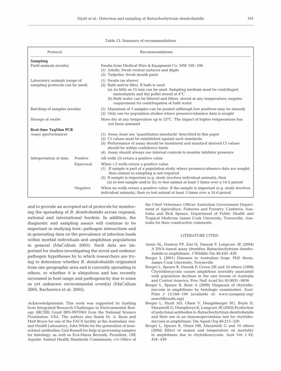
SUMMARY AND RECOMMENDATIONS
This paper has defined the sensitivity, specificity,repeatability and reproducibility of the real-timeTaqMan diagnostic assay for the detection of Batra-chochytrium dendrobatidis. Comparisons between thismolecular assay and conventional histological andimmuno-histochemical assays have also been per-formed; the use of the latter techniques in relation totheir advantages and limitations has been defined. Dif-ferent sampling protocols for the collection of B. den-drobatidis from amphibian skin have also beendescribed and compared. A summary of the recom-mended use of these techniques, together with theinterpretation of the associated data, can be found inTable 13. With the use of validated assays and sam-pling protocols for the detection of B. dendrobatidis, itis now possible to undertake validated testing and sur-veys within both individual animals and populations.This can include studies involving the pathogenesis ofchytridiomycosis, assessing the absence of B. dendro-batidis in captive breeding programs and in endan-gered species, in addition to the screening of the envi-ronment for the fungus by swabbing endemicamphibians, other organisms and inanimate objects.
Finally, it is now possible to recommend to the OIEan internationally recognised set of protocols that canbe used to detect Batrachochytrium dendrobatidis inamphibians. This will be important in classifying ani-mals and regions with or without of chytridiomycosis,
190
Hyatt et al.: Detection and sampling of Batrachochytrium dendrobatidis
and to provide an accepted set of protocols for monitor-ing the spreading of B. dendrobatidis across regional,national and international borders. In addition, thediagnostic and sampling assays will continue to beimportant in studying host–pathogen interactions andin generating data on the prevalence of infection loadswithin morbid individuals and amphibian populationsin general (MaCallum 2005). Such data are im-portant for studies investigating the novel and endemicpathogen hypotheses by to which researchers are try-ing to determine whether B. dendrobatidis originatedfrom one geographic area and is currently spreading toothers, or whether it is ubiquitous and has recentlyincreased in host range and pathogenicity due to someas yet unknown environmental event(s) (MaCallum2005, Rachowicz et al. 2005).
Acknowledgements. This work was supported by fundingfrom Integrated Research Challenges in Environmental Biol-ogy (IRCEB) Grant IBN-9977063 from the National ScienceFoundation, USA. The authors also thank Dr. A. Bean andMatt Bruce for use of the FACS facility at the Australian Ani-mal Health Laboratory; John White for the generation of mon-oclonal antibodies; Gail Russell for help in processing samplesfor histology; as well as Eva-Maria Bernoth, President, OIEAquatic Animal Health Standards Commission, c/o Office of
the Chief Veterinary Officer Australian Government Depart-ment of Agriculture, Fisheries and Forestry, Canberra, Aus-tralia and Rick Speare, Department of Public Health andTropical Medicine James Cook University, Townsville, Aus-tralia for their constructive comments.
LITERATURE CITED
Annis SL, Dastoor FP, Ziel H, Daszak P, Longcore JE (2004)A DNA-based assay identifies Batrachochytrium dendro-batidis in amphibians. J Wildlife Dis 40:420–428
Berger L (2001) Diseases in Australian frogs. PhD thesis,James Cook University, Townsville
Berger L, Speare R, Daszak P, Green DE and 10 others (1998)Chytridiomycosis causes amphibian mortality associatedwith population declines in the rain forests of Australiaand Central America. Proc Natl Acad Sci 95:9031–9036
Berger L, Speare R, Kent A (2000) Diagnosis of chytridio-mycosis in amphibians by histologic examination. Zoos’Print J 15:184–190 (available at: www.zoosprint.org/searchResults.asp)
Berger L, Hyatt AD, Olsen V, Hengstberger SG, Boyle D,Marantelli G, Humphreys K, Longcore JE (2002) Productionof polyclonal antibodies to Batrachochytrium dendrobatidisand their use in an immunoperoxidase test for chytridio-mycosis in amphibians. Dis Aquat Org 48:213–220
Berger L, Speare R, Hines HB, Marantelli G and 10 others(2004) Effect of season and temperature on mortalityin amphibians due to chytridiomycosis. Aust Vet J 82:434–439
191
Protocol Recommendations
SamplingField animals (swabs) Swabs from Medical Wire & Equipment Co. MW 100–100
(1) Adults: Swab ventral surfaces and digits(2) Tadpoles: Swab mouth parts
Laboratory animals (range of (1) Swabs (as above)sampling protocols can be used) (2) Bath and/or filter. If bath is used
(a) As little as 15 min can be used. Sampling medium must be centrifugedimmediately and the pellet stored at 4°C
(b) Bath water can be filtered and filters stored at any temperature; negatesrequirement for centrifugation of bath water
Batching of samples (swabs) (1) Maximum of 5 samples can be pooled (although low positives may be missed)(2) Only use for population studies where presence/absence data is sought
Storage of swabs Store dry at any temperature up to 23°C. The impact of higher temperatures hasnot been assessed
Real-time TaqMan PCRAssay (performance) (1) Assay must use ‘quantitation standards’ described in this paper
(2) Ct values must be established against such standards(3) Performance of assay should be monitored and standard derived Ct values
should be within confidence limits(4) Assay should always use internal controls to monitor inhibitor presence
Interpretation of data Positive All wells (3) return a positive value
Equivocal When <3 wells return a positive value(1) If sample is part of a population study where presence/absence data are sought,
then animal re-sampling is not required(2) If sample is important (e.g. study involves individual animals), then
(a) re-test sample and/or (b) re-test animal at least 3 times over a 14 d period
Negative When no wells return a positive value. If the sample is important (e.g. study involvesindividual animals), then re-test animal at least 3 times over a 14 d period
Table 13. Summary of recommendations

Dis Aquat Org 73: 175–192, 2007
Berger L, Hyatt AD, Speare R, Longcore JE (2005a) Life cyclestages of the amphibian chytrid Batrachochytrium dendro-batidis. Dis Aquat Org 68:51–63
Berger L, Speare R, Skerratt LF (2005b) Distribution of Batra-chochytrium dendrobatidis and pathology in the skin ofgreen tree frogs Litoria caerulae with severe chytridio-mycosis. Dis Aquat Org 68:65–70
Bosch J, Martínez-Solano I, García-París M (2001) Evi-dence of a chytrid fungus infection involved in thedecline of the common midwife toad (Alytes obstetri-cans) in protected areas of central Spain. Biol Conserv97:331–337
Boyle DG, Boyle DB, Olsen V, Morgan JAT, Hyatt AD(2004) Rapid quantitative detection of chytridiomycosis(Batrachochytrium dendrobatidis) in amphibian samplesusing real-time Taqman PCR assay. Dis Aquat Org 60:141–148
Bradley GA, Rosen PC, Sredl MJ, Jones TR, Longcore JE(2002) Chytridiomycosis in native Arizona frogs. J WildlDis 38:206–212
Briggs C, Burgin S (2003) A rapid technique to detect chytridinfection in adult frogs. Herpetol Rev 34(2):124–126
Daszak P, Berger L, Cunningham AA, Hyatt AD, Green DE,Speare R (1999) Emerging infectious diseases and am-phibian population declines. Emerg Infect Dis 5(6):735–748
Daszak P, Cunningham AA, Hyatt AD (2000) Emerging in-fectious diseases of wildlife—threats to biodiversity andhuman health. Science 287(5452):443–449
Fellers GM, Green DE, Longcore JE (2001) Oral chytridio-mycosis in the mountain yellow-legged frog (Rana mus-cosa). Copeia 4:945–953
Green DE, Converse KA, Schrader AK (2002) Epizootiology ofsixty-four amphibian morbidity and mortality events in theUSA, 1996–2001. Ann NY Acad Sci 969:323–339
Heid CA, Stevens J, Livak KJ, Williams PM (1996) Real timequantitative PCR. Genome Res 6:986–994
Johnson ML, Speare R (2003) Survival of Batrachochytriumdedrobatidis in water: quarantine and disease controlimplications. Emerg Infect Dis 9:922–925
Kriger KM, Hines HB, Hero JM, Hyatt AD, Boyle DG (2006)Techniques for detecting chytridiomycosis in wild frogs:comparing histology with real-time Taqman PCR. DisAquat Org 71:141–148
Lie YS, Petropoulos CJ (1998) Advances in quantitative PCRtechnology: 5’ nuclease assays. Curr Opin Biotechnol 9:43–48
Lips K (1999) Mass mortality and population declines of anu-rans at an upland site in western Panama. Conserv Biol 13:117–125
MaCallum H (2005) Inconclusiveness of chytridiomycosis asthe agent in widespread frog declines. Conserv Biol 19(5):1421–1430
McCarthy MA, Parris KM (2004) Clarifying the effect of toe clip-ping on frogs with Bayesian statistics. J Appl Ecol 41:780–786
May RM (2004) Ethics and amphibians. Nature 431:403Mutschmann F, Berger L, Zwart P, Gaedicke C (2000) Chytri-
diomycosis in amphibians—first report in Europe. BerlMuench Tieraerztl Wochenschr 113:380–383
Olsen V, Hyatt AD, Boyle D, Mendez D (2004) Co-localisationof Batrachochytrium dendrobatidis and keratin for en-hanced diagnosis of chytridiomycosis in frogs. Dis AquatOrg 61:85–88
Pessier AP, Nichols DK, Longcore JE, Fuller MS (1999) Cuta-neous chytridiomycosis in poison dart frogs (Dendrobatesspp.) and White’s tree frogs (Litoria caerulae). J Vet DiagnInvest 11:194–199
Rachowicz LJ, Hero JM, Alford RA, Taylor JW, Morgan JAT,Vredenburg VT, Collins JP, Briggs CJ (2005) The noveland endemic pathogen hypotheses: competing explana-tions for the origin of emerging infectious diseases ofwildlife. Conserv Biol 19(5):1441–1448
Ron SR, Duellman WE, Coloma LA, Bustamante MR (2003)Population decline of the Jambato toad Atelopus ignescens(Anura: Bufonidae) in the Andes of Ecuador. J Herpetol37(1):116–126
Stuart SN, Chanson JS, Cox NA, Young BE, Rodrigues AS,Fischman DL, Waller RW (2004) Status and trends ofamphibian declines and extinctions worldwide. Science306(5702):1783–1786
Van Ells T, Stanton J, Strieby A, Daszak P, Hyatt AD, Brown C(2003) Use of immunohistochemistry to diagnose chytrid-iomycosis in dying poison dart frogs (Dendrobates tincto-rius). J Wildl Dis 39(3):742–745
Weldon C, du Preez LH, Hyatt AD, Muller R, Speare R (2004)Origin of the amphibian chytrid fungus. Emerg Infect Dis10(12):2100–2105
192
Editorial responsibility: Managing Editor Submitted: August 21, 2006; Accepted: November 14, 2006Proofs received from author(s): January 8, 2007


![Radioligand Binding Assays: Application of [125I]Angiotensin II Receptor Binding](https://img.dokumen.tips/doc/110x75/635cf688095e4caf22057ea4/radioligand-binding-assays-application-of-125iangiotensin-ii-receptor-binding.jpg)


