Embed Size (px)
Citation preview

RESEARCH ARTICLE Open Access
Prevalence of the pathogenic chytrid fungus,Batrachochytrium dendrobatidis, in anendangered population of northern leopardfrogs, Rana pipiensMaarten J Voordouw1*, Doug Adama2, Barb Houston3, Purnima Govindarajulu4, John Robinson5
Abstract
Background: Emerging infectious diseases threaten naïve host populations with extinction. Chytridiomycosis, anemerging infectious disease of amphibians, is caused by the pathogenic fungus Batrachochytrium dendrobatidis (Bd)and has been linked to global declines in amphibians.
Results: We monitored the prevalence of Bd for four years in the Northern leopard frog, Rana pipiens, which iscritically imperiled in British Columbia (BC), Canada. The prevalence of Bd initially increased and then remainedconstant over the last three years of the study. Young of the year emerging from breeding ponds in summer wererarely infected with Bd. Some individuals cleared their Bd infections and the return rate between infected anduninfected individuals was not significantly different.
Conclusions: The BC population of R. pipiens appears to have evolved a level of resistance that allows it to co-existwith Bd. However, this small population of R. pipiens remains vulnerable to extinction.
BackgroundInfectious diseases can have devastating consequencesfor immunologically naïve host populations. Humaninfectious diseases such as measles, tuberculosis, andsmallpox are believed to have killed as many as 95% ofall Native Americans in the two centuries following thefirst contact with Europeans [1]. In the UK, the declineof the native red squirrel and its replacement by theintroduced grey squirrel is believed to be mediated bythe parapoxvirus [2]. Small, inbred host populations areespecially vulnerable to new pathogens due to a lack ofgenetic variation [3]. For example, the introduction ofavian malaria has decimated populations of Hawaiianbirds [4]. The Christmas Island rat, Rattus macleari,went extinct in less than five years following contactwith the black rat, Rattus rattus, which carried a patho-genic trematode [5]. In this study, we investigated therole of chytridiomycosis, a recently emerged infectious
disease of amphibians, in the decline of a small, endan-gered population of the northern leopard frog, Ranapipiens.Chytridiomycosis is a skin disease of amphibians that is
caused by the chytridiomycete fungus, Batrachochytriumdendrobatidis [6,7]. The waterborne zoospores of thisfungus attack keratinized tissues including the skin ofpost-metamorphic individuals and the mouthparts of tad-poles. In post-metamorphic individuals, chytridiomycosiscauses hyperkeratosis (a marked thickening of the stra-tum corneum) and excessive skin sloughing, which canimpair cutaneous respiration and osmoregulation andresult in death [6]. Chytrid zoospores have limited swim-ming ability (~2 cm) [8] and the fungus appears todepend on water flow or host movement for long dis-tance dispersal [9]. The fungus grows best between 17and 25°C and cannot grow at air temperatures higherthan 28°C [7]. Recent work suggests that Bd may producetiny, non-pathogenic resting spores that attach to theamphibian skin surface but without causing disease [10].Chytridiomycosis is believed to be responsible for the
mass mortality and extinction events of amphibian
* Correspondence: [email protected] of Biology, University of Victoria, PO Box 3020, Station CSC,Victoria, British Columbia, V8W 3N5, Canada
Voordouw et al. BMC Ecology 2010, 10:6http://www.biomedcentral.com/1472-6785/10/6
© 2010 Voordouw et al; licensee BioMed Central Ltd. This is an Open Access article distributed under the terms of the CreativeCommons Attribution License (http://creativecommons.org/licenses/by/2.0), which permits unrestricted use, distribution, andreproduction in any medium, provided the original work is properly cited.

populations in Australia [11], Panama and Costa Rica[12,13]. Most of the chytridiomycosis-related die-offshave occurred in amphibians that breed in permanentwater bodies reflecting the aquatic nature of the disease[12]. In Queensland, Australia, amphibians that breed inephemeral water bodies or terrestrial environments wereseldom infected with Bd [14]. Similarly, a survey inMaine, USA, found that infection prevalence in speciesthat hibernate in terrestrial habitats was almost threetimes lower than that in species that hibernate in aqua-tic habitats [15]. The disease is generally less virulent intadpoles than post-metamorphic individuals. Mass mor-tality events in Arizona and California have foundapparently healthy larvae in the presence of dead ordying adult frogs [16,17]. There is variation among andwithin species in susceptibility to the disease [18-21].The northern leopard frog, Rana pipiens, is a med-
ium-sized, semi-terrestrial frog that is widely distributedin North America [22,23]. R. pipiens emerges from itsoverwintering habitat in early spring and adults move tothe breeding ponds. Mating and egg laying occurs frommid-April to early June. Tadpoles transform into post-metamorphic froglets in late July and disperse awayfrom their natal pond over the next several weeks. Afterbreeding, adults venture into adjacent upland areas toforage. In late August to September, adults, juvenilesand young of the year head to their overwintering habi-tat. We therefore expect R. pipiens to be most vulner-able to Bd during breeding (April to June) andoverwintering (October to March) due to their aquaticnature at these times.The Committee on the Status of Endangered Wildlife
in Canada (COSEWIC) has listed R. pipiens as Endan-gered in British Columbia because only two small popu-lations remain. Both populations are in the southeasternpart of the province, one naturally occurring and onerecently reintroduced as part of a recovery effort [24].Surveyors estimated that between 2000 to 2005 the BCpopulation declined by 50% and found that R. pipienswas infected with Bd [24,25]. In 2003, we began a mark-recapture study of R. pipiens. The purpose of this studywas to monitor the prevalence of Bd over time and todetermine whether season and stage class influencedinfection levels. The mark-recapture design of the studyallowed us to test whether R. pipiens can clear their Bdinfection as demonstrated in previous studies in Austra-lia [26,27]. In this study we also compared the sensitivityof three different tissue-sampling methods in determin-ing whether a frog was infected with Bd.
MethodsStudy areaThe study area included the Creston Valley and Bum-mers Flats Wildlife Management Areas (CVWMA and
BFWMA) in southeast British Columbia, Canada and isdescribed in detail by Adama and Beaucher [24]. TheCVWMA occupies 6,885 ha and the BFWMA occupies850 ha.
Survey MethodsThe survey started in the fall of 2003. From 2004 to2007 surveyors visited the CVWMA and the BFWMAin the spring, summer and fall. The annual samplingeffort in 2003, 2004, 2005, 2006, and 2007 (for theCVWMA and the BFWMA combined) was 119, 160,200, 159, and 49 visits, respectively, which took 199,308, 417, 314, and 108 person hours. Surveyors encoun-tered and captured 320 R. pipiens during nocturnal call-ing surveys, egg mass surveys and visual encountersurveys [for details see [24]] and took 401 tissuesamples.For each R. pipiens capture, surveyors took a photo of
the dorsal side, which has a unique pattern of large,dark circular spots. These photos were used to identifyindividuals and to determine recaptures within andamong subsequent years. Other studies have used spotpatterns to successfully identify individual leopard frogs[28].For each captured animal, surveyors recorded its GPS
coordinates and measured its snout-vent length andbody weight. We used body weight and season of cap-ture to assign individuals to one of three stage classes:young of the year, juvenile, and adult. The three stageclasses were categorized as follows: young of the yearweighed less than 35 grams in the summer and fall. Ajuvenile weighed between 35 and 50 grams in the sum-mer and fall or weighed less than 50 grams in thespring. An adult was any frog that weighed more than50 grams. Captured animals were also checked forsymptoms of chytridiomycosis, which include sloughingskin, redness, lethargy, abnormal body positioning, lossof righting reflex, and vascularization.
Tissue sampling and PCR test for BdTo determine whether the animal was infected with Bd,we collected tissue samples for PCR analysis. Tissuesamples were collected using three different methodsincluding toe clips, bag rinses, and swabs. In the toe clipmethod, we cut the terminal phalange of the fourth toeof the animal’s right hind foot. In the bag rinse method,an animal was lightly “massaged” within a (single use)zip lock bag to collect tissue and the bag was subse-quently rinsed out with ethanol. In the swab method,we used a sterile cotton tip swab (#018-460 AMG Medi-cal Inc) to swab the abdomen, thighs, groin and feet ofthe animal 10 to 20 times. The tissue sampling methodchanged over the course of the study. In 2003 we usedtoe clips because Bd was primarily diagnosed using
Voordouw et al. BMC Ecology 2010, 10:6http://www.biomedcentral.com/1472-6785/10/6
Page 2 of 10

histological techniques [6]. In 2004, we switched to lessinvasive swab and bag rinse methods because PCRbecame the standard method of identifying Bd [29] andbecause R. pipiens is an endangered species. From 2003to 2006 all swabs were preserved in ethanol. In 2007,swabs were stored in tubes without ethanol (i.e., dryswabs). We changed the swab storage protocol becauseextracting DNA from dry swabs was less time consum-ing than extracting DNA from swabs stored in ethanol.Hyatt et al [30] demonstrated that dry swabs and swabsstored in ethanol are equally effective at detecting BdDNA. Tissue samples were sent to the Animal HealthCentre of the Ministry of Agriculture in Abbotsford, BCwhere they were tested for Bd using PCR. We followedthe methods of Boyle et al. [29] except that we did notconstruct a standard curve. We therefore cannot deter-mine zoospore load and the PCR data consisted ofwhether a frog was infected or not. Unfortunately, thetissue samples from 2004 were lost and so we have PCRdata for 2003, 2005, 2006 and 2007.
Statistical MethodsIndependence of data and pseudo-replicationThere are three levels of replication in the survey: (1)animal, (2) capture occasion, and (3) tissue sample. Ananimal refers to a unique R. pipiens individual. A cap-ture occasion refers to the date that an animal was cap-tured (i.e. the same animal can be captured on multipleoccasions). A tissue sample refers to the fact that some-times we obtained multiple tissue samples from thesame capture occasion using different methods (i.e. bagrinse, swab, toe clip). The recapture rate of R. pipienswas low (31 recaptures/320 captures) and we thereforetreated all 320 captures as independent. For captureoccasions with multiple tissue samples, the animal wasconsidered Bd-positive if at least one of the tissue sam-ples tested positive for Bd.Sensitivity of 3 tissue-sampling methodsThe subset of capture occasions with multiple tissuesamples allowed us to compare the sensitivity of thethree tissue sampling methods. We used the Chi-squaretest to determine statistical significance.Prevalence of Batrachochytrium dendrobatidis inRana pipiensThe prevalence of Bd (i.e. the proportion of infectedanimals) is binomial data because an animal is eitherinfected or not. We used generalized linear models(GLM) with a binomial error distribution to model theprevalence of Bd in R. pipiens as a function of the fol-lowing five factors: season, year, stage class, location andtissue-sampling method. The levels of each factor are asfollows: season (spring = April to June, summer = Julyto August, fall = September to October), year (2003,2005, 2006, 2007), stage class (young of year, juveniles,
adults), location (CVWMA, BFWMA), and tissue-sam-pling method. The tissue-sampling method had twolevels: (1) capture occasions that were tissue-sampledwith either a bag rinse or a toe clip, or (2) capture occa-sions that were tissue-sampled with only the swabmethod. The justification for these two levels was thatbag rinses and toe clips were 3.6 times more sensitivethan swabs (see results).We tested all possible combinations of the main
effects and the two-way and three-way interaction termsfor a total of 2728 models. We used Akaike’s informa-tion criterion (AIC) to guide model selection. The bestmodel was the one with the fewest parameters andwithin 1 unit of the lowest AIC score. We used log like-lihood ratio tests to determine the statistical significanceof the terms in the best model. R (version 2.7.0) wasused to analyze the data.To determine whether temperature influences the pre-
valence of Bd, we obtained mean monthly air tempera-ture data from the Creston Campbell Scientific weatherstation for the period from 2003 to 2007. The meanmonthly air temperature is an average of the mean dailyair temperatures for that month. The mean daily airtemperature is the average of the maximum and mini-mum daily air temperature. The Creston CampbellScientific weather station is less than 20 km from theCVWMA.Survival of Bd-infected R. pipiensMark-recapture analysis estimates the probability of cap-turing an animal and the probability of survival betweencapture sessions. Unfortunately there were only 31recaptures in this study, of which 6 were among years, 3were among seasons within year, and the remaining 22were within season (i.e. <24 days). Hence there were notenough recaptures to warrant a proper mark-recaptureanalysis [31]. Instead, we used a chi-square test to deter-mine whether the return rate (defined as the percentageof captures that are recaptures and which includes boththe survival and recapture rate) was different betweenBd-infected and uninfected R. pipiens.
ResultsSensitivity of 3 tissue-sampling methodsThere were 72 capture occasions where we used morethan one tissue sampling method on the same animal.Of these 72 capture occasions, 26 had at least one tissuesample that tested positive for Bd (Table 1). Of the 4Bd-positive captures that were both bag rinsed and toeclipped, the bag rinse and toe clip both tested positivein 3 out of 4 captures; in the fourth case only the bagrinse tested positive (Table 1). For lack of more data, weconsidered the bag rinse and toe clip method to beequally sensitive. There were 25 captures that testedpositive for Bd according to either the bag rinse or the
Voordouw et al. BMC Ecology 2010, 10:6http://www.biomedcentral.com/1472-6785/10/6
Page 3 of 10

toe clip method (Table 1). These 25 animals were alsoswabbed but only 28% of the swabs (7/25) tested posi-tive (Table 1) suggesting that the bag rinse and toe clipmethods were 3.6 times more sensitive than the swabmethod. It was this result that motivated us to includethe tissue sampling method in the GLM model of Bdprevalence in R. pipiens (see Table 2).
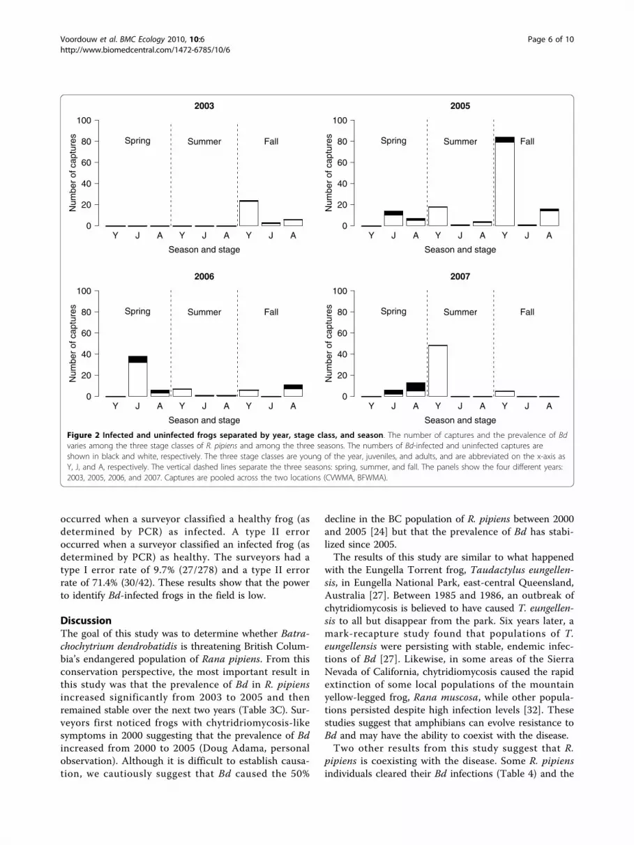
Prevalence of Batrachochytrium dendrobatidis inRana pipiensOver the whole study, there were 320 captures of R.pipiens, of which 13.1% (42/320; 95% confidence interval(CI) = 10.0 to 17.8%) tested positive for Bd. Bd preva-lence increased with stage class: young of the year (3.1%= 6/192; 95% CI = 1.3 to 7.0%), juvenile (25.0% = 16/64;95% CI = 15.4 to 37.7%), and adult (31.3% = 20/64; 95%CI = 20.6 to 44.2%; Figure 1). Bd prevalence in thespring (32.1% = 27/84; 95% CI = 22.6 to 43.3%) wasmuch higher than that in the summer (1.3% = 1/80;95% CI = 0.1 to 7.7%) and fall (9.0% = 14/156; 95% CI =5.2 to 14.9%; Figure 2). Stage class biased this seasonalpattern as follows. All of the captures in the spring werejuveniles and adults whereas most of the captures in the
summer and fall were young of the year, which wereless likely to be infected with Bd (Figure 2). The seaso-nal pattern of Bd prevalence was inversely related withthe mean monthly air temperature across the 7 monthsof sampling (Figure 3). The prevalence of Bd in R.pipiens was 6.1% (2/33; 95% CI = 1.1 to 21.6%) in 2003,10.3% (15/145; 95% CI = 6.1 to 16.8%) in 2005, 18.6%(13/70; 95% CI = 10.6 to 30.0%) in 2006, and 16.7% (12/72; 95% CI = 9.3 to 27.7%) in 2007 (Figure 2).
GLM model of Bd prevalence in R. pipiensThe best model according to AIC included the tissuesampling method, season, year, stage class and the sea-son:stage class interaction (model 3 in Table 2) but notlocation. This model provided a good fit to the data(residual df = 308, residual deviance = 165.77). We usedlog likelihood ratio tests to compare this model tonested models (Table 3A) to determine the statisticalsignificance of each factor (Table 3B) and the parameterestimates (Table 3C). The log likelihood ratio testsfound that the tissue-sampling method, season, year,and stage class accounted for a significant portion of thedeviance in the prevalence of Bd but that the season:
Table 1 PCR results for R. pipiens individuals that were sampled with multiple tissue sampling methods on the samedate.
Tissue samples that test positive for Bd
Method br/sw/tc br/sw br/tc sw/tc br sw tc none Total
BR, SW & TC 1 0 2 0 0 0 0 6 9
BR & SW *** 2 *** *** 2 0 *** 6 10
BR & TC *** *** 0 *** 1 *** 0 1 2
SW & TC *** *** *** 4 *** 0 14 33 51
Total 1 2 2 4 3 0 14 46 72
The outcomes of the PCR assay for the 72 individuals that were tested with multiple tissue-sampling methods on the same date. The three different tissuesampling methods are bag rinse (BR), swab (SW), and toe clip (TC). The combination of methods used to sample an individual is shown in the far-left columnwhereas the possible outcomes of the PCR assay are shown in the top-row. For example, of the 9 individuals that were sampled with all three methods (row BR,SW & TC), 1 individual tested positive for all 3 samples (column br/sw/tc), 2 individuals tested positive for both the bag rinse and toe clip (column br/tc), and 6animals tested negative for all 3 methods (column ‘none’). The triple asterisks (***) denote PCR outcomes that are not possible for that combination of tissuesampling methods.
Table 2 Generalized linear models (GLM) of the prevalence of Bd in R. pipiens
# Model Structure df dev AIC
1 pcr~M + S + Y + T + L + S:T 307 163.19 189.19
2 pcr~M + S + Y + T + L + M:T + M:L + S:T + S:L + T:L + M:T:L + S:T:L 300 149.20 189.20
3 pcr~M + S + Y + T + S:T 308 165.77 189.77
4 pcr~M + S + Y + T + L + M:L + S:T 306 162.15 190.15
5 pcr~M + S + Y + T + L 310 170.36 190.36
6 pcr~M + S + Y + T 311 172.95 190.95
7 pcr~M + S + Y + T + L + M:Y + S:T 305 161.10 191.10
8 pcr~M + S + Y + T + L + M:T + S:T 305 161.11 191.11
9 pcr~M + S + Y + T + L + M:L 309 169.32 191.32
10 pcr~M + S + Y + T + L + M:T + M:L + T:L + M:T:L 304 159.35 191.35
We used GLM with binomial errors to model the prevalence of Bd in R. pipiens as a function of the tissue-sampling method (M), season (S), year (Y), stage class(T), location (L) and their two-way and three-way interactions. Of the 2728 models tested, we show the 10 models with the lowest Akaike Information Criterion(AIC) scores. Also shown are the residual degrees of freedom (df) and the residual deviance (dev) for each model.
Voordouw et al. BMC Ecology 2010, 10:6http://www.biomedcentral.com/1472-6785/10/6
Page 4 of 10

stage class interaction was marginally non-significant(Table 3B). The change in deviance from deleting eachfactor (expressed as a percent of the residual devianceof the best model = 165.77) was as follows: tissue-sam-pling method (10.9%), season (4.6%), year (5.3%), stageclass (6.4%), and season:stage class (4.3%).The parameter estimates (Table 3C) confirmed that
the prevalence of Bd was (1) significantly higher for bagrinses and toe clips than for swab tissue samples, (2) sig-nificantly higher in the spring and fall than in the sum-mer, and (3) significantly higher in 2005, 2006, and 2007than in 2003. After excluding 2003 from the analysis,the factor year was no longer significant, indicating thatthere were no significant differences in Bd prevalenceamong the last three years of the study.
Clearance of Bd infection in R. pipiensWe examined the infection history of recaptured ani-mals to determine whether R. pipiens can clear their Bdinfection. Of the 26 R. pipiens individuals that were cap-tured multiple times, 8 frogs tested positive for Bd on atleast one of their captures. Of these 8 recaptured and
Bd-infected R. pipiens, there were 3 individuals thatapparently cleared their Bd infection. These individualstested positive and then negative for Bd between conse-cutive capture occasions (this happened twice in indivi-dual RC.06010; Table 4). There were two convincingcases of individuals clearing their Bb infection (RC07003 and RC 06010) where the second negative testwas determined by a bag rinse (Table 4). The time inter-val over which the three individuals cleared their Bdinfections ranged between 1 and 23 days (Table 4).
Survival of Bd-infected R. pipiensThe return rates (includes both survival and recapture) ofBd-infected (9.52% = 4/42; 95% CI = 3.1 to 23.5%) anduninfected R. pipiens (9.44% = 27/286; 95% CI = 6.4 to13.6%) were not significantly different (c2 = 0.07, df = 1,p = 0.791).
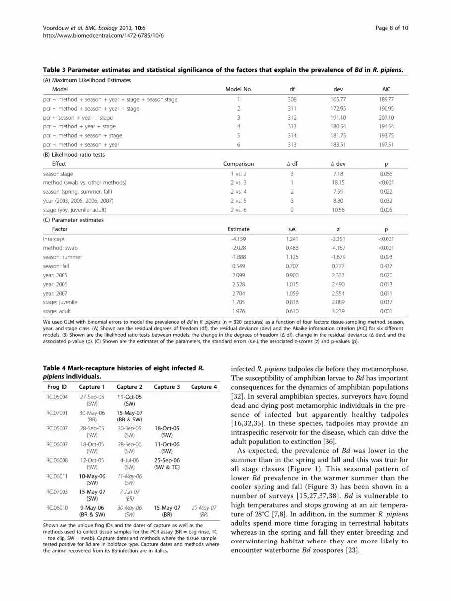
Ability of surveyors to identify chytridiomycosis inthe fieldTo evaluate the performance of the surveyors we haveto consider both type I and type II errors. A type I error
0.0
0.2
0.4
0.6
0.8
1.0
Season
Pre
vale
nce
of B
d
L S F L S F L S F
YOY Juveniles Adults
Figure 1 The prevalence of Bd among stage classes and seasons. The prevalence of Batrachochytrium dendrobatidis (Bd) in the three stageclasses of Rana pipiens varies across the three seasons. Shown are the mean prevalence of Bd and the 95% confidence limits. The three stageclasses are young of the year (YOY), juveniles, and adults and are separated by the vertical dashed lines. The three seasons are spring, summer,and fall and are abbreviated on the x-axis as L, S and F, respectively. Captures are pooled across all years and both locations (CVWMA, BFWMA).
Voordouw et al. BMC Ecology 2010, 10:6http://www.biomedcentral.com/1472-6785/10/6
Page 5 of 10
occurred when a surveyor classified a healthy frog (asdetermined by PCR) as infected. A type II erroroccurred when a surveyor classified an infected frog (asdetermined by PCR) as healthy. The surveyors had atype I error rate of 9.7% (27/278) and a type II errorrate of 71.4% (30/42). These results show that the powerto identify Bd-infected frogs in the field is low.
DiscussionThe goal of this study was to determine whether Batra-chochytrium dendrobatidis is threatening British Colum-bia’s endangered population of Rana pipiens. From thisconservation perspective, the most important result inthis study was that the prevalence of Bd in R. pipiensincreased significantly from 2003 to 2005 and thenremained stable over the next two years (Table 3C). Sur-veyors first noticed frogs with chytridriomycosis-likesymptoms in 2000 suggesting that the prevalence of Bdincreased from 2000 to 2005 (Doug Adama, personalobservation). Although it is difficult to establish causa-tion, we cautiously suggest that Bd caused the 50%
decline in the BC population of R. pipiens between 2000and 2005 [24] but that the prevalence of Bd has stabi-lized since 2005.The results of this study are similar to what happened
with the Eungella Torrent frog, Taudactylus eungellen-sis, in Eungella National Park, east-central Queensland,Australia [27]. Between 1985 and 1986, an outbreak ofchytridiomycosis is believed to have caused T. eungellen-sis to all but disappear from the park. Six years later, amark-recapture study found that populations of T.eungellensis were persisting with stable, endemic infec-tions of Bd [27]. Likewise, in some areas of the SierraNevada of California, chytridiomycosis caused the rapidextinction of some local populations of the mountainyellow-legged frog, Rana muscosa, while other popula-tions persisted despite high infection levels [32]. Thesestudies suggest that amphibians can evolve resistance toBd and may have the ability to coexist with the disease.Two other results from this study suggest that R.
pipiens is coexisting with the disease. Some R. pipiensindividuals cleared their Bd infections (Table 4) and the
Y J A Y J A Y J A
2003
Season and stage
Num
ber
of c
aptu
res
0
20
40
60
80
100
Spring Summer Fall
Y J A Y J A Y J A
2005
Season and stage
Num
ber
of c
aptu
res
0
20
40
60
80
100
Spring Summer Fall
Y J A Y J A Y J A
2006
Season and stage
Num
ber
of c
aptu
res
0
20
40
60
80
100
Spring Summer Fall
Y J A Y J A Y J A
2007
Season and stage
Num
ber
of c
aptu
res
0
20
40
60
80
100
Spring Summer Fall
Figure 2 Infected and uninfected frogs separated by year, stage class, and season. The number of captures and the prevalence of Bdvaries among the three stage classes of R. pipiens and among the three seasons. The numbers of Bd-infected and uninfected captures areshown in black and white, respectively. The three stage classes are young of the year, juveniles, and adults, and are abbreviated on the x-axis asY, J, and A, respectively. The vertical dashed lines separate the three seasons: spring, summer, and fall. The panels show the four different years:2003, 2005, 2006, and 2007. Captures are pooled across the two locations (CVWMA, BFWMA).
Voordouw et al. BMC Ecology 2010, 10:6http://www.biomedcentral.com/1472-6785/10/6
Page 6 of 10

return rates were not significantly different between Bd-infected and uninfected R. pipiens suggesting that theyhad similar survival. Similar results were found in amark-recapture study on the Stony Creek frog, Litoriawilcoxii, where 7 individuals cleared their Bd infectionsand the return rate was not significantly differentbetween Bd-infected and uninfected frogs [26]. Whilethe BC population of R. pipiens may be coexisting withBd, this does not mean that the population is doingwell. Between 2000 and 2005, surveyors have found anaverage of 10 egg masses per year [24]. Hence R. pipiensremains endangered in British Columbia.The prevalence of Bd was higher in juveniles and adults
than young of the year, (Table 3C; Figure 1), perhaps
because the overwintering ponds are infected with Bdand/or sexual transmission between adults in the spring.The results of this study contradict studies on Australianfrogs, which found that small frogs were more likely to beinfected and carried more intense infections than largerfrogs [33,34]. We were surprised that so few young of theyear were infected (Figure 1) because they emerge fromthe same ponds where highly infected adults mate in thespring. One possibility is that the young of the year losetheir infections because they emerge from the breedingponds during the hottest months of the year (July andAugust; Figure 3). Alternatively, the low proportion ofinfected young of the year suggests two other possibilities:(1) R. pipiens tadpoles are rarely infected with Bd or (2)
Month
Pre
vale
nce
of B
d
Apr May Jun Jul Aug Sep Oct
0.0
0.2
0.4
0.6
0.8
1.0
−10
0
10
20
30
40
Tem
pera
ture
(de
gree
s C
elsi
us)
Figure 3 Prevalence of Bd versus the mean monthly temperature. The prevalence of Bd in R. pipiens changes across the 7 months ofsampling. The red circles show the mean prevalence of Bd for each month and the red lines indicate the 95% confidence limits. The solid anddotted black lines show the mean, mean maximum, and mean minimum air temperatures for each month (in °C; averaged for 2003, 2005, 2006and 2007). Captures are pooled across the three tissue sampling methods (bag rinses, toe clips, and swabs), the three stage classes (young ofthe year, juveniles, and adults), the four years (2003, 2005, 2006, and 2007), and the two locations (CVWMA, BFWMA).
Voordouw et al. BMC Ecology 2010, 10:6http://www.biomedcentral.com/1472-6785/10/6
Page 7 of 10
infected R. pipiens tadpoles die before they metamorphose.The susceptibility of amphibian larvae to Bd has importantconsequences for the dynamics of amphibian populations[32]. In several amphibian species, surveyors have founddead and dying post-metamorphic individuals in the pre-sence of infected but apparently healthy tadpoles[16,32,35]. In these species, tadpoles may provide anintraspecific reservoir for the disease, which can drive theadult population to extinction [36].As expected, the prevalence of Bd was lower in the
summer than in the spring and fall and this was true forall stage classes (Figure 1). This seasonal pattern oflower Bd prevalence in the warmer summer than thecooler spring and fall (Figure 3) has been shown in anumber of surveys [15,27,37,38]. Bd is vulnerable tohigh temperatures and stops growing at an air tempera-ture of 28°C [7,8]. In addition, in the summer R. pipiensadults spend more time foraging in terrestrial habitatswhereas in the spring and fall they enter breeding andoverwintering habitat where they are more likely toencounter waterborne Bd zoospores [23].
Table 3 Parameter estimates and statistical significance of the factors that explain the prevalence of Bd in R. pipiens.
(A) Maximum Likelihood Estimates
Model Model No. df dev AIC
pcr ~ method + season + year + stage + season:stage 1 308 165.77 189.77
pcr ~ method + season + year + stage 2 311 172.95 190.95
pcr ~ season + year + stage 3 312 191.10 207.10
pcr ~ method + year + stage 4 313 180.54 194.54
pcr ~ method + season + stage 5 314 181.75 193.75
pcr ~ method + season + year 6 313 183.51 197.51
(B) Likelihood ratio tests
Effect Comparison Δ df Δ dev p
season:stage 1 vs. 2 3 7.18 0.066
method (swab vs. other methods) 2 vs. 3 1 18.15 <0.001
season (spring, summer, fall) 2 vs. 4 2 7.59 0.022
year (2003, 2005, 2006, 2007) 2 vs. 5 3 8.80 0.032
stage (yoy, juvenile, adult) 2 vs. 6 2 10.56 0.005
(C) Parameter estimates
Factor Estimate s.e. z p
Intercept -4.159 1.241 -3.351 <0.001
method: swab -2.028 0.488 -4.157 <0.001
season: summer -1.888 1.125 -1.679 0.093
season: fall 0.549 0.707 0.777 0.437
year: 2005 2.099 0.900 2.333 0.020
year: 2006 2.528 1.015 2.490 0.013
year: 2007 2.704 1.059 2.554 0.011
stage: juvenile 1.705 0.816 2.089 0.037
stage: adult 1.976 0.610 3.239 0.001
We used GLM with binomial errors to model the prevalence of Bd in R. pipiens (n = 320 captures) as a function of four factors: tissue-sampling method, season,year, and stage class. (A) Shown are the residual degrees of freedom (df), the residual deviance (dev) and the Akaike information criterion (AIC) for six differentmodels. (B) Shown are the likelihood ratio tests between models, the change in the degrees of freedom (Δ df), change in the residual deviance (Δ dev), and theassociated p-value (p). (C) Shown are the estimates of the parameters, the standard errors (s.e.), the associated z-scores (z) and p-values (p).
Table 4 Mark-recapture histories of eight infected R.pipiens individuals.
Frog ID Capture 1 Capture 2 Capture 3 Capture 4
RC.05004 27-Sep-05(SW)
11-Oct-05(SW)
RC.07001 30-May-06(BR)
15-May-07(BR & SW)
RC.05007 28-Sep-05(SW)
30-Sep-05(SW)
18-Oct-05(SW)
RC.06007 18-Oct-05(SW)
28-Sep-06(SW)
11-Oct-06(SW)
RC.06008 12-Oct-05(SW)
4-Jul-06(SW)
25-Sep-06(SW & TC)
RC.06011 10-May-06(SW)
11-May-06(SW)
RC.07003 15-May-07(SW)
7-Jun-07(BR)
RC.06010 9-May-06(BR & SW)
30-May-06(SW)
15-May-07(BR)
29-May-07(BR)
Shown are the unique frog IDs and the dates of capture as well as themethods used to collect tissue samples for the PCR assay (BR = bag rinse, TC= toe clip, SW = swab). Capture dates and methods where the tissue sampletested positive for Bd are in boldface type. Capture dates and methods wherethe animal recovered from its Bd-infection are in italics.
Voordouw et al. BMC Ecology 2010, 10:6http://www.biomedcentral.com/1472-6785/10/6
Page 8 of 10

This study would have been much improved if we hadalways used the same tissue-sampling method. We havetried to correct for the tissue-sampling method in ourstatistical analysis but it is possible that some of ourresults were biased by the inconsistent tissue-samplingstrategy. The bag rinse was equally sensitive at detectingBd as the toe clip method. Together, these two methodswere 3.6 times more sensitive at detecting Bd than theswab method (Table 1). Surveyors in the present studyobtained swabs by rubbing the tip of a sterile cottonswab on the abdomen, thighs, groin, and feet 10 to 20times [24]. In contrast, Kriger et al. swab each frog 70times and have detected some of the highest prevalencesof Bd to date [14,26,34,37]. Hence our low swab successrate may be due to the difference in swabbing effort.
ConclusionsChytridiomycosis may have caused the recent decline inthe BC population of R. pipiens. The prevalence of Bdappears to have stabilized over the last three years ofthe study but the population of R. pipiens has not recov-ered. Young of the year emerging from breeding pondswere rarely infected with Bd and the prevalence of Bdin R. pipiens decreased in warmer months. Some indivi-duals cleared their Bd infection and the return rate ofBd-infected and uninfected individuals was the samesuggesting that R. pipiens may have evolved resistanceto chytrid-related mortality. However, the BC populationof R. pipiens remains endangered.
AcknowledgementsThe authors would like to acknowledge the many contributions that madethis project possible. We would like to thank John Krebs of the Fish andWildlife Compensation Program for administering the project. Financialsupport was provided by the Fish and Wildlife Compensation Program(Columbia Basin), Columbia Basin Trust, Creston Valley WildlifeManagement Area, Fortis BC, BC Ministry of Environment, and the Centrefro Coastal Health. Also greatly appreciated were the contributions of theNorthern Leopard Frog Recovery Team: John Krebs, Marc-André Beaucher,Penny Ohanjanian, Ted Antifeau, Dan Wigle, Dave Fraser, DavidCunnington, and Laura Friis. Without the hard work of all the surveyorswho worked on collecting samples for the project from 2003-2007 thisproject would not have been possible, and we wish to thank AndreaDavidson, Anneli Schadeli, Carla Haegele, Justin Lang, Tuomas Kukkonen,Kate Lansley, Sarah Herring, Melissa Hogg, Taryn Woodnote-Saberwing,Julie Mathews, and Brendan Wilson and participating students at SelkirkCollege, Castlegar, BC. Additional contributions were made by HeatherWaye, Kris Kendell (Alberta Conservation Association), Dr. Trent Bollinger(Prairie Diagnostic Services, Western College of Veterinary Medicine), Dr.Helen Schwantje, Dr. Stephen Raverty and staff at the Animal HealthCentre Laboratory (Ministry of Agriculture, Foods, and Fisheries), CrestonValley Wildlife Management Area staff, Beth Woodbridge (FWCPadministrative assistant), and Amy Waterhouse (FWCP GIS). We also thankGodefroy Devevey and one anonymous reviewer whose comments greatlyimproved this manuscript. Animal Care certificates and permits wereobtained under provisions of the Wildlife Act from the BC Ministry ofEnvironment.
Author details1Department of Biology, University of Victoria, PO Box 3020, Station CSC,Victoria, British Columbia, V8W 3N5, Canada. 2BC Hydro, Unit 1 1007 11th
Avenue, Golden, British Columbia, V0A 1H0, Canada. 3Fish and WildlifeCompensation Program - Columbia Basin, 103-333 Victoria Street, Nelson,British Columbia, V1L 4K3, Canada. 4BC Ministry of Environment, PO Box9338 Stn Prov Govt, Victoria, British Columbia, V8W 9M1, Canada. 5AnimalHealth Centre, BC Ministry of Agriculture & Lands, 1767 Angus CampbellRoad, Abbotsford, British Columbia, V3G 2M3, Canada.
Authors’ contributionsMJV analyzed the data and wrote the manuscript. DA and BH designed thesurvey protocol and conducted most of the fieldwork. JR supervised the PCRassay of the tissue samples. PG provided expertise on amphibians andchytridiomycosis. DA, BH, and PG helped to interpret the data and write themanuscript. All authors read and approved the final manuscript.
Received: 30 March 2009Accepted: 4 March 2010 Published: 4 March 2010
References1. Diamond J: Guns, germs, and steel: the fates of human societies. New
York: W. W. Norton & Company 1999.2. Tompkins DM, White AR, Boots M: Ecological replacement of native red
squirrels by invasive greys driven by disease. Ecology Letters 2003,2003(6):189-196.
3. Soule M: Conservation biology: the science of scarcity and diversity.Sunderland, Massachusetts: Sinauer Associates 1986.
4. van Riper C III, van Riper SG, Goff ML, Laird M: The epizootiology andecological significance of malaria in Hawaiian land birds. EcologicalMonographs 1986, 56(4):327-344.
5. Wyatt KB, Campos PF, Gilbert MTP, Kolokotronis SO, Hynes WH, DeSalle R,Daszak P, MacPhee RDE, Greenwood AD: Historical mammal extinction onChristmas Island (Indian Ocean) correlates with introduced infectiousdisease. PloS ONE 2008, 3(11):e3602.
6. Berger L, Speare R, Daszak P, Greens DE, Cunningham AA, Goggin L,Slocombe R, Ragan MA, Hyatt AD, McDonald KR, et al: Chytridiomycosiscauses amphibian mortality associated with population declines in therain forests of Australia and Central America. Proceedings of the NationalAcademy of Sciences of the USA 1998, 95:9031-9036.
7. Longcore JE, Pessier AP, Nichols DK: Batrachochytrium dendrobatidis gen.et sp. nov., a chytrid pathogenic to amphibians. Mycologia 1999,91(2):219-227.
8. Piotrowski JS, Annis SL, Longcore JE: Physiology of Batrachochytriumdendrobatidis, a Chytrid pathogen of amphibians. Mycologia 2004,96(1):9-15.
9. Johnson M, Speare R: Possible modes of dissemination of the amphibianchytrid Batrachochytrium dendrobatidis in the environment. Diseases ofAquatic Organisms 2005, 65:181-186.
10. Di Rosa I, Simoncelli F, Fagotti A, Pascolini R: The proximate cause of frogdeclines?. Nature 2007, 447:E4-E5.
11. Schloegel LM, Hero JM, Berger L, Speare R, McDonald KR, Daszak P: Thedecline of the sharp-snouted day frog (Taudactylus acutirostris): the firstdocumented case of extinction by infection in a free-ranging wildlifespecies?. EcoHealth 2006, 3:35-40.
12. Lips KR, Brem F, Brenes R, Reeve JD, Alford RA, Voyles J, Carey C, Livo L,Pessier AP, Collins JP: Emerging infectious disease and the loss ofbiodiversity in a neotropical amphibian community. Proceedings of theNational Academy of Sciences of the USA 2006, 103(9):3165-3170.
13. Lips KR, Green DE, Papendick R: Chytridiomycosis in wild frogs fromSouthern Costa Rica. Journal of Herpetology 2003, 37(1):215-218.
14. Kriger KM, Hero JM: The chytrid fungus Batrachochytrium dendrobatidis isnon-randomly distributed across amphibian habitats. Diversity andDistributions 2007, 13:781-788.
15. Longcore JR, Longcore JE, Pessier AP, Halteman WA: Chytridiomycosiswidespread in anurans of Northeastern United States. Journal of WildlifeManagement 2007, 71(2):435-444.
16. Bradley GA, Rosen PC, Sredl MJ, Jones TR, Longcore JE: Chytridiomycosis innative Arizona frogs. Journal of Wildlife Diseases 2002, 38(1):206-212.
17. Rachowicz LJ, Knapp RA, Morgan JAT, Stice MJ, Vredenburg VT, Parker JM,Briggs CJ: Emerging infectious disease as a proximate cause ofamphibian mass mortality. Ecology 2006, 87:1671-1683.
18. Blaustein AR, Romansic JM, Scheessele EA, Han BA, Pessier AP, Longcore JE:Interspecific variation in susceptibility of frog tadpoles to the
Voordouw et al. BMC Ecology 2010, 10:6http://www.biomedcentral.com/1472-6785/10/6
Page 9 of 10

pathogenic fungus Batrachochytrium dendrobatidis. Conservation Biology2005, 19(5):1460-1468.
19. Daszak P, Strieby A, Cunningham AA, Longcore JE, Brown CC, Porter D:Experimental evidence that the bullfrog (Rana catesbeiana) is a potentialcarrier of chytridiomycosis, an emerging fungal disease of amphibians.Herpetological Journal 2004, 14:201-207.
20. Davidson EW, Parris M, Collins JP, Longcore JE, Pessier AP, Brunner J:Pathogenicity and transmission of chytridiomycosis in tiger salamanders(Ambystoma tigrinum). Copeia 2003, 3:601-607.
21. Lamirande EW, Nichols DK: Effects of host age on susceptibility tocutaneous chytridiomycosis in blue-and-yellow poison dart frogs(Dendrobates tinctorius). Sixth International Symposium on the Pathology ofReptiles and Amphibians: 2002 University of Minnesota Printing Service, SaintPaul, Minnesota, USA 2002, 3-13.
22. Leonard WP, McAllister KR, Friesz RC: Survey and assessment of Northernleopard frog (Rana pipiens) populations in Washington State.Northwestern Naturalist 1999, 80(2):51-60.
23. Seburn CNL, Seburn DC: COSEWIC status report on the northern leopardfrog Rana pipiens (Southern Mountain and Prairie populations) inCanada. COSEWIC assessment and status report on the northern leopard frogRana pipiens in Canada Ottawa: Committee on the Status of EndangeredWildlife in Canada 1998, 1-40.
24. Adama DB, Beaucher MA: Population monitoring and recovery of thenorthern leopard frog (Rana pipiens) in southeast British Columbia.Report to the Columbia Basin Fish and Wildlife Compensation Program.Nelson, BC 2006, 1-28.
25. Waye HL, Cooper JM: Status of the Northern leopard frog (Rana pipiens)in the Creston Valley Wildlife Management Area 1999. Report to theColumbia Basin Fish and Wildlife Compensation Program. Nelson, BC 2000.
26. Kriger KM, Hero JM: Survivorship in wild frogs infected withchytridiomycosis. EcoHealth 2006, 3:171-177.
27. Retallick RWR, McCallum H, Speare R: Endemic infection of the amphibianchytrid fungus in a frog community post-decline. PLoS Biology 2004,2(11):1965-1971.
28. Nace GW, Richards CM, Hazen GM: Information control in the amphibianfacility: the use of R. pipiens disruptive patterning for individualidentification and genetic studies. American Zoologist 1973, 13(1):115-137.
29. Boyle DG, Boyle DB, Olsen V, Morgan JAT, Hyatt AD: Rapid quantitativedetection of chytridiomycosis (Batrachochytrium dendrobatidis) inamphibian samples using real-time Taqman PCR assay. Diseases ofAquatic Organisms 2004, 60:141-148.
30. Hyatt AD, Boyle DG, Olsen V, Boyle DB, Berger L, Obendorf D, Dalton A,Kriger K, Hero M, Hines H, et al: Diagnostic assays and sampling protocolsfor the detection of Batrachochytrium dendrobatidis. Diseases of AquaticOrganisms 2007, 73:175-192.
31. Lebreton J-D, Burnham KP, Clobert J, Anderson DR: Survival and testingbiological hypothesis using marked animals: a unified approach withcase studies. Ecological Monographs 1992, 62(2):67-118.
32. Briggs CJ, Vredenburg VT, Knapp RA, Rachowicz LJ: Investigating thepopulation-level effects of chytridiomycosis: an emerging infectiousdisease of amphibians. Ecology 2005, 86(12):3149-3159.
33. Kriger KM, Hines H, Hyatt AD, Boyle DB, Hero JM: Techniques for detectingchytridiomycosis in wild frogs: comparing histology with real-timeTaqman PCR. Diseases of Aquatic Organisms 2006, 71:141-148.
34. Kriger KM, Pereoglou F, Hero JM: Latitudinal variation in the prevalenceand intensity of chytrid (Batrachochytrium dendrobatidis) infection inEastern Australia. Conservation Biology 2006, 21(5):1280-1290.
35. Bosch J, Martinez-Solano I, Garcia-Paris M: Evidence of a chytrid fungusinfection involved in the decline of the common mid-wife toad (Alytesobstetricans) in protected areas of central Spain. Biological Conservation2001, 97:331-337.
36. Daszak P, Berger L, Cunningham AA, Hyatt AD, Green DE, Speare R:Emerging infectious diseases and amphibian population declines.Emerging Infectious Diseases 1999, 5(6):735-748.
37. Kriger KM, Hero JM: Large-scale seasonal variation in the prevalence andseverity of chytridiomycosis. Journal of Zoology 2007, 271:352-359.
38. Ouellet M, Mikaelian I, Pauli BD, Rodrigue J, Green DE: Historical evidenceof widespread chytrid infection in North American amphibianpopulations. Conservation Biology 2005, 19(5):1431-1440.
doi:10.1186/1472-6785-10-6Cite this article as: Voordouw et al.: Prevalence of the pathogenicchytrid fungus, Batrachochytrium dendrobatidis, in anendangered population of northern leopard frogs, Rana pipiens. BMCEcology 2010 10:6.
Submit your next manuscript to BioMed Centraland take full advantage of:
• Convenient online submission
• Thorough peer review
• No space constraints or color figure charges
• Immediate publication on acceptance
• Inclusion in PubMed, CAS, Scopus and Google Scholar
• Research which is freely available for redistribution
Submit your manuscript at www.biomedcentral.com/submit
Voordouw et al. BMC Ecology 2010, 10:6http://www.biomedcentral.com/1472-6785/10/6
Page 10 of 10